EP1088517B1 - Method and apparatus for motion-free cardiac CT imaging - Google Patents
Method and apparatus for motion-free cardiac CT imaging Download PDFInfo
- Publication number
- EP1088517B1 EP1088517B1 EP00308538A EP00308538A EP1088517B1 EP 1088517 B1 EP1088517 B1 EP 1088517B1 EP 00308538 A EP00308538 A EP 00308538A EP 00308538 A EP00308538 A EP 00308538A EP 1088517 B1 EP1088517 B1 EP 1088517B1
- Authority
- EP
- European Patent Office
- Prior art keywords
- images
- sequence
- reference point
- image
- body part
- Prior art date
- Legal status (The legal status is an assumption and is not a legal conclusion. Google has not performed a legal analysis and makes no representation as to the accuracy of the status listed.)
- Expired - Lifetime
Links
- 238000000034 method Methods 0.000 title claims description 26
- 238000013170 computed tomography imaging Methods 0.000 title claims description 20
- 230000000747 cardiac effect Effects 0.000 title description 18
- 230000008859 change Effects 0.000 claims description 8
- 230000002861 ventricular Effects 0.000 description 12
- 238000003384 imaging method Methods 0.000 description 10
- 239000008280 blood Substances 0.000 description 6
- 210000004369 blood Anatomy 0.000 description 6
- 238000010894 electron beam technology Methods 0.000 description 6
- 230000006870 function Effects 0.000 description 6
- 230000002308 calcification Effects 0.000 description 5
- 238000005259 measurement Methods 0.000 description 5
- 238000002591 computed tomography Methods 0.000 description 3
- 230000008602 contraction Effects 0.000 description 3
- 238000002405 diagnostic procedure Methods 0.000 description 3
- 210000005240 left ventricle Anatomy 0.000 description 3
- 230000007246 mechanism Effects 0.000 description 3
- 230000005855 radiation Effects 0.000 description 3
- 230000002238 attenuated effect Effects 0.000 description 2
- 238000004891 communication Methods 0.000 description 2
- 238000012545 processing Methods 0.000 description 2
- 238000012546 transfer Methods 0.000 description 2
- 238000002583 angiography Methods 0.000 description 1
- 210000000709 aorta Anatomy 0.000 description 1
- 210000001765 aortic valve Anatomy 0.000 description 1
- 230000005540 biological transmission Effects 0.000 description 1
- 239000002131 composite material Substances 0.000 description 1
- 238000007796 conventional method Methods 0.000 description 1
- 230000003247 decreasing effect Effects 0.000 description 1
- 230000001419 dependent effect Effects 0.000 description 1
- 238000010586 diagram Methods 0.000 description 1
- 230000003205 diastolic effect Effects 0.000 description 1
- 238000002546 full scan Methods 0.000 description 1
- 210000005246 left atrium Anatomy 0.000 description 1
- 210000004072 lung Anatomy 0.000 description 1
- 239000002184 metal Substances 0.000 description 1
- 230000002107 myocardial effect Effects 0.000 description 1
- 230000008569 process Effects 0.000 description 1
- 210000001147 pulmonary artery Anatomy 0.000 description 1
- 210000003492 pulmonary vein Anatomy 0.000 description 1
- 230000004044 response Effects 0.000 description 1
- 238000012552 review Methods 0.000 description 1
- 238000012360 testing method Methods 0.000 description 1
Images


Classifications
-
- A—HUMAN NECESSITIES
- A61—MEDICAL OR VETERINARY SCIENCE; HYGIENE
- A61B—DIAGNOSIS; SURGERY; IDENTIFICATION
- A61B6/00—Apparatus or devices for radiation diagnosis; Apparatus or devices for radiation diagnosis combined with radiation therapy equipment
- A61B6/54—Control of apparatus or devices for radiation diagnosis
- A61B6/541—Control of apparatus or devices for radiation diagnosis involving acquisition triggered by a physiological signal
-
- A—HUMAN NECESSITIES
- A61—MEDICAL OR VETERINARY SCIENCE; HYGIENE
- A61B—DIAGNOSIS; SURGERY; IDENTIFICATION
- A61B6/00—Apparatus or devices for radiation diagnosis; Apparatus or devices for radiation diagnosis combined with radiation therapy equipment
- A61B6/02—Arrangements for diagnosis sequentially in different planes; Stereoscopic radiation diagnosis
- A61B6/03—Computed tomography [CT]
- A61B6/032—Transmission computed tomography [CT]
-
- A—HUMAN NECESSITIES
- A61—MEDICAL OR VETERINARY SCIENCE; HYGIENE
- A61B—DIAGNOSIS; SURGERY; IDENTIFICATION
- A61B6/00—Apparatus or devices for radiation diagnosis; Apparatus or devices for radiation diagnosis combined with radiation therapy equipment
- A61B6/52—Devices using data or image processing specially adapted for radiation diagnosis
- A61B6/5288—Devices using data or image processing specially adapted for radiation diagnosis involving retrospective matching to a physiological signal
-
- Y—GENERAL TAGGING OF NEW TECHNOLOGICAL DEVELOPMENTS; GENERAL TAGGING OF CROSS-SECTIONAL TECHNOLOGIES SPANNING OVER SEVERAL SECTIONS OF THE IPC; TECHNICAL SUBJECTS COVERED BY FORMER USPC CROSS-REFERENCE ART COLLECTIONS [XRACs] AND DIGESTS
- Y10—TECHNICAL SUBJECTS COVERED BY FORMER USPC
- Y10S—TECHNICAL SUBJECTS COVERED BY FORMER USPC CROSS-REFERENCE ART COLLECTIONS [XRACs] AND DIGESTS
- Y10S378/00—X-ray or gamma ray systems or devices
- Y10S378/901—Computer tomography program or processor
Definitions
- This invention relates generally to methods and apparatus for computerized tomographic imaging, and more particularly to methods and apparatus for retrospectively generating computerized tomographic (CT) images of a moving body part without gating signals.
- CT computerized tomographic
- the x-ray source and the detector array are rotated with a gantry within the imaging plane and around the object to be imaged so that the angle at which the x-ray beam intersects the object constantly changes.
- a group of x-ray attenuation measurements, i.e., projection data, from the detector array at one gantry angle is referred to as a "view”.
- a "scan" of the object comprises a set of views made at different gantry angles, or view angles, during one revolution of the x-ray source and detector.
- the projection data is processed to construct an image that corresponds to a two dimensional slice taken through the object.
- CT numbers integers called "CT numbers” or “Hounsfield units”, which are used to control the brightness of a corresponding pixel on a cathode ray tube display.
- CT images of a moving body part For some diagnostic procedures, it is necessary to obtain CT images of a moving body part. For example, cardiac calcification scoring requires CT images of the heart without motion-induced artifacts.
- One known technique for acquiring CT without motion-induced artifacts is to generate x-rays with a scanning electron beam. The scanning electron beam strikes a metal surface and produces a directed beam of x-rays. The beam of x-rays scan a patient's body so rapidly that motion-induced artifacts resulting from motion during a cardiac cycle are avoided.
- electron beam CT imaging systems are more expensive than CT imaging systems having rotating gantries and are not widely available in all hospitals.
- Another known technique for acquiring CT images of a heart is to use EKG gating to select times when a best image of the heart is available.
- An EKG machine is connected to a patient.
- a cardiac cycle period is determined, for example, as a time between R-peaks of the EKG.
- R-peak as a reference and the determined cardiac cycle period
- image acquisition during a scan is gated so that image data is acquired only during periods of a cardiac cycle for which the heart is nearly stationary.
- a disadvantage of this technique is that it requires electronic communication between the CT imaging apparatus and the EKG machine.
- gating times must be estimated in advance. Unfamiliar surroundings and equipment observed by a patient during a CT scan can induce stress in a patient, resulting in variations of a cardiac cycle during a test. Other anomalies, such as preventricular contractions, may also interrupt a steady cardiac cycle. All of these irregularities reduce the accuracy of the estimated gating times, and can result in unacceptable motion-induced artifacts in the acquired images.
- US-A-5 533 085 describes a method and system for identifying end systole and end diastole frames within an angiography sequence by determining the total edge length in each frame.
- a method for generating CT images of a moving body part using a CT imaging system including steps of: scanning a portion of a patient's body including the moving body part utilizing the CT imaging system; collecting image data representative of a sequence of images of the scanned portion of the patient's body; selecting a fixed reference point in images represented by the image data; and selecting at least one image from the sequence of images according to a function of relative positions of the moving body part and the fixed reference point in the sequence of images.
- a computed tomograph (CT) imaging system 10 is shown as including a gantry 12 representative of a "third generation" CT scanner.
- Gantry 12 has an x-ray source 14 that projects a beam of x-rays 16 toward a detector array 18 on the opposite side of gantry 12.
- Detector array 18 is formed by detector elements 20 which together sense the projected x-rays that pass through an object 22, for example a medical patient.
- Detector array 18 may be fabricated in a single slice or multi-slice configuration.
- Each detector element 20 produces an electrical signal that represents the intensity of an impinging x-ray beam and hence the attenuation of the beam as it passes through patient 22.
- gantry 12 and the components mounted thereon rotate about a center of rotation 24.
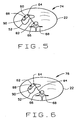
- CT imaging system 10 tracks and measures an area of ventricular wall 54 relative to a fixed reference point 66, for example, a point 66 on spine 68 of patient 22.
- a fixed reference point 66 for example, a point 66 on spine 68 of patient 22.
- ventricular wall 54 begins to expand 56, 58, 60, 62.
- a distance D between spine 68 and point 64 on ventricular myocardial wall 54 increases 56, 58, 60, 62, as revealed in consecutive CT cine images. This distance continually and gradually increases until the aforementioned threshold potential is reached 70.
- Heart 52 is at a moment prior to systole, its relatively quietest moment in the cardiac cycle. As heart 52 enters its systolic phase, left ventricular wall 62 contracts to expel blood. A marked change in a spinal-myocardial measured distance D + ⁇ D to D is observed. This distance drastically changes and diminishes during the systolic phase of the heart.
- These 44 images include at least one complete cardiac cycle. Additional two-second cine scans are taken as needed. Table 46 is stepped between each two-second cine scan so that image slices from a subsequent scan do not overlap a volume imaged in slices from previous scans. In one embodiment, table 46 is stepped an amount equal to a total thickness of the image slices acquired, to obtain a set of slices adjacent to, but not overlapping, slices obtained prior to each step.
- the i th image 78 provides a satisfactory image with reduced motion-induced artifacts from which to measure calcification 88 of pulmonary arteries 90.
- a plurality of images are used for cardiac calcification scoring. These images are the i th image 78, and a selected number of images in sequence. For example, images are used back to a final image 76 at which line 84 between point 66 on spine 68 and point 64 on left ventricular wall 62 is still slightly increasing. Depending upon in which image this occurs, the i -1th image 76 and possibly the i -2nd image 74 are scored in conjunction with the i th image 78. In this embodiment, therefore, the selected number of images of the sequence is no greater than two.
- images 78, 76, and 74 are used for calcification scoring when it is determined that a single image is adequate for such use.
- This image e.g., image 76, is one in which little, if any, change in line length 84 is observed relative to adjacent images 74, 78.
- CT imaging system 10 provide gantry 12 rotation speeds that are a substantial fraction of a cardiac cycle, each of the images in both cine scan and segmented helical scan embodiments are reconstructed from less that a full 360° view angle of data. These views are known as segmented images, and the data representing them is known as segmented image data. For this reason, in one embodiment, imaging system 10 is said to collect segmented image data. Because segmented image data is collected in a relatively short time for each image, motion-induced image artifacts are reduced relative to images reconstructed from longer, full scans. However, embodiments having gantry 12 rotation speeds sufficiently fast to permit reconstruction of views from a full 360° view angle with reduced image artifacts are possible.
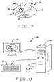
- computer 36 is programmed to display successive images, for example, images 72, 74, 76, 78 of Figures 4, 5, 6, and 7, on display 42. Lines and successive images are manipulated via console 40 or another suitable input device or devices. Computer 36 is also programmed to calculate and display line lengths drawn by an operator on images displayed on display 42.
- image data acquired by CT imaging system 10 is downloaded or transferred to a separate workstation 92, shown in Figure 8.
- Workstation 92 includes, in one embodiment, a system unit 94 having a processor and memory, a display 96, and one or more operator input devices such as a keyboard 98 and a mouse 100.
- the processor is programmed to display and manipulate images on display 96 and to calculate and display line lengths drawn on the screen by an operator using input devices 98 and 100.
- a method in accordance with the invention is defined in claim 1.
- Scanning a portion of a patient's body may comprise the step of scanning a portion of the patient's body including a heart (50) and a spine (68), the moving body part being the heart and the selected fixed reference point being a point (66) on the spine.
- a CT imaging system in accordance with the present invention is defined in claim 7.
- Said system being configured to scan a portion of a patient's body may comprise said system being configured to cine scan the portion of the patient's body.
- Said system being configured to select the at least one image may comprise said system being configured to select at least one image of the sequence of images within a selected number of images of an image in which a change in the distances reverses its sign or becomes constant. In one embodiment, the selected number of images is no greater than two.
- Said system being configured to scan a portion of a patient's body may comprise said system being configured to scan a portion of the patient's body including a heart (50) and a spine (68), the moving body part being the heart and the selected fixed reference point being a point (66) on the spine.
- Said workstation being configured to select the at least one image may comprise said workstation being configured to select at least one image of the sequence of images within a selected number of images in which a change in the distances reverses sign or becomes constant. In one embodiment, the selected number of images is no greater than 2.
Landscapes
- Health & Medical Sciences (AREA)
- Life Sciences & Earth Sciences (AREA)
- Engineering & Computer Science (AREA)
- Medical Informatics (AREA)
- Radiology & Medical Imaging (AREA)
- Biomedical Technology (AREA)
- Biophysics (AREA)
- High Energy & Nuclear Physics (AREA)
- Veterinary Medicine (AREA)
- Nuclear Medicine, Radiotherapy & Molecular Imaging (AREA)
- Optics & Photonics (AREA)
- Pathology (AREA)
- Public Health (AREA)
- Physics & Mathematics (AREA)
- Heart & Thoracic Surgery (AREA)
- Molecular Biology (AREA)
- Surgery (AREA)
- Animal Behavior & Ethology (AREA)
- General Health & Medical Sciences (AREA)
- Physiology (AREA)
- Pulmonology (AREA)
- Theoretical Computer Science (AREA)
- Computer Vision & Pattern Recognition (AREA)
- Apparatus For Radiation Diagnosis (AREA)
Applications Claiming Priority (2)
| Application Number | Priority Date | Filing Date | Title |
|---|---|---|---|
| US09/409,465 US6252924B1 (en) | 1999-09-30 | 1999-09-30 | Method and apparatus for motion-free cardiac CT imaging |
| US409465 | 1999-09-30 |
Publications (2)
| Publication Number | Publication Date |
|---|---|
| EP1088517A1 EP1088517A1 (en) | 2001-04-04 |
| EP1088517B1 true EP1088517B1 (en) | 2007-05-09 |
Family
ID=23620610
Family Applications (1)
| Application Number | Title | Priority Date | Filing Date |
|---|---|---|---|
| EP00308538A Expired - Lifetime EP1088517B1 (en) | 1999-09-30 | 2000-09-28 | Method and apparatus for motion-free cardiac CT imaging |
Country Status (5)
| Country | Link |
|---|---|
| US (1) | US6252924B1 (enExample) |
| EP (1) | EP1088517B1 (enExample) |
| JP (1) | JP4576037B2 (enExample) |
| DE (1) | DE60034748T2 (enExample) |
| IL (1) | IL138566A0 (enExample) |
Families Citing this family (80)
| Publication number | Priority date | Publication date | Assignee | Title |
|---|---|---|---|---|
| US6370217B1 (en) | 1999-05-07 | 2002-04-09 | General Electric Company | Volumetric computed tomography system for cardiac imaging |
| US6256368B1 (en) | 1999-10-15 | 2001-07-03 | General Electric Company | Methods and apparatus for scout-based cardiac calcification scoring |
| US6505065B1 (en) * | 1999-10-29 | 2003-01-07 | Koninklijke Philips Electronics, N.V. | Methods and apparatus for planning and executing minimally invasive procedures for in-vivo placement of objects |
| US6393091B1 (en) | 1999-12-13 | 2002-05-21 | General Electric Company | Methods and apparatus for non-uniform temporal cardiac imaging |
| US6421552B1 (en) | 1999-12-27 | 2002-07-16 | Ge Medical Systems Global Technology Company, Llc | Methods and apparatus for estimating cardiac motion using projection data |
| US6442228B1 (en) * | 2000-04-20 | 2002-08-27 | Ge Medical Systems Global Technology Company, Llc | Data acquisition modifications for improved reconstruction with conventional CT |
| US8489176B1 (en) | 2000-08-21 | 2013-07-16 | Spectrum Dynamics Llc | Radioactive emission detector equipped with a position tracking system and utilization thereof with medical systems and in medical procedures |
| US8565860B2 (en) | 2000-08-21 | 2013-10-22 | Biosensors International Group, Ltd. | Radioactive emission detector equipped with a position tracking system |
| WO2005119025A2 (en) | 2004-06-01 | 2005-12-15 | Spectrum Dynamics Llc | Radioactive-emission-measurement optimization to specific body structures |
| US8909325B2 (en) | 2000-08-21 | 2014-12-09 | Biosensors International Group, Ltd. | Radioactive emission detector equipped with a position tracking system and utilization thereof with medical systems and in medical procedures |
| US7031504B1 (en) | 2000-09-26 | 2006-04-18 | Vital Images, Inc. | Image data based retrospective temporal selection of medical images |
| DE10052870B4 (de) * | 2000-10-20 | 2005-06-23 | Siemens Ag | Vorrichtung zur automatischen Sortierung periodischer Datensätze |
| US6708052B1 (en) * | 2001-04-11 | 2004-03-16 | Harbor Ucla Research And Education Institute | Method and apparatus for cardiac imaging with minimized cardiac motion artifact |
| US6438196B1 (en) | 2001-06-28 | 2002-08-20 | General Electric Company | EKG driven CT image reconstruction for cardiac imaging |
| US7209779B2 (en) * | 2001-07-17 | 2007-04-24 | Accuimage Diagnostics Corp. | Methods and software for retrospectively gating a set of images |
| US7142703B2 (en) * | 2001-07-17 | 2006-11-28 | Cedara Software (Usa) Limited | Methods and software for self-gating a set of images |
| US7006862B2 (en) * | 2001-07-17 | 2006-02-28 | Accuimage Diagnostics Corp. | Graphical user interfaces and methods for retrospectively gating a set of images |
| US8061006B2 (en) * | 2001-07-26 | 2011-11-22 | Powderject Research Limited | Particle cassette, method and kit therefor |
| US7286866B2 (en) * | 2001-11-05 | 2007-10-23 | Ge Medical Systems Global Technology Company, Llc | Method, system and computer product for cardiac interventional procedure planning |
| US6628981B2 (en) | 2001-11-09 | 2003-09-30 | Ge Medical Systems Information Technologies, Inc. | Adaptive heart rate prediction algorithm for computed tomography imaging |
| US6526117B1 (en) * | 2001-11-09 | 2003-02-25 | Ge Medical Systems Global Technology Company, Llc | Method and apparatus to minimize phase misregistration artifacts in gated CT images |
| US7311705B2 (en) | 2002-02-05 | 2007-12-25 | Medtronic, Inc. | Catheter apparatus for treatment of heart arrhythmia |
| US7346381B2 (en) * | 2002-11-01 | 2008-03-18 | Ge Medical Systems Global Technology Company Llc | Method and apparatus for medical intervention procedure planning |
| US7499743B2 (en) * | 2002-03-15 | 2009-03-03 | General Electric Company | Method and system for registration of 3D images within an interventional system |
| US6721386B2 (en) * | 2002-03-15 | 2004-04-13 | Ge Medical Systems Global Technology Co., Llc | Method and apparatus of cardiac CT imaging using ECG and mechanical motion signals |
| US7778686B2 (en) * | 2002-06-04 | 2010-08-17 | General Electric Company | Method and apparatus for medical intervention procedure planning and location and navigation of an intervention tool |
| US6904118B2 (en) * | 2002-07-23 | 2005-06-07 | General Electric Company | Method and apparatus for generating a density map using dual-energy CT |
| US7927275B2 (en) * | 2002-08-26 | 2011-04-19 | The Cleveland Clinic Foundation | System and method of aquiring blood-vessel data |
| US20040085046A1 (en) * | 2002-11-01 | 2004-05-06 | General Electric Company | Power conditioning system for turbine motor/generator |
| FR2848093B1 (fr) * | 2002-12-06 | 2005-12-30 | Ge Med Sys Global Tech Co Llc | Procede de detection du cycle cardiaque a partir d'angiogramme de vaisseaux coronaires |
| GB2397738B (en) † | 2003-01-21 | 2007-08-29 | Elekta Ab | Computed tomography scanning |
| US7747047B2 (en) | 2003-05-07 | 2010-06-29 | Ge Medical Systems Global Technology Company, Llc | Cardiac CT system and method for planning left atrial appendage isolation |
| US7565190B2 (en) * | 2003-05-09 | 2009-07-21 | Ge Medical Systems Global Technology Company, Llc | Cardiac CT system and method for planning atrial fibrillation intervention |
| US7343196B2 (en) * | 2003-05-09 | 2008-03-11 | Ge Medical Systems Global Technology Company Llc | Cardiac CT system and method for planning and treatment of biventricular pacing using epicardial lead |
| US20050010105A1 (en) * | 2003-07-01 | 2005-01-13 | Sra Jasbir S. | Method and system for Coronary arterial intervention |
| US7813785B2 (en) * | 2003-07-01 | 2010-10-12 | General Electric Company | Cardiac imaging system and method for planning minimally invasive direct coronary artery bypass surgery |
| US7344543B2 (en) * | 2003-07-01 | 2008-03-18 | Medtronic, Inc. | Method and apparatus for epicardial left atrial appendage isolation in patients with atrial fibrillation |
| US20050054918A1 (en) * | 2003-09-04 | 2005-03-10 | Sra Jasbir S. | Method and system for treatment of atrial fibrillation and other cardiac arrhythmias |
| US20060009755A1 (en) * | 2003-09-04 | 2006-01-12 | Sra Jasbir S | Method and system for ablation of atrial fibrillation and other cardiac arrhythmias |
| US7308299B2 (en) | 2003-10-22 | 2007-12-11 | General Electric Company | Method, apparatus and product for acquiring cardiac images |
| US7308297B2 (en) * | 2003-11-05 | 2007-12-11 | Ge Medical Systems Global Technology Company, Llc | Cardiac imaging system and method for quantification of desynchrony of ventricles for biventricular pacing |
| US20050137661A1 (en) * | 2003-12-19 | 2005-06-23 | Sra Jasbir S. | Method and system of treatment of cardiac arrhythmias using 4D imaging |
| US20050143777A1 (en) * | 2003-12-19 | 2005-06-30 | Sra Jasbir S. | Method and system of treatment of heart failure using 4D imaging |
| US7639774B2 (en) * | 2003-12-23 | 2009-12-29 | General Electric Company | Method and apparatus for employing multiple axial-sources |
| US7968851B2 (en) | 2004-01-13 | 2011-06-28 | Spectrum Dynamics Llc | Dynamic spect camera |
| US8586932B2 (en) | 2004-11-09 | 2013-11-19 | Spectrum Dynamics Llc | System and method for radioactive emission measurement |
| WO2008010227A2 (en) | 2006-07-19 | 2008-01-24 | Spectrum Dynamics Llc | Imaging protocols |
| US9470801B2 (en) | 2004-01-13 | 2016-10-18 | Spectrum Dynamics Llc | Gating with anatomically varying durations |
| CN1981210A (zh) | 2004-01-13 | 2007-06-13 | 光谱动力学有限责任公司 | 多维图像重构 |
| US8571881B2 (en) | 2004-11-09 | 2013-10-29 | Spectrum Dynamics, Llc | Radiopharmaceutical dispensing, administration, and imaging |
| WO2006051531A2 (en) | 2004-11-09 | 2006-05-18 | Spectrum Dynamics Llc | Radioimaging |
| US7454248B2 (en) * | 2004-01-30 | 2008-11-18 | Ge Medical Systems Global Technology, Llc | Method, apparatus and product for acquiring cardiac images |
| US7333587B2 (en) * | 2004-02-27 | 2008-02-19 | General Electric Company | Method and system for imaging using multiple offset X-ray emission points |
| US7170972B2 (en) * | 2004-03-16 | 2007-01-30 | General Electric Company | Methods and systems for multi-modality imaging |
| DE102004017478B4 (de) * | 2004-04-08 | 2012-01-19 | Siemens Ag | Vorrichtung für die Gewinnung von Strukturdaten eines sich bewegenden Objekts |
| US8280124B2 (en) * | 2004-06-01 | 2012-10-02 | Spectrum Dynamics Llc | Methods of view selection for radioactive emission measurements |
| US8515527B2 (en) * | 2004-10-13 | 2013-08-20 | General Electric Company | Method and apparatus for registering 3D models of anatomical regions of a heart and a tracking system with projection images of an interventional fluoroscopic system |
| US7327872B2 (en) * | 2004-10-13 | 2008-02-05 | General Electric Company | Method and system for registering 3D models of anatomical regions with projection images of the same |
| US9943274B2 (en) | 2004-11-09 | 2018-04-17 | Spectrum Dynamics Medical Limited | Radioimaging using low dose isotope |
| US9316743B2 (en) | 2004-11-09 | 2016-04-19 | Biosensors International Group, Ltd. | System and method for radioactive emission measurement |
| US8615405B2 (en) | 2004-11-09 | 2013-12-24 | Biosensors International Group, Ltd. | Imaging system customization using data from radiopharmaceutical-associated data carrier |
| US8423125B2 (en) * | 2004-11-09 | 2013-04-16 | Spectrum Dynamics Llc | Radioimaging |
| WO2008059489A2 (en) | 2006-11-13 | 2008-05-22 | Spectrum Dynamics Llc | Radioimaging applications of and novel formulations of teboroxime |
| FR2887429B1 (fr) * | 2005-06-24 | 2007-10-26 | Gen Electric | Procede d'imagerie radiologique d'un organe en mouvement |
| EP1909853B1 (en) | 2005-07-19 | 2015-03-18 | Biosensors International Group, Ltd. | Imaging protocols |
| US8111886B2 (en) * | 2005-07-19 | 2012-02-07 | Spectrum Dynamics Llc | Reconstruction stabilizer and active vision |
| US8837793B2 (en) | 2005-07-19 | 2014-09-16 | Biosensors International Group, Ltd. | Reconstruction stabilizer and active vision |
| JP2007054372A (ja) * | 2005-08-25 | 2007-03-08 | Ge Medical Systems Global Technology Co Llc | X線ct装置 |
| CN1955725B (zh) * | 2005-10-27 | 2010-12-15 | Ge医疗系统环球技术有限公司 | X射线ct系统 |
| US8894974B2 (en) | 2006-05-11 | 2014-11-25 | Spectrum Dynamics Llc | Radiopharmaceuticals for diagnosis and therapy |
| US7835486B2 (en) * | 2006-08-30 | 2010-11-16 | General Electric Company | Acquisition and reconstruction of projection data using a stationary CT geometry |
| US7706499B2 (en) * | 2006-08-30 | 2010-04-27 | General Electric Company | Acquisition and reconstruction of projection data using a stationary CT geometry |
| US7616731B2 (en) * | 2006-08-30 | 2009-11-10 | General Electric Company | Acquisition and reconstruction of projection data using a stationary CT geometry |
| US20080056432A1 (en) * | 2006-08-30 | 2008-03-06 | General Electric Company | Reconstruction of CT projection data |
| WO2008075362A2 (en) | 2006-12-20 | 2008-06-26 | Spectrum Dynamics Llc | A method, a system, and an apparatus for using and processing multidimensional data |
| US8521253B2 (en) | 2007-10-29 | 2013-08-27 | Spectrum Dynamics Llc | Prostate imaging |
| US7706912B2 (en) * | 2007-11-30 | 2010-04-27 | Caterpillar Inc. | Orifice formation control system |
| US8396248B2 (en) * | 2008-09-16 | 2013-03-12 | Varian Medical Systems, Inc. | Sequential stereo imaging for estimating trajectory and monitoring target position |
| JP2010069099A (ja) * | 2008-09-19 | 2010-04-02 | Toshiba Corp | 画像処理装置及びx線コンピュータ断層撮影装置 |
| US8338788B2 (en) | 2009-07-29 | 2012-12-25 | Spectrum Dynamics Llc | Method and system of optimized volumetric imaging |
Family Cites Families (6)
| Publication number | Priority date | Publication date | Assignee | Title |
|---|---|---|---|---|
| JP2529949B2 (ja) | 1986-08-12 | 1996-09-04 | 株式会社東芝 | 同期画像再構成装置 |
| JPH0354612U (enExample) * | 1989-09-30 | 1991-05-27 | ||
| US5457728A (en) | 1990-11-14 | 1995-10-10 | Cedars-Sinai Medical Center | Coronary tracking display |
| JP3446964B2 (ja) * | 1993-07-12 | 2003-09-16 | 株式会社日立メディコ | 医用画像表示装置 |
| US5533085A (en) * | 1995-02-27 | 1996-07-02 | University Of Washington | Automatic indexing of cine-angiograms |
| DE19854939C2 (de) * | 1998-11-27 | 2001-11-22 | Siemens Ag | Verfahren und Gerät zur Erzeugung von CT-Bildern |
-
1999
- 1999-09-30 US US09/409,465 patent/US6252924B1/en not_active Expired - Fee Related
-
2000
- 2000-09-19 IL IL13856600A patent/IL138566A0/xx not_active IP Right Cessation
- 2000-09-28 DE DE60034748T patent/DE60034748T2/de not_active Expired - Lifetime
- 2000-09-28 EP EP00308538A patent/EP1088517B1/en not_active Expired - Lifetime
- 2000-09-29 JP JP2000297806A patent/JP4576037B2/ja not_active Expired - Fee Related
Non-Patent Citations (1)
| Title |
|---|
| None * |
Also Published As
| Publication number | Publication date |
|---|---|
| JP4576037B2 (ja) | 2010-11-04 |
| JP2001137229A (ja) | 2001-05-22 |
| IL138566A0 (en) | 2001-10-31 |
| DE60034748T2 (de) | 2008-01-17 |
| US6252924B1 (en) | 2001-06-26 |
| EP1088517A1 (en) | 2001-04-04 |
| DE60034748D1 (de) | 2007-06-21 |
Similar Documents
| Publication | Publication Date | Title |
|---|---|---|
| EP1088517B1 (en) | Method and apparatus for motion-free cardiac CT imaging | |
| JP4630440B2 (ja) | スカウト画像をベースとした心臓石灰化計数のための方法及び装置 | |
| US6370217B1 (en) | Volumetric computed tomography system for cardiac imaging | |
| JP4487095B2 (ja) | 撮像システムの回顧的心臓ゲーティングを用いた冠状動脈の石灰化の検出 | |
| US6504894B2 (en) | Phase-driven multisector reconstruction for multislice helical CT imaging | |
| US5459769A (en) | Procedure for monitoring contrast agent application in a CT imaging system | |
| JP4436601B2 (ja) | ゲート式ct画像中の時相整合不良によるアーティファクトを最小にするための方法及び装置 | |
| JP4130088B2 (ja) | Ct投影データを用いて運動ゲ−ティングするための方法及び装置 | |
| EP1466559A2 (en) | Methods and apparatus for cardiac imaging with conventional computed tomography | |
| US6434215B1 (en) | EKG-less cardiac image reconstruction | |
| JP2000210282A (ja) | 物体の画像を形成する方法及びイメージング・システム | |
| JP3950849B2 (ja) | 拡張再構成ウィンドウを用いた高ピッチ心臓ヘリカルスキャン | |
| JP2004121840A (ja) | 周期的に運動する器官のct画像形成方法およびこの方法を実施するためのct装置 | |
| JP2001224588A (ja) | 被曝を低減したコンピュータ断層撮影イメージング方法及び装置 | |
| US6597803B1 (en) | Hybrid reconstruction for high pitch multi-slice helical cardiac imaging | |
| EP1072224A2 (en) | Retrospective cardiac gating with cine scans on a multislice scanner | |
| EP1101444A2 (en) | Method and apparatus for controlling x-ray exposure during gated cardiac scanning | |
| JP4007928B2 (ja) | X線ct装置 | |
| WO2006029336A2 (en) | Projection gating of x-ray ct scan | |
| US7023958B2 (en) | Radiation image-acquiring apparatus, and radiation image-acquiring method |
Legal Events
| Date | Code | Title | Description |
|---|---|---|---|
| PUAI | Public reference made under article 153(3) epc to a published international application that has entered the european phase |
Free format text: ORIGINAL CODE: 0009012 |
|
| AK | Designated contracting states |
Kind code of ref document: A1 Designated state(s): DE NL |
|
| AX | Request for extension of the european patent |
Free format text: AL;LT;LV;MK;RO;SI |
|
| 17P | Request for examination filed |
Effective date: 20011004 |
|
| AKX | Designation fees paid |
Free format text: DE NL |
|
| GRAP | Despatch of communication of intention to grant a patent |
Free format text: ORIGINAL CODE: EPIDOSNIGR1 |
|
| GRAS | Grant fee paid |
Free format text: ORIGINAL CODE: EPIDOSNIGR3 |
|
| GRAA | (expected) grant |
Free format text: ORIGINAL CODE: 0009210 |
|
| AK | Designated contracting states |
Kind code of ref document: B1 Designated state(s): DE NL |
|
| REF | Corresponds to: |
Ref document number: 60034748 Country of ref document: DE Date of ref document: 20070621 Kind code of ref document: P |
|
| PGFP | Annual fee paid to national office [announced via postgrant information from national office to epo] |
Ref country code: NL Payment date: 20070923 Year of fee payment: 8 |
|
| PLBE | No opposition filed within time limit |
Free format text: ORIGINAL CODE: 0009261 |
|
| STAA | Information on the status of an ep patent application or granted ep patent |
Free format text: STATUS: NO OPPOSITION FILED WITHIN TIME LIMIT |
|
| 26N | No opposition filed |
Effective date: 20080212 |
|
| PG25 | Lapsed in a contracting state [announced via postgrant information from national office to epo] |
Ref country code: NL Free format text: LAPSE BECAUSE OF NON-PAYMENT OF DUE FEES Effective date: 20090401 |
|
| NLV4 | Nl: lapsed or anulled due to non-payment of the annual fee |
Effective date: 20090401 |
|
| PGFP | Annual fee paid to national office [announced via postgrant information from national office to epo] |
Ref country code: DE Payment date: 20100929 Year of fee payment: 11 |
|
| REG | Reference to a national code |
Ref country code: DE Ref legal event code: R119 Ref document number: 60034748 Country of ref document: DE Effective date: 20130403 |
|
| PG25 | Lapsed in a contracting state [announced via postgrant information from national office to epo] |
Ref country code: DE Free format text: LAPSE BECAUSE OF NON-PAYMENT OF DUE FEES Effective date: 20130403 |