EP1015869B1 - Mikroabbildungssystem - Google Patents
Mikroabbildungssystem Download PDFInfo
- Publication number
- EP1015869B1 EP1015869B1 EP97941059A EP97941059A EP1015869B1 EP 1015869 B1 EP1015869 B1 EP 1015869B1 EP 97941059 A EP97941059 A EP 97941059A EP 97941059 A EP97941059 A EP 97941059A EP 1015869 B1 EP1015869 B1 EP 1015869B1
- Authority
- EP
- European Patent Office
- Prior art keywords
- sample
- detector
- lens
- optical
- area
- Prior art date
- Legal status (The legal status is an assumption and is not a legal conclusion. Google has not performed a legal analysis and makes no representation as to the accuracy of the status listed.)
- Expired - Lifetime
Links
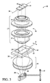
Images


Classifications
-
- G—PHYSICS
- G02—OPTICS
- G02B—OPTICAL ELEMENTS, SYSTEMS OR APPARATUS
- G02B21/00—Microscopes
- G02B21/02—Objectives
Definitions
- the present invention relates to microscopy. Specifically, the present invention pertains to an imaging system ideally suited for full-field imaging.
- U.S. Pat. No. 4,680,635 to Khuruna discloses an emission microscope for monitoring anomalies, such as pattern defects and particulate contamination, during the manufacture of semiconductor wafers.
- the emission microscope includes, in pertinent part, an objective lens system optically coupled to an image intensifier.
- the image intensifier is electronically coupled to a solid state detector and functions to magnify the image detected by the objective lens.
- An image processing computer is coupled to receive the information produced by the solid state detector.
- a lens system is optically coupled to a detector.
- the detector is electronically coupled to a processor.
- the lens system comprises of an objective lens having both high numerical aperture and high magnification.
- a secondary lens is employed to reduce the overall magnification of the lens system.
- Information corresponding to the image sensed by the detector is sent to the processor, whereby a visual display of the information may be obtained.
- Various lens systems may be provided to afford different magnifications.
- U.S. Pat. No. 5,500,770 to Zinter et al. discloses a macrolens system for an emission microscopy instrument which includes two multiple lens groups. One group is disposed proximate to a sample, defining the object group. The remaining group is disposed proximate to a detector, defining a detector. group. The object group is spaced apart from the detector group, forming an axial gap therebetween. The object group defines a focal plane proximate to the sample, and the detector group defines a focal plane proximate to the detector. Light from the sample passes through the object group and enters the axial gap. The object group collimates the light from the sample so that light passes through the axial gap as a bundle of collimated rays. The collimated light enters the detector group which focuses the light onto the detector.
- the axial gap is discussed as providing the advantage of allowing the inclusion of auxiliary components therein, such as a beam splitter.
- Two conflicting goals of light microscopy inspection systems concern providing a high speed imaging system with high resolution.
- the resolution of a light microscope is inversely proportional to the imaging speed.
- the greater the resolution the lower the inspection rate.
- One technique to accommodate the aforementioned conflict is to selectively choose the resolution of the system. To that end, providing a light microscope with varying magnification is beneficial.
- U.S. Pat. No. 4,755,874 to Esrig et al. discloses an emission microscopy system that provides image processing techniques at both visible wavelengths and near infra-red wavelengths of light.
- the system includes, in pertinent part, a lens assembly optically coupled to both a sample under test and a light intensifier.
- a primary video camera, or other solid state optical-to-electronic analog converter, is in optical communication with the intensifier.
- a video image signal processing means is in electrical communication with the video camera to process information corresponding to the image sensed by the camera.
- a display means is electronically coupled to the processing means to form a visual display of the information processed by the processing means.
- the lens assembly is described as including micro-optics and macro-optics, each of which may be interchanged to provide the system differing levels of magnification. In one embodiment, the macro-optics are shown as including back-to-back photo lenses.
- U.S. Patent No. 5,141,609 to Sweedler et al. discloses a detection method and apparatus useful in capillary electrophoresis and capillary chromatography that employs an array of solid state detectors, such as a charge-coupled device (CCD).
- CCD charge-coupled device
- the system is described as operating in a time delay integration (TDI) mode which allows more exposure time of moving analyte zones.
- TDI time delay integration
- the CCD is a two-dimensional array which affords area-wise imaging by employing a spectrograph which disperses the image of a sample spectrally.
- spatial information is collected along a first axis of the CCD array, with the spectral information being collected along a second axis of the CCD array, orthogonal to the first axis.
- a laser is focused via a lens axially into a capillary housing the sample.
- Light, transmitted from the sample is reflected by a cylindrical mirror onto a pair of achromats and cylindrical lenses which image the light from the sample onto the CCD via the spectrograph.
- a drawback with the aforementioned systems is that each sacrifices image resolution or collection efficiency in order to provide differing levels of magnification.
- Another drawback is that each prior art imaging system is suitable for use with only a limited number of microscopic techniques and is not optimized to provide maximum field size with maximum resolution and collection efficiency as well as providing quantitative information.
- What is needed is an optical system that provides high-resolution imaging at varying degrees of magnification to afford quantitative imaging over the entire field of view and is suitable for use with many different microscopic techniques.
- the present invention features a double objective lens assembly which provides a constant image definition so that a number of resolvable points across the field correspond to a number of pixel elements of a detector.
- the lens assembly includes two telecentric, infinite conjugate, flat field achromatic objectives, each of which has an external pupil lying in a common plane and axially aligned, defining a mechanically accessible central pupil of an imaging system.
- Each of the objectives are afocal in the common plane, with one of the lenses, the sample lens, forming a focal plane proximate to a sample.
- An array detector is positioned proximate to a focal plane formed by the remaining objective, defining the detector lens.
- a dimension along one axis (d) of the array detector matches a corresponding dimension (D) along a parallel axis of the sample and the central pupil (p) at unity magnification.
- a full-field view of the sample along D may be obtained by impinging light upon the same to illuminate a scan line thereon.
- High definition imaging is achieved with the objective by having the number of resolvable points across the field coinciding with the number of pixels in the detector.
- the sample may be moved in a direction transverse to the scan line to obtain a full image of the sample or the line may be scanned across the sample accordingly.
- the entire sample may be imaged, with the dimensional area of the detector and pupil corresponding to the area of the sample, at unity magnification.
- the double objective lens assembly simultaneously images the sample on substantially all of the sensing elements of the detector in two dimensions. This avoids having to move the sample or scan a line as described above.
- the light source may be configured for transillumination in which the double objective lens assembly is disposed proximate to one side of the sample and the light source is positioned proximate to an opposing side. This allows the double objective lens assembly to collect light passing through the sample and image the same on the detector.
- epi-illumination may be employed in which a wavelength separating means, such as a beam splitter, part-aperture-filling mirror or wavelength discrimination mirror may be disposed at the central pupil forming a retro-path over which light emitted from an illuminated area of the sample traverses. In this fashion, the light source is disposed off an optical axis defined by the double objective lens assembly.
- the light source directs light over the incident light path to impinge upon the wavelength separating means.
- the wavelength separating means directs the light through the sample lens and onto the sample. Light emerging from the sample is collected by the sample lens and directed along the retro-path through the wavelength separating means.
- the detector lens collects the light passing through the wavelength separating means and images the same upon the detector. In both of the aforementioned imaging systems, the detector produces electrical signals corresponding to the light sensed, and the system includes a signal processor and a video display system to form visual images of the signals.
- a transillumination system 10 is shown as including a double objective lens assembly 12, optical detector 14, light source 16 and condenser lenses 18, all of which lie along an optical path 20.
- a sample 22 is disposed in optical path 20, between lens assembly 12 and light source 16.
- light 24 emitted from source 16 passes through sample 22 and is collected by lens assembly 12.
- Lens assembly 12 then directs the collected light 25 onto detector 14, imaging thereon an area 26 of sample 22 which is illuminated.
- double objective lens assembly 12 is corrected for axial and lateral chromatic aberrations and includes two infinite conjugate objective lenses.
- Each objective lens has a flat field and lateral color correction and an external pupil lying along a common axis and a common plane 28, defining a mechanically accessible central pupil 30 of system 10.
- One of the objective lenses, a sample lens 32 forms a focal plane proximate to sample 22.
- Detector 14 is positioned proximate to a focal plane formed by the remaining objective lens, the detector lens 34. It is preferred that system 10 be capable of providing full-field imaging of sample 22 in at least one dimension.
- detector 14 is typically an array detector having a plurality of sensing elements 42, e.g., photo-diodes, photo-capacitors or photo-conductors aligned along an axis.
- sensing elements 42 e.g., photo-diodes, photo-capacitors or photo-conductors aligned along an axis.
- CCD charge-coupled device
- a dimension (d) of detector 14, along longitudinal axis 36, will be equivalent to or greater than a corresponding dimension (D) of sample 22 along a parallel axis 38.
- the diameter of central pupil 30 defines the numerical aperture of system 10
- central pupil 30 allow a maximum quantity of photons therethrough to permit efficient imaging, so as to minimize optical loss.
- lens 32 and lens 34 may be identical, and, thus, form an optical relay of unity magnification. It is preferred that each lens 32 and 34 be afocal in the common plane 28 and be symmetric in design approach so that double objective lens assembly 12 is symmetric about central pupil 30. By having the pupils of two afocal lenses aligned along a common axis and lying in common plane 28, and containing symmetry in design, optical aberrations, such as coma and distortion, are reduced. To reduce optical loss at the periphery of the field, both lenses 32 and 34 may be telecentric in their respective focal planes, which renders assembly 12's magnification relatively insensitive to errors in focus position.
- lenses 32 and 34 may be achromatic or may have equal and opposite axial chromatic aberrations, allowing lens assembly 12 to operate over a broad band of wavelengths of light, e.g., the primary wavelength plus approximately 300 nm, or greater, while maintaining axial and lateral aberrations below the effective resolution of the system.
- any light source may be employed dependent upon the application.
- light source 16 may be a laser that produces a collimated beam of coherent light, a halogen light or a light-emitting diode producing non-coherent light. This makes system 10 suitable for use in several applications, e.g., OBIC or chemoluminescence.
- lenses 32 and 34 are as follows: Sample Lens Detector Lens Primary Wavelength 488 488-600 Focal Length (mm) 27 27 Field Size (cm) 1 1 Telecentricity (degrees) 1 1 Numerical Aperture .2 .2 Resolution (microns) 5 5 Working Distance (mm) 3 3 Pupil Size (mm) 10.8 10.8
- lens assembly 12 and detector 14 be configured to provide a full-field image of sample 22 along at least one dimension D.
- system 10 may include a translation stage 40 upon which sample 22 rests. The position of sample 22 on stage 40 is maintained in any convenient manner, e.g., vacuum suction or resilient members. Stage 40 moves sample 22 in a direction A transverse to axis 38, allowing successive striped areas of sample 22 to image upon sensing elements 42 of detector 14.
- sensing elements 42 of detector 14 may be connected to a single register 44 via transfer gate 46. In operation, a signal is integrated on sensing elements 42 for a line time. Gate 46 is then activated and the charge from all sensing elements 42 is simultaneously transferred to register 44.
- stage 40 positions sample 22 so that the next area may be imaged on sensing elements 42.
- Processing circuitry 45 produces signals corresponding to the image sensed which is then transmitted to a visual display 47.
- the movement of stage 40 is synchronized with the integration and readout time of detector 14 so that the entire sample may be imaged upon sensing elements 42.
- Antiblooming may be provided in situations where the flux of light from source 16 is not easily controlled.
- electronic shuttering may be employed which allows an integration time of sensing elements 42 to be less than the readout time for register 44. If a higher pixel density is required, a double-sided readout CCD or a staggered liner array CCD may be employed.
- magnification of the system 10 may be easily adjusted without substantial optical loss. Adjustment of the magnification is achieved by merely changing the focal length of sample lens 32, while maintaining a constant distance between central pupil 30 and sample 22.
- d corresponds to an area of detector 14
- D corresponds to an area of sample 22.
- area D corresponds to 5 mm square of sample 22
- area d corresponds to 5mm square of detector length, providing a one-to-one correspondence between detector area and sample area, i.e., unity magnification.
- the magnification of the system may be easily increased by increasing the ratio of detector area to sample area. Based upon equation (1), this ratio of detector area to sample area may be easily increased by simply changing the focal length of sample lens 32.
- sample lens 32 is releasably attached to system 10 so that it may be interchanged with other sample lens 32 to alter the magnification of system 10.
- lens 32 may be a zoom lens which provides incremental, or continuous, adjustment of the focal length of the same.
- system 10 may include a scanning mechanism 58, disposed between sample 22 and light source 16, with the position of stage 40 with respect to detector 14 remaining fixed. In this fashion, sample 22 may be illuminated with a scanning line.
- Any type of scanning mechanism could be employed, so long as a one dimensional scan is provided along direction A transverse to both axis 36 and 38.
- scanning mechanism 58 could include a rotating planar, convex, concave or polygonal mirror, as well as a rotating holographic scanner, or oscillating prisms.
- an acousto-optic deflector or a penta-prism scanning deflector may be employed.
- a benefit of illuminating sample 22 with a scan line is that uniform illumination of the sample 22 is easily achieved as compared to full-field imaging, because the illumination needs to be uniform in only one axis.
- a two-dimensional detector array 114 is shown providing a full-field image of sample 22 in two dimensions, which decreases imaging time of sample 22.
- One such detector is a frame transfer CCD image detector 114 that consists of a plurality of sensing elements 52a-c, 54a-c and 56a-c arranged in a plurality of rows 52, 54 and 56, respectively. Although three rows are shown, any number of rows and elements per row may be employed, dependent upon the application.
- Each row 52, 54 and 56 extends parallel to axis 38, orthogonally to direction A.
- the total area of sensing elements 52a-c, 54a-c and 56a-c may be at least as large as area 126 of sample 22 to be viewed under unity magnification.
- Electronic or mechanical shutter techniques may be employed with detector 214.
- lens assembly 12 is structured so that the field of view of the same allows simultaneous imaging of the entire area of sample 22 onto detector 214.
- shuttering is deactivated, allowing the entire area of detector 214 to be exposed to light 25.
- a charge is collected in each of sensing elements 242.
- Shuttering is activated and the charge collected in sensing elements 242 are clocked therefrom and sent to processing circuitry 245, creating signals corresponding to the image detected. Thereafter, a visual representation of the image may be formed on display 247.
- a prism (not shown) or other wavelength dispersing mechanism may be disposed between lens 34 and detector 214.
- one axis of CCD detector 114, parallel to direction A, may provide spatial information concerning light detected, and axis 36, orthogonal thereto, provides wavelength information concerning the same.
- Time Delay Integration techniques may be employed. For example, an area of the sample may be illuminated, but one row of pixels of the CCD would be activated to determine position information.
- TDI CCD 114 is oriented so that the parallel shift direction B is parallel to the direction A along which sample 22 is moved. Initially, light emerging from sample 22 is collected by lens 32 and directed onto row 52 of TDI CCD detector 114 by lens 34. As the sample 22 moves along direction A, light emerging therefrom images row 52, row 54 and then row 56 of TDI CCD detector 114. This information is transmitted to processing circuitry and used to form a visual representation of the image sensed by CCD 114 on a display, as discussed above.
- the double objective lens assembly includes lenses 334 and 332 that form a central pupil 330, as discussed above with respect to Figs. 1-4, and is included in a system 310 employing epi-illumination techniques.
- a light source 316 directs incident light 324 along an optical path 320 which passes through wave shaping optics, such as condenser lenses 318 and, optionally, an excitation filter 360.
- wave shaping optics such as condenser lenses 318 and, optionally, an excitation filter 360.
- any light source may be employed dependent upon the application.
- light source 316 may be laser that produces a collimated beam of coherent light, a halogen light or a light emitting diode producing non-coherent light.
- beam splitter 362 After passing through excitation filter 360, light 324 impinges upon a beam splitter 362 which fills central pupil 330 of double objective lens assembly 312. Beamsplitter 362 defines a retro-path 364 along which light emitted from sample 322 will travel, passing through detector lens 334 and imaging sample 322 onto detector 314. In this fashion, beam splitter 362 will separate light 324 from light 325 emitted from sample 322 so that light 324 reflected from sample 322 travels along path 320, but in an opposite direction. It is to be understood that any type of beamsplitter may be employed, so long as it is capable of separating incident light 324 from emitted light 325.
- a dichroic filter for example, a fresnel reflector or a 50% beamsplitter may be employed.
- a polarization sensitive beamsplitter may be used to achieve the separation. This embodiment could include a 1/4 waveplate positioned between the beamsplitter and the objective. This would cause incident beam 19 exiting the 1/4 waveplate to be circularly polarized.
- Sample lens 332 directs incident light 324 to illuminate an area 326 of sample 322, thereby stimulating the sample 322 to emit light.
- area 326 should provide a full-field image in at least one dimension.
- area 326 may be a line which is either linear or arcuate that extends completely across sample 322 when lens assembly provides unity magnification. If lens assembly provides magnification greater than unity, area 326 will extend only partially across sample 322.
- Area 326 is imaged onto detector 314 by lens 332 collecting light 325 and directing the same through beamsplitter 362, detection filter 365 and lens 334. In this design, lens 332 affords coaxial illumination and collection.
- Lens 134 directs light 325 onto detector 314 where a visual representation is formed on display 347 by processing circuitry 345 as discussed above.
- sample 322 may be moved along a direction transverse to the longitudinal axis of area 126, as discussed above with respect to Figs. 1 and 2.
- system 310 may include a scanning mechanism (not shown) which provides a one dimensional scan, as discussed above with respect to Fig. 3.
- the entire sample 322 may be illuminated at a given time so that area 326 includes the entire sample 322.
- a two-dimensional detector array may be employed to reduce the imaging time of sample 322, as discussed above with respect to Fig. 4.
- system 310 is suitable for use in many applications, including fluorescence and reflection imaging.
- central pupil 30 can be used to accomplish various imaging modes used in microscopy applications. For instance, in reflection Nomarski DIC imaging a Wollaston prism is placed at the pupil of the system, a polarizer is placed in front of the excitation beam and an analyzer (2nd polarizer) is placed in front of the detector, with the imaging physics being the same as in a microscope.
- a pupil stop 31 may be placed at central pupil 30 as shown in Fig. 4. This improves intensity uniformity as a function of field position.
- Pupil stop 31 may be adjustable by using a common diaphragm type aperture so that more performance areas of the system can be controlled. By closing pupil stop 31 the system can be made less confocal and therefore the Z (axial) resolution will be decreased. Closing the diaphragm is also desirable if the amount of light from the sample overexposes the detector. Closing pupil stop 31 is beneficial when looking at a thick sample and when it is desirable to image a full volume in two dimensions with a single pass. Also, when imaging a monolayer sample in an automated system, it is desirable to decrease the intensity fall off errors with changes in Z position. This reduces the difficulty in positioning the sample.
Claims (17)
- Optisches System zum Abbilden einer Probe, wobei das System umfasst:einen optischen Detektor;eine Lichtquelle, die angeordnet ist, um Licht entlang eines optischen Weges zu richten und eine Fläche der Probe zu beleuchten, wobei der Detektor angeordnet ist, um Licht entsprechend der Fläche zu erfassen und entsprechende elektrische Signale zu erzeugen;eine Objektivlinsenanordnung, die im optischen Weg zwischen dem Detektor und der Probe angeordnet ist, wobei die Linsenanordnung eine erste Linse mit einer ersten Brennebene, die durch eine Brennweite (F) der ersten Linse festgelegt ist, wobei die Probe nahe der ersten Brennebene angeordnet ist, wobei die erste Linse eine erste externe Pupille bildet, die entgegengesetzt zur ersten Brennebene angeordnet ist; und eine zweite Linse mit einer zweiten Brennebene, die durch eine Brennweite (f) der zweiten Linse festgelegt ist, wobei der Detektor nahe der zweiten Brennebene angeordnet ist, wobei die zweite Linse eine zweite externe Pupille bildet, die entgegengesetzt zur zweiten Brennebene angeordnet ist, umfasst, wobei jede der ersten und der zweiten externen Pupille axial ausgerichtet ist und in einer gemeinsamen Ebene liegt, die eine zentrale Pupille festlegt, wobei die erste und die zweite Linse eine Vergrößerung (M) des Systems als f/F festlegen; undein zum Empfangen der elektrischen Signale gekoppeltes Mittel zum Erzeugen einer visuellen Anzeige der Fläche.
- Optisches System nach Anspruch 1, wobei der Detektor eine vorbestimmte Anzahl von Pixeln umfasst und die erste und die zweite Linse ein Blickfeld mit einer Vielzahl von auflösbaren Punkten festlegen, wobei eine Anzahl der Vielzahl von auflösbaren Punkten mit der vorbestimmten Anzahl übereinstimmt.
- Optisches System nach Anspruch 1, welches ferner eine Pupillenblende umfasst, die an der zentralen Pupille angeordnet ist.
- Optisches System nach Anspruch 1, wobei die Probe einen äußeren Umfang aufweist und die Fläche mit dem äußeren Umfang flächengleich ist, wobei die zentrale Pupille ausreichende Abmessungen aufweist, um den optischen Verlust durch Maximieren einer Menge von Photonen, die durch diese hindurchtreten können, zu minimieren, und der Detektor ausreichende Abmessungen aufweist, um das volle Blickfeld der Probe abzubilden.
- Optisches System nach Anspruch 1, wobei sowohl die erste als auch die zweite Linse telezentrisch sind.
- Optisches Abtastsystem nach Anspruch 1, wobei die Probe eine erste und eine zweite entgegengesetzte Hauptfläche umfasst, wobei die erste Linse nahe der ersten Hauptfläche angeordnet ist und die Lichtquelle nahe der zweiten Hauptfläche angeordnet ist, wodurch die Durchlassbeleuchtung der Probe erleichtert wird.
- Optisches Abtastsystem nach Anspruch 1, wobei der Detektor ein ladungsgekoppeltes Bauelement ist.
- Optisches Abtastsystem nach Anspruch 1, wobei der Detektor ein Matrixdetektor ist.
- Optisches Abtastsystem nach Anspruch 1, wobei der Detektor eine MOS-Flächenmatrix ist.
- Optisches Abtastsystem nach Anspruch 1, wobei der Detektor ein Bildabtastelement aus der Gruppe umfasst, die aus einer Flächenphotodiode, einem MOS-Kondensator, einer mit Anschlussstiften versehenen Photodiode oder einem Photoleiter besteht.
- Optisches Abtastsystem nach Anspruch 1, welches ferner ein in dem Weg an der zentralen Pupille angeordnetes Wellenlängentrennmittel zum Trennen von Licht von der Lichtquelle entsprechend der Fläche umfasst, wobei das Trennmittel einen Rückweg festlegt, den Licht entsprechend der Fläche durchläuft.
- Optisches Abtastsystem nach Anspruch 11, wobei die Probe einen äußeren Umfang aufweist und die Fläche mit dem äußeren Umfang flächengleich ist, wobei die zentrale Pupille ausreichende Abmessungen aufweist, um den optischen Verlust durch Maximieren einer Menge an Photonen, die durch diese hindurchtreten können, zu minimieren, und der Detektor ausreichende Abmessungen aufweist, um das volle Blickfeld der Probe abzubilden.
- Optisches Abtastsystem nach Anspruch 11, wobei das Trennmittel ein dichroitisches Filter ist.
- Optisches Abtastsystem nach Anspruch 11, wobei das Trennmittel ein Fresnel-Reflektor ist.
- Optisches Abtastsystem nach Anspruch 11, wobei das Trennmittel ein Strahlteiler mit 50% ist.
- Optisches Abtastsystem nach Anspruch 11, wobei das Trennmittel ein polarisationsempfindlicher Strahlteiler ist.
- Optisches Abtastsystem nach Anspruch 1, welches ferner ein Mittel in elektrischer Verbindung mit dem Detektor zum Verarbeiten der entsprechenden elektrischen Signale umfasst, um eine visuelle Anzeige der Fläche zu erzeugen.
Applications Claiming Priority (3)
| Application Number | Priority Date | Filing Date | Title |
|---|---|---|---|
| US08/716,858 US5754291A (en) | 1996-09-19 | 1996-09-19 | Micro-imaging system |
| US716858 | 1996-09-19 | ||
| PCT/US1997/016224 WO1998012536A1 (en) | 1996-09-19 | 1997-09-12 | Micro-imaging system |
Publications (3)
| Publication Number | Publication Date |
|---|---|
| EP1015869A1 EP1015869A1 (de) | 2000-07-05 |
| EP1015869A4 EP1015869A4 (de) | 2000-07-05 |
| EP1015869B1 true EP1015869B1 (de) | 2004-04-07 |
Family
ID=24879741
Family Applications (1)
| Application Number | Title | Priority Date | Filing Date |
|---|---|---|---|
| EP97941059A Expired - Lifetime EP1015869B1 (de) | 1996-09-19 | 1997-09-12 | Mikroabbildungssystem |
Country Status (5)
| Country | Link |
|---|---|
| US (1) | US5754291A (de) |
| EP (1) | EP1015869B1 (de) |
| JP (1) | JP2001500986A (de) |
| DE (1) | DE69728572T2 (de) |
| WO (1) | WO1998012536A1 (de) |
Cited By (1)
| Publication number | Priority date | Publication date | Assignee | Title |
|---|---|---|---|---|
| DE102011055294A1 (de) * | 2011-11-11 | 2013-05-16 | Leica Microsystems Cms Gmbh | Mikroskopische Einrichtung und Verfahren zur dreidimensionalen Lokalisierung von punktförmigen Objekten in einer Probe |
Families Citing this family (85)
| Publication number | Priority date | Publication date | Assignee | Title |
|---|---|---|---|---|
| US5940545A (en) * | 1996-07-18 | 1999-08-17 | International Business Machines Corporation | Noninvasive optical method for measuring internal switching and other dynamic parameters of CMOS circuits |
| US7498164B2 (en) * | 1998-05-16 | 2009-03-03 | Applied Biosystems, Llc | Instrument for monitoring nucleic acid sequence amplification reaction |
| JP2003524754A (ja) * | 1998-05-16 | 2003-08-19 | ピーイー コーポレイション (エヌワイ) | Dnaのポリメラーゼ連鎖反応をモニタする装置 |
| US6818437B1 (en) | 1998-05-16 | 2004-11-16 | Applera Corporation | Instrument for monitoring polymerase chain reaction of DNA |
| CA2352156A1 (en) | 1998-11-25 | 2000-06-02 | Gary Cantu | Single-head phosphor screen scanning systems |
| US6246046B1 (en) | 1999-01-21 | 2001-06-12 | University Of Pittsburgh | Method and apparatus for electronically controlled scanning of micro-area devices |
| US8005314B2 (en) * | 2005-12-09 | 2011-08-23 | Amnis Corporation | Extended depth of field imaging for high speed object analysis |
| US6671044B2 (en) | 1999-01-25 | 2003-12-30 | Amnis Corporation | Imaging and analyzing parameters of small moving objects such as cells in broad flat flow |
| US6249341B1 (en) * | 1999-01-25 | 2001-06-19 | Amnis Corporation | Imaging and analyzing parameters of small moving objects such as cells |
| US20060257884A1 (en) * | 2004-05-20 | 2006-11-16 | Amnis Corporation | Methods for preparing and analyzing cells having chromosomal abnormalities |
| US6608682B2 (en) | 1999-01-25 | 2003-08-19 | Amnis Corporation | Imaging and analyzing parameters of small moving objects such as cells |
| US8885913B2 (en) | 1999-01-25 | 2014-11-11 | Amnis Corporation | Detection of circulating tumor cells using imaging flow cytometry |
| US6975400B2 (en) * | 1999-01-25 | 2005-12-13 | Amnis Corporation | Imaging and analyzing parameters of small moving objects such as cells |
| US8131053B2 (en) | 1999-01-25 | 2012-03-06 | Amnis Corporation | Detection of circulating tumor cells using imaging flow cytometry |
| US7057732B2 (en) * | 1999-01-25 | 2006-06-06 | Amnis Corporation | Imaging platform for nanoparticle detection applied to SPR biomolecular interaction analysis |
| US7450229B2 (en) * | 1999-01-25 | 2008-11-11 | Amnis Corporation | Methods for analyzing inter-cellular phenomena |
| US6473176B2 (en) | 1999-01-25 | 2002-10-29 | Amnis Corporation | Imaging and analyzing parameters of small moving objects such as cells |
| US6707551B2 (en) * | 2000-01-24 | 2004-03-16 | Amnis Corporation | Multipass cavity for illumination and excitation of moving objects |
| US8406498B2 (en) * | 1999-01-25 | 2013-03-26 | Amnis Corporation | Blood and cell analysis using an imaging flow cytometer |
| WO2001053783A1 (en) * | 2000-01-24 | 2001-07-26 | Amnis Corporation | Imaging and analyzing parameters of small moving objects such as cells |
| US6862142B2 (en) * | 2000-03-10 | 2005-03-01 | Kla-Tencor Technologies Corporation | Multi-detector microscopic inspection system |
| US7738688B2 (en) | 2000-05-03 | 2010-06-15 | Aperio Technologies, Inc. | System and method for viewing virtual slides |
| US7668362B2 (en) | 2000-05-03 | 2010-02-23 | Aperio Technologies, Inc. | System and method for assessing virtual slide image quality |
| US7518652B2 (en) | 2000-05-03 | 2009-04-14 | Aperio Technologies, Inc. | Method and apparatus for pre-focus in a linear array based slide scanner |
| US6711283B1 (en) | 2000-05-03 | 2004-03-23 | Aperio Technologies, Inc. | Fully automatic rapid microscope slide scanner |
| US6690467B1 (en) * | 2000-05-05 | 2004-02-10 | Pe Corporation | Optical system and method for optically analyzing light from a sample |
| JP3818867B2 (ja) * | 2000-05-12 | 2006-09-06 | リオン株式会社 | 光散乱式粒子検出器 |
| DE10029680B4 (de) * | 2000-06-23 | 2016-06-16 | Leica Microsystems Cms Gmbh | Mikroskop-Aufbau |
| US6583865B2 (en) * | 2000-08-25 | 2003-06-24 | Amnis Corporation | Alternative detector configuration and mode of operation of a time delay integration particle analyzer |
| US6778263B2 (en) * | 2000-08-25 | 2004-08-17 | Amnis Corporation | Methods of calibrating an imaging system using calibration beads |
| US6875973B2 (en) * | 2000-08-25 | 2005-04-05 | Amnis Corporation | Auto focus for a flow imaging system |
| WO2002017219A1 (en) | 2000-08-25 | 2002-02-28 | Amnis Corporation | Measuring the velocity of small moving objects such as cells |
| US6934408B2 (en) * | 2000-08-25 | 2005-08-23 | Amnis Corporation | Method and apparatus for reading reporter labeled beads |
| US6608680B2 (en) * | 2000-08-25 | 2003-08-19 | Amnis Corporation | TDI imaging system for kinetic studies |
| AU2001297843A1 (en) | 2000-10-12 | 2002-12-23 | Amnis Corporation | Imaging and analyzing parameters of small moving objects such as cells |
| JP2002168787A (ja) * | 2000-12-04 | 2002-06-14 | Fuji Photo Film Co Ltd | 画像読み取り方法および装置 |
| CA2445960A1 (en) * | 2001-02-21 | 2002-12-19 | Amnis Corporation | Method and apparatus for labeling and analyzing cellular components |
| WO2002086416A2 (en) * | 2001-04-25 | 2002-10-31 | Amnis Corporation | Method and apparatus for correcting crosstalk and spatial resolution for multichannel imaging |
| US6716683B1 (en) * | 2001-06-22 | 2004-04-06 | Advanced Mircor Devices, Inc. | Optical analysis for SOI integrated circuits |
| WO2003009579A2 (en) | 2001-07-17 | 2003-01-30 | Amnis Corporation | Computational methods for the segmentation of images of objects from background in a flow imaging instrument |
| EP1412724B1 (de) * | 2001-07-25 | 2016-03-23 | Life Technologies Corporation | Zeitverzögerungintegration in elektrophoretischen nachweissystemen |
| US7280207B2 (en) * | 2001-07-25 | 2007-10-09 | Applera Corporation | Time-delay integration in a flow cytometry system |
| US7265833B2 (en) * | 2001-07-25 | 2007-09-04 | Applera Corporation | Electrophoretic system with multi-notch filter and laser excitation source |
| US6891363B2 (en) * | 2002-09-03 | 2005-05-10 | Credence Systems Corporation | Apparatus and method for detecting photon emissions from transistors |
| US6943572B2 (en) * | 2002-09-03 | 2005-09-13 | Credence Systems Corporation | Apparatus and method for detecting photon emissions from transistors |
| DE10246521B4 (de) * | 2002-10-05 | 2005-11-10 | Karl Storz Gmbh & Co. Kg | Endoskop |
| US7610942B2 (en) * | 2003-01-15 | 2009-11-03 | Amnis Corporation | Cell suspension rotating fluidic pump |
| WO2004102627A2 (en) * | 2003-05-08 | 2004-11-25 | Alara, Inc. | Method and apparatus for radiation image erasure |
| US20040259270A1 (en) * | 2003-06-19 | 2004-12-23 | Wolf David E. | System, device and method for exciting a sensor and detecting analyte |
| DE102004012161B3 (de) * | 2004-03-12 | 2005-11-03 | Nanofilm Technologie Gmbh | Abbildendes Ellipsometer mit synchronisiertem Probenvorschub und ellipsometrisches Messverfahren |
| US8103080B2 (en) | 2004-03-16 | 2012-01-24 | Amnis Corporation | Method for imaging and differential analysis of cells |
| US8953866B2 (en) | 2004-03-16 | 2015-02-10 | Amnis Corporation | Method for imaging and differential analysis of cells |
| US8150136B2 (en) | 2004-03-16 | 2012-04-03 | Amnis Corporation | Image based quantitation of molecular translocation |
| JP5134365B2 (ja) | 2004-05-27 | 2013-01-30 | アペリオ・テクノロジーズ・インコーポレイテッド | 三次元仮想スライドを生成しかつ可視化するためのシステム及び方法 |
| EP1787157B1 (de) * | 2004-07-23 | 2014-09-24 | GE Healthcare Niagara Inc. | Vorrichtung zur fluoreszenz-konfokalmikroskopie |
| US7768638B2 (en) * | 2005-03-18 | 2010-08-03 | Illumina, Inc. | Systems for and methods of facilitating focusing an optical scanner |
| US8164622B2 (en) | 2005-07-01 | 2012-04-24 | Aperio Technologies, Inc. | System and method for single optical axis multi-detector microscope slide scanner |
| US7483127B1 (en) * | 2005-08-08 | 2009-01-27 | Sru Biosystems, Inc. | Method and apparatus for generating an image of biomolecular sensor target area |
| JP4666619B2 (ja) * | 2005-09-29 | 2011-04-06 | 株式会社栃木ニコン | テラヘルツ波イメージング装置 |
| US7329860B2 (en) | 2005-11-23 | 2008-02-12 | Illumina, Inc. | Confocal imaging methods and apparatus |
| US7813013B2 (en) * | 2006-11-21 | 2010-10-12 | Illumina, Inc. | Hexagonal site line scanning method and system |
| US7791013B2 (en) * | 2006-11-21 | 2010-09-07 | Illumina, Inc. | Biological microarray line scanning method and system |
| US7692162B2 (en) * | 2006-12-21 | 2010-04-06 | Bio-Rad Laboratories, Inc. | Imaging of two-dimensional arrays |
| GB0625775D0 (en) * | 2006-12-22 | 2007-02-07 | Isis Innovation | Focusing apparatus and method |
| US8179526B2 (en) * | 2007-01-25 | 2012-05-15 | Renishaw Plc | Spectroscopic apparatus with dispersive device for collecting sample data in synchronism with relative movement of a focus |
| GB0708582D0 (en) * | 2007-05-03 | 2007-06-13 | Renishaw Plc | Spectroscope apparatus and methods |
| US8834797B2 (en) * | 2008-04-04 | 2014-09-16 | Life Technologies Corporation | Scanning system and method for imaging and sequencing |
| WO2010048584A2 (en) | 2008-10-24 | 2010-04-29 | Aperio Technologies, Inc. | Whole slide fluorescence scanner |
| US8570370B2 (en) * | 2009-08-31 | 2013-10-29 | Bio-Rad Laboratories, Inc. | Compact automated cell counter |
| US8451524B2 (en) * | 2009-09-29 | 2013-05-28 | Amnis Corporation | Modifying the output of a laser to achieve a flat top in the laser's Gaussian beam intensity profile |
| EP2513635A4 (de) | 2009-12-18 | 2017-06-28 | FPInnovations | Online-analysegerät und -verfahren für makroverunreinigungen |
| US8817115B1 (en) | 2010-05-05 | 2014-08-26 | Amnis Corporation | Spatial alignment of image data from a multichannel detector using a reference image |
| US9029103B2 (en) | 2010-08-27 | 2015-05-12 | Illumina Cambridge Limited | Methods for sequencing polynucleotides |
| US8575071B2 (en) | 2010-11-03 | 2013-11-05 | Illumina, Inc. | Reducing adapter dimer formation |
| DE102011087454A1 (de) * | 2011-11-30 | 2013-06-06 | Leica Microsystems (Schweiz) Ag | Optische Vorrichtung zum Einsatz in der Medizintechnik |
| WO2013117595A2 (en) | 2012-02-07 | 2013-08-15 | Illumina Cambridge Limited | Targeted enrichment and amplification of nucleic acids on a support |
| WO2015103225A1 (en) | 2013-12-31 | 2015-07-09 | Illumina, Inc. | Addressable flow cell using patterned electrodes |
| SG10201811725SA (en) * | 2015-02-06 | 2019-02-27 | Life Technologies Corp | Systems and methods for assessing biological samples |
| US10766242B2 (en) * | 2017-08-24 | 2020-09-08 | General Electric Company | System and methods for fabricating a component using a consolidating device |
| SG11202012748TA (en) | 2018-12-14 | 2021-01-28 | Illumina Cambridge Ltd | Decreasing phasing with unlabeled nucleotides during sequencing |
| AU2019411267A1 (en) | 2018-12-17 | 2021-01-07 | Illumina Cambridge Limited | Primer oligonucleotide for sequencing |
| WO2020126593A1 (en) | 2018-12-17 | 2020-06-25 | Illumina Cambridge Limited | Compositions for use in polyunucleotide sequencing |
| JP7432946B2 (ja) | 2019-03-19 | 2024-02-19 | オメック オプティクス エルティーディー. | 単位倍率顕微鏡 |
| US20230288330A1 (en) * | 2020-07-02 | 2023-09-14 | Konica Minolta, Inc. | Optical system for measuring optical characteristics and device for measuring optical characteristics |
| US20230096386A1 (en) | 2021-09-30 | 2023-03-30 | Illumina Cambridge Limited | Polynucleotide sequencing |
Family Cites Families (16)
| Publication number | Priority date | Publication date | Assignee | Title |
|---|---|---|---|---|
| US1777262A (en) * | 1927-09-07 | 1930-09-30 | Hasselkus John William | Objective suitable for photographic purposes |
| JPS5896206A (ja) * | 1981-12-04 | 1983-06-08 | Oki Electric Ind Co Ltd | 表面荒さの評価装置 |
| JPS6189501A (ja) * | 1984-10-08 | 1986-05-07 | Hitachi Ltd | 境界面測定装置 |
| US4680635A (en) * | 1986-04-01 | 1987-07-14 | Intel Corporation | Emission microscope |
| US4755874A (en) * | 1987-08-31 | 1988-07-05 | Kla Instruments Corporation | Emission microscopy system |
| US5034613A (en) * | 1989-11-14 | 1991-07-23 | Cornell Research Foundation, Inc. | Two-photon laser microscopy |
| JPH03221912A (ja) * | 1990-01-29 | 1991-09-30 | Fuji Photo Film Co Ltd | 走査型顕微鏡における撮像方法 |
| CA2034162A1 (en) * | 1990-02-23 | 1991-08-24 | Akira Inoue | Method and apparatus for measuring the thickness of a coating |
| US5141609A (en) * | 1990-11-16 | 1992-08-25 | The Trustees Of The Leland Stanford Junior University | Method and device employing time-delayed integration for detecting sample components after separation |
| US5159412A (en) * | 1991-03-15 | 1992-10-27 | Therma-Wave, Inc. | Optical measurement device with enhanced sensitivity |
| JP3082346B2 (ja) * | 1991-09-12 | 2000-08-28 | 株式会社ニコン | 蛍光コンフォーカル顕微鏡 |
| EP0626578B1 (de) * | 1993-05-26 | 1998-07-29 | Hitachi Electronics Engineering Co., Ltd. | Gelelektrophoresegerät |
| IL107549A (en) * | 1993-11-09 | 1996-01-31 | Nova Measuring Instr Ltd | Device for measuring the thickness of thin films |
| JP3495797B2 (ja) * | 1994-11-29 | 2004-02-09 | 東レエンジニアリング株式会社 | 光学定数測定方法およびその装置 |
| JP3544019B2 (ja) * | 1994-12-02 | 2004-07-21 | 株式会社キーエンス | 光学顕微鏡及び光学顕微鏡の深度測定方法 |
| US5500770A (en) * | 1994-12-30 | 1996-03-19 | Amarel Precision Instruments | Macrolens system for emission microscopy |
-
1996
- 1996-09-19 US US08/716,858 patent/US5754291A/en not_active Expired - Lifetime
-
1997
- 1997-09-12 WO PCT/US1997/016224 patent/WO1998012536A1/en active IP Right Grant
- 1997-09-12 DE DE69728572T patent/DE69728572T2/de not_active Expired - Lifetime
- 1997-09-12 JP JP10514777A patent/JP2001500986A/ja active Pending
- 1997-09-12 EP EP97941059A patent/EP1015869B1/de not_active Expired - Lifetime
Cited By (3)
| Publication number | Priority date | Publication date | Assignee | Title |
|---|---|---|---|---|
| DE102011055294A1 (de) * | 2011-11-11 | 2013-05-16 | Leica Microsystems Cms Gmbh | Mikroskopische Einrichtung und Verfahren zur dreidimensionalen Lokalisierung von punktförmigen Objekten in einer Probe |
| DE102011055294B4 (de) * | 2011-11-11 | 2013-11-07 | Leica Microsystems Cms Gmbh | Mikroskopische Einrichtung und Verfahren zur dreidimensionalen Lokalisierung von punktförmigen Objekten in einer Probe |
| US9179131B2 (en) | 2011-11-11 | 2015-11-03 | Leica Microsystems Cms Gmbh | Microscopic device and method for three-dimensional localization of point-like objects in a specimen |
Also Published As
| Publication number | Publication date |
|---|---|
| EP1015869A1 (de) | 2000-07-05 |
| DE69728572T2 (de) | 2005-03-03 |
| WO1998012536A1 (en) | 1998-03-26 |
| JP2001500986A (ja) | 2001-01-23 |
| DE69728572D1 (de) | 2004-05-13 |
| EP1015869A4 (de) | 2000-07-05 |
| US5754291A (en) | 1998-05-19 |
Similar Documents
| Publication | Publication Date | Title |
|---|---|---|
| EP1015869B1 (de) | Mikroabbildungssystem | |
| US6072624A (en) | Apparatus and method for scanning laser imaging of macroscopic samples | |
| US5672880A (en) | Fluoresecence imaging system | |
| US5248876A (en) | Tandem linear scanning confocal imaging system with focal volumes at different heights | |
| US6031661A (en) | Confocal microscopic equipment | |
| CA1304612C (en) | Solid state microscope | |
| US7525649B1 (en) | Surface inspection system using laser line illumination with two dimensional imaging | |
| US5646411A (en) | Fluorescence imaging system compatible with macro and micro scanning objectives | |
| CN105203507B (zh) | 远心、宽场荧光扫描成像系统和方法 | |
| US11204330B1 (en) | Systems and methods for inspection of a specimen | |
| EP0880690A1 (de) | Mit makro- und mikroabtastobjektiven kompatibles fluoreszenzabbildungssystem | |
| GB2299235A (en) | Confocal fluorescence microscopy | |
| WO1995006895A2 (en) | Scanning laser imaging system | |
| US8633432B2 (en) | Reflective focusing and transmissive projection device | |
| US6987259B2 (en) | Imaging system with an integrated source and detector array | |
| CA2571473A1 (en) | Method and apparatus for dark field chemical imaging | |
| US20130087718A1 (en) | Confocal fluorescence lifetime imaging system | |
| US20080204766A1 (en) | Method and microscope device for observing a moving specimen | |
| EP2533033A1 (de) | Vorrichtung zur analyse von lumineszenten biomikrochips | |
| JP5190603B2 (ja) | 光学顕微鏡、及び観察方法 | |
| KR100371560B1 (ko) | 유전자 판독기 | |
| GB2596145A (en) | A optical imaging method | |
| JP2000136982A (ja) | アレイ素子検査方法およびアレイ素子検査装置 | |
| JP2022514666A (ja) | 顕微鏡 | |
| RU2413263C1 (ru) | Микроскоп отраженного света |
Legal Events
| Date | Code | Title | Description |
|---|---|---|---|
| PUAI | Public reference made under article 153(3) epc to a published international application that has entered the european phase |
Free format text: ORIGINAL CODE: 0009012 |
|
| 17P | Request for examination filed |
Effective date: 19990416 |
|
| A4 | Supplementary search report drawn up and despatched |
Effective date: 19990625 |
|
| AK | Designated contracting states |
Kind code of ref document: A4 Designated state(s): CH DE FR GB LI Kind code of ref document: A1 Designated state(s): CH DE FR GB LI |
|
| RAP1 | Party data changed (applicant data changed or rights of an application transferred) |
Owner name: AMERSHAM BIOSCIENCES SV CORP. |
|
| RAP1 | Party data changed (applicant data changed or rights of an application transferred) |
Owner name: AMERSHAM BIOSCIENCES (SV) CORP. |
|
| GRAP | Despatch of communication of intention to grant a patent |
Free format text: ORIGINAL CODE: EPIDOSNIGR1 |
|
| GRAS | Grant fee paid |
Free format text: ORIGINAL CODE: EPIDOSNIGR3 |
|
| GRAA | (expected) grant |
Free format text: ORIGINAL CODE: 0009210 |
|
| AK | Designated contracting states |
Kind code of ref document: B1 Designated state(s): CH DE FR GB LI |
|
| REG | Reference to a national code |
Ref country code: GB Ref legal event code: FG4D |
|
| REG | Reference to a national code |
Ref country code: CH Ref legal event code: EP |
|
| REF | Corresponds to: |
Ref document number: 69728572 Country of ref document: DE Date of ref document: 20040513 Kind code of ref document: P |
|
| REG | Reference to a national code |
Ref country code: CH Ref legal event code: NV Representative=s name: PATENTANWAELTE BREITER + WIEDMER AG |
|
| ET | Fr: translation filed | ||
| PLBE | No opposition filed within time limit |
Free format text: ORIGINAL CODE: 0009261 |
|
| STAA | Information on the status of an ep patent application or granted ep patent |
Free format text: STATUS: NO OPPOSITION FILED WITHIN TIME LIMIT |
|
| 26N | No opposition filed |
Effective date: 20050110 |
|
| REG | Reference to a national code |
Ref country code: CH Ref legal event code: PFA Owner name: GE HEALTHCARE (SV) CORP. Free format text: AMERSHAM BIOSCIENCES (SV) CORP.#928 EAST ARQUES AVENUE#SUNNYVALE, CA 94086-4520 (US) -TRANSFER TO- GE HEALTHCARE (SV) CORP.#800 CENTENNIAL AVENUE#PISCATAWAY, NJ 08855 (US) Ref country code: CH Ref legal event code: NV Representative=s name: BREITER + PARTNER AG PATENT - UND MARKENBUERO |
|
| REG | Reference to a national code |
Ref country code: FR Ref legal event code: CD |
|
| REG | Reference to a national code |
Ref country code: CH Ref legal event code: PFA Owner name: GE HEALTHCARE (SV) CORP. Free format text: GE HEALTHCARE (SV) CORP.#800 CENTENNIAL AVENUE#PISCATAWAY, NJ 08855 (US) -TRANSFER TO- GE HEALTHCARE (SV) CORP.#800 CENTENNIAL AVENUE#PISCATAWAY, NJ 08855 (US) |
|
| PGFP | Annual fee paid to national office [announced via postgrant information from national office to epo] |
Ref country code: DE Payment date: 20100929 Year of fee payment: 14 |
|
| PGFP | Annual fee paid to national office [announced via postgrant information from national office to epo] |
Ref country code: CH Payment date: 20110926 Year of fee payment: 15 |
|
| PGFP | Annual fee paid to national office [announced via postgrant information from national office to epo] |
Ref country code: FR Payment date: 20111005 Year of fee payment: 15 Ref country code: GB Payment date: 20110926 Year of fee payment: 15 |
|
| REG | Reference to a national code |
Ref country code: CH Ref legal event code: PL |
|
| GBPC | Gb: european patent ceased through non-payment of renewal fee |
Effective date: 20120912 |
|
| REG | Reference to a national code |
Ref country code: FR Ref legal event code: ST Effective date: 20130531 |
|
| PG25 | Lapsed in a contracting state [announced via postgrant information from national office to epo] |
Ref country code: LI Free format text: LAPSE BECAUSE OF NON-PAYMENT OF DUE FEES Effective date: 20120930 Ref country code: GB Free format text: LAPSE BECAUSE OF NON-PAYMENT OF DUE FEES Effective date: 20120912 Ref country code: DE Free format text: LAPSE BECAUSE OF NON-PAYMENT OF DUE FEES Effective date: 20130403 Ref country code: CH Free format text: LAPSE BECAUSE OF NON-PAYMENT OF DUE FEES Effective date: 20120930 |
|
| REG | Reference to a national code |
Ref country code: DE Ref legal event code: R119 Ref document number: 69728572 Country of ref document: DE Effective date: 20130403 |
|
| PG25 | Lapsed in a contracting state [announced via postgrant information from national office to epo] |
Ref country code: FR Free format text: LAPSE BECAUSE OF NON-PAYMENT OF DUE FEES Effective date: 20121001 |