EP0994352A2 - Imagerie de tissus myocardiques par résonance magnétique amplifiée par un agent de contraste - Google Patents
Imagerie de tissus myocardiques par résonance magnétique amplifiée par un agent de contraste Download PDFInfo
- Publication number
- EP0994352A2 EP0994352A2 EP99307707A EP99307707A EP0994352A2 EP 0994352 A2 EP0994352 A2 EP 0994352A2 EP 99307707 A EP99307707 A EP 99307707A EP 99307707 A EP99307707 A EP 99307707A EP 0994352 A2 EP0994352 A2 EP 0994352A2
- Authority
- EP
- European Patent Office
- Prior art keywords
- study
- heart
- myocardial tissue
- patient
- contrast
- Prior art date
- Legal status (The legal status is an assumption and is not a legal conclusion. Google has not performed a legal analysis and makes no representation as to the accuracy of the status listed.)
- Withdrawn
Links
Images
Classifications
-
- G—PHYSICS
- G01—MEASURING; TESTING
- G01R—MEASURING ELECTRIC VARIABLES; MEASURING MAGNETIC VARIABLES
- G01R33/00—Arrangements or instruments for measuring magnetic variables
- G01R33/20—Arrangements or instruments for measuring magnetic variables involving magnetic resonance
- G01R33/44—Arrangements or instruments for measuring magnetic variables involving magnetic resonance using nuclear magnetic resonance [NMR]
- G01R33/48—NMR imaging systems
- G01R33/54—Signal processing systems, e.g. using pulse sequences ; Generation or control of pulse sequences; Operator console
- G01R33/56—Image enhancement or correction, e.g. subtraction or averaging techniques, e.g. improvement of signal-to-noise ratio and resolution
- G01R33/5601—Image enhancement or correction, e.g. subtraction or averaging techniques, e.g. improvement of signal-to-noise ratio and resolution involving use of a contrast agent for contrast manipulation, e.g. a paramagnetic, super-paramagnetic, ferromagnetic or hyperpolarised contrast agent
-
- A—HUMAN NECESSITIES
- A61—MEDICAL OR VETERINARY SCIENCE; HYGIENE
- A61B—DIAGNOSIS; SURGERY; IDENTIFICATION
- A61B5/00—Measuring for diagnostic purposes; Identification of persons
- A61B5/05—Detecting, measuring or recording for diagnosis by means of electric currents or magnetic fields; Measuring using microwaves or radio waves
- A61B5/055—Detecting, measuring or recording for diagnosis by means of electric currents or magnetic fields; Measuring using microwaves or radio waves involving electronic [EMR] or nuclear [NMR] magnetic resonance, e.g. magnetic resonance imaging
-
- G—PHYSICS
- G01—MEASURING; TESTING
- G01R—MEASURING ELECTRIC VARIABLES; MEASURING MAGNETIC VARIABLES
- G01R33/00—Arrangements or instruments for measuring magnetic variables
- G01R33/20—Arrangements or instruments for measuring magnetic variables involving magnetic resonance
- G01R33/44—Arrangements or instruments for measuring magnetic variables involving magnetic resonance using nuclear magnetic resonance [NMR]
- G01R33/48—NMR imaging systems
- G01R33/54—Signal processing systems, e.g. using pulse sequences ; Generation or control of pulse sequences; Operator console
- G01R33/56—Image enhancement or correction, e.g. subtraction or averaging techniques, e.g. improvement of signal-to-noise ratio and resolution
- G01R33/561—Image enhancement or correction, e.g. subtraction or averaging techniques, e.g. improvement of signal-to-noise ratio and resolution by reduction of the scanning time, i.e. fast acquiring systems, e.g. using echo-planar pulse sequences
Definitions
- the invention relates to cardiology, and more particularly relates to diagnosis of diseases of the myocardium. In its most immediate sense, the invention relates to diagnosis of myocardial disease using magnetic resonance imaging (MRI).
- MRI magnetic resonance imaging
- myocardial tissue is living tissue that is capable of normal movement during the normal expansion and contraction of the heart during the cardiac cycle. For the purposes of this patent application, the determination whether myocardial tissue is normal is made when the patient is at rest.
- myocardial tissue is "injured” tissue that does not move normally during the cardiac cycle.
- This category includes tissue that is poorly perfused (“ischemic”), and also includes tissue that may be “stunned” and temporarily dysfunctional as a result cf an earlier ischemic event.
- myocardial tissue can be categorized as "infarcted”. Infarcted tissue is dead. It cannot be treated or brought to life again. As used herein, "infarcted" myocardium includes acutely necrotic myocardium and scar tissue that eventually replaces acutely necrotic myocardium.
- ischemic heart disease When a physician has diagnosed a patient as having ischemic heart disease, it is important to know whether the myocardium is injured or infarcted, and where. Once the existence and extent of injury and/or infarction has been determined, the physician can decide whether e.g. to treat the patient with drugs or whether to carry out a surgical intervention.
- a myocardial radionuclide study is a technique whereby the patient's blood is radiolabelled using a radioisotope of a type that is taken up by myocardial tissue (e.g. Thallium). The patient's heart is then imaged using a scintillation camera in a nuclear medicine or positron emission tomography ("PET") study. If a particular region of the myocardium takes up the radioisotope, that region is assumed to contain living tissue; if not, the region is assumed to contain infarcted tissue. However, because both perfusion and viability are necessary for uptake, it may be difficult to distinguish the relative contributions that ischemia and infarction make to the defect.
- PET positron emission tomography
- Nuclear medicine studies also have very poor spatial resolution. As a result, such studies do not precisely show where tissue is dead, where tissue is injured, and where tissue is normal. Furthermore, nuclear medicine studies may take a long time (a conventional multi-scan myocardial radionuclide study may require five hours or more including the time between scans).
- MRI studies on the other hand, have excellent spatial resolution and can be completed quickly (in, e.g., less than one hour), but such studies have not heretofore been able to distinguish between normal, injured, and infarcted myocardial tissue.
- Another object is to provide methodology to distinguish between normal, injured, and infarcted myocardial tissue using a study having a comparatively rapid duration.
- Another object is, in general, to improve on known methodology of this general type.
- the invention proceeds from a discovery that Gadolinium (Gd) based MR contrast agents (such as Gd-DTPA, which is a chelate of Gadolinium) preferentially hyperenhance infarcted myocardial tissue after the passage of a predetermined interval of time (advantageously, between approximately 10 and 90 minutes).
- Gd Gadolinium
- DTPA a chelate of Gadolinium
- the mechanism for this is not yet precisely known, it is likely that the contrast agent does not enter living myocardial cells, but does enter dead myocardial cells via broken cell membranes. In the case of scar tissue, it may be that the contrast agent accumulates in the increased extracellular space of the collagen matrix.
- the precise location of infarcted tissue can be visualized in an MR image acquired e.g.
- the invention proceeds from the realization that after a predetermined waiting period, myocardial regions hyperenhanced with MR contrast agent are exclusively dead.
- infarcted tissue can be precisely localized using MRI, faster and with higher spatial resolution than a nuclear medicine study.
- hyperenhanced region occasionally has an unenhanced subregion at its center. This unenhanced subregion is also infarcted and it only shows up as unenhanced because the contrast agent has not reached it at the time the MR study has been carried out.
- a T1-weighted MR pulse sequence (specifically a segmented T1-weighted inversion recovery turbo FLASH implementation) is used to produce a T1-weighted MR image of the patient's heart. This makes the location of the contrast agent particularly apparent and makes it easier to identify infarcted tissue.
- the dogs were subjected to surgery to produce two different types of cardiac injuries: transient ischemia and infarction.
- one of the dog's coronary arteries was temporarily occluded by a hydraulic occluder and thereby caused to become seriously (but reversibly) ischemic.
- another one of the dog's cardiac arteries was sewed shut, producing an infarct.
- the dogs were injected with Gd-DPTA and imaged in vivo using an MR imager. Thereafter, the dogs were sacrificed and dissected, permitting pathological findings to be compared with the MR images.
- the hyperenhancement that occurs in infarcted myocardium also appears in myocardial scar tissue.
- the animals were imaged in vivo at 8 weeks.
- a cine MR study which produces a cardiac-gated series of images showing cardiac motion during the cardiac cycle, can be used to identify non-normal regions of the myocardium. This is because injured and infarcted regions of the myocardium are not capable of contracting, and do not move normally during expansions and contractions of the chambers of the heart. It is therefore possible to identify those non-normal myocardial regions that are either injured or infarcted. If a particular region shows as abnormal in a cine MR study and is not hyperenhanced after a delay of e.g. 30 minutes after administration of Gd-DTPA, the region is injured (ischemic or stunned). If a particular region shows as abnormal in a cine MR study and is hyperenhanced after a delay of e.g. 30 minutes after administration of Gd-DTPA, the region is infarcted.
- the MR study is carried out by producing a T1-weighted MR image.
- This makes optimum use of the currently available Gd-based contrast agents and makes hyperenhancement particularly apparent in the MR image.
- Suitable T1-weighted MR pulse sequences are a) magnetization driven spoiled gradient acquisitions in the steady state (“MD-SPGR” sequences) and b) inversion-recovery fast low-angle shot pulse sequences ("IR-FLASH" sequences). Typical parameters were as follows:
- Voxel Size 1 x 1 x 6 mm k-space data segmented over 3 cardiac cycles (33 lines/cycle) 4 averages for each line of k-space data 60 dummy RF pulses prior to acquisition of MR image data
- the waiting period is at least about 10 minutes and at most about 90 minutes, and is advantageously about 30 minutes. These periods of time are not required; they are chosen for convenience. It may alternatively be possible for the wait period to be less than 10 minutes or more than 90 minutes.
- the cine MR acquisition is taken along the long and short axes of the left ventricle.
- ARGUS a commercially available software package
- the use of ARGUS software provides a more objective manner of assessing abnormalities in local and regional wall motion.
- the manner in which the cine MR data are acquired is not part of the invention, and other acquisition methodologies can be used instead.
- wall motion need not necessarily be assessed using ARGUS software or any other quantitative techniques.
- T1-weighted MR image data are acquired using the empirically-measured inversion time of the patient's heart.
- the inversion time is the time between an RF inversion pulse and an MR data acquisition in which the normal myocardium appears black.
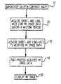
- step 10 Gd-DPTA MR contrast agent is administered in a living patient. Then, to make use of the necessary waiting time, multiple short- and long-axis cine MR data acquisitions are carried out in step 20. After completing the cine acquisitions, the inversion time required to null the signal from normal myocardium is empirically determined by using a series of T1-weighted MR data acquisitions.
- short- and long-axis T1-weighted MR image data are acquired from the patient's heart in step 30.
- this is done by gating the RF inversion pulse to the patient's ECG signal, waiting the empirically determined inversion time, and then acquiring some or all the lines of MR image data. If image acquisition is carried out by segmenting the k-space matrix, the application of RF-gated inversion pulses, the waiting for the empirically-determined inversion time and the acquisition of the MR image data is repeated until the entire k-space matrix has been filled with lines of data. If image acquisition is carried out in a single-shot fashion, all the lines of MR image data are acquired following a single RF-gated inversion pulse.
Landscapes
- Health & Medical Sciences (AREA)
- Physics & Mathematics (AREA)
- Life Sciences & Earth Sciences (AREA)
- Nuclear Medicine, Radiotherapy & Molecular Imaging (AREA)
- General Health & Medical Sciences (AREA)
- Radiology & Medical Imaging (AREA)
- Engineering & Computer Science (AREA)
- High Energy & Nuclear Physics (AREA)
- Signal Processing (AREA)
- Medical Informatics (AREA)
- Condensed Matter Physics & Semiconductors (AREA)
- Biophysics (AREA)
- Pathology (AREA)
- Biomedical Technology (AREA)
- Heart & Thoracic Surgery (AREA)
- General Physics & Mathematics (AREA)
- Molecular Biology (AREA)
- Surgery (AREA)
- Animal Behavior & Ethology (AREA)
- Public Health (AREA)
- Veterinary Medicine (AREA)
- Medicines Containing Antibodies Or Antigens For Use As Internal Diagnostic Agents (AREA)
- Magnetic Resonance Imaging Apparatus (AREA)
Applications Claiming Priority (2)
| Application Number | Priority Date | Filing Date | Title |
|---|---|---|---|
| US162548 | 1993-12-06 | ||
| US09/162,548 US6205349B1 (en) | 1998-09-29 | 1998-09-29 | Differentiating normal living myocardial tissue, injured living myocardial tissue, and infarcted myocardial tissue in vivo using magnetic resonance imaging |
Publications (2)
| Publication Number | Publication Date |
|---|---|
| EP0994352A2 true EP0994352A2 (fr) | 2000-04-19 |
| EP0994352A3 EP0994352A3 (fr) | 2002-03-13 |
Family
ID=22586103
Family Applications (1)
| Application Number | Title | Priority Date | Filing Date |
|---|---|---|---|
| EP99307707A Withdrawn EP0994352A3 (fr) | 1998-09-29 | 1999-09-29 | Imagerie de tissus myocardiques par résonance magnétique amplifiée par un agent de contraste |
Country Status (3)
| Country | Link |
|---|---|
| US (1) | US6205349B1 (fr) |
| EP (1) | EP0994352A3 (fr) |
| JP (1) | JP4422830B2 (fr) |
Cited By (5)
| Publication number | Priority date | Publication date | Assignee | Title |
|---|---|---|---|---|
| WO2006033066A1 (fr) * | 2004-09-23 | 2006-03-30 | Koninklijke Philips Electronics N.V. | Appareil, logiciel et procede pour traiter des images du coeur d'un patient |
| DE102006014882A1 (de) * | 2006-03-30 | 2007-11-15 | Siemens Ag | Verfahren zum Abbilden des Myokards eines Infarktpatienten und Verfahren zum Unterstützen eines therapeutischen Eingriffs am Herzen |
| DE102006045174A1 (de) * | 2006-09-25 | 2008-04-03 | Siemens Ag | Verfahren sowie Bildverarbeitungseinheit und medizinisches Bildaufnahmegerät zum Herstellen eines kontrastverbesserten Bilddatensatzes eines Untersuchungsbereichs eines Patienten |
| EP1538981A4 (fr) * | 2002-07-29 | 2009-04-01 | Univ Wake Forest | Diagnostics cardiaques par imagerie irm cardiaque de perfusion et de deplacement de paroi et systemes de diagnostics cardiaques |
| EP4051376A4 (fr) * | 2019-10-29 | 2023-11-22 | Elekta Ltd. | Ablation cardiaque au moyen d'un rm-linac |
Families Citing this family (29)
| Publication number | Priority date | Publication date | Assignee | Title |
|---|---|---|---|---|
| US6526307B2 (en) * | 2000-12-29 | 2003-02-25 | Ge Medical Systems Global Technology Co., Llc | Method and apparatus to improve myocardial infarction detection with blood pool signal suppression |
| US7327862B2 (en) * | 2001-04-30 | 2008-02-05 | Chase Medical, L.P. | System and method for facilitating cardiac intervention |
| US7526112B2 (en) * | 2001-04-30 | 2009-04-28 | Chase Medical, L.P. | System and method for facilitating cardiac intervention |
| DE10200371B4 (de) * | 2002-01-08 | 2009-10-15 | Siemens Ag | Verfahren zur Durchführung einer dynamischen Magnetresonanz Messung unter Anwendung von Kontrastmittel |
| US7877130B2 (en) * | 2002-10-03 | 2011-01-25 | Siemens Medical Solutions Usa, Inc. | System and method for using delayed enhancement magnetic resonance imaging and artificial intelligence to identify non-viable myocardial tissue |
| WO2004068406A2 (fr) * | 2003-01-30 | 2004-08-12 | Chase Medical, L.P. | Procede et systeme de traitement d'image et d'evaluation de contour |
| US20050043609A1 (en) * | 2003-01-30 | 2005-02-24 | Gregory Murphy | System and method for facilitating cardiac intervention |
| JP4446049B2 (ja) * | 2003-11-06 | 2010-04-07 | 株式会社三重ティーエルオー | 心筋血流の定量化装置 |
| US20070014452A1 (en) * | 2003-12-01 | 2007-01-18 | Mitta Suresh | Method and system for image processing and assessment of a state of a heart |
| US7333643B2 (en) * | 2004-01-30 | 2008-02-19 | Chase Medical, L.P. | System and method for facilitating cardiac intervention |
| US7809426B2 (en) | 2004-04-29 | 2010-10-05 | The Cleveland Clinic Foundation | Acquiring contrast-enhanced, T1 weighted, cine magnetic resonance images |
| US8112145B2 (en) * | 2004-06-03 | 2012-02-07 | Wisconsin Alumni Research Foundation | MRI method for assessing myocardial viability |
| US7640046B2 (en) * | 2004-06-18 | 2009-12-29 | Cardiac Pacemakers, Inc. | Methods and apparatuses for localizing myocardial infarction during catheterization |
| US20070110666A1 (en) * | 2005-09-30 | 2007-05-17 | Paul Pevsner | Methods for preparation of live body tissues for examination |
| JP2009517118A (ja) * | 2005-11-29 | 2009-04-30 | コーニンクレッカ フィリップス エレクトロニクス エヌ ヴィ | 磁気共鳴を使用して結合している及び結合していない造影剤を識別する方法 |
| WO2008049109A2 (fr) * | 2006-10-19 | 2008-04-24 | The Ohio State University | Système et procédé pour IRM de tension d'exercice cardiovasculaire |
| US8086297B2 (en) * | 2007-01-31 | 2011-12-27 | Duke University | Dark blood delayed enhancement magnetic resonance viability imaging techniques for assessing subendocardial infarcts |
| JP5242672B2 (ja) * | 2007-04-11 | 2013-07-24 | オレゴン ヘルス アンド サイエンス ユニバーシティ | 心臓における線維症の非侵襲性の定量的検出のためのシステム |
| US20120288481A1 (en) | 2009-11-09 | 2012-11-15 | The Brigham And Women's Hospital, Inc. | Treatment of heart disease |
| US20120321595A1 (en) | 2009-11-09 | 2012-12-20 | The Brigham And Women's Hospital, Inc. | Treatment of heart disease |
| WO2011114264A1 (fr) | 2010-03-18 | 2011-09-22 | Koninklijke Philips Electronics N.V. | Détermination simultanée et dynamique des durées de relaxation longitudinale et transversale dans un système de spin nucléaire |
| US9149203B2 (en) | 2010-05-05 | 2015-10-06 | Duke University | Blood signal suppressed enhanced magnetic resonance imaging |
| US9131870B2 (en) | 2010-05-05 | 2015-09-15 | Duke University | Blood signal suppressed contrast enhanced magnetic resonance imaging |
| US8682054B2 (en) | 2010-11-15 | 2014-03-25 | Siemens Aktiengesellschaft | Method and system for propagation of myocardial infarction from delayed enhanced cardiac imaging to cine magnetic resonance imaging using hybrid image registration |
| JP6214563B2 (ja) | 2012-01-27 | 2017-10-18 | コーニンクレッカ フィリップス エヌ ヴェKoninklijke Philips N.V. | 定量的t1マッピングを用いた、リスクに晒されたエリアの自動化された検出 |
| DE102012201832B4 (de) | 2012-02-08 | 2014-06-05 | Siemens Aktiengesellschaft | Verfahren und Magnetresonanzeinrichtung zur Ermittlung einer elektrischen Leitungsweginformation in einer Kammerwand einer Herzkammer |
| JP5971684B2 (ja) | 2012-02-15 | 2016-08-17 | 東芝メディカルシステムズ株式会社 | 磁気共鳴撮像装置 |
| WO2014071249A1 (fr) | 2012-11-02 | 2014-05-08 | The Regents Of The University Of California | Imagerie par résonance magnétique (irm) d'amélioration de gadolinium tardif cardiaque améliorée pour des patients ayant des dispositifs cardiaques implantés |
| EP3477324B1 (fr) * | 2017-10-31 | 2025-03-26 | Pie Medical Imaging BV | Amélioration de la segmentation du ventricule gauche dans des ensembles de données ciné irm à contraste amélioré |
Family Cites Families (6)
| Publication number | Priority date | Publication date | Assignee | Title |
|---|---|---|---|---|
| US5387410A (en) * | 1983-03-18 | 1995-02-07 | Mallinckrodt, Inc. | Method for enhancing magnetic resonance with compositions containing paramagnetic elements carried by liposomes |
| US4859450A (en) * | 1984-08-13 | 1989-08-22 | The General Hospital Corporation | Method of NMR imaging using antibody to cardiac myosin |
| US5377681A (en) * | 1989-11-13 | 1995-01-03 | University Of Florida | Method of diagnosing impaired blood flow |
| IL98744A0 (en) * | 1990-07-06 | 1992-07-15 | Gen Hospital Corp | Method of studying biological tissue using monocrystalline particles |
| US5111820A (en) * | 1990-11-02 | 1992-05-12 | The Trustees Of The University Of Pennsylvania | System and method for magnetic resonance imaging of 3-dimensional heart wall motion with spatial modulation of magnetization |
| US5910112A (en) * | 1996-11-08 | 1999-06-08 | Northwestern University | 23 NA and 39 K imaging of the heart |
-
1998
- 1998-09-29 US US09/162,548 patent/US6205349B1/en not_active Expired - Lifetime
-
1999
- 1999-09-29 EP EP99307707A patent/EP0994352A3/fr not_active Withdrawn
- 1999-09-29 JP JP27684799A patent/JP4422830B2/ja not_active Expired - Lifetime
Non-Patent Citations (1)
| Title |
|---|
| No relevant documents disclosed * |
Cited By (10)
| Publication number | Priority date | Publication date | Assignee | Title |
|---|---|---|---|---|
| EP1538981A4 (fr) * | 2002-07-29 | 2009-04-01 | Univ Wake Forest | Diagnostics cardiaques par imagerie irm cardiaque de perfusion et de deplacement de paroi et systemes de diagnostics cardiaques |
| US7818043B2 (en) | 2002-07-29 | 2010-10-19 | Wake Forest University Health Sciences | Cardiac diagnostics using wall motion and perfusion cardiac MRI imaging and systems for cardiac diagnostics |
| EP2430976A1 (fr) * | 2002-07-29 | 2012-03-21 | Wake Forest University | Diagnostics cardiaques par imagerie IRM cardiaque de perfusion et de déplacement de paroi et systèmes de diagnostics cardiaques |
| US8290567B2 (en) | 2002-07-29 | 2012-10-16 | Wake Forest University Health Sciences | Cardiac diagnostics using wall motion and perfusion cardiac MRI imaging and systems for cardiac diagnostics |
| US8874190B2 (en) | 2002-07-29 | 2014-10-28 | Wake Forest University Health Sciences | Cardiac diagnostics using wall motion and perfusion cardiac MRI imaging and systems for cardiac diagnostics |
| WO2006033066A1 (fr) * | 2004-09-23 | 2006-03-30 | Koninklijke Philips Electronics N.V. | Appareil, logiciel et procede pour traiter des images du coeur d'un patient |
| DE102006014882A1 (de) * | 2006-03-30 | 2007-11-15 | Siemens Ag | Verfahren zum Abbilden des Myokards eines Infarktpatienten und Verfahren zum Unterstützen eines therapeutischen Eingriffs am Herzen |
| DE102006045174A1 (de) * | 2006-09-25 | 2008-04-03 | Siemens Ag | Verfahren sowie Bildverarbeitungseinheit und medizinisches Bildaufnahmegerät zum Herstellen eines kontrastverbesserten Bilddatensatzes eines Untersuchungsbereichs eines Patienten |
| US8077941B2 (en) | 2006-09-25 | 2011-12-13 | Siemens Aktiengesellschaft | Method and imaging processing unit and medical imaging device for producing a contrast enhanced image data record of an examination region of a patient |
| EP4051376A4 (fr) * | 2019-10-29 | 2023-11-22 | Elekta Ltd. | Ablation cardiaque au moyen d'un rm-linac |
Also Published As
| Publication number | Publication date |
|---|---|
| US6205349B1 (en) | 2001-03-20 |
| JP2000116621A (ja) | 2000-04-25 |
| EP0994352A3 (fr) | 2002-03-13 |
| JP4422830B2 (ja) | 2010-02-24 |
Similar Documents
| Publication | Publication Date | Title |
|---|---|---|
| US6205349B1 (en) | Differentiating normal living myocardial tissue, injured living myocardial tissue, and infarcted myocardial tissue in vivo using magnetic resonance imaging | |
| US7047060B1 (en) | Multiple preparatory excitations and readouts distributed over the cardiac cycle | |
| US7809426B2 (en) | Acquiring contrast-enhanced, T1 weighted, cine magnetic resonance images | |
| Gefter et al. | Pulmonary thromboembolism: recent developments in diagnosis with CT and MR imaging. | |
| US9008753B2 (en) | Look-locker IR-SSFP for cardiac MR imaging with simultaneous generation of cardiac T1 maps, cine images and IR-prepared images | |
| US6295465B1 (en) | Myocardial perfusion studies using magnetic resonance imaging | |
| JP2005515809A (ja) | 導出されたパラメトリック画像に基づく造影剤取り込みの評価を行う方法及び装置 | |
| Chapon et al. | High field magnetic resonance imaging evaluation of superparamagnetic iron oxide nanoparticles in a permanent rat myocardial infarction | |
| Penzkofer et al. | Assessment of myocardial perfusion using multisection first-pass MRI and color-coded parameter maps:: A comparison to 99mTc Sesta MIBI SPECT and systolic myocardial wall thickening analysis | |
| US8112145B2 (en) | MRI method for assessing myocardial viability | |
| Takase et al. | Whole-heart dipyridamole stress first-pass myocardial perfusion MRI for the detection of coronary artery disease | |
| WO2014196732A1 (fr) | Nouvelle technique d'image par résonance magnétique pour une imagerie et une évaluation de circulation collatérale | |
| US6009341A (en) | Three-dimensional magnetic resonance angiography of coronary arteries | |
| Dewey et al. | Myocardial viability: assessment with three-dimensional MR imaging in pigs and patients | |
| Lim et al. | Novel application of breath‐hold turbo spin‐echo T2 MRI for detection of acute myocardial infarction | |
| Been et al. | Characterisation of acute myocardial infarction by gated magnetic resonance imaging | |
| Park et al. | Usefulness of multidetector-row CT in the evaluation of reperfused myocardial infarction in a rabbit model | |
| Peukert et al. | 3D and 2D delayed-enhancement magnetic resonance imaging for detection of myocardial infarction: preclinical and clinical results | |
| Matsunaka et al. | First-pass myocardial perfusion defect and delayed contrast enhancement in hypertrophic cardiomyopathy assessed with MRI | |
| Wintersperger et al. | Multislice MR perfusion imaging and regional myocardial function analysis: complimentary findings in chronic myocardial ischemia | |
| Thiele et al. | Color-encoded semiautomatic analysis of multi-slice first-pass magnetic resonance perfusion: comparison to tetrofosmin single photon emission computed tomography perfusion and X-ray angiography | |
| Aisen et al. | Visualization of myocardial infarction and subsequent coronary reperfusion with MRI using a dog model | |
| van Ooijen et al. | Noninvasive coronary imaging: CT versus MR | |
| Lombardi et al. | MRI for the evaluation of regional myocardial perfusion in an experimental animal model | |
| Gnyawali et al. | Remodeling of the ischemia-reperfused murine heart: 11.7-T cardiac magnetic resonance imaging of contrast-enhanced infarct patches and transmurality |
Legal Events
| Date | Code | Title | Description |
|---|---|---|---|
| PUAI | Public reference made under article 153(3) epc to a published international application that has entered the european phase |
Free format text: ORIGINAL CODE: 0009012 |
|
| AK | Designated contracting states |
Kind code of ref document: A2 Designated state(s): AT BE CH CY DE DK ES FI FR GB GR IE IT LI LU MC NL PT SE |
|
| AX | Request for extension of the european patent |
Free format text: AL;LT;LV;MK;RO;SI |
|
| PUAL | Search report despatched |
Free format text: ORIGINAL CODE: 0009013 |
|
| AK | Designated contracting states |
Kind code of ref document: A3 Designated state(s): AT BE CH CY DE DK ES FI FR GB GR IE IT LI LU MC NL PT SE |
|
| AX | Request for extension of the european patent |
Free format text: AL;LT;LV;MK;RO;SI |
|
| RIC1 | Information provided on ipc code assigned before grant |
Free format text: 7G 01N 33/28 A, 7G 01R 33/28 B |
|
| STAA | Information on the status of an ep patent application or granted ep patent |
Free format text: STATUS: THE APPLICATION HAS BEEN WITHDRAWN |
|
| 18W | Application withdrawn |
Withdrawal date: 20020527 |