EP0985028B1 - Transplants for myocardial scars and method and cellular preparations therefor - Google Patents
Transplants for myocardial scars and method and cellular preparations therefor Download PDFInfo
- Publication number
- EP0985028B1 EP0985028B1 EP98923950A EP98923950A EP0985028B1 EP 0985028 B1 EP0985028 B1 EP 0985028B1 EP 98923950 A EP98923950 A EP 98923950A EP 98923950 A EP98923950 A EP 98923950A EP 0985028 B1 EP0985028 B1 EP 0985028B1
- Authority
- EP
- European Patent Office
- Prior art keywords
- cells
- transplanted
- tissue
- scar
- cardiomyocytes
- Prior art date
- Legal status (The legal status is an assumption and is not a legal conclusion. Google has not performed a legal analysis and makes no representation as to the accuracy of the status listed.)
- Expired - Lifetime
Links
- 231100000241 scar Toxicity 0.000 title claims abstract description 284
- 230000002107 myocardial effect Effects 0.000 title claims description 82
- 238000002360 preparation method Methods 0.000 title claims description 18
- 238000000034 method Methods 0.000 title abstract description 64
- 208000032544 Cicatrix Diseases 0.000 title description 26
- 230000037387 scars Effects 0.000 title description 26
- 230000001413 cellular effect Effects 0.000 title description 15
- 210000004027 cell Anatomy 0.000 claims abstract description 245
- 210000001519 tissue Anatomy 0.000 claims abstract description 238
- 210000004413 cardiac myocyte Anatomy 0.000 claims abstract description 203
- 210000002216 heart Anatomy 0.000 claims abstract description 179
- 210000002889 endothelial cell Anatomy 0.000 claims abstract description 99
- 210000002950 fibroblast Anatomy 0.000 claims abstract description 52
- 210000000329 smooth muscle myocyte Anatomy 0.000 claims abstract description 38
- 230000004217 heart function Effects 0.000 claims description 44
- 230000000747 cardiac effect Effects 0.000 claims description 19
- 239000007924 injection Substances 0.000 claims description 19
- 238000002347 injection Methods 0.000 claims description 19
- 102000005789 Vascular Endothelial Growth Factors Human genes 0.000 claims description 15
- 108010019530 Vascular Endothelial Growth Factors Proteins 0.000 claims description 15
- 239000003102 growth factor Substances 0.000 claims description 15
- 239000008194 pharmaceutical composition Substances 0.000 claims description 13
- 238000002278 reconstructive surgery Methods 0.000 claims description 7
- 230000001225 therapeutic effect Effects 0.000 claims description 6
- 239000003364 biologic glue Substances 0.000 claims description 5
- 108010073929 Vascular Endothelial Growth Factor A Proteins 0.000 claims description 4
- 102000003974 Fibroblast growth factor 2 Human genes 0.000 claims description 3
- 108090000379 Fibroblast growth factor 2 Proteins 0.000 claims description 3
- 108010019674 Proto-Oncogene Proteins c-sis Proteins 0.000 claims description 3
- 238000002689 xenotransplantation Methods 0.000 claims description 3
- NOESYZHRGYRDHS-UHFFFAOYSA-N insulin Chemical compound N1C(=O)C(NC(=O)C(CCC(N)=O)NC(=O)C(CCC(O)=O)NC(=O)C(C(C)C)NC(=O)C(NC(=O)CN)C(C)CC)CSSCC(C(NC(CO)C(=O)NC(CC(C)C)C(=O)NC(CC=2C=CC(O)=CC=2)C(=O)NC(CCC(N)=O)C(=O)NC(CC(C)C)C(=O)NC(CCC(O)=O)C(=O)NC(CC(N)=O)C(=O)NC(CC=2C=CC(O)=CC=2)C(=O)NC(CSSCC(NC(=O)C(C(C)C)NC(=O)C(CC(C)C)NC(=O)C(CC=2C=CC(O)=CC=2)NC(=O)C(CC(C)C)NC(=O)C(C)NC(=O)C(CCC(O)=O)NC(=O)C(C(C)C)NC(=O)C(CC(C)C)NC(=O)C(CC=2NC=NC=2)NC(=O)C(CO)NC(=O)CNC2=O)C(=O)NCC(=O)NC(CCC(O)=O)C(=O)NC(CCCNC(N)=N)C(=O)NCC(=O)NC(CC=3C=CC=CC=3)C(=O)NC(CC=3C=CC=CC=3)C(=O)NC(CC=3C=CC(O)=CC=3)C(=O)NC(C(C)O)C(=O)N3C(CCC3)C(=O)NC(CCCCN)C(=O)NC(C)C(O)=O)C(=O)NC(CC(N)=O)C(O)=O)=O)NC(=O)C(C(C)CC)NC(=O)C(CO)NC(=O)C(C(C)O)NC(=O)C1CSSCC2NC(=O)C(CC(C)C)NC(=O)C(NC(=O)C(CCC(N)=O)NC(=O)C(CC(N)=O)NC(=O)C(NC(=O)C(N)CC=1C=CC=CC=1)C(C)C)CC1=CN=CN1 NOESYZHRGYRDHS-UHFFFAOYSA-N 0.000 claims 2
- 102000004877 Insulin Human genes 0.000 claims 1
- 108090001061 Insulin Proteins 0.000 claims 1
- 108010094028 Prothrombin Proteins 0.000 claims 1
- AGVAZMGAQJOSFJ-WZHZPDAFSA-M cobalt(2+);[(2r,3s,4r,5s)-5-(5,6-dimethylbenzimidazol-1-yl)-4-hydroxy-2-(hydroxymethyl)oxolan-3-yl] [(2r)-1-[3-[(1r,2r,3r,4z,7s,9z,12s,13s,14z,17s,18s,19r)-2,13,18-tris(2-amino-2-oxoethyl)-7,12,17-tris(3-amino-3-oxopropyl)-3,5,8,8,13,15,18,19-octamethyl-2 Chemical compound [Co+2].N#[C-].[N-]([C@@H]1[C@H](CC(N)=O)[C@@]2(C)CCC(=O)NC[C@@H](C)OP(O)(=O)O[C@H]3[C@H]([C@H](O[C@@H]3CO)N3C4=CC(C)=C(C)C=C4N=C3)O)\C2=C(C)/C([C@H](C\2(C)C)CCC(N)=O)=N/C/2=C\C([C@H]([C@@]/2(CC(N)=O)C)CCC(N)=O)=N\C\2=C(C)/C2=N[C@]1(C)[C@@](C)(CC(N)=O)[C@@H]2CCC(N)=O AGVAZMGAQJOSFJ-WZHZPDAFSA-M 0.000 claims 1
- BTCSSZJGUNDROE-UHFFFAOYSA-N gamma-aminobutyric acid Chemical compound NCCCC(O)=O BTCSSZJGUNDROE-UHFFFAOYSA-N 0.000 claims 1
- 229940125396 insulin Drugs 0.000 claims 1
- 230000002608 insulinlike Effects 0.000 claims 1
- 230000001131 transforming effect Effects 0.000 claims 1
- 238000002054 transplantation Methods 0.000 abstract description 109
- 210000005003 heart tissue Anatomy 0.000 abstract description 27
- 238000012258 culturing Methods 0.000 abstract description 23
- 210000004683 skeletal myoblast Anatomy 0.000 abstract description 9
- 241000700159 Rattus Species 0.000 description 78
- 230000002861 ventricular Effects 0.000 description 76
- 230000001605 fetal effect Effects 0.000 description 49
- 241001465754 Metazoa Species 0.000 description 48
- LOKCTEFSRHRXRJ-UHFFFAOYSA-I dipotassium trisodium dihydrogen phosphate hydrogen phosphate dichloride Chemical compound P(=O)(O)(O)[O-].[K+].P(=O)(O)([O-])[O-].[Na+].[Na+].[Cl-].[K+].[Cl-].[Na+] LOKCTEFSRHRXRJ-UHFFFAOYSA-I 0.000 description 45
- 239000002953 phosphate buffered saline Substances 0.000 description 45
- 239000000243 solution Substances 0.000 description 42
- 210000004165 myocardium Anatomy 0.000 description 41
- 230000033115 angiogenesis Effects 0.000 description 38
- 208000027418 Wounds and injury Diseases 0.000 description 37
- 239000001963 growth medium Substances 0.000 description 37
- 208000014674 injury Diseases 0.000 description 37
- FAPWRFPIFSIZLT-UHFFFAOYSA-M Sodium chloride Chemical compound [Na+].[Cl-] FAPWRFPIFSIZLT-UHFFFAOYSA-M 0.000 description 27
- 230000001965 increasing effect Effects 0.000 description 27
- 230000001746 atrial effect Effects 0.000 description 26
- 230000006870 function Effects 0.000 description 26
- 208000037891 myocardial injury Diseases 0.000 description 26
- 108091003079 Bovine Serum Albumin Proteins 0.000 description 21
- OKKJLVBELUTLKV-UHFFFAOYSA-N Methanol Chemical compound OC OKKJLVBELUTLKV-UHFFFAOYSA-N 0.000 description 21
- 230000017531 blood circulation Effects 0.000 description 21
- 239000012091 fetal bovine serum Substances 0.000 description 21
- 239000002609 medium Substances 0.000 description 20
- LFQSCWFLJHTTHZ-UHFFFAOYSA-N Ethanol Chemical compound CCO LFQSCWFLJHTTHZ-UHFFFAOYSA-N 0.000 description 18
- WSFSSNUMVMOOMR-UHFFFAOYSA-N Formaldehyde Chemical compound O=C WSFSSNUMVMOOMR-UHFFFAOYSA-N 0.000 description 17
- 206010061218 Inflammation Diseases 0.000 description 17
- 230000004054 inflammatory process Effects 0.000 description 17
- 239000007760 Iscove's Modified Dulbecco's Medium Substances 0.000 description 16
- 230000000694 effects Effects 0.000 description 16
- 108090000631 Trypsin Proteins 0.000 description 15
- 102000004142 Trypsin Human genes 0.000 description 15
- 238000004458 analytical method Methods 0.000 description 15
- 239000012588 trypsin Substances 0.000 description 15
- IJGRMHOSHXDMSA-UHFFFAOYSA-N Atomic nitrogen Chemical compound N#N IJGRMHOSHXDMSA-UHFFFAOYSA-N 0.000 description 14
- 210000004748 cultured cell Anatomy 0.000 description 14
- 206010020871 hypertrophic cardiomyopathy Diseases 0.000 description 14
- 230000010016 myocardial function Effects 0.000 description 14
- 108090000623 proteins and genes Proteins 0.000 description 14
- 239000011780 sodium chloride Substances 0.000 description 14
- UCSJYZPVAKXKNQ-HZYVHMACSA-N streptomycin Chemical compound CN[C@H]1[C@H](O)[C@@H](O)[C@H](CO)O[C@H]1O[C@@H]1[C@](C=O)(O)[C@H](C)O[C@H]1O[C@@H]1[C@@H](NC(N)=N)[C@H](O)[C@@H](NC(N)=N)[C@H](O)[C@H]1O UCSJYZPVAKXKNQ-HZYVHMACSA-N 0.000 description 14
- 238000001356 surgical procedure Methods 0.000 description 14
- 210000004379 membrane Anatomy 0.000 description 13
- 239000012528 membrane Substances 0.000 description 13
- 210000003205 muscle Anatomy 0.000 description 13
- WZUVPPKBWHMQCE-UHFFFAOYSA-N Haematoxylin Natural products C12=CC(O)=C(O)C=C2CC2(O)C1C1=CC=C(O)C(O)=C1OC2 WZUVPPKBWHMQCE-UHFFFAOYSA-N 0.000 description 12
- 108090000723 Insulin-Like Growth Factor I Proteins 0.000 description 12
- 210000004204 blood vessel Anatomy 0.000 description 12
- 230000037396 body weight Effects 0.000 description 12
- 239000006285 cell suspension Substances 0.000 description 12
- 239000008188 pellet Substances 0.000 description 12
- 239000006228 supernatant Substances 0.000 description 12
- WQZGKKKJIJFFOK-GASJEMHNSA-N Glucose Natural products OC[C@H]1OC(O)[C@H](O)[C@@H](O)[C@@H]1O WQZGKKKJIJFFOK-GASJEMHNSA-N 0.000 description 11
- 238000004113 cell culture Methods 0.000 description 11
- 239000006143 cell culture medium Substances 0.000 description 11
- 239000008103 glucose Substances 0.000 description 11
- 206010061216 Infarction Diseases 0.000 description 10
- 108010005774 beta-Galactosidase Proteins 0.000 description 10
- 238000005259 measurement Methods 0.000 description 10
- 239000004005 microsphere Substances 0.000 description 10
- 229910052757 nitrogen Inorganic materials 0.000 description 10
- 102000029816 Collagenase Human genes 0.000 description 9
- 108060005980 Collagenase Proteins 0.000 description 9
- PEDCQBHIVMGVHV-UHFFFAOYSA-N Glycerine Chemical compound OCC(O)CO PEDCQBHIVMGVHV-UHFFFAOYSA-N 0.000 description 9
- 102000004218 Insulin-Like Growth Factor I Human genes 0.000 description 9
- 239000000872 buffer Substances 0.000 description 9
- 229960002424 collagenase Drugs 0.000 description 9
- 230000035487 diastolic blood pressure Effects 0.000 description 9
- 230000006862 enzymatic digestion Effects 0.000 description 9
- 230000007574 infarction Effects 0.000 description 9
- 210000005240 left ventricle Anatomy 0.000 description 9
- 210000004698 lymphocyte Anatomy 0.000 description 9
- WEXRUCMBJFQVBZ-UHFFFAOYSA-N pentobarbital Chemical compound CCCC(C)C1(CC)C(=O)NC(=O)NC1=O WEXRUCMBJFQVBZ-UHFFFAOYSA-N 0.000 description 9
- 239000013612 plasmid Substances 0.000 description 9
- 102000004169 proteins and genes Human genes 0.000 description 9
- 239000000725 suspension Substances 0.000 description 9
- 210000003556 vascular endothelial cell Anatomy 0.000 description 9
- 102000004190 Enzymes Human genes 0.000 description 8
- 108090000790 Enzymes Proteins 0.000 description 8
- 230000006378 damage Effects 0.000 description 8
- 230000003205 diastolic effect Effects 0.000 description 8
- 229940088598 enzyme Drugs 0.000 description 8
- YQGOJNYOYNNSMM-UHFFFAOYSA-N eosin Chemical compound [Na+].OC(=O)C1=CC=CC=C1C1=C2C=C(Br)C(=O)C(Br)=C2OC2=C(Br)C(O)=C(Br)C=C21 YQGOJNYOYNNSMM-UHFFFAOYSA-N 0.000 description 8
- 239000008363 phosphate buffer Substances 0.000 description 8
- 230000035488 systolic blood pressure Effects 0.000 description 8
- CURLTUGMZLYLDI-UHFFFAOYSA-N Carbon dioxide Chemical compound O=C=O CURLTUGMZLYLDI-UHFFFAOYSA-N 0.000 description 7
- 229930182555 Penicillin Natural products 0.000 description 7
- JGSARLDLIJGVTE-MBNYWOFBSA-N Penicillin G Chemical compound N([C@H]1[C@H]2SC([C@@H](N2C1=O)C(O)=O)(C)C)C(=O)CC1=CC=CC=C1 JGSARLDLIJGVTE-MBNYWOFBSA-N 0.000 description 7
- 238000010171 animal model Methods 0.000 description 7
- 102000005936 beta-Galactosidase Human genes 0.000 description 7
- 230000008602 contraction Effects 0.000 description 7
- 239000007788 liquid Substances 0.000 description 7
- 239000000463 material Substances 0.000 description 7
- 210000000663 muscle cell Anatomy 0.000 description 7
- 230000003680 myocardial damage Effects 0.000 description 7
- 239000012188 paraffin wax Substances 0.000 description 7
- 229940049954 penicillin Drugs 0.000 description 7
- 229960005322 streptomycin Drugs 0.000 description 7
- DGVVWUTYPXICAM-UHFFFAOYSA-N β‐Mercaptoethanol Chemical compound OCCS DGVVWUTYPXICAM-UHFFFAOYSA-N 0.000 description 7
- WZUVPPKBWHMQCE-XJKSGUPXSA-N (+)-haematoxylin Chemical compound C12=CC(O)=C(O)C=C2C[C@]2(O)[C@H]1C1=CC=C(O)C(O)=C1OC2 WZUVPPKBWHMQCE-XJKSGUPXSA-N 0.000 description 6
- 108010051609 Cardiac Myosins Proteins 0.000 description 6
- 102000013602 Cardiac Myosins Human genes 0.000 description 6
- CSNNHWWHGAXBCP-UHFFFAOYSA-L Magnesium sulfate Chemical compound [Mg+2].[O-][S+2]([O-])([O-])[O-] CSNNHWWHGAXBCP-UHFFFAOYSA-L 0.000 description 6
- 241000283973 Oryctolagus cuniculus Species 0.000 description 6
- 102000003992 Peroxidases Human genes 0.000 description 6
- UIIMBOGNXHQVGW-UHFFFAOYSA-M Sodium bicarbonate Chemical compound [Na+].OC([O-])=O UIIMBOGNXHQVGW-UHFFFAOYSA-M 0.000 description 6
- 208000033774 Ventricular Remodeling Diseases 0.000 description 6
- 230000001154 acute effect Effects 0.000 description 6
- 230000000735 allogeneic effect Effects 0.000 description 6
- 229910002092 carbon dioxide Inorganic materials 0.000 description 6
- 238000002695 general anesthesia Methods 0.000 description 6
- 238000002955 isolation Methods 0.000 description 6
- 208000010125 myocardial infarction Diseases 0.000 description 6
- 229960001412 pentobarbital Drugs 0.000 description 6
- 108040007629 peroxidase activity proteins Proteins 0.000 description 6
- 238000011160 research Methods 0.000 description 6
- 210000003491 skin Anatomy 0.000 description 6
- 238000010186 staining Methods 0.000 description 6
- 230000004083 survival effect Effects 0.000 description 6
- 210000000115 thoracic cavity Anatomy 0.000 description 6
- 230000007998 vessel formation Effects 0.000 description 6
- 108010047303 von Willebrand Factor Proteins 0.000 description 6
- 102100036537 von Willebrand factor Human genes 0.000 description 6
- 238000005406 washing Methods 0.000 description 6
- LCSKNASZPVZHEG-UHFFFAOYSA-N 3,6-dimethyl-1,4-dioxane-2,5-dione;1,4-dioxane-2,5-dione Chemical group O=C1COC(=O)CO1.CC1OC(=O)C(C)OC1=O LCSKNASZPVZHEG-UHFFFAOYSA-N 0.000 description 5
- 241000283707 Capra Species 0.000 description 5
- 102000008186 Collagen Human genes 0.000 description 5
- 108010035532 Collagen Proteins 0.000 description 5
- PMATZTZNYRCHOR-CGLBZJNRSA-N Cyclosporin A Chemical compound CC[C@@H]1NC(=O)[C@H]([C@H](O)[C@H](C)C\C=C\C)N(C)C(=O)[C@H](C(C)C)N(C)C(=O)[C@H](CC(C)C)N(C)C(=O)[C@H](CC(C)C)N(C)C(=O)[C@@H](C)NC(=O)[C@H](C)NC(=O)[C@H](CC(C)C)N(C)C(=O)[C@H](C(C)C)NC(=O)[C@H](CC(C)C)N(C)C(=O)CN(C)C1=O PMATZTZNYRCHOR-CGLBZJNRSA-N 0.000 description 5
- 108010036949 Cyclosporine Proteins 0.000 description 5
- 239000007836 KH2PO4 Substances 0.000 description 5
- 241000700157 Rattus norvegicus Species 0.000 description 5
- 210000000709 aorta Anatomy 0.000 description 5
- 230000008901 benefit Effects 0.000 description 5
- 229920001436 collagen Polymers 0.000 description 5
- 210000002808 connective tissue Anatomy 0.000 description 5
- 230000008828 contractile function Effects 0.000 description 5
- 230000007547 defect Effects 0.000 description 5
- 230000029087 digestion Effects 0.000 description 5
- 229920000669 heparin Polymers 0.000 description 5
- 230000002757 inflammatory effect Effects 0.000 description 5
- 230000003993 interaction Effects 0.000 description 5
- 239000007928 intraperitoneal injection Substances 0.000 description 5
- 229910000402 monopotassium phosphate Inorganic materials 0.000 description 5
- 210000000107 myocyte Anatomy 0.000 description 5
- GNSKLFRGEWLPPA-UHFFFAOYSA-M potassium dihydrogen phosphate Chemical compound [K+].OP(O)([O-])=O GNSKLFRGEWLPPA-UHFFFAOYSA-M 0.000 description 5
- 238000000746 purification Methods 0.000 description 5
- 238000007634 remodeling Methods 0.000 description 5
- 238000003860 storage Methods 0.000 description 5
- 238000001890 transfection Methods 0.000 description 5
- 238000010792 warming Methods 0.000 description 5
- XLYOFNOQVPJJNP-UHFFFAOYSA-N water Substances O XLYOFNOQVPJJNP-UHFFFAOYSA-N 0.000 description 5
- YBJHBAHKTGYVGT-ZKWXMUAHSA-N (+)-Biotin Chemical compound N1C(=O)N[C@@H]2[C@H](CCCCC(=O)O)SC[C@@H]21 YBJHBAHKTGYVGT-ZKWXMUAHSA-N 0.000 description 4
- QTBSBXVTEAMEQO-UHFFFAOYSA-N Acetic acid Chemical compound CC(O)=O QTBSBXVTEAMEQO-UHFFFAOYSA-N 0.000 description 4
- 108010085238 Actins Proteins 0.000 description 4
- 102000007469 Actins Human genes 0.000 description 4
- UXVMQQNJUSDDNG-UHFFFAOYSA-L Calcium chloride Chemical compound [Cl-].[Cl-].[Ca+2] UXVMQQNJUSDDNG-UHFFFAOYSA-L 0.000 description 4
- 229930105110 Cyclosporin A Natural products 0.000 description 4
- KCXVZYZYPLLWCC-UHFFFAOYSA-N EDTA Chemical compound OC(=O)CN(CC(O)=O)CCN(CC(O)=O)CC(O)=O KCXVZYZYPLLWCC-UHFFFAOYSA-N 0.000 description 4
- 108010054218 Factor VIII Proteins 0.000 description 4
- 102000001690 Factor VIII Human genes 0.000 description 4
- 108010080379 Fibrin Tissue Adhesive Proteins 0.000 description 4
- SXRSQZLOMIGNAQ-UHFFFAOYSA-N Glutaraldehyde Chemical compound O=CCCCC=O SXRSQZLOMIGNAQ-UHFFFAOYSA-N 0.000 description 4
- 108010043121 Green Fluorescent Proteins Proteins 0.000 description 4
- 102000004144 Green Fluorescent Proteins Human genes 0.000 description 4
- TWRXJAOTZQYOKJ-UHFFFAOYSA-L Magnesium chloride Chemical compound [Mg+2].[Cl-].[Cl-] TWRXJAOTZQYOKJ-UHFFFAOYSA-L 0.000 description 4
- 102000005604 Myosin Heavy Chains Human genes 0.000 description 4
- 108010084498 Myosin Heavy Chains Proteins 0.000 description 4
- 206010028851 Necrosis Diseases 0.000 description 4
- 229910019142 PO4 Inorganic materials 0.000 description 4
- 102000046299 Transforming Growth Factor beta1 Human genes 0.000 description 4
- 101800002279 Transforming growth factor beta-1 Proteins 0.000 description 4
- SXEHKFHPFVVDIR-UHFFFAOYSA-N [4-(4-hydrazinylphenyl)phenyl]hydrazine Chemical compound C1=CC(NN)=CC=C1C1=CC=C(NN)C=C1 SXEHKFHPFVVDIR-UHFFFAOYSA-N 0.000 description 4
- 238000004026 adhesive bonding Methods 0.000 description 4
- QVGXLLKOCUKJST-UHFFFAOYSA-N atomic oxygen Chemical compound [O] QVGXLLKOCUKJST-UHFFFAOYSA-N 0.000 description 4
- 238000001574 biopsy Methods 0.000 description 4
- 229960002685 biotin Drugs 0.000 description 4
- 239000011616 biotin Substances 0.000 description 4
- 210000004369 blood Anatomy 0.000 description 4
- 239000008280 blood Substances 0.000 description 4
- 239000001110 calcium chloride Substances 0.000 description 4
- 229910001628 calcium chloride Inorganic materials 0.000 description 4
- 238000007675 cardiac surgery Methods 0.000 description 4
- 229960001265 ciclosporin Drugs 0.000 description 4
- 230000004087 circulation Effects 0.000 description 4
- 230000002950 deficient Effects 0.000 description 4
- 238000001493 electron microscopy Methods 0.000 description 4
- 238000002474 experimental method Methods 0.000 description 4
- 229960000301 factor viii Drugs 0.000 description 4
- 239000005090 green fluorescent protein Substances 0.000 description 4
- 238000003125 immunofluorescent labeling Methods 0.000 description 4
- 230000006872 improvement Effects 0.000 description 4
- 238000011534 incubation Methods 0.000 description 4
- 230000006698 induction Effects 0.000 description 4
- 229910052760 oxygen Inorganic materials 0.000 description 4
- 239000001301 oxygen Substances 0.000 description 4
- 230000010412 perfusion Effects 0.000 description 4
- 239000002831 pharmacologic agent Substances 0.000 description 4
- NBIIXXVUZAFLBC-UHFFFAOYSA-K phosphate Chemical compound [O-]P([O-])([O-])=O NBIIXXVUZAFLBC-UHFFFAOYSA-K 0.000 description 4
- 239000010452 phosphate Substances 0.000 description 4
- 230000008569 process Effects 0.000 description 4
- 230000003252 repetitive effect Effects 0.000 description 4
- 239000000523 sample Substances 0.000 description 4
- 210000002027 skeletal muscle Anatomy 0.000 description 4
- 238000012453 sprague-dawley rat model Methods 0.000 description 4
- 238000007619 statistical method Methods 0.000 description 4
- 210000002784 stomach Anatomy 0.000 description 4
- 238000010257 thawing Methods 0.000 description 4
- QKNYBSVHEMOAJP-UHFFFAOYSA-N 2-amino-2-(hydroxymethyl)propane-1,3-diol;hydron;chloride Chemical compound Cl.OCC(N)(CO)CO QKNYBSVHEMOAJP-UHFFFAOYSA-N 0.000 description 3
- 206010002383 Angina Pectoris Diseases 0.000 description 3
- 206010007572 Cardiac hypertrophy Diseases 0.000 description 3
- 208000032170 Congenital Abnormalities Diseases 0.000 description 3
- 229920004934 Dacron® Polymers 0.000 description 3
- 102000010834 Extracellular Matrix Proteins Human genes 0.000 description 3
- 108010037362 Extracellular Matrix Proteins Proteins 0.000 description 3
- 206010020880 Hypertrophy Diseases 0.000 description 3
- 206010062049 Lymphocytic infiltration Diseases 0.000 description 3
- CTQNGGLPUBDAKN-UHFFFAOYSA-N O-Xylene Chemical compound CC1=CC=CC=C1C CTQNGGLPUBDAKN-UHFFFAOYSA-N 0.000 description 3
- 244000203593 Piper nigrum Species 0.000 description 3
- 229920000954 Polyglycolide Polymers 0.000 description 3
- 102000013275 Somatomedins Human genes 0.000 description 3
- 239000006180 TBST buffer Substances 0.000 description 3
- 108090000190 Thrombin Proteins 0.000 description 3
- -1 and Proteins 0.000 description 3
- 239000003242 anti bacterial agent Substances 0.000 description 3
- 229940088710 antibiotic agent Drugs 0.000 description 3
- 238000013459 approach Methods 0.000 description 3
- BVGLIYRKPOITBQ-ANPZCEIESA-N benzylpenicillin benzathine Chemical compound C=1C=CC=CC=1C[NH2+]CC[NH2+]CC1=CC=CC=C1.N([C@H]1[C@H]2SC([C@@H](N2C1=O)C([O-])=O)(C)C)C(=O)CC1=CC=CC=C1.N([C@H]1[C@H]2SC([C@@H](N2C1=O)C([O-])=O)(C)C)C(=O)CC1=CC=CC=C1 BVGLIYRKPOITBQ-ANPZCEIESA-N 0.000 description 3
- WHRVRSCEWKLAHX-LQDWTQKMSA-N benzylpenicillin procaine Chemical compound [H+].CCN(CC)CCOC(=O)C1=CC=C(N)C=C1.N([C@H]1[C@H]2SC([C@@H](N2C1=O)C([O-])=O)(C)C)C(=O)CC1=CC=CC=C1 WHRVRSCEWKLAHX-LQDWTQKMSA-N 0.000 description 3
- 230000036770 blood supply Effects 0.000 description 3
- 230000003683 cardiac damage Effects 0.000 description 3
- 238000005119 centrifugation Methods 0.000 description 3
- 210000000038 chest Anatomy 0.000 description 3
- 210000004351 coronary vessel Anatomy 0.000 description 3
- 230000003247 decreasing effect Effects 0.000 description 3
- 230000001419 dependent effect Effects 0.000 description 3
- 238000010790 dilution Methods 0.000 description 3
- 239000012895 dilution Substances 0.000 description 3
- 201000010099 disease Diseases 0.000 description 3
- 208000037265 diseases, disorders, signs and symptoms Diseases 0.000 description 3
- BNIILDVGGAEEIG-UHFFFAOYSA-L disodium hydrogen phosphate Chemical compound [Na+].[Na+].OP([O-])([O-])=O BNIILDVGGAEEIG-UHFFFAOYSA-L 0.000 description 3
- 229910000397 disodium phosphate Inorganic materials 0.000 description 3
- 230000002526 effect on cardiovascular system Effects 0.000 description 3
- 238000011156 evaluation Methods 0.000 description 3
- 230000003176 fibrotic effect Effects 0.000 description 3
- 230000008014 freezing Effects 0.000 description 3
- 238000007710 freezing Methods 0.000 description 3
- 230000014509 gene expression Effects 0.000 description 3
- 102000006602 glyceraldehyde-3-phosphate dehydrogenase Human genes 0.000 description 3
- 108020004445 glyceraldehyde-3-phosphate dehydrogenase Proteins 0.000 description 3
- 229960002897 heparin Drugs 0.000 description 3
- 229940027941 immunoglobulin g Drugs 0.000 description 3
- 238000002513 implantation Methods 0.000 description 3
- 238000007918 intramuscular administration Methods 0.000 description 3
- 239000004816 latex Substances 0.000 description 3
- 229920000126 latex Polymers 0.000 description 3
- 229910052943 magnesium sulfate Inorganic materials 0.000 description 3
- 230000007246 mechanism Effects 0.000 description 3
- 210000004115 mitral valve Anatomy 0.000 description 3
- 230000000877 morphologic effect Effects 0.000 description 3
- 239000013642 negative control Substances 0.000 description 3
- 235000019371 penicillin G benzathine Nutrition 0.000 description 3
- 238000007747 plating Methods 0.000 description 3
- 239000005020 polyethylene terephthalate Substances 0.000 description 3
- 239000004633 polyglycolic acid Substances 0.000 description 3
- 229920000642 polymer Polymers 0.000 description 3
- 108700038606 rat Smooth muscle Proteins 0.000 description 3
- 210000003752 saphenous vein Anatomy 0.000 description 3
- 238000007390 skin biopsy Methods 0.000 description 3
- 210000002460 smooth muscle Anatomy 0.000 description 3
- 229910000030 sodium bicarbonate Inorganic materials 0.000 description 3
- 230000006641 stabilisation Effects 0.000 description 3
- 238000011105 stabilization Methods 0.000 description 3
- 238000012360 testing method Methods 0.000 description 3
- 210000000779 thoracic wall Anatomy 0.000 description 3
- 229960004072 thrombin Drugs 0.000 description 3
- 229940099456 transforming growth factor beta 1 Drugs 0.000 description 3
- 229960001005 tuberculin Drugs 0.000 description 3
- 210000004509 vascular smooth muscle cell Anatomy 0.000 description 3
- 238000009423 ventilation Methods 0.000 description 3
- 238000005303 weighing Methods 0.000 description 3
- 239000008096 xylene Substances 0.000 description 3
- JKMHFZQWWAIEOD-UHFFFAOYSA-N 2-[4-(2-hydroxyethyl)piperazin-1-yl]ethanesulfonic acid Chemical compound OCC[NH+]1CCN(CCS([O-])(=O)=O)CC1 JKMHFZQWWAIEOD-UHFFFAOYSA-N 0.000 description 2
- UPXRTVAIJMUAQR-UHFFFAOYSA-N 4-(9h-fluoren-9-ylmethoxycarbonylamino)-1-[(2-methylpropan-2-yl)oxycarbonyl]pyrrolidine-2-carboxylic acid Chemical compound C1C(C(O)=O)N(C(=O)OC(C)(C)C)CC1NC(=O)OCC1C2=CC=CC=C2C2=CC=CC=C21 UPXRTVAIJMUAQR-UHFFFAOYSA-N 0.000 description 2
- OYPRJOBELJOOCE-UHFFFAOYSA-N Calcium Chemical compound [Ca] OYPRJOBELJOOCE-UHFFFAOYSA-N 0.000 description 2
- 206010007559 Cardiac failure congestive Diseases 0.000 description 2
- 206010010356 Congenital anomaly Diseases 0.000 description 2
- 108010073385 Fibrin Proteins 0.000 description 2
- 102000009123 Fibrin Human genes 0.000 description 2
- BWGVNKXGVNDBDI-UHFFFAOYSA-N Fibrin monomer Chemical compound CNC(=O)CNC(=O)CN BWGVNKXGVNDBDI-UHFFFAOYSA-N 0.000 description 2
- 239000007995 HEPES buffer Substances 0.000 description 2
- 206010019280 Heart failures Diseases 0.000 description 2
- HTTJABKRGRZYRN-UHFFFAOYSA-N Heparin Chemical compound OC1C(NC(=O)C)C(O)OC(COS(O)(=O)=O)C1OC1C(OS(O)(=O)=O)C(O)C(OC2C(C(OS(O)(=O)=O)C(OC3C(C(O)C(O)C(O3)C(O)=O)OS(O)(=O)=O)C(CO)O2)NS(O)(=O)=O)C(C(O)=O)O1 HTTJABKRGRZYRN-UHFFFAOYSA-N 0.000 description 2
- 108090001117 Insulin-Like Growth Factor II Proteins 0.000 description 2
- 102000048143 Insulin-Like Growth Factor II Human genes 0.000 description 2
- YQEZLKZALYSWHR-UHFFFAOYSA-N Ketamine Chemical compound C=1C=CC=C(Cl)C=1C1(NC)CCCCC1=O YQEZLKZALYSWHR-UHFFFAOYSA-N 0.000 description 2
- 239000007983 Tris buffer Substances 0.000 description 2
- 230000005856 abnormality Effects 0.000 description 2
- 229960000583 acetic acid Drugs 0.000 description 2
- 238000011316 allogeneic transplantation Methods 0.000 description 2
- 229940035676 analgesics Drugs 0.000 description 2
- 238000000540 analysis of variance Methods 0.000 description 2
- 239000000730 antalgic agent Substances 0.000 description 2
- 210000002565 arteriole Anatomy 0.000 description 2
- 210000001008 atrial appendage Anatomy 0.000 description 2
- 238000010009 beating Methods 0.000 description 2
- 230000015572 biosynthetic process Effects 0.000 description 2
- 235000020958 biotin Nutrition 0.000 description 2
- 230000000903 blocking effect Effects 0.000 description 2
- 230000008081 blood perfusion Effects 0.000 description 2
- RMRJXGBAOAMLHD-IHFGGWKQSA-N buprenorphine Chemical compound C([C@]12[C@H]3OC=4C(O)=CC=C(C2=4)C[C@@H]2[C@]11CC[C@]3([C@H](C1)[C@](C)(O)C(C)(C)C)OC)CN2CC1CC1 RMRJXGBAOAMLHD-IHFGGWKQSA-N 0.000 description 2
- 229960001736 buprenorphine Drugs 0.000 description 2
- 229910052791 calcium Inorganic materials 0.000 description 2
- 239000011575 calcium Substances 0.000 description 2
- 238000006243 chemical reaction Methods 0.000 description 2
- 230000001684 chronic effect Effects 0.000 description 2
- 230000035602 clotting Effects 0.000 description 2
- 238000007405 data analysis Methods 0.000 description 2
- 230000008021 deposition Effects 0.000 description 2
- 238000001514 detection method Methods 0.000 description 2
- 238000006073 displacement reaction Methods 0.000 description 2
- 238000001035 drying Methods 0.000 description 2
- 210000001174 endocardium Anatomy 0.000 description 2
- 210000002744 extracellular matrix Anatomy 0.000 description 2
- 229950003499 fibrin Drugs 0.000 description 2
- MHMNJMPURVTYEJ-UHFFFAOYSA-N fluorescein-5-isothiocyanate Chemical compound O1C(=O)C2=CC(N=C=S)=CC=C2C21C1=CC=C(O)C=C1OC1=CC(O)=CC=C21 MHMNJMPURVTYEJ-UHFFFAOYSA-N 0.000 description 2
- 239000012634 fragment Substances 0.000 description 2
- 239000012520 frozen sample Substances 0.000 description 2
- 238000001415 gene therapy Methods 0.000 description 2
- 239000012362 glacial acetic acid Substances 0.000 description 2
- 239000003292 glue Substances 0.000 description 2
- ZFGMDIBRIDKWMY-PASTXAENSA-N heparin Chemical compound CC(O)=N[C@@H]1[C@@H](O)[C@H](O)[C@@H](COS(O)(=O)=O)O[C@@H]1O[C@@H]1[C@@H](C(O)=O)O[C@@H](O[C@H]2[C@@H]([C@@H](OS(O)(=O)=O)[C@@H](O[C@@H]3[C@@H](OC(O)[C@H](OS(O)(=O)=O)[C@H]3O)C(O)=O)O[C@@H]2O)CS(O)(=O)=O)[C@H](O)[C@H]1O ZFGMDIBRIDKWMY-PASTXAENSA-N 0.000 description 2
- 229960001008 heparin sodium Drugs 0.000 description 2
- 210000005260 human cell Anatomy 0.000 description 2
- 230000001969 hypertrophic effect Effects 0.000 description 2
- 230000002055 immunohistochemical effect Effects 0.000 description 2
- 238000011532 immunohistochemical staining Methods 0.000 description 2
- 238000007901 in situ hybridization Methods 0.000 description 2
- 238000000338 in vitro Methods 0.000 description 2
- 238000001727 in vivo Methods 0.000 description 2
- 230000008595 infiltration Effects 0.000 description 2
- 238000001764 infiltration Methods 0.000 description 2
- 229960003299 ketamine Drugs 0.000 description 2
- 229960004184 ketamine hydrochloride Drugs 0.000 description 2
- 238000011694 lewis rat Methods 0.000 description 2
- 230000007774 longterm Effects 0.000 description 2
- 210000002540 macrophage Anatomy 0.000 description 2
- 229910001629 magnesium chloride Inorganic materials 0.000 description 2
- 108020004999 messenger RNA Proteins 0.000 description 2
- 229910052751 metal Inorganic materials 0.000 description 2
- 239000002184 metal Substances 0.000 description 2
- 230000002438 mitochondrial effect Effects 0.000 description 2
- 230000035772 mutation Effects 0.000 description 2
- 230000017074 necrotic cell death Effects 0.000 description 2
- 230000001338 necrotic effect Effects 0.000 description 2
- 210000000440 neutrophil Anatomy 0.000 description 2
- 238000001543 one-way ANOVA Methods 0.000 description 2
- 238000012261 overproduction Methods 0.000 description 2
- 210000003516 pericardium Anatomy 0.000 description 2
- 229920000747 poly(lactic acid) Polymers 0.000 description 2
- 230000026341 positive regulation of angiogenesis Effects 0.000 description 2
- 239000002244 precipitate Substances 0.000 description 2
- 230000000750 progressive effect Effects 0.000 description 2
- 210000002235 sarcomere Anatomy 0.000 description 2
- 230000037390 scarring Effects 0.000 description 2
- 210000002966 serum Anatomy 0.000 description 2
- AJPJDKMHJJGVTQ-UHFFFAOYSA-M sodium dihydrogen phosphate Chemical compound [Na+].OP(O)([O-])=O AJPJDKMHJJGVTQ-UHFFFAOYSA-M 0.000 description 2
- 229910000162 sodium phosphate Inorganic materials 0.000 description 2
- 238000012353 t test Methods 0.000 description 2
- LENZDBCJOHFCAS-UHFFFAOYSA-N tris Chemical compound OCC(N)(CO)CO LENZDBCJOHFCAS-UHFFFAOYSA-N 0.000 description 2
- 210000004881 tumor cell Anatomy 0.000 description 2
- 210000004291 uterus Anatomy 0.000 description 2
- 210000000264 venule Anatomy 0.000 description 2
- 238000012800 visualization Methods 0.000 description 2
- OPIFSICVWOWJMJ-AEOCFKNESA-N 5-bromo-4-chloro-3-indolyl beta-D-galactoside Chemical compound O[C@@H]1[C@@H](O)[C@@H](O)[C@@H](CO)O[C@H]1OC1=CNC2=CC=C(Br)C(Cl)=C12 OPIFSICVWOWJMJ-AEOCFKNESA-N 0.000 description 1
- 206010002329 Aneurysm Diseases 0.000 description 1
- 206010007513 Cardiac aneurysm Diseases 0.000 description 1
- 108090000056 Complement factor B Proteins 0.000 description 1
- 102000003712 Complement factor B Human genes 0.000 description 1
- 208000002330 Congenital Heart Defects Diseases 0.000 description 1
- 102000000634 Cytochrome c oxidase subunit IV Human genes 0.000 description 1
- 108090000365 Cytochrome-c oxidases Proteins 0.000 description 1
- 206010052337 Diastolic dysfunction Diseases 0.000 description 1
- SHIBSTMRCDJXLN-UHFFFAOYSA-N Digoxigenin Natural products C1CC(C2C(C3(C)CCC(O)CC3CC2)CC2O)(O)C2(C)C1C1=CC(=O)OC1 SHIBSTMRCDJXLN-UHFFFAOYSA-N 0.000 description 1
- 206010013801 Duchenne Muscular Dystrophy Diseases 0.000 description 1
- 108010069091 Dystrophin Proteins 0.000 description 1
- 102000001039 Dystrophin Human genes 0.000 description 1
- 206010015719 Exsanguination Diseases 0.000 description 1
- 108050007372 Fibroblast Growth Factor Proteins 0.000 description 1
- 102000018233 Fibroblast Growth Factor Human genes 0.000 description 1
- 102000016359 Fibronectins Human genes 0.000 description 1
- 108010067306 Fibronectins Proteins 0.000 description 1
- 208000013875 Heart injury Diseases 0.000 description 1
- 206010048858 Ischaemic cardiomyopathy Diseases 0.000 description 1
- PIWKPBJCKXDKJR-UHFFFAOYSA-N Isoflurane Chemical compound FC(F)OC(Cl)C(F)(F)F PIWKPBJCKXDKJR-UHFFFAOYSA-N 0.000 description 1
- 241000124008 Mammalia Species 0.000 description 1
- 206010028311 Muscle hypertrophy Diseases 0.000 description 1
- 229920002274 Nalgene Polymers 0.000 description 1
- 239000000020 Nitrocellulose Substances 0.000 description 1
- 208000018737 Parkinson disease Diseases 0.000 description 1
- 229920001213 Polysorbate 20 Polymers 0.000 description 1
- GOOHAUXETOMSMM-UHFFFAOYSA-N Propylene oxide Chemical compound CC1CO1 GOOHAUXETOMSMM-UHFFFAOYSA-N 0.000 description 1
- 208000007718 Stable Angina Diseases 0.000 description 1
- 238000000692 Student's t-test Methods 0.000 description 1
- 229930006000 Sucrose Natural products 0.000 description 1
- CZMRCDWAGMRECN-UGDNZRGBSA-N Sucrose Chemical compound O[C@H]1[C@H](O)[C@@H](CO)O[C@@]1(CO)O[C@@H]1[C@H](O)[C@@H](O)[C@H](O)[C@@H](CO)O1 CZMRCDWAGMRECN-UGDNZRGBSA-N 0.000 description 1
- 206010071436 Systolic dysfunction Diseases 0.000 description 1
- 239000013504 Triton X-100 Substances 0.000 description 1
- 229920004890 Triton X-100 Polymers 0.000 description 1
- 108010051583 Ventricular Myosins Proteins 0.000 description 1
- 239000002253 acid Substances 0.000 description 1
- 208000037919 acquired disease Diseases 0.000 description 1
- 230000010398 acute inflammatory response Effects 0.000 description 1
- 230000036592 analgesia Effects 0.000 description 1
- 239000002870 angiogenesis inducing agent Substances 0.000 description 1
- 230000000692 anti-sense effect Effects 0.000 description 1
- 210000002403 aortic endothelial cell Anatomy 0.000 description 1
- 206010002906 aortic stenosis Diseases 0.000 description 1
- 206010003119 arrhythmia Diseases 0.000 description 1
- 230000001174 ascending effect Effects 0.000 description 1
- 210000002072 atrial myocyte Anatomy 0.000 description 1
- 230000009286 beneficial effect Effects 0.000 description 1
- 229920002988 biodegradable polymer Polymers 0.000 description 1
- 239000004621 biodegradable polymer Substances 0.000 description 1
- 230000005540 biological transmission Effects 0.000 description 1
- 210000000601 blood cell Anatomy 0.000 description 1
- 239000007975 buffered saline Substances 0.000 description 1
- 229960001889 buprenorphine hydrochloride Drugs 0.000 description 1
- UAIXRPCCYXNJMQ-RZIPZOSSSA-N buprenorphine hydrochlorie Chemical compound [Cl-].C([C@]12[C@H]3OC=4C(O)=CC=C(C2=4)C[C@@H]2[C@]11CC[C@]3([C@H](C1)[C@](C)(O)C(C)(C)C)OC)C[NH+]2CC1CC1 UAIXRPCCYXNJMQ-RZIPZOSSSA-N 0.000 description 1
- 239000001506 calcium phosphate Substances 0.000 description 1
- 229910000389 calcium phosphate Inorganic materials 0.000 description 1
- 235000011010 calcium phosphates Nutrition 0.000 description 1
- 239000001569 carbon dioxide Substances 0.000 description 1
- 230000003915 cell function Effects 0.000 description 1
- 230000006727 cell loss Effects 0.000 description 1
- 230000003833 cell viability Effects 0.000 description 1
- 230000030570 cellular localization Effects 0.000 description 1
- 230000008859 change Effects 0.000 description 1
- 238000000975 co-precipitation Methods 0.000 description 1
- 230000000052 comparative effect Effects 0.000 description 1
- 239000003184 complementary RNA Substances 0.000 description 1
- 239000003636 conditioned culture medium Substances 0.000 description 1
- 208000028831 congenital heart disease Diseases 0.000 description 1
- 210000001608 connective tissue cell Anatomy 0.000 description 1
- 238000007887 coronary angioplasty Methods 0.000 description 1
- 238000012937 correction Methods 0.000 description 1
- 238000002316 cosmetic surgery Methods 0.000 description 1
- 239000013078 crystal Substances 0.000 description 1
- 238000012136 culture method Methods 0.000 description 1
- 229930182912 cyclosporin Natural products 0.000 description 1
- 230000007812 deficiency Effects 0.000 description 1
- 229920006237 degradable polymer Polymers 0.000 description 1
- 238000010217 densitometric analysis Methods 0.000 description 1
- 238000013461 design Methods 0.000 description 1
- 210000001047 desmosome Anatomy 0.000 description 1
- 230000001066 destructive effect Effects 0.000 description 1
- 230000006866 deterioration Effects 0.000 description 1
- 238000011161 development Methods 0.000 description 1
- 230000018109 developmental process Effects 0.000 description 1
- 238000010586 diagram Methods 0.000 description 1
- QONQRTHLHBTMGP-UHFFFAOYSA-N digitoxigenin Natural products CC12CCC(C3(CCC(O)CC3CC3)C)C3C11OC1CC2C1=CC(=O)OC1 QONQRTHLHBTMGP-UHFFFAOYSA-N 0.000 description 1
- SHIBSTMRCDJXLN-KCZCNTNESA-N digoxigenin Chemical compound C1([C@@H]2[C@@]3([C@@](CC2)(O)[C@H]2[C@@H]([C@@]4(C)CC[C@H](O)C[C@H]4CC2)C[C@H]3O)C)=CC(=O)OC1 SHIBSTMRCDJXLN-KCZCNTNESA-N 0.000 description 1
- 230000003292 diminished effect Effects 0.000 description 1
- 238000002224 dissection Methods 0.000 description 1
- 238000010494 dissociation reaction Methods 0.000 description 1
- 230000005593 dissociations Effects 0.000 description 1
- 238000002565 electrocardiography Methods 0.000 description 1
- 230000003511 endothelial effect Effects 0.000 description 1
- 238000005516 engineering process Methods 0.000 description 1
- 230000002708 enhancing effect Effects 0.000 description 1
- 230000002255 enzymatic effect Effects 0.000 description 1
- 238000013401 experimental design Methods 0.000 description 1
- 210000003195 fascia Anatomy 0.000 description 1
- 229940126864 fibroblast growth factor Drugs 0.000 description 1
- 238000011010 flushing procedure Methods 0.000 description 1
- 238000001476 gene delivery Methods 0.000 description 1
- 238000007429 general method Methods 0.000 description 1
- 230000035876 healing Effects 0.000 description 1
- 230000036541 health Effects 0.000 description 1
- 210000002837 heart atrium Anatomy 0.000 description 1
- 210000002064 heart cell Anatomy 0.000 description 1
- 208000019622 heart disease Diseases 0.000 description 1
- 210000001308 heart ventricle Anatomy 0.000 description 1
- 230000000004 hemodynamic effect Effects 0.000 description 1
- 230000036732 histological change Effects 0.000 description 1
- 108700038605 human Smooth muscle Proteins 0.000 description 1
- 102000043827 human Smooth muscle Human genes 0.000 description 1
- 238000010191 image analysis Methods 0.000 description 1
- 238000007654 immersion Methods 0.000 description 1
- 230000001900 immune effect Effects 0.000 description 1
- 238000012760 immunocytochemical staining Methods 0.000 description 1
- 210000004969 inflammatory cell Anatomy 0.000 description 1
- 230000028709 inflammatory response Effects 0.000 description 1
- 239000007927 intramuscular injection Substances 0.000 description 1
- 238000010255 intramuscular injection Methods 0.000 description 1
- 238000007912 intraperitoneal administration Methods 0.000 description 1
- 238000011835 investigation Methods 0.000 description 1
- 229960002725 isoflurane Drugs 0.000 description 1
- 210000005246 left atrium Anatomy 0.000 description 1
- 230000003902 lesion Effects 0.000 description 1
- 230000000527 lymphocytic effect Effects 0.000 description 1
- 238000013160 medical therapy Methods 0.000 description 1
- 239000000203 mixture Substances 0.000 description 1
- 230000004660 morphological change Effects 0.000 description 1
- 230000012042 muscle hypertrophy Effects 0.000 description 1
- 210000003098 myoblast Anatomy 0.000 description 1
- 208000031225 myocardial ischemia Diseases 0.000 description 1
- 230000004776 neurological deficiency Effects 0.000 description 1
- 229920001220 nitrocellulos Polymers 0.000 description 1
- 235000015097 nutrients Nutrition 0.000 description 1
- 210000000056 organ Anatomy 0.000 description 1
- 229910000489 osmium tetroxide Inorganic materials 0.000 description 1
- 239000012285 osmium tetroxide Substances 0.000 description 1
- 238000007254 oxidation reaction Methods 0.000 description 1
- 230000035699 permeability Effects 0.000 description 1
- 230000000144 pharmacologic effect Effects 0.000 description 1
- 230000035479 physiological effects, processes and functions Effects 0.000 description 1
- 239000004033 plastic Substances 0.000 description 1
- 229920003023 plastic Polymers 0.000 description 1
- 239000000256 polyoxyethylene sorbitan monolaurate Substances 0.000 description 1
- 235000010486 polyoxyethylene sorbitan monolaurate Nutrition 0.000 description 1
- 235000010482 polyoxyethylene sorbitan monooleate Nutrition 0.000 description 1
- 229920000053 polysorbate 80 Polymers 0.000 description 1
- 230000035935 pregnancy Effects 0.000 description 1
- 102000004196 processed proteins & peptides Human genes 0.000 description 1
- 108090000765 processed proteins & peptides Proteins 0.000 description 1
- 239000000047 product Substances 0.000 description 1
- 230000035755 proliferation Effects 0.000 description 1
- 238000002731 protein assay Methods 0.000 description 1
- 230000004088 pulmonary circulation Effects 0.000 description 1
- 230000002285 radioactive effect Effects 0.000 description 1
- 230000036647 reaction Effects 0.000 description 1
- 238000002271 resection Methods 0.000 description 1
- 230000036387 respiratory rate Effects 0.000 description 1
- 230000004044 response Effects 0.000 description 1
- 230000000250 revascularization Effects 0.000 description 1
- 238000003757 reverse transcription PCR Methods 0.000 description 1
- 230000001020 rhythmical effect Effects 0.000 description 1
- 230000036573 scar formation Effects 0.000 description 1
- 238000007790 scraping Methods 0.000 description 1
- 210000002363 skeletal muscle cell Anatomy 0.000 description 1
- 210000001626 skin fibroblast Anatomy 0.000 description 1
- 238000013222 sprague-dawley male rat Methods 0.000 description 1
- 239000008223 sterile water Substances 0.000 description 1
- 230000000638 stimulation Effects 0.000 description 1
- 238000005728 strengthening Methods 0.000 description 1
- 239000005720 sucrose Substances 0.000 description 1
- 230000002889 sympathetic effect Effects 0.000 description 1
- 208000024891 symptom Diseases 0.000 description 1
- 230000009897 systematic effect Effects 0.000 description 1
- 230000002537 thrombolytic effect Effects 0.000 description 1
- QORWJWZARLRLPR-UHFFFAOYSA-H tricalcium bis(phosphate) Chemical compound [Ca+2].[Ca+2].[Ca+2].[O-]P([O-])([O-])=O.[O-]P([O-])([O-])=O QORWJWZARLRLPR-UHFFFAOYSA-H 0.000 description 1
- 230000007306 turnover Effects 0.000 description 1
- 238000002604 ultrasonography Methods 0.000 description 1
- 230000002792 vascular Effects 0.000 description 1
- 210000003462 vein Anatomy 0.000 description 1
- 230000035899 viability Effects 0.000 description 1
Images


Classifications
-
- A—HUMAN NECESSITIES
- A61—MEDICAL OR VETERINARY SCIENCE; HYGIENE
- A61K—PREPARATIONS FOR MEDICAL, DENTAL OR TOILETRY PURPOSES
- A61K38/00—Medicinal preparations containing peptides
- A61K38/16—Peptides having more than 20 amino acids; Gastrins; Somatostatins; Melanotropins; Derivatives thereof
- A61K38/17—Peptides having more than 20 amino acids; Gastrins; Somatostatins; Melanotropins; Derivatives thereof from animals; from humans
- A61K38/18—Growth factors; Growth regulators
-
- A—HUMAN NECESSITIES
- A61—MEDICAL OR VETERINARY SCIENCE; HYGIENE
- A61K—PREPARATIONS FOR MEDICAL, DENTAL OR TOILETRY PURPOSES
- A61K35/00—Medicinal preparations containing materials or reaction products thereof with undetermined constitution
- A61K35/12—Materials from mammals; Compositions comprising non-specified tissues or cells; Compositions comprising non-embryonic stem cells; Genetically modified cells
- A61K35/33—Fibroblasts
-
- A—HUMAN NECESSITIES
- A61—MEDICAL OR VETERINARY SCIENCE; HYGIENE
- A61K—PREPARATIONS FOR MEDICAL, DENTAL OR TOILETRY PURPOSES
- A61K35/00—Medicinal preparations containing materials or reaction products thereof with undetermined constitution
- A61K35/12—Materials from mammals; Compositions comprising non-specified tissues or cells; Compositions comprising non-embryonic stem cells; Genetically modified cells
- A61K35/34—Muscles; Smooth muscle cells; Heart; Cardiac stem cells; Myoblasts; Myocytes; Cardiomyocytes
-
- A—HUMAN NECESSITIES
- A61—MEDICAL OR VETERINARY SCIENCE; HYGIENE
- A61K—PREPARATIONS FOR MEDICAL, DENTAL OR TOILETRY PURPOSES
- A61K35/00—Medicinal preparations containing materials or reaction products thereof with undetermined constitution
- A61K35/12—Materials from mammals; Compositions comprising non-specified tissues or cells; Compositions comprising non-embryonic stem cells; Genetically modified cells
- A61K35/44—Vessels; Vascular smooth muscle cells; Endothelial cells; Endothelial progenitor cells
-
- A—HUMAN NECESSITIES
- A61—MEDICAL OR VETERINARY SCIENCE; HYGIENE
- A61P—SPECIFIC THERAPEUTIC ACTIVITY OF CHEMICAL COMPOUNDS OR MEDICINAL PREPARATIONS
- A61P9/00—Drugs for disorders of the cardiovascular system
-
- A—HUMAN NECESSITIES
- A61—MEDICAL OR VETERINARY SCIENCE; HYGIENE
- A61P—SPECIFIC THERAPEUTIC ACTIVITY OF CHEMICAL COMPOUNDS OR MEDICINAL PREPARATIONS
- A61P9/00—Drugs for disorders of the cardiovascular system
- A61P9/04—Inotropic agents, i.e. stimulants of cardiac contraction; Drugs for heart failure
-
- C—CHEMISTRY; METALLURGY
- C12—BIOCHEMISTRY; BEER; SPIRITS; WINE; VINEGAR; MICROBIOLOGY; ENZYMOLOGY; MUTATION OR GENETIC ENGINEERING
- C12N—MICROORGANISMS OR ENZYMES; COMPOSITIONS THEREOF; PROPAGATING, PRESERVING, OR MAINTAINING MICROORGANISMS; MUTATION OR GENETIC ENGINEERING; CULTURE MEDIA
- C12N5/00—Undifferentiated human, animal or plant cells, e.g. cell lines; Tissues; Cultivation or maintenance thereof; Culture media therefor
- C12N5/06—Animal cells or tissues; Human cells or tissues
- C12N5/0602—Vertebrate cells
- C12N5/0652—Cells of skeletal and connective tissues; Mesenchyme
- C12N5/0656—Adult fibroblasts
-
- C—CHEMISTRY; METALLURGY
- C12—BIOCHEMISTRY; BEER; SPIRITS; WINE; VINEGAR; MICROBIOLOGY; ENZYMOLOGY; MUTATION OR GENETIC ENGINEERING
- C12N—MICROORGANISMS OR ENZYMES; COMPOSITIONS THEREOF; PROPAGATING, PRESERVING, OR MAINTAINING MICROORGANISMS; MUTATION OR GENETIC ENGINEERING; CULTURE MEDIA
- C12N5/00—Undifferentiated human, animal or plant cells, e.g. cell lines; Tissues; Cultivation or maintenance thereof; Culture media therefor
- C12N5/06—Animal cells or tissues; Human cells or tissues
- C12N5/0602—Vertebrate cells
- C12N5/0652—Cells of skeletal and connective tissues; Mesenchyme
- C12N5/0657—Cardiomyocytes; Heart cells
-
- A—HUMAN NECESSITIES
- A61—MEDICAL OR VETERINARY SCIENCE; HYGIENE
- A61K—PREPARATIONS FOR MEDICAL, DENTAL OR TOILETRY PURPOSES
- A61K48/00—Medicinal preparations containing genetic material which is inserted into cells of the living body to treat genetic diseases; Gene therapy
-
- C—CHEMISTRY; METALLURGY
- C12—BIOCHEMISTRY; BEER; SPIRITS; WINE; VINEGAR; MICROBIOLOGY; ENZYMOLOGY; MUTATION OR GENETIC ENGINEERING
- C12N—MICROORGANISMS OR ENZYMES; COMPOSITIONS THEREOF; PROPAGATING, PRESERVING, OR MAINTAINING MICROORGANISMS; MUTATION OR GENETIC ENGINEERING; CULTURE MEDIA
- C12N2502/00—Coculture with; Conditioned medium produced by
- C12N2502/28—Vascular endothelial cells
-
- C—CHEMISTRY; METALLURGY
- C12—BIOCHEMISTRY; BEER; SPIRITS; WINE; VINEGAR; MICROBIOLOGY; ENZYMOLOGY; MUTATION OR GENETIC ENGINEERING
- C12N—MICROORGANISMS OR ENZYMES; COMPOSITIONS THEREOF; PROPAGATING, PRESERVING, OR MAINTAINING MICROORGANISMS; MUTATION OR GENETIC ENGINEERING; CULTURE MEDIA
- C12N2509/00—Methods for the dissociation of cells, e.g. specific use of enzymes
-
- C—CHEMISTRY; METALLURGY
- C12—BIOCHEMISTRY; BEER; SPIRITS; WINE; VINEGAR; MICROBIOLOGY; ENZYMOLOGY; MUTATION OR GENETIC ENGINEERING
- C12N—MICROORGANISMS OR ENZYMES; COMPOSITIONS THEREOF; PROPAGATING, PRESERVING, OR MAINTAINING MICROORGANISMS; MUTATION OR GENETIC ENGINEERING; CULTURE MEDIA
- C12N2510/00—Genetically modified cells
Definitions
- the present invention relates to the preparation of a pharmaceutical composition for cell transplantation into scar tissue in the heart in order to improve heart function, stimulate angiogenesis, and to salvage myocardium. Also disclosed herein is the preparation and culturing of the subject cells prior to transplantation, a mechanism for the delivery of gene therapy using such transplants, and grafts comprising such cells.
- Organ transplantation and surgical resection have been used to replace or remove diseased non-functional myocardial tissue. Recently, fetal cellular transplantation has been used to improve neurological deficiencies found in Parkinson's disease (Tompson, L. et al., Science 257:868-870, 1992). In a similar approach, normal myoblasts have been transplanted into the skeletal muscle of patients with Duchenne muscular dystrophy (Gussoni; E. et al., Nature 356:435-438, 1992), where the transplanted cells expressed dystrophin.
- Fetal ventricular cardiomyocytes, atrial tumor cells, and skeletal myoblasts have been transplanted into normal myocardium (Koh, GY et al., Journal of Clinical Investigation 92:1548-54, 1993; Soonpaa, MH et al., Science 264:98-101, 1994; U.S. Patent No. 5,602,301).
- the cells were transplanted into the middle and thickest layer of the heart, composed of cardiac muscle, which has an excellent blood supply.
- Transplanted atrial tumor cells formed intercalated disc junctions with the host cardiomyocytes. Myocardial function was not assessed.
- Cardiac scar tissue is formed after the ventricular wall of the heart necroses due to damage. In contrast to myocardial tissue, cardiac scar tissue contains no cardiac muscle cells. Instead, it is composed of connective tissue cells, such as fibroblasts, and non-cellular components, such as collagen and fibronectin. Cardiac scar tissue is non-contractile, and, therefore, interferes with normal cardiac function. Mature scar tissue is thought to be an inert tissue having a limited blood supply. Accordingly, the prior art suggests that cultured cells could not be successfully transplanted into mature scar tissue.
- Scar tissue is much thinner than normal myocardium.
- cellular grafts are introduced into the myocardium by injection.
- this method if applied to the much thinner scar tissue, would result in tissue ballooning and an accompanying increase in pressure within the region of cell injection.
- the transplanted cellular material would leak from the puncture point of the injection needle upon withdrawal, and the efficiency of such transplants would be reduced.
- the present disclosure illustrates that atrial myocytes, smooth muscle cells, endothelial cells, and fibroblasts can be successfully transplanted into the scar tissue formed after ventricular necrosis and into tissue membranes and porous synthetic membranes.
- the cell grafts form tissue that survived the three month duration of the study, improved myocardial function, limited myocardial remodeling, and stimulated angiogenesis. The presence of the grafts did not induce overt cardiac arrhythmias. When auto-cell transplantation occurred, immunorejection did not occur.
- a method of forming a stable myocardial graft in a mammal comprising, transplanting cells into myocardial tissue or scar tissue in the heart;
- Cells are chosen from the group consisting of: adult cardiomyocytes, fetal cardiomyocytes, adult fibroblasts, fetal fibroblasts, smooth muscle cells, endothelial cells, and skeletal myoblasts.
- the cells may be chosen from adult or fetal smooth muscle cells and fibroblasts, adult cardiomyocytes and endothelial cells may be co-transplanted, adult cardiomyocytes may be derived from atrial tissue, the graft may be derived from auto-, allo- or xenotransplantation, and the graft may comprise adult cardiomyocytes derived from autotransplantation, such as cardiomyocytes derived from atrial tissue.
- the cells may be directly introduced into the myocardial tissue or the scar tissue, for example, by injection, and the injection site may be sealed with a biological adhesive to prevent leakage of the cells.
- the cells may be suspended on a biodegradable or non-degradable mesh, or may be transfected to deliver recombinant molecules to the myocardial tissue or the scar tissue.
- the cells may be used in myocardial reconstructive surgery, and may be attached to the outer surface of the myocardial tissue or the scar tissue with a biological adhesive, or may be transplanted following an inflammatory response in the myocardial tissue.
- growth factors may be co-transplanted with the cells. Growth factors are chosen from the group consisting of: insulin-like growth factors I and II; transforming growth factor- ⁇ 1, platelet-derived growth factor-B, basic fibroblast growth factor, and, vascular endothelial growth factor.
- the cells are transplanted into scar tissue, and at least 10%, 20%, or 30% of the scar tissue is occupied by transplanted cells four weeks after transplantation.
- a therapeutic graft for application in mammalian myocardial tissue or scar tissue in the heart, comprising transplanted cells chosen from the group consisting of: adult cardiomyocytes, fetal cardiomyocytes, adult fibroblasts, fetal fibroblasts, smooth muscle cells, endothelial cells, and skeletal myoblasts.
- the graft may comprise adult cardiomyocytes and endothelial cells, the transplanted cells may be chosen from smooth muscle cells and fetal fibroblasts, the adult cardiomyocytes may be derived from atrial tissue, or the graft may be derived from auto-, allo- or xenotransplantation.
- the graft may comprise adult cardiomyocytes derived from autotransplantation and the cardiomyocytes may be derived from atrial tissue.
- the cells of the graft may be introduced into myocardial tissue or scar tissue by injection, and the cells may be transfected to deliver recombinant molecules to myocardial tissue or scar tissue.
- the graft may further comprise growth factors, for example, insulin-like growth factors I and II, transforming growth factor- ⁇ 1, platelet-derived growth factor -B, basic fibroblast growth factor, and, vascular endothelial growth factor.
- growth factors for example, insulin-like growth factors I and II, transforming growth factor- ⁇ 1, platelet-derived growth factor -B, basic fibroblast growth factor, and, vascular endothelial growth factor.
- Cells of the graft also may be suspended on a biodegradable mesh.
- a therapeutic graft for implantation into mammalian myocardial tissue or scar tissue in the heart, comprising a suitable biodegradable or non-biodegradable scaffolding having cells supported thereon.
- the cells are chosen from the group consisting of: adult cardiomyocytes, fetal cardiomyocytes, adult fibroblasts, fetal fibroblasts, smooth muscle cells, endothelial cells, and skeletal myoblasts.
- adult cardiomyocytes may be derived from atrial tissue, and the graft may comprise adult cardiomyocytes and adult endothelial cells.
- the graft may be used in cardiomyoplasty.
- the scaffolding of the graft may comprise Dacron or polyglycolic acid polymers with or without polylactic acid polymers, the cellular material may consist of cardiomyocytes, smooth muscle cells or endothelial cells, and the graft may further include an implantable pacemaker.
- Grafts may be used for closing cardiac defects, and for myocardial reconstructive surgery.
- Also disclosed herein is a method of culturing cardiomyocytes from pediatric mammalian myocardial tissue comprising: a) comminuting said myocardial tissue; b) digesting said tissue for 15 minutes in a digesting solution containing 0.2% trypsin and 0.1 % collagenase dissolved in phosphate buffered saline and separating the digested tissue solution from the remaining myocardial tissue; c) adding to the digested tissue solution a culture medium comprising Iscove's modified Dulbecco's medium (IMDM), 10% fetal bovine serum, and 0.1mM ⁇ -mercaptoethanol; culture medium being added in a ratio of 20 volumes of culture medium to 1 volume of digesting solution; d) centrifuging the resulting solution at 581 x g for 5 minutes and discarding the supernatant; e) re-suspending the pellet in fresh culture medium; f) culturing the suspension in 10% fetal bovine serum and 0.1
- Examples of the method may further include passaging cardiomyocytes by sub-culturing with a sub-culturing enzyme solution comprising 0.01% trypsin, 0.02% glucose, and 0.5 mM EDTA.
- the method of the fourth aspect may further include storing the cardiomyocytes by a) dissociating cultured cardiomyocytes from the culture plate using sub-culturing enzyme solution; b) adding culture medium in a ratio of 5 volumes of culture medium to I volume of sub-culturing enzyme solution; c) centrifuging the solution at 581 x g for 5 minutes; d) discarding the supernatant and re-suspending the pellet in 1 mL IMDM containing 20% fetal bovine serum and 20% glycerol; and, e) freezing and storing the resulting suspension in liquid nitrogen.
- the method may further include thawing the frozen sample at 37°C and culturing the cardiomyocytes for 3 to 5 days in a solution of IMDM containing 20% fetal
- Also disclosed herein is a method of culturing cardiomyocytes from adult mammalian myocardial tissue comprising: a) comminuting said myocardial tissue; b) digesting the tissue for 15 minutes in a digesting solution containing 0.2% trypsin and 0.1 % collagenase dissolved in phosphate buffered saline; c) separating the digested tissue solution and digesting the remaining tissue with fresh digesting solution for 10 minutes; d) combining both digested tissue solutions from steps (b) and (c) and adding a culture medium comprising Iscove's modified Dulbecco's medium (IMDM, containing 10% fetal bovine serum, and, 0.1 mM ⁇ -mercaptoethanol) in a ratio of 20 volumes of culture medium to 1 volume of said digesting solution; e) centrifuging the resulting solution at 581 x g for 5 minutes and discarding the supernatant; f) re-suspending the pellet in fresh culture medium
- Examples of this method may further include passaging the cardiomyocytes using sub-culturing enzyme solution comprising 0.01% trypsin, 0.02% glucose, and 0.5 mM EDTA.
- the method may further include storing cardiomyocytes by a) dissociating cultured cardiomyocytes from the culture plate using sub-culturing enzyme solution; b) adding culture medium in a ratio of 5 volumes of culture medium to 1 volume of sub-culturing enzyme solution; c) centrifuging the solution at 581 x g for 5 minutes; d) discarding the supernatant and re-suspending the pellet in 1 mL IMDM containing 20% fetal bovine serum and 20% glycerol; and, e) freezing and storing the resulting suspension in liquid nitrogen.
- the method may further include thawing the frozen sample at 37°C and culturing the cardiomyocytes for 3 to 5 days in a solution of IMDM containing 20% fetal bovine serum.
- Also disclosed herein is a method of treating defective, damaged or scarified heart tissue, comprising transplanting into the tissue a graft of cells chosen from the group consisting of: adult cardiomyocytes, fetal cardiomyocytes, adult fibroblasts, fetal fibroblasts, smooth muscle cells, endothelial cells, and skeletal myoblasts.
- the adult cardiomyocytes may be derived from atrial tissue
- cells in the graft may be adult cardiomyocytes and endothelial cells
- the cells may be directly introduced into heart tissue
- the graft may be a patch comprising cells suspended on a biologically acceptable biodegradable or non-biodegradable scaffolding.
- the cells are transplanted into scar tissue, and at least 10%, 20%, or 30% of the scar tissue is occupied by transplanted cells four weeks after transplantation.
- the method may comprise the steps of: (a) surgically removing defective heart tissue thereby creating an opening; and, (b) attaching the graft to the opening to form a water tight seal.
- isolated cells for transplantation into myocardial scar tissue selected from the group consisting of: adult cardiomyocytes, adult fibroblasts, fetal fibroblasts, smooth muscle cells, endothelial cells, and skeletal myoblasts, wherein the cells survive in myocardial scar tissue after transplantation and improve cardiac function, relative to cardiac function of a heart having similar myocardial scar tissue that is not transplanted with cells.
- Cardiac function is assessed by at least one of the criteria in the group consisting of: area occupied by scar tissue; vascularization of scar tissue; blood flow to scar tissue; developed pressure, systolic pressure; end diastolic pressure; and dp/dt.
- the cells for transplantation may consist of a combination of adult cardiomyocytes plus endothelial cells.
- Also disclosed herein is a method for testing a pharmacological agent that is intended to prevent or ameliorate cardiac damage during cardiac surgery.
- the method comprises exposing the pharmacological agent to isolated cells selected from the group consisting of: adult cardiomyocytes, adult fibroblasts, fetal fibroblasts, smooth muscle cells, endothelial cells, and skeletal myoblasts, wherein the cells survive in myocardial scar tissue after transplantation and improve cardiac function, relative to cardiac function of a heart having similar myocardial scar tissue that is not transplanted with cells (cardiac function is assessed by at least one of the criteria in the group consisting of: area occupied by scar tissue; vascularization of scar tissue; blood flow to scar tissue; developed pressure, systolic pressure; end diastolic pressure; and dp/dt), wherein cells exposed to the pharmacological agent prevents or ameliorates cardiac damage during cardiac surgery, compared to cells not exposed to the pharmacological agent.
- the present invention provides for the use of cells comprising adult cardiomyocytes, adult fibroblasts, adult smooth muscle cells, or adult endothelial cells, for the preparation of a pharmaceutical composition for forming a therapeutic graft in a scarred mammalian heart.
- cells comprising adult cardiomyocytes, adult fibroblasts, adult smooth muscle cells, or adult endothelial cells, for the preparation of a pharmaceutical composition for forming a therapeutic graft in a scarred mammalian heart.
- Preferred embodiments of the invention are described herein and set out in the appendant claims.
- any of the following examples that relate to the administration of other cell types in the absence of such cells are not to be considered as pertaining to the invention, but are solely provided as comparative examples that are useful for understanding the invention.
- the other technique used for culture purification was the clonal dilution technique.
- viable cells form individual colonies. Any cell not of the desired type that is adjacent to a colony of interest is killed with a sterile needle. Desired colonies are then collected using a sterile Pasteur pipette, and transferred to new culture dishes for culturing and passaging.
- Human cardiomyocytes were used without a purification step.
- Cardiomyocytes The purity of primary cultures of cardiomyocyte was assessed by immunofluorescent staining for cardiac myosin heavy chain (Rougier Bio-Tech Ltd, Quebec) (Li R-K, et al., Cardiovascular Research 32:362-73, 1996).
- the cultured cells were fixed with methanol at -20°C for 15 minutes, washed with phosphate buffered saline, incubated with a monoclonal antibody against cardiac myosin heavy chain for 45 minutes at 37°C, washed three times with phosphate buffered saline for 5 minutes each at room temperature, and then under humid and dark conditions incubated with rabbit anti-mouse IgG conjugated with fluorescein isothiocyanate for 45 minutes at 37°C.
- the cells were washed with phosphate buffered saline, mounted and photographed using a light and UV microscope.
- the purity of cardiomyocyte cultures was determined by counting the percentage of stained cells in 8 random fields/dish. Eight dishes of cardiomyocyte cultures were used in each cardiomyocyte preparation.
- Smooth Muscle Cells The purity of smooth muscle cell cultures was determined by immunofluorescent staining for -smooth muscle cell actin (Sigma) as described in the previous paragraph.
- Vascular Endothelial Cells The purity of endothelial cell cultures was determined by immunofluorescent staining for factor VIII as described in the cardiomyocyte section above.
- Fibroblasts The purity of fibroblast cell cultures was determined by examining cell morphology with a microscope.
- Subject animals were grouped into three categories: sham, control and transplantation.
- the criteria for such grouping was as follows: Group Surgical exposure Scar generation Cell transplantation Sham X Control X X Transplantation X X X
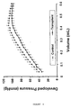
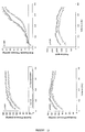
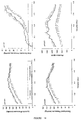
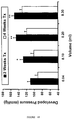
- a latex balloon was passed into the left ventricle through the mitral valve and connected to a pressure transducer (Model p10EZ, Viggo-Spectramed, CA) and transducer amplifier and differentiator amplifier (Model 11-G4113-01, Gould Instrument System Inc., Ohio). After 30 minutes of stabilization, coronary flow in the heart was measured in triplicate by timed collection in the emptying beating state. The balloon size was increased by addition of saline in 0.02 ml increments from 0.04 to 0.8 ml, or the volume at which end diastolic pressure reached to 30 mm Hg, or whichever came first. The systolic and diastolic pressures were recorded at each balloon volume and developed pressure was calculated as the difference between the systolic and diastolic pressures.
- LVFW left ventricular free wall
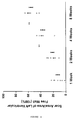
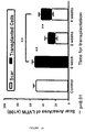
- the area of normal tissue, scar tissue, and transplanted tissue in the left ventricular free wall were traced onto a transparency and quantified using computerized planimetry (Jandal Scientific Sigma Scan, USA) as described by Wu et al. (Wu TW, et al., Cardiovascular Research 27:736-39, 1993).
- the lengths of left ventricular free wall and scar tissue on both the endocardial and epicardial surfaces of each section were measured.
- the surface areas of the epicardial and endocardial scar tissue and left ventricular free wall were measured as: ( endocardial length + epicardial length ) ⁇ section thickness ( 3 mm ) .
- the surface area percentage of scar tissue in the left ventricular free wall (LVFW) was calculated as: ( epicardial scar size + endocardial scar size ) ( endocardial LVFW 1 + epicardial LVFW ) ⁇ 100
- Example 1 On day 30 or 45 post-transplantation, the animals were anesthetized as in Example 1 and transplant, control, and sham hearts were exposed through a midline sternotomy and quickly harvested. The animals were euthanised by exsanguination under general anesthesia.
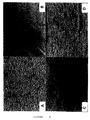
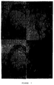
- the transplanted and control myocardial scar tissues were fixed in 5% glacial acetic acid in methanol.
- the tissue was embedded in paraffin and cut into 10 ⁇ m thick sections. After removal of the paraffin by immersing the sections for 3 minutes in xylene and then in 100, 95, 90, 85, and 70% ethanol for 3 minutes each, the samples were stained with haematoxylin and eosin as described by the manufacturer (Sigma Diagnostics, St. Louis, MO), and photographed.
- the heart sections were fixed at 4°C for 12 hours in 2% formaldehyde and 2% glutaraldehyde in phosphate buffer (0.15 M NaCl and 0.015 M NaH 2 PO 4 , pH 7.2).
- the transplanted cardiomyocytes were localized by staining for ⁇ -galactosidase activity as described earlier in Example 2.
- the stained tissue was embedded in paraffin and cut into 10 ⁇ m thick sections that were stained with haematoxylin and eosin as described in the last paragraph.
- tissue sections were washed three times with phosphate buffered saline and fixed with 2 ml 100% cold methanol at -20°C for 15 minutes. After washing three times with phosphate buffered saline and drying by draining, the tissue sections were exposed for 45 minutes at 37°C to monoclonal antibodies against cardiac myosin heavy chain (Rougier Bio-Tech, Montreal, Canada), diluted 1:20 with saline. Control tissues were incubated under the same conditions with phosphate buffered saline.
- the tissues were washed three times with phosphate buffered saline for 15 minutes at room temperature with gentle shaking, after which the secondary antibody, rabbit anti-mouse IgG conjugated with fluorescein isothiocyanate at a concentration of 1:32 dilution with phosphate buffered saline, was added.
- the tissues were incubated with the second antibody under dark and humid conditions for 45 minutes at 37°C. After washing with phosphate buffered saline, the cells in the transplant control tissues were visualized under ultraviolet light using an epi-microscope with a blue filter.
- the smooth muscle cell transplants were identified by staining immuno-fluorescently using a monoclonal antibody for ⁇ -smooth muscle actin as the primary antibody. Endothelial cell transplants were identified by immunofluorescent staining for factor VIII, as described in the next section.
- Fibroblast transplants were identified by the presence of a localized immunorejection within the ventricular scar.
- tissue sections processed as in Section E above were incubated with xylene twice for 5 minutes each, 100% ethanol twice for 2 minutes each and then with 70% ethanol twice for 1 minute each.
- the sections were incubated with rabbit IgG against factor VIII-related antigen (Dimension Lab. Inc., Ontario).
- the control samples were incubated with phosphate buffered saline under the same conditions.
- the test and control samples were incubated with goat anti-rabbit IgG conjugated with peroxidase.
- Cultured cardiomyocytes, smooth muscle cells, endothelial cells, and/or fibroblasts were seeded onto biological mesh, such as a collagen membrane, and on non-biological membranes, such as non-degradable membranes (Dacron) or degradable membranes (polyglycolic acid polymers), and the mesh and cells were cultured in cell culture medium.
- biological mesh such as a collagen membrane
- non-biological membranes such as non-degradable membranes (Dacron) or degradable membranes (polyglycolic acid polymers
- the cell-containing mesh was fixed in 2% formaldehyde and 2% glutaraldehyde in phosphate buffer (0.15 M NaCl and 0.015 M NaH 2 O 4 , pH 7.2) at 4°C for 12 hours, embedded in paraffin, and cut into 10 ⁇ m thick sections, which were stained with haematoxylin and eosin as described in Section E, and photographed.
- cardiomyocytes must be implanted into the infarcted myocardium. Although this can be done by multiple syringe injections, injecting the cells, unfortunately, limits the number of cells which can be transplanted into myocardial scar tissue.
- Thrombin and cryoprecipitate which are derived from human blood, clot rapidly.
- Our in vitro results showed survival and contraction of cardiomyocytes in fibrin clots. We used this fibrin glue for cell transplantation.
- Function data were evaluated for the sham, control and transplant groups by an analysis of covariance using intracavitary balloon volume as the covariate and systolic, diastolic and developed pressure as dependent variables. Main effects were group, volume, and the interaction between group x volume. If there was an overall difference in the analysis of covariance, multiple pair-wise comparisons were performed to specify which groups were different. Because there were multiple pair-wise comparisons, a Bonferroni correction was performed and the critical alpha level was set at 0.01 for the analysis of covariance.
- Idiopathic hypertrophic cardiomyopathy is a primary cardiac abnormality characterized by regional asymmetrical myocardial hypertrophy.
- the hypertrophic myocardium can result in obstruction of left ventricular ejection as well as systolic and diastolic dysfunction and myocardial ischemia. Symptoms unresponsive to medical therapy can necessitate surgery.
- HCM is described for the most part as a heterogeneous disease of the sarcomeres. At least 34 missense mutations have been described in the ⁇ -myosin heavy chain gene, and 7 mutations in candidate loci also exist.
- family studies suggest that the autosomal dominant trait accounts for only 50% of HCM patients. The remaining HCM patients show no familial transmission and the disease occurs sporadically.
- Myocardial calcium kinetics and sympathetic stimulation have been studied because of diastolic functional abnormalities.
- none of these findings explains the regional myocardial hypertrophy (cardiomyocyte hypertrophy and over production of extracellular matrix proteins) observed in most HCM patients. The etiology of this disease remains unknown. It is thought that growth factors may play an important role in cardiomyocyte proliferation, cell hypertrophy and the overproduction of extracellular matrix.
- TGF ⁇ 1 transforming growth factor ⁇ 1
- IGF-I, -II insulin-like growth factors
- PDGF-B platelet-derived factor-B
- TGF ⁇ 1, IGF-I, IGF-II and PDGF-B transcripts were quantitated using multiplex RT-PCR.
- Glyceraldehyde 3-phosphate dehydrogenase (G3PDH) was used as an internal standard.
- Antibodies against TGF ⁇ 1 and IGF-I were used to localize their peptides within the myocardium.
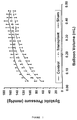
- mRNA levels (densitometric ratio of GF/G3PDH) of TGF ⁇ 1 and IGF-I in HCM myocardium (0.75 ⁇ 0.05, 0.85 ⁇ 0.15, mean ⁇ 1SE) were significantly (p ⁇ 0.01 for all groups) elevated in comparison to non-HCM myocardium (AS: 0.38 ⁇ 0.07, 0.29 ⁇ 0.06; SA: 0.32 ⁇ 0.04, 0.18 ⁇ 0.05; TM: 0.25 ⁇ 0.03, 0.15 ⁇ 0.03).
- CMs fetal rat cardiomyocytes
- CMs were isolated from 18-day gestation, 5-, 22-, 32-, and 62-day old Sprague-Dawley rat hearts and cultured for 1 day. 2 to 5 x 10 6 cells in saline were injected into the skeletal muscle of one adult rat leg. The other leg (control) was injected with saline alone. Cell viability and function were assessed visually and by ultrasound and electrocardiography (ECG).
- ECG ultrasound and electrocardiography
- Donor age is important for maintaining contractility of CMs after transplantation into skeletal muscle.
- Angiogenesis was induced in the scar tissue by endothelial cell transplantation. Since no myocytes were present in the scar, function did not improve. Endothelial cell transplantation in patients with incomplete infarction may restore perfusion, relieve angina and restore function.
- mice All procedures performed on animals were approved by the Animal Care Committee of The Toronto Hospital. Experiments were performed according to the "Guide to the Care and Use of Experimental Animals" published by the National Institutes of Health (NIH publication 85-23, revised 1985). Sprague-Dawley rats (Charles River Canada Inc, Quebec, Canada) were used for allogeneic transplantation. Male rats, weighing 330 to 360 g, were used as recipients and donors of endothelial cells. Sygenic Lewis rats (Male, 250-300 g, Charles River Canada Inc, Quebec, Canada) were used for autologus transplantation.
- Endothelial cells from rat aorta were isolated and cultured as previously described (Mickle et al., J . Mol. Cell. Cardiol. 22:1297-1304, 1990). In brief, descending thoracic aortic segments were obtained from rats under general anesthesia as described in the next section. The blood vessels were washed with phosphate buffered saline solution (PBS; composition in mmol/L: NaCl 136.9, KCl 2.7, Na 2 HPO 4 8.1, KH 2 PO4 1.5, pH 7.3) and connective tissue was removed.
- PBS phosphate buffered saline solution
- aortic segment was then flushed with enzyme solution (0.1 % collagenase and 0.2% trypsin in PBS) and incubated with the enzyme solution for 30 minutes at 37°C.
- enzyme solution 0.1 % collagenase and 0.2% trypsin in PBS
- endothelial cells were isolated by flushing the inside of aortic segments with culture medium (Medium 199 containing 20% fetal bovine serum, 100U/ml of penicillin, 100ug/ml of streptomycin, and 0.5% heparin salt) three to five times.
- culture medium Medium containing 20% fetal bovine serum, 100U/ml of penicillin, 100ug/ml of streptomycin, and 0.5% heparin salt
- the cells were washed three times (15 minutes per wash) with PBS, then incubated with goat anti- rabbit immunoglobulin G conjugated with peroxidase at 37°C for 45 minutes, washed three times (15 minutes per wash) with PBS, and immersed in diaminobenzidine-H 2 O 2 (2mg/ml diaminobenzidine, 0.03% H 2 O 2 in 0.02ml/l phosphate buffer) solution for 15 minutes. After a final wash in PBS, the samples were photographed.
- VEGF protein levels in culture medium and endothelial cells were quantitated by chemiluminescent slot blot analysis.
- Culture medium incubated without cells was used as a control.
- Forty micrograms of protein from culture medium or from cultured endothelial cells were loaded onto a 0.2 ⁇ m nitrocellulose membrane (Schleicher & Schuell Inc., Keene, NH) using the Minifold II Slot Blotting System (Schleicher & Schuell Inc., Keene, NH).
- Standards of VEGF (2, 5, 10, 25 ng) (Sigma, Mississauga, ONT) were loaded on the same membrane.
- TTBS buffer 50 mM Tris-HCl buffer pH 7.4 and 0.1% of Tween-20
- blocking buffer Boehringer Mannhein, GmbH, German.
- Monoclonal antibodies against VEGF (Sigma, Mississauga, ONT) (1:3000, diluted with 0.5 x block buffer) were added and incubated overnight at 4°C.
- TTBS Tris-HCl buffer pH 7.4 and 0.1% of Tween-20
- VEGF proteins were detected by chemiluminescence using the Boehringer Mannheim detection kit (Quebec, Canada).
- a densitometric analysis of the standard and sample bands was performed with a Bio-Rad image analysis system (Bio-Rad Lab. Hercules, CA). Standard curves of VEGF were generated and VEGF quantity in culture medium and cultured endothelial cells was calculated based on the standard curve. The results (mean ⁇ SD) are expressed as ng of VEGF/mg protein.
- Myocardial Scar Formation Rats were anesthesized by intramuscular injection of ketamine hydrochloride (20mg/kg body weight) followed by intraperitoneal injection of sodium pentobarbital (30mg/kg body weight). The anesthesized rats were intubated and positive pressure ventilation was performed with room air supplemented with oxygen and isoflurane (0.2-1.0%) using a Harvard ventilator. The electrocardiogram was monitored during operation.
- the heart was exposed through a 2- to 3-cm left lateral thoracotomy incision. Cryoinjury was produced in the left ventricular free wall(LVFW) with a metal probe (8 X 10 mm in diameter) cooled to -190°C by immersion in liquid nitrogen. It was applied to the LVFW for one minute and this procedure was repeated ten times. The muscle and skin were closed with 3-0 silk sutures. Penlong XL (benzathine penicillin G 150,000 U/ml and procaine penicillin G 150,000 U/ml) was given intramuscularly (1 ml/kg) and buprenorphine hydrochloride (0.01mg/kg) was administered after each operation. The cryoinjured rats were randomly divided into two groups: transplantation and control.
- Endothelial Cell Transplantation At two weeks after cryoinjury, cell transplantation was performed. Under general anesthesia, the heart was exposed through a midline sternotomy. Endothelial cell suspension (60ul, 6X10 6 cells) was injected into the center of the scar tissue in the transplantation group using a tuberculin syringe, and same amount of culture media was injected into the scar in the control group. The chest was closed with 3-0 silk sutures. Antibiotics and analgesics were given as previously described. Cyclosporine A (25mg/kg/day) was administered subcutaneously to both transplanted and control groups.
- Ventricles were dissected into normal tissue, scar tissue, and tissue in the borderline area. Each section was weighed and the radioactivity was then determined using a gamma counter at the window setting of 110 to 138 KeV. The data was expressed as counts per minute (cpm)/mg of tissue. The ratio of cpm/mg in scar and borderline tissue to normal tissue was calculated, expressed as the percentage, and compared between transplanted and control hearts.
- Krebs-Hanseleit buffer mmol/L: NaCl 118, KCl 4.7, KH 2 PO 4 1.2, CaCl 2 2.5, MgSO 4 1.2, NaHCO 3 25, glucose 11, pH 7.
- a latex balloon was inserted into the left ventricle through the mitral valve and connected to a pressure transducer (Model p10EZ; Viggo-Spectramed, Oxnard CA) and a transducer amplifier and differentiator amplifier (Model 11-G4113-01; Gould Instrument System Inc, Valley View OH).
- a pressure transducer Model p10EZ; Viggo-Spectramed, Oxnard CA
- a transducer amplifier and differentiator amplifier Model 11-G4113-01; Gould Instrument System Inc, Valley View OH.
- the coronary flow was measured by the timed collection in the empty beating state.
- the balloon size was increased in 0.02 ml increments from 0.04 to 0.6 ml by the addition of saline solution.
- the systolic and end-diastolic pressure and maximal and minimal dp/dt were recorded at each balloon volume.
- the developed pressure was calculated as the difference between the systolic and end-diastolic pressure.
- the epicardial and endocardial surfaces of the normal and scar tissue in the LVFW were measured by the technique of Pfeffer (Pfeffer Am J Physiol 1991;260:H1406-14) and Jugdutt and Khan Circulation 1994;89:2297-307). Briefly, the hearts were fixed in distension (30 mmHg) with 10% phosphate-buffered formalin solution for 2 days. The atria were excised, the ventricular weight and the size of the scar on epicardial surface were measured, and ventricular volume was measured by water displacement. After that, the hearts were cut into sections of 3 mm thickness.
- the area of normal and scar tissue in the LVFW were traced onto a transparency and quantified using computed planimetry (Jandal Scientific Sigma-Scan, Corte Madera, CA).
- the lengths of LVFW and scar tissue on both the endocardial and epicardial surfaces of each section were measured.
- the surface areas of the endocardial and epicardial scar tissue and the LVFW were measured as the sum of the endocardial length and epicardial length times the section thickness (3mm).
- the surface area percentage of scar tissue in the LVFW was calculated as follows: (endocardial scar size+epicardial scar size)/(endocardial LVFW+epicardial LVFW) X 100.
- the heart sections were used for immunohistochemical staining of endothelial cells.
- the sections were incubated with xylene twice for 5 minutes each, 100% ethanol twice for 2 minutes each, and then with 70% ethanol twice for 1 minute each.
- the sections were incubated with rabbit immunoglobulin G against factor VIII-related antigen and endothelial cells were identified as described in the Cell Identification section above.
- the stained vasculoendothelial cells in the transplanted and control groups were counted using a light microscope at 400X magnification. The result was expressed as the number of capillary vessels/field.
- Cultured endothelial cells were distinguished from fibroblasts and vascular smooth muscle cells by morphologic criteria and growth characteristics. The endothelial cells were oval shaped, in contrast to the spindle-shape of cultured fibroblasts and vascular smooth muscle cells. Cultured endothelial cells grew in a "cobblestone" pattern whereas fibroblasts grew in a "whirling" pattern and vascular smooth muscle cells grew in a "hill and valley” pattern. In addition, cultured endothelial cells stained positively for factor VIII-related antigen. The purity of the culture to be transplanted was more than 95%.
- transplanted cells In syngeneic cell transplantation the transplanted cells also induced angiogenesis in the myocardial scar tissue.
- the density of capillaries in the scar tissue of the transplanted group was 2.6 ⁇ 1.5 vessels/0.8mm 2 , which was significantly (p ⁇ 0.01) greater than that in the control group (1.1 ⁇ 0.6).
- Transplanted endothelial cells were incorporated into capillaries at the transplant site.
- the transplanted endothelial cells stimulate angiogenesis, demonstrated by an increase in the number of capillaries in the scar tissue and increased blood perfusion in scar tissue as illustrated by microsphere perfusion studies.
- the induced angiogenesis in transmural scar tissue did not improve infarcted heart function.
- angiogenesis by endothelial transplantation: (1) formation of blood vessels by transplanted endothelial cells; (2) angiogenesis stimulated by growth factors secreted by transplanted endothelial cells; (3) angiogenesis stimulated by an inflammatory reaction induced by transplanted endothelial cells.
- Endothelial cells are the most important component for blood vessel formation.
- Our in vivo study showed that transplanted endothelial cells become part of newly formed capillaries in syngeneic cell transplantation. These data suggested that transplanted endothelial cells are involved in blood vessel formation.
- the data obtained from allogeneic endothelial cell transplantation suggested that newly formed capillaries in the scar tissue were grown from recipient heart endothelial cells.
- transplanted cells at 1 and 7 days after transplantation, but did not observe transplanted cells after 14 days post transplantation. Additional evidence of this conclusion is that there was no immunologic reaction around capillary vessels in the scar.
- transplanted allogeneic endothelial cells become part of the newly formed capillaries, lymphocyte infiltration should be observed around the blood vessel.
- Angiogenesis in our animal model might also be stimulated by inflammatory reactions.
- syngeneic endothelial cell transplantation has shown that angiogenesis occurs without inflammatory reaction, factors released by inflammatory cells might play a role in blood vessel formation induced by allogeneic endothelial cell transplantation.
- Capillary density in scar tissue subjected to allogeneic cell transplantation was 1.3 times greater than that subjected to syngeneic cell transplantation. This difference in capillary density may be due to lymphocyte infiltration.
- the left ventricular free wall was divided into three parts: normal area, transmural infarct area, and borderline area.
- normal area normal area
- transmural infarct area transmural infarct area
- borderline area The blood flow to the borderline areas was always between that of the normal and complete transmural infarct area.
- myocardial infarction damaged myocardium undergoes both acute and chronic inflammatory reactions.
- necrosed cardiac cells are replaced with fibrotic tissue and ventricular remodeling progresses.
- Ventricular pressure stretches and thins the healing area and ventricular dilatation occurs.
- a ventricular aneurysm may form and congestive heart failure may result.
- Limitation of post infarction ventricular remodeling is important to prevent expansion and thinning of the infarct region and thinning, and progressive ventricular dilatation, which results in congestive heart failure.
- the addition of cardiac tissure to replace the scar tissue may prevent infarct thinning and preserve chamber size and ventricular function.
- Cardiomyocyte transplantation was most successful after the inflammatory reaction resolved and before significant scar expansion and ventricular dilatation occurred.
- mice All procedures performed on animals were approved by the Animal Care Committee of The Toronto Hospital. Experimental animals used were male Sprague-Dawley rats (Lewis, Charles River Canada Inc. Quebec, Canada), weighing 450 grams. Cardiomyocytes obtained from 18-day gestational rat hearts were cultured prior to transplantation. All experiments were performed according to "A Guide to the Care and Use of Experimental Animals" of the Canadian Council on Animal Care and the "Guide for the Care and Use of Laboratory Animals" NIH publication 85-23, revised 1985).
- Myocardial scar tissue was generated as described in Example I above. Briefly, rats were anesthetized with ketamine (22 mg/kg body weight, intramuscular) followed by an intraperitoneal injection of pentobarbital (30 mg/kg body weight). The anesthetized rats were intubated and positive pressure ventilation was maintained with room air supplemented with oxygen (6 L/minute) using a Harvard ventilator (Model 683, South Natick, MA, USA). The rat heart was exposed through a two cm left lateral thoracotomy. Cryo-injury was produced at the left ventricular free wall (LVFW) with a liquid nitrogen probe. The muscle layer and skin were closed with 5-0 vicryl sutures.
- LVFW left ventricular free wall
- Penlog XL benzathine penicillin G 150,000 U/ml and procaine penicillin G 150,000 U/ml
- Penlog XL benzathine penicillin G 150,000 U/ml and procaine penicillin G 150,000 U/ml
- buprenorphine 0.01-0.05 mg/kg body weight
- the LVFWs of 35 animals were cryo-injured. Immediately after cryo-injury and at 1, 2, 4, and 8 weeks after injury, 7 animals chosen at random were sacrificed under general anesthesia (see Fig. 5 for a schematic diagram of the experimental design). The hearts were then fixed in distension (30 mm Hg) with 10% phosphate-buffered formalin solution for 24-48 hours and then cut into 3 mm thick sections. All sections were used for assessment of myocardial and scar sizes. For each section, the area of scar tissue in the LVFW was traced onto a transparency and quantified using computerized planimetry (Jandal Scientific Sigma Scan, Fairfield, CT., USA). Scar length was calculated as the average of endocardial scar length and epicardial scar length.
- Scar area was calculated as the scar length X section thickness (3mm).
- the total scar area in size was calculated as the sum of the scar areas for all of the tissue sections showing scars.
- the LVFW myocardial size was calculated using the same equation.
- the percentage of the LVFW occupied by the scar was calculated as scar size/LVFW size X 100.
- Histological studies The fixed heart sections were embedded in paraffin and cut to yield 10 ⁇ m thick sections. The sections were stained with haematoxylin and eosin as described by the manufacturer (Sigma Diagnostics, St. Louis, MO).
- Cardiomyocytes from fetal rat hearts were isolated, purified and cultured as previously described (Li, R-K, et al. J Tissue Culture Methods 14:93-100, 1992). Briefly, the cells were cultured for 24 hours in Iscove's modified Dulbecco's medium containing 10% fetal bovine serum, 100 U/ml penicillin and 100 ug/ml streptomycin at 37EC, 5% CO 2 and 95% air. The cultured cardiomyocytes were detached from the cell culture dish with 0.05% trypsin in phosphate-buffered saline (PBS). After centrifugation at 580 X g for 3 minutes, the cell pellet was resuspended in culture medium at a concentration of 16 X 10 6 cells/ml. A 0.25 ml cell suspension was used for each transplantation.
- PBS phosphate-buffered saline
- the hearts were quickly isolated from rats and perfused in a Langendorff apparatus with filtered Krebs Heinseleit buffer (mmol/L NaCl 118, KCl 4.7,KH 2 PO 4 1.2, CaCl 2 2.5, MgSO 4 1.2, NaHCO 3 25, glucose 11; pH 7.4) equilibrated with a 5% CO 2 and 95% O 2 .
- a latex balloon was passed into the left ventricle through the mitral valve and connected to a pressure transducer (Model p10EZ, Viggo-Spectramed, CA) and transducer amplifier and differentiator amplifier (Model 11-G4113-01, Gould Instrument System Inc., Ohio).
- the balloon size was increased in 0.02 ml increments from 0.04 to a volume, at which end diastolic pressure was 30 mm Hg or more by the addition of saline.
- the systolic and diastolic pressures were recorded at each balloon volume and developed pressure was calculated as the difference between the systolic and diastolic pressures.
- Histological identification of transplanted muscle cells in the scar tissue Fixed heart sections were embedded in paraffin, and cut into 10 ⁇ m thick sections. The sections were stained with hematoxylin and eosin as described in the "Histological Studies" section above.
- Heart sections containing transplanted cells also were stained for myosin heavy chain (MHC) (Rougier Bio-Tech, Montreal). After deparaffination and rehydration, samples were incubated for 30 minutes with a solution of 3% H 2 O 2 in 70% methanol to inhibit endogenous myocardial peroxidase. Triton X-100 (0.2%) was used to treat samples for 10 minutes to enhance cell permeability. After blocking nonspecific protein binding with 2% normal goat serum in 0.05 M Tris buffer (pH 7.4) for 15 minutes, primary antibodies against HCM (1:1000) were added and the samples were incubated at 37°C for 30 minutes followed by an overnight incubation at 4°C. Negative control samples were incubated in PBS under the same conditions.
- MHC myosin heavy chain
- Data Analysis Data are expressed as the mean ⁇ standard error.
- LVFW LVFW fibrotic tissue
- Fig. 6A a predominantly mononuclear inflammatory infiltrate was present in the affected area
- Fig. 6B At two weeks the inflammatory infiltrate had almost disappeared and fibroblasts and collagen deposition were evident.
- Fig. 6D At four and eight weeks, the scar was composed of fibrotic tissue (Fig. 6D). The tissue was less cellular and lymphocytes were not observed.
- the myocardial scar size of the left ventricle expanded over the 8 week study period in the damaged hearts (Figs. 7 and 8). Although the scar sizes at 1 and 2 weeks (13 ⁇ 6 % and 21 ⁇ 4 of LVFW) were not statistically different, the size of 4-week-old scars (39 ⁇ 5 % of LVFW) was larger (p ⁇ 0.01). At 8 weeks there was a further increase (p ⁇ 0.01) in scar size (55 ⁇ 3% of LVFW).
- Cardiomyocytes transplanted at 4 weeks after myocardial injury also improved (p ⁇ 0.001) myocardial function (Figure 14).
- Figure 15 shows that hearts transplanted at 2 weeks had higher developed pressures at balloon volumes 0.1 ml (p ⁇ 0.05), 0.2 ml (p ⁇ 0.01) and 0.3 ml (p ⁇ 0.01) than hearts transplanted at 4 weeks.
- transplant tissue visibly contracted, but at different rates than did host myocardium.
- the contraction of the transplanted tissue persisted after dissection of the recipient heart, but its rate of contraction continued to be different.
- necrosed ventricular tissue occurs by 2 days after cyro-injury of the myocardium. At this time there is an acute inflammatory reaction with neutrophils accumulating first in the periphery and later in the center of the necrotic region. A chronic inflammatory reaction involving macrophages and lymphocytes follows the acute reaction.
- the inflammatory reaction is less intense two weeks after cryo-injury (and is absent by three weeks): two weeks after cryo-injury, the scars of the control animal hearts showed minimal or no inflammatory reaction.
- Two-week-old scars were less mature and contained more blood vessels and fibroblasts than 4-week-old scars, which comprised firm connective tissue. Compared to one-week-old scars, there was less cardiac tissue in older scars. It is possible that progressive inflammation and lack of oxygen and nutrient supply decreased the myocardial tissue over time.
- the cardiomyocytes transplanted immediately after cryo-injury did not survive in any of the animals studied. We believe the activated neutrophils and macrophages in the inflamed scar area destroyed the transplanted cells.
- the left ventricular chamber of the animals transplanted at the time of cryo-necrosis was similar to that of the control scar. The chamber was dilated due to thinning of the scar. The remaining viable muscle of the left ventricle was hypertrophied. No inflammatory reaction was seen. The function of these transplanted hearts was similar to that of the control hearts.
- a cardiomyocyte transplant forms the equivalent of a viable epicardial rim and prevents ventricular dilatation and over-stretching of the cardiomyocytes during systole. With over-stretching of the cardiomyocytes, cardiac function is diminished (Frank-Starling Law).
- the elastic properties of the contractile apparatus of the transplanted cardiomyocytes may prevent host fibroblast and cardiomyocyte stretching and ventricular enlargement.
- angiogenesis occurred in the course of forming cardiac tissue from transplanted cardiomyocytes. Angiogenesis is most likely necessary to maintain the viability of the transplanted muscle cells. However, an increased blood supply in the scar could also facilitate fibroblast turnover and strengthening of the scar in response to left ventricular free wall stretch.
- the transplanted cardiomyocytes should also increase the contractile capacity of the scar tissue and decrease the contractile requirements of the non-affected host myocardium.
- Patients with angina that cannot be corrected by bypass surgery may benefit from angiogenesis secondary to cardiomyocyte transplantation.
- it may be beneficial to stimulate angiogenesis with growth factors prior to autotransplantation.
- transplantation of fetal cardiomyocytes form cardiac tissue in scar tissue which limits ventricular dilatation and scar thinning.
- the optimal time for transplantation for improved myocardial function is after the acute inflammatory reaction and before significant ventricular dilatation has occurred.
- Cardiac transmural scars were induced by cryo-injury in the left ventricular free wall (LVFW) of Sprague-Dawley rat hearts, as described in the previous examples. Three weeks after cryo-injury, the rats were divided into three groups (eight rats per group), and cardiac transmural scars were transplanted with either medium (no cells), adult cardiomyocytes alone, or adult cardiomyocytes plus endothelial cells. Cardiac function was assessed using a Langandorff preparation five weeks after cell transplantation (Stephen et al., Annals of Thoracic Surgery 59: 1127-1133, 1995).
- Fig. 16 shows a graph of developed pressure in infarcted rat hearts injected with either medium (negative control), adult cardiomyocytes alone (CM), or adult cardiomyocytes plus endothelial cells (CM + EC).
- CM adult cardiomyocytes alone
- CM + EC adult cardiomyocytes plus endothelial cells
Landscapes
- Health & Medical Sciences (AREA)
- Life Sciences & Earth Sciences (AREA)
- Engineering & Computer Science (AREA)
- Chemical & Material Sciences (AREA)
- Biomedical Technology (AREA)
- Zoology (AREA)
- Cell Biology (AREA)
- Biotechnology (AREA)
- General Health & Medical Sciences (AREA)
- Bioinformatics & Cheminformatics (AREA)
- Organic Chemistry (AREA)
- Veterinary Medicine (AREA)
- Public Health (AREA)
- Animal Behavior & Ethology (AREA)
- Medicinal Chemistry (AREA)
- Pharmacology & Pharmacy (AREA)
- Epidemiology (AREA)
- Wood Science & Technology (AREA)
- Immunology (AREA)
- Developmental Biology & Embryology (AREA)
- Genetics & Genomics (AREA)
- Cardiology (AREA)
- Virology (AREA)
- Rheumatology (AREA)
- Microbiology (AREA)
- Biochemistry (AREA)
- General Engineering & Computer Science (AREA)
- Vascular Medicine (AREA)
- Chemical Kinetics & Catalysis (AREA)
- Heart & Thoracic Surgery (AREA)
- Gastroenterology & Hepatology (AREA)
- General Chemical & Material Sciences (AREA)
- Nuclear Medicine, Radiotherapy & Molecular Imaging (AREA)
- Proteomics, Peptides & Aminoacids (AREA)
- Hospice & Palliative Care (AREA)
- Materials For Medical Uses (AREA)
- Micro-Organisms Or Cultivation Processes Thereof (AREA)
- Medicines Containing Material From Animals Or Micro-Organisms (AREA)
- Medicinal Preparation (AREA)
- Medicines Containing Plant Substances (AREA)
Priority Applications (1)
| Application Number | Priority Date | Filing Date | Title |
|---|---|---|---|
| EP06002350A EP1690546B1 (en) | 1997-05-28 | 1998-05-28 | Transplants for myocardial scars and method and cellular preparations therefor |
Applications Claiming Priority (3)
| Application Number | Priority Date | Filing Date | Title |
|---|---|---|---|
| US863882 | 1997-05-28 | ||
| US08/863,882 US6110459A (en) | 1997-05-28 | 1997-05-28 | Transplants for myocardial scars and methods and cellular preparations |
| PCT/CA1998/000520 WO1998054301A2 (en) | 1997-05-28 | 1998-05-28 | Transplants for myocardial scars and method and cellular preparations therefor |
Related Child Applications (1)
| Application Number | Title | Priority Date | Filing Date |
|---|---|---|---|
| EP06002350A Division EP1690546B1 (en) | 1997-05-28 | 1998-05-28 | Transplants for myocardial scars and method and cellular preparations therefor |
Publications (2)
| Publication Number | Publication Date |
|---|---|
| EP0985028A2 EP0985028A2 (en) | 2000-03-15 |
| EP0985028B1 true EP0985028B1 (en) | 2006-07-26 |
Family
ID=25342008
Family Applications (2)
| Application Number | Title | Priority Date | Filing Date |
|---|---|---|---|
| EP98923950A Expired - Lifetime EP0985028B1 (en) | 1997-05-28 | 1998-05-28 | Transplants for myocardial scars and method and cellular preparations therefor |
| EP06002350A Expired - Lifetime EP1690546B1 (en) | 1997-05-28 | 1998-05-28 | Transplants for myocardial scars and method and cellular preparations therefor |
Family Applications After (1)
| Application Number | Title | Priority Date | Filing Date |
|---|---|---|---|
| EP06002350A Expired - Lifetime EP1690546B1 (en) | 1997-05-28 | 1998-05-28 | Transplants for myocardial scars and method and cellular preparations therefor |
Country Status (8)
| Country | Link |
|---|---|
| US (2) | US6110459A (enExample) |
| EP (2) | EP0985028B1 (enExample) |
| JP (1) | JP2002501513A (enExample) |
| AT (2) | ATE488244T1 (enExample) |
| AU (1) | AU753388B2 (enExample) |
| CA (1) | CA2291138C (enExample) |
| DE (2) | DE69835354T2 (enExample) |
| WO (1) | WO1998054301A2 (enExample) |
Families Citing this family (103)
| Publication number | Priority date | Publication date | Assignee | Title |
|---|---|---|---|---|
| US20040087498A1 (en) * | 1991-02-28 | 2004-05-06 | Novo Nordisk Health Care Ag | Modified factor VII |
| US6443974B1 (en) * | 1996-07-28 | 2002-09-03 | Biosense, Inc. | Electromagnetic cardiac biostimulation |
| US6110459A (en) * | 1997-05-28 | 2000-08-29 | Mickle; Donald A. G. | Transplants for myocardial scars and methods and cellular preparations |
| US6099832A (en) * | 1997-05-28 | 2000-08-08 | Genzyme Corporation | Transplants for myocardial scars |
| US6775574B1 (en) * | 1997-11-07 | 2004-08-10 | Medtronic, Inc. | Method and system for myocardial infarction repair |
| US7115417B1 (en) | 1998-05-01 | 2006-10-03 | Chancellor Michael B | Soft tissue and bone augmentation and bulking utilizing muscle-derived progenito compositions, and treatments thereof |
| US6866842B1 (en) | 1998-05-01 | 2005-03-15 | University Of Pittsburgh | Muscle-derived cells (MDCs) for treating muscle-or bone-related injury or dysfunction |
| AU5545499A (en) * | 1998-07-31 | 2000-02-21 | Genzyme Corporation | Improvement of cardiac function by mesenchymal stem cell transplantation |
| CN1420933A (zh) * | 1999-06-25 | 2003-05-28 | 耶路撒冷希伯来大学伊森姆研究发展公司 | 应用微器官诱导血管生成的方法 |
| US6673604B1 (en) | 1999-07-23 | 2004-01-06 | Diacrin, Inc. | Muscle cells and their use in cardiac repair |
| US20030113301A1 (en) * | 1999-07-23 | 2003-06-19 | Albert Edge | Muscle cells and their use in cardiac repair |
| DE10003521A1 (de) * | 2000-01-27 | 2001-08-09 | Medigene Ag | Vorrichtung zum Herstellen eines dreidimensionalen Matrixkörpers, Multi-Well-Platte, Lösung zum Kultivieren von Säugerkardiomyocyten, Verfahren zum Kultivieren einer Zellkultur, Vorrichtung für die Messung isometrischer Kraftparameter von Zellkulturen sowie Verfahren zum meßbaren Verfolgen von Kontraktionen eines in eine Trägersubstanz eingelagerten Zellgewebes |
| ES2288946T3 (es) | 2000-04-14 | 2008-02-01 | University Of Pittsburgh | Aumento y abultamiento de tejido blando y hueso utilizando celulas progenitoras derivadas de musculo, sus composiciones y tratamientos. |
| FR2810045B1 (fr) * | 2000-06-07 | 2004-09-03 | Assist Publ Hopitaux De Paris | Procede d'obtention de population cellulaires caracterisees d'origine musculaire et utilisations |
| US20110091428A1 (en) * | 2000-07-31 | 2011-04-21 | New York Medical College | Compositions of adult organ stem cells and uses thereof |
| US7862810B2 (en) * | 2000-07-31 | 2011-01-04 | New York Medical College | Methods and compositions for the repair and/or regeneration of damaged myocardium |
| US20020061587A1 (en) * | 2000-07-31 | 2002-05-23 | Piero Anversa | Methods and compositions for the repair and/or regeneration of damaged myocardium |
| US7547674B2 (en) | 2001-06-06 | 2009-06-16 | New York Medical College | Methods and compositions for the repair and/or regeneration of damaged myocardium |
| US7294333B1 (en) * | 2000-10-20 | 2007-11-13 | Genegrafts Ltd. | Nucleic acid constructs and cells, and methods utilizing same for modifying the electrophysiological function of excitable tissues |
| US7491385B2 (en) * | 2001-09-05 | 2009-02-17 | Genegrafts Ltd. | Nucleic acid constructs and cells, and methods utilizing same for modifying the electrophysiological function of excitable tissues |
| US6659995B1 (en) * | 2000-11-17 | 2003-12-09 | Syde A. Taheri | Autologous myocyte micro granual retrieval and implantation (AMMGRI) |
| DE10106512C1 (de) * | 2001-02-13 | 2002-05-23 | Axel Haverich | Verfahren zur Herstellung eines biologischen Gewebes unter Verwendung einer Kollagenunterlage und zugehöriges Gewebekonstrukt |
| CA2438904C (en) | 2001-02-23 | 2012-09-04 | The University Of Pittsburgh | Rapid preparation of stem cell matrices for use in tissue and organ treatment and repair |
| US7341062B2 (en) * | 2001-03-12 | 2008-03-11 | Bioheart, Inc. | Method of providing a dynamic cellular cardiac support |
| AU2002303343A1 (en) * | 2001-04-13 | 2002-10-28 | Anterogen Co., Ltd. | Methods and reagents for cell transplantation |
| CA2443984A1 (en) * | 2001-04-13 | 2002-10-24 | Anterogen Co., Ltd. | Encapsulated cell indicator system |
| US20070038244A1 (en) * | 2001-06-08 | 2007-02-15 | Morris Edward J | Method and apparatus for sealing access |
| EP2796098A3 (en) * | 2001-06-08 | 2015-01-07 | Morris Innovative Research, Inc. | Method and apparatus for sealing access |
| US7993365B2 (en) | 2001-06-08 | 2011-08-09 | Morris Innovative, Inc. | Method and apparatus for sealing access |
| IL159580A0 (en) * | 2001-07-12 | 2004-06-01 | Geron Corp | Cells of the cardiomyocyte lineage produced from human pluripotent stem cells |
| US7732199B2 (en) | 2001-07-12 | 2010-06-08 | Geron Corporation | Process for making transplantable cardiomyocytes from human embryonic stem cells |
| AU2003228808A1 (en) * | 2002-05-02 | 2003-11-17 | Regents Of The University Of Minnesota | Fibrin-based biomatrix |
| US20040106896A1 (en) * | 2002-11-29 | 2004-06-03 | The Regents Of The University Of California | System and method for forming a non-ablative cardiac conduction block |
| US7361368B2 (en) | 2002-06-28 | 2008-04-22 | Advanced Cardiovascular Systems, Inc. | Device and method for combining a treatment agent and a gel |
| AU2003259152A1 (en) * | 2002-07-23 | 2004-02-09 | Boston Scientific Limited | Cell therapy for regeneration |
| AU2003257203A1 (en) * | 2002-08-06 | 2004-02-23 | Genvec, Inc. | Improved injection system |
| US7829694B2 (en) | 2002-11-26 | 2010-11-09 | Medtronic, Inc. | Treatment of neurodegenerative disease through intracranial delivery of siRNA |
| US7618948B2 (en) | 2002-11-26 | 2009-11-17 | Medtronic, Inc. | Devices, systems and methods for improving and/or cognitive function through brain delivery of siRNA |
| US7605249B2 (en) | 2002-11-26 | 2009-10-20 | Medtronic, Inc. | Treatment of neurodegenerative disease through intracranial delivery of siRNA |
| US7627373B2 (en) | 2002-11-30 | 2009-12-01 | Cardiac Pacemakers, Inc. | Method and apparatus for cell and electrical therapy of living tissue |
| US7732591B2 (en) * | 2003-11-25 | 2010-06-08 | Medtronic, Inc. | Compositions, devices and methods for treatment of huntington's disease through intracranial delivery of sirna |
| US7994149B2 (en) | 2003-02-03 | 2011-08-09 | Medtronic, Inc. | Method for treatment of Huntington's disease through intracranial delivery of sirna |
| US8383158B2 (en) | 2003-04-15 | 2013-02-26 | Abbott Cardiovascular Systems Inc. | Methods and compositions to treat myocardial conditions |
| CN103230415B (zh) | 2003-04-25 | 2016-05-04 | 匹兹堡大学联邦制高等教育 | 用于促进和增强神经修复和再生的肌肉来源的细胞(mdc) |
| US20050180957A1 (en) * | 2004-01-16 | 2005-08-18 | Scharp David W. | Method of using fibrin-bound angiogenic factors to stimulate vascularization of transplant site of encapsulated cells |
| US7840263B2 (en) | 2004-02-27 | 2010-11-23 | Cardiac Pacemakers, Inc. | Method and apparatus for device controlled gene expression |
| US20050208090A1 (en) * | 2004-03-18 | 2005-09-22 | Medtronic, Inc. | Methods and systems for treatment of neurological diseases of the central nervous system |
| US7452718B2 (en) * | 2004-03-26 | 2008-11-18 | Geron Corporation | Direct differentiation method for making cardiomyocytes from human embryonic stem cells |
| US20050214938A1 (en) * | 2004-03-26 | 2005-09-29 | Gold Joseph D | Cardiac bodies: clusters of spontaneously contracting cells for regenerating cardiac function |
| US7764995B2 (en) * | 2004-06-07 | 2010-07-27 | Cardiac Pacemakers, Inc. | Method and apparatus to modulate cellular regeneration post myocardial infarct |
| US7981065B2 (en) | 2004-12-20 | 2011-07-19 | Cardiac Pacemakers, Inc. | Lead electrode incorporating extracellular matrix |
| US8060219B2 (en) | 2004-12-20 | 2011-11-15 | Cardiac Pacemakers, Inc. | Epicardial patch including isolated extracellular matrix with pacing electrodes |
| US20060134071A1 (en) * | 2004-12-20 | 2006-06-22 | Jeffrey Ross | Use of extracellular matrix and electrical therapy |
| US8874204B2 (en) * | 2004-12-20 | 2014-10-28 | Cardiac Pacemakers, Inc. | Implantable medical devices comprising isolated extracellular matrix |
| US20060263338A1 (en) * | 2005-03-04 | 2006-11-23 | Jacoby Douglas B | Catheter-based delivery of Skeletal Myoblasts to the Myocardium of Damaged Hearts |
| JP4775663B2 (ja) * | 2005-03-30 | 2011-09-21 | 地方独立行政法人 大阪府立病院機構 | 血管新生制御方法 |
| EP1865992A2 (en) * | 2005-03-31 | 2007-12-19 | Mytogen Inc. | Treatment for heart disease |
| US8303972B2 (en) | 2005-04-19 | 2012-11-06 | Advanced Cardiovascular Systems, Inc. | Hydrogel bioscaffoldings and biomedical device coatings |
| US8187621B2 (en) | 2005-04-19 | 2012-05-29 | Advanced Cardiovascular Systems, Inc. | Methods and compositions for treating post-myocardial infarction damage |
| US20080125745A1 (en) | 2005-04-19 | 2008-05-29 | Shubhayu Basu | Methods and compositions for treating post-cardial infarction damage |
| US8828433B2 (en) | 2005-04-19 | 2014-09-09 | Advanced Cardiovascular Systems, Inc. | Hydrogel bioscaffoldings and biomedical device coatings |
| US9539410B2 (en) | 2005-04-19 | 2017-01-10 | Abbott Cardiovascular Systems Inc. | Methods and compositions for treating post-cardial infarction damage |
| US20060253068A1 (en) * | 2005-04-20 | 2006-11-09 | Van Bilsen Paul | Use of biocompatible in-situ matrices for delivery of therapeutic cells to the heart |
| US7902352B2 (en) * | 2005-05-06 | 2011-03-08 | Medtronic, Inc. | Isolated nucleic acid duplex for reducing huntington gene expression |
| WO2006121960A2 (en) * | 2005-05-06 | 2006-11-16 | Medtronic, Inc. | Methods and sequences to suppress primate huntington gene expression |
| JP2008540536A (ja) * | 2005-05-09 | 2008-11-20 | マイトジェン, インコーポレイテッド | 心疾患の患者における補助治療としての細胞性心筋形成術 |
| KR20130099253A (ko) | 2005-06-22 | 2013-09-05 | 제론 코포레이션 | 영장류 다능성 줄기 세포의 심근세포 계통 세포로의 분화 |
| US9133517B2 (en) * | 2005-06-28 | 2015-09-15 | Medtronics, Inc. | Methods and sequences to preferentially suppress expression of mutated huntingtin |
| US20080280843A1 (en) * | 2006-05-24 | 2008-11-13 | Van Bilsen Paul | Methods and kits for linking polymorphic sequences to expanded repeat mutations |
| US8038595B2 (en) * | 2006-01-25 | 2011-10-18 | Beth Israel Deaconess Medical Center | Devices and methods for tissue transplant and regeneration |
| US20090317441A1 (en) * | 2006-02-07 | 2009-12-24 | Bilbo Patrick R | Bioengineered tissue constructs and cardiac uses thereof |
| US9273356B2 (en) | 2006-05-24 | 2016-03-01 | Medtronic, Inc. | Methods and kits for linking polymorphic sequences to expanded repeat mutations |
| US20080039415A1 (en) * | 2006-08-11 | 2008-02-14 | Gregory Robert Stewart | Retrograde transport of sirna and therapeutic uses to treat neurologic disorders |
| US9242005B1 (en) | 2006-08-21 | 2016-01-26 | Abbott Cardiovascular Systems Inc. | Pro-healing agent formulation compositions, methods and treatments |
| US8728819B2 (en) | 2006-08-29 | 2014-05-20 | Fibrocell Technologies, Inc. | Methods for culturing minimally-passaged fibroblasts and uses thereof |
| KR101470472B1 (ko) | 2006-09-14 | 2014-12-09 | 메드제닉스 메디칼 이스라엘 리미티드 | 장기 지속형 약물 제형 |
| US20080112927A1 (en) * | 2006-10-23 | 2008-05-15 | Genegrafts Ltd. | Cells and methods utilizing same for modifying the electrophysiological function of excitable tissues |
| US8324367B2 (en) | 2006-11-03 | 2012-12-04 | Medtronic, Inc. | Compositions and methods for making therapies delivered by viral vectors reversible for safety and allele-specificity |
| US9375440B2 (en) * | 2006-11-03 | 2016-06-28 | Medtronic, Inc. | Compositions and methods for making therapies delivered by viral vectors reversible for safety and allele-specificity |
| US9005672B2 (en) | 2006-11-17 | 2015-04-14 | Abbott Cardiovascular Systems Inc. | Methods of modifying myocardial infarction expansion |
| US7988668B2 (en) * | 2006-11-21 | 2011-08-02 | Medtronic, Inc. | Microsyringe for pre-packaged delivery of pharmaceuticals |
| US7819842B2 (en) * | 2006-11-21 | 2010-10-26 | Medtronic, Inc. | Chronically implantable guide tube for repeated intermittent delivery of materials or fluids to targeted tissue sites |
| US7754486B2 (en) * | 2006-12-26 | 2010-07-13 | Institut De Recherche En Hematologie Et Transplantation | Culture medium named MV06 for both endothelial and myocardiac cells |
| US20100303770A1 (en) * | 2006-12-28 | 2010-12-02 | John Maslowski | Methods for culturing dermal cells for treatment of skin injuries such as burns |
| AU2008205278B2 (en) | 2007-01-11 | 2014-06-12 | University Of Pittsburgh | Muscle derived cells for the treatment of urinary tract pathologies and methods of making and using the same |
| US20080171906A1 (en) * | 2007-01-16 | 2008-07-17 | Everaerts Frank J L | Tissue performance via hydrolysis and cross-linking |
| US9283302B2 (en) * | 2011-12-16 | 2016-03-15 | Cormatrix Cardiovascular, Inc. | Extracellular matrix encasement structures and methods |
| US9066993B2 (en) * | 2007-05-10 | 2015-06-30 | Cormatrix Cardiovascular, Inc. | Extracellular matrix encasement structures and methods |
| US20090024106A1 (en) * | 2007-07-17 | 2009-01-22 | Morris Edward J | Method and apparatus for maintaining access |
| AU2008323719B2 (en) | 2007-11-09 | 2013-04-04 | New York Medical College | Methods for the repair and/or regeneration of damaged myocardium using variants of hepatocyte growth factor |
| SG188098A1 (en) | 2008-01-30 | 2013-03-28 | Geron Corp | Synthetic surfaces for culturing stem cell derived cardiomyocytes |
| US8118832B1 (en) | 2008-06-16 | 2012-02-21 | Morris Innovative, Inc. | Method and apparatus for sealing access |
| CA2734858C (en) | 2008-08-18 | 2019-01-15 | University Of Pittsburgh - Of The Commonwealth System Of Higher Education | Bone augmentation utilizing muscle-derived progenitor compositions in biocompatible matrix, and treatments thereof |
| US8529883B2 (en) | 2010-05-07 | 2013-09-10 | Fibrocell Technologies, Inc. | Dosage unit formulations of autologous dermal fibroblasts |
| WO2012038923A1 (en) | 2010-09-22 | 2012-03-29 | Centre Hospitalier Universitaire Vaudois | Anti-fibrotic response provided by fetal cells to implants and delivery systems |
| WO2012051505A1 (en) | 2010-10-14 | 2012-04-19 | Fibrocell Science, Inc. | Treatment of vocal cords with autologous dermal fibroblast formulation |
| WO2012111000A1 (en) * | 2011-02-14 | 2012-08-23 | Technion Research And Development Foundation Ltd | Tissue engineering construct comprising fibrin |
| CA2857930A1 (en) * | 2011-12-08 | 2013-06-13 | Yeda Research And Development Co, Ltd. | Mammalian fetal pulmonary cells and therapeutic use of same |
| AU2014277754B2 (en) * | 2012-09-24 | 2017-04-13 | Cormatrix Cardiovascular, Inc. | Extracellular matrix encasement structures and methods |
| WO2016203477A1 (en) | 2015-06-18 | 2016-12-22 | Yeda Research And Development Co. Ltd. | Conditioning protocols and use of same for tissue regeneration |
| GB2546978B (en) * | 2016-02-01 | 2019-01-23 | Siemens Healthcare Gmbh | Methods for visualising heart scar tissue |
| WO2019189545A1 (ja) * | 2018-03-30 | 2019-10-03 | 国立大学法人京都大学 | 細胞の製造方法 |
| CN110358723A (zh) * | 2019-05-08 | 2019-10-22 | 广州医科大学附属第二医院 | 一种成年小鼠心脏成纤维细胞的培养方法 |
Family Cites Families (14)
| Publication number | Priority date | Publication date | Assignee | Title |
|---|---|---|---|---|
| US4963489A (en) * | 1987-04-14 | 1990-10-16 | Marrow-Tech, Inc. | Three-dimensional cell and tissue culture system |
| US5202120A (en) * | 1987-09-11 | 1993-04-13 | Case Western Reserve University | Methods of reducing glial scar formation and promoting axon and blood vessel growth and/or regeneration through the use of activated immature astrocytes |
| US5197985A (en) * | 1990-11-16 | 1993-03-30 | Caplan Arnold I | Method for enhancing the implantation and differentiation of marrow-derived mesenchymal cells |
| US5199942A (en) * | 1991-06-07 | 1993-04-06 | Immunex Corporation | Method for improving autologous transplantation |
| CA2111347A1 (en) * | 1991-06-12 | 1992-12-23 | David A. Smith | Method for inducing human myocardial cell proliferation |
| US5543318A (en) * | 1991-06-12 | 1996-08-06 | Smith; David A. | Method of isolation, culture and proliferation of human atrial myocytes |
| WO1993024151A1 (en) * | 1992-05-29 | 1993-12-09 | The General Hospital Corporation | Arterial introduction of myoblasts |
| WO1995012979A1 (en) * | 1993-11-08 | 1995-05-18 | The University Of Southern California | Compositions and methods for transduction of cells |
| US5602301A (en) * | 1993-11-16 | 1997-02-11 | Indiana University Foundation | Non-human mammal having a graft and methods of delivering protein to myocardial tissue |
| US5785966A (en) * | 1994-06-15 | 1998-07-28 | Coles; John G. | Inhibition of human xenogenic or allogenic antibodies to reduce xenograft or allograft rejection in human recipients |
| US5919449A (en) * | 1995-05-30 | 1999-07-06 | Diacrin, Inc. | Porcine cardiomyocytes and their use in treatment of insufficient cardiac function |
| US6099832A (en) * | 1997-05-28 | 2000-08-08 | Genzyme Corporation | Transplants for myocardial scars |
| US6110459A (en) * | 1997-05-28 | 2000-08-29 | Mickle; Donald A. G. | Transplants for myocardial scars and methods and cellular preparations |
| US6673604B1 (en) * | 1999-07-23 | 2004-01-06 | Diacrin, Inc. | Muscle cells and their use in cardiac repair |
-
1997
- 1997-05-28 US US08/863,882 patent/US6110459A/en not_active Expired - Lifetime
-
1998
- 1998-05-28 JP JP50004099A patent/JP2002501513A/ja not_active Ceased
- 1998-05-28 CA CA2291138A patent/CA2291138C/en not_active Expired - Lifetime
- 1998-05-28 AU AU76331/98A patent/AU753388B2/en not_active Ceased
- 1998-05-28 AT AT06002350T patent/ATE488244T1/de not_active IP Right Cessation
- 1998-05-28 AT AT98923950T patent/ATE334191T1/de not_active IP Right Cessation
- 1998-05-28 EP EP98923950A patent/EP0985028B1/en not_active Expired - Lifetime
- 1998-05-28 WO PCT/CA1998/000520 patent/WO1998054301A2/en not_active Ceased
- 1998-05-28 DE DE69835354T patent/DE69835354T2/de not_active Expired - Lifetime
- 1998-05-28 EP EP06002350A patent/EP1690546B1/en not_active Expired - Lifetime
- 1998-05-28 DE DE69842009T patent/DE69842009D1/de not_active Expired - Lifetime
-
2007
- 2007-08-10 US US11/891,387 patent/US20080159997A1/en not_active Abandoned
Also Published As
| Publication number | Publication date |
|---|---|
| WO1998054301A3 (en) | 1999-04-01 |
| DE69835354D1 (de) | 2006-09-07 |
| AU753388B2 (en) | 2002-10-17 |
| EP1690546B1 (en) | 2010-11-17 |
| DE69835354T2 (de) | 2007-08-02 |
| DE69842009D1 (de) | 2010-12-30 |
| ATE488244T1 (de) | 2010-12-15 |
| EP0985028A2 (en) | 2000-03-15 |
| ATE334191T1 (de) | 2006-08-15 |
| US20080159997A1 (en) | 2008-07-03 |
| JP2002501513A (ja) | 2002-01-15 |
| US6110459A (en) | 2000-08-29 |
| CA2291138C (en) | 2010-10-26 |
| WO1998054301B1 (en) | 1999-05-06 |
| EP1690546A2 (en) | 2006-08-16 |
| EP1690546A3 (en) | 2006-10-11 |
| WO1998054301A2 (en) | 1998-12-03 |
| AU7633198A (en) | 1998-12-30 |
| CA2291138A1 (en) | 1998-12-03 |
Similar Documents
| Publication | Publication Date | Title |
|---|---|---|
| EP0985028B1 (en) | Transplants for myocardial scars and method and cellular preparations therefor | |
| US6099832A (en) | Transplants for myocardial scars | |
| Li et al. | Cardiomyocyte transplantation improves heart function | |
| EP1100870B1 (en) | Improvement of cardiac function by mesenchymal stem cell transplantation | |
| Li et al. | Optimal time for cardiomyocyte transplantation to maximize myocardial function after left ventricular injury | |
| US7396537B1 (en) | Cell delivery patch for myocardial tissue engineering | |
| WO2009103818A1 (en) | Methods for obtaining progenitor cells and uses thereof in the treatment of tissue or organ damage | |
| JP5395672B2 (ja) | 心臓病理学を処置するための筋肉由来の細胞およびその作製法および使用法 | |
| RU2312141C2 (ru) | Среда для культивирования аутологических человеческих стволовых клеток-предшественников и способы их применения | |
| RU2662675C1 (ru) | Клетки, производные от кардиальной ткани | |
| WO2014039995A1 (en) | Fibroblast compositions for treating cardial damage after an infarct | |
| CN114984219A (zh) | Pd1抑制剂在制备心脏成纤维细胞转分化抑制剂中的用途 | |
| Krupnick et al. | Myocardial tissue engineering and regeneration as a therapeutic alternative to transplantation | |
| EP1894997A1 (en) | Improvement of cardiac function by mesenchymal stem cell transplantation |
Legal Events
| Date | Code | Title | Description |
|---|---|---|---|
| PUAI | Public reference made under article 153(3) epc to a published international application that has entered the european phase |
Free format text: ORIGINAL CODE: 0009012 |
|
| 17P | Request for examination filed |
Effective date: 19991216 |
|
| AK | Designated contracting states |
Kind code of ref document: A2 Designated state(s): AT BE CH CY DE DK ES FI FR GB GR IE IT LI LU MC NL PT SE |
|
| 17Q | First examination report despatched |
Effective date: 20030613 |
|
| GRAP | Despatch of communication of intention to grant a patent |
Free format text: ORIGINAL CODE: EPIDOSNIGR1 |
|
| RIN1 | Information on inventor provided before grant (corrected) |
Inventor name: WEISEL, RICHARD D. Inventor name: LI, REN-KE Inventor name: MICKLE, DONALD A., G. |
|
| RAP1 | Party data changed (applicant data changed or rights of an application transferred) |
Owner name: GENZYME CORPORATION |
|
| GRAS | Grant fee paid |
Free format text: ORIGINAL CODE: EPIDOSNIGR3 |
|
| GRAA | (expected) grant |
Free format text: ORIGINAL CODE: 0009210 |
|
| AK | Designated contracting states |
Kind code of ref document: B1 Designated state(s): AT BE CH CY DE DK ES FI FR GB GR IE IT LI LU MC NL PT SE |
|
| PG25 | Lapsed in a contracting state [announced via postgrant information from national office to epo] |
Ref country code: LI Free format text: LAPSE BECAUSE OF FAILURE TO SUBMIT A TRANSLATION OF THE DESCRIPTION OR TO PAY THE FEE WITHIN THE PRESCRIBED TIME-LIMIT Effective date: 20060726 Ref country code: IT Free format text: LAPSE BECAUSE OF FAILURE TO SUBMIT A TRANSLATION OF THE DESCRIPTION OR TO PAY THE FEE WITHIN THE PRE;WARNING: LAPSES OF ITALIAN PATENTS WITH EFFECTIVE DATE BEFORE 2007 MAY HAVE OCCURRED AT ANY TIME BEFORE 2007. THE CORRECT EFFECTIVE DATE MAY BE DIFFERENT FROM THE ONE RECORDED.SCRIBED TIME-LIMIT Effective date: 20060726 Ref country code: FI Free format text: LAPSE BECAUSE OF FAILURE TO SUBMIT A TRANSLATION OF THE DESCRIPTION OR TO PAY THE FEE WITHIN THE PRESCRIBED TIME-LIMIT Effective date: 20060726 Ref country code: CH Free format text: LAPSE BECAUSE OF FAILURE TO SUBMIT A TRANSLATION OF THE DESCRIPTION OR TO PAY THE FEE WITHIN THE PRESCRIBED TIME-LIMIT Effective date: 20060726 Ref country code: AT Free format text: LAPSE BECAUSE OF FAILURE TO SUBMIT A TRANSLATION OF THE DESCRIPTION OR TO PAY THE FEE WITHIN THE PRESCRIBED TIME-LIMIT Effective date: 20060726 |
|
| REG | Reference to a national code |
Ref country code: GB Ref legal event code: FG4D |
|
| REG | Reference to a national code |
Ref country code: CH Ref legal event code: EP |
|
| REG | Reference to a national code |
Ref country code: IE Ref legal event code: FG4D |
|
| REF | Corresponds to: |
Ref document number: 69835354 Country of ref document: DE Date of ref document: 20060907 Kind code of ref document: P |
|
| PG25 | Lapsed in a contracting state [announced via postgrant information from national office to epo] |
Ref country code: SE Free format text: LAPSE BECAUSE OF FAILURE TO SUBMIT A TRANSLATION OF THE DESCRIPTION OR TO PAY THE FEE WITHIN THE PRESCRIBED TIME-LIMIT Effective date: 20061026 Ref country code: DK Free format text: LAPSE BECAUSE OF FAILURE TO SUBMIT A TRANSLATION OF THE DESCRIPTION OR TO PAY THE FEE WITHIN THE PRESCRIBED TIME-LIMIT Effective date: 20061026 |
|
| PG25 | Lapsed in a contracting state [announced via postgrant information from national office to epo] |
Ref country code: ES Free format text: LAPSE BECAUSE OF FAILURE TO SUBMIT A TRANSLATION OF THE DESCRIPTION OR TO PAY THE FEE WITHIN THE PRESCRIBED TIME-LIMIT Effective date: 20061106 |
|
| PG25 | Lapsed in a contracting state [announced via postgrant information from national office to epo] |
Ref country code: PT Free format text: LAPSE BECAUSE OF FAILURE TO SUBMIT A TRANSLATION OF THE DESCRIPTION OR TO PAY THE FEE WITHIN THE PRESCRIBED TIME-LIMIT Effective date: 20061226 |
|
| ET | Fr: translation filed | ||
| PGFP | Annual fee paid to national office [announced via postgrant information from national office to epo] |
Ref country code: MC Payment date: 20070502 Year of fee payment: 10 |
|
| PLBE | No opposition filed within time limit |
Free format text: ORIGINAL CODE: 0009261 |
|
| STAA | Information on the status of an ep patent application or granted ep patent |
Free format text: STATUS: NO OPPOSITION FILED WITHIN TIME LIMIT |
|
| PGFP | Annual fee paid to national office [announced via postgrant information from national office to epo] |
Ref country code: LU Payment date: 20070605 Year of fee payment: 10 |
|
| 26N | No opposition filed |
Effective date: 20070427 |
|
| PG25 | Lapsed in a contracting state [announced via postgrant information from national office to epo] |
Ref country code: GR Free format text: LAPSE BECAUSE OF FAILURE TO SUBMIT A TRANSLATION OF THE DESCRIPTION OR TO PAY THE FEE WITHIN THE PRESCRIBED TIME-LIMIT Effective date: 20061027 |
|
| PG25 | Lapsed in a contracting state [announced via postgrant information from national office to epo] |
Ref country code: MC Free format text: LAPSE BECAUSE OF NON-PAYMENT OF DUE FEES Effective date: 20080531 |
|
| PG25 | Lapsed in a contracting state [announced via postgrant information from national office to epo] |
Ref country code: CY Free format text: LAPSE BECAUSE OF FAILURE TO SUBMIT A TRANSLATION OF THE DESCRIPTION OR TO PAY THE FEE WITHIN THE PRESCRIBED TIME-LIMIT Effective date: 20060726 |
|
| PG25 | Lapsed in a contracting state [announced via postgrant information from national office to epo] |
Ref country code: LU Free format text: LAPSE BECAUSE OF NON-PAYMENT OF DUE FEES Effective date: 20080528 |
|
| REG | Reference to a national code |
Ref country code: FR Ref legal event code: PLFP Year of fee payment: 18 |
|
| REG | Reference to a national code |
Ref country code: FR Ref legal event code: PLFP Year of fee payment: 19 |
|
| REG | Reference to a national code |
Ref country code: FR Ref legal event code: PLFP Year of fee payment: 20 |
|
| PGFP | Annual fee paid to national office [announced via postgrant information from national office to epo] |
Ref country code: BE Payment date: 20170324 Year of fee payment: 20 |
|
| PGFP | Annual fee paid to national office [announced via postgrant information from national office to epo] |
Ref country code: NL Payment date: 20170512 Year of fee payment: 20 |
|
| PGFP | Annual fee paid to national office [announced via postgrant information from national office to epo] |
Ref country code: GB Payment date: 20170524 Year of fee payment: 20 Ref country code: DE Payment date: 20170523 Year of fee payment: 20 Ref country code: FR Payment date: 20170413 Year of fee payment: 20 Ref country code: IE Payment date: 20170510 Year of fee payment: 20 |
|
| REG | Reference to a national code |
Ref country code: DE Ref legal event code: R071 Ref document number: 69835354 Country of ref document: DE |
|
| REG | Reference to a national code |
Ref country code: NL Ref legal event code: MK Effective date: 20180527 |
|
| REG | Reference to a national code |
Ref country code: GB Ref legal event code: PE20 Expiry date: 20180527 |
|
| REG | Reference to a national code |
Ref country code: IE Ref legal event code: MK9A |
|
| REG | Reference to a national code |
Ref country code: BE Ref legal event code: MK Effective date: 20180528 |
|
| PG25 | Lapsed in a contracting state [announced via postgrant information from national office to epo] |
Ref country code: IE Free format text: LAPSE BECAUSE OF EXPIRATION OF PROTECTION Effective date: 20180528 Ref country code: GB Free format text: LAPSE BECAUSE OF EXPIRATION OF PROTECTION Effective date: 20180527 |