EP0979279B1 - Hemoglobin mutanten mit erniedrigtem stickstoffoxid entfernungsvermögen - Google Patents
Hemoglobin mutanten mit erniedrigtem stickstoffoxid entfernungsvermögen Download PDFInfo
- Publication number
- EP0979279B1 EP0979279B1 EP98920113A EP98920113A EP0979279B1 EP 0979279 B1 EP0979279 B1 EP 0979279B1 EP 98920113 A EP98920113 A EP 98920113A EP 98920113 A EP98920113 A EP 98920113A EP 0979279 B1 EP0979279 B1 EP 0979279B1
- Authority
- EP
- European Patent Office
- Prior art keywords
- leu
- trp
- val
- phe
- hemoglobin
- Prior art date
- Legal status (The legal status is an assumption and is not a legal conclusion. Google has not performed a legal analysis and makes no representation as to the accuracy of the status listed.)
- Expired - Lifetime
Links
Images


Classifications
-
- C—CHEMISTRY; METALLURGY
- C07—ORGANIC CHEMISTRY
- C07K—PEPTIDES
- C07K14/00—Peptides having more than 20 amino acids; Gastrins; Somatostatins; Melanotropins; Derivatives thereof
- C07K14/795—Porphyrin- or corrin-ring-containing peptides
- C07K14/805—Haemoglobins; Myoglobins
-
- A—HUMAN NECESSITIES
- A61—MEDICAL OR VETERINARY SCIENCE; HYGIENE
- A61P—SPECIFIC THERAPEUTIC ACTIVITY OF CHEMICAL COMPOUNDS OR MEDICINAL PREPARATIONS
- A61P35/00—Antineoplastic agents
-
- A—HUMAN NECESSITIES
- A61—MEDICAL OR VETERINARY SCIENCE; HYGIENE
- A61P—SPECIFIC THERAPEUTIC ACTIVITY OF CHEMICAL COMPOUNDS OR MEDICINAL PREPARATIONS
- A61P43/00—Drugs for specific purposes, not provided for in groups A61P1/00-A61P41/00
-
- A—HUMAN NECESSITIES
- A61—MEDICAL OR VETERINARY SCIENCE; HYGIENE
- A61P—SPECIFIC THERAPEUTIC ACTIVITY OF CHEMICAL COMPOUNDS OR MEDICINAL PREPARATIONS
- A61P7/00—Drugs for disorders of the blood or the extracellular fluid
- A61P7/06—Antianaemics
-
- A—HUMAN NECESSITIES
- A61—MEDICAL OR VETERINARY SCIENCE; HYGIENE
- A61P—SPECIFIC THERAPEUTIC ACTIVITY OF CHEMICAL COMPOUNDS OR MEDICINAL PREPARATIONS
- A61P7/00—Drugs for disorders of the blood or the extracellular fluid
- A61P7/08—Plasma substitutes; Perfusion solutions; Dialytics or haemodialytics; Drugs for electrolytic or acid-base disorders, e.g. hypovolemic shock
-
- A—HUMAN NECESSITIES
- A61—MEDICAL OR VETERINARY SCIENCE; HYGIENE
- A61P—SPECIFIC THERAPEUTIC ACTIVITY OF CHEMICAL COMPOUNDS OR MEDICINAL PREPARATIONS
- A61P9/00—Drugs for disorders of the cardiovascular system
-
- A—HUMAN NECESSITIES
- A61—MEDICAL OR VETERINARY SCIENCE; HYGIENE
- A61P—SPECIFIC THERAPEUTIC ACTIVITY OF CHEMICAL COMPOUNDS OR MEDICINAL PREPARATIONS
- A61P9/00—Drugs for disorders of the cardiovascular system
- A61P9/08—Vasodilators for multiple indications
-
- A—HUMAN NECESSITIES
- A61—MEDICAL OR VETERINARY SCIENCE; HYGIENE
- A61P—SPECIFIC THERAPEUTIC ACTIVITY OF CHEMICAL COMPOUNDS OR MEDICINAL PREPARATIONS
- A61P9/00—Drugs for disorders of the cardiovascular system
- A61P9/10—Drugs for disorders of the cardiovascular system for treating ischaemic or atherosclerotic diseases, e.g. antianginal drugs, coronary vasodilators, drugs for myocardial infarction, retinopathy, cerebrovascula insufficiency, renal arteriosclerosis
-
- A—HUMAN NECESSITIES
- A61—MEDICAL OR VETERINARY SCIENCE; HYGIENE
- A61K—PREPARATIONS FOR MEDICAL, DENTAL OR TOILETRY PURPOSES
- A61K38/00—Medicinal preparations containing peptides
Definitions
- the invention relates to novel hemoglobin mutants having one or more desired functions, including reduced nitric oxide scavenging.
- Hemoglobin is the oxygen-carrying component of blood that circulates through the bloodstream inside small enucleate cells known as erythrocytes or red blood cells. It is a protein comprised of four associated polypeptide chains that bear prosthetic groups known as hemes. The structure of hemoglobin is well known and described in Bunn & Forget; eds., Hemoglobin: Molecular, Genetic and Clinical Aspects (W.B. Saunders Co., Philadelphia, PA: 1986 ) and Fermi & Perutz "Hemoglobin and Myoglobin," in Phillips and Richards, Atlas of Molecular Structures in Biology (Clarendon Press: 1981 ).
- the process cost of producing recombinant hemoglobin is affected by the yield of soluble protein. Economic production of heterologous protein in E .coli is especially challenging when the protein must not only be soluble and functional, and it is also composed of multiple subunits as for recombinant hemoglobin.
- recombinant hemoglobins require the enhanced presence of essential co-factors (prosthetic groups) such as heme and flavins through supplementation or increased endogenous production.
- essential co-factors prosthetic groups
- soluble accumulation of recombinant hemoglobin is limited by heme availability as indicated by the fact that without heme supplementation, addition of ⁇ -ALA, a heme precursor, increases heme and protein accumulation. Consequently, methods of increasing soluble yield are highly desirable.
- NO nitric oxide
- NO nitric oxide
- Extravasation of the hemoglobin into endothelial cells or interstitial spaces may cause significant consumption of NO ( Gould et al., World J. Surg. 20: 1200-1207 (1996) ).
- a recent study also suggests that the oxidative reaction of NO with the bound O 2 of oxyhemoglobin may be of greater significance in vivo than simple binding to the iron atom as reported in Eich et al., Biochemistry 35: 6976-6983 (1996) .
- Eich et al. showed that steric hinderance introduced by substitution of amino acids adjacent to bound oxygen can markedly lower the rate of NO-induced oxidation.
- Nitric oxide acts as a chemical messenger in the control of many important processes in vivo , including neurotransmission, inflammation, platelet aggregation, and regulation of gastrointestinal and vascular smooth muscle tone.
- the biological actions of nitric oxide are mediated by binding to and activation of soluble guanylyl cyclase, which initiates a biochemical cascade resulting in a variety of tissue-specific responses ( Feldman et al., Chem. Eng. News Dec: 26-38 (1993 )).
- nitric oxide synthase Elucidating the functions of nitric oxide has depended largely on inhibition of the NO-generating enzyme, nitric oxide synthase. Most conclusions about the effects of cell-free hemoglobin have been drawn based on experiments involving NO synthase inhibitors and/or NO donors.
- nitric oxide is produced and consumed on a continuous basis, there is a natural turnover of NO in vivo.
- the most relevant parameter for NO scavenging by oxyhemoglobin is the rate of reaction with NO, not the position of the Hb allosteric (R/T) equilibrium.
- the oxidative reaction is irreversible, and NO binding to deoxyhemoglobin is effectively irreversible on physiologic timescales since the half-life for dissociation of nitrosylhemoglobin is 5-6 hours ( Moore et al., J. Biol. Chem. 251: 2788-2794 (1976) .
- an NO molecule reacts with oxyhemoglobin or deoxyhemoglobin, it is eliminated from the pool of signal molecules causing certain adverse conditions.
- hemoglobin can bind nitric oxide causing the prevention of vascular relaxation and potentially leading to hypertension that is sometimes observed after administration of certain extracellular hemoglobin solutions.
- the ability of NO to oxidize oxyhemoglobin producing peroxynitrite and methemoglobin could also lower free concentrations of NO and lead to hypertension.
- Nitric oxide is also needed to mediate certain inflammatory responses.
- nitric oxide produced by the endothelium inhibits platelet aggregation. Consequently, as nitric oxide is bound by cell-free hemoglobin, platelet aggregation may be increased. As platelets aggregate, they release potent vasoconstrictor compounds such as thromboxane A 2 and serotinin. These compounds may act synergistically with the reduced nitric oxide levels caused by hemoglobin scavenging resulting in an significant vasoconstriction.
- nitric oxide In addition to inhibiting platelet aggregation, nitric oxide also inhibits neutrophil attachment to cell walls, which in turn can lead to cell wall damage. Endothelial cell wall damage has been observed with the infusion of certain hemoglobin solutions ( White et al., J. Lab. Clin. Med. 108:121-181 (1986) ).
- the invention relates to the hemoglobin mutants as defined in claims 1 and 2. These mutant hemoglobin molecules confer the desired function of reduced NO scavenging.
- the invention further provides recombinant DNA molecules as defined in claim 3.
- the mutant hemoglobin molecules of the invention can be made by introducing a mutation into one or more beta and/or alpha subunits of recombinant hemoglobin. This can be accomplished by:
- such mutations have rate constants for reaction of NO with oxyhemoglobin of less than 25 ⁇ M -1 S -1 , more preferably between 12 and 15 ⁇ M -1 s -1 , and most preferably less than 5 ⁇ M -1 S -1 .
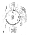
- Figure 1 shows the plasmid pSGE728 which was used for certain mutagenesis and expression experiments.
- Figure 2 shows the partial restriction map of the alpha gene inserted into pSGE728.
- Figure 3 shows the partial restriction map of the beta gene inserted into pSGE728.
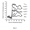
- Figure 4 shows the normalized time course of the NO-induced oxidation of recombinant oxyhemoglobin.
- the recombinant hemoglobin constructs rHb0.0, rHb0.1, rHb1.1 and rHb Bethesda all have wild-type amino acids in their heme pockets and an NO-oxidation rate constant of 60 ⁇ M -1 s -1 .
- Distal heme pocket mutants rHb2, rHb3 and rHb4 have rate constants of 24, 15 and 2 ⁇ M -1 S -1 , respectively.
- Conditions after mixing were 0.2 ⁇ M oxyhemoglobin (heme), 1.0 ⁇ M NO, 20°C.
- the heme pocket substitutions in rHb2 are ⁇ E11(Val ⁇ Phe), ⁇ E11(Val ⁇ Phe), ⁇ G8(Leu ⁇ Ile).
- the substitutions are ⁇ E11 (Val ⁇ Leu), ⁇ E11 (Val ⁇ Phe) and in rHb4 they are ⁇ B10 (Leu ⁇ Phe), ⁇ E11 (Val ⁇ Trp).
- Figure 5 shows the pressor effects of recombinant hemoglobins and human serum albumin (HSA). Changes in mean arterial pressure from pre-administration values are plotted versus time from administration. All hemoglobins contained a genetically fused dialpha subunit to prevent dissociation into ⁇ dimers. The rate constant for reaction of nitric oxide with each rHb is noted beside each data set. HSA indicates the pressor data collected when 5% HSA was administered as a volume control. Protein doses were 350 mg/kg for each rHb.
- HSA human serum albumin
- Figure 6 shows the pressor responses to rHb1.1 and rHb Bethesda. Both molecules contained a genetically fused dialpha subunit to prevent dissociation into alpha/beta dimers. Mean arterial pressure data are plotted versus time from administration.
- rHb1.1 has a P 50 of 32 mmHg
- rHb Bethesda has a P 50 of 2.7 mmHg.
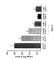
- Figure 7 shows the direct relationship between the magnitude of the pressor response and the rate of NO oxidation.
- the magnitude of the pressor response was calculated by averaging the post-infusion values for each time course in Figure 8 , and plotted against the measured value of k' NO, ox for the corresponding hemoglobin.
- the data point at the y-intercept was collected following administration of an equivalent volume of 5% HSA.
- FIG 8 shows .the average total peripheral resistance (TPR) response to recombinant hemoglobins.
- the average TPR was calculated from data collected starting 20 minutes and ending 90 minutes following administration. The dose used in each case was 350 mg Hb/kg body weight.
- “Mono Hb” refers to single-tetramer Hb species (MW about 64,000), and “di Hb” indicates two Hb tetramers that are genetically fused (MW about 128,000).
- the rate constant for NO-oxidation of each rHb is noted on the abscissa.
- FIG. 9 shows the TPR responses to three recombinant hemoglobins. Changes in TPR are plotted as percent of pre-administration values versus time from administration. All hemoglobins contained a genetically fused dialpha subunit to prevent dissociation into ⁇ dimers. The rate constant for reaction of nitric oxide with each rHb is noted beside each data set. Hemoglobin doses were always 350 mg/kg.
- Figure 10 shows the results of the gastric emptying model of G1 motility. Greater scavenging of nitric oxide, or inhibition of synthesis of nitric oxide, decreases emptying of the stomach.
- the dose used for each protein was 750 mg Hb/kg body weight (10% solution) and the dose for L-NAME was 10 mg/kg.
- the invention generally relates to hemoglobin mutations that confer reduced NO scavenging.
- D731 means that aspartate has been substituted with isoleucine at residue number 73.
- the present invention relates to novel mutant hemoglobins that have significantly reduced rates of reaction with nitric oxide or NO scavenging as defined in claims 1 and 2.
- the mutant hemoglobins of the present invention are also referred to herein as "NO mutants.”
- the present invention further relates to recombinant DNA molecules encoding NO mutants, as defined in claim 3.
- the NO mutants have one or more mutations in or around the heme pocket.
- Heme is an iron-containing porphyrin that serves as a prosthetic group in proteins such as hemoglobin, myoglobin and the cytochromes.
- the heme appears in a cleft between the E and F helices in the globin subunits.
- the heme iron is linked covalently to the imidazole nitrogen of the "proximal" F8 histidine, while the distal E7 histidine and E11 valine appear near the access of oxygen to the heme pocket.
- the residues of the heme pocket include those residues that are on a nearest atom-to-nearest atom basis within 6 angstroms, and preferably within 4 angstroms, of the heme moiety ( Fermi, et al. (1984) J. Mol. Biol. 175: 159-174 ).
- the heme pocket residues include: Distal residues: First shell Second Shell B10 Leu B 13 Met CE1 Phe CE3 His E7 His CE4 Phe E11 Val E10 Lys G8 Leu E14 Ala G12 Leu B14 Phe Proximal residues: First shell Second Shell F8 His C7 Tyr F4 Leu F7 Leu FG3 Leu FG5 Val G4 Asn G5 Phe H15 Val H19 Leu and for beta globin: Distal residues: First shell Second Shell B10 Leu B 13 Leu CD1 Phe CD3 Ser E7 His B14 Leu E11 Val CD4 Phe G8 Leu E10 Lys E14 Ala G12 Leu Proximal residues: First shell Second Shell F8 His C7 Phe F4 Leu F7 Leu FG3 Leu FG5 Val G4 Asn G5 Phe G12 Leu H15 Val H 19 Leu
- first shell residues are those residues in close or direct contact with the heme iron atom and/or the bound ligand
- second shell residues are those amino acids which are not in direct contact with the heme or the bound ligand, but are in direct contact with first shell residues.
- heme pocket residues include these first and second shell residues.
- naturally occurring human hemoglobin refers to human hemoglobin A o whose alpha and beta globin amino acid sequences are given in Figure 1 of U.S. Patent No. 5,028,588 .
- the non-helical segments are identified by letter pairs, indicating which helical segments they connect, for example, non-helical segment BC connects helix B and helix C.
- the helical notation and corresponding amino acids for alpha and beta globin are shown in Table 4 of U.S. Patent No. 5,028,588 .
- Recombinant hemoglobin means hemoglobin, whether native or mutant, comprising alpha-like globin proteins and/or beta-like globin proteins, at least one of which is obtained by expression of a globin gene carried by a recombinant DNA molecule in a cell other than the cell in which that hemoglobin gene and/or hemoglobin protein is naturally found.
- the hemoglobin gene is heterologous to the host in which it is expressed.
- the expression of any human hemoglobin gene in any cell other than a human blood cell would be considered to be a recombinant hemoglobin.
- an "alpha globin” has at least about 75% sequence identity with native human alpha globin.
- a polypeptide of lesser sequence identity may still be considered substantially homologous with native human alpha globin, and thus may be an alpha globin, if it has a greater sequence identity than would be expected from chance and also has the characteristic higher structure of native human alpha globin and similar biological activity.
- a "beta globin” has at least about 75% sequence identity with native human beta globin.
- a polypeptide of lesser sequence identity may still be considered substantially homologous with native human beta globin, and thus may be a beta globin, if it has a greater sequence identity than would be expected from chance and also has the characteristic higher structure of native human beta globin and similar biological activity.
- Ligand hemoglobin means hemoglobin to which a ligand is bound at the heme groups.
- Common preferred ligands include, but are not limited to O 2 , CO, NO and the like.
- Oxyhemoglobin means hemoglobin in which an oxygen molecule is bound to the functional oxygen binding sites in each subunit.
- Deoxyhemoglobin or "unliganded hemoglobin” means any hemoglobin to which no ligand is bound to the alpha globin, the beta globin, and/or any functional heme prosthetic group.
- Metal hemoglobin or "oxidized hemoglobin” means any hemoglobin in which the iron has been oxidized to the ferric state.
- R-state hemoglobin is the high affinity state of hemoglobin and is the dominant form of hemoglobin when a ligand is bound at the heme pockets.
- the ligand is typically oxygen, thus this state is known as the “oxy” or “R” (for relaxed) state.
- R for relaxed
- intersubunit distances are increased relative to the distances in T-state hemoglobin.
- T-state hemoglobin is the low affinity state of hemoglobin and is the dominant form of hemoglobin when it is deoxygenated ("deoxy", or "T" for "tense").
- Heme pocket means that pocket formed around the heme of each globin subunit described by the surrounding residues and is meant to include residues within about 6 ⁇ of the heme moiety and as described above.
- Distal heme pocket means that portion of the heme pocket above the plane of the heme that contains the free coordination site of the iron where ligand molecules can combine reversibly with the iron atom and which contains such residues as histidine E7 and valine E11. Likewise, the proximal side of the heme pocket is described by those residues below the plane of the heme and contains such residues as the proximal histidine at position F8.
- Oxidation means the oxidation of the iron in the heme of any or all of the subunits making up the hemoglobin tetramer from the ferrous (Fe +2 ) to the ferric form. (Fe +3 ). Autooxidation occurs spontaneously without the addition of exogenous oxidants, however oxidation can be induced by the presence of exogenous oxidizing agents, most notably NO and hydrogen peroxide.
- “Mutations” are substitutions, deletions or additions of one or more amino acids to the amino acid sequence that constitutes naturally occurring human hemoglobin.
- Affinity refers to the equilibrium binding of a ligand to hemoglobin, and is described by the thermodynamic equilibrium constant, Keq. Affinity is the ratio of the ligand association rate and the hemoglobin - ligand dissociation rate, and thus changes in association rate, dissociation rate or both can lead to changes in ligand affinity.
- “Altered affinity” means the affinity of a recombinant hemoglobin for a gaseous ligand that is at least 10% different from the affinity of naturally occurring human hemoglobin for that same gaseous ligand under the same measurement conditions.
- mutant alpha subunits were first paired with wild-type beta subunits and vice versa. Additional mutants formed by combination of mutations in the alpha and beta globins were also constructed.
- Any of the mutations described herein can be accomplished by a number of methods that are known in the art. Mutations can be made at the codon level by alteration of the nucleotide sequence that codes for a given amino acid. Substitution of an amino acid at any given position in a protein can be achieved by altering the codon for that particular amino acid. This substitution can be accomplished by site directed mutagenesis using, for example: (1) the Amersham technique (Amersham mutagenesis kit, Amersham, Inc., Cleveland, Ohio) based on the methods of Taylor et al., Nucl. Acids Res. (1985) 13: 8749-8764 ; Taylor et al., (1985) Nucl. Acids Res.
- Site directed mutagenesis can also be accomplished using PCR based mutagenesis such as that described in Zhengbin et al., pages 205-207 in PCR Methods and Applications Cold Spring Harbor Laboratory Press, New York (1992 ); Jones and Howard, (1990) BioTechniques 8(2):178 (1990 ); Jones and Howard, BioTechniques 10: 62-66 (1991) , or as described in the Examples below. Site directed mutagenesis can also be accomplished using cassette mutagenesis with techniques that are known to those of skill in the art.
- Any suitable host cell can be transformed with a plasmid containing the desired mutation(s) by methods known to those skilled in the art or as described in the Examples below.
- Suitable host cells include, for example, bacterial, yeast, plant, mammalian and insect cells.
- E. coli cells are particularly useful for expressing the novel mutant hemoglobins.
- Mutants marked with an asterisk are part of the present invention.
- the remaining mutants, which are not part of the invention, are listed for reference purposes.
- any of the above alpha globin mutations can be combined with any of the above beta globin mutations, or any of the above alpha globins can be combined with a known beta globin and vice versa to obtain desirable properties.
- other mutations may be added to adjust oxygen affinity. The following combinations have the desired NO reaction kinetics.
- Mutants marked with an asterisk are part of the present invention.
- the remaining mutants, which are not part of the invention, are listed for reference purposes.
- Combinations of these mutations can also be placed in larger sized hemoglobins having more than one tetramer such as, for example, di-hemoglobins (di-di alpha constructs).
- Trp and Phe substitutions at Leu-G8 significantly reduced the NO scavenging rate in beta (6 times slower for Trp) but had no detectable effect on the NO scavenging rate in alpha.
- substitutions at Val-E11 gave reduced NO scavenging rates for both alpha and beta subunits the results were not strictly consistent with the size of the substituted amino acid (Table 1).
- alpha subunits the smallest amino acid (leucine) produces the greatest reduction in NO scavenging rate.
- beta subunits the results are more consistent with the prediction that increasing bulk and hydrophobicity will reduce the NO scavenging rate.
- Amino acid substitutions obtained from the mutant libraries were also generally consistent with the theme of large hydrophobic residues promoting reduced rates of NO scavenging.
- mutants identified in this screen as having significantly reduced rates of NO catalyzed oxidation, there was a preponderance of substitutions such as Trp, Phe, Leu, Met, Val and Ile.
- substitutions such as Trp, Phe, Leu, Met, Val and Ile.
- some mutant heme pockets containing unusual hydrophilic residues such as Thr, Ser, Tyr and His were also observed to have substantially reduced NO scavenging rates. While such substitutions might be expected to lead to unstable molecules due to rapid autooxidation, it is possible that some of these molecules could exhibit novel and useful properties.
- the NO mutants of the present invention should have a rate constant for reaction of NO with oxyhemoglobin (k' NO,OX ) less than conventional hemoglobin, preferably in the range of about 0.1 ⁇ M -1 s -1 to less than about 60 ⁇ M -1 s -1 .
- the rate constant is less than 25 ⁇ M -1 s -1 , more preferably between 12 and 15 ⁇ M -1 s -1 , and most preferably less than 5 ⁇ M -1 s -1 .
- the results demonstrate a linear correlation between the magnitude of the pressor effect in vivo and the in vitro rate of NO scavenging.
- reducing the NO reactivity of the heme groups significantly reduced the magnitude of the pressor response in conscious rats.
- the data support the hypothesis that the pressor response to extracellular hemoglobins is due to a decrease in steady-state levels of NO in the region of the endothelial and smooth muscle cells lining the vasculature.
- a decrease in NO concentration results in lesser activation of guanylyl cyclase, which ultimately increases the tone of vascular smooth muscle.
- guanylyl cyclase results in lesser activation of guanylyl cyclase, which ultimately increases the tone of vascular smooth muscle.
- guanylyl cyclase results in lesser activation of guanylyl cyclase, which ultimately increases the tone of vascular smooth muscle.
- guanylyl cyclase results in lesser activation of guanylyl cyclase, which ultimately increases the tone of vascular smooth muscle.
- Those recombinant hemoglobins that have lower rates of reaction with NO are less potent competitors for nitric oxide. Consequently, the NO mutants of the present invention are useful
- each subunit can be modified by varying the degree of steric hindrance near the iron atom, or by varying the strength of hydrogen bonding between the E7 residue and the bound oxygen.
- Steric hindrance affects the association and dissociation rate constants by altering the entry and exit of oxygen to and from the heme pocket.
- steric hindrance also affects the rate of oxidation by NO.
- the E7 histidine stabilizes bound oxygen through hydrogen bonding interactions. Substitution of the E7 histidine can decrease the degree of stabilization of bound oxygen and enhance oxygen dissociation kinetics. E7 substitutions can also affect reactivity of NO with Hb.
- the recombinant hemoglobins of the present invention can be used for a number of in vitro or in vivo applications.
- Such in vitro applications include, for example, the delivery of oxygen by compositions of the instant invention for the enhancement of cell growth in cell culture by maintaining oxygen levels in vitro ( DiSorbo and Reeves, PCT publication WO 94/22482 .
- the hemoglobins of the instant invention can be used to remove oxygen from solutions requiring the removal of oxygen (Bonaventura and Bonaventura, US Patent 4,343,715 ) and as reference standards for analytical assays and instrumentation (Chiang, US Patent 5,320,965 ) and other such in vitro applications known to those of skill in the art.
- the recombinant hemoglobins can be formulated for use in various therapeutic applications.
- Example formulations suitable for the recombinant hemoglobin of the instant invention are described in Milne, et al., WO 95/14038 and Gerber et al., WO 96/27388 .
- Pharmaceutical compositions can be administered by, for example, subcutaneous, intravenous, or intramuscular injection, topical or oral administration, large volume parenteral solutions, aerosol, transdermal or mucus membrane adsorption and the like.
- the recombinant hemoglobins of the present invention can be used in compositions useful as substitutes for red blood cells in any application that red blood cells are used or for any application in which oxygen delivery is desired.
- the recombinant hemoglobins can also be formulated as oxygen' carrying therapeutics and used for the treatment of hemorrhages, traumas and surgeries where blood volume is lost and either fluid volume or oxygen carrying capacity or both must be replaced.
- the recombinant hemoglobins of the instant invention can be made pharmaceutically acceptable, they can be used not only as blood substitutes that deliver oxygen but also as simple volume expanders that provide oncotic pressure due to the presence of the large hemoglobin protein molecule.
- the recombinant hemoglobins of the instant invention can be crosslinked by methods known in the art and used in situations where it is desirable to limit the extravasation or reduce the colloid osmotic pressure of the hemoglobin-based blood substitute.
- the recombinant hemoglobins can act to transport oxygen as a red blood cell substitute, while reducing the adverse effects that can be associated with excessive extravasation.
- a typical dose of recombinant hemoglobin as an oxygen delivery agent can be from 2 mg to 5 grams of hemoglobin per kilogram of patient body weight.
- a typical dose for a human patient might be from a few grams to over 350 grams.
- the unit content of active ingredients contained in an individual dose of each dosage form need not in itself constitute an effective amount since the necessary effective amount could be reached by administration of a number of administrations. The selection of dosage depends upon the dosage form utilized, the condition being treated, and the particular purpose to be achieved according to the determination of those skilled in the art.
- Administration of recombinant hemoglobin can occur for a period of seconds to hours depending on the purpose of the hemoglobin usage.
- an oxygen carrier the usual time course of administration is as rapid as possible.
- Typical infusion rates for hemoglobin solutions as oxygen therapeutics can be from about 100 ml to 3000 ml/hour.
- the hemoglobins of the instant invention can be used to treat anemia, both by providing additional oxygen carrying capacity in a patient that is suffering from anemia, and/or by stimulating hematopoiesis as described in PCT publication WO 95/24213 , incorporated herein by reference.
- administration rates can be slow because the dosage of hemoglobin is much smaller than dosages that can be required to treat hemorrhage. Therefore the recombinant hemoglobins of the instant invention can be used for applications requiring administration to a patient of high volumes of hemoglobin as well as in situations where only a small volume of the hemoglobin of the instant invention is administered.
- the hemoglobins of the present invention can be used to deliver oxygen to areas that red blood cells cannot penetrate. These areas can include any tissue areas that are located downstream of obstructions to red blood cell flow, such as areas downstream of thrombi, sickle cell occlusions, arterial occlusions, angioplasty balloons, surgical instrumentation, any tissues that are suffering from oxygen starvation or are hypoxic, and the like. Additionally, all types of tissue ischemia can be treated using the hemoglobins of the instant invention.
- tissue ischemias include, for example, stroke, emerging stroke, transient ischemic attacks, myocardial stunning and hibernation, acute or unstable angina, emerging angina, infarct, and the like.
- Recombinant hemoglobin can also be used as an adjunct with radiation or chemotherapy for the treatment of cancer.
- the recombinant hemoglobins of the instant invention can also be used to deliver drugs and for in vivo imaging as described in WO 93/08842 .
- Recombinant hemoglobins can also be used as replacement for blood that is removed during surgical procedures where the patient's blood is removed and saved for reinfusion at the end of surgery or during recovery (acute normovolemic hemodilution or hemoaugmentation).
- the recombinant hemoglobins of the instant invention can be used to increase the amount of blood that can be predonated prior to surgery, by acting to replace some of the oxygen carrying capacity that is donated.
- Examples 1 - 4 are studies relating to the expression of recombinant hemoglobins in Ecoli, while Examples 5-10 are studies relating to hemoglobin mutants having reduced NO scavenging.
- PCR based cassette mutagenesis was used to incorporate desired amino acid substitutions.
- DNA sequence for variant codons was based on codons used in proteins that are highly expressed in E.coli as described in Sharp et al., Nucl. Acids Res., 16:8207-8211 (1988) .
- strains were constructed by transformation of the plasmid DNAs into E . coli strains lacking a plasmid using the procedure of Chung, et al., Proc. Natl. Acad. Sci. USA, 86:2172-2175 (1989) , or Hanahan, DNA Cloning: A Practical Approach, V. 1, pp. 109-135 (IRL Press, 1985 ).
- E. coli strains were used, including those listed in Tables 2 and 3. The construction of these strains, especially those containing different oligomerizing mutants or domains, is described in WO 97/04110 , incorporated herein by reference. Tables 4 and 5 summarize the strain background and plasmid backbone used for the expression of these molecules. Plasmid copy number has been shown to influence soluble expression yields as well. The medium copy plasmid pSGE705 served as a platform for some of the molecules tested. Plasmids pSGE715 and pSGE720 are high copy number plasmids, and expression from these plasmids should exceed that observed from pSGE705-based plasmids.
- pSGE720 contains a synthetic operon composed of the di- ⁇ -globin and ⁇ Presbyterian-globin genes transcribed from the tac promoter on a tetracycline resistant plasmid with the pUC high copy number origin of replication ( Weickert & Curry, Arch. Biochem. Biophys. 348:337-346 (1997) ). Table 2 SGE plasmid bkgrd.
- SGE1675 none none gyrA96 (Na1 R ), lacI Q,I , endA, hsdR17, relA1, supE44, recJ (a deriv. of JM107)
- SGE1464 pSGE720 1.1 SGE1675
- SGE1480 pSGE726 1.0 SGE1675
- SGE1483 pSGE728 0.0 SGE1675
- SGE2706 pSGE733 0.1 SGE1675 SGE2761 none none SGE1675 + rpsL (strR) by P1 transduction SGE2782 pSGE767 9+1.1 SGE1675 SGE2784 pSGE768 9.1 SGE1675 SGE3083 pSGE1001 mut.
- pSGE728 was constructed by Xho I digestion and deletion from pSGE720 of one alpha subunit and the di-alpha glycine linker.
- the resulting plasmid, pSGE726, contains a single alpha gene rather than a di-alpha gene (rHb1.0).
- the Presbyterian mutation in beta was replaced by digestion with Bg l II and Hind III, and ligation to introduce wild-type beta and create pSGE728 (rHb0.0).
- the Presbyterian mutation in the beta gene of pSGE720 was replaced by digestion and ligation as for pSGE728, to introduce wild-type beta and created pSGE733 (di-alpha and wild type beta; rHb0.1).
- the Buffalo mutation ( ⁇ Lys82 ⁇ Asp) was introduced into the rHb1.1 background to create rHb9+1.1.
- the Lys82 ⁇ Asp mutation was created by PCR amplification of a portion of the ⁇ -globin gene using an oligonucleotide containing an Asp codon in place of the Lys82.
- CBG124 wild-type beta coding sequence 5' primer near Bsp EI site
- CBG119 (3' primer containing the ⁇ K82D mutation downstream of Asp 718 site) were used to amplify a small DNA fragment from pSGE761 as template.
- CBG119 5'AGC GAA GGT ACC GTC CAG GTT (SEQ.ID.NO: 1)
- CBG124 5'CCT GAC TCC GGA AGA AAA ATC C (SEQ.ID.NO: 2)
- the PCR product and vector were digested with Bsp EI and Asp 718 and ligated.
- DNA sequencing of plasmid isolated from transformants was performed with Sequenase ® kit reagents and protocols (United States Biochemical, 33 P (Amersham, Inc.), and primers synthesized on an Applied Biosystems 380B DNA synthesizer. Sequencing confirmed the Buffalo and Presbyterian mutations and the plasmid (pSGE767) was transformed into SGE1675 to produce SGE2782.
- the Buffalo mutation was introduced into the rHb0.1 background to create rHb9.1.
- a Bam HI/ Asp 718 fragment from pSGE767 was isolated and ligated into digested pSGE733 (rHb0.1). Sequencing confirmed the mutation and the plasmid (pSGE768) was transformed into SGE1675 to produce SGE2784, or SGE3138 to produce SGE3261. Similar steps were used to introduce Buffalo (asp) and the combination of Buffalo (asp) and Presbyterian in a plasmid with a di-alpha Lys 158 ⁇ Cys mutation (Table 5: SGE3083, SGE3084 and SGE3172).
- the 0.2 ml aliquots were transferred to a 96 well flat bottom microtiter plate (Immunlon 4 from Dynatech). The optical density of each well was recorded at 650 nm using the Spectral Dynamics microplate reader and Softmax software package. The cells were pelleted to the bottom of the microplates at 3000 rpm at 4°C for 10 minutes using the Beckman RK6 centrifuge, 3.7 rotor and microplate adapter buckets. The spent media was removed and the cells were resuspended in 0.1 ml 25 mM Borax with light vortexing of microplate.
- microplates were covered with parafilm and stored at -80°C overnight and then thawed at 30°C in a water incubator until just thawed.
- Lysozyme (Ameresco Ultra pure grade) was added to 0.17 mg/ml final volume from a 1 mg/ml stock containing 0.05 M NaCl.
- the samples were mixed by light vortexing of microplate, covered and incubated at room temperature for 30 minutes.
- DNAase I Boehinger Mannheim grade II from bovine pancreas
- DNAase I Boehinger Mannheim grade II from bovine pancreas
- the samples were mixed by light vortexing of microplate, covered and incubated at room temperature for 15 minutes.
- the microplates were covered with parafilm and stored at -80°C for 90 min. to 20 hours and then placed at 30°C in a water incubator until just thawed.
- the cell debris were pelleted to the bottom of the microplates at 3000 rpm at 4°C for 10 minutes using the Beckman RK6 centrifuge, 3.7 rotor and microplate adapter buckets.
- the cleared lysate was transferred to wells of a fresh microplate and stored at 4°C for approximately 12 hours.
- casein blocker Pieris
- ELISA wash buffer diluted samples and standards were then added and the plates were incubated at 37°C for 60 minutes.
- biotin labeled Goat anti-rHb1.1 antibody 50 ng/ml in PBS with 1% casein were added to each well.
- the plates were covered and incubated at 37°C for 60 minutes and were washed as above.
- Fermentations were performed in a defined medium in 15L Biolaffite fermentors, generally as described in Looker et al. "Expression of Recombinant Human. Hemoglobin in Escherichia coli.” Meth. Enzymol. 231:364-374.(1994 ) using DM59(60) medium under Glucose excess (BAR) conditions with induction for 16 hours at 28°C, except where noted.
- BAR Glucose excess
- Induction of expression was achieved by addition of IPTG between 10 and 200 ⁇ M on attaining a cell density of an O.D. 600 of approximately 30. Incubation was continued for 16 hours post induction, and hemin was added in five shots of 10, 13, 17, 17, and 17 ml at 0, 3, 6, 9, and 12 hours post induction respectively, delivering a total concentration of 0.37g/L of hemin.
- One ml fermentation samples were withdrawn at 4, 8, 12, 14 and 16 hours post induction and assayed for soluble rHb1.1. In other studies, hemin was added in five shots at 25 ml each and collected at 12, 14 and 16 hours.
- Mutations were introduced into cloned human alpha and beta genes via site directed PCR-based mutagenesis as described in general by Innis et al., PCR Protocols: A Guide to Methods and Applications (1990 ).
- the desired mutations were introduced into synthetic DNA oligonucleotides which were synthesized according to the manufacturer's instructions on an Applied Biosystems 392 DNA synthesizer. Following standard deblocking procedure, oligonucleotides were dried by vacuum-centrifugation, resuspended in the desired buffer and diluted to 10-50 pmol/ ⁇ l in sterile water.
- oligonucleotides were used as primers in PCR reactions where a recombinant plasmid carrying cloned wild type alpha and beta genes, such as pSGE728 ( Fig. 1 ), was used as template DNA.
- mutagenic oligonucleotide primers were chosen to span the site at which the mutation(s) was to be introduced and a nearby restriction endonuclease recognition site to facilitate cloning of resulting PCR products carrying mutations of interest.
- a second oligonucleotide primer was also required in the PCR reaction to allow DNA amplification.
- This primer also could be designed to contain globin gene mutation(s) or alternatively could consist of wild-type globin gene sequence from a neighboring region of the alpha or beta globin gene.
- This second primer was also chosen to contain a restriction endonuclease recognition site so that the resulting PCR product could be cloned into appropriately digested pSGE728 for subsequent expression of the mutated alpha or beta globin. Partial restriction maps of the alpha and beta genes from pSGE728 are shown in Figures 2 and 3 .
- the lengths of the mutagenic oligonucleotides were determined by the distance between the site to be mutated and the closest restriction site that could be incorporated into the mutagenic oligonucleotide at a position between the 5-prime end of the oligo and the site to be mutated.
- the mutagenic oligos were typically 30 to 45 nucleotides in length and contained mutations affecting one or two codons although more positions potentially could also be altered if desired. It was generally desirable to place the mutated DNA sequences as far as feasible from the 3-prime end of the oligo so as to avoid or minimize potential problems during the primer annealing step of the PCR reaction.
- the mutated nucleotides were generally placed 5-10 nucleotides upstream of the 3-prime end of the mutagenic primer.
- the globin gene restriction site incorporated near the 5-prime end of the mutagenic oligonucleotide was generally placed 5-12 nucleotides downstream of the 5-prime end to facilitate subsequent digestion of PCR products.
- Oligonucleotides which were employed solely as primers in PCR i. e. did not contain mutations
- PCR reactions were generally performed in an Applied Biosystems GeneAmp 9600. PCR reaction conditions were empirically determined: denaturation was typically at 95° C for 15-60 seconds, generally annealing temperatures ranged from 45-60° C for 15-30 seconds with many reactions being run in 50-55° C range for annealing, and extensions were done at 72° C for 15-120 seconds.
- the annealing temperature was raised during the course of the reaction: e.g. a reaction might consist of 5 rounds with an annealing temperature of 45° C followed by 20 rounds at an annealing temperature of 60° C.
- a reaction might consist of 5 rounds with an annealing temperature of 45° C followed by 20 rounds at an annealing temperature of 60° C.
- Typically reactions consisted of a total of 25-30 cycles. The reactions were typically performed in 10 mM Tris-HCl (pH 8.3), 50 mM KCl, 1.5 mM MgCl 2 0.2 mM dNTPs (Pharmacia) and 0.001% gelatin. Oligonucleotide primers were added at concentrations usually 0.5-1.0 ⁇ M. Purified plasmid DNA (about 0.1-10 ng per reaction) such as pSGE728 was generally used as template. AmpliTaq ® DNA Polymerase (Perkin Elmer) was typically used at 1-10 units per reaction and reaction volumes ranged
- reaction products were purified using the QIAquick PCR Purification Kit (QIAGEN Inc. Santa Clarita, CA). The purified products were then subjected to restriction endonuclease digestion with the appropriate enzymes to generate DNA fragments suitable for cloning into similarly cut pSGE728. Restriction digests were performed according to vendor protocols.
- the digested PCR fragments could be cloned directly or first subjected to agarose gel electrophoresis and purified out of the agarose gels.
- Gel composition and electrophoresis conditions were chosen based on the DNA fragment size. Many fragments were about 60-250 base pairs in length and for these fragments resolution in gel electrophoresis is optimal with gels such as 3% NuSeive agarose or 4% Metaphor agarose, both obtained from FMC BioProducts (Rockland, ME) and used according to vendor protocols.
- DNA fragments were purified out of agarose gel slices using the QIAEX II Gel Extraction Kit (QIAGEN Inc. Santa Clarita, CA) according to the vendor protocols.
- the vector pSGE728 was also digested with the enzymes appropriate for cloning the mutagenized PCR fragment(s) of interest and similarly gel-purified following more conventional electrophoresis.
- E coli strain JM109 obtained as competent cells from Promega (Madison, WI) was often used for this purpose although other strains of E coli could also be employed as well as other various methods for preparation of competent cells ( Sambrook et al., Molecular Cloning: A Laboratory Manual (Cold Spring Harbor, 1989 )).
- Transformants were selected for tetracycline-resistance and could subsequently be sequenced to identify the mutations of interest and confirm the sequence of the mutagenized cloned PCR segment. Plasmid templates were prepared using the QIAprep Spin Miniprep Kit (QIAGEN) and sequenced using the AmpliCycleTM Sequencing Kit (Perkin Elmer, Foster City, CA) according to the vendor protocol.
- sequencing was performed manually using Sequenase (United States Biochemical, Cleveland, OH) but the preferred method is automated sequencing using the AmpliCycleTM Sequencing Kit (Perkin Elmer, Foster City, CA) to perform sequence reactions according to the vendor protocol and analysis on an ABI Prism 377 DNA Sequencer (Applied Biosystems Division of Perkin Elmer, Foster City, CA).
- V62 codon was mutated on a PCR fragment spanning the Mae III through Mlu I segment of the alpha gene and this fragment following Mae III digestion was ligated to a gel-purified Bam HI - MaeIII fragment of pSGE728.
- This ligation product was digested with Bam HI and MluI (both of which are unique cutters within pSGE728) and the Bam HI-Mlu I fragment was gel purified and ligated to Bam HI- Mlu I- and gel-purified vector fragment of pSGE728.
- V62 mutations could have been incorporated into longer oligonucleotides that spanned unique sites such as Mlu I.
- two or more mutagenized PCR segments were ligated together to create segments containing as many as four mutagenized sites prior to cloning into appropriately digested pSGE728 vector.
- the appropriately sized ligation products were identified and subsequently purified by agarose gel electrophoresis.
- the purification step was preceded or followed by PCR amplification using primers that would specifically amplify the ligation product of interest.
- mutagenic oligos were synthesized with degeneracies at positions of interest. For "randomization" of a given position two degenerate oligos were synthesized, one of which contained the sequence N(T/A/C)T at the codon to be randomized while the other contained the sequence (A/T)(T/G)(T/G) at the same position. These two oligos could be pooled prior to PCR but more usually two independent PCR reactions were used with such pairs and the PCR products roughly quantitated (using an Alphalmager TM 2000 Documentation & Analysis System from Alpha Innotech Corp San Leandro, CA) by visualization following gel electrophoresis.
- Plasmid templates were prepared using the QIAprep Spin Miniprep Kit (QIAGEN) and sequenced using the AmpliCycleTM Sequencing Kit (Perkin Elmer, Foster City, CA) according to the vendor protocol. Sequences were run and analyzed on an ABI Prism 377 DNA Sequencer (Applied Biosystems Division of Perkin Elmer, Foster City, CA). Sequences were analyzed to assess the distribution of amino acid substitutions within a given library and the frequencies of PCR-induced and synthetic oligo-induced errors in DNA sequence. Subsequently clones from libraries were picked and analyzed as described below.
- alpha and beta mutations can be combined in derivatives of pSGE728.
- such combinations can be achieved by cutting mutant derivatives of pSGE728 with appropriate restriction endonucleases that separate the alpha and beta sequences, gel-purifying the restriction fragment containing the beta gene of the mutant beta derivative of pSGE728, gel-purifying the restriction fragment containing the alpha gene of the mutant alpha derivative, ligating these two fragments together, transforming E coli and analyzing the resulting transformants to verify the presence of both alpha and beta mutations.
- alpha and beta mutations at residues B10, E11, G8 and E7 such combinations can be made by digesting the mutant derivatives of pSGE728 with Bsp HI which cuts within the tetracycline-resistance gene and SacII which cuts within the beta gene, about 28 base pairs from the start of the beta coding sequence.
- the 937 bp fragment derived from the beta mutant derivative of pSGE728 can be excised out of the agarose gel and purified using the QIAEX II Gel Extraction Kit (QIAGEN Inc. Santa Clarita, CA) according to the vendor protocols.
- the 2318 bp fragment from the pSGE728 derivative carrying the alpha mutation can also be excised from the gel and purified. These two purified fragments can be ligated together using T4 DNA ligase (New England BioLabs Beverly, MA) according to the vendor protocols and ligation products were used to transform E coli.
- E coli strain JM 109 obtained as competent cells from Promega (Madison, WI) can be used for this purpose although other strains of E coli could also be employed as well as other various methods for preparation of competent cells (Sambrook et al., supra).
- tetracycline resistant transformants selects for reconstitution of the tetracycline-resistance gene and this is nearly always associated with reconstitution of the beta gene at the SacII site.
- individual transformants thus obtained are analyzed by determining DNA sequence for alpha and beta genes and gross plasmid structure, more than 90% are found to be the desired recombinants which have both the alpha and beta mutations.
- sequence analysis plasmid templates can be prepared using the QIAprep Spin Miniprep Kit (QIAGEN) and sequenced using the AmpliCycle TM Sequencing Kit (Perkin Elmer, Foster City, CA) according to the vendor protocol. Sequences were run and analyzed on an ABI Prism 377 DNA Sequencer (Applied Biosystems Division of Perkin Elmer, foster City, CA).
- E coli strains containing recombinant plasmids such as derivatives of pSGE728, which encode variant hemoglobins were typically grown in shake flasks usually at volumes of about 50 ml.
- recombinant plasmids such as derivatives of pSGE728, which encode variant hemoglobins
- Generally defined media supplemented with about 0.2% yeast extract were used for cell growth and tetracycline was added, generally at 15 ⁇ g/ml to select for maintenance of the recombinant plasmid.
- Expression of the hemoglobin genes was induced by addition of IPTG, usually at a concentration of 100 ⁇ M and hemin was added to a final concentration of 50 ⁇ g/ml generally at the time of induction. Cells were generally grown and induced at 28° C.
- Cells grown to stationary phase could be directly inoculated (generally at a dilution ranging from 1/50 to 1/1000) into media containing IPTG and hemin or such cultures could be inoculated into media lacking IPTG and hemin, grown to log phase, e.g. 0.4-0.7 OD @ A 600 and then induced by the addition of IPTG with hemin typically being added to the cultures at the time of induction.
- Cultures were generally grown under inducing conditions overnight ( ⁇ 14-20 hours) although shorter times, e.g. about 6 hours could also be employed. At the end of this time, cells were pelleted by centrifugation and the cell pelleted were either frozen and stored at -80° C or processed immediately.
- Recombinant hemoglobins were purified by small-scale column chromatography using Fast Flow Zn-Chelating Sepharose (Pharmacia). During the purification, cells, lysates, all buffers and eluted hemoglobins were kept cold on ice as much as possible. Typically, a pellet of a 50 ml culture was resuspended with 1.0 ml ice-cold 25 mM sodium tetraborate and transferred to a 1.7 ml eppendorf tube. Cells were usually lysed by sonication, although enzymatic lysis by lsysozyme could also be employed.
- Sonicated lysates were clarified by centrifugation (generally about 14,000 x g for 15-20 minutes at 4°C) following addition of 20 ul of 20 mM ZnAcetate. Supernatants were loaded onto a ⁇ 150-200 ul column that had previously been equilibrated as follows:
- the column was washed with at least 9 column volumes 0.5 M NaCl, 20 mM Tris-HCl pH 8.1 at 0°C followed by at least 3 column volumes 0.05 M NaCl, 20 mM Tris-HCl pH 8.1 at 0°C and then eluted with -1.0 ml of the desired buffer (e.g. 0.1 M Na phosphate pH 7.0) containing 30 mM EDTA. Hemoglobin was typically recovered in a volume of -200-400 ul. These samples were used in kinetic assays such as NO oxidation and O 2 dissociation. If not used immediately, samples were frozen and stored -80° C.
- the desired buffer e.g. 0.1 M Na phosphate pH 7.0
- the protein is then eluted in two to three volumes of 12.3mM Tris (pH7.6) or if the pI of the protein is below 7.5 it is eluted in a Bis-Tris buffer at the appropriate pH. pI was used to determine proper wash and elution conditions for each protein, both monomeric and dimeric. Certain mutations on the surface of some of the heme pocket mutants were found to effect this value.
- the appropriate load buffer was 10 mM KPi (pH 7.0) for loading onto a ceramic hydroxyapatite (CHT) column (BioRad) or 20 mM Tris (pH 8.0) for loading onto a hydrophobic interaction chromatography (HIC) column (BioRad).
- CHT ceramic hydroxyapatite
- HIC hydrophobic interaction chromatography
- the protein is loaded onto the CHT column at 20gm/L the column is then washed with eight column volumes of 30-40mM KPi (7.0). Five column volumes of 85-90mM Kpi (pH7.0) are used to elute the protein from the column.
- the anion exchange step is designed to wash away remaining monomeric hemoglobin from the di-hemoglobin yielding a di-hemoglobin pool that is 98% pure on a size basis.
- the anion exchange column is a Super Q 650M (TosoHass). The column is equilibrated with 20mM Tris (pH9.0) and 15gm/L of protein is loaded on the column. The column is then washed with three column volumes of 10-15mM Tris (pH 7.6-7.8) and the protein is eluted in three column volumes of 15-30mM Tris (pH 7.6-7.8) or if pH is between 7.3-7.6 then a Bis-Tris buffer is used. The protein from this point on is handled as described in WO 96/15151 .
- Nitric oxide gas was passed through a column of NaOH pellets and used to thoroughly flush a tonometer.
- Anaerobic buffer (0.1 M sodium phosphate, pH 7.4) was injected into the tonometer and equilibrated with the NO to make a stock solution. Dilutions of the stock solution were made in glass syringes containing anaerobic buffer. Time courses of the reaction of oxyhemoglobin with NO were collected at 420 and 402 nm using an Applied Photophysics stopped-flow device. Temperature was 20° C. Data were collected and analyzed using the software program !SX.17MV supplied by Applied Photophysics.
- Substituted alpha and beta subunits having approximately equal values of k' NO , ox were combined into tetrameric hemoglobin constructs for use in animal models.
- These "paired-mutant" rHbs e.g. rHb2, rHb3, and rHb4; Table 4
- rHbs were constructed with a genetically fused, mutated di-alpha subunit to prevent dissociation of the hemoglobins into alpha/beta dimers as described above.
- the mutations are designated, for example, as ⁇ N108K, which means the native human beta globin sequence residue number 108, with wildtype Asn (N) replaced by Lys (K).
- the resulting recombinant oxyhemoglobins have monophasic reactions with nitric oxide ( Figure 7 ), simplifying interpretation of in vivo experiments by having only one rate constant to consider.
- the values of k' NO, ox for rHb2, rHb3, and rHb4 are 24, 15, and 2 ⁇ M -1 s -1 , respectively.
- Recombinant hemoglobins rHb1.1, rHb0.1 (des val, di- ⁇ ), rHb0.0 (des val, wildtype type), and rHb Bethesda have a wide range of P 50 values (32, 10, 15, and 2.7 mm Hg, respectively) due to different tendencies to form the low-affinity T-state quaternary structure.
- the latter four proteins have wild-type amino acids in the heme pockets and identical high values of the rate of NO-induced oxidation ( ⁇ 60 ⁇ M -1 s -1 ).
- the data suggest that, regardless of the position of the allosteric (R/T) equilibrium, only substitutions within the heme pocket affect the NO reactivity of oxyhemoglobin.
- Hemodynamic responses to hemoglobin administration were obtained in conscious, unrestrained rats. Male Sprague-Dawley rats were chronically instrumented with indwelling arterial and venous catheters at least 48 hours prior to experimentation. Top-load doses of 350 mg/kg of the rHb solutions (5 g/dl) were administered to separate groups of rats via intravenous infusion at a rate of 0.5 ml/min. Human serum albumin (HSA, 5 g/dl) was administered to another group of rats as a volume control. Arterial pressure was monitored continuously for 30 minutes prior to and 90 minutes following administration. All data are shown as mean ⁇ standard error. Statistical comparisons between rHb1.1 and the other hemoglobins or HSA were made by repeated measures analysis of variance, p-values ⁇ 0.05 were considered significant.
- MAP mean arterial pressure
Claims (3)
- Hämoglobin-Mutante, ausgewählt aus:einem Hämoglobin, umfassend die α-Globin-Mutationen:B10(Leu → Trp) und E7(His → Gln); undeinem Hämoglobin, umfassend die β-Globin-Mutationen:G8(Leu → Phe) und G12(Leu → Trp),G8(Leu → Phe) und G12(Leu → Met),E11 (Val → Trp) und E7(His → Gln),E11 (Val → Leu) und G8(Leu → Trp),E11 (Val → Met) und G8(Leu → Trp), oderE11(Val → Met) und G8(Leu → Phe).
- Hämogtobin-Mutante nach Anspruch 1, umfassend die Globin-Mutationen:αB10(Leu → Trp), αE7(His → Gin) und. (βE11 (Val → Trp).
- Rekombinantes DNA-Molekül, ausgewählt aus:einem rekombinanten DNA-Molekül, Welches ein α-Globin, umfassend die Mutationen:B10(Leu → Trp) und E7(His → Gln), kodiert; undeinem rekombinanten DNA-Molekül, welches ein β-Globin, umfassend die Mutationen:G8(Leu → Phe) und G12(Leu → Trp),G8(Leu → Phe) und G12(Leu → Met),E11 (Val → Trp) und E7(His → Gln),E11 (Val → Leu) und G8(Leu → Trp),E11 (Val → Met) und G8(Leu → Trp), oderE11 (Val → Met) und G8(Leu→ Phe), kodiert.
Priority Applications (1)
| Application Number | Priority Date | Filing Date | Title |
|---|---|---|---|
| EP08075054A EP1950298A3 (de) | 1997-05-02 | 1998-05-01 | Hämoglobinmutanten mit erhöhtem Löslichkeitswert und/oder reduzierter Stickoxidspülung |
Applications Claiming Priority (5)
| Application Number | Priority Date | Filing Date | Title |
|---|---|---|---|
| US4536497P | 1997-05-02 | 1997-05-02 | |
| US45364P | 1997-05-02 | ||
| US5798697P | 1997-09-05 | 1997-09-05 | |
| US57986P | 1997-09-05 | ||
| PCT/US1998/008861 WO1998050430A2 (en) | 1997-05-02 | 1998-05-01 | Hemoglobin mutants with increased soluble expression and/or reduced nitric oxide scavenging |
Related Child Applications (1)
| Application Number | Title | Priority Date | Filing Date |
|---|---|---|---|
| EP08075054A Division EP1950298A3 (de) | 1997-05-02 | 1998-05-01 | Hämoglobinmutanten mit erhöhtem Löslichkeitswert und/oder reduzierter Stickoxidspülung |
Publications (2)
| Publication Number | Publication Date |
|---|---|
| EP0979279A2 EP0979279A2 (de) | 2000-02-16 |
| EP0979279B1 true EP0979279B1 (de) | 2008-03-19 |
Family
ID=26722688
Family Applications (1)
| Application Number | Title | Priority Date | Filing Date |
|---|---|---|---|
| EP98920113A Expired - Lifetime EP0979279B1 (de) | 1997-05-02 | 1998-05-01 | Hemoglobin mutanten mit erniedrigtem stickstoffoxid entfernungsvermögen |
Country Status (7)
| Country | Link |
|---|---|
| US (2) | US6455676B1 (de) |
| EP (1) | EP0979279B1 (de) |
| JP (2) | JP4512673B2 (de) |
| AU (1) | AU750295B2 (de) |
| CA (1) | CA2299049C (de) |
| DE (1) | DE69839263T2 (de) |
| WO (1) | WO1998050430A2 (de) |
Families Citing this family (29)
| Publication number | Priority date | Publication date | Assignee | Title |
|---|---|---|---|---|
| AU784195B2 (en) * | 1999-11-12 | 2006-02-16 | Baxter Biotech Technology S.A.R.L. | Reduced side-effect hemoglobin compositions |
| CA2467245A1 (en) * | 2001-12-06 | 2003-06-19 | Duke University | Prevention of flap necrosis in plastic surgery |
| DE10224750A1 (de) | 2002-06-04 | 2003-12-24 | Fresenius Medical Care De Gmbh | Vorrichtung zur Behandlung einer medizinischen Flüssigkeit |
| US20070166792A1 (en) * | 2004-09-15 | 2007-07-19 | Olson John S | Increasing hemoglobin and other heme protein production in bacteria by co-expression of heme transport genes |
| US7642233B2 (en) * | 2004-09-15 | 2010-01-05 | William Marsh Rice University | Enhancing recombinant hemoglobin production by co-expression with alpha hemoglobin stabilizing protein |
| EP1797193A4 (de) * | 2004-09-15 | 2009-08-26 | Univ Rice William M | Erhöhte produktion von hämoglobin und anderen hämproteinen in bakterien durch koexpression von hämtransportgenen |
| US7935074B2 (en) | 2005-02-28 | 2011-05-03 | Fresenius Medical Care Holdings, Inc. | Cassette system for peritoneal dialysis machine |
| US20060195064A1 (en) * | 2005-02-28 | 2006-08-31 | Fresenius Medical Care Holdings, Inc. | Portable apparatus for peritoneal dialysis therapy |
| US8197231B2 (en) | 2005-07-13 | 2012-06-12 | Purity Solutions Llc | Diaphragm pump and related methods |
| CA2647564C (en) * | 2006-04-03 | 2016-08-30 | Pharmatherm Chemicals Inc. | Thermal extraction method and product |
| EP2032156B1 (de) | 2006-05-22 | 2012-11-21 | The Regents of the University of California | Zusammensetzungen und verfahren zur abgabe von sauerstoff |
| US7905990B2 (en) | 2007-11-20 | 2011-03-15 | Ensyn Renewables, Inc. | Rapid thermal conversion of biomass |
| US8192401B2 (en) | 2009-03-20 | 2012-06-05 | Fresenius Medical Care Holdings, Inc. | Medical fluid pump systems and related components and methods |
| EP2453946B1 (de) | 2009-07-15 | 2013-02-13 | Fresenius Medical Care Holdings, Inc. | Medizinische Flüssigkeitskassetten sowie entsprechende Systeme |
| US8720913B2 (en) * | 2009-08-11 | 2014-05-13 | Fresenius Medical Care Holdings, Inc. | Portable peritoneal dialysis carts and related systems |
| US8499702B2 (en) | 2010-07-15 | 2013-08-06 | Ensyn Renewables, Inc. | Char-handling processes in a pyrolysis system |
| DE102010053973A1 (de) | 2010-12-09 | 2012-06-14 | Fresenius Medical Care Deutschland Gmbh | Medizinisches Gerät mit einer Heizung |
| US9694125B2 (en) | 2010-12-20 | 2017-07-04 | Fresenius Medical Care Holdings, Inc. | Medical fluid cassettes and related systems and methods |
| US9624915B2 (en) | 2011-03-09 | 2017-04-18 | Fresenius Medical Care Holdings, Inc. | Medical fluid delivery sets and related systems and methods |
| EP3006059B1 (de) | 2011-04-21 | 2017-09-27 | Fresenius Medical Care Holdings, Inc. | Medizinische flüssigkeitspumpsysteme sowie zugehörige vorrichtungen und verfahren |
| US9186449B2 (en) | 2011-11-01 | 2015-11-17 | Fresenius Medical Care Holdings, Inc. | Dialysis machine support assemblies and related systems and methods |
| US9572833B2 (en) | 2011-11-07 | 2017-02-21 | The General Hospital Corporation | Treatment of red blood cells |
| US9109177B2 (en) | 2011-12-12 | 2015-08-18 | Ensyn Renewables, Inc. | Systems and methods for renewable fuel |
| US9610392B2 (en) | 2012-06-08 | 2017-04-04 | Fresenius Medical Care Holdings, Inc. | Medical fluid cassettes and related systems and methods |
| US9500188B2 (en) | 2012-06-11 | 2016-11-22 | Fresenius Medical Care Holdings, Inc. | Medical fluid cassettes and related systems and methods |
| FR3002146B1 (fr) * | 2013-02-15 | 2016-03-04 | Hemarina | Utilisation d'hemoglobine d'annelides pour traiter les cancers |
| US9561323B2 (en) | 2013-03-14 | 2017-02-07 | Fresenius Medical Care Holdings, Inc. | Medical fluid cassette leak detection methods and devices |
| US10117985B2 (en) | 2013-08-21 | 2018-11-06 | Fresenius Medical Care Holdings, Inc. | Determining a volume of medical fluid pumped into or out of a medical fluid cassette |
| US10752672B1 (en) * | 2019-02-01 | 2020-08-25 | Cheer Global Limited | Recombinant hemoglobins and methods of preparation and use thereof |
Family Cites Families (13)
| Publication number | Priority date | Publication date | Assignee | Title |
|---|---|---|---|---|
| US5449759A (en) | 1987-05-16 | 1995-09-12 | Somatogen, Inc. | Hemoglobins with intersubunit desulfide bonds |
| GB8711614D0 (en) * | 1987-05-16 | 1987-06-24 | Medical Res Council | Proteins |
| US6022849A (en) * | 1987-05-16 | 2000-02-08 | Baxter Biotech Technology Saarl | Mutant recombinant hemoglobins containing heme pocket mutations |
| US5545727A (en) | 1989-05-10 | 1996-08-13 | Somatogen, Inc. | DNA encoding fused di-alpha globins and production of pseudotetrameric hemoglobin |
| US5844090A (en) | 1994-05-09 | 1998-12-01 | Somatogen, Inc. | Modified hemoglobin-like compounds |
| US5599907A (en) | 1989-05-10 | 1997-02-04 | Somatogen, Inc. | Production and use of multimeric hemoglobins |
| US5173426A (en) * | 1989-10-06 | 1992-12-22 | Yale University | DNAs encoding genetically engineered low oxygen affinity mutants of human hemoglobin |
| WO1995007932A1 (en) | 1993-09-14 | 1995-03-23 | Medical Research Council | Improvements in or relating to haemoglobin |
| US5665869A (en) | 1993-11-15 | 1997-09-09 | Somatogen, Inc. | Method for the rapid removal of protoporphyrin from protoporphyrin IX-containing solutions of hemoglobin |
| US5843888A (en) * | 1995-05-01 | 1998-12-01 | Carnegie Mellon University | Low oxygen affinity mutant hemoglobin |
| AU6489996A (en) * | 1995-07-14 | 1997-02-18 | Somatogen, Inc. | Methods for increasing protein expression |
| JPH11514237A (ja) * | 1995-10-23 | 1999-12-07 | ライス ユニバーシティ | ヘム損失を減少させるヘモグロビン変異体 |
| WO1997023631A2 (en) | 1995-12-22 | 1997-07-03 | Somatogen, Inc. | Globins containing binding domains |
-
1998
- 1998-05-01 JP JP54825298A patent/JP4512673B2/ja not_active Expired - Fee Related
- 1998-05-01 DE DE69839263T patent/DE69839263T2/de not_active Expired - Lifetime
- 1998-05-01 CA CA002299049A patent/CA2299049C/en not_active Expired - Fee Related
- 1998-05-01 EP EP98920113A patent/EP0979279B1/de not_active Expired - Lifetime
- 1998-05-01 WO PCT/US1998/008861 patent/WO1998050430A2/en active IP Right Grant
- 1998-05-01 AU AU72758/98A patent/AU750295B2/en not_active Ceased
- 1998-05-01 US US09/403,208 patent/US6455676B1/en not_active Expired - Fee Related
-
2002
- 2002-03-27 US US10/107,871 patent/US7049406B2/en not_active Expired - Fee Related
-
2008
- 2008-03-31 JP JP2008094054A patent/JP4399634B2/ja not_active Expired - Fee Related
Also Published As
| Publication number | Publication date |
|---|---|
| JP2002507115A (ja) | 2002-03-05 |
| JP4512673B2 (ja) | 2010-07-28 |
| JP4399634B2 (ja) | 2010-01-20 |
| WO1998050430A3 (en) | 1999-04-01 |
| WO1998050430A2 (en) | 1998-11-12 |
| JP2008194051A (ja) | 2008-08-28 |
| DE69839263D1 (de) | 2008-04-30 |
| CA2299049A1 (en) | 1998-11-12 |
| AU750295B2 (en) | 2002-07-11 |
| US7049406B2 (en) | 2006-05-23 |
| CA2299049C (en) | 2008-07-15 |
| DE69839263T2 (de) | 2009-04-30 |
| AU7275898A (en) | 1998-11-27 |
| US20030017537A1 (en) | 2003-01-23 |
| US6455676B1 (en) | 2002-09-24 |
| EP0979279A2 (de) | 2000-02-16 |
Similar Documents
| Publication | Publication Date | Title |
|---|---|---|
| EP0979279B1 (de) | Hemoglobin mutanten mit erniedrigtem stickstoffoxid entfernungsvermögen | |
| US7211560B2 (en) | Reduced side-effect hemoglobin compositions | |
| US5844089A (en) | Genetically fused globin-like polypeptides having hemoglobin-like activity | |
| JP3991057B2 (ja) | 改変ヘモグロビン化合物およびその精製方法 | |
| US6828125B1 (en) | DNA encoding fused di-alpha globins and use thereof | |
| US6218513B1 (en) | Globins containing binding domains | |
| US6022849A (en) | Mutant recombinant hemoglobins containing heme pocket mutations | |
| EP1950298A2 (de) | Hämoglobinmutanten mit erhöhtem Löslichkeitswert und/oder reduzierter Stickoxidspülung | |
| WO1997023631A2 (en) | Globins containing binding domains | |
| CA2618690A1 (en) | Hemoglobin mutants with increased soluble expression and/or reduced nitric oxide scavenging | |
| US6150506A (en) | Modified hemoglobin-like compounds and methods of purifying same | |
| US6204009B1 (en) | Nucleic acids encoding mutant recombinant hemoglobins containing heme pocket mutations | |
| US6812207B1 (en) | Hemoglobin mutants that reduce heme loss | |
| WO1991013158A1 (en) | Protein production in yeast |
Legal Events
| Date | Code | Title | Description |
|---|---|---|---|
| PUAI | Public reference made under article 153(3) epc to a published international application that has entered the european phase |
Free format text: ORIGINAL CODE: 0009012 |
|
| 17P | Request for examination filed |
Effective date: 19991202 |
|
| AK | Designated contracting states |
Kind code of ref document: A2 Designated state(s): DE FR GB NL |
|
| 17Q | First examination report despatched |
Effective date: 20040708 |
|
| 17Q | First examination report despatched |
Effective date: 20040708 |
|
| RTI1 | Title (correction) |
Free format text: HEMOGLOBIN MUTANTS WITH REDUCED NITRIC OXIDE SCAVENGING |
|
| GRAP | Despatch of communication of intention to grant a patent |
Free format text: ORIGINAL CODE: EPIDOSNIGR1 |
|
| GRAS | Grant fee paid |
Free format text: ORIGINAL CODE: EPIDOSNIGR3 |
|
| GRAA | (expected) grant |
Free format text: ORIGINAL CODE: 0009210 |
|
| RAP1 | Party data changed (applicant data changed or rights of an application transferred) |
Owner name: WILLIAM MARSH RICE UNIVERSITY Owner name: BAXTER BIOTECH TECHNOLOGY S.A.R.L. |
|
| AK | Designated contracting states |
Kind code of ref document: B1 Designated state(s): DE FR GB NL |
|
| REG | Reference to a national code |
Ref country code: GB Ref legal event code: FG4D |
|
| REF | Corresponds to: |
Ref document number: 69839263 Country of ref document: DE Date of ref document: 20080430 Kind code of ref document: P |
|
| NLV1 | Nl: lapsed or annulled due to failure to fulfill the requirements of art. 29p and 29m of the patents act | ||
| PG25 | Lapsed in a contracting state [announced via postgrant information from national office to epo] |
Ref country code: NL Free format text: LAPSE BECAUSE OF FAILURE TO SUBMIT A TRANSLATION OF THE DESCRIPTION OR TO PAY THE FEE WITHIN THE PRESCRIBED TIME-LIMIT Effective date: 20080319 |
|
| ET | Fr: translation filed | ||
| PLBE | No opposition filed within time limit |
Free format text: ORIGINAL CODE: 0009261 |
|
| STAA | Information on the status of an ep patent application or granted ep patent |
Free format text: STATUS: NO OPPOSITION FILED WITHIN TIME LIMIT |
|
| 26N | No opposition filed |
Effective date: 20081222 |
|
| PGFP | Annual fee paid to national office [announced via postgrant information from national office to epo] |
Ref country code: GB Payment date: 20130528 Year of fee payment: 16 Ref country code: DE Payment date: 20130530 Year of fee payment: 16 |
|
| PGFP | Annual fee paid to national office [announced via postgrant information from national office to epo] |
Ref country code: FR Payment date: 20130606 Year of fee payment: 16 |
|
| REG | Reference to a national code |
Ref country code: DE Ref legal event code: R119 Ref document number: 69839263 Country of ref document: DE |
|
| GBPC | Gb: european patent ceased through non-payment of renewal fee |
Effective date: 20140501 |
|
| REG | Reference to a national code |
Ref country code: FR Ref legal event code: ST Effective date: 20150130 |
|
| REG | Reference to a national code |
Ref country code: DE Ref legal event code: R119 Ref document number: 69839263 Country of ref document: DE Effective date: 20141202 |
|
| PG25 | Lapsed in a contracting state [announced via postgrant information from national office to epo] |
Ref country code: DE Free format text: LAPSE BECAUSE OF NON-PAYMENT OF DUE FEES Effective date: 20141202 |
|
| PG25 | Lapsed in a contracting state [announced via postgrant information from national office to epo] |
Ref country code: FR Free format text: LAPSE BECAUSE OF NON-PAYMENT OF DUE FEES Effective date: 20140602 Ref country code: GB Free format text: LAPSE BECAUSE OF NON-PAYMENT OF DUE FEES Effective date: 20140501 |