EP0938674B1 - Mikroelementenanordnung, verfahren zum kontaktieren von in einer flüssigen umgebung befindlichen zellen und verfahren zum herstellen einer mikroelementenanordnung - Google Patents
Mikroelementenanordnung, verfahren zum kontaktieren von in einer flüssigen umgebung befindlichen zellen und verfahren zum herstellen einer mikroelementenanordnung Download PDFInfo
- Publication number
- EP0938674B1 EP0938674B1 EP97950174A EP97950174A EP0938674B1 EP 0938674 B1 EP0938674 B1 EP 0938674B1 EP 97950174 A EP97950174 A EP 97950174A EP 97950174 A EP97950174 A EP 97950174A EP 0938674 B1 EP0938674 B1 EP 0938674B1
- Authority
- EP
- European Patent Office
- Prior art keywords
- arrangement
- cells
- microelements
- base plate
- force
- Prior art date
- Legal status (The legal status is an assumption and is not a legal conclusion. Google has not performed a legal analysis and makes no representation as to the accuracy of the status listed.)
- Expired - Lifetime
Links
- 238000000034 method Methods 0.000 title claims abstract description 38
- 239000007788 liquid Substances 0.000 title claims abstract description 17
- 238000004519 manufacturing process Methods 0.000 title claims abstract description 10
- 239000000758 substrate Substances 0.000 claims abstract description 30
- 238000005370 electroosmosis Methods 0.000 claims description 10
- 239000003792 electrolyte Substances 0.000 claims description 8
- 230000005684 electric field Effects 0.000 claims description 7
- 238000012545 processing Methods 0.000 claims description 7
- 239000011521 glass Substances 0.000 claims description 3
- 230000031700 light absorption Effects 0.000 claims description 3
- 239000000463 material Substances 0.000 claims description 3
- 239000004033 plastic Substances 0.000 claims description 3
- 229920003023 plastic Polymers 0.000 claims description 3
- 239000010453 quartz Substances 0.000 claims description 3
- 239000010703 silicon Substances 0.000 claims description 3
- 229910052710 silicon Inorganic materials 0.000 claims description 3
- VYPSYNLAJGMNEJ-UHFFFAOYSA-N silicon dioxide Inorganic materials O=[Si]=O VYPSYNLAJGMNEJ-UHFFFAOYSA-N 0.000 claims description 3
- 230000000638 stimulation Effects 0.000 claims description 3
- 239000004642 Polyimide Substances 0.000 claims description 2
- 239000004793 Polystyrene Substances 0.000 claims description 2
- 230000003213 activating effect Effects 0.000 claims description 2
- 238000004020 luminiscence type Methods 0.000 claims description 2
- 238000000386 microscopy Methods 0.000 claims description 2
- 229920003229 poly(methyl methacrylate) Polymers 0.000 claims description 2
- 229920001721 polyimide Polymers 0.000 claims description 2
- 239000004926 polymethyl methacrylate Substances 0.000 claims description 2
- 229920002223 polystyrene Polymers 0.000 claims description 2
- 238000001228 spectrum Methods 0.000 claims description 2
- 210000004027 cell Anatomy 0.000 description 92
- 230000008901 benefit Effects 0.000 description 25
- 239000000725 suspension Substances 0.000 description 9
- 239000010410 layer Substances 0.000 description 6
- 238000007789 sealing Methods 0.000 description 6
- 238000000576 coating method Methods 0.000 description 5
- BASFCYQUMIYNBI-UHFFFAOYSA-N platinum Chemical compound [Pt] BASFCYQUMIYNBI-UHFFFAOYSA-N 0.000 description 5
- 239000011248 coating agent Substances 0.000 description 4
- 239000004020 conductor Substances 0.000 description 4
- 238000012402 patch clamp technique Methods 0.000 description 4
- 230000000694 effects Effects 0.000 description 3
- PCHJSUWPFVWCPO-UHFFFAOYSA-N gold Chemical compound [Au] PCHJSUWPFVWCPO-UHFFFAOYSA-N 0.000 description 3
- 230000005484 gravity Effects 0.000 description 3
- 108090000765 processed proteins & peptides Proteins 0.000 description 3
- 238000005086 pumping Methods 0.000 description 3
- 241000287828 Gallus gallus Species 0.000 description 2
- 230000001464 adherent effect Effects 0.000 description 2
- 230000015572 biosynthetic process Effects 0.000 description 2
- 238000010073 coating (rubber) Methods 0.000 description 2
- 238000002474 experimental method Methods 0.000 description 2
- 239000010931 gold Substances 0.000 description 2
- 229910052737 gold Inorganic materials 0.000 description 2
- 230000003993 interaction Effects 0.000 description 2
- 150000002632 lipids Chemical class 0.000 description 2
- 239000011148 porous material Substances 0.000 description 2
- 239000010970 precious metal Substances 0.000 description 2
- 102000004196 processed proteins & peptides Human genes 0.000 description 2
- 239000005871 repellent Substances 0.000 description 2
- 230000004936 stimulating effect Effects 0.000 description 2
- 239000006228 supernatant Substances 0.000 description 2
- 102000016289 Cell Adhesion Molecules Human genes 0.000 description 1
- 108010067225 Cell Adhesion Molecules Proteins 0.000 description 1
- 102000003886 Glycoproteins Human genes 0.000 description 1
- 108090000288 Glycoproteins Proteins 0.000 description 1
- 206010029260 Neuroblastoma Diseases 0.000 description 1
- ATJFFYVFTNAWJD-UHFFFAOYSA-N Tin Chemical compound [Sn] ATJFFYVFTNAWJD-UHFFFAOYSA-N 0.000 description 1
- 230000036982 action potential Effects 0.000 description 1
- 230000004913 activation Effects 0.000 description 1
- 230000006978 adaptation Effects 0.000 description 1
- 238000003491 array Methods 0.000 description 1
- 230000000712 assembly Effects 0.000 description 1
- 238000000429 assembly Methods 0.000 description 1
- 238000005415 bioluminescence Methods 0.000 description 1
- 230000029918 bioluminescence Effects 0.000 description 1
- 210000004556 brain Anatomy 0.000 description 1
- 239000007853 buffer solution Substances 0.000 description 1
- 230000005779 cell damage Effects 0.000 description 1
- 208000037887 cell injury Diseases 0.000 description 1
- 210000000170 cell membrane Anatomy 0.000 description 1
- 239000000919 ceramic Substances 0.000 description 1
- 238000006243 chemical reaction Methods 0.000 description 1
- 238000010276 construction Methods 0.000 description 1
- 230000008878 coupling Effects 0.000 description 1
- 238000010168 coupling process Methods 0.000 description 1
- 238000005859 coupling reaction Methods 0.000 description 1
- 230000001419 dependent effect Effects 0.000 description 1
- 238000009795 derivation Methods 0.000 description 1
- HTXDPTMKBJXEOW-UHFFFAOYSA-N dioxoiridium Chemical compound O=[Ir]=O HTXDPTMKBJXEOW-UHFFFAOYSA-N 0.000 description 1
- 239000003814 drug Substances 0.000 description 1
- 239000003344 environmental pollutant Substances 0.000 description 1
- 238000005530 etching Methods 0.000 description 1
- 230000004907 flux Effects 0.000 description 1
- FDWREHZXQUYJFJ-UHFFFAOYSA-M gold monochloride Chemical compound [Cl-].[Au+] FDWREHZXQUYJFJ-UHFFFAOYSA-M 0.000 description 1
- 230000002209 hydrophobic effect Effects 0.000 description 1
- 238000011835 investigation Methods 0.000 description 1
- 229910052741 iridium Inorganic materials 0.000 description 1
- GKOZUEZYRPOHIO-UHFFFAOYSA-N iridium atom Chemical compound [Ir] GKOZUEZYRPOHIO-UHFFFAOYSA-N 0.000 description 1
- 229910000457 iridium oxide Inorganic materials 0.000 description 1
- 231100000518 lethal Toxicity 0.000 description 1
- 230000001665 lethal effect Effects 0.000 description 1
- 238000005259 measurement Methods 0.000 description 1
- 230000005012 migration Effects 0.000 description 1
- 238000013508 migration Methods 0.000 description 1
- 210000002569 neuron Anatomy 0.000 description 1
- 230000003287 optical effect Effects 0.000 description 1
- 239000012044 organic layer Substances 0.000 description 1
- 238000000206 photolithography Methods 0.000 description 1
- 229910052697 platinum Inorganic materials 0.000 description 1
- 231100000719 pollutant Toxicity 0.000 description 1
- 229920000642 polymer Polymers 0.000 description 1
- 102000004169 proteins and genes Human genes 0.000 description 1
- 108090000623 proteins and genes Proteins 0.000 description 1
- 230000002940 repellent Effects 0.000 description 1
- 230000000284 resting effect Effects 0.000 description 1
- 238000012216 screening Methods 0.000 description 1
- 239000000243 solution Substances 0.000 description 1
- 230000003595 spectral effect Effects 0.000 description 1
- 239000000126 substance Substances 0.000 description 1
- 229910052718 tin Inorganic materials 0.000 description 1
- 210000002105 tongue Anatomy 0.000 description 1
Images

Classifications
-
- G—PHYSICS
- G01—MEASURING; TESTING
- G01N—INVESTIGATING OR ANALYSING MATERIALS BY DETERMINING THEIR CHEMICAL OR PHYSICAL PROPERTIES
- G01N33/00—Investigating or analysing materials by specific methods not covered by groups G01N1/00 - G01N31/00
- G01N33/48—Biological material, e.g. blood, urine; Haemocytometers
- G01N33/483—Physical analysis of biological material
- G01N33/487—Physical analysis of biological material of liquid biological material
- G01N33/48707—Physical analysis of biological material of liquid biological material by electrical means
- G01N33/48728—Investigating individual cells, e.g. by patch clamp, voltage clamp
-
- C—CHEMISTRY; METALLURGY
- C12—BIOCHEMISTRY; BEER; SPIRITS; WINE; VINEGAR; MICROBIOLOGY; ENZYMOLOGY; MUTATION OR GENETIC ENGINEERING
- C12M—APPARATUS FOR ENZYMOLOGY OR MICROBIOLOGY; APPARATUS FOR CULTURING MICROORGANISMS FOR PRODUCING BIOMASS, FOR GROWING CELLS OR FOR OBTAINING FERMENTATION OR METABOLIC PRODUCTS, i.e. BIOREACTORS OR FERMENTERS
- C12M23/00—Constructional details, e.g. recesses, hinges
- C12M23/02—Form or structure of the vessel
- C12M23/12—Well or multiwell plates
-
- C—CHEMISTRY; METALLURGY
- C12—BIOCHEMISTRY; BEER; SPIRITS; WINE; VINEGAR; MICROBIOLOGY; ENZYMOLOGY; MUTATION OR GENETIC ENGINEERING
- C12M—APPARATUS FOR ENZYMOLOGY OR MICROBIOLOGY; APPARATUS FOR CULTURING MICROORGANISMS FOR PRODUCING BIOMASS, FOR GROWING CELLS OR FOR OBTAINING FERMENTATION OR METABOLIC PRODUCTS, i.e. BIOREACTORS OR FERMENTERS
- C12M35/00—Means for application of stress for stimulating the growth of microorganisms or the generation of fermentation or metabolic products; Means for electroporation or cell fusion
- C12M35/02—Electrical or electromagnetic means, e.g. for electroporation or for cell fusion
-
- C—CHEMISTRY; METALLURGY
- C12—BIOCHEMISTRY; BEER; SPIRITS; WINE; VINEGAR; MICROBIOLOGY; ENZYMOLOGY; MUTATION OR GENETIC ENGINEERING
- C12M—APPARATUS FOR ENZYMOLOGY OR MICROBIOLOGY; APPARATUS FOR CULTURING MICROORGANISMS FOR PRODUCING BIOMASS, FOR GROWING CELLS OR FOR OBTAINING FERMENTATION OR METABOLIC PRODUCTS, i.e. BIOREACTORS OR FERMENTERS
- C12M41/00—Means for regulation, monitoring, measurement or control, e.g. flow regulation
- C12M41/46—Means for regulation, monitoring, measurement or control, e.g. flow regulation of cellular or enzymatic activity or functionality, e.g. cell viability
-
- G—PHYSICS
- G01—MEASURING; TESTING
- G01N—INVESTIGATING OR ANALYSING MATERIALS BY DETERMINING THEIR CHEMICAL OR PHYSICAL PROPERTIES
- G01N21/00—Investigating or analysing materials by the use of optical means, i.e. using sub-millimetre waves, infrared, visible or ultraviolet light
- G01N21/75—Systems in which material is subjected to a chemical reaction, the progress or the result of the reaction being investigated
- G01N21/76—Chemiluminescence; Bioluminescence
- G01N21/763—Bioluminescence
Definitions
- the invention relates to a microelement arrangement with a Variety of microelements arranged on a substrate for contacting in a liquid, preferably biological Surrounding cells.
- the invention further relates to a method for contacting in a liquid, preferably biological environment above a substrate located cells, in which a contact between the cells and microelements is produced.
- the invention relates to a method for manufacturing a microelement array having a plurality of microelectrodes, in which the microelements are arranged on a substrate become.
- microelectrode arrangement which are for receiving in a liquid environment Cells and for contacting them is suitable to To be able to measure cell potentials.
- To feed in one liquid surrounding cells to the microelectrodes For example, pipettes or the like are used.
- WO-A-85 02 201 is a system and a method for Cell selection known.
- a carrier By using a carrier, of an exact array of wells that are an individual Single cell can own be stored exactly locatable places.
- Microelectrode arrangements for examining biological Cells serve e.g. to stimulate the cells or to derive of potentials.
- the investigations can be carried out in a biological Environment be carried out or even in a commercial Surroundings.
- This can e.g. a suspension with artificial Vesicles of lipids, wherein in the vesicle sheath pores are incorporated as a model system for biological cells.
- the arrangements For this purpose, on a substrate a plurality of Microelectrodes whose dimensions are approximately of the order of magnitude The cells are in the range of a few microns to some 10 ⁇ m.
- German Patent Application P 195 29 371 is such a microelectrode arrangement known.
- Bioluminescence / chemiluminescence e.g. as reaction to chemical stimuli (pollutants, pharmaceuticals) and to measure changes the absorption of light by such a stimulus when used a light source above the cells
- photosensitive Microelements e.g. Micro photodiodes which are sensitive to certain spectral ranges are.
- Microelectrodes, microphotodiodes and the like are used in the context of collectively referred to as "microelements" in the present invention.
- a biological environment i.e. a liquid, z.b.
- a biological environment in which there are cells, one is more or less dependent on chance, one or the other Cell settles on a particular electrode.
- the cells are generally only partially Cover down on an electrode so that the stimulation the cell or the derivation of a cell potential on this Partial area is limited.
- stimulating the cell goes while, for example, a part of the stimulation energy in the electrolyte lost effective suspension.
- the cells sit only loosely on the electrodes on. This can cause problems with the sealing resistance lead to the reference electrode.
- the contact very sensitive and is already at very low mechanical Influences disturbed again because of the cell detached from contact.
- the invention is therefore based on the object, a microelement arrangement, a method for contacting as well Method for producing such a microelement arrangement to further develop that the above-mentioned problems be avoided.
- microelements i. preferably microelectrodes or Microphotodiodes without being restricted to this application be. This also applies in particular to those described below Embodiments in which of microelectrodes the Speech is, but the comments mostly for Microphotodiodes and similar microelements apply.
- the so-called patch-clamp technique Although it is known to apply cells to a pipette with negative pressure (see US-Z "Nature", Vol. 260, pp. 799-801, 1976), in the patch-clamp technique, however, the pipette must be targeted a single cell be introduced. In the patch-clamp technique the cells to be contacted are not moved because they usually adhere to the substrate. Contacting Cells with patch-clamp pipettes are greatly facilitated when the cells are immobilized by adhesion. translocalization Adherent cells almost always lead to lethal cell damage. The main disadvantage of the patch-clamp technique is in restricting the number of simultaneously contacted cells, because of space reasons not as many pipettes in the Culture chamber can be introduced. The invention has In contrast, the advantage that a plurality of cells at the same time can be contacted without the mentioned space problems occur.
- base plate and one of them separate Cover plate has in the context of the present invention in particular Advantages of repeated use of the invention Arrangement.
- the base plate and the cover plate be reused individually or together several times.
- the two plates can also different Manufacturing process and materials are used.
- the means exert a negative pressure force on the cells out.
- This measure has the advantage that by purely mechanical Means, namely by generating a negative pressure or vacuum, the required attraction can be generated.
- This measure has the advantage that by generating a Flow in the liquid biological environment the desired Force can also be generated in a simple manner.
- the means comprise channels which are in a contact surface of the microelements open.
- This measure has the advantage that the cells to the microelements can be introduced and held there, where at the same time practically a centering of the cells on the Microelements is possible.
- the Channels are connectable to a vacuum source.
- the cell can be replaced by means of a Low-pressure force brought to the contact surface and there be held.
- the channels are with connected to a pumping device for the liquid environment.
- the Pumping device is preferably as an electro-osmosis device educated.
- the pump device comprises two as an electro-osmosis device Electrodes acting at opposite ends of the channels are, with a voltage connected between the electrodes is.
- This measure has the advantage that the desired electro-osmosis device is realized with extremely simple components.
- the means are an electrostatic Exert force on the cells.
- This measure has the advantage that with simple technical Means the desired force exerted on the cells is to lead them to singulate or attract.
- This measure has the advantage that individual cells targeted can be brought to the microelements so that when contacting the cells defined ratios result.
- the means comprise funnel-like microcuvettes in the Substrate, wherein the microelements at the bottom of the microcuvettes are located.
- This measure has the advantage that the cells of the funnel-like Microcuvettes captured and targeted to the preferably annular electrodes are placed. This happens regardless of whether the cells are attracted or whether they are passively sink due to gravity. The cells will be thereby also mechanically centered on the microelectrodes.
- This measure has the advantage that a passive drop in the Cells due to gravity on the spaces between the Cells can be prevented by placing the surface in the area these spaces with a repulsive, i. a cell repellent Substrate is coated.
- the cells sink then preferably in the funnels, i. the cuvettes, and stick then on the cuvette floors. This is especially true if these additionally coated with an attractive substrate are.
- the substrate is preferably at least from a base plate and an overlying cover plate.
- This measure has the advantage that the means for mechanical Donning the cells as individual elements, for example as a channel system, as electrodes, microcuvettes, etc. defined on the base plate and / or the cover plate can be provided.
- the base plate and / or the cover plate made Quartz, glass, silicon or plastic, in particular of polystyrene, PMMA or polyimide.
- the base plate and / or the Cover plate consist of a light-permeable material, the wavelength of the light being in one for microscopy techniques accessible range of the spectrum.
- This measure has the advantage that an optical observation the experiments on a microscope or the like. Is possible.
- This measure has the advantage that a simple electrical Access to the microelements from the outside is possible and that the arrangement can be easily standardized in the usual way electrical Meßupten can be integrated.
- the base plate at least from a lower signal processing plate and an overlying elemental plate.
- This measure has the advantage that the very weak of the Cells derived measuring signals already processed by a short path can be, so that a high signal-to-noise ratio can be achieved.
- microelectrodes Ableitelektroden and stimulating electrodes and / or Include reference electrodes.
- Such a multiple electrode arrangement has the advantage that very different experiments under reproducible Conditions can be performed.
- the plurality of electrodes are concentric arranged to each other.
- a reference electrode is provided, this will preferably at a distance above that at the bottom of the microcuvette arranged Ableitelektrode arranged.
- Arrangement is in contact with the environment Surface of the microelectrode larger than that with the cell in Contact surface.
- Helmholtz capacity is namely the surface between electrode and electrolyte, i. the environment, relevant, but not the surface, the in contact with the cell.
- the microelectrode is formed as a chamber in a substrate, the chamber being surrounded by an opening in the substrate surrounding it Outside space opens.
- This measure has the advantage that in the form of a channel or a sealed cavity electrodes of high surface area can be sunk by the appropriate surface of the Channel or cavity e.g. is gilded.
- the sealing resistance against the reference electrode is doing by the seal the cell-side channel or cavity opening determines the can be kept small.
- a cavity or a canal with gilded Surface can e.g. also a sponge made of a precious metal, For example, a platinum sponge, used. This arrangement is can also be used outside the scope of the present invention.
- the force as already mentioned, preferably as a negative pressure force or as hydrody- namic force, the latter preferably by means of electroosmosis, or as electrostatic Power exercised.
- the cells are transferred over the microelectrodes are stimulated or it will be via the microelectrodes Potentials derived from the cells.
- the micro-elements formed as microphotodiodes the luminescence of the cells and / or their light absorption be measured, as already explained above.
- a channel system formed in the cover plate are microcuvettes formed and the base plate is with the cover plate assembled in such a way that openings at the bottom of the microcuvettes are arranged in contact surfaces of the microelements and communicate with the channel system.
- This measure has the advantage that the mounting of base plate and cover plate on each other in a precise manner is possible, wherein to mechanical fasteners and the like. Be waived can.
- step b) the base plate and the cover plate are adjusted relative to each other.
- the external stimulus exerted preferably as temperature, light or as electric field.
- Both the base plate and the cover plate are anodized or metallic bonding connected.
- Fig. 1 designates as a whole a microelement array.
- the arrangement consists essentially of a two-layered Substrate with a base plate 15 and a cover plate 16.
- the following are described Microelectrodes only as examples of different microelements kind of understanding. The invention is therefore not on the Scope of the microelectrodes limited.
- the cover plate 16 In the cover plate 16 are funnel-like microcuvettes 20th appropriate.
- the microcuvettes 20 run on their underside in ring electrodes 21.
- the ring electrodes 21 open with their central opening into a common channel 23 or can individually led to the outside.
- the channel 23 is preferably using microtechnical methods shaped so that a trench is etched into the base plate. Of the Channel 23 is then formed by placing the cover plate.
- the top of the ring electrodes 21 serve as contact surfaces 24.
- the ring electrodes 21 may be e.g. by means of conductor tracks 25 in the parting plane between the base plate 15 and cover plate 16 be connectable, but also other conductor guides are possible, as indicated by 25a.
- liquid biological environment or suspension or buffer solution in which biological cells 31 are located.
- biological environment as an electrolyte only by way of example to understand. It is possible in the context of the present invention also the use of a suspension with artificial vesicles from lipids, where in the vesicle sheath pores as a model system are incorporated for biological cells. The suspension stops then no liquid biological but rather a liquid artificial environment.
- the cells 31 are disordered in the area 30.
- the microcuvettes 20 thereby cause the cells 31 on the Ring electrodes 21 and the contact surfaces 24 centered become. In this way, the contact area between the cells and the microelectrodes are particularly large.
- Fig. 2 is a conventional arrangement shown. On a substrate 40 sit isolated electrodes 41. More or less coincidentally, cells 42 now settle the electrodes 41 from. A cell 42a in Fig. 2 sits, for example, only on the substrate 40 and has no contact with any electrode 41. Cells 42b and 42c sit, for example, under partial overlap on electrodes 41, where the overlap ratio is also is random.
- Fig. 3 is another embodiment of the invention shown.
- a microelectrode assembly 50 in turn comprises a baseplate 51 and a cover plate 52.
- Microcuvettes 60 formed at the bottom ring electrodes 61 are located with contact surfaces 64.
- the base plate 51 comprises a channel system with branch channels 62, which open out centrally in the ring electrodes 61.
- the stitch channels 62 are in turn connected to a common channel 63.
- the common channel 63 can also here (see Fig. 1) as a trench be formed without stitch channels.
- FIG. 3 corresponds to the as shown in FIGS. 1A and 1B.
- the common channel 63 is to a reservoir 65 connected.
- a first electrode 66 In the reservoir 65 is a second Electrode 67. Between the electrodes 66, 67 is a voltage switched, which is indicated by "+" and "-".
- the cells 71 then sit centered on the ring electrodes 61, as already described in FIG. 1B for the one described therein Embodiment has been illustrated.
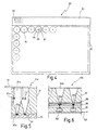
- Fig. 4 shows in plan view a further embodiment a microelectrode assembly 80.
- This consists of a Plate assembly 81, from the side of a connector strip 82nd projects with contact tongues 83.
- the cuvettes 84 are a discharge electrode 85, a Irritating electrode 86 and a reference electrode 87 indicated.
- the electrodes 85, 86, 87 are preferably concentric arranged to each other.
- the plate assembly 80 is multi-layered in construction, as already was explained above.
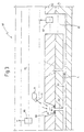
- Figs. 5 and 6 show in section two variants of the layer structure.
- a base plate 90 and a cover plate 91 is provided. Located in the base plate 90 At least the discharge electrodes 85, while in the cuvette 84a, a reference electrode 87a at a distance above the Ableitelektrode 85a is arranged.
- the diverting electrodes 85a are connected to a line 93.
- the reference electrodes 87a with a line 92. It is understood itself, that the wiring here and also in the other Figures is only to be understood very schematically.
- the wires can as single lines. Multiple lines or as in Multiplexing lines used to be formed.
- Fig. 5 is also an electric field E with an arrow hinted that can also be used to one to exert electrostatic force on cells, which then turns on let the sloping surfaces of the cuvette 84a down and finally sink to the drain electrodes 85a. In general but the effect of gravity will suffice.
- an at least three-layered Arrangement used on a signal processing board 95 there is an electrode plate 96. Above this is - optionally via a seal 98 - a cover plate 97 arranged.
- amplifiers 100 In the signal processing board 95 are amplifiers 100, if necessary including impedance converters, filters, signal analyzers or adaptation devices, the amplifiers 100 are connected via lines 101 to the environment.
- the electrode plate 96 are at least Ableitelektroden 85b, 85b ', which - as shown - flat or can be formed rod-shaped or the like.
- the electrode assembly 10 or 50 or 80 each from a base plate 15; 51; 90; 95, 96 and a cover plate 16; 52; 91; 97th
- the plates can be provided with suitable structures (strip conductors, Electrodes, etc.) and then bonded together. This can be done either by conventional metallic bonding taking advantage of the conductor tracks (see Fig. 25 in Fig. 1A) or done with the help of thin organic layers.
- the plates can be made of a polymer using a stamping technique be shaped. You can also by usual microstructure techniques getting produced.
- the ring electrodes provided at the bottom of the microcuvettes 20 and 60, respectively 21 and 61 are preferably made. TiN, iridium, Iridium oxide, platinum, platinum black or gold. You can with one thin layer be chemically functionalized, so that preferably a specific interaction with the adherent Cells is induced.
- microelectrodes are ion-sensitive Electrodes are executed.
- the electrodes with a special surface coating In a specific way this leads to a electrically sealing interaction with the cell membrane.
- lipid-like Molecules cell adhesion proteins or peptides, glycoproteins or peptides and hydrophobic coatings.
- FIG. 7 shows a further exemplary embodiment a microelectrode arrangement 103 according to the invention.
- a substrate 104 is a chamber 105 of e.g. frustoconical shape.
- the chamber 105 is on its walls provided with a conductive coating 106, in particular gilded.
- the chamber 105 can, as shown, down be completed and provided with a connection. she can but alternatively take the place of one of the channels, as described above (eg, channel 62 in FIG. 3).
- a cell 107 is located on top of the opening 108 of the chamber 105 on. Since the chamber 105 before resting the cell 107 after above was the environment used in the arrangement 103 109, i. the electrolyte used, in the chamber 105th penetrated.
- FIG. 7 also only to be understood as an example. Instead of the one shown there Arrangement may as well be a sponge made of a precious metal used, for example, a platinum sponge.
- microcuvettes In one plate were 96 microcuvettes with sloping walls appropriate. In the bottom of the microcuvettes were electrodes cast. The electrodes were made of gold wire, roughened by etching, with 20 ⁇ m diameter and 10 ⁇ m supernatant. Of the Supernatant on the bottom was 200 microns.
- a signal processing plate below the cuvettes Cover plate was with SMD impedance converters as well as amplifiers Mistake. Reference electrodes with an impedance of 1 k ⁇ all contacted on one point. There were nerve cells out embryonic chicken brain is enzymatically dissociated and into the Cuvettes pipetted. The cells dropped to the electrodes and formed there aggregates with networked cell structures. The Signal amplitude was 200 ⁇ V.
- a cover plate was made with 192 microcuvettes with conical walls Mistake. The bottom opening was 100 ⁇ m in diameter. The Walls of the cuvettes were siliconized. At the bottom of the cuvettes were electrode plates with surface electrodes of 1 mm Diameter. These were on a ceramic plate thickfilmtechnisch produced. The surface of the electrodes was galvanically plated. By connecting the cover plate with the Electrode plate became the effective electrode surfaces 100 ⁇ m reduced, i. from 10 k ⁇ to 1 M ⁇ . After the introduction of chicken cells, the formation of aggregates and the sinking the cells on the electrodes (complete coverage) was a sufficient signal-to-noise ratio up to a signal voltage achieved by 4 mV.
- microcuvettes were with a 0.5 mm diameter hole Mistake.
- the oblique walls of the microcuvettes were siliconized. Between the plates was a rubber coating for sealing used.
- An electrode plate was used to form Surface electrodes with 2 mm diameter galvanic with Goldmoor coated (10 k ⁇ ).
- the microcuvettes have a volume between 1 ⁇ l and 100 ml.
- the electrode surface can have a Have diameter between 1 micron and 1 mm.
- the invention enables individual cells or cell aggregates active to certain cells of a multi-plate electrode array and / or a multi-cell array.
- Potential applications of the invention are in the field Pharmacology, pharmaceutical screening, neurobiology and the biosensorics.
Landscapes
- Health & Medical Sciences (AREA)
- Life Sciences & Earth Sciences (AREA)
- Engineering & Computer Science (AREA)
- Chemical & Material Sciences (AREA)
- Bioinformatics & Cheminformatics (AREA)
- Organic Chemistry (AREA)
- Zoology (AREA)
- Wood Science & Technology (AREA)
- Biomedical Technology (AREA)
- Biochemistry (AREA)
- General Health & Medical Sciences (AREA)
- Physics & Mathematics (AREA)
- Biotechnology (AREA)
- Genetics & Genomics (AREA)
- Microbiology (AREA)
- Sustainable Development (AREA)
- Analytical Chemistry (AREA)
- General Engineering & Computer Science (AREA)
- Pathology (AREA)
- Immunology (AREA)
- General Physics & Mathematics (AREA)
- Cell Biology (AREA)
- Chemical Kinetics & Catalysis (AREA)
- Plasma & Fusion (AREA)
- Biophysics (AREA)
- Hematology (AREA)
- Molecular Biology (AREA)
- Urology & Nephrology (AREA)
- Food Science & Technology (AREA)
- Medicinal Chemistry (AREA)
- Electromagnetism (AREA)
- Clinical Laboratory Science (AREA)
- Apparatus Associated With Microorganisms And Enzymes (AREA)
Abstract
Description
- Fig. 1A und 1B
- eine äußerst schematisierte Querschnittsdarstellung eines Ausführungsbeispiels einer erfindungsgemäßen Mikroelektrodenanordnung in zwei unterschiedlichen Betriebsphasen;
- Fig. 2
- eine Darstellung, ähnlich Fig. 1A und 1B, jedoch für eine Mikroelektrodenanordnung nach dem Stand der Technik;
- Fig. 3
- eine weitere Darstellung, ähnlich Fig. 1A und 1B, jedoch für ein anderes Ausführungsbeispiel der Erfindung;
- Fig. 4
- eine Draufsicht auf ein weiteres Ausführungsbeispiel einer erfindungsgemäßen Mikroelektrodenanordnung;
- Fig. 5 und 6
- in vergrößertem Maßstab zwei Darstellungen von Schnitten durch Mikroküvetten, wie sie bei der Anordnung gemäß Fig. 5 verwendet werden können; und
- Fig. 7
- in einer Querschnittsdarstellung eine weitere Ausführungsform einer erfindungsgemäßen Mikroelektrode.
Claims (42)
- Mikroelementennanordnung mit einer Vielzahl von auf einem Substrat (15, 16; 40; 51, 52; 90, 91; 95, 96, 97; 104) angeordneten elektrischen Mikroelementen zum Kontaktieren von in einer flüssigen Umgebung (30; 70; 109) befindlichen Zellen (31; 42; 71), gekennzeichnet durch Mittel zum Führen und/oder Vereinzeln und/oder mechanischen Anziehen der Zellen (31; 71) an die Mikroelemente (21; 61; 85, 86, 87).
- Anordnung nach Anspruch 1, dadurch gekennzeichnet, daß die Mikroelemente (21; 41; 61; 85, 86, 87; 103) zum Ableiten von bioelektrischen Potentialen und/oder zum bioelektrischen Stimulieren der Zellen (31; 42; 71; 107) vorgesehen sind.
- Anordnung nach Anspruch 1, dadurch gekennzeichnet, daß die Mikroelemente als lichtempfindliche Elemente ausgebildet sind.
- Anordnung nach einem oder mehreren der Ansprüche 1 bis 3, dadurch gekennzeichnet, daß die Mittel eine Unterdruck-Kraft auf die Zellen (31) ausüben.
- Anordnung nach einem oder mehreren der Ansprüche 1 bis 3, dadurch gekennzeichnet, daß die Mittel eine hydrodynamische Kraft auf die Zellen (71) ausüben.
- Anordnung nach Anspruch 4 oder 5, dadurch gekennzeichnet, daß die Mittel Kanäle (22; 62) umfassen, die in einer Kontaktoberfläche (24; 64) der Mikroelektroden (21; 61) ausmünden.
- Anordnung nach Anspruch 4 und 6, dadurch gekennzeichnet, daß die Kanäle (22) mit einer Unterdruckquelle verbindbar sind.
- Anordnung nach Anspruch 5 und 6, dadurch gekennzeichnet, daß die Kanäle (62) mit einer Pumpeinrichtung (65, 66, 67) für die flüssige biologische Umgebung (70) verbunden sind.
- Anordnung nach Anspruch 8, dadurch gekennzeichnet, daß die Pumpeinrichtung als Elektroosmose-Einrichtung ausgebildet ist.
- Anordnung nach Anspruch 9, dadurch gekennzeichnet, daß die Pumpeinrichtung (65, 66, 67) als Elektroosmose-Einrichtung zwei Elektroden (66, 67) umfaßt, die an entgegengesetzten Enden der Kanäle (62) wirksam sind, und daß zwischen die Elektroden (66, 67) eine Spannung geschaltet ist.
- Anordnung nach Anspruch 10, dadurch gekennzeichnet, daß die Mittel eine elektrostatische Kraft (E) auf die Zellen ausüben.
- Anordnung, nach einem oder mehreren der Ansprüche 1 bis 11, gekennzeichnet durch Mittel zum Führen und/oder Vereinzeln der Zellen (31; 71) vor dem mechanischen Anziehen an die Mikroelemente.
- Anordnung nach einem oder mehreren der Ansprüche 1 bis 12, dadurch gekennzeichnet, daß die Mittel trichterartige Mikroküvetten (20; 60; 84) im Substrat (15, 16; 51, 52; 90, 91; 95, 96, 97) umfassen, wobei sich die Mikroelemente am Boden der Mikroküvetten (20; 60; 84) befinden.
- Anordnung nach Anspruch 13, dadurch gekennzeichnet, daß Oberflächenbereiche zwischen den Mikroküvetten mit einem zellabweisenden Substrat und/oder die Mikroküvetten mit einem zellanziehenden Substrat beschichtet sind.
- Anordnung nach einem oder mehreren der Ansprüche 1 bis 14, dadurch gekennzeichnet, daß das Substrat (15, 16; 51, 52; 90, 91; 95, 96, 97) mindestens aus einer Grundplatte (15; 51; 90; 95, 96) und einer darüberliegenden Deckplatte (16; 52; 91; 97) besteht.
- Anordnung nach Anspruch 15, dadurch gekennzeichnet, daß die Grundplatte (15; 51; 90; 95, 96) aus Glas, Quarz, Silizium oder Kunststoff besteht.
- Anordnung nach Anspruch 15 oder 16, dadurch gekennzeichnet, daß die Deckplatte (16; 52; 91; 97) aus Glas, Quarz, Silizium oder Kunststoff, insbesondere aus Polystyrol, PMMA oder Polyimid besteht.
- Anordnung nach einem oder mehreren der Ansprüche 15 bis 17, dadurch gekennzeichnet, daß die Grundplatte (15; 51; 90; 95, 96) und/oder die Deckplatte (16; 52; 91; 97) aus einem für Licht durchlässigen Material besteht, wobei die Wellenlänge des Lichtes in einem für Mikroskopietechniken zugänglichen Bereich des Spektrums liegt.
- Anordnung nach einem oder mehreren der Ansprüche 15 bis 18, dadurch gekennzeichnet, daß die mit den Mikroelementen versehene Grundplatte (90) seitlich als Steckerleiste (82) herausgeführt ist.
- Anordnung nach einem oder mehreren der Ansprüche 15 bis 18, dadurch gekennzeichnet, daß die Grundplatte (95) mindestens einer unteren Signalverarbeitungs-Platte (95) und einer darüberliegenden Elementen-Platte (96) besteht.
- Anordnung nach Anspruch 20, dadurch gekennzeichnet, daß die Signalverarbeitungs-Platte (95) seitlich als Steckerleiste (82) herausgeführt ist.
- Anordnung nach einem oder mehreren der Ansprüche 2 bis 21, dadurch gekennzeichnet, daß die Mikroelektroden (85, 86, 87) Ableitelektroden (85) sowie Reizelektroden (86) und/oder Referenzelektroden (87) umfassen.
- Anordnung nach Anspruch 22, dadurch gekennzeichnet, daß die Mikroelektroden (85, 86, 87) mehrere konzentrisch angeordnete Einzelelektroden umfassen.
- Anordnung nach Anspruch 13 und 22, dadurch gekennzeichnet, daß die Referenzelektrode (87a) im Abstand oberhalb der am Boden der Mikroküvette (84a) angeordneten Ableitelektrode (85a) angeordnet ist.
- Anordnung nach Anspruch 2, dadurch gekennzeichnet, daß die mit der Umgebung (109) in Kontakt befindliche Oberfläche der Mikroelektrode (103) größer ist als die mit der Zelle (107) in Kontakt befindliche Oberfläche.
- Anordnung nach Anspruch 25, dadurch gekennzeichnet, daß die Mikroelektrode (103) als Kammer (105) in einem Substrat (104) ausgebildet ist, wobei die Kammer (105) über eine Öffnung (108) in den das Substrat (104) umgebenden Außenraum mündet.
- Verfahren zum Kontaktieren von in einer flüssigen Umgebung oberhalb eines Substrates (15, 16; 40; 51, 52; 90, 91; 95, 96, 97) befindlichen Zellen (31; 42; 71), bei dem ein Kontakt zwischen den Zellen (31; 42; 71) und elektrischen Mikroelementen hergestellt wird, dadurch gekennzeichnet, daß eine Führungs- und/oder Anziehungskraft zwischen den Zellen (31; 71) und den Mikroelementen oder dem Substrat (15, 16; 51, 52; 90, 91; 95, 96, 97) erzeugt wird.
- Verfahren nach Anspruch 27, dadurch gekennzeichnet, daß die Kraft als Unterdruck-Kraft ausgeübt wird.
- Verfahren nach Anspruch 28, dadurch gekennzeichnet, daß die Kraft als hydrodynamische Kraft ausgeübt wird.
- Verfahren nach Anspruch 29, dadurch gekennzeichnet, daß die hydrodynamische Kraft mittels Elektroosmose, insbesondere durch einen mittels Elektroosmose erzeugten Elektrolytfluß, ausgeübt wird.
- Verfahren nach Anspruch 27, dadurch gekennzeichnet, daß die Kraft als Anziehungskraft aufgrund elektrischer Ladung der Zellen und eines in Richtung der Elektroden (85a) wirkenden elektrischen Feldes (E) ausgeübt wird.
- Verfahren nach einem oder mehreren der Ansprüche 27 bis 31, dadurch gekennzeichnet, daß die Anziehungskraft als Kontaktkraft, insbesondere durch Ansaugen, zwischen Zellen (31; 71) und Mikroelementen ausgeübt wird.
- Verfahren nach einem oder mehreren der Ansprüche 27 bis 32, dadurch gekennzeichnet, daß die Kraft für eine gerichtete Bewegung der Zellen (31; 71) auf die Mikroelemente zu ausgeübt wird.
- Verfahren nach einem oder mehreren der Ansprüche 27 bis 33, dadurch gekennzeichnet, daß die Zellen (31; 71) über die als Mikroelektroden (21; 61; 86) ausgebildeten Mikroelemente stimuliert werden.
- Verfahren nach einem oder mehreren der Ansprüche 27 bis 33, dadurch gekennzeichnet, daß über die als Mikroelektroden (21; 61; 85) ausgebildeten Mikroelemente Potentiale von den Zellen (31; 71) abgeleitet werden.
- Verfahren nach einem oder mehreren der Ansprüche 27 bis 33, dadurch gekennzeichnet, daß über die als Mikrophotodioden ausgebildeten Mikroelemente die Lumineszenz der Zellen und/oder deren Lichtabsorption gemessen wird.
- Verfahren zum Herstellen einer Mikroelementenanordnung mit einer Vielzahl von elektrischen Mikroelementen gemäß Ansprüchen 1 bis 26, bei dem die Mikroelemente auf einem Substrat (15, 16; 40; 51, 52; 90, 91; 95, 96, 97) angeordnet werden, dadurch gekennzeichnet, daß das Substrat (15, 16; 51, 52; 90, 91; 95, 96, 97) mindestens aus einer Grundplatte (15; 51; 90; 95, 96) und einer darüberliegenden Deckplatte (16; 52; 91; 97) hergestellt wird.
- Verfahren nach Anspruch 37, dadurch gekennzeichnet, daß in der Grundplatte (15; 51) ein Kanalsystem (23; 62, 63) ausgebildet wird, daß in der Deckplatte (16; 52) Mikroküvetten (20; 60) ausgeformt werden, und daß die Grundplatte (15; 51) mit der Deckplatte (16; 52) derart zusammengefügt wird, daß Öffnung (22) am Boden der Mikroküvetten (20; 60) in Kontaktoberflächen (24; 64) der Mikroelemente angeordnet sind und mit dem Kanalsystem (23; 62, 63) kommunizieren.
- Verfahren nach Anspruch 36 oder 37, gekennzeichnet durch die Schritte:a) Versehen von Grundplatte (15; 51; 90; 95, 96) und/oder Deckplatte (16; 52; 91; 97) auf ihren einander zu weisenden Oberflächen mit einer Schicht von Molekülen mit reaktiver Endgruppe;b) Zusammenfügen von Grundplatte (15; 51; 90; 95, 96) und Deckplatte (16; 52; 91; 97); undc) Aktivieren einer kovalenten Bindung der Schichten mittels eines äußeren Stimulus.
- Verfahren nach Anspruch 39, dadurch gekennzeichnet, daß nach Schritt b) die Grundplatte (15; 51; 90; 95, 96) und die Deckplatte (16; 52; 91; 97) relativ zueinander justiert werden.
- Verfahren nach Anspruch 39 oder 40, dadurch gekennzeichnet, daß der äußere Stimulus als Temperatur, Licht oder elektrisches Feld ausgeübt wird.
- Verfahren nach Anspruch 38, dadurch gekennzeichnet, daß die Grundplatte (15; 51; 90; 95, 96) und die Deckplatte (16; 52; 91; 97) durch anodisches oder metallisches Bonden miteinander verbunden werden.
Applications Claiming Priority (5)
| Application Number | Priority Date | Filing Date | Title |
|---|---|---|---|
| DE19647525 | 1996-11-16 | ||
| DE19647525 | 1996-11-16 | ||
| DE19712309 | 1997-03-24 | ||
| DE19712309A DE19712309A1 (de) | 1996-11-16 | 1997-03-24 | Mikroelementenanordnung, Verfahren zum Kontaktieren von in einer flüssigen Umgebung befindlichen Zellen und Verfahren zum Herstellen einer Mikroelementenanordnung |
| PCT/EP1997/006285 WO1998022819A1 (de) | 1996-11-16 | 1997-11-11 | Mikroelementenanordnung, verfahren zum kontaktieren von in einer flüssigen umgebung befindlichen zellen und verfahren zum herstellen einer mikroelementenanordnung |
Publications (2)
| Publication Number | Publication Date |
|---|---|
| EP0938674A1 EP0938674A1 (de) | 1999-09-01 |
| EP0938674B1 true EP0938674B1 (de) | 2005-06-01 |
Family
ID=26031375
Family Applications (1)
| Application Number | Title | Priority Date | Filing Date |
|---|---|---|---|
| EP97950174A Expired - Lifetime EP0938674B1 (de) | 1996-11-16 | 1997-11-11 | Mikroelementenanordnung, verfahren zum kontaktieren von in einer flüssigen umgebung befindlichen zellen und verfahren zum herstellen einer mikroelementenanordnung |
Country Status (3)
| Country | Link |
|---|---|
| US (2) | US6315940B1 (de) |
| EP (1) | EP0938674B1 (de) |
| WO (1) | WO1998022819A1 (de) |
Cited By (1)
| Publication number | Priority date | Publication date | Assignee | Title |
|---|---|---|---|---|
| WO2019121162A1 (de) | 2017-12-19 | 2019-06-27 | ChanPharm GmbH | Messgerät, messverfahren, hochdurchsatz-testgerät und messkit für elektrophysiologische messungen, insbesondere an zellaggregaten |
Families Citing this family (124)
| Publication number | Priority date | Publication date | Assignee | Title |
|---|---|---|---|---|
| AU9786798A (en) * | 1997-10-10 | 1999-05-03 | Biosepra Inc. | Aligned multiwell multiplate stack and method for processing biological/chemicalsamples using the same |
| US20020144905A1 (en) | 1997-12-17 | 2002-10-10 | Christian Schmidt | Sample positioning and analysis system |
| US6758961B1 (en) | 1997-12-17 | 2004-07-06 | Ecole Polytechnique Federale De Lausanne | Positioning and electrophysiological characterization of individual cells and reconstituted membrane systems on microstructured carriers |
| US7244349B2 (en) | 1997-12-17 | 2007-07-17 | Molecular Devices Corporation | Multiaperture sample positioning and analysis system |
| GB9812783D0 (en) * | 1998-06-12 | 1998-08-12 | Cenes Ltd | High throuoghput screen |
| IL143213A0 (en) * | 1998-12-05 | 2002-04-21 | Cenes Ltd | Interface patch clamping |
| US6488829B1 (en) * | 1999-08-05 | 2002-12-03 | Essen Instruments Inc | High-throughput electrophysiological measurement apparatus |
| US6682649B1 (en) * | 1999-10-01 | 2004-01-27 | Sophion Bioscience A/S | Substrate and a method for determining and/or monitoring electrophysiological properties of ion channels |
| DE19948473A1 (de) * | 1999-10-08 | 2001-04-12 | Nmi Univ Tuebingen | Verfahren und Vorrichtung zum Messen an in einer flüssigen Umgebung befindlichen Zellen |
| US20060128006A1 (en) * | 1999-11-10 | 2006-06-15 | Gerhardt Antimony L | Hydrodynamic capture and release mechanisms for particle manipulation |
| US6692952B1 (en) * | 1999-11-10 | 2004-02-17 | Massachusetts Institute Of Technology | Cell analysis and sorting apparatus for manipulation of cells |
| FR2802078B1 (fr) * | 1999-12-14 | 2003-10-03 | Univ Joseph Fourier | Microelectrode support de cellules a membrane excitable |
| GB9930719D0 (en) * | 1999-12-24 | 2000-02-16 | Central Research Lab Ltd | Apparatus for and method of making electrical measurements on an object in a m edium |
| GB9930718D0 (en) * | 1999-12-24 | 2000-02-16 | Central Research Lab Ltd | Apparatus for and method of making electrical measurements on objects |
| DE10032568A1 (de) * | 2000-07-05 | 2002-01-24 | Nmi Univ Tuebingen | Vorrichtung und Verfahren zum elektrischen Kontaktieren von in einer Flüssigkeit in Suspension befindlichen biologischen Zellen |
| JP3909738B2 (ja) * | 2000-07-13 | 2007-04-25 | 松下電器産業株式会社 | 細胞外記録用一体化複合電極 |
| EP1178315A1 (de) * | 2000-07-31 | 2002-02-06 | Albrecht Dr.med. Priv.Doz. Lepple-Wienhues | Verfahren und Vorrichtung zur Untersuchung von Zellen mit Hilfe der Patch Clamp-Methode |
| US7067046B2 (en) | 2000-08-04 | 2006-06-27 | Essen Instruments, Inc. | System for rapid chemical activation in high-throughput electrophysiological measurements |
| US7270730B2 (en) * | 2000-08-04 | 2007-09-18 | Essen Instruments, Inc. | High-throughput electrophysiological measurement system |
| AU2001287472A1 (en) * | 2000-09-19 | 2002-04-02 | Cytion Sa | Sample positioning and analysis system |
| AU9367601A (en) * | 2000-10-02 | 2002-04-15 | Sophion Bioscience As | System for electrophysiological measurements |
| US6932893B2 (en) * | 2000-10-02 | 2005-08-23 | Sophion Bioscience A/S | System for electrophysiological measurements |
| JP3729729B2 (ja) * | 2000-11-30 | 2005-12-21 | 日立ソフトウエアエンジニアリング株式会社 | 生体高分子検出装置及びカートリッジ |
| US6913697B2 (en) | 2001-02-14 | 2005-07-05 | Science & Technology Corporation @ Unm | Nanostructured separation and analysis devices for biological membranes |
| CA2441163A1 (en) * | 2001-03-16 | 2002-09-26 | Memorec Biotec Gmbh | Analysing device for analysing chemical and/or biological samples |
| US20060029955A1 (en) | 2001-03-24 | 2006-02-09 | Antonio Guia | High-density ion transport measurement biochip devices and methods |
| US20040146849A1 (en) * | 2002-01-24 | 2004-07-29 | Mingxian Huang | Biochips including ion transport detecting structures and methods of use |
| AU2002307218A1 (en) | 2001-03-24 | 2002-10-08 | Aviva Biosciences Corporation | Biochips including ion transport detecting structures and methods of use |
| US20050058990A1 (en) * | 2001-03-24 | 2005-03-17 | Antonio Guia | Biochip devices for ion transport measurement, methods of manufacture, and methods of use |
| US20050009004A1 (en) * | 2002-05-04 | 2005-01-13 | Jia Xu | Apparatus including ion transport detecting structures and methods of use |
| FR2823999B1 (fr) * | 2001-04-27 | 2003-06-27 | Commissariat Energie Atomique | Dispositif miniature de separation et d'isolement d'objets biologiques et utilisations |
| DE10128574A1 (de) * | 2001-06-13 | 2003-01-02 | Infineon Technologies Ag | Vorrichtung und Verfahren zur Manipulation von Vesikeln |
| US6899800B2 (en) * | 2001-06-20 | 2005-05-31 | Axon Instruments, Inc. | Polymeric electrode for electrophysiological testing |
| DE60215029T2 (de) * | 2001-06-29 | 2007-10-25 | The Board Of Trustees Of The Leland Stanford Junior University, Palo Alto | Kunstmässige chip-schnittstellenverbindung für elektronische reitinaimplantat |
| US7147865B2 (en) | 2001-06-29 | 2006-12-12 | The Board Of Trustees Of The Leland Stanford University | Artificial synapse chip |
| US7704745B2 (en) * | 2001-08-06 | 2010-04-27 | Vanderbilt University | Apparatus and methods for monitoring the status of a metabolically active cell |
| EP2332651A3 (de) * | 2001-10-25 | 2011-08-31 | Bar Ilan University | Interaktiver transparenter Biochip-Prozessor für einzeln adressierbare Zellen |
| AU2002353741A1 (en) * | 2001-11-27 | 2003-06-10 | Cellectricon Ab | A method for combined sequential agent delivery and electroporation for cell structures and use thereof |
| US7318902B2 (en) * | 2002-02-04 | 2008-01-15 | Colorado School Of Mines | Laminar flow-based separations of colloidal and cellular particles |
| ATE516879T1 (de) * | 2002-02-12 | 2011-08-15 | Cellectricon Ab | Systeme und verfahren zur schnellen änderung der lösungsumgebung von sensoren |
| WO2003085379A2 (en) | 2002-04-01 | 2003-10-16 | Fluidigm Corporation | Microfluidic particle-analysis systems |
| US20040120854A1 (en) * | 2002-04-04 | 2004-06-24 | Heath James R | Silicon-wafer based devices and methods for analyzing biological material |
| AU2003231300A1 (en) * | 2002-05-04 | 2003-11-17 | Aviva Biosciences Corporation | Apparatus including ion transport detecting structures and methods of use |
| US20030215872A1 (en) * | 2002-05-20 | 2003-11-20 | Clark-Mxr, Inc. | Screening apparatus and method for making |
| US20030215815A1 (en) * | 2002-05-20 | 2003-11-20 | Clark William G. | Screening method |
| JP4691407B2 (ja) * | 2005-06-29 | 2011-06-01 | パナソニック株式会社 | 細胞電位測定用容器 |
| WO2006132116A1 (ja) * | 2005-06-07 | 2006-12-14 | Matsushita Electric Industrial Co., Ltd. | 細胞電気生理測定デバイスおよびその製造方法 |
| WO2007001091A1 (en) * | 2005-06-29 | 2007-01-04 | Matsushita Electric Industrial Co., Ltd. | Cellular potential measurement container |
| WO2009034697A1 (ja) | 2007-09-11 | 2009-03-19 | Panasonic Corporation | シリコン構造体およびその製造方法並びにセンサチップ |
| US8202439B2 (en) * | 2002-06-05 | 2012-06-19 | Panasonic Corporation | Diaphragm and device for measuring cellular potential using the same, manufacturing method of the diaphragm |
| JP4552423B2 (ja) * | 2003-11-21 | 2010-09-29 | パナソニック株式会社 | 細胞外電位測定デバイスおよびこれを用いた細胞外電位の測定方法 |
| US7214348B2 (en) * | 2002-07-26 | 2007-05-08 | Applera Corporation | Microfluidic size-exclusion devices, systems, and methods |
| EP1801586B1 (de) * | 2002-08-21 | 2010-10-13 | Cellectricon Ab | System zur Gewinnung und Erhaltung hochresistenter Verschlüsse bei Patch-Clamp-Aufzeichnungen |
| JP4386835B2 (ja) * | 2002-08-21 | 2009-12-16 | セレクトリコン アーベー | パッチクランプ記録において高抵抗固着を獲得して維持するシステム及び方法 |
| FR2844052B1 (fr) * | 2002-08-28 | 2005-07-01 | Commissariat Energie Atomique | Dispositif de mesure de l'activite electrique d'elements biologiques et ses applications |
| CA2500392C (en) | 2002-09-27 | 2012-11-27 | The General Hospital Corporation | Microfluidic device for cell separation and uses thereof |
| AU2003278461A1 (en) | 2002-10-16 | 2004-05-04 | Cellectricon Ab | Nanoelectrodes and nanotips for recording transmembrane currents in a plurality of cells |
| US20040209352A1 (en) * | 2002-10-28 | 2004-10-21 | Nobuhiko Ozaki | Integrated electrode and cell immobilization device equipped with the integrated electrode |
| US7932098B2 (en) * | 2002-10-31 | 2011-04-26 | Hewlett-Packard Development Company, L.P. | Microfluidic system utilizing thin-film layers to route fluid |
| US20040086872A1 (en) * | 2002-10-31 | 2004-05-06 | Childers Winthrop D. | Microfluidic system for analysis of nucleic acids |
| US20040251145A1 (en) * | 2003-02-21 | 2004-12-16 | Robertson Janet Kay | High throughput screening (HTS) method and apparatus for monitoring ion channels |
| IL154677A0 (en) * | 2003-02-27 | 2003-09-17 | Univ Bar Ilan | A method and apparatus for manipulating an individual cell |
| US7501279B2 (en) * | 2003-04-03 | 2009-03-10 | University Of Washington | Microwell arrays with nanoholes |
| US7112433B2 (en) * | 2003-04-24 | 2006-09-26 | Hewlett-Packard Development Company, L.P. | Electrical analysis of biological membranes |
| US8597597B2 (en) | 2003-06-26 | 2013-12-03 | Seng Enterprises Ltd. | Picoliter well holding device and method of making the same |
| US9200245B2 (en) | 2003-06-26 | 2015-12-01 | Seng Enterprises Ltd. | Multiwell plate |
| DE602004016009D1 (de) * | 2003-09-03 | 2008-10-02 | Cedi Diagnostics B V | Verfahren zur erfassung von mehreren analyten |
| US7547381B2 (en) * | 2003-09-26 | 2009-06-16 | Agency For Science, Technology And Research And National University Of Singapore | Sensor array integrated electrochemical chip, method of forming same, and electrode coating |
| US8058056B2 (en) * | 2004-03-12 | 2011-11-15 | The Regents Of The University Of California | Method and apparatus for integrated cell handling and measurements |
| DK1745144T3 (da) | 2004-05-11 | 2011-02-07 | Axiogenesis Ag | Analysefremgangsmåde til opsporing af lægemidler på grundlag af in vitro-differentierede celler |
| WO2006005370A1 (en) * | 2004-07-15 | 2006-01-19 | Nano S Biotechnologie Gmbh | Substrate for a device for electrophysiological measurements |
| JPWO2006022092A1 (ja) * | 2004-08-25 | 2008-05-08 | 松下電器産業株式会社 | 細胞電位測定プローブ |
| EP1820001A1 (de) * | 2004-11-12 | 2007-08-22 | Luminex Corporation | Verfahren und systeme zur positionierung von mikrokugeln für die bilderzeugung |
| JP2006149227A (ja) * | 2004-11-25 | 2006-06-15 | Fujitsu Ltd | 微小物体の捕獲装置及び方法 |
| US20060171846A1 (en) * | 2005-01-10 | 2006-08-03 | Marr David W M | Microfluidic systems incorporating integrated optical waveguides |
| DE602005023534D1 (de) | 2005-01-25 | 2010-10-21 | Seng Entpr Ltd | Mikrofluidische vorrichtung zur untersuchung zellen |
| US20070196820A1 (en) | 2005-04-05 | 2007-08-23 | Ravi Kapur | Devices and methods for enrichment and alteration of cells and other particles |
| US20060246571A1 (en) * | 2005-04-29 | 2006-11-02 | Fujitsu Limited | Apparatus and method for automatically producing substance-introduced particles |
| US20090126459A1 (en) * | 2005-05-19 | 2009-05-21 | Koninklijke Philips Electronics, N.V. | Functional assembly and method of obtaining it |
| US8921102B2 (en) | 2005-07-29 | 2014-12-30 | Gpb Scientific, Llc | Devices and methods for enrichment and alteration of circulating tumor cells and other particles |
| WO2007052245A1 (en) | 2005-11-03 | 2007-05-10 | Seng Enterprises Ltd. | Method and device for studying floating, living cells |
| JP4470999B2 (ja) | 2005-12-20 | 2010-06-02 | パナソニック株式会社 | 細胞電気生理センサ |
| US8119976B2 (en) * | 2007-07-03 | 2012-02-21 | Colorado School Of Mines | Optical-based cell deformability |
| US9878326B2 (en) * | 2007-09-26 | 2018-01-30 | Colorado School Of Mines | Fiber-focused diode-bar optical trapping for microfluidic manipulation |
| US9487812B2 (en) | 2012-02-17 | 2016-11-08 | Colorado School Of Mines | Optical alignment deformation spectroscopy |
| US9885644B2 (en) | 2006-01-10 | 2018-02-06 | Colorado School Of Mines | Dynamic viscoelasticity as a rapid single-cell biomarker |
| GB2436145A (en) * | 2006-03-16 | 2007-09-19 | Sophion Bioscience As | A system for monitoring properties of a single cell |
| US8293524B2 (en) * | 2006-03-31 | 2012-10-23 | Fluxion Biosciences Inc. | Methods and apparatus for the manipulation of particle suspensions and testing thereof |
| US20100019756A1 (en) * | 2006-05-17 | 2010-01-28 | Matsushita Electric Industrial Co., Ltd. | Device for measuring cellular potential, substrate used for the same and method of manufacturing substrate for device for measuring cellular potential |
| EP2037258A4 (de) * | 2006-07-06 | 2013-12-04 | Panasonic Corp | Vorrichtung für zellelektrophysiologiesensor, zellelektrophysiologiesensor unter verwendung der vorrichtung sowie verfahren zur herstellung der zellelektrophysiologiesensorvorrichtung |
| EP2019317A1 (de) * | 2007-07-24 | 2009-01-28 | Evotec AG | Mikrofluidisches System zur Partikelanalyse |
| US20090062828A1 (en) * | 2007-09-04 | 2009-03-05 | Colorado School Of Mines | Magnetic field-based colloidal atherectomy |
| US10722250B2 (en) | 2007-09-04 | 2020-07-28 | Colorado School Of Mines | Magnetic-field driven colloidal microbots, methods for forming and using the same |
| US9975118B2 (en) * | 2007-11-15 | 2018-05-22 | Seng Enterprises Ltd. | Device for the study of living cells |
| US9145540B1 (en) | 2007-11-15 | 2015-09-29 | Seng Enterprises Ltd. | Device for the study of living cells |
| GB0724736D0 (en) | 2007-12-19 | 2008-01-30 | Oxford Nanolabs Ltd | Formation of layers of amphiphilic molecules |
| US8008063B2 (en) * | 2008-06-24 | 2011-08-30 | Bio-Rad Laboratories, Inc. | Individual-cell electroporation using area-focused electric fields |
| US9137895B2 (en) | 2008-12-24 | 2015-09-15 | Stmicroelectronics S.R.L. | Micro-electro-mechanical systems (MEMS) and corresponding manufacturing process |
| DE102009061008B4 (de) | 2009-03-20 | 2014-09-25 | Retina Implant Ag | Verwendung einer Testvorrichtung und Verfahren zum Test von Zellen, Zellkulturen und/oder organotypischen Zellverbänden |
| EP2457991A4 (de) * | 2009-07-24 | 2016-10-26 | Nipro Corp | Behälter zur messung eines zellpotentials |
| US8367018B2 (en) * | 2009-12-07 | 2013-02-05 | Southern Taiwan University Of Technology | Chip with tri-layer electrode and micro-cavity arrays for control of bioparticle and manufacturing method thereof |
| CN102097385B (zh) * | 2009-12-15 | 2014-05-07 | 中芯国际集成电路制造(上海)有限公司 | 双位快闪存储器的制作方法 |
| AU2011271087B2 (en) * | 2010-06-21 | 2014-07-31 | The Regents Of The University Of California | Ultra-high photosensitivity vertical nanowire arrays for retinal prosthesis |
| US9199080B2 (en) | 2011-09-12 | 2015-12-01 | Okuvision Gmbh | Method for treating an eye |
| GB201202519D0 (en) | 2012-02-13 | 2012-03-28 | Oxford Nanopore Tech Ltd | Apparatus for supporting an array of layers of amphiphilic molecules and method of forming an array of layers of amphiphilic molecules |
| WO2014022828A1 (en) | 2012-08-02 | 2014-02-06 | The Regents Of The University Of California | Integrated nanowire array devices for detecting and/or applying electrical signals to tissue |
| GB201313121D0 (en) | 2013-07-23 | 2013-09-04 | Oxford Nanopore Tech Ltd | Array of volumes of polar medium |
| CN103087908B (zh) * | 2013-01-17 | 2014-03-05 | 杭州电子科技大学 | 细胞剪切强度测量方法与装置 |
| US9279801B2 (en) | 2013-07-26 | 2016-03-08 | Axion Biosystems, Inc. | Devices, systems and methods for high-throughput electrophysiology |
| EP3040410B1 (de) * | 2013-08-30 | 2018-04-25 | Japan Science and Technology Agency | Flache patch-clamp-vorrichtung und verfahren verwendend die patch-clamp vorrichtung |
| US9757707B2 (en) | 2014-06-12 | 2017-09-12 | Takara Bio Usa, Inc. | Single cell capture with capture chips |
| EP3155396A4 (de) | 2014-06-12 | 2017-12-27 | Wafergen, Inc. | Einzelzellenerfassung mit polymererfassungsfilmen |
| US10067117B2 (en) | 2014-08-12 | 2018-09-04 | Axion Biosystems, Inc. | Cell-based biosensor array and associated methods for manufacturing the same |
| WO2016161081A1 (en) | 2015-04-03 | 2016-10-06 | Fluxion Biosciences, Inc. | Molecular characterization of single cells and cell populations for non-invasive diagnostics |
| GB201611770D0 (en) | 2016-07-06 | 2016-08-17 | Oxford Nanopore Tech | Microfluidic device |
| GB2561173A (en) | 2017-04-03 | 2018-10-10 | Univ Dublin City | Microfluidic device for detection of analytes |
| GB2568895B (en) | 2017-11-29 | 2021-10-27 | Oxford Nanopore Tech Ltd | Microfluidic device |
| EP3721217A4 (de) * | 2017-12-08 | 2021-12-29 | Enuvio Inc. | Mikrofluidischer chip und verfahren zur herstellung davon |
| GB2574048B (en) | 2018-05-24 | 2021-06-16 | Oxford Nanopore Tech Ltd | Nanopore sensor component with electrostatic discharge protection |
| JP7492200B2 (ja) | 2019-03-12 | 2024-05-29 | オックスフォード ナノポール テクノロジーズ ピーエルシー | ナノ細孔センシングデバイスと操作方法およびその成形方法 |
| WO2020223900A1 (en) * | 2019-05-07 | 2020-11-12 | Boe Technology Group Co., Ltd. | Method of classifying and counting cells in a sample, bioassay kit, and micropore array plate |
| EP4675256A3 (de) | 2020-07-17 | 2026-01-28 | Oxford Nanopore Technologies PLC | Nanoporenmessvorrichtung |
| KR102799481B1 (ko) * | 2023-01-09 | 2025-04-23 | 한국화학연구원 | 세포 스페로이드 임피던스 측정 장치를 위한 측정 플레이트 |
| DE102023117490B4 (de) * | 2023-07-03 | 2025-02-06 | ChanPharm GmbH | Zellsubstrat zur Aufnahme biologischer Zellen und Verfahren zu dessen Verwendung |
Family Cites Families (31)
| Publication number | Priority date | Publication date | Assignee | Title |
|---|---|---|---|---|
| US5310674A (en) * | 1982-05-10 | 1994-05-10 | Bar-Ilan University | Apertured cell carrier |
| IL68507A (en) * | 1982-05-10 | 1986-01-31 | Univ Bar Ilan | System and methods for cell selection |
| DE3485462D1 (de) * | 1983-11-08 | 1992-02-27 | Scient Diagnostics Inc | System und verfahren fuer zellenauslese. |
| JP2662215B2 (ja) * | 1986-11-19 | 1997-10-08 | 株式会社日立製作所 | 細胞保持装置 |
| JP2559760B2 (ja) * | 1987-08-31 | 1996-12-04 | 株式会社日立製作所 | 細胞搬送方法 |
| JP2564322B2 (ja) * | 1987-10-07 | 1996-12-18 | 株式会社日立製作所 | 細胞融合装置 |
| US5183744A (en) * | 1988-10-26 | 1993-02-02 | Hitachi, Ltd. | Cell handling method for cell fusion processor |
| JP2966904B2 (ja) * | 1990-08-08 | 1999-10-25 | 株式会社日立製作所 | 細胞の処理方法、および処理装置 |
| US5173158A (en) * | 1991-07-22 | 1992-12-22 | Schmukler Robert E | Apparatus and methods for electroporation and electrofusion |
| US6017696A (en) * | 1993-11-01 | 2000-01-25 | Nanogen, Inc. | Methods for electronic stringency control for molecular biological analysis and diagnostics |
| US5849486A (en) * | 1993-11-01 | 1998-12-15 | Nanogen, Inc. | Methods for hybridization analysis utilizing electrically controlled hybridization |
| US5632957A (en) * | 1993-11-01 | 1997-05-27 | Nanogen | Molecular biological diagnostic systems including electrodes |
| IL103674A0 (en) * | 1991-11-19 | 1993-04-04 | Houston Advanced Res Center | Method and apparatus for molecule detection |
| US5846708A (en) * | 1991-11-19 | 1998-12-08 | Massachusetts Institiute Of Technology | Optical and electrical methods and apparatus for molecule detection |
| US5489506A (en) * | 1992-10-26 | 1996-02-06 | Biolife Systems, Inc. | Dielectrophoretic cell stream sorter |
| WO1994013303A1 (en) * | 1992-12-14 | 1994-06-23 | Start Technology Partnership | Administration of oligonucleotides antisense to dopamine receptor mrna for diagnosis and treatment of neurological pathologies |
| GB9306729D0 (en) * | 1993-03-31 | 1993-05-26 | British Tech Group | Improvements in separators |
| US5730850A (en) * | 1993-04-23 | 1998-03-24 | Hitachi, Ltd. | Capillary array electrophoresis system |
| DK1157743T3 (da) * | 1993-10-28 | 2009-07-06 | Houston Advanced Res Ct | Mikrofremstillet poröst gennemströmningsapparat til diskret påvisning af bindingsreaktioner |
| US6099803A (en) * | 1994-07-07 | 2000-08-08 | Nanogen, Inc. | Advanced active electronic devices for molecular biological analysis and diagnostics |
| US6225059B1 (en) * | 1993-11-01 | 2001-05-01 | Nanogen, Inc. | Advanced active electronic devices including collection electrodes for molecular biological analysis and diagnostics |
| US5489515A (en) * | 1993-12-09 | 1996-02-06 | Avl Medical Instruments Ag | Device for analyzing the metabolism of cells |
| US5774581A (en) | 1994-02-16 | 1998-06-30 | U.S. Philips Corporation | Device for segmenting a discrete assembly of data |
| US5563067A (en) * | 1994-06-13 | 1996-10-08 | Matsushita Electric Industrial Co., Ltd. | Cell potential measurement apparatus having a plurality of microelectrodes |
| US6001229A (en) * | 1994-08-01 | 1999-12-14 | Lockheed Martin Energy Systems, Inc. | Apparatus and method for performing microfluidic manipulations for chemical analysis |
| DE19512117A1 (de) | 1995-04-04 | 1996-10-10 | Itt Ind Gmbh Deutsche | Meßeinrichtung |
| FR2733055B1 (fr) * | 1995-04-12 | 1997-12-19 | Chemodyne Sa | Nouveau dispositif d'etude de cultures organotypiques et ses applications en electrophysiologie |
| JP3201965B2 (ja) * | 1995-12-28 | 2001-08-27 | アロカ株式会社 | 試薬処理用の試料容器及び生物組織の処理装置 |
| DE19601488C1 (de) * | 1996-01-17 | 1997-05-28 | Itt Ind Gmbh Deutsche | Verfahren zum Herstellen einer Meßeinrichtung sowie danach hergestellte Meßeinrichtung |
| DE19646505A1 (de) | 1996-11-12 | 1998-05-14 | Itt Ind Gmbh Deutsche | Vorrichtung zur Durchführung von Untersuchungen an Zellproben und dergleichen |
| US5981268A (en) * | 1997-05-30 | 1999-11-09 | Board Of Trustees, Leland Stanford, Jr. University | Hybrid biosensors |
-
1997
- 1997-11-11 WO PCT/EP1997/006285 patent/WO1998022819A1/de not_active Ceased
- 1997-11-11 EP EP97950174A patent/EP0938674B1/de not_active Expired - Lifetime
-
1999
- 1999-05-13 US US09/311,780 patent/US6315940B1/en not_active Expired - Fee Related
-
2001
- 2001-10-26 US US10/045,380 patent/US6989089B2/en not_active Expired - Fee Related
Cited By (2)
| Publication number | Priority date | Publication date | Assignee | Title |
|---|---|---|---|---|
| WO2019121162A1 (de) | 2017-12-19 | 2019-06-27 | ChanPharm GmbH | Messgerät, messverfahren, hochdurchsatz-testgerät und messkit für elektrophysiologische messungen, insbesondere an zellaggregaten |
| DE102017130518B4 (de) | 2017-12-19 | 2024-04-18 | ChanPharm GmbH | Messgerät, Messverfahren, Hochdurchsatz-Testgerät und Messkit für elektrophysiologische Messungen, insbesondere an Zellaggregaten |
Also Published As
| Publication number | Publication date |
|---|---|
| US6989089B2 (en) | 2006-01-24 |
| EP0938674A1 (de) | 1999-09-01 |
| US20020074227A1 (en) | 2002-06-20 |
| US6315940B1 (en) | 2001-11-13 |
| WO1998022819A1 (de) | 1998-05-28 |
Similar Documents
| Publication | Publication Date | Title |
|---|---|---|
| EP0938674A1 (de) | Mikroelementenanordnung, verfahren zum kontaktieren von in einer flüssigen umgebung befindlichen zellen und verfahren zum herstellen einer mikroelementenanordnung | |
| DE19712309A1 (de) | Mikroelementenanordnung, Verfahren zum Kontaktieren von in einer flüssigen Umgebung befindlichen Zellen und Verfahren zum Herstellen einer Mikroelementenanordnung | |
| EP1218736B1 (de) | Vorrichtung zum messen an in einer flüssigen umgebung befindlichen zellen | |
| DE112005001781B4 (de) | Verfahren und Vorrichtung zum Messen eines zeitveränderlichen, durch einen Ionenkanal fließenden Stroms mit einer kapazitiven Messelektrode | |
| DE19822123C2 (de) | Verfahren und Vorrichtung zum Nachweis von Analyten | |
| DE19841337C1 (de) | Verfahren und Vorrichtung zur intrazellulären Manipulation einer biologischen Zelle | |
| EP1311655B1 (de) | Vorrichtung und verfahren zum elektrischen kontaktieren von in einer flüssigkeit in suspension befindlichen biologischen zellen | |
| DE60029209T2 (de) | Vorrichtung und verfahren zur einbringung von substanzen in eine zelle durch elektroporation | |
| DE102010022929B4 (de) | Verfahren zum Herstellen einer Bilipidschicht sowie Mikrostruktur und Messanordnung | |
| DE60120396T2 (de) | Verfahren und vorrichtung für patch-clamp-messungen an zellen | |
| EP1349916A2 (de) | Vorrichtung und verfahren zur untersuchung von ionenkanälen in membranen | |
| DE112007001257T5 (de) | Vorrichtung und Verfahren zum Sensieren eines zeitvarianten Ionenstroms in einem elektrolytischen System | |
| DE102011120394B4 (de) | Verfahren und Mikrostrukturvorrichtung zur elektrischen Kontaktierung biologischer Zellen | |
| EP4052033B1 (de) | Elektrophysiologisches messgerät und messverfahren zur erfassung mindestens eines elektrischen messwerts an einer biologischen zellprobe | |
| EP1623224A2 (de) | Biokompatible sensorelektrodenanordnung und verfahren zu deren herstellung | |
| DE19628052C1 (de) | Sensor- und/oder Trennelement sowie Verfahren zu dessen Herstellung und Anwendung desselben | |
| DE102023117490B4 (de) | Zellsubstrat zur Aufnahme biologischer Zellen und Verfahren zu dessen Verwendung | |
| DE102014008606B3 (de) | Elektrophysiologische Messanordnung und elektrophysiologisches Messverfahren | |
| DE102005030858A1 (de) | Elektrodenanordnung, deren Verwendung sowie Verfahren zu deren Herstellung | |
| DE102014203280A1 (de) | Vorrichtung zur Bestimmung von Messgrößen an Membranen | |
| DE102023209046A1 (de) | Vorrichtung mit nanopipetten-array zum durchführen von elektrophysiologischen messungen und verfahren zum herstellen dieser vorrichtung | |
| DE102008012760B4 (de) | Vorrichtung zur elektrischen Kontaktierung biologischer Zellen | |
| DE102004025269A1 (de) | Biochipzelle mit Biochipsubstrat und Abdeckung für Aufnahme und optische Analyse biologischer Proben und Verfahren zur Präparation und Durchführung der Analyse | |
| DE102005028245A1 (de) | Biosensoranordnung und Verfahren zu deren Herstellung | |
| WO2003006163A1 (de) | Analysevorrichtung |
Legal Events
| Date | Code | Title | Description |
|---|---|---|---|
| PUAI | Public reference made under article 153(3) epc to a published international application that has entered the european phase |
Free format text: ORIGINAL CODE: 0009012 |
|
| 17P | Request for examination filed |
Effective date: 19990424 |
|
| AK | Designated contracting states |
Kind code of ref document: A1 Designated state(s): CH DE FR GB LI NL |
|
| GRAP | Despatch of communication of intention to grant a patent |
Free format text: ORIGINAL CODE: EPIDOSNIGR1 |
|
| RAP1 | Party data changed (applicant data changed or rights of an application transferred) |
Owner name: NMI NATURWISSENSCHAFTLICHES UND MEDIZINISCHES INST |
|
| GRAS | Grant fee paid |
Free format text: ORIGINAL CODE: EPIDOSNIGR3 |
|
| GRAA | (expected) grant |
Free format text: ORIGINAL CODE: 0009210 |
|
| AK | Designated contracting states |
Kind code of ref document: B1 Designated state(s): CH DE FR GB LI NL |
|
| PG25 | Lapsed in a contracting state [announced via postgrant information from national office to epo] |
Ref country code: NL Free format text: LAPSE BECAUSE OF FAILURE TO SUBMIT A TRANSLATION OF THE DESCRIPTION OR TO PAY THE FEE WITHIN THE PRESCRIBED TIME-LIMIT Effective date: 20050601 |
|
| REG | Reference to a national code |
Ref country code: GB Ref legal event code: FG4D Free format text: NOT ENGLISH |
|
| REG | Reference to a national code |
Ref country code: CH Ref legal event code: NV Representative=s name: TROESCH SCHEIDEGGER WERNER AG Ref country code: CH Ref legal event code: EP |
|
| REF | Corresponds to: |
Ref document number: 59712333 Country of ref document: DE Date of ref document: 20050707 Kind code of ref document: P |
|
| GBT | Gb: translation of ep patent filed (gb section 77(6)(a)/1977) |
Effective date: 20050917 |
|
| NLV1 | Nl: lapsed or annulled due to failure to fulfill the requirements of art. 29p and 29m of the patents act | ||
| ET | Fr: translation filed | ||
| PLBE | No opposition filed within time limit |
Free format text: ORIGINAL CODE: 0009261 |
|
| STAA | Information on the status of an ep patent application or granted ep patent |
Free format text: STATUS: NO OPPOSITION FILED WITHIN TIME LIMIT |
|
| 26N | No opposition filed |
Effective date: 20060302 |
|
| REG | Reference to a national code |
Ref country code: GB Ref legal event code: 732E |
|
| REG | Reference to a national code |
Ref country code: CH Ref legal event code: PUE Owner name: CYTOCENTRICS AG Free format text: NMI NATURWISSENSCHAFTLICHES UND MEDIZINISCHES INSTITUT AN DER UNIVERSITAET TUEBINGEN IN REUTLINGEN STIFTUNG BUERGERLICHEN RECHTS#MARKWIESENSTRASSE 55#72770 REUTLINGEN (DE) -TRANSFER TO- CYTOCENTRICS AG#JOACHIM-JUNGIUS-STRASSE 9#18059 ROSTOCK (DE) |
|
| PGFP | Annual fee paid to national office [announced via postgrant information from national office to epo] |
Ref country code: CH Payment date: 20081114 Year of fee payment: 12 |
|
| PGFP | Annual fee paid to national office [announced via postgrant information from national office to epo] |
Ref country code: FR Payment date: 20081113 Year of fee payment: 12 |
|
| PGFP | Annual fee paid to national office [announced via postgrant information from national office to epo] |
Ref country code: GB Payment date: 20081117 Year of fee payment: 12 |
|
| PGFP | Annual fee paid to national office [announced via postgrant information from national office to epo] |
Ref country code: DE Payment date: 20091223 Year of fee payment: 13 |
|
| REG | Reference to a national code |
Ref country code: CH Ref legal event code: PL |
|
| GBPC | Gb: european patent ceased through non-payment of renewal fee |
Effective date: 20091111 |
|
| REG | Reference to a national code |
Ref country code: FR Ref legal event code: ST Effective date: 20100730 |
|
| PG25 | Lapsed in a contracting state [announced via postgrant information from national office to epo] |
Ref country code: LI Free format text: LAPSE BECAUSE OF NON-PAYMENT OF DUE FEES Effective date: 20091130 Ref country code: FR Free format text: LAPSE BECAUSE OF NON-PAYMENT OF DUE FEES Effective date: 20091130 Ref country code: CH Free format text: LAPSE BECAUSE OF NON-PAYMENT OF DUE FEES Effective date: 20091130 |
|
| PG25 | Lapsed in a contracting state [announced via postgrant information from national office to epo] |
Ref country code: GB Free format text: LAPSE BECAUSE OF NON-PAYMENT OF DUE FEES Effective date: 20091111 |
|
| REG | Reference to a national code |
Ref country code: DE Ref legal event code: R119 Ref document number: 59712333 Country of ref document: DE Effective date: 20110601 Ref country code: DE Ref legal event code: R119 Ref document number: 59712333 Country of ref document: DE Effective date: 20110531 |
|
| PG25 | Lapsed in a contracting state [announced via postgrant information from national office to epo] |
Ref country code: DE Free format text: LAPSE BECAUSE OF NON-PAYMENT OF DUE FEES Effective date: 20110531 |