EP0935672B1 - In situ-hybridisierungsverfahren auf objektträgern - Google Patents
In situ-hybridisierungsverfahren auf objektträgern Download PDFInfo
- Publication number
- EP0935672B1 EP0935672B1 EP97910799A EP97910799A EP0935672B1 EP 0935672 B1 EP0935672 B1 EP 0935672B1 EP 97910799 A EP97910799 A EP 97910799A EP 97910799 A EP97910799 A EP 97910799A EP 0935672 B1 EP0935672 B1 EP 0935672B1
- Authority
- EP
- European Patent Office
- Prior art keywords
- slide
- hybridization
- providing
- nucleic acid
- solution
- Prior art date
- Legal status (The legal status is an assumption and is not a legal conclusion. Google has not performed a legal analysis and makes no representation as to the accuracy of the status listed.)
- Expired - Lifetime
Links
- 238000000034 method Methods 0.000 title claims description 120
- 238000007901 in situ hybridization Methods 0.000 title claims description 27
- 239000000523 sample Substances 0.000 claims description 72
- 239000000243 solution Substances 0.000 claims description 72
- 238000009396 hybridization Methods 0.000 claims description 68
- 210000004027 cell Anatomy 0.000 claims description 49
- FAPWRFPIFSIZLT-UHFFFAOYSA-M Sodium chloride Chemical compound [Na+].[Cl-] FAPWRFPIFSIZLT-UHFFFAOYSA-M 0.000 claims description 36
- 108020004707 nucleic acids Proteins 0.000 claims description 30
- 102000039446 nucleic acids Human genes 0.000 claims description 30
- 150000007523 nucleic acids Chemical class 0.000 claims description 30
- PEDCQBHIVMGVHV-UHFFFAOYSA-N Glycerine Chemical compound OCC(O)CO PEDCQBHIVMGVHV-UHFFFAOYSA-N 0.000 claims description 27
- ZHNUHDYFZUAESO-UHFFFAOYSA-N Formamide Chemical compound NC=O ZHNUHDYFZUAESO-UHFFFAOYSA-N 0.000 claims description 24
- 239000006059 cover glass Substances 0.000 claims description 24
- 238000004925 denaturation Methods 0.000 claims description 24
- 230000036425 denaturation Effects 0.000 claims description 23
- 210000001519 tissue Anatomy 0.000 claims description 21
- 239000011780 sodium chloride Substances 0.000 claims description 20
- 108020004711 Nucleic Acid Probes Proteins 0.000 claims description 14
- 239000002853 nucleic acid probe Substances 0.000 claims description 14
- 238000005406 washing Methods 0.000 claims description 12
- 229960000633 dextran sulfate Drugs 0.000 claims description 11
- 239000000834 fixative Substances 0.000 claims description 11
- 150000003839 salts Chemical class 0.000 claims description 11
- 108020004999 messenger RNA Proteins 0.000 claims description 9
- 239000000203 mixture Substances 0.000 claims description 8
- 239000007984 Tris EDTA buffer Substances 0.000 claims description 6
- 239000000872 buffer Substances 0.000 claims description 6
- 230000001351 cycling effect Effects 0.000 claims description 5
- 239000008188 pellet Substances 0.000 claims description 5
- 239000001103 potassium chloride Substances 0.000 claims description 5
- 239000000020 Nitrocellulose Substances 0.000 claims description 4
- 239000004677 Nylon Substances 0.000 claims description 4
- 210000004748 cultured cell Anatomy 0.000 claims description 4
- 238000007865 diluting Methods 0.000 claims description 4
- 229920001220 nitrocellulos Polymers 0.000 claims description 4
- 229920001778 nylon Polymers 0.000 claims description 4
- 239000000565 sealant Substances 0.000 claims description 3
- 239000001509 sodium citrate Substances 0.000 claims description 3
- NLJMYIDDQXHKNR-UHFFFAOYSA-K sodium citrate Chemical compound O.O.[Na+].[Na+].[Na+].[O-]C(=O)CC(O)(CC([O-])=O)C([O-])=O NLJMYIDDQXHKNR-UHFFFAOYSA-K 0.000 claims description 3
- 239000011248 coating agent Substances 0.000 claims 5
- 238000000576 coating method Methods 0.000 claims 5
- 210000000805 cytoplasm Anatomy 0.000 claims 3
- WCUXLLCKKVVCTQ-UHFFFAOYSA-M Potassium chloride Chemical compound [Cl-].[K+] WCUXLLCKKVVCTQ-UHFFFAOYSA-M 0.000 claims 2
- FFYPMLJYZAEMQB-UHFFFAOYSA-N diethyl pyrocarbonate Chemical compound CCOC(=O)OC(=O)OCC FFYPMLJYZAEMQB-UHFFFAOYSA-N 0.000 claims 2
- 239000012141 concentrate Substances 0.000 claims 1
- 238000002509 fluorescent in situ hybridization Methods 0.000 claims 1
- OKKJLVBELUTLKV-UHFFFAOYSA-N Methanol Chemical compound OC OKKJLVBELUTLKV-UHFFFAOYSA-N 0.000 description 48
- 108020004414 DNA Proteins 0.000 description 32
- 239000003298 DNA probe Substances 0.000 description 27
- 108020003215 DNA Probes Proteins 0.000 description 15
- 238000001514 detection method Methods 0.000 description 14
- 239000011521 glass Substances 0.000 description 14
- QTBSBXVTEAMEQO-UHFFFAOYSA-N Acetic acid Chemical compound CC(O)=O QTBSBXVTEAMEQO-UHFFFAOYSA-N 0.000 description 11
- 210000000349 chromosome Anatomy 0.000 description 10
- 210000001776 amniocyte Anatomy 0.000 description 8
- 238000005516 engineering process Methods 0.000 description 8
- 210000004940 nucleus Anatomy 0.000 description 8
- 108091032973 (ribonucleotides)n+m Proteins 0.000 description 7
- 238000002360 preparation method Methods 0.000 description 7
- 230000035945 sensitivity Effects 0.000 description 7
- YBJHBAHKTGYVGT-ZKWXMUAHSA-N (+)-Biotin Chemical compound N1C(=O)N[C@@H]2[C@H](CCCCC(=O)O)SC[C@@H]21 YBJHBAHKTGYVGT-ZKWXMUAHSA-N 0.000 description 6
- 108020005544 Antisense RNA Proteins 0.000 description 5
- 239000003184 complementary RNA Substances 0.000 description 5
- 230000016507 interphase Effects 0.000 description 5
- 210000005259 peripheral blood Anatomy 0.000 description 5
- 239000011886 peripheral blood Substances 0.000 description 5
- 229960000583 acetic acid Drugs 0.000 description 4
- 208000036878 aneuploidy Diseases 0.000 description 4
- 231100001075 aneuploidy Toxicity 0.000 description 4
- LOKCTEFSRHRXRJ-UHFFFAOYSA-I dipotassium trisodium dihydrogen phosphate hydrogen phosphate dichloride Chemical compound P(=O)(O)(O)[O-].[K+].P(=O)(O)([O-])[O-].[Na+].[Na+].[Cl-].[K+].[Cl-].[Na+] LOKCTEFSRHRXRJ-UHFFFAOYSA-I 0.000 description 4
- 239000002953 phosphate buffered saline Substances 0.000 description 4
- 238000012545 processing Methods 0.000 description 4
- XJMOSONTPMZWPB-UHFFFAOYSA-M propidium iodide Chemical compound [I-].[I-].C12=CC(N)=CC=C2C2=CC=C(N)C=C2[N+](CCC[N+](C)(CC)CC)=C1C1=CC=CC=C1 XJMOSONTPMZWPB-UHFFFAOYSA-M 0.000 description 4
- 108090000623 proteins and genes Proteins 0.000 description 4
- MPLHNVLQVRSVEE-UHFFFAOYSA-N texas red Chemical compound [O-]S(=O)(=O)C1=CC(S(Cl)(=O)=O)=CC=C1C(C1=CC=2CCCN3CCCC(C=23)=C1O1)=C2C1=C(CCC1)C3=[N+]1CCCC3=C2 MPLHNVLQVRSVEE-UHFFFAOYSA-N 0.000 description 4
- FWBHETKCLVMNFS-UHFFFAOYSA-N 4',6-Diamino-2-phenylindol Chemical compound C1=CC(C(=N)N)=CC=C1C1=CC2=CC=C(C(N)=N)C=C2N1 FWBHETKCLVMNFS-UHFFFAOYSA-N 0.000 description 3
- 108010067770 Endopeptidase K Proteins 0.000 description 3
- 229930040373 Paraformaldehyde Natural products 0.000 description 3
- 238000004458 analytical method Methods 0.000 description 3
- 229960002685 biotin Drugs 0.000 description 3
- 235000020958 biotin Nutrition 0.000 description 3
- 239000011616 biotin Substances 0.000 description 3
- 230000001413 cellular effect Effects 0.000 description 3
- 230000002759 chromosomal effect Effects 0.000 description 3
- 230000002559 cytogenic effect Effects 0.000 description 3
- 238000003745 diagnosis Methods 0.000 description 3
- MHMNJMPURVTYEJ-UHFFFAOYSA-N fluorescein-5-isothiocyanate Chemical compound O1C(=O)C2=CC(N=C=S)=CC=C2C21C1=CC=C(O)C=C1OC1=CC(O)=CC=C21 MHMNJMPURVTYEJ-UHFFFAOYSA-N 0.000 description 3
- 230000031864 metaphase Effects 0.000 description 3
- 229920002866 paraformaldehyde Polymers 0.000 description 3
- PYWVYCXTNDRMGF-UHFFFAOYSA-N rhodamine B Chemical compound [Cl-].C=12C=CC(=[N+](CC)CC)C=C2OC2=CC(N(CC)CC)=CC=C2C=1C1=CC=CC=C1C(O)=O PYWVYCXTNDRMGF-UHFFFAOYSA-N 0.000 description 3
- 239000012488 sample solution Substances 0.000 description 3
- 239000006228 supernatant Substances 0.000 description 3
- BUOYTFVLNZIELF-UHFFFAOYSA-N 2-phenyl-1h-indole-4,6-dicarboximidamide Chemical compound N1C2=CC(C(=N)N)=CC(C(N)=N)=C2C=C1C1=CC=CC=C1 BUOYTFVLNZIELF-UHFFFAOYSA-N 0.000 description 2
- 108090000790 Enzymes Proteins 0.000 description 2
- 102000004190 Enzymes Human genes 0.000 description 2
- 206010028980 Neoplasm Diseases 0.000 description 2
- 108020005187 Oligonucleotide Probes Proteins 0.000 description 2
- 210000001766 X chromosome Anatomy 0.000 description 2
- 210000002593 Y chromosome Anatomy 0.000 description 2
- 150000001298 alcohols Chemical class 0.000 description 2
- 239000003146 anticoagulant agent Substances 0.000 description 2
- 229940127219 anticoagulant drug Drugs 0.000 description 2
- 230000006378 damage Effects 0.000 description 2
- VHJLVAABSRFDPM-QWWZWVQMSA-N dithiothreitol Chemical compound SC[C@@H](O)[C@H](O)CS VHJLVAABSRFDPM-QWWZWVQMSA-N 0.000 description 2
- 230000001605 fetal effect Effects 0.000 description 2
- 230000014509 gene expression Effects 0.000 description 2
- 238000007654 immersion Methods 0.000 description 2
- 210000000265 leukocyte Anatomy 0.000 description 2
- 239000000463 material Substances 0.000 description 2
- 230000008774 maternal effect Effects 0.000 description 2
- 239000002751 oligonucleotide probe Substances 0.000 description 2
- 210000003819 peripheral blood mononuclear cell Anatomy 0.000 description 2
- 238000011160 research Methods 0.000 description 2
- 231100000331 toxic Toxicity 0.000 description 2
- 230000002588 toxic effect Effects 0.000 description 2
- 102000040650 (ribonucleotides)n+m Human genes 0.000 description 1
- VLEIUWBSEKKKFX-UHFFFAOYSA-N 2-amino-2-(hydroxymethyl)propane-1,3-diol;2-[2-[bis(carboxymethyl)amino]ethyl-(carboxymethyl)amino]acetic acid Chemical compound OCC(N)(CO)CO.OC(=O)CN(CC(O)=O)CCN(CC(O)=O)CC(O)=O VLEIUWBSEKKKFX-UHFFFAOYSA-N 0.000 description 1
- 108090001008 Avidin Proteins 0.000 description 1
- 108010077544 Chromatin Proteins 0.000 description 1
- 208000026350 Inborn Genetic disease Diseases 0.000 description 1
- CTQNGGLPUBDAKN-UHFFFAOYSA-N O-Xylene Chemical compound CC1=CC=CC=C1C CTQNGGLPUBDAKN-UHFFFAOYSA-N 0.000 description 1
- 108020004518 RNA Probes Proteins 0.000 description 1
- 239000003391 RNA probe Substances 0.000 description 1
- 239000007983 Tris buffer Substances 0.000 description 1
- 238000007605 air drying Methods 0.000 description 1
- 230000003321 amplification Effects 0.000 description 1
- 238000000137 annealing Methods 0.000 description 1
- 238000013459 approach Methods 0.000 description 1
- 210000004369 blood Anatomy 0.000 description 1
- 239000008280 blood Substances 0.000 description 1
- 230000017531 blood circulation Effects 0.000 description 1
- 238000010322 bone marrow transplantation Methods 0.000 description 1
- 108091092356 cellular DNA Proteins 0.000 description 1
- 108091092328 cellular RNA Proteins 0.000 description 1
- 239000004568 cement Substances 0.000 description 1
- 238000005119 centrifugation Methods 0.000 description 1
- 239000003153 chemical reaction reagent Substances 0.000 description 1
- 239000003795 chemical substances by application Substances 0.000 description 1
- 210000003483 chromatin Anatomy 0.000 description 1
- 239000003086 colorant Substances 0.000 description 1
- 230000001010 compromised effect Effects 0.000 description 1
- 230000003247 decreasing effect Effects 0.000 description 1
- 230000018044 dehydration Effects 0.000 description 1
- 238000006297 dehydration reaction Methods 0.000 description 1
- 230000001419 dependent effect Effects 0.000 description 1
- 238000011161 development Methods 0.000 description 1
- 238000009826 distribution Methods 0.000 description 1
- 230000000694 effects Effects 0.000 description 1
- 230000008030 elimination Effects 0.000 description 1
- 238000003379 elimination reaction Methods 0.000 description 1
- 238000011156 evaluation Methods 0.000 description 1
- 230000007717 exclusion Effects 0.000 description 1
- 230000004720 fertilization Effects 0.000 description 1
- 239000012530 fluid Substances 0.000 description 1
- 238000001917 fluorescence detection Methods 0.000 description 1
- 239000012634 fragment Substances 0.000 description 1
- 208000016361 genetic disease Diseases 0.000 description 1
- 230000002068 genetic effect Effects 0.000 description 1
- 239000012362 glacial acetic acid Substances 0.000 description 1
- 210000003714 granulocyte Anatomy 0.000 description 1
- 231100000206 health hazard Toxicity 0.000 description 1
- 238000002513 implantation Methods 0.000 description 1
- 238000007850 in situ PCR Methods 0.000 description 1
- 238000000338 in vitro Methods 0.000 description 1
- 208000015181 infectious disease Diseases 0.000 description 1
- 150000002540 isothiocyanates Chemical class 0.000 description 1
- 230000000155 isotopic effect Effects 0.000 description 1
- 239000003446 ligand Substances 0.000 description 1
- 230000004807 localization Effects 0.000 description 1
- 210000004698 lymphocyte Anatomy 0.000 description 1
- 238000004519 manufacturing process Methods 0.000 description 1
- 230000000813 microbial effect Effects 0.000 description 1
- 238000000386 microscopy Methods 0.000 description 1
- 230000000877 morphologic effect Effects 0.000 description 1
- 238000003199 nucleic acid amplification method Methods 0.000 description 1
- 238000005457 optimization Methods 0.000 description 1
- 230000002018 overexpression Effects 0.000 description 1
- 239000012188 paraffin wax Substances 0.000 description 1
- 230000002093 peripheral effect Effects 0.000 description 1
- 108091033319 polynucleotide Proteins 0.000 description 1
- 102000040430 polynucleotide Human genes 0.000 description 1
- 239000002157 polynucleotide Substances 0.000 description 1
- 238000003793 prenatal diagnosis Methods 0.000 description 1
- 238000004321 preservation Methods 0.000 description 1
- 238000012216 screening Methods 0.000 description 1
- 238000007789 sealing Methods 0.000 description 1
- 238000000926 separation method Methods 0.000 description 1
- 238000004904 shortening Methods 0.000 description 1
- 239000002904 solvent Substances 0.000 description 1
- 238000003892 spreading Methods 0.000 description 1
- 238000010561 standard procedure Methods 0.000 description 1
- 239000000126 substance Substances 0.000 description 1
- 231100000462 teratogen Toxicity 0.000 description 1
- 239000003439 teratogenic agent Substances 0.000 description 1
- LENZDBCJOHFCAS-UHFFFAOYSA-N tris Chemical compound OCC(N)(CO)CO LENZDBCJOHFCAS-UHFFFAOYSA-N 0.000 description 1
- 230000003612 virological effect Effects 0.000 description 1
- 238000012800 visualization Methods 0.000 description 1
- 239000008096 xylene Substances 0.000 description 1
Images


Classifications
-
- G—PHYSICS
- G02—OPTICS
- G02B—OPTICAL ELEMENTS, SYSTEMS OR APPARATUS
- G02B21/00—Microscopes
- G02B21/34—Microscope slides, e.g. mounting specimens on microscope slides
-
- C—CHEMISTRY; METALLURGY
- C12—BIOCHEMISTRY; BEER; SPIRITS; WINE; VINEGAR; MICROBIOLOGY; ENZYMOLOGY; MUTATION OR GENETIC ENGINEERING
- C12Q—MEASURING OR TESTING PROCESSES INVOLVING ENZYMES, NUCLEIC ACIDS OR MICROORGANISMS; COMPOSITIONS OR TEST PAPERS THEREFOR; PROCESSES OF PREPARING SUCH COMPOSITIONS; CONDITION-RESPONSIVE CONTROL IN MICROBIOLOGICAL OR ENZYMOLOGICAL PROCESSES
- C12Q1/00—Measuring or testing processes involving enzymes, nucleic acids or microorganisms; Compositions therefor; Processes of preparing such compositions
- C12Q1/68—Measuring or testing processes involving enzymes, nucleic acids or microorganisms; Compositions therefor; Processes of preparing such compositions involving nucleic acids
- C12Q1/6813—Hybridisation assays
- C12Q1/6841—In situ hybridisation
Definitions
- This invention relates to in situ hybridization.
- ISH In situ hybridization
- DNA and RNA nucleic acids
- ISH is widely used for research and potentially for diagnosis in the areas of prenatal genetic disorders, and molecular cytogenetics.
- ISH is used to detect gene expression and over-expression, to map genes, to identify sites of gene expression, to localize target genes, and to identify and to localize various viral and microbial infections.
- ISH technology research is being expanded into tumor diagnosis, preimplantation genetic diagnosis for in vitro fertilization, evaluation of bone marrow transplantation, and analysis of chromosome aneuploidy in interphase and metaphase nuclei.
- ISH labeled nucleic acids
- DNA or anti-sense RNA are hybridized to chromosomes or mRNAs in cells which are immobilized on microscope glass slides
- In Situ Hybridization Medical Applications (eds. G.R. Coulton and J. de Belleroche), Kluwer Academic Publishers, Boston (1992 ); In Situ Hybridization: In Neurobiology; Advances in Methodology (eds. J.H. Eberwine, K.L. Valentino, and J.D. Barchas), Oxford University Press Inc., England (1994 ); and In Situ Hybridization: A Practical Approach (ed. D.G. Wilkinson), Oxford University Press Inc., England (1992 )).
- Fluorochrome-directly-labeled nucleic acid probes eliminate the need for multi-layer detection procedures (e.g ., antibody-based systems), which allows fast processing and also reduces non-specific background signals. Therefore, fluorescence in situ hybridization (FISH) has become an increasingly popular and valuable tool in both basic and clinical sciences.
- FISH fluorescence in situ hybridization
- FISH technology for DNA (or RNA) chromosomes is dependent on four major factors: (a) optimal temperature for effective denaturation of double-strand DNAs (separation of two DNA strands); (b) optimal temperature for annealing or hybridization between target DNA (or RNA) and labeled DNA or RNA probes (i.e ., DNA or anti-sense RNA fragments with which enzymes, fluorochromes, chromophores, chemiluminescers, bioluminescers, radioisotopes, biotin or avidin are conjugated); (c) selection of suitable solutions to enhance both the denaturation and the hybridization processes; and (d) effective post-hybridization washing conditions.
- FISH Fluorescence In situ hybridization
- FISH procedures performed by many laboratories around the world are generally very similar to those of Kuo, et al., ("Detection of Aneuploidy Involving Chromosomes 13, 18 or 21, by Fluorescence in Situ Hybridization to Interphase and Metaphase Amniocytes," Am. J. Hum. Genet. 99:112-119 (1991 )); Klinger, et al., ("Rapid Detection of Chromosome Aneuploidies in Uncultured Amniocytes by Using Fluorescence in Situ Hybridization (FISH)," Am J. Hum. Genet. 51:55-65 (1992 )); and Ward, B.E., et al.
- Scoring fluorescence signals using the FISH procedures described above generally requires a 100X oil-immersion objective lens with a triple bandpass filter due to lower signal sensitivity.
- the use of a high concentration of formamide during the FISH process appears to incur, morphological destruction of cellular, nuclear or chromosomal structure.
- all of these processes involve the use of formamide during hybridization or the post-hybridization process.
- Formamide is an expensive, toxic solvent and also a teratogen. Therefore, a formamide-free FISH process is environmentally and hygienically desirable.
- US-A-4,886,741 discloses methods for using volume exclusion agents for the enhancement of in situ hybridization.
- US-A-5,527,510 relates to in situ PCR amplification systems.
- US-A-4,722,598 discloses a diagnostic microscope slide having multiple sample wells and cover.
- WO-A-93/01311 relates to enhancement of polynucleotide hybridization.
- US-A-5,491,224 relates to direct label transaminated DNA probe compositions for chromosome identification and methods for their manufacture.
- FISH fluorescence in situ hybridization
- a rapid, simple, and highly sensitive fluorescence in situ hybridization (FISH) procedure was developed on the basis of a preferred denaturation-hybridization solutions : a formamide-free, 10% dextran sulfate/20% glycerol/0.9% NaCl or KCl solution.
- Labeled nucleic acid (DNA or anti-sense RNA) probes were dissolved in this denaturation-hybridization solutions.
- the solution containing the labeled probes was applied to nuclei or appropriately treated cells and tissue sections which were immobilized on microscopic glass slides and then glass coverslips were gently placed to allow uniform spreading of the probe solution.
- Labeled nucleic acid probes and nucleic acids in chromosomes and appropriately treated cells and tissue sections on the glass slides were simultaneously denatured for approximately 1.5 ⁇ 0.5 minutes in an oven of approximately 100° C ⁇ 5°C with or without a sealant between the coverslip and the glass slide, and then immediately hybridized in an oven at a temperature of approximately 55°C ⁇ 5°C for 5 minutes.
- the hybridized slides were washed in 50% formamide in 0.45% NaCl for 3 minutes at 38°C, and then for 5 minutes in 0.9% NaCl at 38°C.
- the hybridized slides were washed in formamide-free 0.1-0.2% NaCl at 60°C for 5 minutes and then for another 3 minutes in new 0.1-0.2% NaCl at 60°C.
- the present invention permits the detection of one copy of target genes or RNA in cultured cells, tissue sections, tumors, and on nitrocellulose or nylon paper for use in association with blotting technology.
- the present invention permits the detection of a signal in interphase nuclei, using more dilute solutions of the probe than suggested in prior art (Oncor, Inc.; Vysis Inc.; BDS/Amersham, Inc.), thus decreasing the cost of the procedure and increasing the likelihood that screening large populations would be cost effective.
- the present invention allows the entire procedure to be accomplished at varying temperatures and times, without negatively affecting signal detection, and makes the whole process more forgiving of minor deviations and thus applicable to processing large numbers of samples.
- the speed and reliability with which FISH can be accomplished with the present invention makes it applicable to those instances where speed is of the essence, e.g ., pre-implantation genetics.
- Prior art FISH technologies available from commercial sources, take at least two hours to more than 12 hours.
- those procedures involve various laborious steps, separate denaturation of target nucleic acids and labeled probes, separate denaturation and hybridization procedures, and repeated dehydration of target nucleic acids with graded alcohols, etc.
- the present invention in contrast, involves three steps and results in a high detection sensitivity: denaturation; hybridization; and post-hybridization wash. It takes a total of approximately two minutes to perform the FISH process of the present invention. Conventional FISH procedures require precise timing and temperature for the denaturing and hybridization processes. The present invention allows variation in the time and temperature with little effect on the high degree of sensitivity.
- SSC saline sodium citrate
- SSC saline sodium citrate
- the pH must be adjusted to around 7.0. Preparation of SSC is a time consuming process.
- SSC may be used instead of the saline with the present invention, in the present invention, SSC may be entirely replaced with saline. There is no need for adjusting the pH of saline for post-hybridization washings, thus shortening and simplifying the FISH process.
- solution G which comprises a formamide-free mixture of 10% ⁇ 2% by weight dextran sulfate and 15%-25% (preferably 20%) by weight glycerol and 0.9% by weight NaCl, KCl or other salt.
- a 10%-20% dextran sulfate solution or 20-50% glycerol solution alone, mixed with labeled nucleic acid probes, will not result in effective hybridization.
- the combination of glycerol and dextran sulfate enhances the hybridization remarkably.
- solution F comprising 10% ⁇ 2% by weight dextran sulfate, 10-30% (preferably 20%) by volume formamide and 0.9% by weight NaCl, KCl or other salt.
- the formamide is only effective for hybridization in conjunction with the dextran sulfate.
- Formamide concentrations lower than 15% or higher than 25% can be used for fluorescence in situ hybridization, but the use of these formamide concentrations requires different denaturation temperature settings, different denaturation times. Therefore, the preferred formamide concentration is 20% ⁇ 5% by volume.
- a higher concentration of formamide (above 35%) promotes structural destruction of cellular and nuclear morphology.
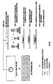
- a defined area of a glass slide is coated with a selected DNA probe.
- Glass slides with defined areas, e.g ., ring(s) are used.
- a defined area(s) on the slide is coated with specific DNA probes. This can be accomplished by diluting the DNA probe in TE buffer (Tris-EDTA) or other buffers in applying it to the slide and allowing it to dry.
- the cell sample (target DNA) solution is placed on top of the DNA probe in the previously defined locations and allowed to dry.
- Fig. 1 illustrates this embodiment of the invention.
- the cells are coated with DNA probe.
- the cell sample (target DNA) solution is applied to the slide within the defined area and allowed to dry.
- the probe DNA in TE or other buffer is applied to the same defined area and is allowed to dry.
- the cells are coated and thus the DNA is concentrated on the cells.
- the previously described solution G is layered on top of the DNA probe-coated cells.
- a coverglass is placed over the defined area, and the sample is simultaneously denatured and hybridized. This embodiment of the invention is shown in Fig. 2A .
- An alternative method to slide method 2A is the following: The cell sample solution is centrifuged to pellet the cells. The fluid is decanted. DNA probe in TE buffer or other buffer is applied to the pellet, and the cells are resuspended. The solution of DNA probe and cells is applied to the defined area on the slide and allowed to dry.
- Solution G is layered on top of the DNA probe coated cells. A coverglass is applied and the sample is simultaneously denatured and hybridized. This embodiment of the invention is shown in Fig. 2B .
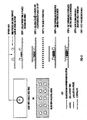
- the DNA probe is not denatured. This is of particular importance when working with unique sequence, short segment, oligonucleotide probes (DNA), e.g ., Vysis, Inc., LSI probes.
- DNA oligonucleotide probes
- Glass slides with defined area(s), i.e. , ring(s), are used.
- the cell sample solution is applied to the slide within a defined area and allowed to dry.
- Solution G is layered on top of the cell sample.
- a coverglass is applied.
- the slide with the coverglassed sample is denatured at appropriate temperatures and times.
- the coverglass is pushed to the side and DNA probe in TE buffer or other buffer is added, the coverglass is reapplied and sample hybridized.
- Fig. 3 illustrates this embodiment of the invention.
- the coverglass is removed, and a two-step post-hybridization wash is performed.
- the sample is placed in a solution of 0.1% to 1% NP-40 (nonidet ® P-40) in 0.15% NaCl for thirty seconds to two minutes at 65°C and second the sample is placed in a solution of 0.15% NaCl for three minutes to ten minutes at 65°C.
- NP-40 nonidet ® P-40
- the above description of the present invention defines the optimal conditions under which the whole FISH process can be completed.
- Other conditions may be used, for example, any temperature for denaturation and hybridization can be used, as long as the temperature does not exceed approximately 110°C.
- Example 1 FISH performed with F and G solutions on various cell and tissue samples with simultaneous denaturation/ hybridization of sample and probes.
- Rhodamine-labeled specific DNA for X chromosome and FITC fluorescent isothiocyanate-labeled specific DNA for Y chromosome probes (or Rhodamine-labeled Y chromosome and FITC-labeled X chromosome DNA probes) were obtained from a commercial source (Oncor, Inc., Vysis, Inc., BDS/Amersham, Inc.). The probes were diluted with denaturation-hybridization solution G.
- labeled probes represent DNA or anti-sense RNA to which relatively heat-stable enzymes and ligands, fluorochromes, (e.g ., FITC, rhodamine, Texas Red) chromophores, chemiluminescers, bioluminescers, or radioisotopes were covalently conjugated.
- the labeled probes were diluted with solution G or TE buffer to an appropriate concentration.
- the "probe solution represents labeled probes.
- slide method 1 (see Fig. 1 ) (DNA-coated slides), 5 to 30 ul of diluted probe solution is placed on the slide and allowed to dry. The cell sample is placed on top of the DNA probe and allowed to dry. Solution G is layered over the cells/DNA probe area and covered with a coverglass.
- the diluted probe solution is added to the pellet of cells in which the supernatant fixative has been decanted.
- the cells are resuspended and applied to the slide and allowed to dry.
- solution G is layered on top of the DNA-covered slide.
- Figs. 4-6 illustrate example cyclings.
- coverslips were removed from glass slides and the slides were immersed in 0.1% to 1% NP-40 for thirty seconds to two minutes and 0.1%-0.2% NaCl solution for three minutes.
- a unique sequence, short segment, oligonucleotide probe(DNA) was used on cultured and uncultured, male, peripheral blood leukocytes. The purpose was to perform FISH procedures.
- paraffin embedded tissue sections were deparaffinized and dehydrated.
- tissue sections were treated as set forth in Example 1, slide method 2A.
- fixatives were used with the direct-labeled fluorescent and indirect labeled DNA probes.
- the purpose was to demonstrate the versatility of the present invention with various fixatives.
- Peripheral blood was collected in an anticoagulant tube and the PBMN and PNM separated as described in prior art and fixed in several fixatives: 1) 3:1 Methanol:Glacial acetic acid (MeOH:Hac) fixative for a minimum of 30 minutes at -20°C; 2) 4% paraformaldehyde for 30 minutes to 3 hours and then applied to slides which were air dried for processing; and 3) Methanol for 30 minutes minimum at -20°C.
- MeOH:Hac Methanol:Glacial acetic acid
- the samples were processed by the optimal methods previously described herein (see Example 1).
- the slides were viewed with a Texas red triple bandpass filter on a fluorescent microscope; signals were observed with a 40X dry objective in the interphase nuclei.
- Maternal peripheral blood was collected in an anticoagulant tube and the fetal cells separated as described in prior art and fixed in 3:1 MeOH:Hac fixative for a minimum of 30 minutes at -20°C.
- the samples were centrifuged at 1000 g's for 10 minutes, and 10 ul of sample applied to each slide until a density of cells was reached sufficient for analysis.
- Example 1 The samples were processed by previously described optimal methods (Examples 1, 2 and 3).
- the slide was viewed with a Texas red triple bandpass filter on an epifluorescent microscope.
- the preparation of sperm and non-cultured amniocytes for FISH were identical. Aliquots of sperm or amniocytes were placed into 15 ml centrifuge tubes. PBS-containing 2mM Dithiothreitol (DTT) was added to sperm or amniocytes at a concentration of 10 to 20x10 6 per ml for 6.5 minutes at room temperature to decondense the chromatin. The samples were then centrifuged at 160gs for 5 minutes. Supernatant was discarded and 8 ml -20°C 3:1 MeOH/Hac were added and the sample vortexed. The samples were fixed at -20°C 3:1 MeOH:Hac for a minimum of 30 minutes.
- DTT Dithiothreitol
- Preferable fixation time is overnight at -20°C.
- the sperm or amniocyte samples were centrifuged at 1000 g's for 10 minutes. The supernatant was removed. samples were resuspended in an appropriate volume of 3:1 MeOH:Hac; and 10 ul of sample suspended in fixatives were placed on each slide (12 mm circle) assuring an even distribution. The slides were air dried for a minimum of 5 minutes.
- the samples were processed by previously described preferred method (see Example 1). The samples were viewed and counted using a fluorescence microscope with a Texas red triple band pass filter 60X dry lens.
Landscapes
- Chemical & Material Sciences (AREA)
- Physics & Mathematics (AREA)
- Life Sciences & Earth Sciences (AREA)
- Organic Chemistry (AREA)
- Analytical Chemistry (AREA)
- Zoology (AREA)
- Proteomics, Peptides & Aminoacids (AREA)
- Health & Medical Sciences (AREA)
- Engineering & Computer Science (AREA)
- Wood Science & Technology (AREA)
- Microbiology (AREA)
- Molecular Biology (AREA)
- Immunology (AREA)
- Biotechnology (AREA)
- Biophysics (AREA)
- Biochemistry (AREA)
- Bioinformatics & Cheminformatics (AREA)
- General Engineering & Computer Science (AREA)
- General Health & Medical Sciences (AREA)
- Genetics & Genomics (AREA)
- General Physics & Mathematics (AREA)
- Optics & Photonics (AREA)
- Measuring Or Testing Involving Enzymes Or Micro-Organisms (AREA)
Claims (53)
- Verfahren zur In-situ-Hybridisierung, das die folgenden Schritte umfasst:a) Bereitstellen eines Objektträgers, umfassend einen erhabenen Ring darauf;b) Bereitstellen einer Nukleinsäuresonde auf dem Objektträger;c) Bereitstellen einer Targetnukleinsäure auf dem Objektträger;d) Bereitstellen einer Hybridisierungslösung auf dem Objektträger, bestehend aus: einem formamidfreien Gemisch aus 10 Gew.-% ± 2 Gew.-% Dextransulfat und 15 Gew.-%-25 Gew.-% Glycerol und 0,9 Gew.-% Salz;e) Anordnen eines Deckgläschens auf dem Ring;f) Anordnen des Objektträgers unter denaturierenden Bedingungen;g) Anordnen des Objektträgers unter hybridisierenden Bedingungen; undh) Durchführen einer Posthybridisierungswäsche des Objektträgers.
- Verfahren nach Anspruch 1, wobei der Schritt des Bereitstellens einer Nukleinsäuresonde ferner das Überziehen des Objektträgers mit der Sonde umfasst, um eine erste Schicht zu bilden, der Schritt des Bereitstellens einer Targetnukleinsäure ferner die Schichtbildung der Targetnukleinsäure auf dem Sondenüberzug umfasst, um eine zweite Schicht zu bilden, und der Schritt des Bereitstellens einer Lösung ferner die Schichtbildung der Lösung auf der ersten Schicht des Sondenüberzugs und der zweiten Schicht der Targetnukleinsäure umfasst.
- Verfahren nach Anspruch 2, wobei die Schritte des Denaturierens und Hybridisierens auf der Sonde und der Probe gleichzeitig durchgeführt werden.
- Verfahren nach Anspruch 1, wobei Schritt (c) vor Schritt (b) durchgeführt wird und ferner das Aufbringen der Targetnukleinsäure auf den Objektträger umfasst, um eine Schicht zu bilden, Schritt (b) ferner das Überziehen der ersten Schicht der Targetnukleinsäure mit der Sonde umfasst, um die Nukleinsäuresonde auf der Targetnukleinsäure zu konzentrieren, und der Schritt des Bereitstellens einer Lösung ferner die Schichtbildung der Lösung auf der Targetnukleinsäure umfasst, die mit der Sonde beschichtet ist.
- Verfahren nach Anspruch 4, wobei die Schritte des Denaturierens und Hybridisierens auf der Sonde und der Probe gleichzeitig durchgeführt werden.
- Verfahren nach Anspruch 1, wobei Schritt (c) vor Schritt (b) durchgeführt wird und ferner das Bereitstellen einer Targetnukleinsäure in Lösung, das Zentrifugieren der die Lösung enthaltenden Targetnukleinsäure, um die Targetnukleinsäure zu pelletieren, und das Dekantieren der Lösung umfasst;
Schritt (b) ferner das Aufbringen der Nukleinsäuresonde auf die pelletierte Targetnukleinsäure, um einen Überzug zu bilden, das Resuspendieren der mit der Sonde überzogenen Targetnukleinsäure in einer ersten Lösung, und das Aufbringen der ersten Lösung auf den Objektträger, um eine erste Schicht zu bilden, umfasst; und
der Schritt des Bereitstellens einer Lösung ferner das Bereitstellen einer zweiten Lösung und die Schichtbildung der zweiten Lösung auf der mit der Sonde überzogenen Targetnukleinsäure, um eine zweite Schicht zu bilden, umfasst. - Verfahren nach Anspruch 6, wobei die Schritte des Denaturierens und Hybridisierens auf der Sonde und der Probe gleichzeitig durchgeführt werden.
- Verfahren nach Anspruch 1, wobei
Schritt (c) vor Schritt (b) durchgeführt wird und ferner das Bereitstellen einer Targetnukleinsäure in Lösung auf dem Objektträger und das Trocknen lassen der aufgebrachten Targetnukleinsäure auf dem Objektträger umfasst,
Schritt (d) vor Schritt (b) durchgeführt wird und ferner das Aufbringen der Lösung als Schicht auf der Targetnukleinsäure auf dem Objektträger umfasst,
die Schritte (e) und (f) vor Schritt (b) durchgeführt werden, und
der Schritt des Bereitstellens einer Nukleinsäuresonde ferner das zur Seite Schieben des Deckgläschens, das Aufbringen der Nukleinsäuresonden auf die denaturierte Targetnukleinsäure und die Lösung und das erneute Anbringen des Deckgläschens umfasst. - Verfahren nach Anspruch 1, wobei die Lösung ferner Diethylpyrocarbonat umfasst.
- Verfahren nach Anspruch 1, wobei die Schritte (a) bis (h) innerhalb von zwei Minuten oder darunter durchgeführt werden.
- Verfahren nach Anspruch 1, wobei der Schritt des Bereitstellens einer Sonde ferner das Verdünnen der Sonde vor dem Aufbringen umfasst.
- Verfahren nach Anspruch 11, wobei der Schritt des Verdünnens der Sonde ferner das Verdünnen der Sonde mit Puffer umfasst.
- Verfahren nach Anspruch 12, wobei der Puffer Tris-EDTA-Puffer umfasst.
- Verfahren nach Anspruch 1, wobei das Salz ausgewählt ist aus der Gruppe bestehend aus NaCl, KCl und Natriumcitrat.
- Verfahren nach Anspruch 1, wobei die Schritte der Denaturierung und Hybridisierung auf der Sonde und der Probe gleichzeitig durchgeführt werden.
- Verfahren nach Anspruch 1, wobei die Schritte der Denaturierung und Hybridisierung in weniger als 5 Minuten ausgeführt werden.
- Verfahren nach Anspruch 1, wobei der Schritt der Denaturierung innerhalb von etwa 1 bis 2 Minuten ausgeführt wird.
- Verfahren nach Anspruch 17, wobei der Schritt der Denaturierung bei einer Temperatur zwischen etwa 95 °C und 105 °C ausgeführt wird.
- Verfahren nach Anspruch 1, wobei der Schritt des Durchführens der Posthybridisierungswäsche innerhalb von etwa 5 bis 10 Minuten ausgeführt wird.
- Verfahren nach Anspruch 19, wobei der Schritt des Durchführens der Posthybridisierungswäsche bei zwischen etwa 55 °C und 65 °C ausgeführt wird.
- Verfahren nach Anspruch 1, wobei der Schritt des Durchführens einer Posthybridisierungswäsche ferner die folgenden Schritte umfasst:Entfernen des Deckgläschens;Waschen des Objektträgers in einer ersten Lösung, die ein Salz umfasst; undWaschen des Objektträgers in einer zweiten Lösung, die ein Salz umfasst.
- Verfahren nach Anspruch 21, wobei die erste Lösung ferner NaCl und Nonidet® P-40 umfasst.
- Verfahren nach Anspruch 22, wobei die erste Lösung ferner 0,15 % NaCl und 0,1-1 % Nonidet® P-40 umfasst.
- Verfahren nach Anspruch 21, wobei der Schritt des Waschens in der ersten Lösung 30 Sekunden bis 2 Minuten lang durchgeführt wird.
- Verfahren nach Anspruch 24, wobei der Schritt des Waschens in der ersten Lösung bei einer Temperatur von 65 °C durchgeführt wird.
- Verfahren nach Anspruch 21, wobei die zweite Lösung ferner NaCl umfasst.
- Verfahren nach Anspruch 26, wobei die zweite Lösung ferner 0,1-0,2 % NaCl umfasst.
- Verfahren nach Anspruch 21, wobei der Schritt des Waschens in einer zweiten Lösung 3-10 Minuten lang durchgeführt wird.
- Verfahren nach Anspruch 28, wobei der Schritt des Waschens in der zweiten Lösung bei einer Temperatur von 65 °C durchgeführt wird.
- Verfahren nach Anspruch 1, wobei der Schritt der Denaturierung in einem Thermocycler 1,5 Minuten lang bei 100 °C ausgeführt wird.
- Verfahren nach Anspruch 1, wobei der Schritt der Hybridisierung in einem Thermocycler ausgeführt wird und ferner die Schritte des schnellen Senkens der Temperatur nach dem Schritt der Denaturierung und das periodische Wechseln der Temperatur umfasst, bis die Hybridisierung abgeschlossen ist.
- Verfahren nach Anspruch 31, wobei der Schritt des periodischen Wechselns der Temperatur etwa 1 Minute bis 1 Stunde lang ausgeführt wird.
- Verfahren nach Anspruch 1, wobei die Schritte der Denaturierung und Hybridisierung bei einer Temperatur ausgeführt werden, die 110 °C nicht übersteigt.
- Verfahren nach Anspruch 1, wobei die Schritte der Denaturierung und Hybridisierung in Abwesenheit eines Schritts des Bereitstellens eines Fixierungsmittels auf dem Objektträger stattfinden.
- Verfahren nach Anspruch 1, wobei der Schritt des Anordnens eines Deckgläschens auf dem Ring in Abwesenheit des Schritts des Bereitstellens eines Dichtstoffs für das Deckgläschen auf dem Objektträger stattfindet.
- Verfahren nach Anspruch 1, wobei der Schritt des Durchführens einer Posthybridisierungswäsche das Waschen in Abwesenheit von Formamid umfasst.
- Verfahren nach Anspruch 1, wobei der Schritt der Hybridisierung unter einer kontrollierten Absenkung von der Denaturierungstemperatur auf umgebende Raumtemperatur ausgeführt wird.
- Verfahren nach Anspruch 1, wobei der Schritt der Hybridisierung unter einer nicht kontrollierten Absenkung von der Denaturierungstemperatur auf umgebende Raumtemperatur ausgeführt wird.
- Verfahren nach Anspruch 1, wobei die Nukleinsäuren in Nuclei oder Cytoplasma aus fixiertem, in Paraffin eingebettetem Gewebe, gefrorenen Gewebeschnitten, kultivierten oder nicht kultivierten Zellen, und auf Nitrocellulose oder Nylonpapier nachgewiesen werden.
- Verfahren zur In-situ-Hybridisierung, das die folgenden Schritte umfasst:a) Bereitstellen eines Objektträgers, umfassend einen erhabenen Ring darauf;b) Bereitstellen einer Nukleinsäuresonde auf dem Objektträger;c) Bereitstellen einer Targetnukleinsäure auf dem Objektträger;d) Bereitstellen einer Hybridisierungslösung auf dem Objektträger, bestehend aus: einem formamidfreien Gemisch aus 10 Gew.-% ± 2 Gew.-% Dextransulfat und 15 Gew.-%-25 Gew.-% Glycerol und 0,9 Gew.-% Salz;e) Anordnen eines Deckgläschens auf dem Ring;f) Anordnen des Objektträgers unter denaturierenden Bedingungen bei einer Temperatur zwischen 95 °C-105 °C;g) Anordnen des Objektträgers unter hybridisierenden Bedingungen bei einer Temperatur zwischen 45 °C-60 °C; undh) Durchführen einer Posthybridisierungswäsche des Objektträgers,wobei sowohl Denaturierung als auch Hybridisierung und Posthybridisierung alle innerhalb von 15 Minuten abgeschlossen sind.
- Verfahren nach Anspruch 40, wobei die Lösung ferner Diethylpyrocarbonat umfasst.
- Verfahren nach Anspruch 40, wobei das Salz ausgewählt ist aus der Gruppe bestehend aus NaCl, KCl und Natriumcitrat.
- Verfahren nach Anspruch 40, wobei die Schritte der Denaturierung und Hybridisierung in Abwesenheit eines Schritts des Bereitstellens eines Fixierungsmittels auf dem Objektträger stattfinden.
- Verfahren nach Anspruch 40, wobei der Schritt des Anordnens eines Deckgläschens auf dem Ring in Abwesenheit des Bereitstellens eines Dichtstoffs für das Deckgläschen auf dem Objektträger stattfindet.
- Verfahren nach Anspruch 40, wobei der Schritt der Posthybridisierung das Waschen in Abwesenheit von Formamid umfasst.
- Verfahren zur In-situ-Hybridisierung zum Nachweis von mRNA, wobei das Verfahren die folgenden Schritte umfasst:a) Bereitstellen eines Objektträgers, umfassend einen erhabenen Ring darauf;b) Bereitstellen einer Nukleinsäuresonde auf dem Objektträger;c) Bereitstellen einer Targetnukleinsäure auf dem Objektträger;d) Bereitstellen einer Hybridisierungslösung auf dem Objektträger, bestehend aus: einem formamidfreien Gemisch aus 10 Gew.-% ± 2 Gew.-% Dextransulfat und 15 Gew.-%-25 Gew.-% Glycerol und 0,9 Gew.-% Salz;e) Anordnen eines Deckgläschens auf dem Ring;f) Vorhybridisieren der Sonde und der Probe, indem der Objektträger bei einer Temperatur zwischen 37 °C-55 °C angeordnet wird;g) Denaturieren der Sonde und der Probe, indem der Objektträger bei einer Temperatur zwischen 95 °C-105 °C angeordnet wird;h) Hybridisieren der Sonde und der Probe, indem der Objektträger für 5 Minuten oder weniger bei einer Temperatur zwischen 45 °C-85 °C angeordnet wird; undi) Durchführen einer Posthybridisierungswäsche des Objektträgers,wobei sowohl Denaturierung als auch Hybridisierung und Posthybridisierung alle innerhalb von 15 Minuten abgeschlossen sind.
- Verfahren zur In-situ-Hybridisierung zum Nachweis von mRNA, wobei das Verfahren die folgenden Schritte umfasst:a) Bereitstellen eines Objektträgers, umfassend einen erhabenen Ring darauf;b) Bereitstellen einer Nukleinsäuresonde auf dem Objektträger;c) Bereitstellen einer Targetnukleinsäure auf dem Objektträger;d) Bereitstellen einer Hybridisierungslösung auf dem Objektträger, bestehend aus: einem formamidfreien Gemisch aus 10 Gew.-% ± 2 Gew.-% Dextransulfat und 15 Gew.-%- 25 Gew.-% Glycerol und 0,9 Gew.-% Salz;e) Anordnen eines Deckgläschens auf dem Ring;f) Anordnen des Objektträgers unter vorhybridisierenden Bedingungen;g) Anordnen des Objektträgers unter Hybridisierungsbedingungen für 5 Minuten oder weniger; undh) Anordnen des Objektträgers unter Posthybridisierungsbedingungen,wobei die Schritte der Vorhybridisierung, Hybridisierung und Posthybridisierung innerhalb von 24 Stunden abgeschlossen sind.
- Verfahren nach Anspruch 47 zum Nachweis von mRNA in den Nuclei von fixiertem, in Paraffin eingebettetem Gewebe.
- Verfahren nach Anspruch 47, wobei die mRNA aus den Nuclei von kultivierten oder nicht kultivierten Zellen abgeleitet ist.
- Verfahren zur Herstellung einer fluoreszierenden In-situ-Hybridisierung zum Nachweis von mRNA nach Anspruch 47, wobei das Verfahren die folgenden Schritte umfasst:a) Bereitstellen eines Objektträgers, umfassend einen erhabenen Ring darauf;b) Bereitstellen einer Nukleinsäuresonde auf dem Objektträger;c) Bereitstellen einer Targetnukleinsäure auf dem Objektträger;d) Bereitstellen einer Hybridisierungslösung auf dem Objektträger, bestehend aus: einem formamidfreien Gemisch aus 10 Gew.-% ± 2 Gew.-% Dextransulfat und 15 Gew.-%-25 Gew.-% Glycerol und 0,9 Gew.-% Salz;e) Anordnen eines Deckgläschens auf dem Ring;f) Vorhybridisierung bei einer Temperatur zwischen 37 °C und 55 °C;g) Hybridisierung für 5 Minuten oder weniger bei einer Temperatur zwischen 45 °C und 85 °C; undh) Posthybridisierung,wobei die Schritte der Vorhybridisierung, Hybridisierung und Posthybridisierung innerhalb von 24 Stunden abgeschlossen sind.
- Verfahren nach Anspruch 50, wobei mRNA in Nuclei oder Cytoplasma mindestens eines Gruppenmitglieds, ausgewählt aus der Gruppe bestehend aus fixiertem, in Paraffin eingebettetem Gewebe, gefrorenen Gewebeschnitten, kultivierten Zellen und nicht kultivierten Zellen nachgewiesen wird.
- Verfahren nach Anspruch 50, wobei mRNA auf mindestens einem Medium, ausgewählt aus der Gruppe bestehend aus Nitrocellulosepapier und Nylonpapier nachgewiesen wird.
- Verfahren nach Anspruch 50, wobei mRNA in Nuclei oder Cytoplasma mindestens eines Gruppenmitglieds, ausgewählt aus der Gruppe bestehend aus fixiertem, in Paraffin eingebettetem Gewebe, gefrorenen Gewebeschnitten, kultivierten Zellen und nicht kultivierten Zellen, und auf mindestens einem Medium, ausgewählt aus der Gruppe bestehend aus Nitrocellulosepapier und Nylonpapier nachgewiesen wird.
Applications Claiming Priority (3)
| Application Number | Priority Date | Filing Date | Title |
|---|---|---|---|
| US08/727,951 US6022689A (en) | 1995-04-07 | 1996-10-09 | Situ hybridization slide processes |
| US727951 | 1996-10-09 | ||
| PCT/US1997/017968 WO1998015656A1 (en) | 1996-10-09 | 1997-10-03 | In situ hybridization slide processes |
Publications (3)
| Publication Number | Publication Date |
|---|---|
| EP0935672A1 EP0935672A1 (de) | 1999-08-18 |
| EP0935672A4 EP0935672A4 (de) | 2004-03-17 |
| EP0935672B1 true EP0935672B1 (de) | 2008-12-17 |
Family
ID=24924786
Family Applications (1)
| Application Number | Title | Priority Date | Filing Date |
|---|---|---|---|
| EP97910799A Expired - Lifetime EP0935672B1 (de) | 1996-10-09 | 1997-10-03 | In situ-hybridisierungsverfahren auf objektträgern |
Country Status (7)
| Country | Link |
|---|---|
| US (1) | US6022689A (de) |
| EP (1) | EP0935672B1 (de) |
| AU (1) | AU4808497A (de) |
| CA (1) | CA2282702A1 (de) |
| DE (1) | DE69739170D1 (de) |
| ES (1) | ES2319135T3 (de) |
| WO (1) | WO1998015656A1 (de) |
Families Citing this family (20)
| Publication number | Priority date | Publication date | Assignee | Title |
|---|---|---|---|---|
| DE50014456D1 (de) * | 1999-05-07 | 2007-08-16 | Vermicon Ag | Verfahren zum nachweisen von mikroorganismen in einer probe |
| ATE364722T1 (de) * | 2000-06-02 | 2007-07-15 | Bayer Corp | Verfahren zum nachweis und zur lokalisierung von genen in situ durch hybridisierung einer verzweigten dna |
| DE10061655A1 (de) * | 2000-12-11 | 2002-06-20 | Vermicon Ag | In situ-Hybridisierungs-Anordnung zum spezifischen Nachweis von Mikroorganismen |
| DE10118043B4 (de) * | 2001-04-11 | 2005-10-27 | Holburn Pathology Products Ltd., Bowmanville | Hochspezifisches Detektionssystem |
| US6773677B2 (en) | 2002-01-09 | 2004-08-10 | Caliper Life Sciences, Inc. | Slide cassette for fluidic injection |
| US7202398B2 (en) * | 2002-08-16 | 2007-04-10 | E. I. Du Pont De Nemours And Company | Chalcone isomerase |
| US20040241734A1 (en) * | 2003-04-28 | 2004-12-02 | Exagen Diagnostics | Methods for in situ hybridization without the need for competitior DNA |
| US7534875B2 (en) * | 2003-11-03 | 2009-05-19 | Exagen Diagnostics, Inc. | Compositions for glioma classification |
| WO2005106043A2 (en) * | 2004-04-23 | 2005-11-10 | Exagen Diagnostics, Inc. | Breast cancer gene expression biomarkers |
| JP2007530075A (ja) * | 2004-04-23 | 2007-11-01 | エグザジェン ダイアグノスティクス インコーポレイテッド | 乳がんの予後診断のための組成物および方法 |
| US20050287578A1 (en) * | 2004-06-28 | 2005-12-29 | Exagen Diagnostics, Inc. | Methods for RNA fluorescence in situ hybridization |
| US20060223075A1 (en) * | 2005-03-29 | 2006-10-05 | Exagen Diagnostics, Inc. | Unique sequence hybridization probes (USP) |
| US7557198B2 (en) * | 2005-05-04 | 2009-07-07 | Exagen Diagnostics, Inc. | Acute myelogenous leukemia biomarkers |
| SG136000A1 (en) * | 2006-03-27 | 2007-10-29 | Univ Singapore | In situ hybridization method |
| US8626337B2 (en) | 2007-04-24 | 2014-01-07 | Pioneer Hi Bred International Inc | Method and computer program product for distinguishing and sorting seeds containing a genetic element of interest |
| US8452445B2 (en) | 2007-04-24 | 2013-05-28 | Pioneer Hi-Bred International, Inc. | Method and computer program product for distinguishing and sorting seeds containing a genetic element of interest |
| US8459463B2 (en) | 2007-04-24 | 2013-06-11 | Pioneer Hi-Bred International, Inc. | Method for sorting resistant seed from a mixture with susceptible seed |
| CN107904283A (zh) * | 2017-11-09 | 2018-04-13 | 武汉康录生物技术股份有限公司 | 一种即用型多探针杂交玻片及其制备方法和应用 |
| CN115418395B (zh) * | 2022-11-07 | 2023-01-17 | 百力格生物科技(上海)股份有限公司 | Mgb探针溶解方法及其溶液 |
| CN119935691B (zh) * | 2025-04-08 | 2025-06-06 | 中山大学 | 一种染色体压片结合原位杂交的简易操作方法 |
Family Cites Families (27)
| Publication number | Priority date | Publication date | Assignee | Title |
|---|---|---|---|---|
| US3883398A (en) * | 1973-05-07 | 1975-05-13 | Bellco Glass Inc | Microculture slide chamber |
| US4302204A (en) * | 1979-07-02 | 1981-11-24 | The Board Of Trustees Of Leland Stanford Junior University | Transfer and detection of nucleic acids |
| US4358535A (en) * | 1980-12-08 | 1982-11-09 | Board Of Regents Of The University Of Washington | Specific DNA probes in diagnostic microbiology |
| ES8600404A1 (es) * | 1983-05-02 | 1985-10-01 | Enzo Biochem Inc | Un metodo para detectar una secuencia seleccionada de acido nucleico en un material biologico. |
| US4647529A (en) * | 1984-06-01 | 1987-03-03 | Rodland Karin D | Hybridization method of detecting nucleic acid sequences with probe containing thionucleotide |
| US5132207A (en) * | 1984-07-05 | 1992-07-21 | Gen-Probe Incorporated | Accelerated nucleic acid reassociation method |
| US4689294A (en) * | 1984-11-19 | 1987-08-25 | Miles Laboratories, Inc. | Enhancement of hybridization of nucleic acids by anionic polymers |
| US4888278A (en) * | 1985-10-22 | 1989-12-19 | University Of Massachusetts Medical Center | In-situ hybridization to detect nucleic acid sequences in morphologically intact cells |
| US5447841A (en) * | 1986-01-16 | 1995-09-05 | The Regents Of The Univ. Of California | Methods for chromosome-specific staining |
| US4722598A (en) * | 1986-12-04 | 1988-02-02 | Max M. Ford | Diagnostic microscope slide having multiple sample wells and cover |
| US4886741A (en) * | 1987-12-09 | 1989-12-12 | Microprobe Corporation | Use of volume exclusion agents for the enhancement of in situ hybridization |
| WO1990002204A1 (en) * | 1988-08-31 | 1990-03-08 | Research Development Foundation | Manual in situ hybridization assay |
| US5225326A (en) * | 1988-08-31 | 1993-07-06 | Research Development Foundation | One step in situ hybridization assay |
| CA2005927A1 (en) * | 1988-12-21 | 1990-06-21 | Chander Bahl | Method of preparing nucleotide probes using a bridging complement |
| US5554500A (en) * | 1989-09-27 | 1996-09-10 | The United States Of America As Represented By The Department Of Health And Human Services | Cloned genes for human dopamine D2 receptors and cell lines expressing same |
| US5232829A (en) * | 1989-09-29 | 1993-08-03 | Hoffmann-La Roche Inc. | Detection of chlamydia trachomatis by polymerase chain reaction using biotin labelled lina primers and capture probes |
| US5491224A (en) * | 1990-09-20 | 1996-02-13 | Bittner; Michael L. | Direct label transaminated DNA probe compositions for chromosome identification and methods for their manufacture |
| US5232831A (en) * | 1991-06-28 | 1993-08-03 | Gen-Probe Incorporated | Nucleic acid probes to streptococcus pyogenes |
| GB9114180D0 (en) * | 1991-07-01 | 1991-08-21 | Amersham Int Plc | Enhancement of polynucleotide hybridisation |
| US5316906A (en) * | 1991-08-23 | 1994-05-31 | Molecular Probes, Inc. | Enzymatic analysis using substrates that yield fluorescent precipitates |
| US5506098A (en) * | 1991-09-04 | 1996-04-09 | Daikin Industries, Ltd. | In situ hybridization method |
| AU675774B2 (en) * | 1991-10-04 | 1997-02-20 | Children's Hospital Of Philadelphia, The | Detecting digeorge syndrome mutations |
| US5521061A (en) * | 1992-07-17 | 1996-05-28 | Aprogenex, Inc. | Enhancement of probe signal in nucleic acid-mediated in-situ hybridization studies |
| US5364790A (en) * | 1993-02-16 | 1994-11-15 | The Perkin-Elmer Corporation | In situ PCR amplification system |
| AU7474394A (en) * | 1993-07-19 | 1995-02-20 | Aprogenex, Inc. | Enriching and identifying fetal cells in maternal blood for in situ hybridization |
| US5571721A (en) * | 1994-05-05 | 1996-11-05 | Erie Scientific Company | Improved biological culture slide and method of making same |
| US5750340A (en) * | 1995-04-07 | 1998-05-12 | University Of New Mexico | In situ hybridization solution and process |
-
1996
- 1996-10-09 US US08/727,951 patent/US6022689A/en not_active Expired - Lifetime
-
1997
- 1997-10-03 DE DE69739170T patent/DE69739170D1/de not_active Expired - Lifetime
- 1997-10-03 ES ES97910799T patent/ES2319135T3/es not_active Expired - Lifetime
- 1997-10-03 WO PCT/US1997/017968 patent/WO1998015656A1/en not_active Ceased
- 1997-10-03 EP EP97910799A patent/EP0935672B1/de not_active Expired - Lifetime
- 1997-10-03 AU AU48084/97A patent/AU4808497A/en not_active Abandoned
- 1997-10-03 CA CA002282702A patent/CA2282702A1/en not_active Abandoned
Also Published As
| Publication number | Publication date |
|---|---|
| CA2282702A1 (en) | 1998-04-16 |
| US6022689A (en) | 2000-02-08 |
| AU4808497A (en) | 1998-05-05 |
| WO1998015656A1 (en) | 1998-04-16 |
| EP0935672A1 (de) | 1999-08-18 |
| EP0935672A4 (de) | 2004-03-17 |
| ES2319135T3 (es) | 2009-05-04 |
| DE69739170D1 (de) | 2009-01-29 |
Similar Documents
| Publication | Publication Date | Title |
|---|---|---|
| EP0935672B1 (de) | In situ-hybridisierungsverfahren auf objektträgern | |
| US5750340A (en) | In situ hybridization solution and process | |
| WO1998015656A9 (en) | In situ hybridization slide processes | |
| Cremer et al. | Multicolor 3D fluorescence in situ hybridization for imaging interphase chromosomes | |
| US6132961A (en) | Methods of biological dosimetry employing chromosome-specific staining | |
| US7326575B2 (en) | Methods and compositions for the preparation and use of fixed-treated cell-lines and tissue in fluorescence in situ hybridization | |
| US20240209422A1 (en) | Hybridization compositions and methods using formamide | |
| EP1172445A1 (de) | Verfahren zur direkten genetischen Analyse von Zielzellen anhand Fluoreszentensonden | |
| US10457981B2 (en) | Hybridization buffers | |
| WO2009144581A1 (en) | Hybridization compositions and methods | |
| WO2010097655A1 (en) | Compositions and methods for rna hybridization applications | |
| US20240018590A1 (en) | Hybridization compositions and methods | |
| US20170067094A1 (en) | Hybridization buffers | |
| Leitch et al. | In situ hybridisation | |
| IL94906A (en) | Methods and preparations for specific chromosome specification | |
| US20040241734A1 (en) | Methods for in situ hybridization without the need for competitior DNA | |
| Difilippantonio et al. | Technicolor genome analysis | |
| Christine et al. | Simultaneous FISH and Fluorescence Immunostaining | |
| CA2413835A1 (en) | Isolation and culturing of fetal cells | |
| Mohaddes Ardebili | Optimisation of interphase fluorescence in situ hybridisation for detection of common aneuploidies | |
| Driscoll et al. | Laser Microdissection as a Technique to Resolve Mixtures and Improve the Analysis of Difficult Evidence Samples |
Legal Events
| Date | Code | Title | Description |
|---|---|---|---|
| PUAI | Public reference made under article 153(3) epc to a published international application that has entered the european phase |
Free format text: ORIGINAL CODE: 0009012 |
|
| 17P | Request for examination filed |
Effective date: 19990510 |
|
| AK | Designated contracting states |
Kind code of ref document: A1 Designated state(s): DE ES FR GB |
|
| RAP1 | Party data changed (applicant data changed or rights of an application transferred) |
Owner name: SCIENCE & TECHNOLOGY CORPORATION UNM |
|
| A4 | Supplementary search report drawn up and despatched |
Effective date: 20040204 |
|
| RIC1 | Information provided on ipc code assigned before grant |
Ipc: 7G 02B 21/34 B Ipc: 7C 12Q 1/68 A |
|
| 17Q | First examination report despatched |
Effective date: 20040429 |
|
| RAP1 | Party data changed (applicant data changed or rights of an application transferred) |
Owner name: STC.UNM |
|
| RAP1 | Party data changed (applicant data changed or rights of an application transferred) |
Owner name: STC.UNM |
|
| RTI1 | Title (correction) |
Free format text: IN SITU HYBRIDIZATION METHODS ON SLIDES |
|
| GRAP | Despatch of communication of intention to grant a patent |
Free format text: ORIGINAL CODE: EPIDOSNIGR1 |
|
| GRAS | Grant fee paid |
Free format text: ORIGINAL CODE: EPIDOSNIGR3 |
|
| GRAA | (expected) grant |
Free format text: ORIGINAL CODE: 0009210 |
|
| AK | Designated contracting states |
Kind code of ref document: B1 Designated state(s): DE ES FR GB |
|
| REG | Reference to a national code |
Ref country code: GB Ref legal event code: FG4D |
|
| REF | Corresponds to: |
Ref document number: 69739170 Country of ref document: DE Date of ref document: 20090129 Kind code of ref document: P |
|
| REG | Reference to a national code |
Ref country code: ES Ref legal event code: FG2A Ref document number: 2319135 Country of ref document: ES Kind code of ref document: T3 |
|
| PLBE | No opposition filed within time limit |
Free format text: ORIGINAL CODE: 0009261 |
|
| STAA | Information on the status of an ep patent application or granted ep patent |
Free format text: STATUS: NO OPPOSITION FILED WITHIN TIME LIMIT |
|
| 26N | No opposition filed |
Effective date: 20090918 |
|
| PGFP | Annual fee paid to national office [announced via postgrant information from national office to epo] |
Ref country code: ES Payment date: 20091026 Year of fee payment: 13 Ref country code: DE Payment date: 20091028 Year of fee payment: 13 |
|
| PGFP | Annual fee paid to national office [announced via postgrant information from national office to epo] |
Ref country code: GB Payment date: 20091026 Year of fee payment: 13 Ref country code: FR Payment date: 20091029 Year of fee payment: 13 |
|
| GBPC | Gb: european patent ceased through non-payment of renewal fee |
Effective date: 20101003 |
|
| PG25 | Lapsed in a contracting state [announced via postgrant information from national office to epo] |
Ref country code: FR Free format text: LAPSE BECAUSE OF NON-PAYMENT OF DUE FEES Effective date: 20101102 |
|
| REG | Reference to a national code |
Ref country code: FR Ref legal event code: ST Effective date: 20110630 |
|
| PG25 | Lapsed in a contracting state [announced via postgrant information from national office to epo] |
Ref country code: GB Free format text: LAPSE BECAUSE OF NON-PAYMENT OF DUE FEES Effective date: 20101003 |
|
| REG | Reference to a national code |
Ref country code: DE Ref legal event code: R119 Ref document number: 69739170 Country of ref document: DE Effective date: 20110502 |
|
| REG | Reference to a national code |
Ref country code: ES Ref legal event code: FD2A Effective date: 20111118 |
|
| PG25 | Lapsed in a contracting state [announced via postgrant information from national office to epo] |
Ref country code: ES Free format text: LAPSE BECAUSE OF NON-PAYMENT OF DUE FEES Effective date: 20101004 |
|
| PG25 | Lapsed in a contracting state [announced via postgrant information from national office to epo] |
Ref country code: DE Free format text: LAPSE BECAUSE OF NON-PAYMENT OF DUE FEES Effective date: 20110502 |