EP0933094A2 - Mund- / Nasenkanüle - Google Patents
Mund- / Nasenkanüle Download PDFInfo
- Publication number
- EP0933094A2 EP0933094A2 EP99200205A EP99200205A EP0933094A2 EP 0933094 A2 EP0933094 A2 EP 0933094A2 EP 99200205 A EP99200205 A EP 99200205A EP 99200205 A EP99200205 A EP 99200205A EP 0933094 A2 EP0933094 A2 EP 0933094A2
- Authority
- EP
- European Patent Office
- Prior art keywords
- oxygen
- cannula
- patient
- nasal
- tube
- Prior art date
- Legal status (The legal status is an assumption and is not a legal conclusion. Google has not performed a legal analysis and makes no representation as to the accuracy of the status listed.)
- Granted
Links
- 0 CCC(*)(C1)CC11CCCC1 Chemical compound CCC(*)(C1)CC11CCCC1 0.000 description 1
Images


Classifications
-
- A—HUMAN NECESSITIES
- A61—MEDICAL OR VETERINARY SCIENCE; HYGIENE
- A61B—DIAGNOSIS; SURGERY; IDENTIFICATION
- A61B5/00—Measuring for diagnostic purposes; Identification of persons
- A61B5/68—Arrangements of detecting, measuring or recording means, e.g. sensors, in relation to patient
- A61B5/6801—Arrangements of detecting, measuring or recording means, e.g. sensors, in relation to patient specially adapted to be attached to or worn on the body surface
- A61B5/6813—Specially adapted to be attached to a specific body part
- A61B5/6814—Head
- A61B5/682—Mouth, e.g., oral cavity; tongue; Lips; Teeth
-
- A—HUMAN NECESSITIES
- A61—MEDICAL OR VETERINARY SCIENCE; HYGIENE
- A61B—DIAGNOSIS; SURGERY; IDENTIFICATION
- A61B5/00—Measuring for diagnostic purposes; Identification of persons
- A61B5/08—Detecting, measuring or recording devices for evaluating the respiratory organs
- A61B5/083—Measuring rate of metabolism by using breath test, e.g. measuring rate of oxygen consumption
-
- A—HUMAN NECESSITIES
- A61—MEDICAL OR VETERINARY SCIENCE; HYGIENE
- A61B—DIAGNOSIS; SURGERY; IDENTIFICATION
- A61B5/00—Measuring for diagnostic purposes; Identification of persons
- A61B5/08—Detecting, measuring or recording devices for evaluating the respiratory organs
- A61B5/097—Devices for facilitating collection of breath or for directing breath into or through measuring devices
-
- A—HUMAN NECESSITIES
- A61—MEDICAL OR VETERINARY SCIENCE; HYGIENE
- A61B—DIAGNOSIS; SURGERY; IDENTIFICATION
- A61B5/00—Measuring for diagnostic purposes; Identification of persons
- A61B5/68—Arrangements of detecting, measuring or recording means, e.g. sensors, in relation to patient
- A61B5/6801—Arrangements of detecting, measuring or recording means, e.g. sensors, in relation to patient specially adapted to be attached to or worn on the body surface
- A61B5/6813—Specially adapted to be attached to a specific body part
- A61B5/6814—Head
- A61B5/6819—Nose
-
- A—HUMAN NECESSITIES
- A61—MEDICAL OR VETERINARY SCIENCE; HYGIENE
- A61M—DEVICES FOR INTRODUCING MEDIA INTO, OR ONTO, THE BODY; DEVICES FOR TRANSDUCING BODY MEDIA OR FOR TAKING MEDIA FROM THE BODY; DEVICES FOR PRODUCING OR ENDING SLEEP OR STUPOR
- A61M16/00—Devices for influencing the respiratory system of patients by gas treatment, e.g. mouth-to-mouth respiration; Tracheal tubes
- A61M16/06—Respiratory or anaesthetic masks
- A61M16/0666—Nasal cannulas or tubing
- A61M16/0672—Nasal cannula assemblies for oxygen therapy
-
- A—HUMAN NECESSITIES
- A61—MEDICAL OR VETERINARY SCIENCE; HYGIENE
- A61M—DEVICES FOR INTRODUCING MEDIA INTO, OR ONTO, THE BODY; DEVICES FOR TRANSDUCING BODY MEDIA OR FOR TAKING MEDIA FROM THE BODY; DEVICES FOR PRODUCING OR ENDING SLEEP OR STUPOR
- A61M16/00—Devices for influencing the respiratory system of patients by gas treatment, e.g. mouth-to-mouth respiration; Tracheal tubes
- A61M16/08—Bellows; Connecting tubes ; Water traps; Patient circuits
- A61M16/0816—Joints or connectors
- A61M16/0841—Joints or connectors for sampling
- A61M16/085—Gas sampling
-
- A—HUMAN NECESSITIES
- A61—MEDICAL OR VETERINARY SCIENCE; HYGIENE
- A61B—DIAGNOSIS; SURGERY; IDENTIFICATION
- A61B5/00—Measuring for diagnostic purposes; Identification of persons
- A61B5/08—Detecting, measuring or recording devices for evaluating the respiratory organs
- A61B5/083—Measuring rate of metabolism by using breath test, e.g. measuring rate of oxygen consumption
- A61B5/0836—Measuring rate of CO2 production
-
- A—HUMAN NECESSITIES
- A61—MEDICAL OR VETERINARY SCIENCE; HYGIENE
- A61M—DEVICES FOR INTRODUCING MEDIA INTO, OR ONTO, THE BODY; DEVICES FOR TRANSDUCING BODY MEDIA OR FOR TAKING MEDIA FROM THE BODY; DEVICES FOR PRODUCING OR ENDING SLEEP OR STUPOR
- A61M16/00—Devices for influencing the respiratory system of patients by gas treatment, e.g. mouth-to-mouth respiration; Tracheal tubes
- A61M16/08—Bellows; Connecting tubes ; Water traps; Patient circuits
- A61M16/0816—Joints or connectors
- A61M16/0833—T- or Y-type connectors, e.g. Y-piece
-
- A—HUMAN NECESSITIES
- A61—MEDICAL OR VETERINARY SCIENCE; HYGIENE
- A61M—DEVICES FOR INTRODUCING MEDIA INTO, OR ONTO, THE BODY; DEVICES FOR TRANSDUCING BODY MEDIA OR FOR TAKING MEDIA FROM THE BODY; DEVICES FOR PRODUCING OR ENDING SLEEP OR STUPOR
- A61M2210/00—Anatomical parts of the body
- A61M2210/06—Head
- A61M2210/0625—Mouth
-
- A—HUMAN NECESSITIES
- A61—MEDICAL OR VETERINARY SCIENCE; HYGIENE
- A61M—DEVICES FOR INTRODUCING MEDIA INTO, OR ONTO, THE BODY; DEVICES FOR TRANSDUCING BODY MEDIA OR FOR TAKING MEDIA FROM THE BODY; DEVICES FOR PRODUCING OR ENDING SLEEP OR STUPOR
- A61M2230/00—Measuring parameters of the user
- A61M2230/40—Respiratory characteristics
- A61M2230/43—Composition of exhalation
- A61M2230/432—Composition of exhalation partial CO2 pressure (P-CO2)
Definitions
- the present invention relates to a nasal cannula and to an oral/nasal cannula, and, more particularly, to a nasal cannula and an oral/nasal cannula which permits both delivery of oxygen and accurate sampling of carbon dioxide.
- oral/nasal cannulas are used to deliver oxygen to hospital patients who require assistance to breathe properly, to collect carbon dioxide samples from patients to monitor respiration, or to perform both functions. Such cannulas are used when direct ventilation is not provided.
- oral/nasal refers to the adaptable configuration of such cannulas which can be in close proximity to the oral cavity or inserted into the nasal cavity of the patient. In either arrangement, a sidestream of the patient's exhaled breath flows through the cannula to a gas analyzer to be analyzed. The results of this non-invasive analysis provide an indication of the patient's condition, such as the state of the patient's pulmonary perfusion, respiratory system and metabolism.
- the accuracy of this non-invasive analysis of exhaled gases depends on the ability of a sampling system to move a gas sample from the patient to the gas analyzer while maintaining a smooth, laminar flow of gases, such that there are as few alterations to the waveform and response time of the concentration of the gases as possible.
- the waveform of the concentration of the gas is critical for accurate analysis. As the gas mixture travels from the patient to the gas analyzer, the concentration of the gases can be affected by mixing of the component gases, which reduces the accuracy of the analysis of the sample by the gas analyzer, and reduces the amount of information obtained from that analysis.
- Prior art nasal or oral/nasal cannulas unfortunately have caused significant alterations to these important features of the internal structure of the stream of exhaled gases. Such alterations have especially arisen as the result of attempts to combine the delivery of oxygen with the sampling of the exhaled breath of the patient.
- the simplest nasal cannula design consisting of a tube with two double hollow prongs for insertion into the nostrils, allows significant mixing of the oxygen which is delivered from the end of one tube, and the exhaled breath which is collected from the end of the second tube.
- Such mixing occurs when oxygen is delivered in a stream with strong force, so that the oxygen stream penetrates deeply into the nasal cavity even during expiration, thereby artifactually altering the composition of the exhaled gases.
- one type of prior art nasal cannula (Salter Labs, Arvin, California, USA) consists of a tube with two openings at either end, and two hollow prongs projecting perpendicularly from the center of the tube with a partition between them. Oxygen enters the tube from one end and exhaled breath leaves the tube from the other end. The two hollow prongs are inserted into the nasal cavity of a patient, one prong in each nostril, so that oxygen could be delivered to, and exhaled breath collected from, the patient.
- this type of cannula usually has significant "void volume", or space in which mixing of gases, and concurrent alteration of the gas waveform, can occur.
- void volume space in which mixing of gases, and concurrent alteration of the gas waveform, can occur.
- Such space is often referred to as “void volume” because it is not part of the pathway for the flow of gases and hence is unproductive.
- void volume arises in this cannula between the septum dividing the main tube and the junction of each prong with that tube. The presence of such void volume is a significant hindrance to the accurate analysis of exhaled gases.
- this prior art nasal cannula has a reduced efficiency for the collection of exhaled gases for analysis.
- a nasal cannula (Hospitak, Lindenhurst, New York, USA) has two parallel overlapping tubes, one for delivering oxygen and one for receiving exhaled gases.
- the tube which receives exhaled gases has two nasal prongs, while the tube which delivers oxygen has two holes parallel to these prongs. Both tubes have two holes, such that the gases can flow freely from the prongs to the holes. This configuration allows delivered oxygen to easily mix with expired gases, even at the end of the expiration period, thereby reducing the accuracy of the gas analysis.
- U.S. Patent No. 5,046,491 discloses another type of nasal cannula which also includes a first tube with two double nasal prongs and a septum placed between the prongs. One prong delivers oxygen and the second prong collects exhaled gases. A second tube is attached to the first tube and has two holes which are placed in or near the oral cavity of the patient for collecting exhaled breath.
- One problem with this cannula is that the exhaled gases are collected through two outputs, which are then connected to two separate tubes. These separate tubes then join together before delivering the gases to the capnograph. If gases are not flowing at exactly the same rate through both tubes, for example due to condensation, then the waveform of the gas concentration is altered and the results of the analysis are affected.
- this cannula has significant void volume because of the large dimension of the tubes and because there are two outputs for collecting the exhaled gases.
- the large void volume also causes mixing of the gases.
- the term “respiratory cavity” refers to the oral cavity, the nasal cavity, or both cavities, of a patient.
- the effectiveness of oxygen delivery by a cannula is determined by two principles, neither of which is completely fulfilled by prior art cannulas.
- the first principle is that the distribution of the delivered oxygen stream should be equal between the two nostrils of the patient. In most prior art cannulas, one nostril receives 1.2 - 2.0 times as much oxygen as the other. However, an equal distribution of oxygen is preferable for the following reasons. First, if one of the nostrils is blocked, the second will continue to deliver oxygen. Second, even flow rates for both nostrils will not cause the patient to feel excess pressure in one nostril, even at high flow rates for the delivered oxygen. Third, producing even flow rates through the presence of oxygen "clouds" near the nostrils of the patient will cause such "clouds" to be the same size at both nostrils, and will permit the more effective use of ambient oxygen present near the nostrils before the inspiration phase.
- the second principle is that the oxygen stream should be delivered at a relatively slow rate, rather than being forced into the nostrils at a high rate, for the following reasons.
- First, an oxygen stream which is delivered at a slow rate will not penetrate deeply into the nostrils of the patient and so will not be collected during the exhalation phase, thereby preventing distortion of the carbon dioxide measurements because of dilution of the exhaled gases.
- a cannula which does not alter the gas waveform, which does not easily become blocked or clogged, which has minimal added void volume, and which can deliver oxygen without disturbing the waveform of exhaled gases, yet which has the flexibility and adaptability of an oral/nasal cannula.
- a nasal cannula for collection of exhaled gases front a patient having nostrils comprising: (a) two nasal prongs for insertion into the nostrils of the patient; and (b) a collection tube for the collection of the exhaled gases from the patient, the nasal prongs and the collection tube being connected at a single junction, such that the exhaled gases flow freely from the nasal prongs to the collection tube.
- the collection tube is a single collection tube.
- the nasal prongs are joined in an are substantially before being connected to the junction.
- the collection tube delivers the exhaled gases to a capnograph for gas analysis.
- a cannula for collection of exhaled gases from a patient having nostrils and an oral cavity including: (a) two nasal prongs for insertion into the nostrils of the patient; (b) an oral prong for being located proximately to the oral cavity of the patient; and (c) a collection tube for the collection of the exhaled gases from the patient, the nasal prongs, the oral prong and the collection tube being connected at a single junction located substantially near the nostrils of the patient, such that the exhaled gases flow freely from the nasal prongs and the oral prong to the collection tube.
- the collection tube is a single collection tube.
- the oral prong features a distal portion, the distal portion being bent at an angle. More preferably, the angle is about 90 degrees, such that the distal portion is located proximately to the oral cavity of the patient.
- the distal portion features a cap, the cap being attached to the distal portion, and the cap being made of a substantially hydrophilic material, such that the cap absorbs condensation from the distal portion.
- the nasal prongs are joined in an arc substantially before being connected to the junction.
- the collection tube delivers the exhaled gases to a capnograph for gas analysis.
- the cannula further includes (d) an oxygen tube for delivery of oxygen, the oxygen tube being located near the nostrils of the patient; and (e) two oxygen inlets connected to the oxygen tube and being disposed such that the oxygen flows from the oxygen tube into the nostrils of the patient.
- the oxygen tube is located either above or below the nostrils of the patient.
- the oxygen tube includes a centrally located input for receiving oxygen being placed substantially equidistant from both oxygen inlets.
- the oxygen inlets are holes. More preferably, the holes have an first diameter at an inner surface of the oxygen tube and the holes have a second diameter at an outer surface of the oxygen tube, the first diameter being smaller than the second diameter.
- the oxygen tube features a screen, the screen being placed within the oxygen tube such that the oxygen flows from the oxygen tube through the screen.
- the screen is constructed of a material selected from the group consisting of a hydrophobic porous material, a wide mesh and a netting.
- the inlets are oxygen prongs for being inserted into the nostrils of the patient. More preferably, the oxygen prongs are substantially shorter in length than the nasal prongs, such that the nasal prongs extend farther into the nostrils than the oxygen prongs. Also more preferably, the oxygen prongs are formed of a substantially porous material, such that the oxygen prongs are permeable to gases. Most preferably, the oxygen prongs are formed from an inner cylinder and an outer cylinder, both cylinders being made from the substantially hydrophobic porous material, and the inner cylinder being substantially shorter in length than the outer cylinder.
- At least a portion of the oxygen tube is formed from a substantially porous material such that the at least a portion of the oxygen tube is permeable to gases. More preferably, the at least a portion of the oxygen tube is located substantially between the oxygen prongs.
- a method of using the cannula of claim 1 for collecting the exhaled gases from the patient including: (a) inserting the nasal prongs into the nostrils of the patient; (b) attaching the collection tube to a conduit for conducting gas; (c) connecting the conduit to a gas analyzer; and (d) applying a force at the gas analyzer, such that the exhaled gases flowing through the cannula moves from the collection tube to the gas analyzer.
- a cannula for collection of exhaled gases from a patient and for delivery of oxygen to a patient the patient having nostrils and an oral cavity, including: (a) two nasal prongs for insertion into the nostrils of the patient; (b) an oral prong for being located proximately to the oral cavity of the patient; (c) a collection tube for the collection of the exhaled gases from the patient, the nasal prongs, the oral prong and the collection tube being connected at a single junction, such that the exhaled gases flow freely from the nasal prongs and the oral prong to the collection tube; (d) an oxygen tube for delivery of oxygen, the oxygen tube being located near the nostrils of the patient; and (e) two oxygen inlets connected to the oxygen tube and being disposed such that the oxygen flows from said oxygen tube into the nostrils of the patient.
- the term “attached” is defined as connected to, or integrally formed with.
- the term “connected” is defined as communicating with.
- the term “prong” refers to a hollow tube with two openings, one at each end of the tube.
- the present invention is of a cannula which can effectively be used to collect samples of gas without reducing the accuracy of the analysis of the collected gas, and which is less likely to become blocked by condensed moisture, or by liquid or solid material, or their mixtures thereof, such as mucous or saliva.
- the present invention has two prongs for insertion into the nostrils of a patient. These two prongs are joined outside the nasal cavity to a single output tube for collection of the exhaled gases.
- a second tube is attached to the two prongs, which is parallel to the nasal prongs, for placement of the distal end of the tube near the oral cavity of the patient, thereby providing an oral/nasal cannula.
- an additional tube is provided for the delivery of oxygen, the additional tube having two additional prongs for insertion into the nostrils of the patient, and the additional tube being perpendicular to the additional nasal prongs.
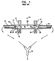
- FIG. 1 shows a prior art oral/nasal carbon dioxide cannula.
- a cannula 10 has two nasal prongs 12 for insertion into the nostrils of a patient (not shown).
- Nasal prongs 12 are connected to a first side 14 of a hollow tube 16 .
- Hollow tube 16 is substantially perpendicular to nasal prongs 12 .
- Two oral prongs 18 are also connected to a second side 20 of tube 16 in a substantially perpendicular orientation, such that gas flow from nasal prongs 12 to oral prongs 18 through tube 16 is substantially free and unimpeded.
- Tube 16 also has two holes 22 , one at each end of tube 16 , for connection to one of a plurality of connectors 24 .
- Each connector 24 is attached to a gas line (not shown) which is then connected to a Y-connector 26 .
- Y-connector 26 is attached to a line which leads to a capnograph (not shown).
- cannula 10 is suitable only for collection of exhaled gases for analysis.
- Prior art cannula 10 unfortunately has a significant void volume 28 (also designated as V 0 ) between nasal prongs 12 , within which gases do not properly circulate.
- Two smaller void volumes 30 (also designated as V 1 and V 2 ) are also present parallel to nasal prongs 12 and oral prongs 18 .
- Such void volumes 28 and 30 and especially the larger void volume 28 , permit the mixing of exhaled gases from a currently exhaled breath with previously exhaled breaths, thereby increasing the response time, altering the waveform and introducing an artifact into the gas analysis.
- the separation of the exhaled gases into two streams from nasal prongs 12 by tube 16 also increases the response time if there is even a slight difference in the flow rate of the gases between tubes 16 .
- Such separation potentially also results in two streams having different flow properties. For example, if one tube 16 accumulated more condensed water than the other, the corresponding stream of exhaled gases would have a lower flow rate, thereby altering the waveform of the gas concentrations and increasing the response time for gases in that tube 16 .
- prior art cannula 10 cannot provide completely accurate collection of gases for analysis.
- Figure 2 shows an exemplary prior art double oxygen/carbon dioxide nasal cannula for the collection of exhaled gases and the delivery of oxygen.
- a prior art nasal cannula 32 again has a first pair of nasal prongs 34 for insertion into nostrils 36 of a patient.
- First nasal prongs 34 are again connected to a first hollow tube 38 .
- First hollow tube 38 is again substantially perpendicular to first nasal prongs 34 .
- nasal cannula 32 has a second pair of nasal prongs 40 for insertion into nostrils 36 .
- Second nasal prongs 40 are attached to a second hollow tube 42 in a substantially perpendicular orientation.
- First nasal prongs 34 and first hollow tube 38 are intended for the collection of exhaled gases from the patient, in a substantially similar configuration as that shown in Figure 1.
- Second nasal prongs 40 and second hollow tube 42 are intended to deliver oxygen to the patient, so that nasal cannula 32 is capable of simultaneous oxygen delivery and gas collection.
- prior art nasal cannula 32 also permits the mixing of delivered oxygen and exhaled gases between first nasal prongs 34 and second nasal prongs 40 in nostrils 36 , thereby diluting the true concentration of expired carbon dioxide.
- prior art nasal cannula 32 also introduces artifacts into the analysis of expired gases.
- the efficiency of oxygen delivery by prior art nasal cannula 32 is not sufficient because the oxygen flow rate varies between nasal prongs 40 .
- nasal prong 40 which is closer to the input of hollow tube 42 will have a higher flow rate than the other nasal prong 40 .
- the strong oxygen stream into the nostrils creates discomfort for the patient, the alleviation of which is especially important for long term oxygen delivery.
- Figure 3 shows a second exemplary prior art divided oxygen/carbon dioxide nasal cannula for the simultaneous delivery of oxygen and collection of exhaled gases.
- a prior art nasal cannula 44 has a single tube 46 for both delivery of oxygen and collection of gases.
- Tube 46 has two nasal prongs 48 and 50 for insertion into nostrils 52 of a patient.
- Oxygen is delivered through nasal prong 48 and exhaled gases are collected from nasal prong 50 .
- a septum 54 is present inside tube 46 between nasal prong 48 and nasal prong 50 to separate the delivered oxygen from the exhaled gases.
- particularly forceful streams of delivered oxygen can pass from nasal prong 48 , penetrate deeply into nostrils 52 , entering nasal prong 50 and dilute the true concentration of exhaled carbon dioxide.
- a significant void volume 56 is present between septum 54 and nasal prong 50 , both increasing the response time and mixing the exhaled gases, which also reduce the accuracy of the analysis of the exhaled gases.
- prior art nasal cannula 44 is still not able to collect gases for a completely accurate analysis.
- the strong oxygen stream into the nostrils creates discomfort for the patient, the alleviation of which is especially important for long term oxygen delivery.
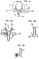
- FIG 4 shows a schematic illustration of an exemplary novel oral/nasal carbon dioxide cannula for collection of exhaled gases according to the present invention.
- An oral/nasal cannula 58 also has a pair of nasal prongs 60 for insertion into the nostrils 62 of a patient.
- Cannula 58 preferably features an oral prong 64 for placement near the oral cavity of the patient (not shown) to form an oral/nasal cannula. If oral prong 64 is absent, then cannula 58 is a nasal cannula according to the present invention.
- Cannula 58 also has a collection tube 66 for collection of the exhaled gases for analysis by a capnograph (not shown).
- Nasal prongs 60 , oral prong 64 and collection tube 66 meet at a single junction 68 , which is preferably minimized to reduce void volume.
- single junction refers to the joining of nasal prongs 60 , oral prong 64 and collection tube 66 at least in close proximity, and preferably at exactly one junction.
- having the single junction 68 between all portions of oral/nasal cannula 58 significantly reduces the void volume, thereby reducing mixing of the gases and maintaining the response time.
- having the single collection tube 66 rather than two such tubes as in prior art cannulas, eliminates the division of the stream of exhaled gases as well as reducing the amount of void volume created.
- FIG. 5A shows a front cross-sectional view of oral/nasal cannula 58 .
- nasal prongs 60 , oral prong 64 and collection tube 66 all meet at a single small junction 68 with a minimum void volume.
- the void volume can be almost completely eliminated through this configuration, because there are no poorly ventilated areas within oral/nasal cannula 58 .
- a portion 70 of collection tube 66 does extend past nasal prongs 60 opposite to the collection point. However, portion 70 is blocked and is only intended to permit the attachment of a symmetrical loop which extends around the head of the patient (not shown).
- Figure 5B shows a side cross-sectional view of the connection between one nasal prong 60 and oral prong 64 .
- a distal end 72 of oral prong 64 is bent, more preferably at approximately a 90 degree angle from the remainder of oral prong 64 , so as to be substantially parallel to the direction of flow of orally exhaled gases from the patient.
- Such an orientation both provides optimal response time for gas analysis and promotes self-clearing of condensation from oral/nasal cannula 58 .
- nasal prongs 60 are joined in an are, so that condensation tends to move into oral prong 64 under dynamic pressure of the nasal exhalation of gases by the patient.
- oral/nasal cannula 58 is designed to eliminate one significant problem with certain prior art oral/nasal cannulas, which is the susceptibility of these prior art cannulas to the intake of ambient air through that portion of the cannula which is not receiving exhaled air. For example, if the patient exhales through the nasal cavity, ambient air can be sucked into the prior art cannula through the opening provided for the nasal cavity. Such ambient air can dilute the concentration of gas in the exhaled breath of the patient, thus giving misleading results for the gas analysis.
- the structure of oral/nasal cannula 58 reduces or eliminates this problem with the presence of single small junction 68 , and the bending of distal end 72 of oral prong 64 .
- the resultant structure substantially prevents ambient air from entering the portion of cannula 58 which is not directly receiving exhaled air from the patient.
- nasal prongs 60 and oral prong 64 have an optimal diameter, sufficiently large to promote rapid and easy removal of condensation from the interior of nasal cannula 58 , yet not so large as to increase the response time.
- an optimal diameter for both nasal prongs 60 and oral prong 64 is in a range of from about 1.6 mm to about 2.0 mm.
- distal end 72 of oral prong 64 features a porous, hydrophilic cap 74 , as shown in cross-section in Figure 5C.
- Porous hydrophilic cap 74 covers distal end 72 and absorbs water droplets formed from condensation which collects in nasal cannula 58 .
- the particular advantage of cap 74 is that the material of cap 74 preferably attracts water away from oral prong 64 , and then provides a relatively large surface area for evaporation of that water. Additionally, cap 74 relieves potential patient discomfort from water dripping from cannula 58 into the mouth of the patient.
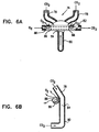
- Figures 6A and 6B show cross-sectional views of a second preferred embodiment of the oral/nasal cannula for oxygen delivery and gas collection of the present invention. Detailed illustrations of portions of the cannula of Figures 6A and 6B are shown in Figures 7A and 7B. Figures 7A and 7B also show the preferred addition of a porous screen to the oxygen tube.
- an oral/nasal cannula 76 again has a pair of nasal prongs 78 for insertion into the nostrils of a patient (not shown).
- Cannula 76 again preferably features an oral prong 80 for placement near the oral cavity of the patient (not shown) to form an oral/nasal cannula.
- Cannula 76 also has a collection tube 82 for collection of the exhaled gases for analysis by a capnograph (not shown).
- Nasal prongs 78 , oral prong 80 and collection tube 82 again meet at a single junction 84 , which is preferably minimized to reduce void volume.
- cannula 76 also features an oxygen tube 86 for lying near the nostrils of the patient (not shown) and more preferably above or below the nostrils of the patient, substantially parallel with the upper lip of the patient (not shown), oxygen is not delivered through a second set of nasal prongs. Instead, oxygen tube 86 has two holes 88 , through which oxygen is delivered to the patient. Holes 88 are placed near the nostrils of the patient yet do not enter the nostrils, thereby preventing the delivered oxygen from entering as a forceful stream of gases which dilutes the exhaled gases and reduces the accuracy of gas analysis.
- Figure 6B shows a side cross-sectional view of junction 84 between one nasal prong 78 and oral prong 80 , as well as a portion of oxygen tube 86 . Oxygen is shown being dispersed from oxygen tube 86 through hole 88 .
- FIG. 7A shows holes 88 in more detail.
- Holes 88 preferably have a relatively large diameter. Most preferably the diameter of holes 88 increases from the inner surface of oxygen tube 86 to the outer surface of oxygen tube 86 , in order to reduce the force of the delivered oxygen stream.
- Holes 88 have a first smaller diameter 90 at the inner surface of oxygen tube 86 , and a second larger diameter 92 at the outer surface of oxygen tube 86 , with the diameter of holes 88 preferably gradually increasing from the inner to the outer surface of oxygen tube 86 .
- oxygen tube 86 preferably features a screen 94 made from a substantially porous material which is permeable to oxygen, such as a wide mesh, a hydrophobic porous screen, netting or cotton wool, for example.
- screen 94 made from a substantially porous material which is permeable to oxygen, such as a wide mesh, a hydrophobic porous screen, netting or cotton wool, for example.
- the advantages of screen 94 are that the force of the delivered oxygen stream is reduced and an oxygen "cloud" is created near the nostrils of the patient.
- the combination of the dispersion of oxygen through screen 94 and hole 88 is shown in a side, cross-sectional view in Figure 7B, which also shows junction 84 .
- Figures 8A and 8B provide a detailed illustration of a portion of a third embodiment of an oral/nasal cannula according to the present invention.
- Figure 8A shows a portion of an oral/nasal cannula 96 , showing a section of a pair of nasal prongs 98 for receiving exhaled carbon dioxide, an oxygen tube 100 and a pair of second nasal prongs 102 .
- oxygen is delivered through oxygen tube 100 and is then dispersed through second nasal prongs 102 .
- second nasal prongs 102 are constructed from two cylinders, in order to ensure that oxygen is delivered to the nostrils of the patient efficiently, yet is quickly dispersed within the nasal cavity.
- the first cylinder is an inner cylinder 104 , preferably made from a substantially porous hydrophobic material. The material is preferably hydrophobic to prevent absorption of moisture.
- Inner cylinder 104 is surrounded by an outer cylinder 106 , also preferably made from a substantially porous hydrophobic material, such that oxygen is dispersed throughout the nostrils of the patient, rather than entering the nasal cavity as a highly pressurized stream of gas.
- Figure 8B shows a side, cross-sectional view of the portion of the cannula illustrated in Figure 8A.
- a junction 108 between one nasal prong 98 and an oral prong 110 is shown, as is one second nasal prong 102 with inner cylinder 104 and outer cylinder 106 .
- the advantage of constructing second nasal prong 102 from a porous material is that such material would be permeable to oxygen, thereby allowing oxygen to disperse evenly from second nasal prong 102 . Such dispersion reduces the force of the delivered oxygen stream.
- Figures 9A-9C show a comparison between a prior art oral/nasal cannula in which oxygen is delivered unequally to the nostrils of the patient ( Figure 9A), and a oral/nasal cannula according to the present invention in which oxygen is delivered at equal flow rates (Figures 9B and 9C).
- Figure 9A shows a cross-sectional view of the oxygen-delivery portion of a typical prior art oral/nasal cannula 112 .
- Prior art cannula 112 has an oxygen delivery tube 114 for delivery oxygen to two outputs 116 and 118 .
- Outputs 116 and 118 could be holes or nasal prongs as shown previously. The problem with this configuration is that oxygen is not distributed evenly between both outputs 116 and 118 .
- Output 116 which is closest to the start of oxygen delivery tube 114 , has a greater flow of oxygen than output 118 , as indicated by the arrows. Such a situation arises because the resistance of outputs 116 and 118 to the flow of oxygen is much lower than the resistance of the connecting portion of oxygen delivery tube 114 .
- Figure 9B shows a cross-sectional view of the oxygen-delivery portion of a first exemplary oral/nasal cannula 120 according to the present invention.
- First cannula 120 has an oxygen delivery tube 122 for delivery oxygen to two sets of outputs 124 and 126 .
- Each set of outputs 124 and 126 includes at least two outputs, although three are shown here for illustrative purposes, without any intention of being limiting. Again, the outputs could be holes, holes with a porous screen, or nasal prongs as shown previously.
- the advantage of this configuration is that oxygen is distributed more evenly between both sets of outputs 124 and 126 . Such a situation arises because the resistance of both sets of outputs 124 and 126 to the flow of oxygen is much greater than the resistance of the connecting portion of oxygen delivery tube 122 .
- FIG. 9C shows a cross-sectional view of the oxygen-delivery portion of a second exemplary oral/nasal cannula 128 according to the present invention.
- Second cannula 128 has an oxygen delivery tube 130 for delivery oxygen to two sets of outputs 132 and 134 .
- Each set of outputs 132 and 134 includes at least one output, although only one is shown here for illustrative purposes, without any intention of being limiting. Again, the outputs could be holes, holes with a porous screen, or nasal prongs as shown previously.
- oxygen delivery tube 130 features a centrally located input 136 for the delivery of oxygen.
- centrally located input 136 is located substantially equidistantly to outputs 132 and 134 .
- the first test performed was the self-cleaning test.
- Self-cleaning is important for preventing the accumulation of condensed water, which can disturb the sampling of carbon dioxide.
- V ex is defined as the minimal volume of expired breath required for self-cleaning of water from the cannula.
- the second test was the response time test, performed in accordance with Regulation prEN 864:1992 (European Union standard) for capnography. All measurements were conducted on a capnograph with low flow rate of 47 ml/min. Response times (in mSec) were tested for nasal cannula blanks only, nasal cannula systems which also included the set of sample lines, and the entire capnograph set which included the nasal cannula system with a typical capnograph flow system.
- the third test determined the accuracy of measurements of expired carbon dioxide (EtCO 2 ).
- EtCO 2 expired carbon dioxide was measured both with and without oxygen delivery.
- the fourth test measured the effectiveness of the delivery of oxygen according to the flow distribution between the two nasal cannula oxygen delivery outputs.
- Oxygen was delivered at the rate of 8 L/min.
- the flow of oxygen from each output, given as Q 1 and Q 2 was measured.
- the efficiency (K eff ) was determined according to the ratio of Q 1 to Q 2 .
- cannulas Three types of cannulas were obtained and tested from Salter Labs (Arvin, California, USA): a nasal cannula (catalog number 4000); a dual oral/nasal cannula (catalog number 4001); and divided oxygen/carbon dioxide nasal cannula (catalog number 4707). Two types of cannulas were obtained and tested from Hudson (Temecula, California, USA): a nasal cannula (catalog number 1103); and an oxygen/carbon dioxide nasal cannula (catalog number 1843).
- An oral/nasal cannula according to the present invention was also tested, in the embodiment of an oxygen/carbon dioxide oral/nasal cannula with inserts of braid or cotton wool for oxygen dispersion as shown in Figure 7B. Results for all tests are shown in Table 1.
- the cannula of the present invention performed at least as well as, and in many respects better than, the prior an cannulas.
- the cannula of the present invention had a much lower response time than any of the other tested prior art cannulas.
- the cannula of the present invention had a response time of 14, while those of the cannulas of Hudson were 97 and 47, and those of the cannulas of Salter Labs were 167, 143 and 239.
- the cannula of the present invention had a far better response time than these tested cannulas.
Landscapes
- Health & Medical Sciences (AREA)
- Life Sciences & Earth Sciences (AREA)
- Veterinary Medicine (AREA)
- Public Health (AREA)
- Animal Behavior & Ethology (AREA)
- Biomedical Technology (AREA)
- Heart & Thoracic Surgery (AREA)
- Pulmonology (AREA)
- Engineering & Computer Science (AREA)
- General Health & Medical Sciences (AREA)
- Emergency Medicine (AREA)
- Physics & Mathematics (AREA)
- Surgery (AREA)
- Otolaryngology (AREA)
- Molecular Biology (AREA)
- Medical Informatics (AREA)
- Pathology (AREA)
- Biophysics (AREA)
- Anesthesiology (AREA)
- Hematology (AREA)
- Physiology (AREA)
- Dentistry (AREA)
- Oral & Maxillofacial Surgery (AREA)
- Obesity (AREA)
- Measurement Of The Respiration, Hearing Ability, Form, And Blood Characteristics Of Living Organisms (AREA)
- Investigating Or Analysing Biological Materials (AREA)
- Medicines Containing Material From Animals Or Micro-Organisms (AREA)
Applications Claiming Priority (2)
| Application Number | Priority Date | Filing Date | Title |
|---|---|---|---|
| IL12312298A IL123122A0 (en) | 1998-01-29 | 1998-01-29 | Oral/nasal cannula |
| IL12312298 | 1998-01-29 |
Publications (3)
| Publication Number | Publication Date |
|---|---|
| EP0933094A2 true EP0933094A2 (de) | 1999-08-04 |
| EP0933094A3 EP0933094A3 (de) | 1999-12-29 |
| EP0933094B1 EP0933094B1 (de) | 2004-05-06 |
Family
ID=11071162
Family Applications (1)
| Application Number | Title | Priority Date | Filing Date |
|---|---|---|---|
| EP99200205A Expired - Lifetime EP0933094B1 (de) | 1998-01-29 | 1999-01-25 | Mund- / Nasenkanüle |
Country Status (7)
| Country | Link |
|---|---|
| US (2) | US6422240B1 (de) |
| EP (1) | EP0933094B1 (de) |
| JP (1) | JP4184520B2 (de) |
| AT (1) | ATE265874T1 (de) |
| CA (1) | CA2257415A1 (de) |
| DE (1) | DE69916907T2 (de) |
| IL (1) | IL123122A0 (de) |
Cited By (11)
| Publication number | Priority date | Publication date | Assignee | Title |
|---|---|---|---|---|
| US6379312B2 (en) * | 1999-12-28 | 2002-04-30 | O'toole James | End tidal carbon dioxide sampling device |
| WO2002102446A1 (en) * | 2001-06-18 | 2002-12-27 | Riggins Michael A | A nasal and oral cannula apnea detection device |
| WO2003030977A1 (en) * | 2001-10-12 | 2003-04-17 | Southmedic Incorporated | Lightweight oxygen delivery device for patients |
| EP1360050A1 (de) * | 2001-01-04 | 2003-11-12 | Salter Labs | Verfahren zur herstellung von atmungserfassungsvorrichtungen mit nasaler und oraler kanüle |
| EP1438086A2 (de) * | 2001-10-25 | 2004-07-21 | Worldwide Medical Technologies Inc. | Nasenkanüle |
| US6938619B1 (en) | 2000-06-13 | 2005-09-06 | Scott Laboratories, Inc. | Mask free delivery of oxygen and ventilatory monitoring |
| EP1694193A2 (de) * | 2003-12-05 | 2006-08-30 | Salter Labs | Nasale und orale zufuhr-, entnahme- und/oder nachweisvorrichtung |
| US7327440B2 (en) | 2004-08-16 | 2008-02-05 | James N. Horn | Distance measuring device |
| EP2814558A4 (de) * | 2012-02-16 | 2015-08-19 | Capnia Inc | Gasverteiler mit einem streuungsmundstück |
| US10716912B2 (en) | 2015-03-31 | 2020-07-21 | Fisher & Paykel Healthcare Limited | User interface and system for supplying gases to an airway |
| US11324908B2 (en) | 2016-08-11 | 2022-05-10 | Fisher & Paykel Healthcare Limited | Collapsible conduit, patient interface and headgear connector |
Families Citing this family (97)
| Publication number | Priority date | Publication date | Assignee | Title |
|---|---|---|---|---|
| US7640932B2 (en) * | 1997-04-29 | 2010-01-05 | Salter Labs | Nasal cannula for acquiring breathing information |
| US20100113956A1 (en) * | 1997-04-29 | 2010-05-06 | Salter Labs | Nasal cannula for acquiring breathing information |
| US20050121033A1 (en) * | 1998-02-25 | 2005-06-09 | Ric Investments, Llc. | Respiratory monitoring during gas delivery |
| US6776162B2 (en) * | 2000-03-13 | 2004-08-17 | Innomed Technologies, Inc. | Ventilation interface for sleep apnea therapy |
| BR0102116B1 (pt) | 2000-05-10 | 2010-09-21 | componente para um membro de circuito de respiração. | |
| US7559324B2 (en) | 2000-06-21 | 2009-07-14 | Fisher & Paykel Healthcare Limited | Conduit with heated wick |
| US7743770B2 (en) * | 2001-01-04 | 2010-06-29 | Salter Labs | Nasal and oral cannula having three or more capabilities and method of producing same |
| US7832400B2 (en) * | 2001-01-04 | 2010-11-16 | Salter Labs | Nasal and oral cannula having two capabilities and method of producing same |
| US6799575B1 (en) * | 2001-04-21 | 2004-10-05 | Aaron Carter | Cannula for the separation of inhaled and exhaled gases |
| US6913017B2 (en) * | 2002-01-04 | 2005-07-05 | Bevely Roberts | Apparatus for delivering inhalant and monitoring exhaled fluid, method of making same, and method of delivering inhalant and monitoring exhaled fluid |
| AU2003207984A1 (en) * | 2002-02-15 | 2003-09-04 | Oridion Medical 1987 Ltd. | Dual function nasal cannula |
| US6986353B2 (en) * | 2002-08-21 | 2006-01-17 | Medical Device Group, Inc. | Divided nasal cannula assembly |
| AU2003244171B2 (en) | 2002-09-09 | 2007-11-15 | Fisher & Paykel Healthcare Limited | Limb for Breathing Circuit |
| US7059322B2 (en) * | 2002-10-11 | 2006-06-13 | Ric Investments, Llc. | Low deadspace airway adapter |
| US7013888B2 (en) | 2002-12-19 | 2006-03-21 | Scadds Incorporated | Self contained aerosol dual delivery system (SCADDS) |
| US7913686B2 (en) * | 2002-12-19 | 2011-03-29 | Scadds Incorporated | Self contained aerosol dual delivery system (SCADDS) |
| JP4247758B2 (ja) * | 2003-02-18 | 2009-04-02 | 日本光電工業株式会社 | 炭酸ガス測定センサ |
| JP4998895B2 (ja) * | 2003-02-18 | 2012-08-15 | 日本光電工業株式会社 | 炭酸ガス測定センサ |
| NL1025769C2 (nl) * | 2003-03-20 | 2006-01-17 | Samsung Electronics Co Ltd | Projectiesysteem. |
| US8555886B2 (en) * | 2003-05-20 | 2013-10-15 | Oridion Medical 1987 Ltd. | Endoscopic bite block |
| US20040231675A1 (en) * | 2003-05-20 | 2004-11-25 | Lyons James R. | Method and apparatus for transnasal ventilation |
| US7493902B2 (en) | 2003-05-30 | 2009-02-24 | Fisher & Paykel Healthcare Limited | Breathing assistance apparatus |
| US7588033B2 (en) | 2003-06-18 | 2009-09-15 | Breathe Technologies, Inc. | Methods, systems and devices for improving ventilation in a lung area |
| EP1646419B1 (de) * | 2003-07-09 | 2013-04-10 | ResMed R&D Germany GmbH | Atemmaskenanordnung |
| AU2003258103A1 (en) * | 2003-08-06 | 2005-03-07 | Innomed Technologies, Inc. | Nasal interface and system including ventilation insert |
| US7472707B2 (en) * | 2003-08-06 | 2009-01-06 | Innomed Technologies, Inc. | Nasal interface and system including ventilation insert |
| JP2007506480A (ja) * | 2003-08-18 | 2007-03-22 | ワンドカ,アンソニー・ディ | 鼻用インターフェイスによる非侵襲的換気のための方法と器具 |
| AU2004203870B2 (en) | 2003-09-17 | 2011-03-03 | Fisher & Paykel Healthcare Limited | Breathable Respiratory Mask |
| US20050092329A1 (en) * | 2003-10-29 | 2005-05-05 | Sta-Maria Rosalinda C. | Nasal continuous positive airway pressure cannula device and securement for infants |
| EP1706025B1 (de) | 2003-12-30 | 2012-05-09 | University of Florida Research Foundation, Inc. | Neues speziell konfiguriertes nasalpulsoxymeter |
| CN101628142B (zh) | 2003-12-31 | 2014-03-26 | 雷斯梅德有限公司 | 小型口鼻病人接口 |
| CN1942215B (zh) | 2004-04-15 | 2012-09-26 | 雷斯梅德有限公司 | 气道正压通气机械导管 |
| WO2005115087A2 (en) * | 2004-05-27 | 2005-12-08 | Oridion Medical 1987 Ltd. | Capnography apparatus |
| US20060042637A1 (en) * | 2004-08-31 | 2006-03-02 | Martin James F | Bite block assembly |
| US20080275357A1 (en) * | 2004-11-22 | 2008-11-06 | Ron Porat | Oral/nasal cannula |
| JP2006198093A (ja) * | 2005-01-19 | 2006-08-03 | Izumi Products Co | ロータリー式電気かみそり |
| WO2007012140A1 (en) * | 2005-07-29 | 2007-02-01 | Resmed Limited | Method and apparatus for managing moisture buildup in pressurised breathing systems |
| EP1924854B1 (de) * | 2005-08-16 | 2016-04-13 | Oridion Medical 1987 Ltd. | Atemprobennahmevorrichtung und verfahren zur verwendung davon |
| US11833301B2 (en) * | 2005-09-12 | 2023-12-05 | ResMed Pty Ltd | High flow therapy device utilizing a non-sealing respiratory interface and related methods |
| EP1926517A2 (de) | 2005-09-20 | 2008-06-04 | Lutz Freitag | Systeme, verfahren und gerät zur atemunterstützung eines patienten |
| US20070113850A1 (en) * | 2005-11-22 | 2007-05-24 | General Electric Company | Respiratory monitoring with cannula receiving respiratory airflows and differential pressure transducer |
| US20080078393A1 (en) * | 2005-11-22 | 2008-04-03 | General Electric Company | Respiratory monitoring with cannula receiving respiratory airflows, differential pressure transducer, and ventilator |
| US7422015B2 (en) * | 2005-11-22 | 2008-09-09 | The General Electric Company | Arrangement and method for detecting spontaneous respiratory effort of a patient |
| US20070113847A1 (en) * | 2005-11-22 | 2007-05-24 | General Electric Company | Respiratory monitoring with cannula receiving first respiratory airflows and second respiratory airflows |
| US20070113848A1 (en) * | 2005-11-22 | 2007-05-24 | General Electric Company | Respiratory monitoring with cannula receiving respiratory airflows and exhaled gases |
| US20070113856A1 (en) * | 2005-11-22 | 2007-05-24 | General Electric Company | Respiratory monitoring with cannula receiving respiratory airflows |
| WO2007063532A2 (en) | 2005-12-01 | 2007-06-07 | Oridion Medical (1987) Ltd. | Endoscopic bite block |
| US20070272247A1 (en) * | 2006-04-25 | 2007-11-29 | Oridion Medical Ltd. | Oral nasal cannula |
| JP5191005B2 (ja) | 2006-05-18 | 2013-04-24 | ブリーズ テクノロジーズ, インコーポレイテッド | 気管切開の方法およびデバイス |
| US8028701B2 (en) | 2006-05-31 | 2011-10-04 | Masimo Corporation | Respiratory monitoring |
| EP2068992B1 (de) | 2006-08-03 | 2016-10-05 | Breathe Technologies, Inc. | Vorrichtung für minimal invasive atmungsunterstützung |
| US7946288B2 (en) * | 2006-11-10 | 2011-05-24 | Encompas Unlimited, Inc. | Bite block system and method |
| JP4968647B2 (ja) * | 2007-03-09 | 2012-07-04 | 日本光電工業株式会社 | 呼気情報収集用アダプタおよび生体情報処理システム |
| WO2008144589A1 (en) | 2007-05-18 | 2008-11-27 | Breathe Technologies, Inc. | Methods and devices for sensing respiration and providing ventilation therapy |
| JP5513392B2 (ja) | 2007-09-26 | 2014-06-04 | ブリーズ・テクノロジーズ・インコーポレーテッド | 睡眠時無呼吸を治療するための方法及び装置 |
| US8567399B2 (en) | 2007-09-26 | 2013-10-29 | Breathe Technologies, Inc. | Methods and devices for providing inspiratory and expiratory flow relief during ventilation therapy |
| JP5758799B2 (ja) | 2008-04-18 | 2015-08-05 | ブリーズ・テクノロジーズ・インコーポレーテッド | 呼吸作用を感知し、人工呼吸器の機能を制御するための方法およびデバイス |
| US8770193B2 (en) | 2008-04-18 | 2014-07-08 | Breathe Technologies, Inc. | Methods and devices for sensing respiration and controlling ventilator functions |
| ES2398942T3 (es) * | 2008-04-29 | 2013-03-22 | Oridion Medical 1987 Ltd. | Capnografia inalámbrica |
| CA2734296C (en) | 2008-08-22 | 2018-12-18 | Breathe Technologies, Inc. | Methods and devices for providing mechanical ventilation with an open airway interface |
| CA2739435A1 (en) | 2008-10-01 | 2010-04-08 | Breathe Technologies, Inc. | Ventilator with biofeedback monitoring and control for improving patient activity and health |
| US9044565B2 (en) * | 2008-10-30 | 2015-06-02 | Oridion Medical (1987) Ltd. | Oral-nasal cannula system enabling CO2 and breath flow measurement |
| US9055888B2 (en) * | 2009-01-05 | 2015-06-16 | Oridion Medical (1987) Ltd. | Exhaled breath sampling with delivery of gas |
| US9132250B2 (en) | 2009-09-03 | 2015-09-15 | Breathe Technologies, Inc. | Methods, systems and devices for non-invasive ventilation including a non-sealing ventilation interface with an entrainment port and/or pressure feature |
| WO2010115170A2 (en) | 2009-04-02 | 2010-10-07 | Breathe Technologies, Inc. | Methods, systems and devices for non-invasive open ventilation for treating airway obstructions |
| US9962512B2 (en) | 2009-04-02 | 2018-05-08 | Breathe Technologies, Inc. | Methods, systems and devices for non-invasive ventilation including a non-sealing ventilation interface with a free space nozzle feature |
| CA2774902C (en) | 2009-09-03 | 2017-01-03 | Breathe Technologies, Inc. | Methods, systems and devices for non-invasive ventilation including a non-sealing ventilation interface with an entrainment port and/or pressure feature |
| IT1396484B1 (it) * | 2009-11-19 | 2012-12-14 | Mezzoli | Dispositivo atto a somministrare per via nasale e/o buccale le soluzioni sotto forma di aerosol, doccia micronizzata o nebulizzazione. |
| JP6017312B2 (ja) | 2009-12-22 | 2016-10-26 | フィッシャー アンド ペイケル ヘルスケア リミテッド | 医療用回路用部品 |
| JP5891226B2 (ja) | 2010-08-16 | 2016-03-22 | ブリーズ・テクノロジーズ・インコーポレーテッド | Loxを使用して換気補助を提供する方法、システム及び装置 |
| WO2012045051A1 (en) | 2010-09-30 | 2012-04-05 | Breathe Technologies, Inc. | Methods, systems and devices for humidifying a respiratory tract |
| US20120157794A1 (en) * | 2010-12-20 | 2012-06-21 | Robert Goodwin | System and method for an airflow system |
| BR112013019445A2 (pt) * | 2011-01-31 | 2020-10-27 | Breathe Technologies, Inc. | métodos sistemas e dispositivos para ventilação utilizando uma máscara de ventilação nasal com um coletor e um tubo interno complacente e conjunto almofada de vedação nasal |
| CA2838529C (en) | 2011-06-07 | 2020-03-24 | Parion Sciences, Inc. | Methods of treatment |
| RU2477088C1 (ru) * | 2011-07-22 | 2013-03-10 | Общество с ограниченной ответственностью "Айсенс Групп" | Эндоназальный активатор |
| US20130092165A1 (en) * | 2011-09-26 | 2013-04-18 | Anthony David Wondka | Nasal Ventilation Cannula System and Methods |
| US10413695B2 (en) * | 2012-03-28 | 2019-09-17 | Robert Tero | Nasal cannula with pressure monitoring |
| US20140014108A1 (en) * | 2012-07-16 | 2014-01-16 | David G. Dillard | Oxygen delivery device for diffusing gas flow |
| US9795756B2 (en) | 2012-12-04 | 2017-10-24 | Mallinckrodt Hospital Products IP Limited | Cannula for minimizing dilution of dosing during nitric oxide delivery |
| DK2928531T3 (en) | 2012-12-04 | 2017-05-22 | Ino Therapeutics Llc | CANNEL FOR MINIMIZING DILUTION DOSAGE DURING NITROGEN OXIDE ADMINISTRATION |
| FR3003176B1 (fr) * | 2013-03-15 | 2015-03-13 | Deltamedics | Canule nasopharyngee pour capnographie en flux secondaire |
| US10010690B1 (en) | 2013-03-15 | 2018-07-03 | Monitoring For Life, Llc | Endotracheal tube apparatus |
| US9888867B2 (en) | 2013-03-15 | 2018-02-13 | Airway Control Technologies, Llc | Medical breathing apparatus |
| US20150119742A1 (en) * | 2013-10-30 | 2015-04-30 | Rutgers, The State University Of New Jersey | Nasal oxygen mask and breathing circuit assembly |
| US10112024B2 (en) | 2014-01-17 | 2018-10-30 | Monitoring For Life Llc | Medical tube apparatus |
| EP4151258A1 (de) * | 2014-04-11 | 2023-03-22 | Fisher & Paykel Healthcare Limited | Gastherapiesystem |
| US20170095630A1 (en) * | 2014-05-30 | 2017-04-06 | Wake Forest University Health Sciences | Oxygen Port Nasal Cannula |
| LT3177207T (lt) * | 2014-09-04 | 2024-04-10 | Fisher & Paykel Healthcare Limited | Iškvepiamų dujų matavimo kompensavimas didelo srautų kvėpavimo terapijas metu |
| JP6721611B2 (ja) | 2015-01-23 | 2020-07-15 | マシモ スウェーデン アーベーMasimo Sweden Ab | 鼻/口カニューレ・システムおよび製造 |
| US10220174B2 (en) | 2016-02-17 | 2019-03-05 | Christine M. Huerta | Septi-cannula |
| AU2017341838A1 (en) | 2016-10-14 | 2019-05-02 | Vapotherm, Inc. | Systems and methods for high velocity nasal insufflation |
| US11052212B2 (en) * | 2017-01-20 | 2021-07-06 | Oridion Medical 1987 Ltd. | Capnoxygen masks |
| CN107320824A (zh) * | 2017-08-08 | 2017-11-07 | 湖南明康中锦医疗科技发展有限公司 | 一种加湿器冷凝水报警系统与呼吸机 |
| US10792449B2 (en) | 2017-10-03 | 2020-10-06 | Breathe Technologies, Inc. | Patient interface with integrated jet pump |
| JP7064920B2 (ja) * | 2018-03-27 | 2022-05-11 | 日本光電工業株式会社 | 呼吸補助具 |
| US20210016036A1 (en) * | 2019-07-16 | 2021-01-21 | Neotech Products Llc | Wearable suction aspirator for nasal, oral and tracheostomy airway secretions |
| CN214050088U (zh) * | 2020-10-30 | 2021-08-27 | 北京怡和嘉业医疗科技股份有限公司 | 用于鼻氧导管的鼻塞、鼻氧导管以及通气治疗设备 |
Citations (1)
| Publication number | Priority date | Publication date | Assignee | Title |
|---|---|---|---|---|
| US5046491A (en) | 1990-03-27 | 1991-09-10 | Derrick Steven J | Apparatus and method for respired gas collection and analysis |
Family Cites Families (11)
| Publication number | Priority date | Publication date | Assignee | Title |
|---|---|---|---|---|
| US759152A (en) * | 1903-09-03 | 1904-05-03 | George L Bennett | Inhaler. |
| US2693800A (en) * | 1951-04-27 | 1954-11-09 | Caldwell Lyle | Nasal cannula |
| US4151843A (en) * | 1976-06-28 | 1979-05-01 | Brekke John H | Apparatus for administration of a gas to a human and the exhausting thereof |
| US4106505A (en) | 1977-01-17 | 1978-08-15 | Salter Labs., Inc. | Nasal cannula assembly |
| US4156426A (en) * | 1977-08-11 | 1979-05-29 | Gold Lawrence W | Head-mounted oxygen-administration device |
| US4367735A (en) * | 1979-12-31 | 1983-01-11 | Novametrix Medical Systems, Inc. | Nasal cannula |
| DE68924121T2 (de) * | 1988-04-15 | 1996-02-01 | Salter Labs | Verfahren und vorrichtung zur inhalation von therapeutischen gasen und zur probeentnahme ausgeatmeter gase zum zwecke der quantitativen analyse. |
| DK171592B1 (da) * | 1993-12-21 | 1997-02-17 | Maersk Medical As | Anordning for tilførsel af ilt og/eller andre gasser til en patient |
| US5375593A (en) * | 1994-02-10 | 1994-12-27 | Press; John R. | Oxygenating pacifier |
| US5495848A (en) * | 1994-11-25 | 1996-03-05 | Nellcar Puritan Bennett | Monitoring system for delivery of therapeutic gas |
| US5794619A (en) * | 1997-02-18 | 1998-08-18 | Edelman; Robert | Nasal cannula mounted solely by frictional engagement with the columella |
-
1998
- 1998-01-29 IL IL12312298A patent/IL123122A0/xx not_active IP Right Cessation
- 1998-12-22 CA CA002257415A patent/CA2257415A1/en not_active Abandoned
-
1999
- 1999-01-25 DE DE69916907T patent/DE69916907T2/de not_active Expired - Lifetime
- 1999-01-25 EP EP99200205A patent/EP0933094B1/de not_active Expired - Lifetime
- 1999-01-25 AT AT99200205T patent/ATE265874T1/de not_active IP Right Cessation
- 1999-01-27 JP JP01794499A patent/JP4184520B2/ja not_active Expired - Lifetime
- 1999-01-28 US US09/239,119 patent/US6422240B1/en not_active Expired - Lifetime
-
2001
- 2001-12-26 US US10/033,387 patent/US20020055685A1/en not_active Abandoned
Patent Citations (1)
| Publication number | Priority date | Publication date | Assignee | Title |
|---|---|---|---|---|
| US5046491A (en) | 1990-03-27 | 1991-09-10 | Derrick Steven J | Apparatus and method for respired gas collection and analysis |
Cited By (18)
| Publication number | Priority date | Publication date | Assignee | Title |
|---|---|---|---|---|
| US6379312B2 (en) * | 1999-12-28 | 2002-04-30 | O'toole James | End tidal carbon dioxide sampling device |
| US6938619B1 (en) | 2000-06-13 | 2005-09-06 | Scott Laboratories, Inc. | Mask free delivery of oxygen and ventilatory monitoring |
| US7152604B2 (en) | 2000-06-13 | 2006-12-26 | Scott Laboratories, Inc. | Apparatus and method for mask free delivery of an inspired gas mixture and gas sampling |
| US7337780B2 (en) * | 2001-01-04 | 2008-03-04 | Salter Labs | Nasal and oral cannula breathing detection device |
| EP1360050A4 (de) * | 2001-01-04 | 2007-04-04 | Salter Labs | Verfahren zur herstellung von atmungserfassungsvorrichtungen mit nasaler und oraler kanüle |
| EP1360050A1 (de) * | 2001-01-04 | 2003-11-12 | Salter Labs | Verfahren zur herstellung von atmungserfassungsvorrichtungen mit nasaler und oraler kanüle |
| AU2002347264B2 (en) * | 2001-06-18 | 2006-11-09 | Pro-Tech Services, Inc. | A nasal and oral cannula apnea detection device |
| WO2002102446A1 (en) * | 2001-06-18 | 2002-12-27 | Riggins Michael A | A nasal and oral cannula apnea detection device |
| WO2003030977A1 (en) * | 2001-10-12 | 2003-04-17 | Southmedic Incorporated | Lightweight oxygen delivery device for patients |
| EP1438086A4 (de) * | 2001-10-25 | 2004-11-17 | Worldwide Medical Technologies | Nasenkanüle |
| EP1438086A2 (de) * | 2001-10-25 | 2004-07-21 | Worldwide Medical Technologies Inc. | Nasenkanüle |
| EP1694193A2 (de) * | 2003-12-05 | 2006-08-30 | Salter Labs | Nasale und orale zufuhr-, entnahme- und/oder nachweisvorrichtung |
| EP1694193A4 (de) * | 2003-12-05 | 2011-09-07 | Salter Labs | Nasale und orale zufuhr-, entnahme- und/oder nachweisvorrichtung |
| US7327440B2 (en) | 2004-08-16 | 2008-02-05 | James N. Horn | Distance measuring device |
| EP2814558A4 (de) * | 2012-02-16 | 2015-08-19 | Capnia Inc | Gasverteiler mit einem streuungsmundstück |
| US10716912B2 (en) | 2015-03-31 | 2020-07-21 | Fisher & Paykel Healthcare Limited | User interface and system for supplying gases to an airway |
| US11904097B2 (en) | 2015-03-31 | 2024-02-20 | Fisher & Paykel Healthcare Limited | User interface and system for supplying gases to an airway |
| US11324908B2 (en) | 2016-08-11 | 2022-05-10 | Fisher & Paykel Healthcare Limited | Collapsible conduit, patient interface and headgear connector |
Also Published As
| Publication number | Publication date |
|---|---|
| DE69916907T2 (de) | 2005-04-28 |
| US20020055685A1 (en) | 2002-05-09 |
| DE69916907D1 (de) | 2004-06-09 |
| EP0933094B1 (de) | 2004-05-06 |
| IL123122A0 (en) | 1998-09-24 |
| US6422240B1 (en) | 2002-07-23 |
| CA2257415A1 (en) | 1999-07-29 |
| ATE265874T1 (de) | 2004-05-15 |
| JP4184520B2 (ja) | 2008-11-19 |
| JPH11267223A (ja) | 1999-10-05 |
| EP0933094A3 (de) | 1999-12-29 |
Similar Documents
| Publication | Publication Date | Title |
|---|---|---|
| US6422240B1 (en) | Oral/nasal cannula | |
| US6799575B1 (en) | Cannula for the separation of inhaled and exhaled gases | |
| CN100361716C (zh) | 无面罩供给吸入气体混合物以及气体取样的装置和方法 | |
| US7640932B2 (en) | Nasal cannula for acquiring breathing information | |
| EP0673265B1 (de) | Vorrichtung und verfahren zum sammeln von ausgeatmetem gas | |
| US7004168B2 (en) | Face mask for gas monitoring during supplemental oxygen delivery | |
| JP4708644B2 (ja) | 新生児用気道アダプタ | |
| EP1849491A1 (de) | Mund-Nasenkanüle | |
| US20140018691A1 (en) | Methods and devices for monitoring carbon dioxide | |
| US20010031929A1 (en) | End tidal carbon dioxide sampling device | |
| EP0364567B1 (de) | Verfahren und vorrichtung zur inhalation von therapeutischen gasen und zur probeentnahme ausgeatmeter gase zum zwecke der quantitativen analyse | |
| US20090173350A1 (en) | Multipurpose cannula | |
| AU2001268349A1 (en) | Apparatus and method for mask free delivery of an inspired gas mixture and gas sampling | |
| EP2163275A1 (de) | Verzweigungseinheit zur Verabreichung von Atemgas an einen Patienten | |
| US9867554B2 (en) | Oral/nasal cannula manifold | |
| DE102007038856A1 (de) | Nicht-Invasive Bestimmung des vom Herzen geförderten Blutvolumens, des Gasaustausches und der Gaskonzentration des arteriellen Blutes | |
| US5555890A (en) | Pharyngeal end-tidal carbon dioxide measuring catheter | |
| US6951216B2 (en) | Apparatus and method for use in non-invasively determining conditions in the circulatory system of a subject | |
| MXPA99001012A (en) | Canula nasal / o | |
| US6484723B2 (en) | Tracheostomy air filtration system | |
| CN218338467U (zh) | 呼出气体的采集装置 | |
| CN213077087U (zh) | 带呼末二氧化碳收集功能的吸氧管 | |
| US20240009414A1 (en) | Airway device | |
| IL145821A (en) | Oral / epic canola |
Legal Events
| Date | Code | Title | Description |
|---|---|---|---|
| PUAI | Public reference made under article 153(3) epc to a published international application that has entered the european phase |
Free format text: ORIGINAL CODE: 0009012 |
|
| AK | Designated contracting states |
Kind code of ref document: A2 Designated state(s): AT BE CH DE DK ES FI FR GB GR IT LI NL PT SE |
|
| AX | Request for extension of the european patent |
Free format text: AL;LT;LV;MK;RO;SI |
|
| PUAL | Search report despatched |
Free format text: ORIGINAL CODE: 0009013 |
|
| AK | Designated contracting states |
Kind code of ref document: A3 Designated state(s): AT BE CH CY DE DK ES FI FR GB GR IE IT LI LU MC NL PT SE |
|
| AX | Request for extension of the european patent |
Free format text: AL;LT;LV;MK;RO;SI |
|
| RIC1 | Information provided on ipc code assigned before grant |
Free format text: 6A 61M 16/06 A, 6A 61B 5/097 B, 6A 61B 5/083 B |
|
| K1C3 | Correction of patent application (complete document) published |
Effective date: 19990804 |
|
| 17P | Request for examination filed |
Effective date: 20000629 |
|
| AKX | Designation fees paid |
Free format text: AT BE CH DE DK ES FI FR GB GR IT LI NL PT SE |
|
| AXX | Extension fees paid |
Free format text: RO PAYMENT 20000629 |
|
| 17Q | First examination report despatched |
Effective date: 20020314 |
|
| GRAH | Despatch of communication of intention to grant a patent |
Free format text: ORIGINAL CODE: EPIDOS IGRA |
|
| GRAS | Grant fee paid |
Free format text: ORIGINAL CODE: EPIDOSNIGR3 |
|
| GRAA | (expected) grant |
Free format text: ORIGINAL CODE: 0009210 |
|
| AK | Designated contracting states |
Kind code of ref document: B1 Designated state(s): AT BE CH DE DK ES FI FR GB GR IT LI NL PT SE |
|
| AX | Request for extension of the european patent |
Extension state: RO |
|
| PG25 | Lapsed in a contracting state [announced via postgrant information from national office to epo] |
Ref country code: NL Free format text: LAPSE BECAUSE OF FAILURE TO SUBMIT A TRANSLATION OF THE DESCRIPTION OR TO PAY THE FEE WITHIN THE PRESCRIBED TIME-LIMIT Effective date: 20040506 Ref country code: LI Free format text: LAPSE BECAUSE OF FAILURE TO SUBMIT A TRANSLATION OF THE DESCRIPTION OR TO PAY THE FEE WITHIN THE PRESCRIBED TIME-LIMIT Effective date: 20040506 Ref country code: FI Free format text: LAPSE BECAUSE OF FAILURE TO SUBMIT A TRANSLATION OF THE DESCRIPTION OR TO PAY THE FEE WITHIN THE PRESCRIBED TIME-LIMIT Effective date: 20040506 Ref country code: CH Free format text: LAPSE BECAUSE OF FAILURE TO SUBMIT A TRANSLATION OF THE DESCRIPTION OR TO PAY THE FEE WITHIN THE PRESCRIBED TIME-LIMIT Effective date: 20040506 Ref country code: BE Free format text: LAPSE BECAUSE OF FAILURE TO SUBMIT A TRANSLATION OF THE DESCRIPTION OR TO PAY THE FEE WITHIN THE PRESCRIBED TIME-LIMIT Effective date: 20040506 Ref country code: AT Free format text: LAPSE BECAUSE OF FAILURE TO SUBMIT A TRANSLATION OF THE DESCRIPTION OR TO PAY THE FEE WITHIN THE PRESCRIBED TIME-LIMIT Effective date: 20040506 |
|
| REG | Reference to a national code |
Ref country code: GB Ref legal event code: FG4D |
|
| REG | Reference to a national code |
Ref country code: CH Ref legal event code: EP |
|
| REF | Corresponds to: |
Ref document number: 69916907 Country of ref document: DE Date of ref document: 20040609 Kind code of ref document: P |
|
| PG25 | Lapsed in a contracting state [announced via postgrant information from national office to epo] |
Ref country code: SE Free format text: LAPSE BECAUSE OF FAILURE TO SUBMIT A TRANSLATION OF THE DESCRIPTION OR TO PAY THE FEE WITHIN THE PRESCRIBED TIME-LIMIT Effective date: 20040806 Ref country code: GR Free format text: LAPSE BECAUSE OF FAILURE TO SUBMIT A TRANSLATION OF THE DESCRIPTION OR TO PAY THE FEE WITHIN THE PRESCRIBED TIME-LIMIT Effective date: 20040806 Ref country code: DK Free format text: LAPSE BECAUSE OF FAILURE TO SUBMIT A TRANSLATION OF THE DESCRIPTION OR TO PAY THE FEE WITHIN THE PRESCRIBED TIME-LIMIT Effective date: 20040806 |
|
| PG25 | Lapsed in a contracting state [announced via postgrant information from national office to epo] |
Ref country code: ES Free format text: LAPSE BECAUSE OF FAILURE TO SUBMIT A TRANSLATION OF THE DESCRIPTION OR TO PAY THE FEE WITHIN THE PRESCRIBED TIME-LIMIT Effective date: 20040817 |
|
| NLV1 | Nl: lapsed or annulled due to failure to fulfill the requirements of art. 29p and 29m of the patents act | ||
| REG | Reference to a national code |
Ref country code: CH Ref legal event code: PL |
|
| ET | Fr: translation filed | ||
| PLBE | No opposition filed within time limit |
Free format text: ORIGINAL CODE: 0009261 |
|
| STAA | Information on the status of an ep patent application or granted ep patent |
Free format text: STATUS: NO OPPOSITION FILED WITHIN TIME LIMIT |
|
| 26N | No opposition filed |
Effective date: 20050208 |
|
| PG25 | Lapsed in a contracting state [announced via postgrant information from national office to epo] |
Ref country code: PT Free format text: LAPSE BECAUSE OF NON-PAYMENT OF DUE FEES Effective date: 20041006 |
|
| REG | Reference to a national code |
Ref country code: FR Ref legal event code: PLFP Year of fee payment: 18 |
|
| REG | Reference to a national code |
Ref country code: FR Ref legal event code: PLFP Year of fee payment: 19 |
|
| REG | Reference to a national code |
Ref country code: FR Ref legal event code: PLFP Year of fee payment: 20 |
|
| PGFP | Annual fee paid to national office [announced via postgrant information from national office to epo] |
Ref country code: FR Payment date: 20171221 Year of fee payment: 20 |
|
| PGFP | Annual fee paid to national office [announced via postgrant information from national office to epo] |
Ref country code: GB Payment date: 20171222 Year of fee payment: 20 |
|
| PGFP | Annual fee paid to national office [announced via postgrant information from national office to epo] |
Ref country code: DE Payment date: 20171218 Year of fee payment: 20 |
|
| PGFP | Annual fee paid to national office [announced via postgrant information from national office to epo] |
Ref country code: IT Payment date: 20180102 Year of fee payment: 20 |
|
| REG | Reference to a national code |
Ref country code: DE Ref legal event code: R071 Ref document number: 69916907 Country of ref document: DE |
|
| REG | Reference to a national code |
Ref country code: GB Ref legal event code: PE20 Expiry date: 20190124 |
|
| PG25 | Lapsed in a contracting state [announced via postgrant information from national office to epo] |
Ref country code: GB Free format text: LAPSE BECAUSE OF EXPIRATION OF PROTECTION Effective date: 20190124 |