EP0876397B1 - Durch hla-b44 moleküle präsentierte tumor-abstossungsantigene und ihre verwendungen - Google Patents
Durch hla-b44 moleküle präsentierte tumor-abstossungsantigene und ihre verwendungen Download PDFInfo
- Publication number
- EP0876397B1 EP0876397B1 EP97906894A EP97906894A EP0876397B1 EP 0876397 B1 EP0876397 B1 EP 0876397B1 EP 97906894 A EP97906894 A EP 97906894A EP 97906894 A EP97906894 A EP 97906894A EP 0876397 B1 EP0876397 B1 EP 0876397B1
- Authority
- EP
- European Patent Office
- Prior art keywords
- cells
- hla
- seq
- peptide
- mel
- Prior art date
- Legal status (The legal status is an assumption and is not a legal conclusion. Google has not performed a legal analysis and makes no representation as to the accuracy of the status listed.)
- Expired - Lifetime
Links
Images


Classifications
-
- C—CHEMISTRY; METALLURGY
- C07—ORGANIC CHEMISTRY
- C07K—PEPTIDES
- C07K7/00—Peptides having 5 to 20 amino acids in a fully defined sequence; Derivatives thereof
- C07K7/04—Linear peptides containing only normal peptide links
- C07K7/06—Linear peptides containing only normal peptide links having 5 to 11 amino acids
-
- A—HUMAN NECESSITIES
- A61—MEDICAL OR VETERINARY SCIENCE; HYGIENE
- A61K—PREPARATIONS FOR MEDICAL, DENTAL OR TOILETRY PURPOSES
- A61K40/00—Cellular immunotherapy
- A61K40/10—Cellular immunotherapy characterised by the cell type used
- A61K40/11—T-cells, e.g. tumour infiltrating lymphocytes [TIL] or regulatory T [Treg] cells; Lymphokine-activated killer [LAK] cells
-
- A—HUMAN NECESSITIES
- A61—MEDICAL OR VETERINARY SCIENCE; HYGIENE
- A61K—PREPARATIONS FOR MEDICAL, DENTAL OR TOILETRY PURPOSES
- A61K40/00—Cellular immunotherapy
- A61K40/40—Cellular immunotherapy characterised by antigens that are targeted or presented by cells of the immune system
- A61K40/41—Vertebrate antigens
- A61K40/42—Cancer antigens
- A61K40/4267—Cancer testis antigens, e.g. SSX, BAGE, GAGE or SAGE
- A61K40/4268—MAGE
-
- A—HUMAN NECESSITIES
- A61—MEDICAL OR VETERINARY SCIENCE; HYGIENE
- A61K—PREPARATIONS FOR MEDICAL, DENTAL OR TOILETRY PURPOSES
- A61K40/00—Cellular immunotherapy
- A61K40/40—Cellular immunotherapy characterised by antigens that are targeted or presented by cells of the immune system
- A61K40/46—Viral antigens
-
- C—CHEMISTRY; METALLURGY
- C07—ORGANIC CHEMISTRY
- C07K—PEPTIDES
- C07K14/00—Peptides having more than 20 amino acids; Gastrins; Somatostatins; Melanotropins; Derivatives thereof
- C07K14/435—Peptides having more than 20 amino acids; Gastrins; Somatostatins; Melanotropins; Derivatives thereof from animals; from humans
- C07K14/46—Peptides having more than 20 amino acids; Gastrins; Somatostatins; Melanotropins; Derivatives thereof from animals; from humans from vertebrates
- C07K14/47—Peptides having more than 20 amino acids; Gastrins; Somatostatins; Melanotropins; Derivatives thereof from animals; from humans from vertebrates from mammals
- C07K14/4701—Peptides having more than 20 amino acids; Gastrins; Somatostatins; Melanotropins; Derivatives thereof from animals; from humans from vertebrates from mammals not used
- C07K14/4748—Tumour specific antigens; Tumour rejection antigen precursors [TRAP], e.g. MAGE
-
- C—CHEMISTRY; METALLURGY
- C07—ORGANIC CHEMISTRY
- C07K—PEPTIDES
- C07K14/00—Peptides having more than 20 amino acids; Gastrins; Somatostatins; Melanotropins; Derivatives thereof
- C07K14/435—Peptides having more than 20 amino acids; Gastrins; Somatostatins; Melanotropins; Derivatives thereof from animals; from humans
- C07K14/705—Receptors; Cell surface antigens; Cell surface determinants
- C07K14/70503—Immunoglobulin superfamily
- C07K14/70539—MHC-molecules, e.g. HLA-molecules
-
- C—CHEMISTRY; METALLURGY
- C12—BIOCHEMISTRY; BEER; SPIRITS; WINE; VINEGAR; MICROBIOLOGY; ENZYMOLOGY; MUTATION OR GENETIC ENGINEERING
- C12N—MICROORGANISMS OR ENZYMES; COMPOSITIONS THEREOF; PROPAGATING, PRESERVING, OR MAINTAINING MICROORGANISMS; MUTATION OR GENETIC ENGINEERING; CULTURE MEDIA
- C12N5/00—Undifferentiated human, animal or plant cells, e.g. cell lines; Tissues; Cultivation or maintenance thereof; Culture media therefor
- C12N5/06—Animal cells or tissues; Human cells or tissues
- C12N5/0602—Vertebrate cells
- C12N5/0634—Cells from the blood or the immune system
- C12N5/0636—T lymphocytes
-
- C—CHEMISTRY; METALLURGY
- C12—BIOCHEMISTRY; BEER; SPIRITS; WINE; VINEGAR; MICROBIOLOGY; ENZYMOLOGY; MUTATION OR GENETIC ENGINEERING
- C12Q—MEASURING OR TESTING PROCESSES INVOLVING ENZYMES, NUCLEIC ACIDS OR MICROORGANISMS; COMPOSITIONS OR TEST PAPERS THEREFOR; PROCESSES OF PREPARING SUCH COMPOSITIONS; CONDITION-RESPONSIVE CONTROL IN MICROBIOLOGICAL OR ENZYMOLOGICAL PROCESSES
- C12Q1/00—Measuring or testing processes involving enzymes, nucleic acids or microorganisms; Compositions therefor; Processes of preparing such compositions
- C12Q1/68—Measuring or testing processes involving enzymes, nucleic acids or microorganisms; Compositions therefor; Processes of preparing such compositions involving nucleic acids
- C12Q1/6876—Nucleic acid products used in the analysis of nucleic acids, e.g. primers or probes
- C12Q1/6881—Nucleic acid products used in the analysis of nucleic acids, e.g. primers or probes for tissue or cell typing, e.g. human leukocyte antigen [HLA] probes
-
- C—CHEMISTRY; METALLURGY
- C12—BIOCHEMISTRY; BEER; SPIRITS; WINE; VINEGAR; MICROBIOLOGY; ENZYMOLOGY; MUTATION OR GENETIC ENGINEERING
- C12Q—MEASURING OR TESTING PROCESSES INVOLVING ENZYMES, NUCLEIC ACIDS OR MICROORGANISMS; COMPOSITIONS OR TEST PAPERS THEREFOR; PROCESSES OF PREPARING SUCH COMPOSITIONS; CONDITION-RESPONSIVE CONTROL IN MICROBIOLOGICAL OR ENZYMOLOGICAL PROCESSES
- C12Q1/00—Measuring or testing processes involving enzymes, nucleic acids or microorganisms; Compositions therefor; Processes of preparing such compositions
- C12Q1/68—Measuring or testing processes involving enzymes, nucleic acids or microorganisms; Compositions therefor; Processes of preparing such compositions involving nucleic acids
- C12Q1/6876—Nucleic acid products used in the analysis of nucleic acids, e.g. primers or probes
- C12Q1/6883—Nucleic acid products used in the analysis of nucleic acids, e.g. primers or probes for diseases caused by alterations of genetic material
- C12Q1/6886—Nucleic acid products used in the analysis of nucleic acids, e.g. primers or probes for diseases caused by alterations of genetic material for cancer
-
- G—PHYSICS
- G01—MEASURING; TESTING
- G01N—INVESTIGATING OR ANALYSING MATERIALS BY DETERMINING THEIR CHEMICAL OR PHYSICAL PROPERTIES
- G01N33/00—Investigating or analysing materials by specific methods not covered by groups G01N1/00 - G01N31/00
- G01N33/48—Biological material, e.g. blood, urine; Haemocytometers
- G01N33/50—Chemical analysis of biological material, e.g. blood, urine; Testing involving biospecific ligand binding methods; Immunological testing
- G01N33/53—Immunoassay; Biospecific binding assay; Materials therefor
- G01N33/569—Immunoassay; Biospecific binding assay; Materials therefor for microorganisms, e.g. protozoa, bacteria, viruses
- G01N33/56966—Animal cells
- G01N33/56977—HLA or MHC typing
-
- G—PHYSICS
- G01—MEASURING; TESTING
- G01N—INVESTIGATING OR ANALYSING MATERIALS BY DETERMINING THEIR CHEMICAL OR PHYSICAL PROPERTIES
- G01N33/00—Investigating or analysing materials by specific methods not covered by groups G01N1/00 - G01N31/00
- G01N33/48—Biological material, e.g. blood, urine; Haemocytometers
- G01N33/50—Chemical analysis of biological material, e.g. blood, urine; Testing involving biospecific ligand binding methods; Immunological testing
- G01N33/53—Immunoassay; Biospecific binding assay; Materials therefor
- G01N33/575—Immunoassay; Biospecific binding assay; Materials therefor for cancer
- G01N33/5758—Immunoassay; Biospecific binding assay; Materials therefor for cancer involving compounds serving as markers for tumours, cancers or neoplasias, e.g. cellular determinants, receptors, heat shock/stress proteins, A-protein, oligosaccharides or metabolites
- G01N33/5759—Immunoassay; Biospecific binding assay; Materials therefor for cancer involving compounds serving as markers for tumours, cancers or neoplasias, e.g. cellular determinants, receptors, heat shock/stress proteins, A-protein, oligosaccharides or metabolites involving compounds localised on the membrane of tumour or cancer cells
-
- C—CHEMISTRY; METALLURGY
- C12—BIOCHEMISTRY; BEER; SPIRITS; WINE; VINEGAR; MICROBIOLOGY; ENZYMOLOGY; MUTATION OR GENETIC ENGINEERING
- C12Q—MEASURING OR TESTING PROCESSES INVOLVING ENZYMES, NUCLEIC ACIDS OR MICROORGANISMS; COMPOSITIONS OR TEST PAPERS THEREFOR; PROCESSES OF PREPARING SUCH COMPOSITIONS; CONDITION-RESPONSIVE CONTROL IN MICROBIOLOGICAL OR ENZYMOLOGICAL PROCESSES
- C12Q2600/00—Oligonucleotides characterized by their use
- C12Q2600/136—Screening for pharmacological compounds
Definitions
- This invention relates to isolated peptides, derived from tumor rejection antigen precursors and presented by HLA molecules, HLA-B44 in particular, and uses thereof. In addition, it relates to the ability to identify those individuals diagnosed with conditions characterized by cellular abnormalities whose abnormal cells present complexes of these peptides and HLA molecules, the presented peptides, and the ramification is thereof.
- the process by which the mammalian immune system recognizes and reacts to foreign or alien materials is a complex one.
- An important facet of the system is the T cell response. This response requires that T cells recognize and interact with complexes of cell surface molecules, referred to as human leukocyte antigens ("HLA”), or major histocompatibility complexes ("MHCs”), and peptides.
- HLA human leukocyte antigens
- MHCs major histocompatibility complexes
- peptides are derived from larger molecules which are processed by the cells which also present the HLA/MHC molecule. See in this regard Male et al., Advanced Immunology (J.P. Lipincott Company, 1987), especially chapters 6-10.
- T cell and complexes of HLA/peptide are restricted, requiring a T cell specific for a particular combination of an HLA molecule and a peptide. If a specific T cell is not present, there is no T cell response even if its partner complex is present. Similarly, there is no response if the specific complex is absent, but the T cell is present. This mechanism is involved in the immune system's response to foreign materials, in autoimmune pathologies, and in responses to cellular abnormalities. Recently, much work has focused on the mechanisms by which proteins are processed into the HLA binding peptides.
- T cells recognize cellular abnormalities has also been implicated in cancer.
- International publication number WO92/20356 published on November 26, 1992, and incorporated by reference, a family of genes is disclosed, which are processed into peptides which, in turn, are expressed on . cell surfaces, which can lead to lysis of the tumor cells by specific CTLs.
- the genes are said to code for "tumor rejection antigen precursors” or "TRAP” molecules, and the peptides derived therefrom are referred to as "tumor rejection antigens" or "TRAs”.
- the enzyme tyrosinase catalyzes the reaction converting tyrosine to dehydroxyphenylalanine or "DOPA" and appears to be expressed selectively in melanocytes (Muller et al., EMBO J 7: 2715 (1988)).
- An early report of cDNA for the human enzyme is found in Kwon, U.S. Patent No. 4,898,814.
- a later report by Bouchard et al., J. Exp. Med. 169: 2029 (1989) presents a slightly different sequence.
- a great deal of effort has gone into identifying inhibitors for this enzyme, as it has been implicated in pigmentation diseases.
- Some examples of this literature include Jinbow, WO9116302; Mishima et al., U.S. Patent No. 5,077,059, and Nazzaropor, U.S. Patent No. 4,818,768. The artisan will be familiar with other references which teach similar materials.
- U.S. Patent No. 5,487,974 teaches that tyrosinase may be treated in a manner similar to a foreign antigen or a TRAP molecule - i.e., it was found that in certain cellular abnormalities, such as melanoma, tyrosinase is processed and a peptide derived therefrom forms a complex with HLA molecules on certain abnormal cells. These complexes were found to be recognized by cytolytic T cells ("CTLs”), which then lyse the presenting cells. The ramifications of this surprising and unexpected phenomenonwere discussed. Additional peptidcs have now been found which also act as tumor rejection antigens presented by HLA-A2 molecules. These are described in U.S. Patent No. 5,530,096.
- U.S. Patent No. 5,519,117 disclosed that tyrosinase is also processed to an antigen presented by HLA-B44 molecules. The finding was of importance, because not all individuals are HLA-A2 + . The fact that tyrosinase is processed to an HLA-B44 presented peptide, however, does not provide for a universal approach to diagnosis and treatment of all HLA-B44 + tumors, because tyrosinase expression is not universal. Further, the fact that tyrosinase is expressed by normal cells as well as tumor cells may suggest some caution in the therapeutic area.
- HLA-B44 and MAGE-3 derived peptides on their surfaces, with clear implications for diagnosis and therapy.
- Five different HLA-B44 alleles have been identified, as can be seen via Fleischhauer, et al., Tissue Antigens 37: 133-137 (1991); Petersdorf, et al., Tissue Antigens 44: 211-216 (1994); Yao, et al., Immunogenetics 40: 310; Yao, et al., Human Immunol 42: 54-60 (1995); Yao, et al., Immunogenetics 41: 387 (1995).
- HLA-B*4402 Among the Caucasian population which expresses HLA-B44, 61% present the HLA-B*4402 allele, and 36% present allele HLA-B*4403. See Yao, et al., Human Immunol. 42: 54-60 1995). There is a single difference between these two alleles which occurs at amino acid 156. In HLA-B*4402, this position is occupied by Asp, while it is occupied by Leu in HLA-B*4403. Bone marrow allograft rejections, and Graft versus Host disease have been observed between donors and recipients which differ with respect to the HLA-B*4402 and HLA-B*4403 alleles. Since amino acid 156 is located in the middle of ⁇ 2 helix and extends into the peptide binding site, it was of interest to determine if these different HLA-B44 alleles present different peptides.
- Khanna et al., J. Exp. Med. 176: 169-179 (July 1992), disclose an HLA-B44 binding peptide, which is discussed further infra .
- the Khanna peptide is not related to the peptides claimed herein.
- the Thorpe disclosure speaks of a negatively charged amino acid at position 2, and one at position 9 which may be hydrophobic, or positively charged.
- the MAGE-3 also expresses a tumor rejection antigen precursor is processed to one tumor rejection antigens presented by HLA-B44 molecules. It is of interest that this peptide differs from a peptide also derived from MAGE-3 and known to bind to HLA-A1, by a single, added amino acid at the N-terminus. This, inter alia, is the subject of the invention disclosure which follows.
- LB33-MEL Melanoma cell line LB33-MEL which has been available to researchers for many years, was used in the following experiments. A clone derived therefrom was also used. The clone is referred to hereafter as LB33-MELc1.
- Samples containing mononuclear blood cells were taken from patient LB33.
- the melanoma cell line was contacted to the mononuclear blood cell containing samples.
- the mixtures were observed for lysis of the melanoma cell lines, this lysis indicating that cytolytic T cells ("CTLs") specific for a complex of peptide and HLA molecule presented by the melanoma cells were present in the sample.
- CTLs cytolytic T cells
- the lysis assay employed was a chromium release assay following Herin et al., Int. J. Cancer 39:390-396 (1987), the disclosure of which is incorporated by reference. The assay, however, is described herein.
- the target melanoma cells were grown in vitro , and then resuspended at 10 7 cells/ml in DMEM, supplemented with 10 mM HEPES and 30% FCS (i.e., from fetal calf serum) and incubated for 45 minutes at 37 ° C with 200 ⁇ Ci/ml of Na( 51 Cr)O 4 . Labelled cells were washed three times with DMEM, supplemented with 10 mM Hepes.
- % 51 Cr release (ER-SR) (MR-SR) x 100 where ER is observed, experimental 51 Cr release, SR is spontaneous release measured by incubating 10 3 labeled cells in 200 ul of medium alone, and MR is maximum release, obtained by adding 100 ul 0.3% Triton X-100 to target cells.
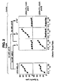
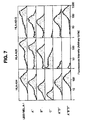
- a second CTL clone i.e., LB33-CTL-159/3 was isolated. These lines will be referred to as "159/5" and "159/3", respectively.
- This second CTL has specificity differing from 159/5. This was ascertained following isolation of two antigen loss variants which (i) are lysed by 159/5 but not 159/3 and (ii) are not lysed by 159/5 and are lysed by 159/3. These variants are referred to as A - and B - , respectively.
- a - variant was then immunoselected with 159/5, and a third variant was obtained, which was not lysed by either 159/5 or 159/3.
- This variant is referred to as A - B - .
- Figure 2 summarizes the results of the lysis assays, leading to isolation of the variants.
- TRIP tumor rejection antigen precursor
- mRNA was isolated from cell line LB33-MELc1.
- the messenger RNA was isolated using an oligo-dT binding kit, following well recognized techniques. Once the messenger RNA was secured, it was transcribed into cDNA, again using standard methodologies.
- the cDNA was then ligated to EcoRI adaptors and cloned into the EcoRI site of plasmid pcDNA-I/Amp, in accordance with manufacturer's instructions.
- the recombinant plasmids were then electrophorated into DH5 ⁇ E . coli (electroporation conditions: 1 pulse at 25 ⁇ farads, 2500 V).
- the transfected bacteria were selected with ampicillin (50 ⁇ g/ml), and then divided into pools of 100 bacteria each. Each pool represented about 50 different cDNAs, as analysis showed that about 50% of plasmids contained an insert. Each pool was amplified to saturation, and plasmid DNA was isolated via alkaline lysis, potassium acetate precipitation and phenol extraction, following Maniatis et al., in Molecular Cloning: A Laboratory Manual (Cold Spring Harbor, N.Y., 1982). Cesium gradient centrifugation was not used.
- the amplified plasmids were then transfected into eukaryotic cells.
- Samples of COS-7 cells were seeded, at 15,000 cells/well into tissue culture flat bottom microwells, in Dulbeco's modified Eagles Medium ("DMEM") supplemented with 10% fetal calf serum.
- DMEM Dulbeco's modified Eagles Medium
- the cells were incubated overnight at 37°C, medium was removed and then replaced by 30 ⁇ l/well of DMEM medium containing 10% Nu serum, 400 ⁇ g/ml DEAE-dextran, 100 ⁇ M chloroquine, and 100 ng of a plasmid containing cDNA for HLA-B44 from LB33.
- the medium was removed, and replaced by 50 ⁇ l of PBS containing 10% DMSO. This medium was removed after two minutes and replaced by 200 ⁇ l of DMEM supplemented with 10% of FCS.
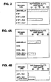
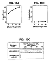
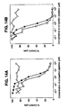
- Plasmid DNA was extracted from the bacteria cloned in Example 4, transfected into a new sample of COS cells in the same manner as described supra , and the cells were again tested for stimulation of 159/5. A positive clone was found in clone 350/2, as demonstrated by data summarized in figure 4A.
- the human choriocarcinoma cell line JAR which is readily available from the American Type Culture Collection, was used. This cell line does not express HLA molecules, nor is it recognized by CTL 159/5. When JAR was transfected with HLA-B44 cDNA, it was still not recognized by CTL 159/5. Co-transfection with HLA-B44 and 350/2 cDNAs, however, led to lysis, as is seen in figure 4B.
- the plasmid from the positive clone was removed, and sequenced following art known techniques. Information shows that the plasmid insert was 1896 base pairs long, and showed no homology with any sequences in data banks.
- the nucleotide sequence is set forth herein as SEQ ID MO: 1.
- Examples 1-6 describe work using the cell line LB33-Melcl. Additional cell lines were also derived from a cutaneous metastasis from patient LB33. One such line is LB33-MEL.A-1, which is used in the example which follows.
- the cell line was used, in the same manner that the cell line of examples 1-6 was used (Herin et al., supra ).
- Blood mononuclear cells (10 6 /well) were stimulated with irradiated tumor cells (3/10 5 cells/well), in 2 ml of Iscove's medium, supplemented with 10% pooled human serum, asparagine-glutamine-arginine (36 mg/ml, 216 mg/ml, 116 mg/ml, respectively), 2-mercaptoethanol (0.05 mM), and 5 U/ml of human IL-4.
- IL-2 (10 U/ml) was added on the third day of cultivation. Sensitivity of the tumor cells to autologous CTLs was determined as in example 1, supra.
- the experiment yielded 82 stable cytolytic T lymphocytes, derived from seven independent cultures. All of these CTLs were CD8 + . They were specific for tumor cells in that they lysed LB33-MEL.A-1 cells, but not K562, or autologous, EBV transformed cells.
- samples of the cell line were selected, four times, with the autologous CTL clone LB33-CTL 159/3, described supra.
- Each round of selection involved incubating, for 2-6 hours, 2-3x10 7 adherent tumor cells with a similar number of CTLs, in the same manner described supra.
- CTLs were washed away following the incubation, and the surviving adherent tumor cells were amplified prior to the next round of selection.
- LB33-MEL.A-1 is considered "A + B + C + D + " for antigen expression (lysed by all of CTL 159/3, 159/5, 204/26, and 202/1);
- MEL.A-1.1 is A - B + C + D + (not lysed by 159/3, lysed by others);
- MEL.A-1.2 is A + B - C + D + (not lysed by 159/5; lysed by others),
- MEL.A-1.3 is A + B + C - D + (not lysed by 204/26; lysed by others), and
- MEL.A-1.4 is A - B + C + D - (not lysed by 202/1 or 159/3).
- cell line MEL.A-1.1.1 was isolated, which was A - B - C + D - (lysed only by 204/26).
- the patient from whom the LB33 cell lines had been developed had been serologically typed, previously, as HLA-A24, A28, B13, B44, Cw6, Cw7. Studies were then carried out to determine the expression of HLA class I genes by the cell lines.
- a "+++” indicates expression corresponding to more than half that of the LB33-MEL.A-1 cells, "++” means that expression was between 1/8 and 1/2 of that of LB33-MEL.A-1, a “+” means that expression was less than 1/8 of that of LB33-MEL.A-1 expressed and "-" means there was no expression.
- LB33-MEL.A-1 LB33-MEL.A tumor cells
- both MEL.A-1 cells, and B - variant expressed similar levels of all six HLA alleles.
- the A - variant showed an approximately 4-fold decrease in expression of Cw7.
- the remaining antigen loss variants showed decreases in expression of sets of three alleles.
- reduced levels of expression for HLA-A24, B13, and Cw6 were found, while A - D - , and A - B - D - variants showed reduction in A28, B44, and Cw7 expression.
- A24-B13-Cw6, and A28-B44-Cw7 constitute two HLA class I haplotypes of patient LB33, and that reduced expression of these haplotype probably accounted for loss of antigen expression by the immunoselected tumor cells.
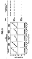
- Table 2 summarizes the results, which are also shown in figure 7.
- the indicated level of HLA expression corresponds to the mean intensity of fluorescence shown in figure 6. Values are expressed relative to levels found in LB33-MEL.A-1 cells.
- Figure 8 depicts these results. Expression of antigen B was restored in A - B - D - cells by transfection with a plasmid carrying HLA-B44, but not with plasmids containing HLA-A28 or HLA-Cw7. The expression of antigen C was restored in C - cells by transfection with HLA B13. Four other anti-C CTL clones also recognized C - cells, but five other anti-C CTL clones, including depicted CTL 179C/50, did not; rather, these CTLs recognized C - cells transfected with HLA-Cw6. Thus, it may be concluded that there are two groups of anti-C CTL clones.
- antigen D A - D - cells were restored to A - D + via transfection with HLA-A28. None of the cDNA restored expression of antigen A (i.e., tested HLA A28, B44, Cw7), although it clearly is presented by HLA-class I molecules, because lysis by anti-A CTLs is completely inhibited by anti-class I monoclonal antibody W6/32.
- this antigen may be presented by a non-A, B, C class I molecule, of which two alleles were present in patient LB33, one of these being lost, together with the A28-B44-Cw7 haplotype in A - D - , A - B - D - cells.
- antigen C has led to a change in nomenclature.
- antigens There are two antigens referred to as antigen, Ca and antigen Cb, hereafter.
- Irradiated LB33-MEL.B.1 cells were used in the same manner as was used, supra (Herin, et al), to stimulate autologous lymphocytes.
- the lymphocytes had been taken from patient LB33 in 1990 or 1994.
- the MAGE-3 TRAP amino acid sequence contains a stretch of amino acids at position 167-176, which corresponds to this motif.
- the amino acid sequence is:
- the HLA-B44 motif is known to contain at least two major subtypes, referred to as HLA-B* 4402 and HLA-B* 4403.
- the MHC molecule appears on 23% of all caucasians. When this figure is combined with standard analyses of melanoma, it is concluded that 15% of caucasian melanoma patients should present HLA-B44 on the surface of their melanoma cells. Thus, it is of great interest to determine if the peptide of SEQ ID NO: 17 or related molecules can in fact be used to identify HLA-B44 cells, and to provoke their lysis following binding to the MHC molecule. As noted in prior examples, the peptide of SEQ ID NO: 2 was shown to bind to HLA-B44 positive cells.
- a peptide was designed which was similar to SEQ ID NO: 2, except for having Ala at position 8, rather than Leu.
- This new peptide i.e.: was tested in a competition assay with SEQ ID NO: 2.
- This peptide was used in view of result obtained in experiments not reported here. Briefly, derivatives of SEQ ID NO: 2 were prepared, wherein each derivative contained an Ala at a position not occupied by Ala in SEQ ID NO: 2.
- CTL clone 159/5 was slightly better at recognizing complexes containing SEQ ID NO: 18 than SEQ ID NO: 2, making it an excellent reagent for competitive assays. Competition was carried out using C1R cells, described by Storkus et al., J.
- C1R cells are MHC class I negative, lymphoblastoid cells.
- the C1R cells were transfected with a cDNA for HLA-B*4402, or with a genomic DNA for HLA-B*4403, using the same methodology given supra .
- the cDNA for HLA-B*4402 is set forth by Fleischhauer, et al, Tissue Antigens 44: 311-317 (1994), while the genomic DNA for HLA-B*4403 is given by Fleischhauer, et al. (1990) New Eng. J. Med 323:1818-1822 (1990). Both papers are incorporated by reference.
- the cells were labelled with 51 Cr for one hour at 37°C, in the presence of anti-HLA class I monoclonal antibody W6/32 (30% (v/v) of culture medium of the hybridoma cells). This increases the ability of the cells to present antigenic peptides to T cells.
- the peptide of SEQ ID NO: 18 was then added in the serum free culture medium at a final concentration of 45 ng/ml, (C1R-B4402 + cells), or 160 ng/ml (C1R-B4403 + cells).
- the cells were incubated for 30 minutes at 20°C, and washed twice in Iscove's medium plus 2% fetal calf serum.
- the CTL clone LB33-CTL 159/5 was added in Iscove's medium and 10% human serum, at an E:T ratio of 20.
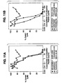
- the release of 51 Cr was measured after three hours, and is shown in figures 11A and 11B, for C1R-B*4402 and C1R-B*4403 cells. The data presented in figure 11, show clear evidence of competition.
- Cytolytic T cell clones were derived from two subjects, referred to as LB 816 and LB 822, respectively. These subjects showed no evidence of cancer.
- BMCs Blood mononuclear cells
- T lymphocytes in the BMCs were purified by rosetting, using sheep red blood cells which had been treated with aminoethylisothiouronium bromide, and then labelled with an anti-CD8 monoclonal antibody coupled to magnetic microbeads.
- the CD8 + cells were sorted by passage through a magnetized area, and then stored at -80°C in Iscove's culture medium, supplemented with 10% human serum, 116 mg/l L-arginine, 36 mg/ml L-asparagine, and 216 mg/l of L-glutamine, 0.05mM 2-mercaptoethanol, and 10% DMSO.
- Any non-rosetting BMC were left to adhere for two hours at 37°C on tissue culture plates. Non-adherent cells were discarded, and adherent cells cultured for seven days in the presence of IL-4 (50 U/ml), and GM-CSF (100 ng/ml). The resulting population was enriched for antigen presenting cells ("APCs"; in this case, dendritic cells or macrophages). Then, from 5x10 5 to 10 6 of these cells were incubated in 2 ml wells for four hours, at 37°C, in 400 ul Iscove's medium supplemented with 2.5 ug/ml of human ⁇ 2 microglobulin, and 50 ug/ml of the peptide of SEQ ID NO: 17.
- APCs antigen presenting cells
- Adherent, peptide pulsed cells were then irradiated at 5000 rads, and washed.
- 2x10 6 autologous CD8 + T cells were added, in culture medium, supplemented with 1000 U/ml of IL-6, and 5 ng/ml of IL-12.
- lymphocytes were restimulated with adherent, autologous BMCs, pulsed with peptide as above.
- 5x10 6 BMCs were left to adhere for two hours at 37°C, in 400 ul Iscove's medium containing ⁇ 2-microglobulin and SEQ ID NO: 17, as discussed above. Any peptide pulsed, adherent cells, were irradiated and washed.
- Responder cells were then added, in culture medium supplemented with 10 U/ml of IL-2, and 5 ng/ml of IL-7.
- the lymphocytes were restimulated with autologous BMCs pulsed with SEQ ID NO: 17.
- the BMCs were incubated, at 2x10 7 cells/ml, in the Iscove's medium containing B 2 -microglobulin and SEQ ID NO: 17. After two hours of incubation (20°C), peptide pulsed BMCs were irradiated, washed, and resuspended at 2x10 6 cells/ml in culture medium augmented with IL-2 and IL-7, as above. Samples of these stimulator cells (2X10 6 ), were added to each well which contained responder cells.
- the responder lymphocytes were cloned on day 21. Anywhere from 10 to 0.3 cells/well were seeded in microwells, in culture medium which had been supplemented with 50 U/ml of IL-2, and 5 U/ml of IL-4. These were then stimulated by adding allogenic EBV transformed B cells (LG2-EBV-B) and irradiated at 10,000 rads, at 20,000 cells per well, and one of (i) peptide pulsed HLA-B4402 + cells, or (ii) peptide pulsed HLA-B4403 + cells. For (i) or (ii), irradiation was at 15,000 rads, at 8000 cells per well.
- LG2-EBV-B allogenic EBV transformed B cells
- Microcultures were restimulated every week in the same way they were on the 21st day.
- the one change was that at days 28 and 35, 40,000 and 60,000 EBV-B cells respectively were added per well, as compared to 20,000 at day 21.
- Any microcultures which showed anti-peptide lytic activity were restimulated with 5x10 4 irradiated, peptide-pulsed B4402 + or B4403 + cells, plus 5x10 5 irradiated LG2-EBV-B cells, in 800 ul of culture medium augmented with 50 U/ml of IL-2, and 5 U/ml of IL-4.
- CTLs LB 816-CTL-340 A/l, and LB822-CTL-346A/1 were obtained. These CTLs are specific for complexes of SEQ ID NO: 17 and either HLA-B*4402, or HLA-B4403, respectively.
- HLA-B4402 + or HLA-B4403 + EBV-transformed B cells which do not express MAGE-3 were labelled with 51 Cr in the presence of monoclonal antibody W6/32, for 1 hour, at 37°C, Brodsky, et al, J. Immunol 128:129-135 (1982). The cells were washed, and incubated for 30 minutes at 20°C in serum free medium, using varied concentrations of SEQ ID NO: 17. Each CTL described in Example 14 was tested in a 51 Cr release assay, also as described, with chromium release being measured after four hours.
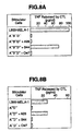
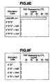
- COS-7 cells were transfected by cDNA encoding MAGE-3 following Gaugler, et al, J. Exp. Med 179:921-930 (1994), in the expression vector pcDNA-1/AMP, and one of HLA-B*4402 cDNA cloned into vector pcDNA3, or HLA-B*4403 cDMA cloned into pcDNAl/AMP.
- Transfectants were incubated for 24 hours, at 37°C, then 3000 CTLs/well were added. Materials were incubated for 18 hours, at 37°C. Supernatants were then collected, and TNF content was determined by testing the cytolytic effect on TNF sensitive WEHI-16 clone 13 cells, following Espevik et al, J. Immunol. Meth 95:99-105 (1986).
- Table 2 which follows, shows the results, wherein TNF release is expressed in pg/ml.
- LB33-MEL and LB494-MEL were incubated with IFN ⁇ at 100 U/ml for 24 hours prior to the assay.
- Tumor cell lines LB33-MEL, LB494-MEL, and M22-MEL were also tested. These cell lines all express MAGE-3 cDNA, and are either HLA-B*4402 + (LB33-MEL, LB494-MEL), or HLA-B*4403 + (MZ2-MEL). Hence, no transfection was necessary for these cells. The results show that TNF was released.
- the cells used in the experiments were C1R cells which had been transformed with Epstein Barr Virus (EBV). These cells differ from other, EBV transformed B cells in that they express the MAGE-3 gene. They are also MHC negative, and hence can serve as subjects for transfection with genes encoding MHC molecules.
- Samples were transfected with either of a cDNA molecule encoding HLA-B*4402, or with genomic DNA encoding HLA-B*4403 in accordance with Fleischhauer, et al., Tissue Antigens 44: 311-317 (1994) or Fleischhauer, et al., N. Eng. J. Med 323: 1828-1822 (1990) both of which are incorporated by reference.
- the transfectants were grown in RPMI-1640 medium which had been supplemented with 10% fetal calf serum (FCS).
- the transfected C1R (C1R-B*4402 as C1R-B*4403) transformed cells were 51 Cr labelled for one hour, at 37°C, in the presence of anti-human class-I MHC monoclonal antibody W6/32 (30% w/v of culture medium of the hybridoma cells), and washed three times. Labelled cells (1000 cells in 80 ul), were incubated in V bottom microwells for 30 minutes at 20°C, in serum free, X-VIVO 10 medium.
- Test peptides were added, at varying concentrations, as explained infra, followed by the addition of the peptide at 50 ng/ml in 40 ul of X-VIVO 10 medium for C1R-B*4402, and 160 ng/ml for C1R-B*4403.
- This peptide is known to bind to both HLA-B*4402 and HLA-B*4403 allotypes. (See, e.g., Coulie, et al., Proc. Natl. Acad. Sci. USA 92: 7976-7980 (1995)).
- CTL clone LB33-159/5 described by Coulie et al as recognizing and lysing cells which present complexes of SEQ ID NO: 18 and HLA-B44 on their surface, were added at 20,000 cells in 150 ul of Iscove's medium supplemented with 10% human serum.
- the competitive peptides were added at varying concentrations, until lysis by CTL 159/5 was inhibited by 50%. Lysis was determined using the formula described supra. When no competitor peptide was used, lysis of C1R-B*4402 transformants was 58%, and that of C1R-B*4403 was 72%.
- SEQ ID NOS: 3 and 19 were used as controls.
- the former i.e.: is derived from tyrosinase and is known to bind to both HLA-B*4402 and HLA-B*4403 (Brichard, et al., Eur. J. Immunol. (1995), while the latter, i.e. binds to HLA-B8 (Burrows, et al., J. Exp. Med. 171: 345-349 (1990)).
- the results, presented below set forth the concentration, in uM, of peptide needed to inhibit lysis of C1R-B44 cells sensitized with SEQ ID NO: 8.
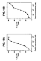
- SEQ ID NOS: 17, 20, 21 and 22 did inhibit lysis of OR-844 cells, which indicates that they bind to HLA-B44 molecules.
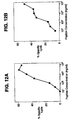
- the peptide of SEQ ID NO: 17 was the best binder for both HLA-B*B4402 and HLA-B*4403. This is shown in figure 14, wherein SEQ ID NOS: 17, 19 and 3 and are plotted as a function of lysis versus concentration of competitor peptides.
- adherent peripheral blood mononuclear cells were isolated from a donor, referred to as LB816, who did not suffer from cancer, and who was HLA-B*4402 positive.
- the adherent cells were isolated by first securing a sample of peripheral blood mononuclear cells ("PBMCs" hereafter), using density gradient centrifugation. T-lymphocytes were purified from the sample via rosetting, using 2-aminoethyl-isothiouronium bromide hydrobromide-treated sheep erythrocytes, in accordance with Mikamo, J. Immunol. Meth. 107: 189-196 (1988), incorporated by reference, and via labelling with anti-CD8 monoclonal antibodies coupled to magnetic microbeads, followed by sorting of the CD8 + cells via passage in a magnetic field. The thus separated cells were frozen for storage. Any non-rosetting PBMCs were left to adhere for two hours at 37°C on NUNC tissue culture wells, with non-adherent cells being discarded.
- PBMCs peripheral blood mononuclear cells
- the adherent cells were then cultured, for seven days, in the presence of IL-4 (50 U/ml), and GM-CSF (100 ng/ml), in RPMI medium supplemented with 10% FCS.
- GM-CSF and IL-4 are used in order to increase the proportion of dendritic cells in the culture. See Romani, et al., J. Exp. Med. 180: 83-93 (1994); Sallusto, et al., J. Exp. Med. 179: 1109-1118 (1994).
- those taken from donor LB816 were found, for the most part, to be CD11c + and CD14 +
- cells from donor LB822 were mostly CD11c + and CD14 - .
- a total of from 5x10 5 to 5x10 6 of these antigen presenting cells were then incubated in 2 ml wells, for four hours at 37°C, in 400 ul of Iscove's medium supplemented with human B2 microglobulin (2.5 ug/ml), and 50 ug/ml of SEQ ID NO: 17. Following this, the cells were irradiated at 50 Gy, and washed. These cells were then used as stimulator cells on the autologous CD8 + cells discussed supra.
- CD8 + wells Six cultures of autologous CD8 + wells were established. For each culture, 2x10 6 CD8 + cells were combined with culture medium (Iscove's medium plus 10% human serum, L-arginine, L-asparagine, L-glutamine, 0.05 mM 2-mercaptoethanol, 1000 U/ml IL-6 and 5 ng/ml IL-12). On days 7 and 14, these responder cells wee stimulated with the stimulator cells. To do this, on day 7 the PBMCs (5x10 6 ) were left to adhere for 2 hours, at 37°C in 400 ul of Iscove's medium containing ⁇ 2 microglobulin, and the peptide of SEQ ID NO: 17, as described supra.
- culture medium Iscove's medium plus 10% human serum, L-arginine, L-asparagine, L-glutamine, 0.05 mM 2-mercaptoethanol, 1000 U/ml IL-6 and 5 ng/ml IL-12.
- the adherent cells were irradiated and washed, also as described supra .
- the responder lymphocytes i.e., the CD8 + cells
- culture medium which had been augmented with 10 U/ml of IL-2 and 5 ng/ml of IL-7 were added.
- the lymphocytes were restimulated with PBMCs which had been pulsed with peptide.
- 2x10 7 PBMCs per ml were incubated for two hours at 20°C in Iscove's medium with ⁇ 2 microglobulin, and the peptide of SEQ ID NO: 17, as described supra.
- these PBMCs were irradiated, washed, and resuspended at 2x10 6 cells/ml in culture medium supplemented with IL-2 and IL-7, and, again as described, were added to the responders.
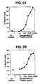
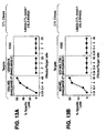
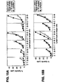
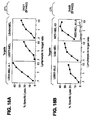
- lytic activity was assessed on LB816 cells (HLA-B*4402 positive cells), which had been sensitized with SEQ ID NO: 17.
- the lytic activity was measured using the 51 Cr release assay as described supra. The results are shown in figure 15A. Effector cells were incubated with unlabelled K562 target cells (50,000 cells/well) for 45 minutes, in order to inhibit lysis by NK-like effectors. 51 Cr-labelled target cells (1000 cells/well) were incubated with 1 ⁇ M of SEQ ID No: 17 (filled in circle) or without it (open circle). 51 Cr release was measured after 4 hours. Only one of the six autologous CD8 + culture preferentially lysed the target cells.
- HLA-B*4403 positive cells (LB822) were tested. Results, presented in figure 15B, show that one out of five cultures tested preferentially lysed the cells.
- the lymphocytes of the positive cultures were cloned by limiting dilution. Specifically, on day 21, these cells were cultured in culture medium supplemented with 50 U/ml of IL-2, and 5 U/ml of IL-4.
- the LB816 cells were stimulated with irradiated C1R-B*4402 cells (150 Gy, 8000 cells/wells), which had been incubated for one hour at 20°C with 1 ug/ml of SEQ ID NO: 17, and washed.
- the LB822 clones were stimulated with irradiated M22-MEL cells which are HLA-B*4403 positive melanoma cell (100 Gy, 8000 cells per well).
- LG2-EBV cells 100 Gy, 20,000 cells/well
- Irradiated allogeneic LG2-EBV cells 100 Gy, 20,000 cells/well
- Microcultures were stimulated each week.
- CTL clones LB816-CTL-340/1 and LB822-CTL-346/8 were isolated in this fashion.
- CTL clones discussed supra were stimulated each week in 2 ml wells with 2x10 5 irradiated C1R-B*4402 or C1R-B*4403 cells, which had been incubated for one hour at 20°C with 1 ug/ml of the peptide of SEQ ID NO: 17, then washed, and with 10 6 irradiated LG2-EBV cells, all in culture medium supplemented with IL-2 and IL-4, as before.
- COS-7 cells (15,000 per well) were cotransfected with 50 ng of pcDNAI/Amp containing cDNA for MAGE-3, and either 50 ng of pcDNAI/Amp containing cDNA for HLA-B*4403, or 50 ng of pcDNA3/Amp containing cDNA for HLA-B*4402.
- the cotransfection was carried out in accordance with the well known protocol of Seed, et al., Proc. Natl. Acad. Sci. USA 84: 3365-3369 (1987).
- NA8-MEL melanoma cells were cotransfected. These cells do not express either of HLA-B44 or MAGE-3 molecules.
- TNF production was determined following Lehmann et al., Eur. J. Immunol. 25: 340-347 (1995), incorporated by reference, but discussed briefly here.
- 3000 CTLs were added in 100 ul of Iscove's medium supplemented with 10% human serum, and 25 U/ml of IL-2. After 20 hours, supernatant was collected and TNF content determined by testing its cytotoxicity on WEHI 164c13 cells, in accordance with Espevik et al., J. Immunol. Meth. 95: 99-105 (1986).
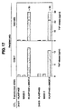
- controls were prepared using single transfections (either MAGE-3 or HLA-B*4402 or HLA-B*4403).
- Figure 17 shows that cells cotransfected with the two constructs stimulated TNF release, while cells transfected with one plasmid did not. This result was observed with both cell types, and both CTLs.
- the recognition of MAGE-3/HLA-B44 complexes did not require the high copy number provided by COS-7 cells, as the NA8-MEL work shows.
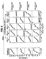
- the CTLs described supra i.e., 340/1 and 346/8, were then tested for their ability to lyse HLA-B44 presenting tumor cells.
- the cells used included lines known to express MAGE-3, including LB33-MEL-A-1, LB373-MEL, LB494-MEL, LB831-BLC, LG2-MEL and MZ2-MEL.43, which are melanoma cells except for LB831-BLC, which is a bladder cancer derived cell line.
- Cell line MZ2-MEL.61.2 is a melanoma cell line which has lost expression of MAGE-3. In figure 18, which is discussed infra, this cell is depicted by open (“O") circles, rather than closed (“O") circles.
- the tumor rejection antigens of the invention are isolated nonapeptides which have a Glu residue at the 2nd position, and a Phe or Tyr residue at the 9th or 10th position, consisting of amino acid sequence: or amino acid sequence: wherein the amino acid sequence is selected from the group consisting of: and These correspond to peptide sequences which are homologous to SEQ ID NO: 17, in that they are found at the corresponding positions of MAGE-1, 2, 4, 5, 6 and 12, respectively. Their ability to bind to HLA-B44 molecules renders them useful, inter alia, in identifying cells which present HLA-B44 molecules in their surfaces.
- peptides of the invention are similar to the peptide disclosed in US Patent No. 5,519,117 so-assigned to the assignee of the subject application, i.e.: Khanna, et al., supra, teaches a decamer, i.e.: but does not discuss how modification of the decamer could lead to an effective nonamer.
- the invention thus involves tumor rejection antigens which bind to HLA-B44 molecules, and then provoke lysis by CTLs.
- the complexes of TRA and HLA molecule provoke a cytolytic T cell response
- isolated complexes of the tumor rejection antigen and an HLA-B44 molecule are also encompassed by the invention, as are isolated tumor rejection antigen precursors coded for by the previously described nucleic acid molecules.
- the peptides may also be used, simply to identify HLA-B44 positive cells.
- the identification of a tumor rejection antigen which is specifically presented by an HLA-B44 molecule, as well as a nucleic acid molecule coding for its parallel tumor rejection antigen precursor permits the artisan to diagnose a disorder characterized by expression of the TRAP.
- These methods involve determining expression of the TRAP gene, and/or TRAs derived therefrom, such as TRA presented by HLA molecules.
- Other TRAs may also be derived from the TRAPS of the invention and presented by different HLA molecules. In the former situation, such determinations can be carried out via any standard nucleic acid determination assay, including the polymerase chain reaction, or assaying with labelled hybridization probes. In the latter situation, assaying with binding partners for complexes of TRA and HLA, such as antibodies, is especially preferred.
- the isolation of the TRAP gene also makes it possible to isolate the TRAP molecule itself, especially TRAP molecules containing the amino acid sequence of SEQ ID NO: 1. Fragments of peptides of these isolated molecules when presented as the TRA, or as complexes of TRA and HLA-B44, may be combined with materials such as adjuvants to produce vaccines useful in treating disorders characterized by expression of the TRAP molecule.
- vaccines can be prepared from cells which present the TRA/HLA complexes on their surface, such as non-proliferative cancer cells, non-proliferative transfectants, etcetera.
- these can be cells transfected with coding sequences for one or both of the components necessary to prove a CTL response, or be cells which express both molecules without transfection.
- the TRAP molecule, its associated TRAs, as well as complexes of TRA and HLA may be used to produce antibodies, using standard techniques well known to the art.
- disorder refers to any pathological condition where the tumor rejection antigen precursor is expressed.
- An example of such a disorder is cancer, melanoma in particular.
- Therapeutic approaches based upon the disclosure are premised on a response by a subject's immune system, leading to lysis of TRA presenting cells, such as cells presenting the relevant HLA molecule.
- One such approach is the administration of CTLs specific to the complex to a subject with abnormal cells of the phenotype at issue, it is within the skill of the artisan to develop such CTLs in vitro.
- a sample of cells such as blood cells
- the target cell can be a transfectant, such as a COS cell of the type described supra.
- These transfectants present the desired complex on their surface and, when combined with a CTL of interest, stimulate its proliferation.
- COS cells such as those used herein are widely available, as are other suitable host cells.
- the foregoing therapy assumes that at least some of the subject's abnormal cells present the HLA/TRA complex. This can be determined very easily, as the art is very familiar with methods for identifying cells which present a particular HLA molecule, as well as how to identify cells expressing DNA containing the indicated sequences. Once isolated, such cells can be used with a sample of a subject's abnormal cells to determine lysis in vitro . If lysis is observed, then the use of specific CTLs in such a therapy may alleviate the condition associated with the abnormal cells. A less involved methodology examines the abnormal cells for HLA phenotyping, using standard assays, and determines expression via amplification using, e.g., PCR.
- Adoptive transfer is not the only form of therapy that is available in accordance with the invention.
- CTLs can also be provoked in vivo , using a number of approaches.
- One approach i.e., the use of non-proliferative cells expressing the complex, has been elaborated upon supra.
- the cells used in this approach may be those that normally express the complex, such as irradiated melanoma cells or cells transfected with one or both of the genes necessary for presentation of the complex. Chen et al., Proc. Natl. Acad. Sci. USA 88: 110-114 (January, 1991) exemplifies this approach, showing the use of transfected cells expressing HPVE7 peptides in a therapeutic regime.
- Various cell types may be used.
- vectors carrying one or both of the genes of interest may be used.
- Viral or bacterial vectors are especially preferred.
- the gene of interest is carried by, e.g., a Vaccinia virus or the bacteria BCG, and the materials de facto "infect" host cells.
- the cells which result present the complex of interest, and are recognized by autologous CTLs, which then proliferate.
- a similar effect can be achieved by combining the tumor rejection antigen or the precursor itself with an adjuvant to facilitate incorporation into cells which present the HLA molecule of interest.
- the TRAP is processed to yield the peptide partner of the HLA molecule while the TRA is presented without the need for further processing.
Landscapes
- Health & Medical Sciences (AREA)
- Life Sciences & Earth Sciences (AREA)
- Chemical & Material Sciences (AREA)
- Immunology (AREA)
- Engineering & Computer Science (AREA)
- Organic Chemistry (AREA)
- General Health & Medical Sciences (AREA)
- Zoology (AREA)
- Molecular Biology (AREA)
- Proteomics, Peptides & Aminoacids (AREA)
- Biochemistry (AREA)
- Genetics & Genomics (AREA)
- Biomedical Technology (AREA)
- Wood Science & Technology (AREA)
- Analytical Chemistry (AREA)
- Cell Biology (AREA)
- Biotechnology (AREA)
- Microbiology (AREA)
- Hematology (AREA)
- Biophysics (AREA)
- Medicinal Chemistry (AREA)
- Physics & Mathematics (AREA)
- Pathology (AREA)
- Bioinformatics & Cheminformatics (AREA)
- Urology & Nephrology (AREA)
- Epidemiology (AREA)
- Animal Behavior & Ethology (AREA)
- General Engineering & Computer Science (AREA)
- Public Health (AREA)
- Veterinary Medicine (AREA)
- Gastroenterology & Hepatology (AREA)
- General Physics & Mathematics (AREA)
- Toxicology (AREA)
- Food Science & Technology (AREA)
- Virology (AREA)
- Tropical Medicine & Parasitology (AREA)
- Oncology (AREA)
- Hospice & Palliative Care (AREA)
- Peptides Or Proteins (AREA)
- Medicines Containing Antibodies Or Antigens For Use As Internal Diagnostic Agents (AREA)
Claims (2)
- Isoliertes Peptid, das an HLA-B44 Moleküle anbindet und aus folgender Aminosäurensequenz besteht:oder der Aminosäurensequenz
 dadurch gekennzeichnet, dass die Aminosäurensequenz aus der Gruppe mit den SEQ ID. Nr.: 26, SEQ ID. Nr.: 27, SEQ ID Nr.: 28, SEQ ID Nr.: 29 und SEQ ID Nr.: 30 gewählt ist.
- Verfahren zur Identifizierung einer HLA-B44 positiven Zelle in einer Probe, gekennzeichnet durch Kontaktieren der Probe mit dem isolierten Peptid nach Anspruch 1 und Ermittlung einer HLA-B44 positiven Zelle in der Probe über die Bestimmung der Anbindung des Peptids.
Applications Claiming Priority (3)
| Application Number | Priority Date | Filing Date | Title |
|---|---|---|---|
| US602506 | 1990-10-24 | ||
| US08/602,506 US6060257A (en) | 1994-06-03 | 1996-02-20 | Tumor rejection antigens presented by HLA-B44 molecules, and uses thereof |
| PCT/US1997/001915 WO1997031017A1 (en) | 1996-02-20 | 1997-02-05 | Tumor rejection antigens presented by hla-b44 molecules, and uses thereof |
Publications (3)
| Publication Number | Publication Date |
|---|---|
| EP0876397A1 EP0876397A1 (de) | 1998-11-11 |
| EP0876397A4 EP0876397A4 (de) | 1999-08-18 |
| EP0876397B1 true EP0876397B1 (de) | 2003-05-02 |
Family
ID=24411635
Family Applications (1)
| Application Number | Title | Priority Date | Filing Date |
|---|---|---|---|
| EP97906894A Expired - Lifetime EP0876397B1 (de) | 1996-02-20 | 1997-02-05 | Durch hla-b44 moleküle präsentierte tumor-abstossungsantigene und ihre verwendungen |
Country Status (12)
| Country | Link |
|---|---|
| US (2) | US6060257A (de) |
| EP (1) | EP0876397B1 (de) |
| JP (1) | JP3256749B2 (de) |
| KR (1) | KR19990087064A (de) |
| CN (1) | CN1214692A (de) |
| AT (1) | ATE239035T1 (de) |
| AU (1) | AU711912B2 (de) |
| CA (1) | CA2246222C (de) |
| DE (1) | DE69721485T2 (de) |
| NZ (1) | NZ331398A (de) |
| WO (1) | WO1997031017A1 (de) |
| ZA (1) | ZA971403B (de) |
Families Citing this family (11)
| Publication number | Priority date | Publication date | Assignee | Title |
|---|---|---|---|---|
| US5977300A (en) * | 1994-06-03 | 1999-11-02 | Ludwig Institute Of Cancer Research | Isolated nonapeptide which bind to HLA-B44 molecules and the uses thereof |
| DE69534683T2 (de) | 1994-12-29 | 2006-07-06 | Canon K.K. | Tintenstrahlkopf mit verschiedenen Heizelementen pro Düse und Tintenstrahldrucker unter Verwendung desselben |
| US6503703B1 (en) | 1995-05-19 | 2003-01-07 | Mount Sinai School Of Medicine Of New York University | Identification and use of antiviral compounds that inhibit interaction of host cell proteins and viral proteins required for viral replication |
| CA2274737A1 (en) * | 1996-12-12 | 1998-06-18 | Visible Genetics, Inc. | Method and kit for hla class i typing |
| CN1402782A (zh) * | 1999-10-19 | 2003-03-12 | 路德维哥癌症研究院 | Mage-a12抗原肽及其应用 |
| US6897288B1 (en) | 1999-10-19 | 2005-05-24 | Ludwig Institute For Cancer Research | Mage-A12 antigenic peptides and uses thereof |
| CA2388337C (en) | 1999-10-22 | 2013-01-08 | Aventis Pasteur Limited | Method of inducing and/or enhancing an immune response to tumor antigens |
| WO2001053833A1 (en) * | 2000-01-20 | 2001-07-26 | Ludwig Institute For Cancer Research | Mage antigenic peptides which bind hla-b35 and hla-b44 |
| WO2001074847A2 (en) * | 2000-03-30 | 2001-10-11 | The Government Of The United States Of America Represented By The Secretary, Department Of Health And Human Services | T-cell epitope of mage-12 and related nucleic acids, vectors, cells, compositions and methods of inducing an immune response to cancer |
| DK1282702T3 (da) | 2000-05-10 | 2007-04-02 | Sanofi Pasteur Ltd | Immunogene polypeptider, som er kodet af KAGE-minigener, og anvendelser deraf |
| US7740871B2 (en) * | 2002-09-19 | 2010-06-22 | Johns Hopkins University School Of Medicine | Cancer immunotherapy with a viral antigen-defined, immunomodulator-secreting cell vaccine |
Family Cites Families (7)
| Publication number | Priority date | Publication date | Assignee | Title |
|---|---|---|---|---|
| US5541104A (en) * | 1991-05-23 | 1996-07-30 | Ludwig Institute For Cancer Research | Monoclonal antibodies which bind to tumor rejection antigen precursor mage-1 |
| US5342774A (en) * | 1991-05-23 | 1994-08-30 | Ludwig Institute For Cancer Research | Nucleotide sequence encoding the tumor rejection antigen precursor, MAGE-1 |
| US6235525B1 (en) * | 1991-05-23 | 2001-05-22 | Ludwig Institute For Cancer Research | Isolated nucleic acid molecules coding for tumor rejection antigen precursor MAGE-3 and uses thereof |
| JP3608788B2 (ja) * | 1992-08-31 | 2005-01-12 | ルドヴィグ・インスティテュート・フォー・キャンサー・リサーチ | Mage−3遺伝子から誘導されてhla−a1により提示される単離されたノナペプチドおよびそれらの用途 |
| US5405940A (en) * | 1992-08-31 | 1995-04-11 | Ludwig Institute For Cancer Research | Isolated nonapeptides derived from MAGE genes and uses thereof |
| US5558995A (en) * | 1993-01-22 | 1996-09-24 | Ludwig Institute For Cancer Research | Peptides which are derived from tumor rejection antigen precursor molecule MAGE-1, which complex to MHC molecule HLA-C clone 10, and uses thereof |
| US5977300A (en) * | 1994-06-03 | 1999-11-02 | Ludwig Institute Of Cancer Research | Isolated nonapeptide which bind to HLA-B44 molecules and the uses thereof |
-
1996
- 1996-02-20 US US08/602,506 patent/US6060257A/en not_active Expired - Lifetime
-
1997
- 1997-02-05 EP EP97906894A patent/EP0876397B1/de not_active Expired - Lifetime
- 1997-02-05 NZ NZ331398A patent/NZ331398A/xx unknown
- 1997-02-05 AT AT97906894T patent/ATE239035T1/de not_active IP Right Cessation
- 1997-02-05 JP JP53018597A patent/JP3256749B2/ja not_active Expired - Fee Related
- 1997-02-05 DE DE69721485T patent/DE69721485T2/de not_active Expired - Fee Related
- 1997-02-05 CN CN97193299A patent/CN1214692A/zh active Pending
- 1997-02-05 KR KR1019980706450A patent/KR19990087064A/ko not_active Withdrawn
- 1997-02-05 WO PCT/US1997/001915 patent/WO1997031017A1/en not_active Ceased
- 1997-02-05 AU AU22614/97A patent/AU711912B2/en not_active Ceased
- 1997-02-05 CA CA002246222A patent/CA2246222C/en not_active Expired - Fee Related
- 1997-02-06 US US08/796,883 patent/US5744353A/en not_active Expired - Lifetime
- 1997-02-19 ZA ZA9701403A patent/ZA971403B/xx unknown
Also Published As
| Publication number | Publication date |
|---|---|
| CA2246222A1 (en) | 1997-08-28 |
| AU2261497A (en) | 1997-09-10 |
| ATE239035T1 (de) | 2003-05-15 |
| US5744353A (en) | 1998-04-28 |
| US6060257A (en) | 2000-05-09 |
| ZA971403B (en) | 1997-10-10 |
| AU711912B2 (en) | 1999-10-21 |
| KR19990087064A (ko) | 1999-12-15 |
| EP0876397A4 (de) | 1999-08-18 |
| CA2246222C (en) | 2002-01-08 |
| JPH11512622A (ja) | 1999-11-02 |
| DE69721485D1 (de) | 2003-06-05 |
| JP3256749B2 (ja) | 2002-02-12 |
| DE69721485T2 (de) | 2003-11-27 |
| EP0876397A1 (de) | 1998-11-11 |
| WO1997031017A1 (en) | 1997-08-28 |
| NZ331398A (en) | 1999-03-29 |
| CN1214692A (zh) | 1999-04-21 |
Similar Documents
| Publication | Publication Date | Title |
|---|---|---|
| EP0711173B1 (de) | Isolierte peptide, die mit mhc mulekül hla-c-klon 10 komplexe bilden und ihre verwendungen | |
| EP0690675B1 (de) | Nukleinsäure, die für einen tumor abstossungsantigenvorläufer kodiert | |
| EP0711355B1 (de) | Verfahren zur diagnose von störungen durch bestimmung der expression von gage-tumorabstossungsantigen-vorläufern | |
| EP0876397B1 (de) | Durch hla-b44 moleküle präsentierte tumor-abstossungsantigene und ihre verwendungen | |
| US5846826A (en) | Isolated cytolytic T cell line specific to complexes of HLA-B44 molecules and specific nonapeptides | |
| AU680236B2 (en) | Method for identifying and treating individuals bearing cancer cells that express HLA-C-Clone 10/MAGE-1 | |
| US5997870A (en) | Isolated peptides which bind to HLA-B44 Molecules | |
| EP0789591B1 (de) | Methoden zur identifizierung von individuen, die an einer zellulären abnormalität leiden | |
| EP0854727B1 (de) | Durch hla-b44 moleküle präsentierte tumor-abstossungsantigene und ihre verwendung | |
| WO1997010837A9 (en) | Tumor rejection antigens presented by hla-b44 molecules, and uses thereof | |
| WO2000050589A1 (en) | TYROSINE KINASE RECEPTOR EphA3 ANTIGENIC PEPTIDES | |
| CA2165435C (en) | Isolated peptides which form complexes with mhc molecule hla-c-clone 10 and uses thereof | |
| US6680056B1 (en) | Method for identifying individuals suffering from a cellular abnormality some of whose abnormal cells present complexes of hla-cw 1601/mage-1 derived peptides, and methods for treating said individuals | |
| CA2213001C (en) | Isolated nucleic acid molecule encoding peptides which form complexes with mhc molecule hla-cw*1601 and uses thereof |
Legal Events
| Date | Code | Title | Description |
|---|---|---|---|
| PUAI | Public reference made under article 153(3) epc to a published international application that has entered the european phase |
Free format text: ORIGINAL CODE: 0009012 |
|
| 17P | Request for examination filed |
Effective date: 19980813 |
|
| AK | Designated contracting states |
Kind code of ref document: A1 Designated state(s): AT BE CH DE DK ES FI FR GB GR IE IT LI LU MC NL PT SE |
|
| RAP1 | Party data changed (applicant data changed or rights of an application transferred) |
Owner name: LUDWIG INSTITUTE FOR CANCER RESEARCH |
|
| RIN1 | Information on inventor provided before grant (corrected) |
Inventor name: LUESCHER, IMMANUEL, LUDWIG INS.FOR CANCER RESEARC Inventor name: VAN DER BRUGGEN, PIERRE Inventor name: BOON-FALLEUR, THIERRY Inventor name: COULIE, PIERRE Inventor name: HERMAN, JEAN |
|
| A4 | Supplementary search report drawn up and despatched |
Effective date: 19990707 |
|
| AK | Designated contracting states |
Kind code of ref document: A4 Designated state(s): AT BE CH DE DK ES FI FR GB GR IE IT LI LU MC NL PT SE |
|
| RIC1 | Information provided on ipc code assigned before grant |
Free format text: 6C 07K 7/06 A, 6C 12N 5/06 B, 6C 12N 5/08 B, 6G 01N 33/574 B, 6C 07K 14/47 B, 6C 07K 14/705 B, 6G 01N 33/569 B, 6A 61K 35/14 B |
|
| 17Q | First examination report despatched |
Effective date: 20011022 |
|
| GRAH | Despatch of communication of intention to grant a patent |
Free format text: ORIGINAL CODE: EPIDOS IGRA |
|
| GRAH | Despatch of communication of intention to grant a patent |
Free format text: ORIGINAL CODE: EPIDOS IGRA |
|
| GRAA | (expected) grant |
Free format text: ORIGINAL CODE: 0009210 |
|
| AK | Designated contracting states |
Designated state(s): AT BE CH DE DK ES FI FR GB GR IE IT LI LU MC NL PT SE |
|
| PG25 | Lapsed in a contracting state [announced via postgrant information from national office to epo] |
Ref country code: NL Free format text: LAPSE BECAUSE OF FAILURE TO SUBMIT A TRANSLATION OF THE DESCRIPTION OR TO PAY THE FEE WITHIN THE PRESCRIBED TIME-LIMIT Effective date: 20030502 Ref country code: IT Free format text: LAPSE BECAUSE OF FAILURE TO SUBMIT A TRANSLATION OF THE DESCRIPTION OR TO PAY THE FEE WITHIN THE PRESCRIBED TIME-LIMIT;WARNING: LAPSES OF ITALIAN PATENTS WITH EFFECTIVE DATE BEFORE 2007 MAY HAVE OCCURRED AT ANY TIME BEFORE 2007. THE CORRECT EFFECTIVE DATE MAY BE DIFFERENT FROM THE ONE RECORDED. Effective date: 20030502 Ref country code: FI Free format text: LAPSE BECAUSE OF FAILURE TO SUBMIT A TRANSLATION OF THE DESCRIPTION OR TO PAY THE FEE WITHIN THE PRESCRIBED TIME-LIMIT Effective date: 20030502 |
|
| REG | Reference to a national code |
Ref country code: GB Ref legal event code: FG4D |
|
| REG | Reference to a national code |
Ref country code: CH Ref legal event code: EP |
|
| REG | Reference to a national code |
Ref country code: CH Ref legal event code: NV Representative=s name: MOINAS & SAVOYE SA |
|
| REF | Corresponds to: |
Ref document number: 69721485 Country of ref document: DE Date of ref document: 20030605 Kind code of ref document: P |
|
| REG | Reference to a national code |
Ref country code: IE Ref legal event code: FG4D |
|
| PG25 | Lapsed in a contracting state [announced via postgrant information from national office to epo] |
Ref country code: SE Free format text: LAPSE BECAUSE OF FAILURE TO SUBMIT A TRANSLATION OF THE DESCRIPTION OR TO PAY THE FEE WITHIN THE PRESCRIBED TIME-LIMIT Effective date: 20030802 Ref country code: GR Free format text: LAPSE BECAUSE OF FAILURE TO SUBMIT A TRANSLATION OF THE DESCRIPTION OR TO PAY THE FEE WITHIN THE PRESCRIBED TIME-LIMIT Effective date: 20030802 Ref country code: DK Free format text: LAPSE BECAUSE OF FAILURE TO SUBMIT A TRANSLATION OF THE DESCRIPTION OR TO PAY THE FEE WITHIN THE PRESCRIBED TIME-LIMIT Effective date: 20030802 |
|
| PG25 | Lapsed in a contracting state [announced via postgrant information from national office to epo] |
Ref country code: PT Free format text: LAPSE BECAUSE OF FAILURE TO SUBMIT A TRANSLATION OF THE DESCRIPTION OR TO PAY THE FEE WITHIN THE PRESCRIBED TIME-LIMIT Effective date: 20030804 |
|
| PG25 | Lapsed in a contracting state [announced via postgrant information from national office to epo] |
Ref country code: ES Free format text: LAPSE BECAUSE OF FAILURE TO SUBMIT A TRANSLATION OF THE DESCRIPTION OR TO PAY THE FEE WITHIN THE PRESCRIBED TIME-LIMIT Effective date: 20030813 |
|
| NLV1 | Nl: lapsed or annulled due to failure to fulfill the requirements of art. 29p and 29m of the patents act | ||
| ET | Fr: translation filed | ||
| PGFP | Annual fee paid to national office [announced via postgrant information from national office to epo] |
Ref country code: IE Payment date: 20031222 Year of fee payment: 8 |
|
| PGFP | Annual fee paid to national office [announced via postgrant information from national office to epo] |
Ref country code: DE Payment date: 20031229 Year of fee payment: 8 |
|
| PGFP | Annual fee paid to national office [announced via postgrant information from national office to epo] |
Ref country code: FR Payment date: 20031230 Year of fee payment: 8 |
|
| PGFP | Annual fee paid to national office [announced via postgrant information from national office to epo] |
Ref country code: BE Payment date: 20040114 Year of fee payment: 8 |
|
| PGFP | Annual fee paid to national office [announced via postgrant information from national office to epo] |
Ref country code: GB Payment date: 20040123 Year of fee payment: 8 |
|
| PG25 | Lapsed in a contracting state [announced via postgrant information from national office to epo] |
Ref country code: LU Free format text: LAPSE BECAUSE OF NON-PAYMENT OF DUE FEES Effective date: 20040205 |
|
| PGFP | Annual fee paid to national office [announced via postgrant information from national office to epo] |
Ref country code: AT Payment date: 20040223 Year of fee payment: 8 |
|
| PGFP | Annual fee paid to national office [announced via postgrant information from national office to epo] |
Ref country code: CH Payment date: 20040227 Year of fee payment: 8 |
|
| PG25 | Lapsed in a contracting state [announced via postgrant information from national office to epo] |
Ref country code: MC Free format text: LAPSE BECAUSE OF NON-PAYMENT OF DUE FEES Effective date: 20040228 |
|
| PLBE | No opposition filed within time limit |
Free format text: ORIGINAL CODE: 0009261 |
|
| STAA | Information on the status of an ep patent application or granted ep patent |
Free format text: STATUS: NO OPPOSITION FILED WITHIN TIME LIMIT |
|
| 26N | No opposition filed |
Effective date: 20040203 |
|
| PG25 | Lapsed in a contracting state [announced via postgrant information from national office to epo] |
Ref country code: GB Free format text: LAPSE BECAUSE OF NON-PAYMENT OF DUE FEES Effective date: 20050205 Ref country code: AT Free format text: LAPSE BECAUSE OF NON-PAYMENT OF DUE FEES Effective date: 20050205 |
|
| PG25 | Lapsed in a contracting state [announced via postgrant information from national office to epo] |
Ref country code: IE Free format text: LAPSE BECAUSE OF NON-PAYMENT OF DUE FEES Effective date: 20050207 |
|
| PG25 | Lapsed in a contracting state [announced via postgrant information from national office to epo] |
Ref country code: LI Free format text: LAPSE BECAUSE OF NON-PAYMENT OF DUE FEES Effective date: 20050228 Ref country code: CH Free format text: LAPSE BECAUSE OF NON-PAYMENT OF DUE FEES Effective date: 20050228 Ref country code: BE Free format text: LAPSE BECAUSE OF NON-PAYMENT OF DUE FEES Effective date: 20050228 |
|
| BERE | Be: lapsed |
Owner name: *LUDWIG INSTITUTE FOR CANCER RESEARCH Effective date: 20050228 |
|
| PG25 | Lapsed in a contracting state [announced via postgrant information from national office to epo] |
Ref country code: DE Free format text: LAPSE BECAUSE OF NON-PAYMENT OF DUE FEES Effective date: 20050901 |
|
| GBPC | Gb: european patent ceased through non-payment of renewal fee |
Effective date: 20050205 |
|
| REG | Reference to a national code |
Ref country code: CH Ref legal event code: PL |
|
| PG25 | Lapsed in a contracting state [announced via postgrant information from national office to epo] |
Ref country code: FR Free format text: LAPSE BECAUSE OF NON-PAYMENT OF DUE FEES Effective date: 20051031 |
|
| REG | Reference to a national code |
Ref country code: IE Ref legal event code: MM4A |
|
| REG | Reference to a national code |
Ref country code: FR Ref legal event code: ST Effective date: 20051031 |
|
| BERE | Be: lapsed |
Owner name: *LUDWIG INSTITUTE FOR CANCER RESEARCH Effective date: 20050228 |