EP0773289A2 - Derivate des Asialoglycoprotein-Rezeptors und deren Verwendung - Google Patents
Derivate des Asialoglycoprotein-Rezeptors und deren Verwendung Download PDFInfo
- Publication number
- EP0773289A2 EP0773289A2 EP96113349A EP96113349A EP0773289A2 EP 0773289 A2 EP0773289 A2 EP 0773289A2 EP 96113349 A EP96113349 A EP 96113349A EP 96113349 A EP96113349 A EP 96113349A EP 0773289 A2 EP0773289 A2 EP 0773289A2
- Authority
- EP
- European Patent Office
- Prior art keywords
- agpr
- protein
- cells
- antibody
- derivative
- Prior art date
- Legal status (The legal status is an assumption and is not a legal conclusion. Google has not performed a legal analysis and makes no representation as to the accuracy of the status listed.)
- Withdrawn
Links
Images

Classifications
-
- C—CHEMISTRY; METALLURGY
- C07—ORGANIC CHEMISTRY
- C07K—PEPTIDES
- C07K14/00—Peptides having more than 20 amino acids; Gastrins; Somatostatins; Melanotropins; Derivatives thereof
- C07K14/435—Peptides having more than 20 amino acids; Gastrins; Somatostatins; Melanotropins; Derivatives thereof from animals; from humans
- C07K14/46—Peptides having more than 20 amino acids; Gastrins; Somatostatins; Melanotropins; Derivatives thereof from animals; from humans from vertebrates
- C07K14/47—Peptides having more than 20 amino acids; Gastrins; Somatostatins; Melanotropins; Derivatives thereof from animals; from humans from vertebrates from mammals
- C07K14/4701—Peptides having more than 20 amino acids; Gastrins; Somatostatins; Melanotropins; Derivatives thereof from animals; from humans from vertebrates from mammals not used
- C07K14/4713—Autoimmune diseases, e.g. Insulin-dependent diabetes mellitus, multiple sclerosis, rheumathoid arthritis, systemic lupus erythematosus; Autoantigens
-
- C—CHEMISTRY; METALLURGY
- C07—ORGANIC CHEMISTRY
- C07K—PEPTIDES
- C07K14/00—Peptides having more than 20 amino acids; Gastrins; Somatostatins; Melanotropins; Derivatives thereof
- C07K14/435—Peptides having more than 20 amino acids; Gastrins; Somatostatins; Melanotropins; Derivatives thereof from animals; from humans
- C07K14/705—Receptors; Cell surface antigens; Cell surface determinants
-
- A—HUMAN NECESSITIES
- A61—MEDICAL OR VETERINARY SCIENCE; HYGIENE
- A61K—PREPARATIONS FOR MEDICAL, DENTAL OR TOILETRY PURPOSES
- A61K38/00—Medicinal preparations containing peptides
Definitions
- the present invention relates to derivatives of asialoglycoprotein receptors H1 and L-H2 (AGPR H1 and AGPR L-H2), to a process for production of the AGPR derivatives or AGPR, and to the use thereof.
- AGPR H1 and AGPR L-H2 asialoglycoprotein receptors H1 and L-H2
- the AGPR derivatives of the invention are useful for the detection of anti-AGPR antibodies, particularly for clinical examinations.
- the autoimmune hepatitis is an active chronic hepatitis, and often progresses to cirrhosis and even to fatal intractable hepatitis if not treated (with immunosuppressants or the like).
- diagnosis is made by comparison tests against criteria established by the Health and Welfare Ministry (hypergammaglobulin plasma, anti-nuclear antibody, etc.), but no markers have existed which can be used for definite diagnosis.
- the diagnostic criteria for AIH are still judged to have AIH, it is clear that the present diagnostic criteria are insufficient, and thus the establishment of markers has been desired as a more certain and appropriate measure for the definite diagnosis of autoimmune hepatitis as well.
- An autoimmune disease is a symptom which occurs when an antigen which should normally be recognized as self is recognized as nonself, and thus the self is attacked by the immune system which is supposed to attack foreign bodies.
- the detection of antibodies against the antigen recognized as nonself is an effective diagnostic criterion.
- AIH anti-nuclear autoantibodies and anti-smooth muscle autoantibodies are detected in such conditions as lupoid hepatitis which are classified as Type I, while anti-Liver-Kidney microsomal type 1 autoantibodies and anti-liver cytosol type 1 autoantibodies, etc. are detected in Type II, and this detection has become one of the criteria of diagnosis.
- detection of these antibodies is also observed in other autoimmune diseases, and no organ specificity has been found. Consequently, definitive diagnosis of autoimmune hepatitis cannot be made based on detection of these antibodies alone, and thus a comprehensive diagnosis is made taking into consideration other symptoms or liver biopsy (invasion of lymph follicles in the liver, etc.). Patients still undergo stress despite progress in, for example, liver biopsy techniques, and therefore it would be preferable to use a simpler method such as blood serum examination. In the light of such circumstances, establishment of an AIH-specific autoantibody detection system has been a desired goal.
- Asialoglycoprotein receptor (hereunder abbreviated to AGPR) is a membrane protein expressed specifically by hepatocytes, and it functions to uptake asialoglycoproteins in the serum for degradation in the liver. Reduction in expression of AGPR has been reported in response to such liver conditions as hepatic cirrhosis, liver cancer and regenerated liver (Stadalnik et al., J. Nucl. Med. 26:1233-1242, 1985). It has also be reported that AGPR itself is present in part of the serum (Katsugi et al., Alcohol Metabolism and Liver: Vol.12, pp.65-68, 1992), and significant research is progressing toward the measurement of serum AGPR. Other published documents teach that labelling compounds binding to AGPR can be used as good indicators of liver function (Kudo, et al., Japan Assoc. of Gastrointest. Pathology: Vol.89, pp.1349-1359, 1992).
- McFarlane et al. have reported that antibodies which react with AGPR purified from rabbit liver are detected at a high level in active AIH patients, suggesting the possibility that AGPR is a target of AIH (McFarlane et al., J. Hepatol. 3:196-205, 1986). These results have been confirmed by Treichel et al. In other words, the fact that antibodies which react with AGPR purified from human liver are found at high levels in AIH patients and primary biliary cirrhosis patients supports the idea that the appearance of autoantibodies against AGPR can be used as an indicator for AIH (Treichel et al., Hepatol. 11:606-612, 1990).
- Treichel et al. conducted a comparative study of the reactivities of AGPR purified from rabbits and rats and AGPR purified from human liver, with AIH patient serum, and reported a difference in the reactivities of the human protein and the protein purified from other animal species. That is, human AGPR must be used to measure antibodies against AGPR.
- AGPR is reduced in response to such liver conditions as hepatic cirrhosis, liver cancer and regenerated liver (Stadalnik et al., J. Nucl. Med. 26:1233-1242, 1985), and therefore healthy liver must be used to obtain AGPR.
- healthy human liver is not easily acquirable, the establishment of a method of preparing AGPR easily and in large amounts has been a desired goal.
- purification from liver there also exists the risk of contamination by liver antigens that would react no specifically in AIH patient, and thus when the proteins purified from liver are used as antigens for antigen/antibody reaction to detect anti-AGPR antibodies, there is the risk that antibodies other than those against AGPR will be detected.
- the present invention provides, therefore, asialoglycoprotein receptor (AGPR) derivatives (AGPR derivatives), i.e. AGPR H1 derivative comprising the extracellular domain of asialoglycoprotein receptor H1 (AGPR H1); AGPR L-H2 derivative comprising the extracellular domain of asialoglycoprotein receptor L-H2 (AGPR L-H2); AGPR H1 derivative comprising both the cytoplasmic domain and the extracellular domain of AGPR H1 (without the membrane spanning domain); and AGPR L-H2 derivative comprising the cytoplasmic domain and extracellular domain of AGPR (without the membrane spanning domain).
- AGPR H1 derivative comprising the extracellular domain of asialoglycoprotein receptor H1
- AGPR L-H2 derivative comprising the extracellular domain of AGPR H1 (without the membrane spanning domain)
- AGPR H1 derivative comprising both the cytoplasmic domain and the extracellular domain of AGPR H1 (without the membrane spanning domain)
- AGPR L-H2 derivative comprising the cytoplasm
- the present invention further provides a method of producing the above-mentioned AGPR derivatives which is characterized by culturing host cells transformed by an expression vector comprising DNA encoding the polypeptide of the derivatives, and collecting the cultured product or further collecting the AGPR derivatives from the cultured product.
- the present invention further provides a method of producing AGPR H1 or AGPR L-H2 which is characterized by culturing host cells transformed by an expression vector comprising DNA encoding the polypeptide amino acid sequence of the AGPR, and collecting the cultured product or further collecting the AGPR from the cultured product.
- the present invention still further provides a method of simultaneously producing AGPR H1 and AGPR L-H2 which is characterized by culturing host cells transformed by an expression vector comprising DNA encoding the polypeptide amino acid sequence of AGPR H1 and DNA coding for the polypeptide amino acid sequence of AGPR L-H2, and collecting the cultured product or further collecting the AGPR H1 and AGPR L-H2 from the cultured product.
- the present invention still further provides a method of detecting or measuring antibodies against AGPR (anti-AGPR antibody) which is characterized by contacting a test sample to be measured for the presence or amount of the anti-AGPR antibody with at least one type of the above-mentioned AGPR derivatives or the AGPR produced by the above-mentioned methods, and detecting whether an antigen/antibody reaction occurs or measuring its amount
- the present invention still further provides a reagent for measuring anti-AGPR antibodies which comprises at least one type of the above-mentioned AGPR derivatives or AGPR obtained by the above-mentioned methods.
- the present invention still further provides a kit for measuring anti-AGPR antibodies which comprises at least one type of the above-mentioned AGPR derivatives or AGPR obtained by the above-mentioned methods, and the anti-AGPR antibody as a standard.
- Fig. 1 is a schematic drawing of the primary structures of AGPR H1 (A) and AGPR L-H2 (B).
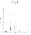
- Fig. 2 is a graph showing the results of ELISA measurement of each specimen, using a membrane spanning domain-removed AGPR H1 derivative according to the invention as the antigen.
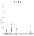
- Fig. 3 is a graph showing the results of ELISA measurement of each specimen, using a membrane spanning domain-removed AGPR L-H2 derivative according to the invention as the antigen.
- AGPR expressed by normal liver is H1, H2 and L-H2.
- HepG2 cells express H1, and H2 and H2' which are believed to be a result of alternative splicing from the same gene as L-H2 (Paietta et al., Hepatol. 15:359-402, 1992).
- the protein to be produced according to the invention reflects AGPR expressed in normal liver, the objects are H1 and L-H2 derivatives.
- AGPR H1 and AGPR L-H2 are shown in Fig. 1.
- One of the AGPR derivatives of the invention approximately comprises the extracellular domain of AGPR H1.
- a specific example thereof is a derivative having the amino acid sequence from the 60th amino acid Gly to the 291st amino acid Leu of the amino acid sequence represented by SEQ ID NO: 19.
- Another derivative according to the invention approximately comprises the cytoplasmic domain and extracellular domain of AGPR H1.
- a specific example thereof comprises the cytoplasmic domain having the amino acid sequence from the 1st amino acid Met to the 42nd amino acid Leu, and the extracellular domain having the amino acid sequence from the 60th amino acid Gly to the 291st amino acid Leu, of the amino acid sequence represented by SEQ ID NO: 19.
- Another AGPR derivative according to the invention approximately comprises the extracellular domain of AGPR L-H2.
- a specific example thereof is one having the amino acid sequence from the 59th amino acid Gly to the 287th amino acid Ala of SEQ ID NO: 20.
- Yet another AGPR derivative according to the invention approximately comprises the cytoplasmic domain and extracellular domain of AGPR L-H2.
- a specific example thereof comprises the cytoplasmic domain having the amino acid sequence from the 1st amino acid Met to the 41st amino acid Ser, and the extracellular domain having the amino acid sequence from the 59th amino acid Gly to the 287th amino acid Ala, of SEQ ID NO: 20.
- the AGPR derivatives of the invention are not limited to those with the amino acid sequences mentioned above. So long as the antigen determinant site of the AGPR is intact, the AGPR derivatives of the invention also include AGPR derivatives modified by a substitution, deletion and/or addition of one or more amino acids of the above-mentioned amino acid sequences, for example less than 20%, or less than 10%, of the amino acids in each domain, which may be fewer than 6 or fewer than 3 amino acids of the cytoplasmic domain, and fewer than 40, fewer than 20 or fewer than 10 amino acids of the extracellular domain.
- the derivatives of the present invention are soluble because they contain no membrane spanning domains, and thus may be easily recovered and purified, while they are also very useful as reagents for assaying anti-AGPR antibodies because they retain the antigen determinant site of AGPR.
- the AGPR derivatives of the invention may be in the form of polypeptides without sugar chains or in the form of polypeptides or glycoproteins with sugar chains.
- the former may be obtained by gene recombination production using bacteria or other prokaryotes, or by chemical synthesis, while the latter may be produced by gene recombination production using animal cells or other eukaryotic cells.
- the AGPR derivatives of the invention may be produced according to conventional methods for producing polypeptides, such as chemical synthesis or gene recombination. However, production by gene recombination is preferred considering the size or the molecular weight.
- the AGPR derivatives comprising parts of the amino acid sequence of SEQ ID NO: 19 or 20 are preferably produced using human-derived cDNA.
- AGPR derivatives having amino acid sequences modified by a substitution, deletion and/or addition of one or more amino acids of the amino acid sequence of SEQ ID NO: 19 or 20 may be produced by commonly used site-specific mutagenesis.
- cDNA fragments encoding AGPR may be isolated from cDNA synthesized using human liver polyA RNA as the template.
- the method of isolation may involve using a nucleotide sequence inferred from a published amino acid sequence (references: Spiess et al., J. Biol. Chem. 260; 1979-1982, 1985, Spiess et al., Proc. Natl. Acad. Sci.
- the desired cDNA for H1 was obtained using a primer having part of the nucleotide sequence reported by Spiess et al. (Spiess et al., J. Biol. Chem., 260: 1979-1982, 1985) for synthesis of cDNA with human liver polyA RNA as the template, and amplification by the polymerase chain reaction (PCR).
- This cDNA was used as the basis to prepare expression vectors for preparation of AGPR polypeptides by a genetic engineering method.
- the genetic engineering method used here encompasses any process by which a DNA fragment encoding a peptide with the amino acid sequence represented by any of Sequence Nos. 1 to 4 or a partial sequence thereof is obtained by chemical synthesis, or a combination of techniques. It also encompasses any procedure for constructing a replicable expression vector including a DNA fragment coding for the above-mentioned peptide, a procedure for introducing the expression vector in host cells to obtain a transformant which expresses the peptide, an expression step whereby the transformant is cultured to obtain the expressed product, and a step of recovering the expressed peptide.
- Expression vectors to be used for expression in the genetic engineering method may be, in addition to those cited in the examples, vectors employing E. coli tryptophan operon as well as tac promoter, tac promoter, lac promoter, PhoA promoter, ⁇ -phage promoter, etc., in the case of an E. coli host.
- Yeast may also be used as the host instead of E.
- coli for expression using an expression vector including a promoter commonly used in yeast, for example a glycolytic gene, such as for glyceroaldehyde-3-phosphate dehydrogenase, alcohol dehydrogenase I and II, pyruvate kinase, phosphoglycerine kinase, triose phosphate isomerase, etc.
- a promoter commonly used in yeast for example a glycolytic gene, such as for glyceroaldehyde-3-phosphate dehydrogenase, alcohol dehydrogenase I and II, pyruvate kinase, phosphoglycerine kinase, triose phosphate isomerase, etc.
- the expression may also be accomplished using an expression system with animal cells, for example mammalian cells such as CHO cells, COS-1 cells or HeLa cells, as the host, with a commonly used integrating expression vector or vaccinia virus as the vector, or an expression system employing adenovirus or another commonly used virus as the vector.
- animal cells for example mammalian cells such as CHO cells, COS-1 cells or HeLa cells
- a commonly used integrating expression vector or vaccinia virus as the vector
- an expression system employing adenovirus or another commonly used virus as the vector.
- the H1 and L-H2 polypeptides were expressed as fused proteins with a portion of an E. coli TrpE gene product.
- they were expressed as H1EM and L-H2EM comprising each of the cellular domain sequences for AGPR H1 and L-H2, and as H1TIM(-) and L-H2TM(-) polypeptides prepared lacking the respective membrane spanning domains and fusing the intracellular domain and extracellular domain.
- the recombinant CHO cell line 9801 was prepared which simultaneously expresses H1 and L-H2, for an expression system using mammalian cells as the host.
- This cell line expresses H1 and L-H2 in a modified form with sugar chains, and the expressed peptides were confirmed to retain receptor activity based on binding to N-galactosamine.
- the anti-AGPR antibody assay method using AGPR prepared from human liver according to the teaching of Treichel et al. was also investigated on patient serum.
- the results matched the discrimination results obtained according to the invention, thus demonstrating that AIH patient serum can be discriminated with a system using a peptide prepared according to the invention.
- anti-AGPR antibodies in patient serum react with both H1 and L-H2 derivative polypeptides and that it largely depends on the epitope present in the extracellular domain.
- the present invention provides a method of detecting anti-AGPR antibodies which is characterized by using at least one type of the aforementioned AGPR derivatives.
- the method involves contacting a test sample such as human or other serum or plasma to be measured for the presence or amount of the anti-AGPR antibodies, with at least one type of AGPR derivative according to the invention, and detecting whether an antigen/antibody reaction occurs between anti-AGPR antibodies in the test sample and the AGPR derivative antigen, or measuring the extent of the antigen/antibody reaction.
- the system of assay by antigen/antibody reaction is not particularly limited, and any conventional immunoassay method may be used.
- the Enzyme-Linked Immunosorbent Assay (ELISA), Western blotting, the indirect fluorescent antibody method, radioimmunoassay (RIA), or the like may be used.
- the labelling used for the assay method may be an enzyme such as peroxidase, alkali phosphatase, digoxigenase or ⁇ -galactosidase, a radioactive isotope such as 125 I, 35 S, 3 H or 14 C, or a fluorescent substance such as FITC, rhodamine, Texas red, etc.
- the present invention further provides a reagent to be used in the above-mentioned method for assaying anti-AGPR antibodies, which comprises the aforementioned AGPR derivative and either or both AGPR H1 and AGPR L-H2.
- the AGPR derivative and AGPR in the reagent may be purified or partially purified, or may be the host cells themselves expressing and bearing the polypeptides or proteins.
- Such a reagent may be used in the form of the AGPR derivative or AGPR dissolved in a buffer solution such as a phosphate buffer, carbonate buffer, Tris-HCl buffer or HEPES buffer solution, one of these solutions which has been dried by lyophilization, the polypeptides or proteins immobilized on a solid carrier such as a filter, glass slide or beads, or the cells themselves expressing and bearing the polypeptide or protein as mentioned above, which have been immobilized on the surface of a filter, glass slide or the like.
- a buffer solution such as a phosphate buffer, carbonate buffer, Tris-HCl buffer or HEPES buffer solution
- the present invention also relates to a kit for assaying anti-AGPR antibodies which comprises at least a reagent such as mentioned above which contains AGPR or AGPR, and standard anti-AGPR antibody.
- the anti-AGPR antibody may be either polyclonal or monoclonal antibody.
- the kit sometimes contains a labelled non-human-derived antibody against human immunoglobulin, as in the case of an ELISA kit.
- the assay method, assay reagent and assay kit described above may use as the antigen at least one type of AGPR derivative of the invention and AGPR produced by recombination, but it is sometimes preferable for the assay method to employ 2 or more, particularly AGPR H1 or its derivative and AGPR L-H2 or its derivative, in combination. In such cases, cells which express both AGPR H1 or its derivative and AGPR L-H2 or its derivative are preferably used.
- AGPR or its derivative which can be produced according to the present invention also allows quantitative and qualitative analysis of asialoglycoprotein in blood. Purification of asialoglycoproteins and asialated sugars is also possible.
- the peptides of the invention may also be used to develop inhibiting agents against incorporation of asialoglycoprotein through AGPR receptors. They may also be used to develop inhibiting agents against invasion of viruses and complexes of viruses and other proteins into liver cells through AGPR receptors.
- the gene necessary to construct a Type I AGPR (H1) recombinant was cloned by the PCR from single-stranded cDNA from human liver.
- the single stranded cDNA from human liver was prepared from 0.5 ⁇ g of human liver poly(A)-RNA (CLONTECH), using a First Strand Synthesis Kit (STRATAGENE). The following protocol was followed. The 0.5 ⁇ g of human liver poly(A)-RNA was dissolved in 32 ⁇ l of DEPC water, and 0.3 ⁇ g/3 ⁇ l oligo dT primer was added. After heating the mixture at 65°C for 5 minutes, it was allowed to stand until reaching room temperature.
- the primers used for the PCR were synthesized from the nucleotide sequence of AGPR H1 determined by M. Spiess et al. (Spiess, M. et al., J. Biol. Chem., 260:1979-1982, 1985), and had the sequences shown below.
- the PCR reaction was conducted twice for cloning of the AGPR H1 gene.
- the first PCR reaction employed the following composition. 10x buffer solution (Ampli Taq PCR kit) 10 ⁇ l 2.5 mM dATP 2 ⁇ l 2.5 mM dCTP 2 ⁇ l 2.5 mM dGTP 2 ⁇ l 2.5 mM dTTP 2 ⁇ l 10 pmol/ ⁇ l primer F1 10 ⁇ l 10 pmol/ ⁇ l primer B9 10 ⁇ l Human liver single stranded cDNA 5 ⁇ l Sterilized water 56 ⁇ l 10 U/ ⁇ l Taq polymerase 1 ⁇ l
- the reaction was conducted in 35 cycles of 30 seconds at 94°C, 1 minute at 50°C and 2 minutes at 72°C, followed by incubation at 72°C for 7 minutes and preservation at 4°C until the next procedure.
- a 50 ⁇ l portion of the first PCR reaction solution was subjected to 1.2% agarose gel electrophoresis with a 1xTAE buffer solution, and an approximately 1.1 kb band was cut out.
- the DNA was recovered from this using Gene Clean (BIO 101), and 1 ⁇ l of the recovered 10 ⁇ l solution was used for the second PCR reaction.
- the second PCR reaction employed the following composition.
- the reaction was conducted in the same manner as the first.
- Four ⁇ l of the reaction solution was separated by electrophoresis on a 2% agarose gel, the darkest band corresponding to approximately 1.1. kb was cut out and the DNA recovered with Gene Clean.
- a 3.5 ⁇ l portion of the recovered 10 ⁇ l solution and 1 ⁇ l of pGEM-T vector (Promega) were subjected to ligation using 60 ⁇ l of solution A and 7.5 ⁇ l of solution B from a ligation kit (Takara Shuzo), and the DNA fragments obtained by PCR were cloned at the multicloning sites of pGEM-T.
- Four ⁇ l of the ligation reaction solution was mixed with 100 ⁇ l E.
- coli DH5 ⁇ competent cells and heating at 42°C for 30 seconds was followed by rapid cooling to transform the DH5 ⁇ .
- the transformed DH5 ⁇ was looped onto a 1.5% agar LB plate containing 100 ⁇ g/ml ampicillin and cultured overnight at 37°C.
- the ampicillin-resistant transformants which had formed colonies on the LB plate containing ampicillin were collected with a pick and were planted into 3 ml of 2 ⁇ YT medium and cultured overnight at 37°C, after which the transformants were collected and the plasmid DNA recovered by the alkalilysis method.
- the obtained plasmid DNA was cut with restriction endonuclease, separated by agarose gel electrophoresis, and the lengths thereof were determined to select a clone in which the desired AGPR H1 gene had been cloned in pGEM-T.
- the transformant carrying this clone was further planted in 100 ml of 2 ⁇ YT medium and cultured overnight at 37°C, and the transformant cells were collected to prepare a large amount of the plasmid DNA by the alkalilysis method.
- the nucleotide sequence of the inserted portion was confirmed with a fluorescent sequencer (ABI Co.).
- the clone obtained in this manner has the sequence corresponding to bases 107 to 1198 of the reported AGPR H1 cDNA sequence at the multicloning site of cloning vector pGEM-T, and it was named pGEM-T-H1.
- first PCR reaction solution 1/100 ⁇ l was used for a separate second PCR reaction.
- This reaction solution had the following composition. 10x buffer solution 10 ⁇ l 2.5 mM dATP 2 ⁇ l 2.5 mM dCTP 2 ⁇ l 2.5 mM dGTP 2 ⁇ l 2.5 mM dTTP 2 ⁇ l 10 pmol/ ⁇ l primer F1.5 10 ⁇ l 10 pmol/ ⁇ l primer B2.5 10 ⁇ l 1/100 dilution of first PCR solution 1 ⁇ l Sterilized water 60 ⁇ l 10 U/ ⁇ l Taq polymerase 1 ⁇ l
- the reaction was conducted in 20 cycles of 30 seconds at 94°C, 1 minute at 60°C and 2 minutes at 72°C, followed by reaction for 7 minutes at 72°C. Forty ⁇ l of the reaction solution was separated by electrophoresis on a 2% agarose gel, and a band corresponding to a size of approximately 900 bases was cut out.
- the DNA was recovered from the gel fragments with Gene Clean Kit[BIO 101], and 3.5 ⁇ l of the recovered 10 ⁇ l DNA solution and 5 ⁇ l of pGEM-T vector were subjected to ligation using 60 ⁇ l of solution A and 7.5 ⁇ l of solution B from a Takara Shuzo ligation kit. Selection was thus made of desired gene cloned in pGEM-T and having bases 164 to 1073 of the reported AGPR H1 cDNA sequence.
- the PmaCI restriction enzyme recognition site of the insert and the SalI recognition site at the 3'-end pGEM-T multicloning site of the insert were recombined with PmaCI/SalI of pGEM-T-H1 to prepare clone pGEM-T-H1-2 having the sequence from bases 164 to 1198 of the reported AGPR H1 cDNA sequence.
- the primer used for the PCR was synthesized from the nucleotide sequence of AGPR L-H2 determined by M. Spiess et al. (Spiess et al., Proc. Natl. Acad. Sci. 82:6465-6469, 1985, Paietta, E., et al. Hepatology 15:395-402, 1992). The following sequences were used.
- the PCR reaction was conducted twice for cloning of the AGPR L-H2 gene.
- the PCR reaction conditions and method of cloning were the same as in Example 1.
- the cloned and amplified sequence obtained using primer 1 and primer 2 in the PCR reaction corresponds to bases 188-1080 of the L-H2 cDNA.
- the cloned and amplified sequence obtained using primer 3 and primer 4 in the PCR reaction corresponds to bases 299-1148 of the L-H2 cDNA.
- the cloned and amplified sequence obtained using primer 7 and primer 8 in the PCR reaction corresponds to bases 34-553 of the L-H2 cDNA.
- the XhoI/SphI region of pGEM-T-LH2-34 (299-1148) was recombined with the XhoI/SphI (3' end multicloning site) of pGEM-T-LH2-12 (188-1080) to construct pGEM-T-L-H2 (188-1148).
- the SphI/EcoRV region of pGEM-T-LH2-5' (34-553) was recombined with SpeI (5' end multicloning site) of pGEM-T-L-H2 to construct pGEM-T-L-H2F.
- This clone includes bases 34 through 1148 of the reported L-H2 cDNA.
- the gene for the extracellular domain of AGPR H1 was cloned and inserted into the expression vector pAT-TrpE.
- the cloning of the extracellular domain of the AGPR H1 gene was accomplished by the PCR using pGEM-T-H1 as the template, cloning the amplified DNA in pGEM-T.
- the following PCR primers were used.
- Primers AG1EcoF and AG1SalR include the EcoRI restriction enzyme recognition site and the SalI recognition site, respectively, and the H1 extracellular domain gene amplified by the two primers has the EcoRI recognition site at the 5' end and the SalI recognition site at the 3' end.
- the PCR reaction was conducted using the following composition.
- the reaction was conducted in 25 cycles of 30 seconds at 94°C, 1 minute at 45°C and 2 minutes at 72°C, followed by reaction for 7 minutes at 72°C.
- An approximately 700 b band obtained by the reaction was cut out, subjected to ligation reaction with a ligation buffer solution (Takara Shuzo), and cloned in pGEM-T.
- the resulting plasmid was dubbed pGEM-T-H1EM.
- the nucleotide sequence was determined using restriction endonuclease and sequencing, and it was then cut at the EcoRI/SalI restriction enzyme recognition site prepared with the PCR primer, and inserted by ligation reaction at the EcoRI/SalI site of pAT-TrpE.
- the resulting expression vector was named pAT-TrpE-H1EM, and after confirming the sequence with restriction endonuclease, it was introduced into XL-1 Blue competent cells to obtain transformants.
- the extracellular domain of AGPR H1 protein was expressed as a fused protein with a polypeptide consisting of the 17 N-terminal amino acids of TrpE. Expression of the protein of interest was confirmed in the following manner. pAT-TrpE-H1EM/XL-1 blue cells were seeded at a 2% concentration in an M9-CA medium containing 100 ⁇ g/ml ampicillin, and shake cultured overnight at 37°C. After collecting the E. coli cultured overnight, they were subjected to SDS-PAGE according to conventional methods to separate the protein, and then stained with Coomassie Brilliant Blue R250 solution to detect the protein.
- H1EM H1EM in a large amount
- culturing was conducted on a 1L scale.
- the pAT-TrpE-H1EM/XL-1 blue colonies were seeded into 2 ml 2 ⁇ YT/100 ⁇ g/ml ampicillin and shake cultured for 8 hours at 37°C. This was reseeded into 30 ml 2 ⁇ YT/100 ⁇ g/ml ampicillin at a proportion of 1/50 and shake cultured at 37°C overnight.
- pAT-TrpE-H1Em XL-1 blue cells cultured in 1 L M9-CA/100 ⁇ g/ml ampicillin at a proportion of 1/50 were seeded and shake cultured at 37°C overnight. After confirming expression of the H1EM protein, the cells were collected by centrifugation and the resulting pellet ( E. coli ) was cryopreserved at -20°C.
- the H1EM protein was purified for use in the subsequent experiment.
- One gram of the cryopreserved E. coli was suspended in 5 ml of dissolving buffer solution (50 mM Tris-HCl, pH 8.0/30 mM NaCl/5 mM EDTA), and 1 mg of lysozyme was added prior to shaking at 27°C for one hour. After sonication twice at 150 W for 90 seconds, the cells were disrupted. The disruption of the cells was confirmed by microscope. These were separated by centrifugation at 15K, 4°C for 30 minutes to obtain a pellet which was then suspended by addition of buffer A (50 mM Tris-HCl, pH 8.0) and homogenized at 1500 rpm, 5 strokes.
- buffer A 50 mM Tris-HCl, pH 8.0
- the pellet obtained by subsequent centrifugation at 15K, 4°C for 15 minutes was washed with buffer A and again subjected to centrifugation.
- the resulting pellet was suspended by addition of buffer A containing 2 M urea, and was then centrifuged at 15K, 4°C for 15 minutes to obtain a supernatant and a pellet.
- the pellet was also subjected to the same procedure using 4M and 6M urea.
- the extract obtained by each procedure was subjected to SDS-PAGE electrophoresis and the protein was detected by CBB staining, which demonstrated the presence of the TrpE-H1EM fused protein.
- the protein of interest was present in the 4-6 M urea extraction solutions together with contaminants.
- the 4-6 M urea extraction solutions were passed through Q Sepharose (Pharmacia) (16/10) and eluted with 0 to 0.3 M NaCl for 15 minutes at a flow rate of 0.4 ml/min.
- the eluted fractions exhibiting an absorption peak at O.D. 280 nm were passed through Sephacryl S-300 (Pharmacia) (26/95) and eluted with buffer B (20 mM Tris-HCl, pH 8.0/6 M urea/0.1 M NaCl/5 mM DTT) at a flow rate of 2 ml/min.
- the eluted fractions were again passed through Sephacryl S-300 (Pharmacia) (26/95) to obtain eluted fractions exhibiting absorption at O.D. 280 nm in fractions 120-160.
- the cytoplasmic and extracellular domains of the AGPR H1 gene were cloned and linked together prior to insertion in expression vector pTac-TrpE.
- the gene cloned in Example 3 was used as the extracellular domain of the AGPR H1 gene.
- the cloning of the cytoplasmic domain of the AGPR H1 gene was accomplished by PCR reaction with pGEM-T-H1 as the template, and cloning the amplified DNA in pGEM-T.
- the following PCR primers were used.
- Primers H1XhoF and H1EcoR include the XhoI restriction enzyme recognition site and the EcoRI recognition site, respectively, and the H1 extracellular domain gene amplified by the two primers has the XhoI recognition site at the 5' end and the EcoRI recognition site at the 3' end.
- the PCR reaction was conducted using the following composition.
- the reaction was conducted in 10 cycles of 30 seconds at 94°C, 1 minute at 45°C and 1 minute at 72°C, followed by reaction for 7 minutes at 72°C.
- An approximately 127 b band obtained by the reaction was cut out, subjected to ligation reaction with a ligation buffer solution (Takara Shuzo), and cloned in pGEM-T.
- the resulting plasmid was dubbed pGEM-T-H1IM.
- the base sequence was determined by confirming the restriction enzyme cleavage sites and by sequencing, and it was then cut at the XhoI/EcoRI restriction enzyme recognition sites prepared with the PCR primer.
- the XhoI/EcoRI-cut fragments of pGEM-T-H1IM and EcoRI/SpeI-cut fragments of the pGEM-T-H1EM obtained in Example 3 were inserted by ligation reaction at the XhoI/SpeI site of the E. coli expression vector pTac-TrpE including the Tac promoter.
- the resulting expression vector was named pTac-H1TM(-), and after confirming the sequence with restriction endonuclease, it was introduced into XL-1 Blue competent cells to obtain transformants.
- the fused protein of the H1 cytoplasmic and extracellular domains was expressed as a fused protein with a polypeptide consisting of the 17 N-terminal amino acids of TrpE. Expression of the protein of interest was confirmed in the following manner. pTac-H1TM(-)/XL-1 blue cells were seeded at a 2% concentration in LB medium containing 100 ⁇ g/ml ampicillin, and shake cultured at 37°C until a value of 40 was reached with a turbidimeter. After addition to the culture solution of IPTG at a final concentration of 0.4 mM, culturing was continued for 6 hours and the E. coli cells were collected. After collecting the E. coli , they were subjected to SDS-PAGE according to conventional methods to separate the protein and then stained with Coomassie Brilliant Blue R250 (CBB) solution to detect the protein.
- CBB Coomassie Brilliant Blue R250
- H1TM(-) In order to obtain H1TM(-) in a large amount, culturing was conducted on a 2L scale.
- the pTac-H1TM(-)/XL-1 blue colonies were seeded into 30 ml LB/100 ⁇ g/ml ampicillin and shake cultured overnight at 37°C.
- pTac-H1TM(-)/XL-1 blue cells cultured in 2 L LB/100 ⁇ g/ml ampicillin at a proportion of 1/100 were seeded and shake cultured at 37°C for 2 hours, after which IPTG at a final concentration of 0.4 mM was added and the culturing was continued overnight.
- the cells After confirming expression of the H1TM(-) protein, the cells were collected by centrifugation and the resulting pellet ( E. coli ) was cryopreserved at -20°C.
- the H1TM(-) protein was purified for use in the subsequent experiment.
- Urea extraction was performed in the same manner as Example 3. Five ml of 1% NP-40 was added to one gram of the E. coli in buffer A. The protein of interest was present in the 6 M urea extraction solution together with contaminants. DTT at a final concentration of 50 mM was added to the 6 M urea extraction solution which was then allowed to stand at 4°C for over an hour.
- the 0.15-0.2 M NaCl elution fraction exhibited an absorption peak at O.D. 280 nm. Upon separation and identification of the protein by SDS-PAGE, almost a single band of protein was detected.
- the eluted fraction containing the desired protein was concentrated with an Amicon YM10 according to the manual, and the DTT was brought to a low concentration with buffer C lacking DTT. The purified H1TM(-) peptide was thus obtained.
- the extracellular domain of the AGPR L-H2 gene was cloned and inserted into expression vector pAT-TrpE.
- the cloning of the extracellular domain of the AGPR L-H2 gene was accomplished by PCR reaction using pGEM-T-L-H2 as the template, in the same manner as the cloning of the H1 extracellular domain.
- the following PCR primers were used.
- Primers AGEcoF and AGEcoR include the EcoRI restriction enzyme recognition site and the BamHI recognition site, respectively, and the L-H2 extracellular domain gene amplified by the two primers has the EcoRI recognition site at the 5' end and the BamHI recognition site at the 3' end.
- the PCR reaction was conducted in the same manner as for H1.
- the procedure after the PCR reaction was also the same as for H1, and the plasmid obtained by inserting the PCR product in vector pGEM-T was named pGEM-T-L-H2EM.
- the L-H2 extracellular domain gene was inserted at EcoRI/BamHI of pAT-TrpE to prepare expression vector pAT-TrpE-L-H2EM. This was introduced into XL-1 Blue competent cells to obtain transformants.
- pAT-TrpE-L-H2EM XL-1 blue cells cultured in 1 L M9-CA/100 ⁇ g/ml ampicillin at a proportion of 1/50 were seeded and shake cultured at 37°C overnight. After confirming expression of the L-H2EM protein, the cells were collected by centrifugation and the resulting pellet ( E. coli ) was cryopreserved at -20°C.
- the L-H2EM protein was purified for use in the subsequent experiment.
- Urea extraction was performed in the same manner as Example 3.
- the protein of interest was present in the 4-6 M urea extraction solution, together with contaminants.
- the 4-6 M urea extraction solution was passed through Q Sepharose (Pharmacia) (16/10) and eluted with 0-0.3 M NaCl for 15 minutes at a flow rate of 0.4 ml/min.
- the eluted fractions exhibiting an absorption peak at O.D. 280 nm were passed through Sephacryl S-300 (Pharmacia) (26/95) and eluted with buffer B at a flow rate of 2 ml/min.
- the eluted fractions were passed through Superdex 75 pg (26/60) and eluted with buffer D (20 mM Tris-HCl, pH 8.0/6 M urea/0.1 M NaCl/5 mM DTT) at a flow rate of 2 ml/min.
- the protein of interest eluted in fractions 55-65. Separation and identification of the protein by SDS-PAGE of the eluted fraction revealed almost a single band of protein.
- the DTT was removed from the buffer solution of the eluted fractions containing the desired protein using Sephadex G25 fine, the solution was concentrated with Centricon-10 (Amicon), and the buffer solution was replaced with PBS(-) in a PD-10 column (Pharmacia) according to the manual. The purified L-H2EM peptide was thus obtained.
- the cytoplasmic and extracellular domains of the AGPR L-H2 gene were cloned and inserted into expression vector pAT-TrpE.
- the cloning of the cytoplasmic domain of the AGPR L-H2 gene was accomplished by PCR reaction using pGEM-T-L-H2 as the template, in the same manner as the cloning of the H1 cytoplasmic domain.
- the following PCR primers were used.
- Primers L-H2XhoF and L-H2EcoR include the XhoI restriction enzyme recognition site and the EcoRI recognition site, respectively, and the L-H2 extracellular domain gene amplified by the two primers has the XhoI recognition site at the 5' end and the EcoRI recognition site at the 3' end.
- the PCR reaction was conducted in the same manner as for H1.
- the procedure after the PCR reaction was also the same as for H1, and the plasmid obtained by inserting the PCR-obtained fragment in vector pGEM-T was dubbed pGEM-T-L-H2TM(-).
- the fragment obtained by cutting pGEM-T-L-H2TM(-) with XhoI/EcoRI restriction enzyme and the fragment obtained by cutting pGEM-T-L-H2EM with EcoRI/BamHI restriction enzyme were inserted at the XhoI/BamHI site of pTac-TrpE to prepare the E. coli expression vector pTac-L-H2TM(-). This was introduced into XL-1 Blue competent cells to obtain transformants.
- the L-H2TM(-) protein was purified for use in the subsequent experiment.
- Urea extraction was performed in the same manner as Example 3. Five ml of 1% NP-40 was added to one gram of the E. coli in buffer A. The protein of interest was present in the 4-6 M urea extraction solution together with contaminants. DTT at a final concentration of 50 mM was added to the 6 M urea extraction solution which was then allowed to stand at 4°C for over an hour. This was passed through equilibrized MonoQ (Pharmacia) (5/5) and eluted with 0-0.3 M NaCl in buffer E (50 mM Tris-HCl, pH 8.0/6 M urea/5 mM DTT) at a flow rate of 1 ml/min.
- the approximately 0.1 M NaCl elution fraction exhibited an absorption peak at O.D. 280 nm.
- the eluted fraction containing the protein of interest was dialyzed in buffer F (50 mM Tris, pH 8.0/6 M urea/0.15 M NaCl) and the DTT was removed.
- the purified L-H2TM(-) peptide was thus obtained.
- the purified recombinant AGPR proteins H1EM and L-H2Em obtained above in Examples 3 and 5 were used to prepare anti-AGPR H1 antibody and anti-AGPR L-H2 antibody.
- Rabbits New Zealand white
- mice were used for the immunization, and Freund's complete adjuvant (FCA) was used as the adjuvant for immunization according to conventional methods.
- FCA Freund's complete adjuvant
- the antibody titer was measured by ELISA, and the reaction specificity was confirmed by Western blotting.
- ELISA was conducted according to the following method.
- the H1EM purified protein or L-H2EM purified protein was coated on a plate (Nunc) to 1 ⁇ g/ml at room temperature for 2 hours. Appropriately diluted serum was then incubated at room temperature for one hour. Peroxidase-bound anti-rabbit IgG was used at 1000-fold dilution as the secondary antibody and incubation was conducted at room temperature for one hour. Coloring was accomplished by incubation at room temperature for 15 minutes in a mixture of 6 ⁇ l hydrogen peroxide water and a solution of an OPD1 tablet (Wako Junyaku) in 35 ml of phosphate/citrate buffer solution, and an equivalent of 10% sulfuric acid was added to suspend the reaction. The detection was accomplished by measurement at OD 492 nm with a microplate reader (Tori).
- the Western blotting was conducted according to conventional methods. SDS-PAGE was performed according to conventional methods using XL-1 blue cells expressing H1EM or L-H2EM, and wild XL-1 blue cells as the samples. These were then transferred onto a PVDF membrane (millipore) by a conventional method. Each of the antiserums was used at 10,000-fold dilution as the primary antibody, and alkali-phosphatase-bound anti-rabbit IgG was used at 1000-fold dilution as the secondary antibody, for Western blotting according to conventional methods.
- the AGPR protein in the antiserums obtained as a result could be detected by ELISA and Western blotting
- the anti-H1 antiserum exhibited an antibody titer of 10 5 against H1EM antigen in ELISA.
- the anti-L-H2 antiserum also exhibited an antibody titer of 10 5 against L-H2EM antigen.
- Western blotting the anti-H1 antiserum exhibited strong reactivity to AGPR H1 protein while the anti-L-H2 antiserum also exhibited strong reactivity to AGPR L-H2 protein, and despite crossing the reactions, each was found to be specific antiserum.
- Plasmid PaSR was used as the vector for expression in the COS-1 cells. Five ⁇ g of PaSR DNA was cut with 25 U XhoI and 25 U BamHI and treated with 5 U of Klenow fragment according to conventional methods to smooth the ends. It was further treated with 10 U BAP according to conventional methods for dephosphorylation of the ends.
- H1 (nucleotides 107-1198) cut from pGEM-T-H1 with SacI/SacII and H1-2 (nucleotides 164-1198) cut out from pGEM-T-H1-2 with SacI/SacII were used as the two AGPR H1 genes for insertion into the expression vector.
- the expression units were cut out by cutting 10 ⁇ g of plasmid with 50 U SacI and 50 U SacII, and the ends were smoothed using a branching kit (Takara Shuzo) according to the attached protocol.
- the vectors and H1 and H1-2 fragments prepared above were ligated using a ligation kit (Takara Shuzo) according to the attached protocol.
- the reaction solution was introduced into competent cells by a conventional method for transformation, and a few ampicillin-resistant colonies were obtained.
- the plasmids were then extracted by a conventional method and cut with restriction endonuclease and identified to obtain the desired expression vectors PaSR-H1 and PaSR-H1-2.
- the expression vectors were introduced into COS-1 cells by transfection using electroporation. Growth-phase COS-1 cells cultured overnight were suspended in PBS(-) to a concentration of 2.5 ⁇ 10 6 cells/ml. A 0.6 ml portion of the cell suspension was placed in an electroporation cuvette (Bio-Rad), mixed with 15 ⁇ g of expression vector and allowed to stand in ice for 30 minutes. Electroporation was then conducted at 125 ⁇ F, 125 V. The cells were allowed to stand in ice for 10 minutes and then seeded into a 100 mm ⁇ tissue culturing dish containing DMEM 10% FBS. After culturing for 24 hours the medium was replaced, and after 48 hours of electroporation the cells were collected.
- the expressed proteins were detected by Western blotting.
- the recovered cells were suspended in SDS-sample buffer according to a conventional method and an amount of 1 ⁇ 10 5 cells per lane was separated by SDS-PAGE.
- the anti-H1 serum obtained in Example 6 was used at a 1000-fold dilution for Western blotting by conventional methods. Detection was accomplished using an ECL Western blotting biotin system (Amersham).
- LH-2 (nucleotides 188-1148) cut from pGEM-T-L-H2 with SpeI/SphI and L-H2F (nucleotides 34-1148) cut out from pGEM-T-L-H2F with SpeI/SfiI were used as the two AGPR LH-2 genes for insertion into the expression vector.
- the expression units were cut out by cutting 10 ⁇ g of plasmid with 50 U SpeI, 50 U SphI or 50 U SfiI, and the ends were smoothed using a branching kit (Takara Shuzo) according to the attached protocol.
- COS-1 cells Introduction of the expression vectors into COS-1 cells was accomplished by transfection using lipofectin (GIBCO BRL).
- the COS-1 cells in an amount of 5 ⁇ 10 5 were seeded into a 100 mm ⁇ tissue culturing dish and cultured overnight.
- Two ⁇ g of DNA prepared according to the lipofectin manual was mixed therewith also according to the manual, and further culturing was conducted overnight.
- 10% FBS was then added to the medium, and after 48 hours the cells were collected.
- the expressed protein was detected by Western blotting in the same manner as the COS-1-expressed H1 protein in Example 8.
- Anti-L-H2 antiserum diluted 1000-fold was used as the primary antibody.
- the AGPR H1 gene was introduced into cells of the established animal cell line CHO-K1 to express AGPR H1 protein.
- the method of expressing the AGPR H1 protein in the CHO-K1 cells is described below.
- the expression vector for introduction of the AGPR H1 gene into the CHO-K1 cells was constructed according to the Marcos method (Saito, et al., Experimental Medicine, Vol.7, p.183-).
- a 30 ⁇ g portion of expression vector PaSR-H1 obtained in Example 8 was cut with 200 U of restriction enzyme SfiI, and the resulting DNA fragments were recovered with a DNA CELL (Daiichi Kagaku).
- a 1.5 ⁇ g portion of vector pCHD2L carrying ampicillin-resistant and hygromycin B-resistant genes was also cut with 20 U SfiI, and phenol/chloroform extraction and ethanol precipitation were followed by dissolution in TE (pH 7.5).
- the Marcos DNA clones with the largest molecular weight were selected, and 30 ⁇ g of plasmid prepared with Quagen tip-500 (Quagen) was introduced into 5 ⁇ 10 5 CHO-K1 cells by the calcium phosphate method using a Mammalian Transfection Kit (STRATAGENE).
- the cells were relooped at 5 ⁇ 10 5 per 100 mm ⁇ tissue culturing dish, and 600 or 800 ⁇ g of hygromycin B was added and selection was made of the CHO-K1 lines which exhibited hygromycin B resistance due to incorporation of the foreign gene into the genome.
- the selection was conducted for about 2 weeks thereafter. A few colonies of CHO-K1 cells exhibiting hygromycin B resistance were cloned, and expression of AGPR H1 protein was confirmed by Western blotting as explained in Example 8.
- the clones confirmed to express AGPR H1 protein were also verified by the indirect fluorescent antibody method.
- the AGPR L-H2 gene was introduced into cells of the established animal cell line CHO-K1 to express AGPR L-H2 protein.
- the method of expressing the AGPR L-H2 protein in the CHO-K1 cells is described below.
- the expression vector for introduction of the AGPR L-H2 gene into the CHO-K1 cells was constructed by the Marcos method as for AGPR H1 in Example 10.
- An approximately 48K Marcos DNA was obtained by the same method as in Example 10.
- the obtained Marcos DNA was verified by digestion with restriction enzyme KpnI present on pCHD2L but not on the expression unit, SfiI located on both ends of the expression unit and SacI on the expression unit.
- the obtained Marcos DNA was confirmed to have a plurality (about 20) of the Sfi fragments of PaSR-H1 introduced in the same direction per pCHD2L vector.
- the AGPR H1 gene and AGPR L-H2 gene was introduced into cells of the established animal cell line CHO-K1 to express both AGPR H1 protein and AGPR L-H2 protein.
- the method of expressing both the AGPR H1 protein and AGPR L-H2 protein in the CHO-K1 cells is described below.
- the expression vector for introduction of the AGPR H1 gene and AGPR L-H2 gene into the CHO-K1 cells was constructed by the Marcos method as for AGPR H1 in Example 10.
- DNA fragments obtained by digesting PaSR-H1 and PaSR-L-H2 with SfiI were collected with a DNA CELL (Daiichi Kagaku) and used, in the same manner as Examples 10 and 11.
- equivalent amounts of H1 and L-H2 SfiI fragments were used.
- An approximately 48K Marcos DNA was obtained by this method.
- the obtained Marcos DNA was confirmed to have a plurality (about 20) of the SfiI fragments of PaSR-H1 and Sfi fragments of PaSR-L-H2 introduced as expression units per pCHD2L vector.
- the AGPR protein-expressing CHO-K1 cells obtained in Examples 10, 11 and 12 were used to detect anti-AGPR antibodies in autoimmune hepatitis patient serum by the indirect fluorescent antibody method. The following procedure was followed.
- AIH patient serum and control serum were diluted 100-fold with PBS(-) and incubated for 30 minutes at 37°C. After washing with PBS(-), anti-human IgG-fluorescein was used as the secondary antibody and incubation was continued at 37°C for 30 minutes. After further washing with PBS(-) and air drying followed by glycerol encapsulation, the cells were observed with a fluorescent microscope.
- Example 14 Assay of anti-AGPR antibody in patient serum by ELISA
- the details regarding the human serum specimens were: 38 cases of autoimmune hepatitis (AIH), 30 cases of primary biliary cirrhosis (PBC), 10 cases of alcoholic liver disease (ALD), 2 cases of systemic lupus erythematosus (SLE), 16 cases of chronic hepatitis B (CH-B) and 19 healthy cases.
- AIH autoimmune hepatitis
- PBC primary biliary cirrhosis
- ALD alcoholic liver disease
- SLE systemic lupus erythematosus
- CH-B chronic hepatitis B
- the anti-AGPR antibodies in the sera were assayed by ELISA, according to the following method.
- the recombinant AGPR proteins H1TM(-) and L-H2TM(-) were diluted with PBS(-) and dispensed at 1 ⁇ g/100 ⁇ l/well, and allowed to stand at room temperature for 2 hours for coating onto an ELISA immunomodule plate (Nunc, Maxisorp). After washing and removal of the antigen, 0.5% casein/PBS(-) was dispensed at 300 ⁇ l/well as a blocking buffer solution, and the mixture was allowed to stand at 4°C overnight. After removal of the blocking buffer solution, diluted samples of the human serum specimens were added at 100 ⁇ l/well prior for incubation at 37°C for 2 hours. The dilution was carried out using 0.05% casein/1% BSA/0.05% Tween20/PBS(-), and the degree of dilution was 1000-fold.
- peroxidase-labelled anti-human IgG polyclonal antibody (BioRad) was diluted with 0.05% casein/1% BSA/0.05% Tween20/PBS(-) to 10,000-fold for antigen H1TM(-) and to 5000-fold for antigen L-H2TM(-), and dispensed at 100 ⁇ l/well prior to incubation at 37°C for 2 hours.
- the AIH and PBC groups showed a distribution at higher values than the ALD, SLE, CH-B and normal groups (Figs. 2 and 3).
- This anti-AGPR antibody detection system distinguished the groups of AIH and PBC, which are autoimmune liver diseases, from the group of healthy persons, the group of ALD and SLE which are other autoimmune diseases, and the group of chronic hepatitis B.
Landscapes
- Health & Medical Sciences (AREA)
- Chemical & Material Sciences (AREA)
- Life Sciences & Earth Sciences (AREA)
- Organic Chemistry (AREA)
- Medicinal Chemistry (AREA)
- Molecular Biology (AREA)
- Gastroenterology & Hepatology (AREA)
- Toxicology (AREA)
- Biochemistry (AREA)
- Biophysics (AREA)
- General Health & Medical Sciences (AREA)
- Genetics & Genomics (AREA)
- Immunology (AREA)
- Zoology (AREA)
- Proteomics, Peptides & Aminoacids (AREA)
- Cell Biology (AREA)
- Diabetes (AREA)
- Hematology (AREA)
- Rehabilitation Therapy (AREA)
- Rheumatology (AREA)
- Peptides Or Proteins (AREA)
- Preparation Of Compounds By Using Micro-Organisms (AREA)
- Micro-Organisms Or Cultivation Processes Thereof (AREA)
- Saccharide Compounds (AREA)
Applications Claiming Priority (3)
| Application Number | Priority Date | Filing Date | Title |
|---|---|---|---|
| JP7212118A JPH0956380A (ja) | 1995-08-21 | 1995-08-21 | アシアロ糖蛋白質受容体誘導体及びその使用 |
| JP212118/95 | 1995-08-21 | ||
| JP21211895 | 1995-08-21 |
Publications (2)
| Publication Number | Publication Date |
|---|---|
| EP0773289A2 true EP0773289A2 (de) | 1997-05-14 |
| EP0773289A3 EP0773289A3 (de) | 2001-06-27 |
Family
ID=16617183
Family Applications (1)
| Application Number | Title | Priority Date | Filing Date |
|---|---|---|---|
| EP96113349A Withdrawn EP0773289A3 (de) | 1995-08-21 | 1996-08-20 | Derivate des Asialoglycoprotein-Rezeptors und deren Verwendung |
Country Status (2)
| Country | Link |
|---|---|
| EP (1) | EP0773289A3 (de) |
| JP (1) | JPH0956380A (de) |
Cited By (5)
| Publication number | Priority date | Publication date | Assignee | Title |
|---|---|---|---|---|
| WO2001066749A3 (en) * | 2000-03-08 | 2002-03-14 | Zymogenetics Inc | A member of the lectin superfamily |
| WO2004081029A3 (en) * | 2003-03-13 | 2008-07-17 | Univ Ramot | Novel non-invasive marker for liver disease |
| US7741065B2 (en) | 2003-03-13 | 2010-06-22 | Ramot At Tel Aviv University Ltd. | Non-invasive marker for liver function and disease |
| US7981627B2 (en) | 2008-07-17 | 2011-07-19 | Ramot At Tel-Aviv University Ltd. | Methods for diagnosing and monitoring liver diseases |
| EP2385131A1 (de) * | 2010-05-06 | 2011-11-09 | Charité - Universitätsmedizin Berlin | Verwendung einer genetisch modifizierten Zelllinie mit funktionaler Asialoglycoprotein Rezeptor-Expression zur Herstellung von hochsialylierten Glycoproteinen |
Families Citing this family (3)
| Publication number | Priority date | Publication date | Assignee | Title |
|---|---|---|---|---|
| KR100394940B1 (ko) * | 1999-11-10 | 2003-08-19 | 주식회사 코비아스 | 간 질환 진단을 위한 혈중 아사이알로당단백질양의측정방법 및 측정용 킷트 |
| KR100455762B1 (ko) * | 2001-11-29 | 2004-11-06 | 네오바이오다임 주식회사 | 아사이알로당단백질 수용체 재조합 단백질을 이용한 아사이알로당단백질의 농도 측정방법 |
| CN1300312C (zh) * | 2004-08-23 | 2007-02-14 | 中国人民解放军军事医学科学院放射与辐射医学研究所 | 靶向去唾液酸糖蛋白受体的抗乙型肝炎病毒反义寡核苷酸的结构和用途 |
Family Cites Families (1)
| Publication number | Priority date | Publication date | Assignee | Title |
|---|---|---|---|---|
| JP3345507B2 (ja) * | 1994-04-20 | 2002-11-18 | 第一化学薬品株式会社 | アシアログリコプロテインレセプターの測定法及びこれに用いる測定試薬 |
-
1995
- 1995-08-21 JP JP7212118A patent/JPH0956380A/ja active Pending
-
1996
- 1996-08-20 EP EP96113349A patent/EP0773289A3/de not_active Withdrawn
Cited By (7)
| Publication number | Priority date | Publication date | Assignee | Title |
|---|---|---|---|---|
| WO2001066749A3 (en) * | 2000-03-08 | 2002-03-14 | Zymogenetics Inc | A member of the lectin superfamily |
| WO2004081029A3 (en) * | 2003-03-13 | 2008-07-17 | Univ Ramot | Novel non-invasive marker for liver disease |
| US7741065B2 (en) | 2003-03-13 | 2010-06-22 | Ramot At Tel Aviv University Ltd. | Non-invasive marker for liver function and disease |
| US8183348B2 (en) | 2003-03-13 | 2012-05-22 | Ramot At Tel-Aviv University Ltd. | Non-invasive marker for liver function and disease |
| US7981627B2 (en) | 2008-07-17 | 2011-07-19 | Ramot At Tel-Aviv University Ltd. | Methods for diagnosing and monitoring liver diseases |
| EP2385131A1 (de) * | 2010-05-06 | 2011-11-09 | Charité - Universitätsmedizin Berlin | Verwendung einer genetisch modifizierten Zelllinie mit funktionaler Asialoglycoprotein Rezeptor-Expression zur Herstellung von hochsialylierten Glycoproteinen |
| WO2011138403A1 (en) | 2010-05-06 | 2011-11-10 | Charité - Universitätsmedizin Berlin | Use of a genetically modified cell line expressing functional asialoglycoprotein receptor in the production of sialylated glycoproteins |
Also Published As
| Publication number | Publication date |
|---|---|
| EP0773289A3 (de) | 2001-06-27 |
| JPH0956380A (ja) | 1997-03-04 |
Similar Documents
| Publication | Publication Date | Title |
|---|---|---|
| JP3220752B2 (ja) | Mts−1遺伝子により転移性癌の診断 | |
| JP2001514889A (ja) | 前立腺腫瘍ポリヌクレオチドおよび抗原組成物 | |
| EP1365022A1 (de) | Adiponectin-assoziiertes protein | |
| US20080138794A1 (en) | Method for detecting or measuring HBV | |
| US20120329669A1 (en) | Use of dkk-1 protein in the cancer diagnosis | |
| WO1998005786A2 (en) | Afc1 and rce1:isoprenylated caax processing enzymes | |
| JPH0956380A (ja) | アシアロ糖蛋白質受容体誘導体及びその使用 | |
| AU684930B2 (en) | DNA encoding CAI resistance proteins and uses thereof | |
| JP2003534765A (ja) | 単離されたvshk−1レセプターポリペプチドおよびそれを使用する方法 | |
| CA2188423A1 (en) | Factor interacting with nuclear proteins | |
| US5849503A (en) | Mutant proteins of human DNA topoisomerase I | |
| AU769825B2 (en) | Gene encoding syntaxin interacting protein | |
| WO1991008217A1 (en) | Human intestinal mucins | |
| JP2003510071A (ja) | P−糖タンパク質およびその使用 | |
| WO2001029079A1 (en) | IMMUNO-INTERACTIVE FRAGMENTS OF THE αC SUBUNIT OF INHIBIN | |
| WO2003004532A2 (en) | Q9ul33/q9nzz4 and q9h5p3 splice variants | |
| US7427666B2 (en) | Antibody directed against a ubiquitin-specific protease occurring in the brain | |
| US20040234537A1 (en) | Carcinoma-related peptides | |
| JP2002539833A (ja) | カリンレギュレーターroc1およびroc2をコードする単離dna、それらによってコードされた単離タンパク質、ならびにそれらの利用法 | |
| US20050171336A1 (en) | Murine ortholog of the human disrupted-in-schizophrenia 1 gene | |
| JP3756974B2 (ja) | ジェノタイプ1bのC型肝炎ウイルスに対する治療の有効性の判定方法 | |
| JPH10179176A (ja) | 新規IgAFc結合タンパク質 | |
| EP1369478A1 (de) | Neues protein der fänger-rezeptor-klasse a | |
| WO1997009432A1 (en) | Cyclin-dependent kinase | |
| US6790936B1 (en) | CAI resistance proteins and uses thereof |
Legal Events
| Date | Code | Title | Description |
|---|---|---|---|
| PUAI | Public reference made under article 153(3) epc to a published international application that has entered the european phase |
Free format text: ORIGINAL CODE: 0009012 |
|
| 17P | Request for examination filed |
Effective date: 19960820 |
|
| AK | Designated contracting states |
Kind code of ref document: A2 Designated state(s): DE ES FR |
|
| RAP1 | Party data changed (applicant data changed or rights of an application transferred) |
Owner name: ADVANCED LIFE SCIENCE INSTITUTE, INC. |
|
| PUAL | Search report despatched |
Free format text: ORIGINAL CODE: 0009013 |
|
| AK | Designated contracting states |
Kind code of ref document: A3 Designated state(s): DE ES FR |
|
| STAA | Information on the status of an ep patent application or granted ep patent |
Free format text: STATUS: THE APPLICATION IS DEEMED TO BE WITHDRAWN |
|
| 18D | Application deemed to be withdrawn |
Effective date: 20030301 |