EP0749442B1 - ISOLIERTES p27 PROTEIN UND NUKLEINSÄURE DAFÜR KODIEREND - Google Patents
ISOLIERTES p27 PROTEIN UND NUKLEINSÄURE DAFÜR KODIEREND Download PDFInfo
- Publication number
- EP0749442B1 EP0749442B1 EP95906793A EP95906793A EP0749442B1 EP 0749442 B1 EP0749442 B1 EP 0749442B1 EP 95906793 A EP95906793 A EP 95906793A EP 95906793 A EP95906793 A EP 95906793A EP 0749442 B1 EP0749442 B1 EP 0749442B1
- Authority
- EP
- European Patent Office
- Prior art keywords
- cyclin
- polypeptide
- cdk2
- cell
- protein
- Prior art date
- Legal status (The legal status is an assumption and is not a legal conclusion. Google has not performed a legal analysis and makes no representation as to the accuracy of the status listed.)
- Expired - Lifetime
Links
Images








Classifications
-
- C—CHEMISTRY; METALLURGY
- C07—ORGANIC CHEMISTRY
- C07K—PEPTIDES
- C07K14/00—Peptides having more than 20 amino acids; Gastrins; Somatostatins; Melanotropins; Derivatives thereof
- C07K14/435—Peptides having more than 20 amino acids; Gastrins; Somatostatins; Melanotropins; Derivatives thereof from animals; from humans
- C07K14/46—Peptides having more than 20 amino acids; Gastrins; Somatostatins; Melanotropins; Derivatives thereof from animals; from humans from vertebrates
- C07K14/47—Peptides having more than 20 amino acids; Gastrins; Somatostatins; Melanotropins; Derivatives thereof from animals; from humans from vertebrates from mammals
- C07K14/4701—Peptides having more than 20 amino acids; Gastrins; Somatostatins; Melanotropins; Derivatives thereof from animals; from humans from vertebrates from mammals not used
- C07K14/4738—Cell cycle regulated proteins, e.g. cyclin, CDC, INK-CCR
-
- A—HUMAN NECESSITIES
- A61—MEDICAL OR VETERINARY SCIENCE; HYGIENE
- A61P—SPECIFIC THERAPEUTIC ACTIVITY OF CHEMICAL COMPOUNDS OR MEDICINAL PREPARATIONS
- A61P1/00—Drugs for disorders of the alimentary tract or the digestive system
- A61P1/04—Drugs for disorders of the alimentary tract or the digestive system for ulcers, gastritis or reflux esophagitis, e.g. antacids, inhibitors of acid secretion, mucosal protectants
-
- A—HUMAN NECESSITIES
- A61—MEDICAL OR VETERINARY SCIENCE; HYGIENE
- A61P—SPECIFIC THERAPEUTIC ACTIVITY OF CHEMICAL COMPOUNDS OR MEDICINAL PREPARATIONS
- A61P35/00—Antineoplastic agents
-
- A—HUMAN NECESSITIES
- A61—MEDICAL OR VETERINARY SCIENCE; HYGIENE
- A61P—SPECIFIC THERAPEUTIC ACTIVITY OF CHEMICAL COMPOUNDS OR MEDICINAL PREPARATIONS
- A61P43/00—Drugs for specific purposes, not provided for in groups A61P1/00-A61P41/00
-
- A—HUMAN NECESSITIES
- A61—MEDICAL OR VETERINARY SCIENCE; HYGIENE
- A61K—PREPARATIONS FOR MEDICAL, DENTAL OR TOILETRY PURPOSES
- A61K38/00—Medicinal preparations containing peptides
Definitions
- Progression through the cell cycle is marked by a series of irreversible transitions that separate discrete tasks necessary for faithful cell duplication. These transitions are negatively regulated by signals that constrain the cell cycle until specific conditions are fulfilled. Entry into mitosis, for example, is inhibited by incompletely replicated DNA or DNA damage (Weinert and Hartwell, 1988). Another feedback pathway delays the transition from M to G1 if the mitotic spindle is defective (Hoyt et al., 1991; Li & Murray, 1991). These restrictions on cell cycle progression are essential for preserving the fidelity of the genetic information during cell division (Hartwell & Weinert, 1989).
- G1 to S phase coordinates cell proliferation with environmental cues, after which the checks on cell cycle progression tend to be cell autonomous (Hartwell et al., 1974; Pardee 1974, 1989).
- extracellular influences that restrict cell cycle progression during G 1 are proteins that inhibit cell proliferation, growth factor or amino acid depletion, and cell-cell contact. Disruption of these signaling pathways uncouples cellular responses from environmental controls and may lead to unrestrained cell proliferation.
- Cdks cyclin-dependent kinases
- the physiological signals controlling the G2 to M transition target a series of steps that activate the mitotic Cdk, Cdc2.
- Cdc2 activation is positively regulated by phosphorylation on threonine-161 (Booher & Beach, 1986; Krek & Nigg, 1991; Gould et al., 1991; Solomon et al., 1990; 1992) and negatively by phosphorylation on tyrosine-15 (Gould & Nurse, 1989).
- Mammalian cells like yeast, require cyclin-dependent kinases for progression through G1 and entry into S phase (D'Urso et al., 1990; Blow & Nurse, 1990; Furukawa et al., 1990; Fang & Newport, 1991; Pagano et al., 1993; Tsai et al., 1993). Both D and E-type cyclins are rate limiting for the G1 to S transition and both reduce, but do not eliminate, the cell's requirement for mitogenic growth factors (Ohtsubo & Roberts, 1993; Jo et al., 1993). There is little information, however, concerning the manner by which these cyclins and Cdks are negatively regulated by extracellular signals that inhibit cell proliferation.
- contact inhibited and TGF- ⁇ treated cells contain a titratable excess of a 27 kD protein that binds to the cyclin E-Cdk2 complex and prevents its activation.
- the inhibitory activity of p27 can be competed by the cyclin D2-Cdk4 complex, suggesting that p27 and cyclin D2-Cdk4 may function within a pathway that transmits growth inhibitory signals to Cdk2.
- the subject invention provides an isolated 27 kD protein capable of binding to and inhibiting the activation of a cyclin E-Cdk2 complex.
- the subject invention further provides related recombinant nucleic acid molecules, host vector systems and methods for making same.
- the subject invention provides methods of identifying agents and using agents which act on or mimic p27 function, so as to exploit the regulatory role of p27 in cell proliferation.
- the subject invention provides an isolated protein having an apparent molecular weight of about 27 kD as measured by SDS polyacrylamide gel electrophoresis, and capable of binding to and inhibiting the activation of a cyclin E-Cdk2 complex.
- the subject invention further provides a recombinant nucleic acid molecule which encodes the protein of the subject invention.
- the subject invention further provides a vector comprising the recombinant nucleic acid molecule of the subject invention.
- the subject invention further provides a host vector system for the production of a protein having an apparent molecular weight of about 27 kD as measured by SDS polyacrylamide gel electrophoresis, and capable of binding to and inhibiting the activation of a cyclin E-Cdk2 complex, which comprises the vector of the subject invention in a suitable host.
- the subject invention further provides a method for producing a protein having an apparent molecular weight of about 27 kD as measured by SDS polyacrylamide gel electrophoresis, and capable of binding to and inhibiting the activation of a cyclin E-Cdk2 complex, which comprises growing the host vector system of the subject invention under conditions permitting the production of the protein and recovering the protein produced thereby.
- the subject invention further provides a method of determining whether an agent is capable of specifically inhibiting the ability of p27 protein to inhibit the activation of cyclin E-Cdk2 complex which comprises: (a) contacting suitable amounts of p27 protein, cyclin E, Cdk2 and the agent under suitable conditions; (b) subjecting the p27, cyclin E, Cdk2, and agent so contacted to conditions which would permit the formation of active cyclin E-Cdk2 complex in the absence of p27 protein; (c) quantitatively determining the amount of active cyclin E-Cdk2 complex so formed; and (d) comparing the amount of active cyclin E-Cdk2 complex so formed with the amount of active cyclin E-Cdk2 complex formed in the absence of the agent, a greater amount of active cyclin E-Cdk2 complex formed in the presence of the agent than in the absence of the agent indicating that the agent is capable of specifically inhibiting the ability of p27 protein to inhibit the activation of cyclin E
- the subject invention further provides a method of determining whether an agent is capable of specifically enhancing the ability of p27 protein to inhibit the activation of cyclin E-Cdk2 complex which comprises: (a) contacting suitable amounts of p27 protein, cyclin E, Cdk2 and the agent under suitable conditions; (b) subjecting the p27, cyclin E, Cdk2, and agent so contacted to conditions which would permit the formation of active cyclin E-Cdk2 complex in the absence of p27 protein; (c) quantitatively determining the amount of active cyclin E-Cdk2 complex so formed; and (d) comparing the amount of active cyclin E-Cdk2 complex so formed with the amount of active cyclin E-Cdk2 complex formed in the absence of the agent, a lesser amount of active cyclin E-Cdk2 complex formed in the presence of the agent than in the absence of the agent indicating that the agent is capable of specifically enhancing the ability of p27 protein to inhibit the activation of cyclin E
- the subject invention further provides a method of treating a subject having a hyperproliferative disorder which comprises administering to the subject a therapeutically effective amount of an agent capable of specifically enhancing the ability of p27 protein to inhibit the activation of cyclin E-Cdk2 complex in the hyperproliferative cells of the subject, so as to thereby treat the subject.
- the subject invention further provides a method of treating a subject having a hypoproliferative disorder which comprises administering to the subject a therapeutically effective amount of an agent capable of specifically inhibiting the ability of p27 protein to inhibit the activation of cyclin E-Cdk2 complex in the hypoproliferative cells of the subject, so as to thereby treat the subject.
- the subject invention further provides a method of diagnosing a hyperproliferative disorder in a subject which disorder is associated with the presence of a p27 protein mutation in the cells of the subject, which comprises determining the presence of a p27 protein mutation in the cells of the subject, said mutation being associated with a hyperproliferative disorder, so as to thereby diagnose a hyperproliferative disorder in the subject.
- the subject invention further provides a pharmaceutical composition which comprises an effective amount of a recombinant virus capable of infecting a suitable host cell, said recombinant virus comprising the nucleic acid molecule of the subject invention, and a pharmaceutically acceptable carrier.
- this invention provides a method for treating a subject suffering from a hyperproliferative disorder associated with the presence of a p27 protein mutation in the cells of the subject, which comprises administering to the subject an amount of the pharmaceutical composition of the subject invention effective to treat the subject.
- the subject invention provides an isolated protein having an apparent molecular weight of about 27 kD as measured by SDS polyacrylamide gel electrophoresis, and capable of binding to and inhibiting the activation of a cyclin E-Cdk2 complex.
- the SDS polyacrylamide gel electrophoresis used to obtain the 27 kD molecular weight is performed under reducing conditions.
- the isolated protein of the subject invention is a mammalian protein.
- the mammalian protein may be a murine protein.
- the mammalian protein may also be a human protein.
- the mammalian protein may further be a mink protein.
- the mink protein is the mink protein derived from Mv1Lu cells and having che partial internal amino acid sequences shown in Table I.
- the protein having an apparent molecular weight of about 27 kD as measured by SDS polyacrylamide gel electrophoresis, and capable of binding to and inhibiting the activation of a cyclin E-Cdk2 complex is referred to synonymously as "p27", “p27 protein”, “inhibitor”, “p27 Kip1 " and "Kip1".
- isolated means free of any other proteins.
- the isolated protein may include nitrocellulose membrane fragments and a buffer.
- capable of binding to a cyclin E-Cdk2 complex means capable of binding to a cyclin E-Cdk2 complex but incapable of binding to Cdk2 alone.
- Inhibition of the activation of a cyclin E-Cdk2 complex may be measured, for example, using assays for (a) the site-specific phosphorylation of the Cdk2 moiety of the cyclin E-Cdk2 complex and (b) histone kinase activity.
- assays are discussed in more detail infra. These assays may be conducted in a kinetic mode (i.e., by measuring the rate of phosphorylation) or as qualitative or quantitative static assays (i.e., measurements made at selected points in time). Those skilled in the art will know that a variety of enzymes and conditions may be used in such assays.
- p27 inhibits the rate of site-specific phosphorylation of the Cdk2 moiety of the complex (as expressed in moles of Cdk2 moiety phosphorylated per minute) if the rate is inhibited by at least 50%.
- the isolated 27 kD protein of the subject invention may be obtained, by way of example, by the heat treatment method and by the cyclin E-Cdk2 complex affinity method described infra.
- the subject invention further provides a protein comprising a portion having amino acid sequence homology with the portion of p27 protein from amino acid residue +28 to, and including, amino acid residue +88 (as shown in Figure 9B).
- the protein has sequence identity with at least one boxed amino acid residue in the portion of p27 protein from amino acid residue +28 to, and including, amino acid residue +88 (as shown in Figure 9B).
- the protein may be naturally occurring or recombinant.
- the degree of homology is 30%.
- the degree of homology is 40%.
- the degree of homology is 44%.
- the degree of homology is 50%.
- the degree of homology is 90%.
- the subject invention further provides recombinant nucleic acid molecules which encodes the proteins of the subject invention.
- a recombinant nucleic acid molecule is a nucleic acid molecule which does not occur in nature and which is obtained through the use of recombinant technology.
- the nucleic acid molecule is a DNA molecule.
- the DNA molecule may be a cDNA molecule or a cloned genomic DNA molecule.
- the nucleic acid molecule is an RNA molecule.
- the RNA molecule may be an mRNA molecule.
- the subject invention further provides a vector comprising the recombinant nucleic acid molecule of the subject invention.
- the vector is a plasmid.
- the vector is a virus.
- vector systems for expression of the protein of the subject invention may be employed.
- one class of vectors utilizes DNA elements which are derived from animal viruses such as bovine papilloma virus, polyoma virus, adenovirus, vaccinia virus, baculovirus, retroviruses (RSV, MMTV or MoMLV), Semliki Forest virus or SV40 virus.
- cells which have stably integrated the DNA into their chromosomes may be selected by introducing one or more markers which allow for the selection of transfected host cells.
- the marker may provide, for example, prototropy to an auxotrophic host, biocide resistance, (e.g., antibiotics) or resistance to heavy metals such as copper or the like.
- the selectable marker gene can be either directly linked to the DNA sequences to be expressed, or introduced into the same cell by cotransformation. Additional elements may also be needed for optimal synthesis of mRNA. These elements may include splice signals, as well as transcriptional promoters, enhancers, and termination signals.
- the subject invention further provides a host vector system for the production of a protein having an apparent molecular weight of about 27 kD as measured by SDS polyacrylamide gel electrophoresis, and capable of binding to and inhibiting the activation of a cyclin E-Cdk2 complex, which comprises the vector of the subject invention in a suitable host.
- the suitable host is a bacterial cell. In another embodiment, the suitable host is an eucaryotic cell.
- the eucaryotic cell may be an insect cell. Insect cells include, by way of example, sf9 cells.
- the subject invention further provides a method for producing a protein having an apparent molecular weight of about 27 kD as measured by SDS polyacrylamide gel electrophoresis, and capable of binding to and inhibiting the activation of a cyclin E-Cdk2 complex, which comprises growing the host vector system of the subject invention under conditions permitting the production of the protein and recovering the protein produced thereby.
- Methods and conditions for growing host vector systems and for recovering the protein so produced are well known to those skilled in the art, and may be varied or optimized depending upon the specific vector and host cell employed.
- recovery methods include, by way of example, gel electrophoresis, ion exchange chromatography, affinity chromatography or combinations thereof.
- the subject invention further provides a method of determining whether an agent is capable of specifically inhibiting the ability of p27 protein to inhibit the activation of cyclin E-Cdk2 complex which comprises: (a) contacting suitable amounts of p27 protein, cyclin E, Cdk2 and the agent under suitable conditions; (b) subjecting the p27, cyclin E, Cdk2, and agent so contacted to conditions which would permit the formation of active cyclin E-Cdk2 complex in the absence of p27 protein; (c) quantitatively determining the amount of active cyclin E-Cdk2 complex so formed; and (d) comparing the amount of active cyclin E-Cdk2 complex so formed with the amount of active cyclin E-Cdk2 complex formed in the absence of the agent, a greater amount of active cyclin E-Cdk2 complex formed in the presence of the agent than in the absence of the agent indicating that the agent is capable of specifically inhibiting the ability of p27 protein to inhibit the activation of cyclin E
- the term "agent” includes both protein and non-protein moieties.
- the agent is a small molecule.
- the agent is a protein.
- the agent may be derived from a library of low molecular weight compounds or a library of extracts from plants or other organisms.
- an agent capable of specifically inhibiting the ability of p27 protein to inhibit the activation of cyclin E-Cdk2 complex interferes with the interaction between p27 protein and cyclin E-Cdk2 complex, but not with the site-specific phosphorylation of the Cdk2 moiety of the cyclin E-Cdk2 complex in the absence of p27 protein.
- Cyclin E may be obtained using methods well known to those skilled in the art based on the nucleic acid sequence encoding same as disclosed in Koff, et al. (1991).
- Cdk2 may be obtained using methods well known to those skilled in the art based on the nucleic acid sequence encoding same as disclosed in Elledge and Spottswood (1991).
- Amounts of p27 protein, cyclin E, Cdk2 and the agent suitable for the method of the subject invention may be determined by methods well known to those skilled in the art.
- An example of suitable conditions i.e., conditions suitable for measuring the effect on p27 function by an agent under which p27 protein, cyclin E, Cdk2 and the agent are contacted appears infra .
- active cyclin E-Cdk2 complex means a cyclin E-Cdk2 complex which is capable of specifically phosphorylating a suitable substrate (e.g., histone H1).
- a suitable substrate e.g., histone H1
- An example of an active cyclin E-Cdk2 complex is provided infra .
- the amount of active cyclin E-Cdk2 complex correlates with its measurable activity.
- quantitatively determining the amount of active cyclin E-Cdk2 complex may be accomplished by measuring the rate at which a substrate of the active cyclin E-Cdk2 complex is phosphorylated. Such methods well known to those skilled in the art, and include, by way of example, a histone H1 kinase assay.
- the cyclin E and Cdk2 proteins may exist as separate proteins, or as a complex, prior to being contacted with the agent.
- the subject invention further provides a method of determining whether an agent is capable of specifically enhancing the ability of p27 protein to inhibit the activation of cyclin E-Cdk2 complex which comprises: (a) contacting suitable amounts of p27 protein, cyclin E, Cdk2 and the agent under suitable conditions; (b) subjecting the p27 protein, cyclin E, Cdk2, and agent so contacted to conditions which would permit the formation of active cyclin E-Cdk2 complex in the absence of p27 protein; (c) quantitatively determining the amount of active cyclin E-Cdk2 complex so formed; and (d) comparing the amount of active cyclin E-Cdk2 complex so formed with the amount of active cyclin E-Cdk2 complex formed in the absence of the agent, a lesser amount of active cyclin E-Cdk2 complex formed in the presence of the agent than in the absence of the agent indicating that the agent is capable of specifically enhancing the ability of p27 protein to inhibit the activation of cyclin
- an agent capable of specifically enhancing the ability of p27 protein to inhibit the activation of cyclin E-Cdk2 complex affects the interaction between p27 protein and cyclin E-Cdk2 complex, but not with the site-specific phosphorylation of the Cdk2 moiety of the cyclin E-Cdk2 complex in the absence of p27 protein.
- the subject invention further provides a method of determining whether an agent is capable of mimicking the ability of p27 protein to inhibit the activation of cyclin E-Cdk2 complex which comprises: (a) contacting suitable amounts of cyclin E, Cdk2 and the agent under conditions which would permit the formation of active cyclin E-Cdk2 complex in the absence of the agent; (b) quantitatively determining the amount of active cyclin E-Cdk2 complex so formed; and (c) comparing the amount of active cyclin E-Cdk2 complex so formed with the amount of active cyclin E-Cdk2 complex formed in the absence of the agent, a lesser amount of active cyclin E-Cdk2 complex formed in the presence of the agent than in the absence of the agent indicating that the agent is capable of mimicking the ability of p27 protein to inhibit the activation of cyclin E-Cdk2 complex.
- the subject invention further provides a method of treating a subject having a hyperproliferative disorder which comprises administering to the subject a therapeutically effective amount of an agent capable of specifically enhancing the ability of p27 protein to inhibit the activation of cyclin E-Cdk2 complex in the hyperproliferative cells of the subject, so as to thereby treat the subject.
- the subject is a human.
- a hyperproliferative disorder is a disorder wherein cells present in the subject having the disorder proliferate at an abnormally high rate, which abnormally high rate of proliferation is a cause of the disorder.
- the hyperproliferative disorder is selected from the group consisting of cancer and hyperplasia.
- the administering of the agent may be effected or performed using any of the various methods known to those skilled in the art.
- the administering comprises administering intravenously.
- the administering comprises administering intramuscularly.
- the administering comprises administering subcutaneously.
- the therapeutically effective amount of the agent may be determined by methods well known to those skilled in the art.
- the subject invention further provides a pharmaceutical composition
- a pharmaceutical composition comprising a therapeutically effective amount of an agent capable of specifically enhancing the ability of p27 protein to inhibit the activation of cyclin E-Cdk2 complex in the hyperproliferative cells of a subject suffering from a hyperproliferative disorder, and a pharmaceutically acceptable carrier.
- Pharmaceutically acceptable carriers are well known to those skilled in the art and include, but are not limited to 0.01-0.1M and preferably 0.05M phosphate buffer or 0.8% saline. Additionally, such pharmaceutically acceptable carriers may be aqueous or non-aqueous solutions, suspensions, and emulsions. Examples of non-aqueous solvents are propylene glycol, polyethylene glycol, vegetable oils such as olive oil, and injectable organic esters such as ethyl oleate.
- Aqueous carriers include water, alcoholic/aqueous solutions, emulsions or suspensions, including saline and buffered media.
- Parenteral vehicles include sodium chloride solution, Ringer's dextrose, dextrose and sodium chloride, lactated Ringer's or fixed oils.
- Intravenous vehicles include fluid and nutrient replenishers, electrolyte replenishers such as those based on Ringer's dextrose, and the like. Preservatives and other additives may also be present, such as, for example, antimicrobials, antioxidants, chelating agents, inert gases and the like.
- the subject invention further provides a method of treating a subject having a hyperproliferative disorder which comprises administering to the subject a therapeutically effective amount of an agent capable of mimicking the ability of p27 protein to inhibit the activation of cyclin E-Cdk2 complex in the hyperproliferative cells of the subject, so as to thereby treat the subject.
- the subject invention further provides a method of treating a subject having a hypoproliferative disorder which comprises administering to the subject a therapeutically effective amount of an agent capable of specifically inhibiting the ability of p27 protein to inhibit the activation of cyclin E-Cdk2 complex in the hypoproliferative cells of the subject, so as to thereby treat the subject.
- the subject is a human.
- a hypoproliferative disorder is a disorder wherein cells present in the subject having the disorder proliferate at an abnormally low rate, which abnormally low rate of proliferation is a cause of the disorder.
- the hypoproliferative disorder is an ulcer.
- Examples of hypoproliferative cells are terminally differentiated cells in normal tissue and organs which, with the exception of the liver and bone marrow, normally lack the ability to regenerate following traumatic injury.
- the method of the subject invention, and agents identified thereby have use in stimulating tissue and organ repair in subjects in need thereof, as well as in establishing tissue cultures of cells from a variety of different tissues.
- the therapeutically effective amount of the agent may be determined by methods well known to those skilled in the art.
- the subject invention further provides a pharmaceutical composition
- a pharmaceutical composition comprising a therapeutically effective amount of an agent capable of specifically inhibiting the ability of p27 protein to inhibit the activation of cyclin E-Cdk2 complex in the hypoproliferative cells of a subject suffering from a hypoproliferative disorder, and a pharmaceutically acceptable carrier.
- the subject invention further provides a method for obtaining partially purified polyclonal antibodies capable of specifically binding to p27 protein which method comprises (a) immunizing a subject with p27 protein, (b) recovering from the immunized subject serum comprising antibodies capable of specifically binding to p27 protein, and (c) partially purifying the antibodies present in the serum, thereby obtaining partially purified polyclonal antibodies capable of specifically binding to p27 protein.
- partially purified antibodies means a composition which comprises antibodies which specifically bind to p27 protein, and consists of fewer protein impurities than does the serum from which the antibodies are derived.
- a protein impurity means a protein other than the antibodies specific for p27 protein.
- the partially purified antibodies might be an IgG preparation.
- Methods of recovering serum from a subject are well known to those skilled in the art.
- Methods of partially purifying antibodies are also well known to those skilled in the art, and include, by way of example, filtration, ion exchange chromatography, and precipitation.
- the subject invention further provides the partially purified antibodies produced by the method of the subject invention.
- the subject invention further provides a method for obtaining a purified monoclonal antibody capable of specifically binding to p27 protein which method comprises (a) immunizing a subject with p27 protein, (b) recovering from the immunized subject a B cell-containing cell sample, (c) contacting the B cell-containing cell sample so recovered with myeloma cells under conditions permitting fusion of the myeloma cells with the B cells therein so as to form hybridoma cells, (d) isolating from the resulting sample a hybridoma cell capable of producing a monoclonal antibody capable of specifically binding to p27 protein, (e) growing the hybridoma cell so isolated under conditions permitting the production of the monoclonal antibody, and (f) recovering the monoclonal antibody so produced, thereby obtaining a purified monoclonal antibody capable of specifically binding to p27 protein.
- Methods of making hybridomas and monoclonal antibodies are well known to those skilled in the art.
- the subject invention further provides the hybridoma cell produced in step (d) of the method of the subject invention.
- the subject invention further provides the purified monoclonal antibody produced by the method of the subject invention.
- a “purified monoclonal antibody” means the monoclonal antibody free of any other antibodies.
- the subject invention further provides an antibody capable of specifically binding to p27 protein, said antibody being labeled with a detectable marker.
- the labeled antibody may be a polyclonal or monoclonal antibody.
- the labeled antibody is a purified labeled antibody.
- the term “antibody” includes, by way of example, both naturally occurring and non-naturally occurring antibodies. Specifically, the term “antibody” includes polyclonal and monoclonal antibodies, and fragments thereof. Furthermore, the term “antibody” includes chimeric antibodies and wholly synthetic antibodies, and fragments thereof.
- the detectable marker may be, for example, radioactive or fluorescent. Methods of labeling antibodies are well known in the art.
- the subject invention further provides a method for quantitatively determining the amount of p27 protein in a sample which comprises contacting the sample with the antibody of the subject invention under conditions permitting the antibody to form a complex with p27 protein present in the sample, quantitatively determining the amount of complex so formed, and comparing the amount so determined with a known standard, so as to thereby quantitatively determine the amount of p27 protein in the sample.
- the sample may be, for example, a cell sample, tissue sample, or protein-containing fluid sample.
- Conditions permitting an antibody to form a complex with its antigen and methods of detecting the presence of complex so formed are well known in the art.
- the amount of p27 protein present in a sample as determined need not be an absolute number, in the sense that it need not be the actual number of p27 protein molecules or moles of p27 protein in the sample. Rather, the amount determined may merely correlate with this number.
- the subject invention further provides a method for quantitatively determining the level of expression of p27 in a cell population, and a method for determining whether an agent is capable of increasing or decreasing the level of expression of p27 in a cell population.
- the method for determining whether an agent is capable of increasing or decreasing the level of expression of p27 in a cell population comprises the steps of (a) preparing cell extracts from control and agent-treated cell populations, (b) isolating p27 from the cell extracts (e.g., by affinity chromatography on, and elution from, a cyclin E-Cdk2 complex solid phase affinity adsorbant), (c) quantifying (e.g., in parallel) the amount of p27 inhibitor activity in the control and agent-treated cell extracts using a cyclin E-Cdk2 kinase assay (e.g., histone H1 assay described infra ).
- a cyclin E-Cdk2 kinase assay
- Agents that induce increased p27 expression may be identified by their ability to increase the amount of p27 inhibitor activity in the treated cell extract in a manner that is dependant on transcription, i.e., the increase in p27 inhibitor activity is prevented when cells are also treated with an inhibitor of transcription (e.g., actinomycin D).
- agents that decrease expression of p27 may be identified by their ability to decrease the amount of p27 inhibitor activity in the treated cell extract in a manner that is dependent upon transcription.
- the subject invention further provides a method of determining whether a cell sample obtained from a subject possesses an abnormal amount of p27 protein which comprises (a) obtaining a cell sample from the subject, (b) quantitatively determining the amount of p27 protein in the sample so obtained, and (c) comparing the amount of p27 protein so determined with a known standard, so as to thereby determine whether the cell sample obtained from the subject possesses an abnormal amount of p27 protein.
- the subject invention further provides a method of determining whether the amount of p27 protein in a cell sample obtained from a subject having a disease is correlative with the disease which comprises determining whether a cell sample obtained from the subject possesses an abnormal amount of p27 protein, an abnormal amount of p27 .protein in the sample indicating that the amount of p27 protein in the cell sample obtained from the subject having the disease is correlative with the disease.
- the subject invention further provides a method of quantitatively determining the specific activity of p27 protein in a sample which comprises quantitatively determining (i) the ability of the p27 protein in the sample to inhibit the activation of cyclin E-Cdk2 complex and (ii) the total amount of p27 protein in the sample, and dividing the ability of the p27 protein so determined by the total amount of p27 protein so determined so as to thereby quantitatively determine the specific activity of p27 protein in the sample.
- the subject invention further provides a kit for practicing the methods of the subject invention.
- the kit comprises suitable amounts of p27 protein, cyclin E and Cdk2.
- the kit may further comprise suitable buffers, and a package insert describing p27 as an inhibitor of cyclin E-Cdk2 complex activity.
- the subject invention further provides a method of diagnosing a hyperproliferative disorder in a subject which disorder is associated with the presence of a p27 protein mutation in the cells of the subject, which comprises determining the presence of a p27 protein mutation in the cells of the subject, said mutation being associated with a hyperproliferative disorder, so as to thereby diagnose a hyperproliferative disorder in the subject.
- diagnosis means determining the presence of a hyperproliferative disorder in a subject. In one embodiment, “diagnosing” additionally means determining the type of hyperproliferative disorder in a subject.
- a "p27 protein mutation” may be any abnormality in the primary sequence of p27 protein resulting from an abnormality in the genomic DNA sequence encoding same or controlling the expression of same.
- the p27 protein mutation may be a point mutation, a deletion mutation of a portion of p27 protein, or an absence of the entire p27 protein resulting from an abnormality in the structural gene encoding same or regulatory DNA sequence controlling the expression of same.
- Determining the presence of a p27 protein mutation may be accomplished according to methods well known to those skilled in the art. Such methods include probing a subject's DNA or RNA with a p27 nucleic acid probe. Such methods also include analyzing a protein sample from the subject for p27 protein structural abnormalities or functional abnormalities resulting therefrom.
- the subject is a human and the hyperproliferative disorder is cancer.
- the subject invention further provides a pharmaceutical composition which comprises an effective amount of a recombinant virus capable of infecting a suitable host cell, said recombinant virus comprising the nucleic acid molecule of the subject invention, and a pharmaceutically acceptable carrier.
- the "suitable host cell” is any cell in which p27 protein would normally be produced in a healthy subject.
- Pharmaceutically acceptable carriers are well known to those skilled in the art and include, but are not limited to, 0.01-0.1M and preferably 0.05M phosphate buffer or 0.8% saline. Additionally, such pharmaceutically acceptable carriers may be aqueous or non-aqueous solutions, suspensions, and emulsions. Examples of non-aqueous solvents are propylene glycol, polyethylene glycol, vegetable oils such as olive oil, and injectable organic esters such as ethyl oleate.
- Aqueous carriers include water, alcoholic/aqueous solutions, emulsions or suspensions, including saline and buffered media.
- Parenteral vehicles include sodium chloride solution, Ringer's dextrose, dextrose and sodium chloride, lactated Ringer's or fixed oils.
- Intravenous vehicles include fluid and nutrient replenishers, electrolyte replenishers such as those based on Ringer's dextrose, and the like. Preservatives and other additives may also be present, such as, for example, antimicrobials, antioxidants, chelating agents, inert gases and the like.
- this invention provides a method for treating a subject suffering from a hyperproliferative disorder associated with the presence of a p27 protein mutation in the cells of the subject, which comprises administering to the subject an amount of the pharmaceutical composition of the subject invention effective to treat the subject.
- the subject is a human and the hyperproliferative disorder is cancer.
- Mv1Lu mink epithelial cells arrested by either mechanism are incapable of assembling active complexes containing the G1 cyclin, cyclin E, and its catalytic subunit, Cdk2.
- These growth inhibitory signals block Cdk2 activation by raising the threshold level of cyclin E necessary to activate Cdk2.
- the threshold is set higher than physiological cyclin E levels, and is determined by an inhibitor that binds to cyclin E-Cdk2 complexes.
- a 27 kD protein that binds to and prevents the activation of cyclin E-Cdk2 complexes can be purified from arrested cells, but not from proliferating cells, using cyclin E-Cdk2 affinity chromatography.
- p27 is present in proliferating cells, but it is sequestered and unavailable to interact with cyclin E-Cdk2 complexes.
- Cyclin D2-Cdk4 complexes competitively bind to and down-regulate the activity of p27 and may thereby act in a pathway that reverses Cdk2 inhibition and enables G1 progression.
- Mv1Lu cells Exponentially growing Mv1Lu cells were growth arrested by culturing them to confluence in the presence of 10% fetal bovine serum. Cells were released from contact inhibition by trypsinization and reseeding in sparse conditions. TGF- ⁇ (100pM) was added to the cells at the indicated times. Cell entry into S phase was routinely confirmed by measuring 125 I deoxyuridine incorporation into DNA (Laiho et al., 1990).
- Cyclin E, Cdk2, Cdk2-HA, and Cdk2K were prepared by the method of Desai et al (1992). Briefly, 100 mm plates of confluent Sf9 cells were infected with the appropriate baculovirus at an m.o.i. of 5-20 p.f.u per cell. After 48 hours of infection the cells were collected and lysed by Dounce homogenization or cup-horn sonication in hypotonic buffer. The extract is clarified by ultracentrifugation and stored at -70° C. The baculoviral vectors containing cyclins D1, D2, D3, Cdk4 and catalytically inactive Cdk4 have been previously described (Matsushime et al., 1992: Kato et al., 1993).
- the purification protocol used as the starting material 200 15-cm dishes containing confluent cultures of contact inhibited Mv1Lu cells ( ⁇ 2 x 10 10 cells).
- Cells were lysed by sonication in 33 ml of hypotonic extraction buffer, and cell debris were removed by centrifugation at 200,000 x g for 1 hour.
- the lysate was heated to 100 °C for 5 min, and precipitated material was removed by centrifugation at 100,000 x g for 15 min.
- the supernatant was adjusted to NP-40 lysis buffer conditions (Polyak et al., 1994) with 4x NP-40 lysis buffer, and precleared by two successive 30 min. incubations with 5 ml of agarose at 4 °C and once with 5 ml of nickel-NTA-agarose under the same conditions.
- the precleared lysate was allowed to bind to an affinity column for 2 hours at 4 °C.
- This affinity column consisted of nickel-NTA-agarose containing baculoviral Cdk2 in complex with baculoviral cyclin E tagged at the N-terminus with a hexahistidine sequence that allows binding to nickel-NTA-agarose.
- the column was washed once with 50 ml of NP-40 lysis buffer, and five times with 50 ml of SDS/RIPA buffer at room temperature. Bound proteins were eluted with 5 ml of a Hopes-buffered solution (pH 7.0) containing 6M guanidium hydrochloride.
- the eluate was dialyzed overnight against Hepes-buffered solution, and proteins were precipitated with 4 volumes of acetone at -20 °C for 30 min. Precipitated proteins were collected by centrifugation at 20,000 x g for 15 min, solubilized in SDS electrophoresis sample buffer containing dithiothreitol, and electrophoresed on a 12% polyacrylamide gel. After electrophoresis, the gel was blotted onto nitrocellulose at 35V overnight in Tris/glycine/methanol transfer buffer.
- the nitrocellulose membrane was stained with Ponceau stain to detect proteins.
- the filters contained only two proteins that were well separated from each other and were of 27 kd and 34 kd, respectively. These proteins were identified as p27 and p34 cdk2 . Two separate preparations gave similar results. The yield of p27 in these two preparation was approximately 0.3 ⁇ g and 1 ⁇ g, respectively, as estimated by from the Ponceau staining and from microsequencing.
- the protein of nitrocellulose containing purified p27 was excised and subjected to tryptic digestion in preparation for microsequencing analysis. After HPLC of the tryptic digests, the following peptides were sequenced:
- Oligonucleotides to be used in obtaining the full-length cDNA sequence of p27 are shown in Table II:
- CAK was purified from Xenopus egg extracts through the Mono Q step exactly as described (Solomon et al., 1993) and was used at a final concentration of 1-2 units per ml.
- Mv1Lu cultures in 150 mm dishes were incubated for 30 minutes in methionine-free medium supplemented with 10% dialyzed fetal bovine serum, followed by incubation for 2 hours in the same medium with 200 ⁇ Ci/ml of 35S-methionine (Trans 35S label, ICN). Cells were collected by trypsinization and centrifuged at 2000g for 5 minutes.
- NP40 lysis buffer 50 mM Tris HCl pH 7.4, 200 mM NaCl, 2 mM EDTA, 0.5% NP40, 0.3 mM Na-orthovanadate, 50 mM NaF, 80 ⁇ M b-glycerophosphate, 20 mM Na pyrophosphate, 0.5 mM DTT and protease inhibitors
- lysates were clarified by centrifugation (10,000g 15 minutes at 4°C). Prior to binding reactions the supernatants were precleared twice with sepharose and once with protein A-sepharose.
- Indicated amounts of baculovirus expressed recombinant cyclin, Cdk, or cyclin-Cdk complex were added to 50 micrograms of extracts prepared by sonication in a hypotonic buffer as previously described (Koff et al 1993). In all cases the exogenous cyclins and Cdks were added in the form of an unfractionated Sf9 cell lysate. Cyclins and Cdks typically comprise at least 1-3% of total cell protein. Uninfected Sf9 cell lysates have been tested in all assays and have no activity. After 30 minutes at 37° C the reaction was adjusted to 0.5% NP40, 250 mM NaCl and immunoprecipitated with the indicated antibody.
- Immunoprecipitates were subsequently assayed for histone H1 kinase activity as described (Koff et al 1993). For experiments in which the effect of the D cyclins and Cdk4 on cyclin E activity were tested, all cyclins and Cdks were added to the cell extract together. Heat treatment of extracts was performed by incubating extracts to 100°C for 5 minutes. Coagulated protein was then pelleted by microcentrifugation.
- Cyclin E-Cdk2 sepharose was prepared by mixing 1.2 ⁇ l of Sf9 cell lysate containing Hemagglutinin tagged Cdk2 (Cdk2-HA) with 12 ⁇ l lysate containing cyclin E in buffer A (30 mM HEPES-KOH pH 7.5, 7.5mM MgCl 2 , 1 mM DTT) containing 10 mM ATP and incubated at room temperature for 30 minutes to allow complete formation. The assembly reaction was then adjusted to 250 mM NaCl and 0.5% NP40. The Cdk2-HA containing complexes were immunoprecipitated with the 12CA5 monoclonal antibody (BABCO) and collected on protein A-sepharose.
- Cdk2 sepharose was prepared in an identical manner except cyclin E was omitted. Immunoprecipitates were washed twice with NP40 RIPA buffer (0.5% NP40, 250 mM NaCl 10 mM EDTA, 20 mM Tris-HCl pH 7.4) and four times with buffer A. The matrix was divided into 4 aliquots and incubated with 100 ⁇ g of cell extract in buffer A containing 3 mM ATP, 20 ⁇ g/ml creatine phosphokinase, 40 mM phosphocreatine for 45 minutes at 37°C. After incubation the supernatant was collected and assayed for Cdk2 activation by addition of recombinant cyclin E as described below. A critical parameter in the execution of this experiment is to ensure that no cyclin, Cdk or complex leaks from the beads into the cell extract. This is unpredictable and must be checked by immunoblotting each time the experiment is performed.
- baculoviral cyclin E with baculoviral Cdk2 containing the influenza virus Hemagglutinin epitope HA1 were formed as described below.
- Cdk2 or cyclin E-Cdk2 adsorbed to protein A-sepharose were incubated with metabolically labeled cell lysates from 10 7 cells for 2 h at 4°C. Unless otherwise indicated, the beads were washed several times with SDS-RIPA buffer, and the proteins were eluted by heating in SDS-PAGE sample buffer and analyzed on 12% polyacrylamide gels followed by fluorography. For heat treatment metabolically labeled cell lysates were heated for 3 minutes at 100 °C, the precipitated proteins were removed by microcentrifugation and the clarified lysates were incubated with protein A-Sepharose bound Cdk2 or cyclin ECdk2 complexes.
- metabolically labeled cell extracts were pre-incubated with 4 ⁇ l of cyclin D2-Cdk4 complex for 30 minutes at 4°C before addition of protein A-sepharose bound Cdk2 or cyclin E-Cdk2. After removing the sepharose beads, cell extracts were immunoprecipitated with Cdk4 antiserum and the immunoprecipitates were analyzed on 12% SDS-PAGE.
- HA-tagged Cdk2 alone or in complex with cyclin E, was bound to HA antibody immobilized on protein A sepharose beads (ImmunoPure Orientation Kit, Pierce,) and used to isolated proteins from metabolically labeled cell lysates. Bound proteins were eluted from the column in 0.1M glycine pH 2.8 and precipitated with 4 volumes of ice-cold acetone and kept at -20°C for 20 minutes.
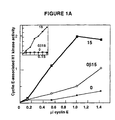
- cyclin E associated H1 kinase inhibition assays aliquots (37.5 11g of protein) from 100,000 x g supernatants of lysates prepared from exponentially growing Mv1Lu cells, were incubated for 30 min. at 37 °C with physiological amounts of baculoviral cyclin E either alone or in the presence of the indicated volumes of renatured eluates. After incubation, samples were precipitated with cyclin E antiserum, and assayed for histone H1 kinase activity. The relative cyclin E-associated H1 kinase activity was quantitated using a Molecular Dynamics Phosphorimager ImageQuant software.
- Non-proliferating cells contain an inhibitor of Cdk2 activation
- extracts from non-proliferating cells demonstrated an elevated threshold level of cyclin E necessary to activate Cdk2.
- Contact inhibited cells appeared to have a higher threshold than cells arrested in G1 by exposure to TGF- ⁇ , but in both cases the cyclin E requirement was substantially greater than the physiological levels of cyclin E achieved in proliferating cells.
- a Cdk2 inhibitor binds to cyclin E-Cdk2 complexes
- Cyclin E-Cdk2 complexes were formed by mixing extracts from Sf9 cells infected with baculoviral vectors expressing either Cdk2 tagged with an influenza virus hemagglutinin (HA) epitope or cyclin E. Although neither extract alone contains significant H1 kinase activity, mixing of the extracts yields high levels of active enzyme (Kato et al., 1993).
- the cyclin E-Cdk2 (HA) complexes were immunoprecipitated with sepharose-linked monoclonal antibody directed against the HA tag on Cdk2.
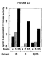
- Cyclin E-Cdk2 complexes were more effective at removing the inhibitory activity than was cyclin E, suggesting that the inhibitor interacted preferentially with complexes. Cyclin E was added to a non-proliferating cell extract at a level below the threshold necessary to activate Cdk2 ( Figure 2C). The assembly of additional cyclin-Cdk2 complexes was then induced by supplementing the extracts with an exogenous Cdk2 protein that was rendered catalytically inactive by a mutation of its ATP binding site (Gu et al., 1992).
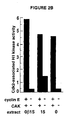
- Cdc2 was activated equally when cyclin B was added to extracts from either proliferating cells or TGF- ⁇ arrested cells (Figure 3A). Therefore, functional CAK was present in extracts from TGF- ⁇ treated cells. CAK was limiting in this experiment since addition of purified CAK to these extracts catalyzed the activation of additional cyclin B-Cdc2 complexes ( Figure 3B). Moreover, the activity of the added CAK was similar in extracts from TGF- ⁇ treated and proliferating cells ( Figure 3B). Thus, exogenous CAK was not inhibited.
- cyclin E was added to non-proliferating cell extracts at sub-threshold levels and the cyclin E-Cdk2 complexes were immunoprecipitated using anti-cyclin E antibodies. No tyrosine phosphorylation of Cdk2 in the inactive cyclin E-Cdk2 complexes was detected by immunoblotting with anti-phosphotyrosine antibodies (not shown). As a positive control, phosphotyrosine was readily detected in Cdc2 immunoprecipitated from human cells.
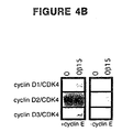
- Cdk4 is a partner of the D-type cyclins and does not form active complexes with cyclins E, A or B. It interacts equally well with each of the D-type cyclins when they are co-expressed in insect cells. Cyclin D-Cdk4 complexes are poorly active on histone H1 but show strong activity using the Rb protein as substrate (Matsushime et al., 1992; Kato et al., 1993). Complexes between Cdk4 and either cyclin D1, D2 or D3 were assembled by co-infection of Sf9 cells with baculoviral vectors and Sf9 lysates were added to extracts from proliferating and non-proliferating Mv1Lu cells.
- cyclin D2-Cdk4 complexes were added to extracts from proliferating cells.
- the cyclin D2-Cdk4 complex did not have CAK activity, since it was unable to substitute for CAK in promoting the activation of cyclin E-Cdk2 complexes assembled from proteins expressed in Sf9 cells (not shown).
- the cyclin D2-Cdk4 complex reversed the inhibition of Cdk2 activation.
- the Cdk2 inhibitor is a 27 kd protein
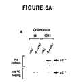
- Mv1Lu cells were metabolically labeled with 35S-methionine, and lysates were incubated with Sepharose beads that contained immunoadsoroed recombinant Cdk2, either alone or in complexes with recombinant cyclin E.
- Denatured 35S-labeled proteins eluted by heating the beads with buffer containing 1% SDS, were visualized by gel electrophoresis and fluorography ( Figure 5A).
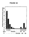
- Extracts from metabolically-labeled TGF- ⁇ treated cells were subjected to chromatography over cyclin E-Cdk2-sepharose or, as a control, Cdk2-sepharose. After washing, the beads were eluted with an acidic buffer, and one portion of the eluate was analyzed by SDS PAGE. This showed that p27 was the predominant labeled species recovered and was present only in the eluate from cyclin E-Cdk2 beads ( Figure 7A). Samples from the same eluates were assayed for the presence of the Cdk2 inhibitor, and this activity was present in the eluate from cyclin E-Cdk2 beads but not Cdk2 beads ( Figure 7B).
- the inhibitor and p27 Kip1 share the following characteristics: they bind to cyclin E-Cdk2 complexes but not to Cdk2 alone; they are only detected in extracts from growth arrested cells; they can be sequestered by cyclin D2-Cdk4 complexes but not by either component alone; they are heat stable; they are latent in extracts of proliferating cells but can be unmasked by brief heat treatment.
- purified p27 Kip1 inhibits Cdk2 activation by cyclin E when added to an extract from proliferating cells.
- p27 Kip1 is at least a component of the Cdk2 inhibitor, it has not been determined whether inhibition is due to p27 Kip1 alone, or whether p27 Kip1 recruits additional proteins to the cyclin E-Cdk2 complex. It has further been determined that p27, as well as cyclin E and Cdk2 are present in other organisms, e.g., mice and humans (data not shown).
- the mechanism of p27 Kip1 inhibition has features that distinguish it from pathways that control activation of the mitotic cyclin-Cdc2 complexes.
- p27 Kip1 appears to act stoichiometrically rather than catalytically.
- tyrosine phosphorylation of Cdk2 was not detected in inactive cyclin E-Cdk2 complexes containing p27 Kip1 , suggesting that p27 Kip1 does not have tyrosine kinase activity or inhibit a tyrosine phosphatase.
- p27 Kip1 links growth inhibitory signals to cell cycle arrest p27 Kip1 was discovered in cells arrested in G1 by either contact inhibition or TGF- ⁇ . A similar activity has also been found to block Cdk2 activation in various cell types deprived of specific growth factors, including serum-starved fibroblasts and IL-2 deprived lymphocytes (unpublished observations). Inhibition of Cdk2 activation by p27 Kip1 or functionally similar proteins, may be a general mechanism through which diverse extracellular and intracellular signals exert control on cell proliferation.
- p27 Kip1 constrains cell proliferation by setting the threshold level of cyclin E necessary to activate Cdk2. If p27 Kip1 acts stoichiometrically, as these data suggest, then the Cdk2 activation threshold is reached soon after the amount of cyclin E in the cell exceeds the amount of active p27 Kip1 . In arrested cells this threshold is set higher than physiological cyclin E levels, and consequently only inactive cyclin E-Cdk2 complexes assemble.
- the cyclin A-Cdk2 complex may be subject to similar control (Koff et al., 1993; Firpo et al., in preparation), and an inabilicy to activate this complex should also contribute to cell cycle arrest (Girard et al. 1991: PacJano et al., 1992: 1993; Tsai et al., 1993).
- the abundance of "free" p27 Kip1 that is able to interact with the cyclin E-Cdk2 complex might, therefore, be modulated by an upstream regulator, such as the cyclin D2-Cdk4 complex, which also binds to p27 Kip1 directly. This prevents association with cyclin E-Cdk2 and enables its functional activation, at least in vitro.
- an upstream regulator such as the cyclin D2-Cdk4 complex
- D-type cyclins are themselves targets of growth inhibitory signals (reviewed in Sherr, 1993). Their synthesis is rapidly reduced in growth factor-deprived cells (Matsushime et al., 1991; Won et al., 1992; Kato et al., in press) and in contact inhibited cells (unpublished observations leading to a reduction in cyclin D-Cdk4 levels (Matsusnime et al., 1992). While D-type cyclin levels are not greatly affected by TGF- ⁇ blockade, TGF- ⁇ does reduce synthesis of Cdk4 so that a net reduction in cyclin D-Cdk4 complexes is achieved nevertheless (Ewen et al., 1993b).
- cyclin D2-Cdk4 It is not likely that the only role of cyclin D2-Cdk4 is to titrate p27 Kip1 , but rather, complex accumulation is likely to trigger the Cdk4-mediated phosphorylation of particular substrates necessary for G1 progression.
- cyclin D complexed with catalytically inactive Cdk4 is sufficient to sequester p27 Kip1 , but is unlikely to fully substitute for all essential Cdk4 functions in vivo.
- p27 Kip1 induced cell cycle arrest is that cells can accumulate inactive cyclin E-Cdk2 complexes. Recovery from cell cycle arrest, therefore, might not require synthesis of new cyclin E and assembly of new cyclin E-Cdk2 complexes. Rather the cell may make use of this latent pool of inactive complexes when cell proliferation resumes. This might be essential under circumstances where the signals that promoted cyclin synthesis were transient, and absent when the growth inhibitory signals ceased. Thus far, however, conditions have not been defined that allow re-activation of inactive cyclin E-Cdk2p27 Kip1 complexes. In vitro, only cyclin E-Cdk2 complexes which assemble after titration of p27 Kip1 are active, and the same may be true in vivo as well.
- p27 Kip1 in proliferating cells suggests that its role may not be restricted to inducing cell cycle arrest in response to extracellular signals. It may also set the cyclin E threshold for execution of the G1 to S transition during each mitotic cycle.
- Cell fusion experiments have indicated that entry into S phase in mammalian fibroblasts is controlled by an activator that accumulates continuously during G1 (Foumier and Pardee, 1975; Rao et al., 1977). By comparing the rate of S phase entry in mono- bi- and tri-nucleate cells it was concluded that the amount of this activator rather than its concentration was critical in determining the start of S phase.
- p27 Kip1 may enforce order during G1 progression
- Cyclin-Cdk complexes appear in a specific order as cells transit G1 (Sherr, 1993). If it is assumed that this temporal order is essential for normal G1 progression, then cells must solve the problem of restoring order during recovery from cell cycle arrest. For example, contact inhibition and TGF- ⁇ interfere with the accumulation of cyclin D-Cdk4 complexes, but do not affect synthesis of cyclin E- and cyclin A-Cdk2 complexes, which act later in the cell cycle. If the cyclin E-Cdk2 and cyclin A-Cdk2 complexes were active during cell cycle arrest, then the normal order of Cdk action would be lost.
- p27 Kip1 might ensure that this does not happen by preventing activation of these pre-existing complexes during cell cycle arrest.
- the activity of p27 Kip1 is itself controlled by cyclin D2-Cdk4, then this would provide an efficient mechanism for maintaining Cdk2 inactive until cyclin D-Cdk4 complexes assemble and execute their functions.
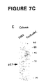
- Mv1Lu cells were synchronized by contact inhibition, treated with TGF-b, metabolically labeled, lysed, immunoprecipitated with anti-Cdk2 antibody or chromatographed on cyclin E-Cdk2 affinity columns.
- the 27kd band present in Cdk2 immunoprecipitates and in the cyclin E-Cdk2 affinity column eluates was cut out from the gels, digested with 0.1 ⁇ g of V8 protease, and resolved on 15-22.5% gradient gels.
- the human cyclin E cDNA (Koff et al., 1991) was tagged at the N-terminus with a hexahistidine sequence. This cDNA was cloned into baculovirus transfer vector pVL1392, and expressed in Sf9 cells as described in the BaculoGold Transfection Kit (Pharmingen). Baculoviral proteins were prepared by the method of Desai et al., 1992.
- Specifically bound proteins were eluted with 6M guanidium hydrochloride solution, dialyzed overnight against 1xHBB buffer (25 mM HEPES-KOH, pH 7.7, 150 mM NaCl, 5 mM MgCl 2 , 0.05% NP-40 and 1 mM DTT) (Kaelin Jr et al., 1992) and acetone-precipitated.
- 1xHBB buffer 25 mM HEPES-KOH, pH 7.7, 150 mM NaCl, 5 mM MgCl 2 , 0.05% NP-40 and 1 mM DTT
- Protein was fractionated by SDS-PAGE, electroblotted onto nitrocellulose, and the Ponceau S-stained 27 kDa band was excised and processed for internal amino acid sequence analysis (Tempst et al., 1990).
- HPLC peak fractions over trypsin background
- MALDI-TOF matrix-assisted laser-desorption
- Mass analysis (on 2% aliquots) was carried out using a model LaserTec Research MALDI-TOF instrument (Vestec), and a-cyano-4-hydroxy cinnamic acid as the matrix.
- Chemical sequencing on 95% of the sample was done using an Applied Biosystems 477A sequenator optimized for femtomole level analysis.
- RT-PCR reactions were performed using degenerate oligonucleotides as primers and total RNA from contact-inhibited Mv1Lu cells as template.
- the combination of one pair of primers yielded a 135 bp fragment that was used to screen a lZAPII cDNA library prepared from Mv1Lu cells.
- the mouse Kip1 cDNA was obtained from a lEXlox mouse embryo cDNA library (Novagen), and the human Kip1 cDNA was obtained from a lgt11 kidney cDNA library (Clontech). Poly(A) + RNA blots were hybridized with a PCR-derived fragment of the mouse Kip1 cDNA labeled by random priming.
- a NdeI-XhoI fragment containing the coding region of the mouse Kip1 cDNA was subcloned to pCITE2a (Novagen). This construct encodes a fusion protein containing a C-terminal hexahistidine sequence and 6 amino acids from the vector at the N-terminus of Kip1. In vitro transcription and translation were performed using Red Nova lysate (Novagen).
- a 591bp PCR generated NheI-XhoI fragment of the mouse Kip1 cDNA containing the full length coding region was subloned into pET21a (Novagen), yielding a construct that encodes Kip1 with a C-terminal hexahistidine sequence.
- the protein was expressed in BL21(DE3) bacteria and purified by sonicating cells in a solution containing 8M urea, 50 mM Tris-HCl (pH 7.4), 20 mM imidazole, clarified by centrifugation and bound to Ni ++ -NTA agarose for 1 h at 4 °C.
- the column was washed with a 6M to 0.75 M urea reverse gradient in 0.5M sodium chloride, 50 mM Tris (pH 7.4), and 20% glycerol and eluted with 200 mM imidazole, 20 mM HEPES pH 7.4, 1M KCl, 100 mM EDTA.
- the eluate was dialysed overnight against 1xHBB buffer and stored at -80°C until use.
- H5 insect cell extracts containing baculovirally-expressed cyclins and Cdks were incubated with recombinant Kip1 for 30 min at 37°C, precipitated with anti-HA antibody, and the histone H1 kinase activity of these complexes was assayed (Koff et al., 1993).
- Rb kinase reactions were done according to Matsushime et al. (1991). The phosphorylation of the histone H1 band and Rb band were quantitated with Phosphorimager (Molecular Dynamics).
- hypotonic cell extracts from exponentially growing A549 cells were incubated with baculoviral His-cyclin E protein, with or without Kip1, at 37°C for 30 min. Mixtures were then diluted 10-fold in 1 x NP40 RIPA buffer containing 20 mM imidazole and incubated with Ni ++ -NTA-agarose at 4°C for 1h. One portion of the samples was run on 12% SDS-PAGE, and immunoblotted with anti-Cdk2 antibody (Koff et al., 1993).
- the mouse Kip1 cDNA (nucleotides -82 to +591) was subcloned into pCMV5 (Attisano et al., 1993).
- R-1B cells were cotransfected with 0.5 ⁇ g/ml of pCEXV-3 containing murine CD16 cDNA (Kurosaki and Ravetch, 1989) and 3 ⁇ g/ml of pCMV5 alone, or with 3 ⁇ g/ml of pCMV5-Kip1 (Attisano et al., 1993).
- CD16 immunostained cells (Wirthmueller et al., 1992) were analyzed by flow cytometry using FACScan (Becton-Dickinson) and Multicycle software (PHOENIX Flow Systems).
- Lysates from contact-inhibited Mv1Lu cells were heated to 100 °C, cleared of insoluble material, and allowed to bind to a cyclin E-Cdk2 affinity column. Elution with 6 M guanidium hydrochloride yielded recombinant Cdk2 released from the column, and the 27 kd protein Kip1. Dialyzed aliquots of this sample had strong inhibitory activity towards cyclin E-Cdk2 in histone H1 kinase assays, and this activity was shown to coelute from SDS-PAGE gel slices with Kip1. The Kip1 yield from two separate preparations ( ⁇ 2x10 10 cells each) was 0.3 ⁇ g and 1 ⁇ g, respectively.
- Kip1 tryptic peptide sequences were obtained by automated Edman degradation and used to design degenerate oligonucleotide primers for cDNA amplification by the reverse transcription-polymerase chain reaction (RT-PCR).
- RT-PCR reverse transcription-polymerase chain reaction
- a PCR product amplified out of reverse-transcribed Mv1Lu mRNA was used to screen a Mv1Lu cDNA library. This yielded one single positive clone that encoded the sequences obtained from the purified protein ( Figure 9A). Screening of cDNA libraries from human kidney and mouse embryo with the Kip1 cDNA yielded clones of highly related sequence.
- the human and mouse Kip1 cDNAs had open reading frames of 594 and 591 bp, respectively, starting with an ATG codon in a favorable translation initiation context and preceded by stop codons (data not shown). Compared to these open reading frames, the mink clone was incomplete, and ended at nucleotide 534 ( Figure 9A).
- the Kip1 cDNA encodes a predicted protein of 198 amino acids (22,257 daltons) in human and 197 amino acids (22,208 daltons) in mouse. These values are smaller than the 27 kd value obtained with the purified mink protein by SDS-PAGE. To resolve this discrepancy, a cDNA encoding the mouse Kip1 sequence was constructed, and tagged at the C-terminus with a hexahistidine sequence ( ⁇ 1 kd mass).
- Kip1 is highly conserved and related to Cip1/WAF1
- Kip1 The predicted human, mouse and mink Kip1 amino acid sequences are highly related, showing ⁇ 90 % identity ( Figure 9A).
- a Genbank search revealed that, at the amino acid level, Kip1 shows significant homology only to Cip1/WAF1. The similarity was largely limited to a 60-amino acid segment in the N-terminal half of the protein. This region was 44% identical to the corresponding region in Cip1/WAF1 ( Figure 9B).
- Kip1 has a putative bipartite nuclear localization signal (Dingwall and Laskey, 1991) near the C-terminus ( Figure 9B).
- the Kip1 sequence does not have a putative zinc finger motif in the N-terminal region, and has a C-terminal extension of 23 amino acids that contains a consensus Cdc2 phosphorylation site ( Figure 9B).
- Kip1 the inhibitory activity of Kip1 resided in the region of similarity to Cip1/WAF1.
- Kip1 prevents Cdk2 activation
- Kip1 was originally identified as a factor whose presence in extracts from quiescent cells rendered Cdk2 refractory to activation by phosphorylation at Thr 160 .
- its effect on cyclin E-dependent Cdk2 activation in extracts from exponentially growing cells was assayed (Koff et al., 1993).
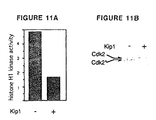
- A549 human lung carcinoma cell extracts were incubated with histidine-tagged cyclin E which was then retrieved and assayed for associated histone H1 kinase activity (Figure 11A).
- Addition of histidine-tagged Kip1 to the cell extracts markedly decreased the level of cyclin E-associated kinase activity ( Figure 11A).
- Kip 1 overexpression inhibits cell entry into S phase
- Mouse Kip1 subcloned into a mammalian expression vector was transfected into Mv1Lu cells under conditions in which up to 65% of the cell population takes up and transiently expresses transfected plasmids (Attisano et al., 1993).
- the rate of 125 I-deoxyuridine incorporation into DNA was reduced 70% in cells transfected with Kip1 compared to cells transfected with vector alone (Table III).
- Kip1 blocks entry into S phase Time after transfection Vector 125 I-deoxyuridine incorporation a Percentage of CD16 + cells in S phase b 24 h pCMV5 27,581 ⁇ 5,126 26 ⁇ 5 pCMV5-Kip1 8,386 ⁇ 1,250 9 ⁇ 2 43 h pCMV5 5,126 ⁇ 47 35 ⁇ 2 pCMV5-Kip1 1,510 ⁇ 140 7 ⁇ 1
- the TGF-b receptor-defective R-1B cell line was cotransfected with a human CD16 expression vector and pCMV5 or pCMV5 containing the mouse Kip1 cDNA. Assays were conducted at the indicated times after transfection.
- Kip1 was cotransfected with a CD16 expression vector (Kurosaki and Ravetch, 1989) that allowed flow cytometric separation of the transfected cells based on CD16 immunofluorescence.
- the CD16 + population cotransfected with Kip1 showed a larger proportion of cells in G1 phase and a smaller proportion in S phase than the CD16 + population cotransfected with vector alone (Table III), suggesting that Kip1 overexpression obstructed cell entry into S phase.
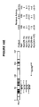
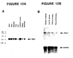
- Kip1 mRNA expression in various human tissues was determined by Northern blot analysis. The only mRNA detected was a species of 2.5 kb present at similar levels in all tissues tested, although it was somewhat higher in skeletal muscle and lower in liver and kidney (Figure 12A). Kip1 mRNA levels were similar in exponentially proliferating and contact-inhibited Mv1Lu cells, and did not change when cells were released from contact inhibition by being plated at low density in the presence of serum (Figure 12B). Addition of TGF-b to cells released from contact-inhibition also did not affect Kip1 mRNA levels ( Figure 12B). These results indicate that the regulation of Kip1 by extracellular antiproliferative signals occurs at a post-transcriptional level.
- Kip1 encodes a protein of 198 amino acids that is highly conserved ( ⁇ 90 % identity) in mouse and mink. Its most distinctive feature is a 60-amino acid region in the N-terminal half that has amino acid sequence similarity to Cip1/WAF1 (El-Deiry et al., 1993; Harper et al., 1993; Xiong et al., 1993). Like Cip1/WAF1, Kip1 contains a potential nuclear localization signal in the C-terminal region. In Kip1, this region also contains a consensus Cdc2 kinase site that might play a role in feed-back regulation by their target kinases.
- Kip1 and Cip1/WAF1 define a family of mammalian Cdk inhibitors with different regulatory properties. Kip1 is involved post-transcriptionally in the action of extracellular signals (present work) and its silencing in exponentially growing cells correlates with binding to a heat-labile component. In contrast, Cip1/WAF1 is regulated transcriptionally by p53, senescence and cell quiescence. Kip1 and Cip1/WAF1 are more effective against G1 Cdks than against mitotic Cdks.
- Kip1 was more effective against cyclin E-Cdk2 than against cyclin A-Cdk2 (or cyclin D2-Cdk4) whereas in similar assays, Cip1/WAF1 was more effective against cyclin A-Cdk2 (Harper et al., 1993). Kip1 effectiveness is likely defined by its binding affinity for a given cyclin-Cdk complex.
- the Kip1 region that is similar to Cip1/WAF1 is sufficient to inhibit Cdk activity when tested as a 52-amino acid peptide in vitro.
- This 52 amino acid segment contains the sequence LFGPVN which corresponds to the longest uninterrupted stretch of identity to Cip1/WAF1 and, interestingly, is similar to the FAR1 sequence LSQPVN located in a region required for interaction with CLN2-CDC28 (Peter et al., 1993).
- Kip1 can inhibit both the process of Cdk activation and the kinase activity of cyclin-Cdk complexes assembled and activated in intact cells. Kip1 was originally identified as a factor whose presence in extracts of quiescent cells rendered them unable to activate Cdk2 by phosphorylation at Thr 160 . Indeed, recombinant Kip1 inhibits Cdk2 Thr 160 phosphorylation and activation in vitro. Although Kip1 could act as an inhibitor of the Cdk-activating kinase, previous results tend to argue against this possibility.
- Kip1 both on Cdk2 activation and Cdk2 activity, might relate to the fact that Thr 160 is located in a loop that closes the substrate-binding cleft in the Cdk2 structure (DeBondt et al., 1993). It is conceivable that binding of Kip1 to this region might interfere with Thr 160 phosphorylation as well as with the catalytic function of activated Cdk2.
- Cyclin E-Cdk2 and cyclin D-Cdk4 are rate limiting for G1 progression (Jiang et al., 1993; Ohtsubo and Roberts, 1993; Jo et al., 1993). Inhibition of these kinases by Kip1 in vivo would render cells unable to reach that transition. The strong reductions in the rate of DNA synthesis and the proportion of cells in S phase caused by Kip1 transfection are consistent with this possibility and with a role of Kip1 as mediator of extracellular growth inhibitory signals.
- Kip1 might be a passive regulator whose uniform levels could ensure that active Cdks become available only when their levels reach the threshold imposed by binding to Kip1. In the latter situation, even small effects of mitogens and antimitogens on cyclin or Cdk protein levels could become amplified by the existence of that threshold.
Landscapes
- Health & Medical Sciences (AREA)
- Chemical & Material Sciences (AREA)
- Life Sciences & Earth Sciences (AREA)
- Organic Chemistry (AREA)
- General Health & Medical Sciences (AREA)
- Medicinal Chemistry (AREA)
- Pharmacology & Pharmacy (AREA)
- Animal Behavior & Ethology (AREA)
- Nuclear Medicine, Radiotherapy & Molecular Imaging (AREA)
- General Chemical & Material Sciences (AREA)
- Public Health (AREA)
- Chemical Kinetics & Catalysis (AREA)
- Veterinary Medicine (AREA)
- Bioinformatics & Cheminformatics (AREA)
- Toxicology (AREA)
- Engineering & Computer Science (AREA)
- Molecular Biology (AREA)
- Genetics & Genomics (AREA)
- Biophysics (AREA)
- Biochemistry (AREA)
- Gastroenterology & Hepatology (AREA)
- Zoology (AREA)
- Proteomics, Peptides & Aminoacids (AREA)
- Cell Biology (AREA)
- Peptides Or Proteins (AREA)
- Medicines That Contain Protein Lipid Enzymes And Other Medicines (AREA)
- Preparation Of Compounds By Using Micro-Organisms (AREA)
- Investigating Or Analysing Biological Materials (AREA)
- Saccharide Compounds (AREA)
- Medicines Containing Material From Animals Or Micro-Organisms (AREA)
- Micro-Organisms Or Cultivation Processes Thereof (AREA)
- Measuring Or Testing Involving Enzymes Or Micro-Organisms (AREA)
Claims (38)
- Isoliertes Polypeptid, umfassend einen Aminosäureteil mit 90 % Homologie zu den Resten +28 bis +88 der in Fig. 9B dargestellten oberen Polypeptidsequenz, wobei das Polypeptid die Aktivierung eines Cyclin E-Cdk2-Komplexes hemmt.
- Rekombinantes Polypeptid, umfassend einen Aminosäureteil mit 90 % Homologie zu den Resten +28 bis +88 der in Fig. 9B dargestellten oberen Polypeptidsequenz, wobei das Polypeptid die Aktivierung eines Cyclin-E-Cdk2-Komplexes hemmt.
- Polypeptid nach Anspruch 1 oder 2, wobei die Polypeptidsequenz identisch mit einer beliebigen der drei in Fig. 9A dargestellten Polypeptidsequenzen ist.
- Polypeptid nach Anspruch 1 oder 2, wobei das Polypeptid eine Sequenz umfasst, die identisch mit den Aminosäureresten +28 bis +88 der in Fig. 9B dargestellten oberen Polypeptidsequenz ist.
- Isoliertes oder rekombinantes Polypeptid, umfassend eine Aminosäuresequenz entsprechend den Resten 28-88 der in Fig. 9B dargestellten oberen Polypeptidsequenz, wobei das Polypeptid die Aktivierung eines Cyclin E-Cdk2-Komplexes hemmt.
- Polypeptid nach einem der vorstehenden Ansprüche, wobei das Polypeptid ein scheinbares Molekulargewicht von etwa 27 kD aufweist.
- Polypeptid nach einem der vorstehenden Ansprüche, wobei das Polypeptid die Zell-Zyklus-Progression von der G1- zur S-Phase hemmt.
- Polypeptid nach einem der vorstehenden Ansprüche, wobei das Polypeptid humanen Ursprungs ist.
- Isoliertes oder rekombinantes Nucleinsäuremolekül, umfassend eine Nucleotidsequenz, die für ein Polypeptid nach einem der vorstehenden Ansprüche kodiert.
- Nucleinsäuremolekül nach Anspruch 9, wobei es sich bei dem Nucleinsäuremolekül um ein DNA-Molekül handelt.
- Nucleinsäuremolekül nach Anspruch 10, wobei es sich bei dem DNA-Molekül um ein cDNA-Molekül handelt.
- Nucleinsäuremolekül nach Anspruch 9, wobei es sich bei dem Nucleinsäuremolekül um ein RNA-Molekül handelt.
- Vektor, umfassend das Nucleinsäuremolekül nach einem der Ansprüche 9 bis 12.
- Vektor nach Anspruch 13, ferner umfassend eine transkriptionale regulatorische Sequenz, die funktionell mit der Nucleotidsequenz im Innern des Vektors verknüpft ist.
- Vektor nach Anspruch 13, wobei es sich bei dem Vektor um ein Plasmid handelt.
- Vektor nach Anspruch 13, wobei es sich bei dem Vektor um ein Virus handelt.
- Wirt-Vektor-System zur Herstellung eines Polypeptids, umfassend den Vektor von Anspruch 13 in einem geeigneten Wirt.
- Wirt-Vektor-System nach Anspruch 17, wobei es sich bei dem geeigneten Wirt um eine bakterielle Zelle handelt.
- Wirt-Vektor-System nach Anspruch 17, wobei es sich bei dem geeigneten Wirt um eine eukaryontische Zelle handelt.
- Wirt-Vektor-System nach Anspruch 19, wobei es sich bei der eukaryontischen Zelle um eine Insektenzelle handelt.
- Isolierter Antikörper oder Fragment davon, der spezifisch immunoreaktiv mit einem Polypeptid nach einem der Ansprüche 1 bis 8 ist.
- Antikörper nach Anspruch 21, wobei es sich bei dem Antikörper um einen monoklonalen Antikörper handelt.
- Antikörper nach Anspruch 21, wobei der Antikörper mit einer nachweisbaren Markierung markiert ist.
- Partiell gereinigtes Präparat von polyklonalen Antikörpern oder einem Fragment davon, das spezifisch mit einem Polypeptid nach einem der Ansprüche 1 bis 8 immunoreaktiv ist.
- Verfahren zum Identifizieren eines Mittels, das zur spezifischen Hemmung der Fähigkeit eines Polypeptids nach einem der Ansprüche 1 bis 8, die Aktivierung des Cyclin E-Cdk2-Komplexes zu hemmen, befähigt ist, umfassend folgende Schritte:(i) geeignete Mengen an Polypeptid, Cyclin E, Cdk2 und dem Mittel werden unter geeigneten Bedingungen in Kontakt gebracht;(ii) das Polypeptid, Cyclin E, Cdk2 und das Mittel werden unter solchen Bedingungen in Kontakt gebracht, die die Bildung eines aktiven Cyclin E-Cdk2-Komplexes in Abwesenheit des Polypeptids ermöglichen würden;(iii) die Menge des auf diese Weise gebildeten aktiven Cyclin E-Cdk2-Komplexes wird quantitativ bestimmt; und(iv) die Menge des auf diese Weise gebildeten aktiven Cyclin E-Cdk2-Komplexes wird mit der Menge des in Abwesenheit des Mittels gebildeten aktiven Cyclin E-Cdk2-Komplexes verglichen, wobei die Tatsache, dass die Menge des in Gegenwart des Mittels gebildeten aktiven Cyclin E-Cdk2-Komplexes größer als die in Abwesenheit des Mittels gebildete Menge ist, darauf hinweist, dass das Mittel zu einer spezifischen Hemmung der Fähigkeit des Polypeptids, die Aktivierung des Cyclin E-Cdk2-Komplexes zu hemmen, befähigt ist.
- Verfahren zur Bestimmung, ob ein Mittel zur spezifischen Verstärkung der Fähigkeit eines Polypeptids nach einem der Ansprüche 1 bis 8, die Aktivierung des Cyclin E-Cdk2-Komplexes zu hemmen, befähigt ist, umfassend die folgenden Schritte:(i) geeignete Mengen an Polypeptid, Cyclin E, Cdk2 und dem Mittel werden unter geeigneten Bedingungen in Kontakt gebracht;(ii) das Polypeptid, Cyclin E, Cdk2 und das Mittel werden unter solchen Bedingungen in Kontakt gebracht, die die Bildung eines aktiven Cyclin E-Cdk2-Komplexes in Abwesenheit des Polypeptids ermöglichen würden;(iii) die Menge des auf diese Weise gebildeten aktiven Cyclin E-Cdk2-Komplexes wird quantitativ bestimmt; und(iv) die Menge des auf diese Weise gebildeten aktiven Cyclin E-Cdk2-Komplexes wird mit der Menge des in Abwesenheit des Mittels gebildeten aktiven Cyclin E-Cdk2-Komplexes verglichen, wobei die Tatsache, dass die Menge des in Gegenwart des Mittels gebildeten aktiven Cyclin E-Cdk2-Komplexes kleiner als die in Abwesenheit des Mittels gebildete Menge ist, darauf hinweist, dass das Mittel zu einer spezifischen Verstärkung der Fähigkeit des Polypeptids, die Aktivierung des Cyclin E-Cdk2-Komplexes zu hemmen, befähigt ist.
- Verfahren zur Diagnose einer Krankheit bei einem Patienten, umfassend folgende Schritte:(i) die Konzentration eines Polypeptids nach einem der Ansprüche 1 bis 8 in einer Probe von Zellen des Patienten wird bestimmt; und(ii) das Vorliegen oder die Abwesenheit einer Krankheit wird unter Heranziehung der festgestellten Konzentration des Polypeptids diagnostiziert, wobei eine verminderte Konzentration des Polypeptids in der Probe relativ zu einer Konzentration eines Wildtyp-Polypeptids in einer Kontrollprobe von normalen Zellen mit dem Vorliegen einer Krankheit korreliert und wobei das Wildtyp-Polypeptid einen Aminosäureteil mit 90 % Homologie zu den Resten +28 bis +88 des in Fig. 9B dargestellten oberen Polypeptids aufweist, wobei dieses Polypeptid die Aktivierung eines Cyclin E Cdk2-Komplexes hemmt.
- Verfahren nach Anspruch 27, wobei die Konzentration des Polypeptids durch einen Immunoassay bestimmt wird.
- Verfahren nach Anspruch 27, wobei das Wildtyp-Polypeptid eine Aminosäuresequenz von einem der drei in Fig. 9A dargestellten Polypeptidsequenzen aufweist.
- Verfahren nach Anspruch 27, wobei es sich bei dem Patienten um einen Menschen handelt.
- Verfahren zur Bestimmung der relativen Menge eines Polypeptids nach einem der Ansprüche 1 bis 8 in einer Zelle oder Probe von Zellen (Zellprobe), umfassend folgende Schritte:(i) die Konzentration des Polypeptids in der Zellprobe wird durch einen Immunoassay unter Verwendung eines Antikörpers nach einem der Ansprüche 21 bis 24 bestimmt; und(ii) die Konzentration des Polypeptids in der Zellprobe wird mit der für eine Kontrollzelle bestimmten Konzentration eines Wildtyp-Polypeptids verglichen, wobei das Wildtyp-Polypeptid einen Aminosäureteil mit 90 % Homologie zu den Resten +28 bis +88 der in Fig. 9B dargestellten oberen Polypeptidsequenz umfasst, wobei das Polypeptid die Aktivierung eines Cyclin E-Cdk2-Komplexes hemmt.
- Verfahren nach Anspruch 31, wobei das Wildtyp-Polypeptid eine Aminosäuresequenz aufweist, die mit einer beliebigen der drei in Fig. 9A dargestellten Polypeptidsequenzen identisch ist.
- Verfahren zur Bestimmung der Menge eines Polypeptids nach einem der Ansprüche 1 bis 8 in einer Zelle oder Zellprobe, umfassend folgende Schritte:(i) die Zelle oder Zellprobe wird mit einem Antikörper, der selektiv mit dem Polypeptid reaktiv ist, unter Bedingungen kontaktiert, die ausreichen, dass der Antikörper das Polypeptid bindet; und(ii) die Menge des durch den Antikörper gebundenen Polypeptids wird bestimmt.
- Verfahren nach Anspruch 33, wobei der Antikörper mit einer nachweisbaren Markierung markiert ist.
- Verfahren nach Anspruch 34, wobei die Markierung unter Radioisotopen und fluoreszierenden Verbindungen ausgewählt ist.
- Verfahren zur Bestimmung der relativen Menge eines Polypeptids nach einem der Ansprüche 1 bis 8 in einer Zelle oder Zellprobe, umfassend folgende Schritte:(i) die Zelle oder Zellprobe wird mit einem Antikörper nach einem der Ansprüche 21 bis 24 behandelt;(ii) die Konzentration eines zwischen dem Antikörper und dem Polypeptid in der Zelle oder Zellprobe gebildeten Immunokomplexes wird quantitativ bestimmt; und(iii) die Konzentration des Immunokomplexes in der Zellprobe wird mit der für eine Kontrollzelle bestimmten Konzentration eines Wildtyp-Polypeptids verglichen.
- Verfahren zur Bestimmung der relativen Aktivität eines Polypeptids nach einem der Ansprüche 1 bis 8 in einer Zelle oder Zellprobe, umfassend folgende Schritte:(i) die Aktivität des Polypeptids in der Zelle oder Zellprobe wird durch Überwachung der Hemmung der Aktivierung des Cyclin E-Cdk2-Komplexes bestimmt; und(ii) die Aktivität des Polypeptids in der Zelle oder Zellprobe wird mit der in einer Kontrollzelle oder Kontrollzellprobe bestimmten Aktivität eines Wildtyp-p27-Polypeptids verglichen.
- Pharmazeutische Zusammensetzung, umfassend eine wirksame Menge eines rekombinanten Virus, der zur Infektion einer geeigneten Wirtszelle befähigt ist, wobei das rekombinante Virus das Nucleinsäuremolekül nach einem der Ansprüche 9 bis 12 umfasst, und einen pharmazeutisch verträglichen Träger.
Applications Claiming Priority (5)
| Application Number | Priority Date | Filing Date | Title |
|---|---|---|---|
| US275983 | 1988-11-25 | ||
| US17904594A | 1994-01-07 | 1994-01-07 | |
| US179045 | 1994-01-07 | ||
| US08/275,983 US5688665A (en) | 1994-01-07 | 1994-07-15 | Isolated nucleic acid molecules encoding the p27 KIP-1 protein |
| PCT/US1995/000247 WO1995018824A1 (en) | 1994-01-07 | 1995-01-09 | ISOLATED p27 PROTEIN AND ITS ENCODING NUCLEIC ACID MOLECULES |
Publications (3)
| Publication Number | Publication Date |
|---|---|
| EP0749442A1 EP0749442A1 (de) | 1996-12-27 |
| EP0749442A4 EP0749442A4 (de) | 2000-06-28 |
| EP0749442B1 true EP0749442B1 (de) | 2004-07-14 |
Family
ID=26874941
Family Applications (1)
| Application Number | Title | Priority Date | Filing Date |
|---|---|---|---|
| EP95906793A Expired - Lifetime EP0749442B1 (de) | 1994-01-07 | 1995-01-09 | ISOLIERTES p27 PROTEIN UND NUKLEINSÄURE DAFÜR KODIEREND |
Country Status (8)
| Country | Link |
|---|---|
| US (1) | US5688665A (de) |
| EP (1) | EP0749442B1 (de) |
| JP (1) | JP4143119B2 (de) |
| AT (1) | ATE271129T1 (de) |
| AU (1) | AU699969B2 (de) |
| CA (1) | CA2180570C (de) |
| DE (1) | DE69533255T2 (de) |
| WO (1) | WO1995018824A1 (de) |
Families Citing this family (27)
| Publication number | Priority date | Publication date | Assignee | Title |
|---|---|---|---|---|
| US6242575B1 (en) | 1994-01-07 | 2001-06-05 | Fred Hutchinson Institute For Cancer Research | Antibodies for detecting p27 protein |
| US6316208B1 (en) | 1994-01-07 | 2001-11-13 | Memorial Sloan-Kettering Cancer Center | Methods for determining isolated p27 protein levels and uses thereof |
| US6355774B1 (en) | 1994-01-07 | 2002-03-12 | Fred Hutchinson Cancer Research Center | Isolated p27 protein |
| AU2770695A (en) | 1994-07-15 | 1996-02-16 | Fred Hutchinson Cancer Research Center | Isolated p27 protein and methods for its production and use |
| AU5297496A (en) * | 1995-02-17 | 1996-09-04 | Government Of The United States Of America, As Represented By The Secretary Of The Department Of Health And Human Services, The | Methods of preparation and use of recombinant adenoviral vectors |
| US5863904A (en) | 1995-09-26 | 1999-01-26 | The University Of Michigan | Methods for treating cancers and restenosis with P21 |
| US7087582B1 (en) | 1995-09-26 | 2006-08-08 | Regents Of The University Of Michigan | Combination for site-specifically transforming cells in vivo comprising a double-balloon catheter and nucleic acid comprising a gene encoding P21 |
| AU3483795A (en) * | 1995-09-29 | 1997-04-28 | Ciba-Geigy Ag | Ring finger protein |
| JP2000503538A (ja) | 1996-01-18 | 2000-03-28 | フレッド ハッチンソン カンサー リサーチ センター | 細胞周期を仲介する組成物および方法 |
| CA2289942C (en) * | 1997-05-15 | 2009-05-05 | Thomas Jefferson University | Determination of cyclin-dependent kinase inhibitor p27 levels as a prognostic factor in cancer patients |
| US6177272B1 (en) * | 1997-07-21 | 2001-01-23 | The Regents Of The University Of Michigan | Method for treating vascular proliferative diseases with p27 and fusions thereof |
| CA2301333C (en) * | 1997-08-21 | 2011-12-06 | Thomas Jefferson University | Prb2/p130 peptide inhibitors of cdk2 kinase activity |
| AU1801499A (en) * | 1997-12-01 | 1999-06-16 | Sloan-Kettering Institute For Cancer Research | Uses of p27 in prostate cancer |
| US6972170B1 (en) | 1997-12-01 | 2005-12-06 | Sloan-Kettering Institute For Cancer Research | Markers for prostate cancer |
| US6479487B1 (en) * | 1998-02-26 | 2002-11-12 | Aventis Pharmaceuticals Inc. | 6, 9-disubstituted 2-[trans-(4-aminocyclohexyl)amino] purines |
| US6413974B1 (en) * | 1998-02-26 | 2002-07-02 | Aventis Pharmaceuticals Inc. | 6,9,-disubstituted 2-[trans-(4-aminocyclohexyl) amino] purines |
| US5973119A (en) * | 1998-06-05 | 1999-10-26 | Amgen Inc. | Cyclin E genes and proteins |
| JP2002517998A (ja) * | 1998-06-18 | 2002-06-25 | キュラジェン コーポレイション | p27(KIP1)のFKBP−12との相互作用 |
| US6589505B1 (en) | 1999-01-29 | 2003-07-08 | St. Jude Children's Research Hospital | Cells that lack p19ink4d and p27kip1 activity and methods of use thereof |
| HK1049845B (zh) | 1999-05-14 | 2009-05-08 | 弗雷德哈钦森癌症研究中心 | 通过功能性地抑制植物细胞周期蛋白抑制剂基因增加植物细胞增殖的方法 |
| EP1512755A3 (de) * | 1999-06-10 | 2007-11-21 | Sloan-Kettering Institute For Cancer Research | Marker für Prostatakrebs |
| IL147144A0 (en) * | 1999-06-18 | 2002-08-14 | Advanced Res & Tech Inst | Cardiomyocytes with enhanced proliferative potential and methods for preparing and using the same |
| US7482506B2 (en) * | 2001-03-26 | 2009-01-27 | University Of South Florida | Ras p27 mouse models and uses thereof |
| GB0117600D0 (en) * | 2001-07-19 | 2001-09-12 | Trikon Holdings Ltd | Semiconductor structure |
| US7601299B2 (en) * | 2004-06-18 | 2009-10-13 | Roche Diagnostics Operations, Inc. | System and method for coding information on a biosensor test strip |
| US20080053911A1 (en) * | 2006-08-30 | 2008-03-06 | Qiagen Gmbh | Separation of an organic phase from a mixture comprising organic and aqueous phases by solid phase systems |
| PL3104939T3 (pl) | 2014-02-10 | 2024-09-30 | Fred Hutchinson Cancer Center | Leczenie jodkiem sodu zawału serca i urazu niedokrwiennego |
-
1994
- 1994-07-15 US US08/275,983 patent/US5688665A/en not_active Expired - Lifetime
-
1995
- 1995-01-09 AU AU15251/95A patent/AU699969B2/en not_active Ceased
- 1995-01-09 CA CA002180570A patent/CA2180570C/en not_active Expired - Fee Related
- 1995-01-09 DE DE69533255T patent/DE69533255T2/de not_active Expired - Lifetime
- 1995-01-09 AT AT95906793T patent/ATE271129T1/de not_active IP Right Cessation
- 1995-01-09 WO PCT/US1995/000247 patent/WO1995018824A1/en not_active Ceased
- 1995-01-09 EP EP95906793A patent/EP0749442B1/de not_active Expired - Lifetime
- 1995-01-09 JP JP51864595A patent/JP4143119B2/ja not_active Expired - Fee Related
Also Published As
| Publication number | Publication date |
|---|---|
| ATE271129T1 (de) | 2004-07-15 |
| AU699969B2 (en) | 1998-12-17 |
| US5688665A (en) | 1997-11-18 |
| DE69533255T2 (de) | 2005-07-28 |
| CA2180570A1 (en) | 1995-07-13 |
| JPH09509311A (ja) | 1997-09-22 |
| WO1995018824A1 (en) | 1995-07-13 |
| CA2180570C (en) | 2002-03-26 |
| EP0749442A4 (de) | 2000-06-28 |
| DE69533255D1 (de) | 2004-08-19 |
| AU1525195A (en) | 1995-08-01 |
| JP4143119B2 (ja) | 2008-09-03 |
| EP0749442A1 (de) | 1996-12-27 |
Similar Documents
| Publication | Publication Date | Title |
|---|---|---|
| US5688665A (en) | Isolated nucleic acid molecules encoding the p27 KIP-1 protein | |
| Polyak et al. | p27Kip1, a cyclin-Cdk inhibitor, links transforming growth factor-beta and contact inhibition to cell cycle arrest. | |
| Polyak et al. | Cloning of p27Kip1, a cyclin-dependent kinase inhibitor and a potential mediator of extracellular antimitogenic signals | |
| US6316208B1 (en) | Methods for determining isolated p27 protein levels and uses thereof | |
| Harper et al. | The p21 Cdk-interacting protein Cip1 is a potent inhibitor of G1 cyclin-dependent kinases | |
| Toyoshima et al. | p27, a novel inhibitor of G1 cyclin-Cdk protein kinase activity, is related to p21 | |
| Serrano et al. | A new regulatory motif in cell-cycle control causing specific inhibition of cyclin D/CDK4 | |
| Bai et al. | Human cyclin F. | |
| Saha et al. | p21 CIP1 and Cdc25A: competition between an inhibitor and an activator of cyclin-dependent kinases | |
| Azzi et al. | Purification of a 15-kDa cdk4-and cdk5-binding protein. | |
| WO1998034121A9 (en) | Isolated p27 protein and methods for its production and use | |
| Okada et al. | p53 gene family p51 (p63)-encoded, secondary transactivator p51B (TAp63α) occurs without forming an immunoprecipitable complex with MDM2, but responds to genotoxic stress by accumulation | |
| JP2008301815A (ja) | 刺激誘導性I(κ)Bキナーゼ[IKK]シグナルソーム | |
| Kaldis et al. | CAK-independent activation of CDK6 by a viral cyclin | |
| Horton et al. | The cyclin box and C-terminus of cyclins A and E specify CDK activation and substrate specificity | |
| Kwon et al. | Staurosporine-induced G1 arrest is associated with the induction and accumulation of cyclin-dependent kinase inhibitors | |
| Higashi et al. | Cyclin‐dependent kinase‐2 (cdk2) forms an inactive complex with cyclin D1 since cdk2 associated with cyclin D1 is not phosphorylated by cdk7–cyclin‐H | |
| US6635450B1 (en) | Isolated P27 protein, nucleic acid molecules encoding same, methods of identifying agents acting on same, and uses of said agents | |
| EP0871655A2 (de) | Pcna bindende substanz | |
| US6355774B1 (en) | Isolated p27 protein | |
| US6242575B1 (en) | Antibodies for detecting p27 protein | |
| CN100482795C (zh) | Iren蛋白,其制备和应用 | |
| US20020110886A1 (en) | Isolated p27 protein and methods for its production and use | |
| US5770720A (en) | Ubiquitin conjugating enzymes having transcriptional repressor activity | |
| Luo | Isolation and functional studies of D. discoideum cyclin B gene during growth and development |
Legal Events
| Date | Code | Title | Description |
|---|---|---|---|
| PUAI | Public reference made under article 153(3) epc to a published international application that has entered the european phase |
Free format text: ORIGINAL CODE: 0009012 |
|
| 17P | Request for examination filed |
Effective date: 19960725 |
|
| AK | Designated contracting states |
Kind code of ref document: A1 Designated state(s): AT BE CH DE DK ES FR GB GR IE IT LI LU MC NL PT SE |
|
| RIC1 | Information provided on ipc code assigned before grant |
Free format text: 7C 12N 15/12 A, 7C 12N 15/86 B, 7C 12N 5/10 B, 7C 12N 1/21 B, 7C 07K 14/47 B, 7G 01N 33/50 B, 7A 61K 38/17 B, 7A 61K 48/00 B, 7C 12Q 1/68 B |
|
| A4 | Supplementary search report drawn up and despatched |
Effective date: 20000512 |
|
| AK | Designated contracting states |
Kind code of ref document: A4 Designated state(s): AT BE CH DE DK ES FR GB GR IE IT LI LU MC NL PT SE |
|
| RA4 | Supplementary search report drawn up and despatched (corrected) |
Effective date: 20000518 |
|
| 17Q | First examination report despatched |
Effective date: 20011026 |
|
| GRAP | Despatch of communication of intention to grant a patent |
Free format text: ORIGINAL CODE: EPIDOSNIGR1 |
|
| GRAS | Grant fee paid |
Free format text: ORIGINAL CODE: EPIDOSNIGR3 |
|
| GRAA | (expected) grant |
Free format text: ORIGINAL CODE: 0009210 |
|
| AK | Designated contracting states |
Kind code of ref document: B1 Designated state(s): AT BE CH DE DK ES FR GB GR IE IT LI LU MC NL PT SE |
|
| PG25 | Lapsed in a contracting state [announced via postgrant information from national office to epo] |
Ref country code: NL Free format text: LAPSE BECAUSE OF FAILURE TO SUBMIT A TRANSLATION OF THE DESCRIPTION OR TO PAY THE FEE WITHIN THE PRESCRIBED TIME-LIMIT Effective date: 20040714 Ref country code: IT Free format text: LAPSE BECAUSE OF FAILURE TO SUBMIT A TRANSLATION OF THE DESCRIPTION OR TO PAY THE FEE WITHIN THE PRE;WARNING: LAPSES OF ITALIAN PATENTS WITH EFFECTIVE DATE BEFORE 2007 MAY HAVE OCCURRED AT ANY TIME BEFORE 2007. THE CORRECT EFFECTIVE DATE MAY BE DIFFERENT FROM THE ONE RECORDED.SCRIBED TIME-LIMIT Effective date: 20040714 Ref country code: FR Free format text: LAPSE BECAUSE OF FAILURE TO SUBMIT A TRANSLATION OF THE DESCRIPTION OR TO PAY THE FEE WITHIN THE PRESCRIBED TIME-LIMIT Effective date: 20040714 Ref country code: BE Free format text: LAPSE BECAUSE OF FAILURE TO SUBMIT A TRANSLATION OF THE DESCRIPTION OR TO PAY THE FEE WITHIN THE PRESCRIBED TIME-LIMIT Effective date: 20040714 Ref country code: AT Free format text: LAPSE BECAUSE OF FAILURE TO SUBMIT A TRANSLATION OF THE DESCRIPTION OR TO PAY THE FEE WITHIN THE PRESCRIBED TIME-LIMIT Effective date: 20040714 |
|
| REG | Reference to a national code |
Ref country code: GB Ref legal event code: FG4D |
|
| REG | Reference to a national code |
Ref country code: CH Ref legal event code: EP |
|
| REF | Corresponds to: |
Ref document number: 69533255 Country of ref document: DE Date of ref document: 20040819 Kind code of ref document: P |
|
| REG | Reference to a national code |
Ref country code: IE Ref legal event code: FG4D |
|
| PG25 | Lapsed in a contracting state [announced via postgrant information from national office to epo] |
Ref country code: SE Free format text: LAPSE BECAUSE OF FAILURE TO SUBMIT A TRANSLATION OF THE DESCRIPTION OR TO PAY THE FEE WITHIN THE PRESCRIBED TIME-LIMIT Effective date: 20041014 Ref country code: GR Free format text: LAPSE BECAUSE OF FAILURE TO SUBMIT A TRANSLATION OF THE DESCRIPTION OR TO PAY THE FEE WITHIN THE PRESCRIBED TIME-LIMIT Effective date: 20041014 Ref country code: DK Free format text: LAPSE BECAUSE OF FAILURE TO SUBMIT A TRANSLATION OF THE DESCRIPTION OR TO PAY THE FEE WITHIN THE PRESCRIBED TIME-LIMIT Effective date: 20041014 |
|
| PG25 | Lapsed in a contracting state [announced via postgrant information from national office to epo] |
Ref country code: ES Free format text: LAPSE BECAUSE OF FAILURE TO SUBMIT A TRANSLATION OF THE DESCRIPTION OR TO PAY THE FEE WITHIN THE PRESCRIBED TIME-LIMIT Effective date: 20041025 |
|
| REG | Reference to a national code |
Ref country code: CH Ref legal event code: NV Representative=s name: R. A. EGLI & CO. PATENTANWAELTE |
|
| NLV1 | Nl: lapsed or annulled due to failure to fulfill the requirements of art. 29p and 29m of the patents act | ||
| PG25 | Lapsed in a contracting state [announced via postgrant information from national office to epo] |
Ref country code: LU Free format text: LAPSE BECAUSE OF NON-PAYMENT OF DUE FEES Effective date: 20050109 |
|
| PG25 | Lapsed in a contracting state [announced via postgrant information from national office to epo] |
Ref country code: IE Free format text: LAPSE BECAUSE OF NON-PAYMENT OF DUE FEES Effective date: 20050110 |
|
| PG25 | Lapsed in a contracting state [announced via postgrant information from national office to epo] |
Ref country code: MC Free format text: LAPSE BECAUSE OF NON-PAYMENT OF DUE FEES Effective date: 20050131 |
|
| PLBE | No opposition filed within time limit |
Free format text: ORIGINAL CODE: 0009261 |
|
| STAA | Information on the status of an ep patent application or granted ep patent |
Free format text: STATUS: NO OPPOSITION FILED WITHIN TIME LIMIT |
|
| 26N | No opposition filed |
Effective date: 20050415 |
|
| EN | Fr: translation not filed | ||
| REG | Reference to a national code |
Ref country code: IE Ref legal event code: MM4A |
|
| PG25 | Lapsed in a contracting state [announced via postgrant information from national office to epo] |
Ref country code: PT Free format text: LAPSE BECAUSE OF NON-PAYMENT OF DUE FEES Effective date: 20041214 |
|
| PGFP | Annual fee paid to national office [announced via postgrant information from national office to epo] |
Ref country code: CH Payment date: 20120112 Year of fee payment: 18 |
|
| PGFP | Annual fee paid to national office [announced via postgrant information from national office to epo] |
Ref country code: DE Payment date: 20120104 Year of fee payment: 18 |
|
| PGFP | Annual fee paid to national office [announced via postgrant information from national office to epo] |
Ref country code: GB Payment date: 20120104 Year of fee payment: 18 |
|
| REG | Reference to a national code |
Ref country code: CH Ref legal event code: PL |
|
| GBPC | Gb: european patent ceased through non-payment of renewal fee |
Effective date: 20130109 |
|
| PG25 | Lapsed in a contracting state [announced via postgrant information from national office to epo] |
Ref country code: DE Free format text: LAPSE BECAUSE OF NON-PAYMENT OF DUE FEES Effective date: 20130801 Ref country code: CH Free format text: LAPSE BECAUSE OF NON-PAYMENT OF DUE FEES Effective date: 20130131 Ref country code: LI Free format text: LAPSE BECAUSE OF NON-PAYMENT OF DUE FEES Effective date: 20130131 |
|
| PG25 | Lapsed in a contracting state [announced via postgrant information from national office to epo] |
Ref country code: GB Free format text: LAPSE BECAUSE OF NON-PAYMENT OF DUE FEES Effective date: 20130109 |
|
| REG | Reference to a national code |
Ref country code: DE Ref legal event code: R119 Ref document number: 69533255 Country of ref document: DE Effective date: 20130801 |