EP0747701B1 - Essai utilisant des micelles de protection - Google Patents
Essai utilisant des micelles de protection Download PDFInfo
- Publication number
- EP0747701B1 EP0747701B1 EP96304228A EP96304228A EP0747701B1 EP 0747701 B1 EP0747701 B1 EP 0747701B1 EP 96304228 A EP96304228 A EP 96304228A EP 96304228 A EP96304228 A EP 96304228A EP 0747701 B1 EP0747701 B1 EP 0747701B1
- Authority
- EP
- European Patent Office
- Prior art keywords
- probe
- label
- salts
- analyte
- amphiphiles
- Prior art date
- Legal status (The legal status is an assumption and is not a legal conclusion. Google has not performed a legal analysis and makes no representation as to the accuracy of the status listed.)
- Expired - Lifetime
Links
Images


Classifications
-
- G—PHYSICS
- G01—MEASURING; TESTING
- G01N—INVESTIGATING OR ANALYSING MATERIALS BY DETERMINING THEIR CHEMICAL OR PHYSICAL PROPERTIES
- G01N33/00—Investigating or analysing materials by specific methods not covered by groups G01N1/00 - G01N31/00
- G01N33/48—Biological material, e.g. blood, urine; Haemocytometers
- G01N33/50—Chemical analysis of biological material, e.g. blood, urine; Testing involving biospecific ligand binding methods; Immunological testing
- G01N33/53—Immunoassay; Biospecific binding assay; Materials therefor
- G01N33/536—Immunoassay; Biospecific binding assay; Materials therefor with immune complex formed in liquid phase
- G01N33/542—Immunoassay; Biospecific binding assay; Materials therefor with immune complex formed in liquid phase with steric inhibition or signal modification, e.g. fluorescent quenching
-
- G—PHYSICS
- G01—MEASURING; TESTING
- G01N—INVESTIGATING OR ANALYSING MATERIALS BY DETERMINING THEIR CHEMICAL OR PHYSICAL PROPERTIES
- G01N33/00—Investigating or analysing materials by specific methods not covered by groups G01N1/00 - G01N31/00
- G01N33/48—Biological material, e.g. blood, urine; Haemocytometers
- G01N33/50—Chemical analysis of biological material, e.g. blood, urine; Testing involving biospecific ligand binding methods; Immunological testing
- G01N33/53—Immunoassay; Biospecific binding assay; Materials therefor
- G01N33/543—Immunoassay; Biospecific binding assay; Materials therefor with an insoluble carrier for immobilising immunochemicals
- G01N33/54313—Immunoassay; Biospecific binding assay; Materials therefor with an insoluble carrier for immobilising immunochemicals the carrier being characterised by its particulate form
- G01N33/5432—Liposomes or microcapsules
-
- C—CHEMISTRY; METALLURGY
- C12—BIOCHEMISTRY; BEER; SPIRITS; WINE; VINEGAR; MICROBIOLOGY; ENZYMOLOGY; MUTATION OR GENETIC ENGINEERING
- C12Q—MEASURING OR TESTING PROCESSES INVOLVING ENZYMES, NUCLEIC ACIDS OR MICROORGANISMS; COMPOSITIONS OR TEST PAPERS THEREFOR; PROCESSES OF PREPARING SUCH COMPOSITIONS; CONDITION-RESPONSIVE CONTROL IN MICROBIOLOGICAL OR ENZYMOLOGICAL PROCESSES
- C12Q1/00—Measuring or testing processes involving enzymes, nucleic acids or microorganisms; Compositions therefor; Processes of preparing such compositions
- C12Q1/68—Measuring or testing processes involving enzymes, nucleic acids or microorganisms; Compositions therefor; Processes of preparing such compositions involving nucleic acids
- C12Q1/6813—Hybridisation assays
- C12Q1/6816—Hybridisation assays characterised by the detection means
-
- G—PHYSICS
- G01—MEASURING; TESTING
- G01N—INVESTIGATING OR ANALYSING MATERIALS BY DETERMINING THEIR CHEMICAL OR PHYSICAL PROPERTIES
- G01N33/00—Investigating or analysing materials by specific methods not covered by groups G01N1/00 - G01N31/00
- G01N33/48—Biological material, e.g. blood, urine; Haemocytometers
- G01N33/50—Chemical analysis of biological material, e.g. blood, urine; Testing involving biospecific ligand binding methods; Immunological testing
- G01N33/58—Chemical analysis of biological material, e.g. blood, urine; Testing involving biospecific ligand binding methods; Immunological testing involving labelled substances
- G01N33/585—Chemical analysis of biological material, e.g. blood, urine; Testing involving biospecific ligand binding methods; Immunological testing involving labelled substances with a particulate label, e.g. coloured latex
- G01N33/586—Liposomes, microcapsules or cells
Definitions
- This invention concerns compositions and methods for detecting and quantifying nucleic acid analytes. More particularly, the invention relates to the detection of nucleic acid analytes using methods employing the applied disciplines of molecular biology and/or immunology. Applications of such detection methods and compositions include the diagnosis of human or animal disease and infection, the testing of foods and other products intended for human consumption, and forensic and environmental testing.
- microorganisms and viruses and biological molecules such as nucleic acids and proteins
- detection or quantification of specific biomolecules from tissue, sputum, urine, blood, semen, saliva and other biological materials plays an increasingly important role in various fields including, without limitation, the identification of criminal suspects and in paternity testing.
- nucleic acid probes and proteins such as antibodies can be "labeled", or linked to a detectable moiety such as a radionuclide, an enzyme (or enzyme substrate) capable of participating in a predetermined chemical reaction which can be independently monitored, or a fluorescent, luminescent or chemiluminescent compound.
- a detectable moiety such as a radionuclide, an enzyme (or enzyme substrate) capable of participating in a predetermined chemical reaction which can be independently monitored, or a fluorescent, luminescent or chemiluminescent compound.
- nucleic acids and antibodies can be used as molecular probes, one of these types of probes may be more suitable than the other in a given application.
- single-stranded nucleic acids are most often used in methods to detect target nucleic acids in a sample having nucleotide sequences complementary to that of the probe.
- nucleotide sequence is meant an order of consecutive nucleotides (e.g., the phosphate esters of adenosine (A), thymidine (T), cytosine (C), guanadine (G), inosine (I) and uracil (U)) comprising a single-stranded nucleic acid, conventionally described from the 5' terminus to the 3' terminus of the subject nucleic acid strand. Under appropriate reaction conditions a single-stranded nucleic acid may hybridize with another single-stranded nucleic acid to form a double-stranded helical structure held together by hydrogen bonds between pairs of complementary bases on opposing strands.
- nucleotides e.g., the phosphate esters of adenosine (A), thymidine (T), cytosine (C), guanadine (G), inosine (I) and uracil (U)
- nucleic acid probes are most commonly used to detect other nucleic acids in a target region of nucleotide sequence complementarity.
- nucleic acid probes are often more suitable for detecting a given species of organism than are antibody probes. Nucleic acid probes can beakily sensitive. For example, small amounts of target nucleic acids can be amplified using methods such as the polymerase chain reaction (e.g., EPO 0 200 362) and transcription-based amplification systems (e.g., WO 91/01384, WO 93/22461 and WO 94/03472), and can then be detected using specific oligonucleotide probes.
- polymerase chain reaction e.g., EPO 0 200 362
- transcription-based amplification systems e.g., WO 91/01384, WO 93/22461 and WO 94/03472
- Nucleic acid probes can also effectively distinguish slight differences (sometimes a single mismatch) between versions of the same gene or nucleotide sequence, which can permit diagnostic screening for genetic defects or mutations. Nucleic acid probes can be used in the identification of a particular individual within a species, for example to discount or even identify a criminal suspect by screening specimens of his or her nucleic acids.
- Antibodies also remain effective tools for the identification and detection of specific cells, viruses, and proteins. Immunodiagnostic methods can thus be used to test for the presence of a specific disease or infection state, genetic defect, or physiological condition connected with the specific antigen or substance sought to be detected.
- probe:analyte complex or “probe:analyte conjugate” is meant a specifically-associated molecular species containing at least one antibody or nucleic acid probe molecule (preferably labeled with a detectable moiety or atom) in stable association with at least one analyte molecule.
- An analyte molecule is a molecular species such as a nucleic acid or antigen sought to be detected, quantitated and/or identified.
- a “hybrid” or “probe:analyte hybrid” is a probe:analyte complex in which probe and analyte are both nucleic acids.
- Assay methods utilizing a physical separation step may employ a solid phase matrix, such as glass, minerals or polymeric materials in the separation process.
- the separation may involve preferentially binding the probe: analyte complex to the solid phase matrix while allowing the unassociated probe molecules to remain in a liquid phase.
- the amount of probe bound to the solid phase support after a washing step is proportional to the amount of analyte in the sample.
- the assay may involve preferentially binding the unassociated probe while leaving the probe:analyte complex to remain in the liquid phase; in this case the amount of probe in the liquid phase, again after a washing step, is proportional to the amount of analyte in the original sample.
- the solid support can include, without limitation, an adsorbent such as hydroxylapatite, a polycationic moiety, a hydrophobic or "reverse phase” material, an ion-exchange matrix such as DEAE, a gel filtration matrix, or a -combination of one or more of these solid phase materials.
- an adsorbent such as hydroxylapatite, a polycationic moiety, a hydrophobic or "reverse phase” material, an ion-exchange matrix such as DEAE, a gel filtration matrix, or a -combination of one or more of these solid phase materials.
- media such as gel filtration, the separation is not due to binding of the oligonucleotide but is caused by molecular sieving of differently sized or shaped molecules.
- a heterogeneous assay method may also involve binding the probe to a solid phase matrix prior to addition of a sample suspected of containing the analyte of interest.
- the sample can be contacted with the label under conditions which would cause the analyte of interest to be labeled, if present in the sample mixture.
- the solid phase matrix may be derivatized or activated so that a covalent bond is formed between the probe and the matrix; alternatively, the probe may be bound to the matrix through strong non-covalent interactions, including, without limitation: ionic, hydrophobic, reverse-phase, immunobinding, chelating, and enzyme-substrate interactions.
- the separation step is accomplished by washing the solid phase matrix free of any unbound labeled analyte.
- the analyte can be bound to the solid phase matrix and contacted with labeled probe, with the excess free probe washed from the matrix before detection.
- HPA hybridization protection assay
- Patent 5,283,174 in which a probe is linked to a chemiluminescent moiety, contacted with a analyte and then subjected to selective chemical degradation or detectable change in stability under conditions which alter the chemiluminescent reagent bound to unhybridized probe without altering the chemiluminescent reagent bound to an analyte:probe conjugate.
- This patent enjoys common ownership with the present application.
- Competition assays in which a labeled probe or analyte competes for binding with its unlabeled analog, are also commonly used in a heterogeneous format. Depending on how such a system is designed, either the amount of bound, labeled probe or the amount of unbound, labeled probe can be correlated with the amount of analyte in a sample. However, such an assay can also be used in a homogeneous format without a physical separation step, or in a format incorporating elements of both a homogeneous and a heterogeneous assay.
- the present invention may be used in a homogeneous format, a heterogeneous format, or a mixture of formats as outlined above. It relates to assays in which there is caused a reversible difference in the detectability of a label when coupled to a nucleic acid probe forming a complex with the nucleic acid analyte, as opposed to when it is not so coupled.
- the invention is concerned with means for establishing an environment in which a labeled nucleic acid is differentially detected in a complexed or bound form, as opposed to a "free" or unbound. form.
- a particular object of the present invention is to provide a method for detecting, identifying or measuring a nucleic acid analyte in which the detection method is based on the relationship of the label to its microenvironment, thus allowing the label to differentiate between a "bound" state and an "unbound” state, relative to the substance to which it is coupled.
- Another object of the present invention is to provide methods for altering the microenvironment of the labeled substance so as to exploit the ability of the label to so differentiate.
- a further object of the invention is to provide an assay method which is simple and minimizes the number of operator steps necessary to obtain a result.
- the present invention is especially useful in a homogenous format wherein detection of the binding pair is accomplished without a physical separation step, although as mentioned above, the methods and compositions described herein may easily be adapted to include such a step if desired.
- an object of the invention to provide a method of decreasing the amount of background signal in a diagnostic assay, thereby increasing the signal-to-noise ratio for the assay which may in turn allow the detection of smaller quantities of analyte in the original sample.
- Such an improvement in the sensitivity of diagnostic assays supplies a clear advantage in the prompt and accurate identification of, for example, trace amounts of pathogenetic bacteria, fungi, or virions in a patient specimen, which in turn can allow for more rapid and accurate diagnosis and treatment,
- quenching is meant to prevent a labeling substance from being detected or detectable, and may act either directly or indirectly.
- quenching is accomplished through the use of a substance which prevents a triggering agent from reacting with a label (such as a chemiluminescent label), resulting in the inability of the quenched label to be made detectable as, for example, by the emission of light.
- the quenching agent may interact with the label thereby absorbing energy emitted by the label, as is the case when, for example, the fluorescent label 1,5-IEDANS (Molecular Probes, Eugene, OR) interacts with the quencher moiety DABCYL (Molecular Probes, Eugene, OR) to prevent the emission of fluorescent light.
- HPA the hybridization protection assay
- assay selectivity is based on the molecular microenvironment of the labeling reagent
- assay format in which the activity of the label is different depending on whether the substance to which it is coupled is bound to the analyte or not typically uses an enzyme as a label.
- the probe-coupled enzyme uses a substrate to elicit a colorometric, fluorometric or chemiluminescent signal, which is then detected.
- Such formats are described in U.S. Patents No. 3,654,090, 3,817,837, and 4,190,496.
- U.S. Patent No. 5,093,270 there is described a method which uses liposome vesicles to encapsulate marker molecules, including fluorescent and chemiluminescent molecules, which are subsequently released from the liposomes using water-miscible alcohols.
- U.S. Patents 4,640,898 and 4,816,419 describe a competition assay for gentamicin utilizing charged SDS micelles to bind the oppositely charged gentamicin moiety of free fluorescein-labeled gentamycin; under the conditions described no such binding occurred when the gentamicin was conjugated with a specific antibody. The fluorescein label's intensity and changes in the polarity of emitted light were then detected.
- the present invention relates to qualitative and quantitative assay methods and compositions wherein a labeled probe's detectability is dependent upon whether the probe is bound to the analyte of interest or is "free" or unbound.
- the method involves the use of amphiphilic molecules, such as detergents (surfactants) or lipids to form micelles or liposomes. It is thought that the label molecules are sequestered within the micelles or liposomes when the probe is unbound, thus functioning to the extinguishing or diminishing the label's detectability. Under the same conditions, however, the label associated with a probe:analyte complex is not sequestered, and is thus available to be detected.
- the present invention provides a simple, sensitive and inexpensive method for causing a labeling compound coupled to a molecular probe to become differentially detectable depending on whether the probe is stably complexed to its target analyte.
- probe is meant any nucleic acid species which will preferentially bind to or become stably associated with a nucleic acid analyte of interest.
- the present invention is directed to a method for determining the presence or amount of a target nucleic acid analyte which involves providing a) a probe capable of specifically binding to the analyte, b) a detectable label linked to the probe, where the detectability of the label is dependant upon whether the probe forms part of a probe analyte conjugate or remains unbound, and c) a sample suspected of containing the analyte of interest. These elements are brought together under conditions which allow the probe and analyte to form a probe:analyte complex.
- amphiphiles are provided to the sample such as a surfactant or lipid sufficient to cause the formation of micelles, liposomes, lipid bilayers or combinations thereof, the amphiphile(s) being capable of diminishing or extinguishing the detectability of label which has not formed part of a probe:analyte complex.
- the label is then detected as a measure of the presence or amount of the analyte.
- the label is a hydrophobic compound, for example a hydrophobic dye or fluorescent or chemiluminescent compound, which is also able to associate or intercalate with a double-stranded nucleic acid.
- the detected compound is an N-substituted-9-acridone formed by oxidation of a precursor label compound, e.g., an acridinium ester derivative, with an oxidizing agent such as hydrogen peroxide, a peroxide-forming compound, or superoxide radical.
- the excited acridone emits electromagnetic radiation in the form of light, which is detectable and quantifiable using a luminometer.
- the label is a chemiluminescent rhodamine derivative which is detectable when the probe is bound to the analyte, but which interacts with the micelles when the labeled probe remains unbound, to cause a quenching of the label's chemiluminescent potential.
- the probe and analyte are single-stranded nucleic acids which hybridize with each other to form a double-stranded nucleic acid hybrid complex.
- lipid bilayers are formed from at least one positively charged lipid wherein the interior of the bilayers are hydrophobic and the exterior of the bilayers are hydrophilic. In further embodiments, the hydrophobicity of the bilayer interior is altered by the addition of at least one other neutral or charged lipid or detergent.
- the surfactant micelles contain at least one charged surfactant species, preferably a cationic detergent having a hydrophobic "tail” and a positively charged “head” under the assay conditions.
- a charged surfactant species preferably a cationic detergent having a hydrophobic "tail” and a positively charged “head” under the assay conditions.
- the surfactant micelles contain at least two detergent molecules having "tail" regions of different hydrophobicities.
- mixing the surfactant molecules in different ratios allows the formation of a range of micelles with hydrophobic regions having hydrophobicities between that of the most hydrophobic and the least hydrophobic "tail" regions of the surfactants comprising them.
- the micelles contain a mixture of at least one cationic surfactant species, particularly those selected from the group consisting of cetyl trimethyl ammonium salts, cetylpyridinium salts, cetyl dimethylethyl ammonium salts, 3-(cyclohexylamino)-2-hydroxyl-1-propane sulfonate salts, benzthonium salts, benzyldimethyl salts, benzyldimethyl stearyl ammonium salts, benzyl trimethyl ammonium salts, benzylcetyldimethyl ammonium salts, benzyldimethyltetradecyl ammonium salts and benzalkonium salts, and may contain at least one other surfactant species, particularly those selected from the group consisting of surfactants of the TRITON® series, the Tween® series, the Brij series and the NP series, most particularly selected from the group consisting of TRITON®
- the label may be coupled to the probe by any stable means, for example by hapten binding, through a chelating group, by antibody-antigen interactions or by covalent linkages.
- a linker moiety for example an N-hydroxy-sucoinate group, may be used to couple the label to the probe.
- linkages having other groups able to join label and probe are known in the art.
- the method of the present invention may be combined with a physical separation step if desired, for example to decrease any residual background of unbound labeled probe thereby increasing the signal-to-noise ratio of the assay system.
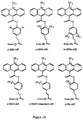
- Figures 1A, 1B and 1C depict the structures of various acridinium ester derivatives.
- Figures 2A and 2B depict the structures of varicus amphiphiles.
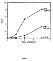
- Figure 3 is a plot of the chemiluminescence of standard AE as a function of CTAB concentration.
- Figure 4 is a plot of the chemiluminescence or probe-linked and hybrid-linked standard AE as a function of CTAB concentration.
- Figure 5 is a plot of the chemiluminescence of free standard AE, probe-linked standard AE, and hybrid-linked standard AE, in the presence of CTAB, as a function of TRITON® X-100 concentration.
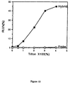
- Figure 6 is a plot of the chemiluminescence or probe linked and hybrid-linked standard AE as a function of CPC concentration.
- Figure 7 is a plot of the chemiluminescence of probe-linked or hybrid-linked standard AE, in the presence of CPC or CPB, as a function of TRITON® X-100 concentration.
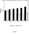
- Figure 8 is a graph showing the effect of different cationic detergents on the chemiluminescence of probe-linked and hybrid-linked standard AE.
- Figure 9 is a graph showing the effect of increasing amounts of TRITON® X-100 on the chemiluminescence of probe-linked and hybrid-linked standard AE.
- Figure 10 is a graph showing the effect of mixed micelles containing CPC and different neutral detergents on the chemiluminescence of probe-linked and hybrid-linked standard AE.
- Figure 11 is a graph showing the effect of homogeneous micelles composed of cationic, anionic and zwitterionic detergents on the chemiluminescence of probe-linked and hybrid-linked standard AE.
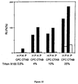
- Figure 12 is a graph showing the effect of- mixed micelles containing fixed concentrations of CPC or CTAB and increasing concentrations of TRITON® X-100 on the chemiluminescence of probe-linked and hybrid-linked standard AE.
- Figure 13 is a graph showing the effect of mixed micelles containing fixed concentrations of TRITON® X-100 and increasing concentrations of CPC on the chemiluminescence of probe-linked and hybrid-linked standard AE.
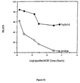
- Figure 14 is a plot of the chemiluminescence of probe-linked or hybrid-linked standard AE as a function of CPC concentration.
- the analyte was ribosomal RNA.
- Figure 15 is a plot of the chemiluminescence of probe-linked or hybrid-linked standard AE, in the presence of a fixed concentration of CPC, as a function of TRITON® X-100 concentration.
- the analyte was ribosomal RNA.
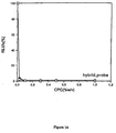
- Figure 16 is a plot of the chemiluminescence of hybrid-linked AE derivatives, in the presence of a fixed concentration of CPC, as a function of TRITON® X-100 concentration.
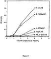
- Figure 17 is a plot of the chemiluminescence of probe-linked AE derivatives, in the presence of a fixed concentration of CPC, as a function of TRITON® X-100 concentration.
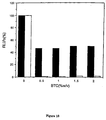
- Figure 18 is a graph showing the effect of increasing amounts of BTC on the chemiluminescence of probe-linked and hybrid-linked 2,7-diMe AE.
- Figure 19 is a plot of the chemiluminescence of probe-linked and hybrid-linked rhodamine, in the presence of a fixed concentration of CTAB, as a function of increasing concentrations of TRITON® X-100.
- Figure 19 is a plot of the chemiluminescence of probe-linked and hybrid-linked rhodamine, in the presence of a fixed concentration of CPC, as a function of increasing concentrations of TRITON® X-100.
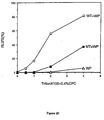
- Figure 20 is a plot of the chemiluminescence of probe-linked (WP), hybrid-linked (WT+WP), and mismatched hybrid-linked standard AE (MT+WP), in the presence of a fixed amount of CPC, as a function of TRITON® X-100 concentration.
- Figure 21 is a plot of the chemiluminescence of probe-linked and hybrid-linked standard AS as a function of mixed lipid concentration.
- the present invention relates to qualitative and quantitative assay methods and compositions wherein a labeled probe's detectability is dependent upon whether the probe is bound to the analyte of interest or is "free" or unbound.
- the method involves the use of amphiphilic molecules, such as detergents (surfactants) or lipids to form micelles, liposomes or lipid bilayers. It is thought that under these conditions, the label molecules are sequestered within the hydrophobic interior of the micelles or bilayers when the probe is unbound, thus extinguishing or diminishing the probe-associated label's detectability, as explained below.
- surfactant and “detergent” shall be used interchangeably herein to mean any of the class of amphiphilic molecules having separate hydrophobic and hydrophilic domains, and able to form homogeneous and/or heterogeneous micelles in solution.
- lipid shall be used to refer to amphiphilic molecules having separate hydrophobic and hydrophilic domains, and able to form homogeneous and/or heterogeneous liposomes or bilayers in solution; these lipids tend not to be as soluble in aqueous solution as are the detergents, and form liposomes or lipid bilayers rather than micelles.
- the lipid bilayers consist of a hydrophobic interior and hydrophilic exterior surfaces; thus these structures are thought to function in a manner analogous to micelles in the present invention.
- Micelles are usually monolayered structures having a hydrophobic interior and a hydrophilic exterior.
- amphiphile shall refer to the group of chemical' compounds, including surfactants and lipids, which have discrete hydrophilic and hydrophobic domains and which are able to form micelles, liposomes, bilayers or combinations of these structures.
- the interior of the micelles or bilayers provides a hydrophobic environment to which labels having a hydrophobic moiety are attracted and held.
- a hydrophobic portion of the label is sequestered by the hydrophobic interior of the micelle or lipid bilayer, which quenches the label's detectable signal.
- the micelle or bilayer is thought to provide a shield from exposure of the label to catalysts, reactants or cofactors necessary for the label's detectability.
- reagents are herein termed triggering agents, and the reaction in which they and the label participate to form.
- a detectable signal is termed a triggering reaction.
- the probe and analyte are both single-stranded nucleic acids
- the label is an N-acridinium phenyl ester derivative
- the acridinium ring may be substituted at one or more positions and the phenyl ring may also be substituted at one or more positions so long as oxidation of the ester linkage results in the production of a light-emitting excited N-acridone.
- AE acridinium ester
- acridinium ester is 4-(2-succinimidyloxycarbonyl ethyl)-phenyl-10-methylacridinium-9-carboxylate fluorosulfonate (hereafter referred to as standard AE), which is preferably linked via a non-nucleotide linker to or between the bases of an oligonucleotide probe. See EPO 0 313 219, supra . When the probe and analyte nucleic acids form a stable double-stranded hybrid there is created a major groove and a minor groove as part of the topology of the double helix.
- standard AE 4-(2-succinimidyloxycarbonyl ethyl)-phenyl-10-methylacridinium-9-carboxylate fluorosulfonate
- chemiluminescent acridinium derivatives having groups other than phenyl, or having additional substituents are known in the art; see, e.g., Corning; McCapra; Hoescht ; such derivatives would be expected by the skilled artisan to function in the present assay by virtue of their chemical similarity to the acridinium phenyl esters described herein.
- an amount of a surfactant e.g., the cationic detergent cetyl trimethyl_ammonium bromide (CTAB) is added to the assay solution at a concentration sufficient to form micelles. Under these conditions the chemiluminescent potential of the acridinium ester label is strongly quenched, even when the label is coupled to the probe.
- a surfactant e.g., the cationic detergent cetyl trimethyl_ammonium bromide (CTAB)
- CTAB cationic detergent cetyl trimethyl_ammonium bromide
- chemiluminescent potential is meant the ability of a compound to emit light upon addition to the reaction mixture of a triggering agent such as a reactant or catalyst.
- a triggering agent such as a reactant or catalyst.
- the triggering agent is any oxidizing agent (such as, without limitation, hydrogen peroxide, a hydrogen peroxide-producing compound, or a superoxide producing compound) able to cause the production of a lightproducing excited N-acridone.
- the addition of a surfactant to solutions containing labeled, hybridized probe can, at low concentrations of- surfactant, result in an initial quenching of the chemiluminescent signal, followed by a gradual restoration of the chemiluminescent potential of hybrid-bound label with increasing detergent or lipid concentration.
- an amount of TRITON® X-100 (polyoxyethylene alcohol) is added to a cationic surfactant such as CPC (cetyl pyridinium chloride) prior to micelle formation.
- CPC cetyl pyridinium chloride
- the chemiluminescence emitted by the acridinium ester label coupled to hybridized probe is initially quenched but then increases to easily detectable levels with the addition of greater concentrations of TRITON® X-100.
- the label coupled to unhybridized probe remains quenched after addition of the oxidizing agent, even with increasing amounts of the non-ionic detergent.
- the Applicant believes the acridinium ester derivatives linked to hybridized probe reside within the hydrophobic "pocket" formed in one or both of the grooves of the double helix. While the label which so resides remains susceptible to oxidation by hydrogen peroxide or superoxide radical (resulting in detectable light emission) the label is associated with the double groove strongly enough that the detergent micelles or lipid bilayers present a higher energy, less desirable environment for the hybrid-associated label. In the absence of the double-stranded environment the label coupled to unhybridized probe lacks this favored microenvironment and preferentially becomes associated-with the hydrophobic interior of-the micelles or bilayers.
- a detectable label may preferentially intercalate between bases in the double helix more readily than become associated with the detergent micelles or lipid bilayers. Again, if the label has no helical structure to intercalate with it is more likely to be quenched by the hydrophobic interiors of the micelles or bilayers.
- the sequestration phenomenon may be manipulated by varying the hydrophobicity of the micelle (or bilayer) interior relative to a specific probe:analyte conjugate.
- the hydrophobicity of the micelle or bilayer interior can be "tuned", as by the mixture of amphiphiles having different types of hydrophobic "tail".
- an amphiphile having long unsubstituted aliphatic chains such as CPC with its - (CH 2 ) 15 -CH 3 tail
- the interior of micelles containing, for example, mixtures of these two surfactant types should therefore be of intermediate hydrophobicity relative to the two surfactant types comprising them.
- the ratios of the detergents one of ordinary skill in the art can, with the guidance of this disclosure, precisely control the hydrophobicity of the micelle interior. This can allow the designing of micelles and bilayers which have interiors less hydrophobic than the probe:analyte conjugate but still hydrophobic enough to sequester the label associated with free probe. This will lead to optimization of the assay.
- micelles may be made with amphiphiles having a charge opposite from that of either or both the probe or analyte molecules.
- the micelles or bilayers may be made with cationic amphiphiles or mixtures of cationic and neutral amphiphiles which are able to concentrate the nucleic acids at the micelle surface. In such a case, the quenching of the label would be expected to be more efficient than in a system utilizing neutral amphiphiles only.
- an effective amount is meant an amount of amphiphile which will increase the signal-to-noise ratio between the labeled probe:analyte complex and free probe in an assay.
- the ratio of signal from the probe:analyte complex versus free probe-associated label "noise" is two-fold or greater, four-fold or greater, ten-fold or greater, twenty-fold or greater, fifty-fold or greater, one hundred-fold or greater and two hundred-fold or greater.
- assay systems can vary greatly in their reproducibility and precision, depending on factors including the type of instrument used to detect the label the nature of the label, and the volume of sample detected.
- CV coefficient of variance
- a very low signal to noise ratio is sufficient to achieve discrimination between a positive result (signal) and. a negative result (noise) with a high.degree of confidence.
- the signal to noise ratio must be greater in order to have the same degree of confidence in the result.
- the target nucleic acid need not necessarily be a single-stranded nucleic acid.
- Applicant envisions embodiments of the present invention wherein a single stranded nucleic acid probe is directed towards a double-stranded nucleic acid analyte, resulting in a triple-stranded probe:analyte complex.
- Labels were coupled to the oligonucleotide probes described in the examples using a non-nucleotide linker as described in EPO 0 313 219, supra .
- a single linker type was used for coupling the acridinium ester derivatives to probe in the following examples; this linker was attached at the para position of the phenyl ring and had the formula: phenyl-(CH 2 ) 2 -CO-NH-(CH 2 ) 5 -CO-NH-CH 2 -CH-(phosphodiester-nucleotide)-CH 2 -phosphodiester-nucleotide, where -CH- represents a two-way branch point and one of the two valences are represented by parentheses.
- the acridinium ester derivative used in each of the Examples was standard AE. All probes labeled with standard AE or AE derivatives had a specific activity of 8 X 10 19 RLU/mole.
- rhodamine was coupled to the oligonucleotide probe in the following manner: a rhodamine conjugate was purchased from Applied Biosystems Inc., (Foster City, CA) having an N-hydroxysuccinimide (NHS) group attached thereto (Catalog No. 400980). A 5'dimethoxytrityl-5-[N-trifluoroacetylaminohexyl)-3-acrylimido]-2'-deoxyuridine, 3'-[(2-cyanoethyl)-(N,N-diisopropyl)]-phosphoramidite modified nucleotide was purchased from Glen Research, Sterling, VA.
- the modified nucleotide was incorporated into the synthetic oligonucleotide; oligonucleotide synthesis was performed using standard phosphoramidite chemistry, see e.g., Carruthers, et al. , Methods in Enzymology , Volume 143, pg. 287 (1987), Bhatt, U.S. Patent No. 5,252,723 (which enjoys common ownership with the present invention) and Klem et al ., PCT Application No. WO 92/07864.
- the trifluoroacetyl moiety of the modified nucleotide was cleaved with 30% ammonium hydroxide and then reacted with the NHS group of the rhodamine conjugate to then link the rhodamine dye to the nuclectide.
- the detergents were dissolved in 0.6 M sodium borate buffer (pH 8.8) at the concentrations indicated in the examples.
- One hundred microliters of the resulting solution was placed in a 12 mm x 75 mm Sarstedt polystyrene tube for each test point.
- RLU relative light units
- Standard AE was coupled to the probe of SEQ ID NO:7 as described above.
- 4 ⁇ l of the labeled probe 160 pmoles was added to 8 ml of 11 mM lithium succinate (pH 5.1).
- One milliliter of this solution was then added to 2 ⁇ l (80 pmoles) of the unlabeled DNA target analyte of SEQ ID NO:8 and allowed to hybridize for 40 minutes at room temperature.
- the other set of samples was treated in exactly the same way but not given the target analyte.
- each tube within a set was then given a different concentration of CTAB, and micelles were formed and chemiluminescence detected as described above. The results are given below and in Figure 4.
- the data indicate that with increasing concentrations of CTAB chemiluminescence is initially decreased to about 3% for the labeled probe and about 3% for the labeled hybrid relative to a control tube having no detergent.
- all tubes contained 0.4%(w/v) CTAB in 0.6 M sodium borate. As shown in the previous example this is the concentration of CTAB at which there is little or no discrimination between labeled probe and labeled probe/analyte hybrid (see Figure 4).
- Example 2 Three sets of tubes were made as in Example 2; one set contained only the free AE label, another set of tubes contained the labeled probe alone, and a third set of tubes contained the labeled probe:DNA analyte hybrid. Hybridization was performed as described in Example 2. The probe and target oligonucleotides were those used in Example 2. Tubes within each set were also given TRITON® X-100 (polyethylene glycol p-isooctylphenyl ether; also in 0.6 M sodium borate) to a final concentration of 0%, 0.5%, 1%, 2% and 4%(v/v). Micelle formation and detection of chemiluminescence was performed as described in Example 1 (free label) and Example 2 (labeled probe and labeled probe:analyte hybrid).
- TRITON® X-100 polyethylene glycol p-isooctylphenyl ether; also in 0.6 M sodium borate
- the chemiluminescent signal obtained from the labeled probe:analyte hybrid increased drastically at concentrations within the range of 0% and 2%(v/v) of added TRITON® X-100; from about 2% to about 100% of the RLUs obtained in the absence of any detergent.
- the ability of the assay to discriminate between labeled hybrid and labeled probe is substantially increased when mixed micelles are used.
- CPC cetylpyridinium chloride
- the effect of the charged surfactant's counter-ion on the degree of discrimination was tested by forming micelles containing either 0.4% CPC or CPB and varying the concentration of TRITON® X-100. The only difference between these compounds is that the counter-ion in the former is chloride and the counter-ion in the latter is bromide.
- the probe and target oligonucleotides were those used in Example 2.
- the chemiluminescence of the labeled probe alone was similarly strongly quenched regardless of the nature of the counter-ion.
- the chemiluminescence of the labeled hybrid in CPC was less quenched with increasing concentrations of TRITON® X-100 than was the labeled hybrid in CPB, with the 0.4% (w/v) CPC/4% (v/v) TRITON® X-100 mixed micelles returning the detectability of the AE-labeled hybrid to about 81% of its value in the absence of any detergent and the 0.4%(w/v) CPB/4%(v/v) TRITON® X-100 mixed micelles restoring only about 36% of the same value.
- a panel of cationic detergents was evaluated for use in conjunction with the methods of the present invention; the detergents tested were CDEAB, BDSAC, BTC, BAC, BDTAC, BTAC, CPB and BCDAC. Each detergent was disolved in 0.6 M sodium borate (pH 8.7). For each detergent the detectability of free AE-labeled probe and the labeled DNA hybrid was determined under three conditions: a) in the presence of 0.05%(w/v) cationic surfactant, b) in the presence of 0.5%(w/v) cationic surfactant, and c) in the presence of 0.5% (w/v) cationic surfactant plus 2%(v/v) TRITON® X-100. Hybridization was as described in Example 2. The micelle formation and detection steps were conducted as described above. Probe and target oligonucleotides were those used in Example 2. The results are shown below and in Figure 8.
- a panel of neutral detergents was evaluated for use in conjunction with the methods of the present invention in mixed micelles with CPC.
- the neutral detergents tested were TRITON® X-100, TRITON® X-305, Brij® 35, Tween 20, and NP-40.
- For each neutral detergent the detectability of free AE-labeled probe and the labeled DNA hybrid was determined in the presence of 0.4%(w/v) CPC and 2%(v/v) of the neutral detergent.
- Hybridization, micelle formation and detection steps were conducted as described in Example 2. Probe and target oligonucleotides were those used in Example 2. The results are given below and in Figure 10.
- Ionic Detergent RLU (labeled probe) (percent) RLU (labeled hybrid) (percent) TRITON® X-100 0.450 58.800 TRITON® X-305 0.210 5.000 Brij® 35 0.350 13.200 Tween® 20 0.280 4.700 NP-40 0.320 5.200
- a panel of different surfactants was evaluated for use in the methods of the present invention.
- homogeneous micelles formed solely of each of the evaluated surfactants were used. These surfactants were neutral (TRITON® X-305, Brij®35, Tween®20, and NP-40) zwitterionic (CAPSO) and anionic (TRITON® QS-44 and sodium dodecyl sulfate (SDS)) in nature. 0.5% (v/v) of each detergent was used except in the case of Brij®35, CAPSO and SDS, where 0.5% (w/v) was used. Micelle formation and detection of the standard AE label was otherwise as described in Example 1. The probe and target oligonucleotides were those used in Example 2.
- anionic, neutral and zwitterionic micelles are capable of preferentially quenching the chemiluminescence of the unhybridized probe-coupled label over that of the hybrid-coupled label in a fashion similar to that seen for cationic micelles.
- the magnitude of discrimination observed is less than that seen with cationic micelles presumably due to the lack of ionic interaction between the negatively charged nucleic acid and the detergent.
- TRITON® X-100 The effect of high concentrations of TRITON® X-100 on label discrimination was determined under conditions of constant concentration of cationic detergent.
- TRITON® X-100 was added to the tubes of each set (containing either CPC or CTAB) at concentrations of 0.5%, 4%, 10%, and 25%(v/v).
- Hybridization was performed as in Example 2; micelle formation and detection steps were as in Example 1.
- the probe and target oligonucleotides were those used in Example 2. The results are shown below and in Figure 12.
- the effect on label discrimination of raising the concentration of ionic surfactant was measured under conditions of a constant concentration of TRITON® X-100.
- the concentration of TRITON® X-100 in each tube was 4%(v/v).
- Two sets of tubes containing increasing concentrations (0%, 0.4%, 0.7%, 1%, 2%, 3%, and 4%(w/v)) of CPC were made; one contained the AE-labeled probe alone, the other contained the AE-labeled hybrid.
- the probe and target oligonucleotides were those used in Example 2. Hybridization was as described in Example 2; micelle formation and detection was as described in Example 1. The results were as shown below and in Figure 13.
- the amount of chemiluminescence detected due to labeled free probe is about 50% of that detected for the labeled hybrid (yielding a signal-to-noise ratio of about 2:1).
- the signal-to-noise ratio is increased to about 91:1.
- the signal-to-noise ratio is increased to 165:1; at CPC concentrations above 1%(w/v) there is no significant improvement of signal-to-noise ratio.
- the first probe and target oligonucleotides were of SEQ ID NOs: 5 and 6, respectively.
- the second probe and target were of SEQ ID NOs: 3 and 4, respectively.
- Each probe was coupled to standard AE by the methodology described above.
- Hybridization reactions were conducted as follows: 1 ⁇ l (1 pmole) of probe was added to 30 ⁇ l of 0.5 M LiCl, 0.1 M lithium succinate (pH 5.1). To tubes representing probe:analyte hybrid was also added 4 ⁇ l target analyte (4 pmole). The tubes were incubated at 60°C for 30 minutes.
- oligonucleotides either labeled probe alone or labeled hybrid
- 100 ⁇ l 0.6M sodium borate pH 8.7
- 1%(v/v) TRITON® X-100 1%(v/v) TRITON® X-100
- the tubes were then given 100 ⁇ l 0.8%(w/v) CPC, 6%(v/v) TRITON® X-100 -and 0.6M sodium borate (pH 8.7) (SEQ ID NOS:5 and 6) or 0.8%(w/v) CPC, 4%(v/v) TRITON® X-100 and 0.6M sodium borate (pH 8.7)(SEQ ID NOS:3 and 4); the same solutions minus TRITON® X-100 were added to the aliquots already collected. Micelle formation and detection was as in Example 1.
- the signal-to-noise ratio of the assay is improved using the methods of the present invention.
- the increase in the signal-to-noise ratio for the micelle-containing assay was 2.4 times the signal-to-noise ratio observed for the same assay without micelles; in the assay using the other probe and analyte pair the signal-to-noise ratio was about 4.5 times greater than that observed in the non-micelle-containing assay.
- results indicate the general utility of the present invention in increasing the signal-to-noise ratio of other assay systems when used in conjunction therewith. Also, experiments such as these have shown that the variation between replicate samples in assays employing differential hydrolysis of AE-labeled probes is decreased using amphiphiles as described herein, thus improving the precision of the assay and the degree of confidence of the quantitative results.
- the target nucleic acid was whole ribosomal RNA (rRNA) purified from a pure culture of Chlamydia trachomatis .
- the probe had a nucleotide sequence of SEQ ID NO:9, designed to be complementary to one subunit of the rRNA.
- Unlabeled helper oligonucleotides were used to facilitate binding of the probe to the rRNA target; helper probes are described in Hogan et al. , U.S. Patent No. 5,030,557.
- Helper probes can be an aid to nucleic acid probe hybridization in certain cases (e.g., when the target analyte has considerable secondary structure) but are not, in and of themselves, part of the invention of this application.
- the probes used in this example were labeled with standard AE as described above.
- Hybridization was conducted as follows: 3 ⁇ l (0.05 pmoles) of probe was added to 6 ⁇ l of water. This was combined with 4 ⁇ l (0.25 pmoles) of target rRNA, 2 ⁇ l (10 pmoles) of unlabeled helper probes and 15 ⁇ l of 200 mM lithium succinate (pH 5.1) and incubated at 60°C for 40 minutes. Micelle formation and detection was performed as described in Example 2.
- Figure 15 shows the results of adding increasing amounts of TRITON® X-100, to the labeled probe or hybrid in 0.4%(w/v) CPC.
- addition of second detergent having a different hydrophobic "tail" to the reaction mixture prior to micelle formation and detection results in a strong increase in the ability of the system to distinguish free probe and labeled DNA:RNA hybrid. While not certain of the mechanism of action, Applicants believe that this increase is the result of "tuning" of the hydrophobicity of the micelle interior.
- acridinium ester derivatives were coupled to the same probe and hybridized to the same target in the presence of a constant concentration of CPC and increasing concentrations of TRITON® X-100 (0%, 0.5%, 1%, 2% and 4%(v/v)).
- Some of these derivatives have substitutions in the heterocyclic acridinium ring (1-methyl-AE, 2,7-dimethyl AE), others have substitutions on the phenyl ring (o-dimethyl-AE, o-dibromo-AE) or replace the phenyl ring with another aryl group (naphthyl-AE); otherwise they are identical to standard AE.
- Hybridization was performed as follows. To a final volume of 30 ⁇ l of 0.1 M lithium succinate (pH 5.1) and 0.5 M LiCl was added 1.7 ⁇ l of probe (0.1 pmole). In the case of the tubes containing labeled hybrid, 4 ⁇ l (1 pmole) of the target oligonucleotide was also added. The tubes were incubated at 60°C for 60 minutes.
- each tube's contents (each tube containing between 20,000 and 200,000 RLU of label) were added to 200 ⁇ l of detergent solution of the concentrations indicated below.
- the tubes were vortexed for 10 seconds, then allowed to sit at room temperature for 10 minutes. Detection was as described in Example 1.
- Figure 17 demonstrates that, under the same conditions, the same AE derivatives coupled to the same probe respond differently under these conditions when the probe is unhybridized. While the standard AE- and the o-dibromo AE-labeled free probe are both strongly quenched at higher concentrations of TRITON® X-100, the o-dimethyl AE and naphthyl AE derivatives are somewhat less strongly quenched and 1-methyl AE and 2,7-dimethyl AE-labeled probes are much less strongly quenched under these conditions.
- surfactant micelles to discriminate between labeled probe and labeled probe:analyte conjugate can depend both on the surfactant used and the structure of the labeling compound. While CPC:TRITON® X-100 mixed micelles do not, under the conditions of Example 14, allow for good discrimination between probe and hybrid when the probe is labeled with 2,7-dimethyl AE, micelles formed from BTC do allow for good discrimination between these species.
- Assay tubes were made up containing either 2,7-dimethyl AE-labeled probe alone or the identically labeled probe:analyte hybrid. Hybridization was conducted as in Example 14. The probe and DNA target oligonucleotide were those used in Example 14. Concentrations of BTC were 0%, 0.5%, 1%, 1.5% and 2%(w/v). Micelle formation and detection was conducted as described in Example 14. Results are shown below and in Figure 18. Tube Number Concentration BTC %(w/v) RLU (labeled hybrid) (percent) RLU (labeled probe) (percent) 1 0.000 100.000 100.000 2 0.500 45.900 1.060 3 1.000 46.200 1.110 4 1.500 49.800 1.730 5 2.000 49.900 1.840
- This example illustrates the ability of labels other than acridinium esters derivatives to function in the method of the present invention.
- An NHS-derivatized rhodamine was purchased from Applied Biosystems Corp. (Foster City, CA) and linked to a nucleotide via a linker as detailed above.
- the labeled nucleotide was then incorporated into a synthetic oligonucleotide probe having the same sequence as the probe used in Example 2 above.
- the target DNA analyte was the same as was used in Example 2.
- Hybridization was conducted as follows: to 8 ml of 11 mM lithium succinate buffer (pH 5.2) was added 100 ⁇ l of probe (640 picomoles).
- rhodamine can be used in the present invention in conjunction with micelles as a method of detecting hybrid probe:analyte complexes.
- the rhodamine-labeled unhybridized probe's chemiluminescent potential is strongly quenched under these conditions while the labeled hybrid remains detectable.
- the method of the present invention can be used with labels other than acridinium esters.
- the linker used to couple the rhodamine label to the probe is different than the linker used to couple AE derivatives to the probes in the previous examples, this example demonstrates that the method of linkage of label to target may be varied in the present assay without defeating the utility of this invention.
- Cationic micelles are known to concentrate anions near their surface through ionic interactions.
- peroxide ion OOH-
- the concentration of peroxide ion required to initiate a chemiluminescent reaction would be expected to be lower than the concentration necessary to initiate a reaction having the same chemiluminescent yield in the absence of cationic micelles.
- Probe and target oligonucleotides were those in Example 2; and the probe was labeled with standard AE as described above. Hybridization was conducted as described in Example 12. Micelle formation was as in Example 1.
- the detection step was also as described in Example 1 with the following differences: the hydrogen peroxide solution was added either at full strength, at a ten-fold dilution or at a thirty-fold dilution.
- the signal-to-noise ratio of the assay determined by comparing the chemiluminescence detected from the labeled hybrid with the chemiluminescence from the free labeled probe, is improved by lowering the peroxide ion concentration.
- a probe oligonucleotide was synthesized SEQ ID NO:12 (WP). Additionally, a pair of target oligonucleotides having a single base mismatch at the same relative position (on the complementary strand) were also synthesized (SEQ ID NOs:10 (WT) and 13 (MT)).
- the standard AE label was linked to the probe molecules as described above; in this experiment the label was placed at a location just to the 3'side of the site of nucleotide substitution, as indicated below by an asterisk. The structures resulting from hybridization of these oligonucleotides are shown below.
- Example 14 Each combination of probe and target indicated below was made in separate tubes; a third tube contained the "wild-type" probe alone. Hybridization and micelle formation was conducted as described in Example 14. Detection was as described in Example 1.
- a standard AE-coupled probe was hybridized to either a target oligonucleotide having an exactly complementary nucleotide sequence or a nucleotide sequence having a single base mismatch, with respect to the probe, at a site corresponding to the probe location at which the AE label is coupled.
- Hybridization was performed as described in Example 14. Three sets of tubes were given 0.4%(w/v) CPC; tubes within each set were given 0%, 0.5% 1%, 2% and 4% TRITON® X-100. Micelle formation was as in Example 1. All tubes within each set were given either the perfectly matched hybrid, the mutant mismatched hybrid, or the labeled probe alone. Detection was as in Example 1. The results are shown below and in Figure 20.
- the chemiluminescence of the labeled unhybridized probe is strongly quenched at all concentrations of TRITON® X-100.
- the chemiluminescence of the perfectly matched labeled hybrid increased with increasing concentrations of TRITON® X-100 to about 80% of the chemiluminescent yield of the hybrid in the absence of detergent micelles.
- lipid preparation consisting of a 1:2.5 molar ratio of the cationic lipid dimethyl dioctadecylammonium bromide (DDAB) to dioleoyl phosphatidyl ethanolamine (DOPE) (LipofectACE® , Gibco/BRL, Gaitersburg, MD) was added to 2 ⁇ l of either the labeled probe alone or the labeled probe:analyte complex. The resulting solution was vortexed gently and allowed to sit for 10 minutes at room temperature to form liposomes. Detection of chemiluminescence was as in Example 2. The results were as indicated below.
- DDAB cationic lipid dimethyl dioctadecylammonium bromide
- DOPE dioleoyl phosphatidyl ethanolamine
- the results show that the chemiluminescent potential of both labeled probe and labeled probe:analyte complex initially decreases upon the addition of low concentrations of the mixed cationic lipid preparation.
- liposome concentrations of about 0.0028% (w/v) and above the chemiluminescence of the labeled probe:analyte complex remains essentially constant (at about 55% of the probe:analyte complex's chemiluminescence in the absence of liposomes), while the chemiluminescence of the labeled probe alone continues to decrease in this concentration range.
Landscapes
- Health & Medical Sciences (AREA)
- Life Sciences & Earth Sciences (AREA)
- Chemical & Material Sciences (AREA)
- Engineering & Computer Science (AREA)
- Immunology (AREA)
- Molecular Biology (AREA)
- Biomedical Technology (AREA)
- Hematology (AREA)
- Urology & Nephrology (AREA)
- Organic Chemistry (AREA)
- Biochemistry (AREA)
- Microbiology (AREA)
- Biotechnology (AREA)
- Physics & Mathematics (AREA)
- General Health & Medical Sciences (AREA)
- Analytical Chemistry (AREA)
- Wood Science & Technology (AREA)
- Zoology (AREA)
- Cell Biology (AREA)
- Proteomics, Peptides & Aminoacids (AREA)
- Food Science & Technology (AREA)
- Medicinal Chemistry (AREA)
- General Physics & Mathematics (AREA)
- Pathology (AREA)
- Genetics & Genomics (AREA)
- General Engineering & Computer Science (AREA)
- Bioinformatics & Cheminformatics (AREA)
- Biophysics (AREA)
- Measuring Or Testing Involving Enzymes Or Micro-Organisms (AREA)
- Investigating Or Analysing Materials By The Use Of Chemical Reactions (AREA)
- Pharmaceuticals Containing Other Organic And Inorganic Compounds (AREA)
- Magnetic Resonance Imaging Apparatus (AREA)
- Medicines Containing Antibodies Or Antigens For Use As Internal Diagnostic Agents (AREA)
Claims (40)
- Méthode pour déterminer la présence ou la quantité d'un analyte acide nucléique cible dans un échantillon consistant essentiellement dans le fait de :a) fournir à l'échantillon une sonde d'acide nucléique marquée de façon détectable dans des conditions telles que la sonde puise se lier spécifiquement à l'analyte, en formant ainsi un complexe sonde:analyte ;b) fournir à l'échantillon un ou plusieurs amphiphiles en une quantité suffisante pour provoquer la formation de micelles, liposomes, bicouches lipidiques ou combinaisons de ceux-ci qui diminuent ou annulent la détectabilité du marqueur associé à la sonde qui n'a pas formé partie d'un complexe sonde:analyte, de telle sorte que la détectabilité de la sonde qui fait partie d'un complexe sonde:analyte peut être distinguée de la détectabilité de la sonde qui ne fait pas partie d'un complexe sonde:analyte ; etc) détecter la sonde qui fait partie d'un complexe sonde:analyte en tant qu'indication de la présence ou de la quantité de l'analyte dans l'échantillon.
- Méthode de la revendication 1, dans laquelle le marqueur de la sonde comprend une partie hydrophobe.
- Méthode de la revendication 1 ou 2, dans laquelle le marqueur de la sonde est détecté grâce à une réaction d'activation entre le marqueur de la sonde et un agent activant.
- Méthode de l'une quelconque des revendications 1 à 3, dans laquelle le marqueur de la sonde est luminescent ou fluorescent, et la luminescence ou la fluorescence du marqueur de la sonde est altérée lorsque la sonde ne fait pas partie d'un complexe sonde:analyte.
- Méthode de la revendication 4, dans laquelle le marqueur de la sonde forme une espèce chimioluminescente ou électrochimioluminescente détectable lorsqu'il est soumis à une réaction d'activation, et le marqueur de la sonde qui ne fait pas partie d'un complexe sonde:analyte est protégé de l'agent activant par la présence du ou des amphiphiles dans des conditions permettant l'activation du marqueur de la sonde qui fait partie d'un complexe sonde:analyte.
- Méthode de la revendication 5, dans laquelle le marqueur de la sonde forme une espèce chimioluminescente détectable après avoir été soumis à la réaction d'activation, l'espèce comprenant une N-acridone excitée éventuellement substituée.
- Méthode de la revendication 6, dans laquelle le marqueur de la sonde comprend un ester phénylique de N-acridinium éventuellement substitué sur le cycle acridinium et sur les carbones 2 à 6 du cycle phényle.
- Méthode de la revendication 7, dans laquelle le marqueur de la sonde est choisi dans l'ensemble consistant en ester d'acridinium standard, ester de o-diméthylacridinium, ester de 1-méthylacridinium, ester de 2,7-diméthylacridinium, ester de naphtylacridinium, et ester de o-dibromoacridinium.
- Méthode de l'une quelconque des revendications 3 à 8, dans laquelle l'agent activant est un agent oxydant.
- Méthode de la revendication 9, dans laquelle l'agent oxydant est choisi dans l'ensemble consistant en un composé peroxyde et un radical superoxyde.
- Méthode de l'une quelconque des revendications 1 à 10, dans laquelle au moins un des amphiphiles a une charge positive dans les conditions permettant la formation d'un complexe sonde:analyte.
- Méthode de l'une quelconque des revendications 1 à 11, dans laquelle le ou les amphiphiles comprennent au moins deux amphiphiles différents.
- Méthode de la revendication 12, dans laquelle le ou les amphiphiles comprennent au moins deux amphiphiles ayant des domaines hydrophobes différents.
- Méthode de la revendication 12 ou 13, dans laquelle le ou les amphiphiles comprennent un premier amphiphile choisi dans l'ensemble consistant en la série des Triton® , la série des Tween® , la série des Brij® , et la série des NP.
- Méthode de la revendication 14, dans laquelle le premier amphiphile est choisi dans l'ensemble consistant en Triton® X-100, Triton® X-305, Tween® 20, Brij® 35 et NP-40.
- Méthode de l'une quelconque des revendications 1 à 13, dans laquelle le ou les amphiphiles comprennent un amphiphile choisi dans l'ensemble consistant en sels de cétyltriméthylammonium, sels de cétylpyridinium, sels de cétyldiméthyléthylammonium, sels de 3-(cyclohexylamino)-2-hydroxy-1-propane sulfonates, sels de benzthonium, sels de benzyldiméthyle, sels de benzyldiméthylstéarylammonium, sels de benzyltriméthylammonium, sels de benzylcétyldiméthylammonium, sels de benzyldiméthyltétradécylammonium, sels de benzalconium et sels dodécylsulfates.
- Méthode de la revendication 15, dans laquelle le premier amphiphile est le Triton® X-100 et un second amphiphile est choisi dans l'ensemble consistant en sels de cétyltriméthyléthylammonium, sels de cétylpyridinium, sels de cétyldiméthyléthyl ammonium, sels de 3-(cyclohexylamino)-2-hydroxy-1-propane sulfonates, sels de benzthonium, sels de benzyldiméthyle, sels de benzyldiméthylstéarylammonium, sels de benzyltrimé-thylammonium, sels de benzylcétyldiméthylammonium, sels de benzyldiméthyltétradécylammonium et sels de benzalconium.
- Méthode de la revendication 17, dans laquelle le second amphiphile est choisi dans l'ensemble consistant en sels de cétyltriméthylammonium et sels de cétylpyridinium.
- Méthode de l'une quelconque des revendications 1 à 18, dans laquelle au moins un du ou des amphiphiles est un détergent fourni en une quantité au moins égale à la concentration micellaire critique du détergent.
- Méthode de l'une quelconque des revendications 5 à 19, dans laquelle le ou les amphiphiles sont fournis en une quantité efficace pour que la chimioluminescence du marqueur de la sonde qui fait partie d'un complexe sonde:analyte soit au moins quatre fois la chimioluminescence du marqueur de la sonde qui ne fait pas partie d'un complexe sonde:analyte.
- Méthode de la revendication 20, dans laquelle le ou les amphiphiles sont fournis en une quantité efficace pour que la chimioluminescence du marqueur de la sonde qui fait partie d'un complexe sonde:analyte soit au moins dix fois la chimioluminescence du marqueur de la sonde qui ne fait pas partie d'un complexe sonde:analyte.
- Méthode de la revendication 21, dans laquelle le ou les amphiphiles sont fournis en une quantité efficace pour que la chimioluminescence du marqueur de la sonde qui fait partie d'un complexe sonde:analyte soit au moins vingt fois la chimioluminescence du marqueur de la sonde qui ne fait pas partie d'un complexe sonde:analyte.
- Méthode de la revendication 22, dans laquelle le ou les amphiphiles sont fournis en une quantité efficace pour que la chimioluminescence du marqueur de la sonde qui fait partie d'un complexe sonde:analyte soit au moins quarante fois la chimioluminescence du marqueur de la sonde qui ne fait pas partie d'un complexe sonde:analyte.
- Méthode de la revendication 23, dans laquelle le ou les amphiphiles sont fournis en une quantité efficace pour que la chimioluminescence du marqueur de la sonde qui fait partie d'un complexe sonde:analyte soit au moins cent fois la chimioluminescence du marqueur de la sonde qui ne fait pas partie d'un complexe sonde:analyte.
- Méthode de la revendication 24, dans laquelle le ou les amphiphiles sont fournis en une quantité efficace pour que la chimioluminescence du marqueur de la sonde qui fait partie d'un complexe sonde:analyte soit au moins deux cent fois la chimioluminescence du marqueur de la sonde qui ne fait pas partie d'un complexe sonde:analyte.
- Composition pour déterminer la présence ou la quantité d'un analyte acide nucléique cible dans un milieu comprenant :a) une sonde d'acide nucléique marqué de façon détectable capable de former un complexe sonde:analyte avec l'analyte etb) un ou plusieurs amphiphiles en une quantité suffisante pour provoquer la formation de micelles, liposomes, bicouches lipidiques ou combinaisons de ceux-ci qui diminuent ou annulent la détectabilité du marqueur associé à la sonde qui n'a pas formé partie d'un complexe sonde:analyte, de telle sorte que la détectabilité de la sonde qui fait partie d'un complexe sonde:analyte puisse être distinguée de la détectabilité de la sonde qui ne fait pas partie d'un complexe sonde:analyte.
- Composition de la revendication 26, dans laquelle le marqueur de la sonde est détecté grâce à une réaction d'activation entre le marqueur de la sonde et un agent activant.
- Composition de la revendication 27, dans laquelle le marqueur de la sonde forme une espèce chimioluminescente ou électrochimioluminescente détectable lorsqu'il est soumis à une réaction d'activation, et le marqueur de la sonde qui ne fait pas partie d'un complexe sonde:analyte est protégé de l'agent activant par la présence du ou des amphiphiles dans des conditions permettant l'activation du marqueur de la sonde qui fait partie d'un complexe sonde:analyte.
- Méthode de la revendication 28, dans laquelle le marqueur de la sonde forme une espèce chimioluminescente détectable après avoir été soumis à la réaction d'activation, l'espèce comprenant une N-acridone excitée éventuellement substituée.
- Composition de la revendication 29, dans laquelle le marqueur de la sonde comprend un ester phénylique de N-acridinium éventuellement substitué sur le cycle acridinium et sur les carbones 2 à 6 du cycle phényle.
- Composition de la revendication 30, dans laquelle le marqueur de la sonde est choisi dans l'ensemble consistant en ester d'acridinium standard, ester de o-diméthylacridinium, ester de 1-méthylacridinium, ester de 2,7-diméthylacridinium, ester de naphtylacridinium, et ester de o-dibromoacridinium.
- Composition de l'une quelconque des revendications 27 à 31, dans laquelle l'agent activant est un agent oxydant.
- Composition de l'une quelconque des revendications 26 à 32, dans laquelle au moins un du ou des amphiphiles a une charge positive dans les conditions permettant la formation d'un complexe sonde:analyte.
- Composition de l'une quelconque des revendications 26 à 33, dans laquelle le ou les amphiphiles comprennent au moins deux amphiphiles différents.
- Composition de la revendication 34, dans laquelle au moins deux du ou des amphiphiles ont des domaines hydrophobes différents.
- Composition de l'une quelconque des revendications 26 à 35, dans laquelle le ou les amphiphiles comprennent un premier amphiphile choisi dans l'ensemble consistant en la série des Triton® , la série des Tween® , la série des Brij® , et la série des NP.
- Composition de la revendication 36, dans laquelle le premier amphiphile est choisi dans l'ensemble consistant en Triton® X-100, Triton® X-305, Tween® 20, Brij® 35 et NP-40.
- Composition de l'une quelconque des revendications 26 à 35, dans laquelle le ou les amphiphiles comprennent un amphiphile choisi dans l'ensemble consistant en sels de cétyltriméthylammonium, sels de cétylpyridinium, sels de cétyldiméthyléthylammonium, sels de 3-(cyclohexylamino)-2-hydroxyl-1-propane sulfonates, sels de benzthonium, sels de benzyldiméthyle, sels de benzyldiméthylstéarylammonium, sels de benzyltriméthylammonium, sels de benzylcétyldiméthylammonium, sels de benzyldiméthyltétradécylammonium, sels de benzalconium et sels dodécylsulfates.
- Composition de la revendication 37, dans laquelle le premier amphiphile est le Triton® X-100 et un second amphiphile est choisi dans l'ensemble consistant en sels de cétyltriméthylammonium, sels de cétylpyridinium, sels de cétyldiméthyléthylammonium, , sels de 3-(cyclohexylamino)-2-hydroxy-1-propane sulfonates, sels de benzthonium, sels de benzyldiméthyle, sels de benzyldiméthylstéarylammonium, sels de benzyltriméthylammonium, sels de benzylcétyldiméthylammonium, sels de benzyldiméthyltétradécylammonium et sels de benzalconium.
- Composition de la revendication 39, dans laquelle le second amphiphile est choisi dans l'ensemble consistant en sels de cétyltriméthylammonium et sels de cétylpyridinium.
Applications Claiming Priority (4)
| Application Number | Priority Date | Filing Date | Title |
|---|---|---|---|
| US48432095A | 1995-06-07 | 1995-06-07 | |
| US08/475,334 US5879885A (en) | 1995-06-07 | 1995-06-07 | Compositions for detecting and quantifying biological samples |
| US475334 | 1995-06-07 | ||
| US484320 | 2000-01-18 |
Publications (2)
| Publication Number | Publication Date |
|---|---|
| EP0747701A1 EP0747701A1 (fr) | 1996-12-11 |
| EP0747701B1 true EP0747701B1 (fr) | 2001-12-05 |
Family
ID=27044749
Family Applications (1)
| Application Number | Title | Priority Date | Filing Date |
|---|---|---|---|
| EP96304228A Expired - Lifetime EP0747701B1 (fr) | 1995-06-07 | 1996-06-06 | Essai utilisant des micelles de protection |
Country Status (10)
| Country | Link |
|---|---|
| EP (1) | EP0747701B1 (fr) |
| JP (1) | JP4236281B2 (fr) |
| KR (1) | KR19990022598A (fr) |
| AT (1) | ATE210293T1 (fr) |
| AU (1) | AU707818B2 (fr) |
| CA (1) | CA2223047C (fr) |
| DE (1) | DE69617532T2 (fr) |
| DK (1) | DK0747701T3 (fr) |
| ES (1) | ES2169211T3 (fr) |
| WO (1) | WO1996041178A1 (fr) |
Families Citing this family (4)
| Publication number | Priority date | Publication date | Assignee | Title |
|---|---|---|---|---|
| US6180340B1 (en) | 1997-10-31 | 2001-01-30 | Gen-Probe Incorporated | Extended dynamic range assays |
| JP4759803B2 (ja) * | 2000-11-29 | 2011-08-31 | ソニー株式会社 | 個人識別用dnaインク及び個人識別システム |
| US10449254B2 (en) | 2013-11-03 | 2019-10-22 | The Regents Of The University Of California | Ionic liquids for transdermal drug delivery |
| US10912834B2 (en) | 2016-08-29 | 2021-02-09 | The Regents Of The University Of California | Topical formulations based on ionic species for skin treatment |
Family Cites Families (9)
| Publication number | Priority date | Publication date | Assignee | Title |
|---|---|---|---|---|
| US4640898A (en) * | 1983-08-01 | 1987-02-03 | University Of Health Sciences/The Chicago Medical School | Homogeneous fluorescence ligang binding assay based upon preferential alteration of the respective intensities of bound and free label by solvent components |
| US5004565A (en) * | 1986-07-17 | 1991-04-02 | The Board Of Governors Of Wayne State University | Method and compositions providing enhanced chemiluminescence from 1,2-dioxetanes |
| US4927769A (en) * | 1987-07-08 | 1990-05-22 | Ciba Corning Diagnostics Corp. | Method for enhancement of chemiluminescence |
| DE3856535T2 (de) * | 1987-09-21 | 2002-11-14 | Gen Probe Inc | Schutztest |
| US5283174A (en) * | 1987-09-21 | 1994-02-01 | Gen-Probe, Incorporated | Homogenous protection assay |
| WO1989004375A1 (fr) * | 1987-10-23 | 1989-05-18 | Siska Diagnostics, Inc. | Sondes d'acides nucleique marquees par des chelates de lanthanide |
| EP0492570B1 (fr) * | 1990-12-24 | 2001-05-02 | Enzo Diagnostics, Inc. | Méthode pour le dépistage d'un polynucléotide cible dans un échantillon en utilisant un réactif réducteur du bruit de fond et composition et kit comprenant ce réactif |
| US5279940A (en) * | 1992-08-03 | 1994-01-18 | Eastman Kodak Company | Chemiluminescent composition containing cationic surfactants or polymers and 4'-hydroxyacetanilide, test kits and their use in analytical methods |
| EP0709466B1 (fr) * | 1994-10-28 | 2006-09-27 | Gen-Probe Incorporated | Compositions et procédés pour la détection et quantification simultanée de plusieurs séquences d'acide nucléique |
-
1996
- 1996-05-14 JP JP50057097A patent/JP4236281B2/ja not_active Expired - Fee Related
- 1996-05-14 CA CA002223047A patent/CA2223047C/fr not_active Expired - Fee Related
- 1996-05-14 WO PCT/US1996/006919 patent/WO1996041178A1/fr not_active Application Discontinuation
- 1996-05-14 AU AU57935/96A patent/AU707818B2/en not_active Ceased
- 1996-05-14 KR KR1019970709079A patent/KR19990022598A/ko not_active Application Discontinuation
- 1996-06-06 EP EP96304228A patent/EP0747701B1/fr not_active Expired - Lifetime
- 1996-06-06 DE DE69617532T patent/DE69617532T2/de not_active Expired - Fee Related
- 1996-06-06 AT AT96304228T patent/ATE210293T1/de not_active IP Right Cessation
- 1996-06-06 ES ES96304228T patent/ES2169211T3/es not_active Expired - Lifetime
- 1996-06-06 DK DK96304228T patent/DK0747701T3/da active
Also Published As
| Publication number | Publication date |
|---|---|
| KR19990022598A (ko) | 1999-03-25 |
| DK0747701T3 (da) | 2001-12-27 |
| CA2223047C (fr) | 2008-11-18 |
| EP0747701A1 (fr) | 1996-12-11 |
| DE69617532D1 (de) | 2002-01-17 |
| JP4236281B2 (ja) | 2009-03-11 |
| DE69617532T2 (de) | 2002-05-02 |
| ATE210293T1 (de) | 2001-12-15 |
| WO1996041178A1 (fr) | 1996-12-19 |
| AU5793596A (en) | 1996-12-30 |
| JPH11506924A (ja) | 1999-06-22 |
| AU707818B2 (en) | 1999-07-22 |
| ES2169211T3 (es) | 2002-07-01 |
| CA2223047A1 (fr) | 1996-12-19 |
Similar Documents
| Publication | Publication Date | Title |
|---|---|---|
| EP0747706B1 (fr) | Essai de protection par adduction | |
| JP3787352B2 (ja) | ハイブリダイゼーション保護検定 | |
| US5948899A (en) | Compositions for homogenous protection assay | |
| US5283174A (en) | Homogenous protection assay | |
| US6004745A (en) | Hybridization protection assay | |
| EP1239049B1 (fr) | Détermination de l'eau oxygenée avec des oxidases et des complexes lanthanoide-ligand | |
| US7635571B2 (en) | Amplified signal in binding assays | |
| EP0664339A1 (fr) | Methode de discrimination des acides nucleiques et necessaire d'essai a cette fin | |
| US6059561A (en) | Compositions and methods for detecting and quantifying biological samples | |
| US7563579B2 (en) | Method for detecting a structured target | |
| AU4135696A (en) | Compositions and methods for the simultaneous detection and quantification of multiple specific nucleic acid sequences | |
| JP3495752B2 (ja) | 生標的個体の検出方法およびプローブ | |
| JPH08501689A (ja) | 改良型の鎖置換アッセイおよびそれに有用な複合体 | |
| EP1444518B1 (fr) | Methode et appareil d'analyse par cytometrie a flux multiplex de substances a analyser melangees provenant d'echantillons de fluides corporels | |
| EP0747701B1 (fr) | Essai utilisant des micelles de protection | |
| EP0660844A4 (fr) | Sequences nucleotidiques isolees destinees a l'identification de neisseria gonorrhoeae. | |
| EP0912765B1 (fr) | Detection d'analytes au moyen de deux marqueurs |
Legal Events
| Date | Code | Title | Description |
|---|---|---|---|
| PUAI | Public reference made under article 153(3) epc to a published international application that has entered the european phase |
Free format text: ORIGINAL CODE: 0009012 |
|
| AK | Designated contracting states |
Kind code of ref document: A1 Designated state(s): AT BE CH DE DK ES FR GB IT LI LU NL SE |
|
| 17P | Request for examination filed |
Effective date: 19970116 |
|
| RAP1 | Party data changed (applicant data changed or rights of an application transferred) |
Owner name: GEN-PROBE INCORPORATED |
|
| 17Q | First examination report despatched |
Effective date: 19991125 |
|
| GRAG | Despatch of communication of intention to grant |
Free format text: ORIGINAL CODE: EPIDOS AGRA |
|
| GRAG | Despatch of communication of intention to grant |
Free format text: ORIGINAL CODE: EPIDOS AGRA |
|
| GRAG | Despatch of communication of intention to grant |
Free format text: ORIGINAL CODE: EPIDOS AGRA |
|
| GRAH | Despatch of communication of intention to grant a patent |
Free format text: ORIGINAL CODE: EPIDOS IGRA |
|
| GRAH | Despatch of communication of intention to grant a patent |
Free format text: ORIGINAL CODE: EPIDOS IGRA |
|
| GRAA | (expected) grant |
Free format text: ORIGINAL CODE: 0009210 |
|
| AK | Designated contracting states |
Kind code of ref document: B1 Designated state(s): AT BE CH DE DK ES FR GB IT LI LU NL SE |
|
| REF | Corresponds to: |
Ref document number: 210293 Country of ref document: AT Date of ref document: 20011215 Kind code of ref document: T |
|
| REG | Reference to a national code |
Ref country code: CH Ref legal event code: EP |
|
| REG | Reference to a national code |
Ref country code: CH Ref legal event code: NV Representative=s name: PATENTANWAELTE SCHAAD, BALASS, MENZL & PARTNER AG |
|
| REG | Reference to a national code |
Ref country code: GB Ref legal event code: IF02 |
|
| REF | Corresponds to: |
Ref document number: 69617532 Country of ref document: DE Date of ref document: 20020117 |
|
| REG | Reference to a national code |
Ref country code: ES Ref legal event code: FG2A Ref document number: 2169211 Country of ref document: ES Kind code of ref document: T3 |
|
| PLBE | No opposition filed within time limit |
Free format text: ORIGINAL CODE: 0009261 |
|
| STAA | Information on the status of an ep patent application or granted ep patent |
Free format text: STATUS: NO OPPOSITION FILED WITHIN TIME LIMIT |
|
| 26N | No opposition filed | ||
| PGFP | Annual fee paid to national office [announced via postgrant information from national office to epo] |
Ref country code: LU Payment date: 20080508 Year of fee payment: 13 Ref country code: ES Payment date: 20080619 Year of fee payment: 13 Ref country code: DK Payment date: 20080508 Year of fee payment: 13 Ref country code: CH Payment date: 20080425 Year of fee payment: 13 |
|
| PGFP | Annual fee paid to national office [announced via postgrant information from national office to epo] |
Ref country code: AT Payment date: 20080507 Year of fee payment: 13 |
|
| PGFP | Annual fee paid to national office [announced via postgrant information from national office to epo] |
Ref country code: BE Payment date: 20080630 Year of fee payment: 13 Ref country code: IT Payment date: 20080618 Year of fee payment: 13 |
|
| PGFP | Annual fee paid to national office [announced via postgrant information from national office to epo] |
Ref country code: SE Payment date: 20080609 Year of fee payment: 13 Ref country code: NL Payment date: 20080530 Year of fee payment: 13 |
|
| PGFP | Annual fee paid to national office [announced via postgrant information from national office to epo] |
Ref country code: DE Payment date: 20090630 Year of fee payment: 14 |
|
| BERE | Be: lapsed |
Owner name: *GEN-PROBE INC. Effective date: 20090630 |
|
| REG | Reference to a national code |
Ref country code: CH Ref legal event code: PL |
|
| REG | Reference to a national code |
Ref country code: DK Ref legal event code: EBP |
|
| NLV4 | Nl: lapsed or anulled due to non-payment of the annual fee |
Effective date: 20100101 |
|
| PG25 | Lapsed in a contracting state [announced via postgrant information from national office to epo] |
Ref country code: LI Free format text: LAPSE BECAUSE OF NON-PAYMENT OF DUE FEES Effective date: 20090630 Ref country code: CH Free format text: LAPSE BECAUSE OF NON-PAYMENT OF DUE FEES Effective date: 20090630 |
|
| PG25 | Lapsed in a contracting state [announced via postgrant information from national office to epo] |
Ref country code: BE Free format text: LAPSE BECAUSE OF NON-PAYMENT OF DUE FEES Effective date: 20090630 Ref country code: AT Free format text: LAPSE BECAUSE OF NON-PAYMENT OF DUE FEES Effective date: 20090606 |
|
| PG25 | Lapsed in a contracting state [announced via postgrant information from national office to epo] |
Ref country code: NL Free format text: LAPSE BECAUSE OF NON-PAYMENT OF DUE FEES Effective date: 20100101 Ref country code: DK Free format text: LAPSE BECAUSE OF NON-PAYMENT OF DUE FEES Effective date: 20090630 |
|
| PGFP | Annual fee paid to national office [announced via postgrant information from national office to epo] |
Ref country code: FR Payment date: 20100617 Year of fee payment: 15 |
|
| REG | Reference to a national code |
Ref country code: ES Ref legal event code: FD2A Effective date: 20090608 |
|
| PG25 | Lapsed in a contracting state [announced via postgrant information from national office to epo] |
Ref country code: ES Free format text: LAPSE BECAUSE OF NON-PAYMENT OF DUE FEES Effective date: 20090608 |
|
| PGFP | Annual fee paid to national office [announced via postgrant information from national office to epo] |
Ref country code: GB Payment date: 20100401 Year of fee payment: 15 |
|
| PG25 | Lapsed in a contracting state [announced via postgrant information from national office to epo] |
Ref country code: IT Free format text: LAPSE BECAUSE OF NON-PAYMENT OF DUE FEES Effective date: 20090606 |
|
| PG25 | Lapsed in a contracting state [announced via postgrant information from national office to epo] |
Ref country code: DE Free format text: LAPSE BECAUSE OF NON-PAYMENT OF DUE FEES Effective date: 20110101 Ref country code: LU Free format text: LAPSE BECAUSE OF NON-PAYMENT OF DUE FEES Effective date: 20090606 |
|
| PG25 | Lapsed in a contracting state [announced via postgrant information from national office to epo] |
Ref country code: SE Free format text: LAPSE BECAUSE OF NON-PAYMENT OF DUE FEES Effective date: 20090607 |
|
| GBPC | Gb: european patent ceased through non-payment of renewal fee |
Effective date: 20110606 |
|
| REG | Reference to a national code |
Ref country code: FR Ref legal event code: ST Effective date: 20120229 |
|
| PG25 | Lapsed in a contracting state [announced via postgrant information from national office to epo] |
Ref country code: FR Free format text: LAPSE BECAUSE OF NON-PAYMENT OF DUE FEES Effective date: 20110630 |
|
| PG25 | Lapsed in a contracting state [announced via postgrant information from national office to epo] |
Ref country code: GB Free format text: LAPSE BECAUSE OF NON-PAYMENT OF DUE FEES Effective date: 20110606 |