EP0726318A1 - Polypeptide accepteur pour la N-acétylgalactosaminyl-transférase - Google Patents
Polypeptide accepteur pour la N-acétylgalactosaminyl-transférase Download PDFInfo
- Publication number
- EP0726318A1 EP0726318A1 EP96104017A EP96104017A EP0726318A1 EP 0726318 A1 EP0726318 A1 EP 0726318A1 EP 96104017 A EP96104017 A EP 96104017A EP 96104017 A EP96104017 A EP 96104017A EP 0726318 A1 EP0726318 A1 EP 0726318A1
- Authority
- EP
- European Patent Office
- Prior art keywords
- enzyme
- protein
- galnac
- seq
- transferase
- Prior art date
- Legal status (The legal status is an assumption and is not a legal conclusion. Google has not performed a legal analysis and makes no representation as to the accuracy of the status listed.)
- Withdrawn
Links
Images


Classifications
-
- C—CHEMISTRY; METALLURGY
- C12—BIOCHEMISTRY; BEER; SPIRITS; WINE; VINEGAR; MICROBIOLOGY; ENZYMOLOGY; MUTATION OR GENETIC ENGINEERING
- C12N—MICROORGANISMS OR ENZYMES; COMPOSITIONS THEREOF; PROPAGATING, PRESERVING, OR MAINTAINING MICROORGANISMS; MUTATION OR GENETIC ENGINEERING; CULTURE MEDIA
- C12N9/00—Enzymes; Proenzymes; Compositions thereof; Processes for preparing, activating, inhibiting, separating or purifying enzymes
- C12N9/10—Transferases (2.)
- C12N9/1048—Glycosyltransferases (2.4)
- C12N9/1051—Hexosyltransferases (2.4.1)
-
- C—CHEMISTRY; METALLURGY
- C07—ORGANIC CHEMISTRY
- C07K—PEPTIDES
- C07K7/00—Peptides having 5 to 20 amino acids in a fully defined sequence; Derivatives thereof
- C07K7/04—Linear peptides containing only normal peptide links
- C07K7/06—Linear peptides containing only normal peptide links having 5 to 11 amino acids
Definitions
- the present invention relates to glycosyltransferase enzymes and the genes corresponding to such enzymes.
- the present invention relates to the enzyme N-acetylgalactosaminyltransferase.
- the invention relates to the isolation and sequencing of the enzyme N-acetylgalactosaminyltransferase.
- the invention also relates to the construction of proteins capable of expressing the acceptor peptide for the enzyme N-acetylgalactosaminyltransferase.
- Carbohydrates are an important class of biological compounds.
- carbohydrates function as structural components where they regulate viscosity, store energy, or are key components of cell surfaces. Nearly all site specific intercellular interactions involve cell surface carbohydrates. For example, union of sperm and egg as well as the implantation of fertilized egg are both mediated by cell surface carbohydrates.
- a number of proteins that function as cell adhesion molecules including GMP-140, ELAM-1, and lymphocyte adhesion molecules like Mel-14, exhibit structural features that mimic lectins, and are thought to bind specific cell surface carbohydrate structures (Stoolman, Cell (1989) 56:907-910). Glycosylated proteins as tumor-associated antigens are now being used to identify the presence of numerous carcinomas. Even isolated oligosaccharides have been found to exhibit biological activity on their own.
- oligosaccharides have an influence on the protein or lipid to which they are conjugated (Rademacher et al., Ann. Rev., Biochem. , (1988), 57:785). Specific oligosaccharides have been shown to influence proteins, stability, rate of proteolysis, rate of in vivo clearance from the bloodstream, thermal stability and solubility. Changes in the oligosaccharide portion of cell surface carbohydrates have been noted in cells which have become cancerous. Other oligosaccharide changes have been detected during cell differentiation (Toone et al., Tetrahedron Report (1989) 45(17):5365-5422). As such, the significance of oligosaccharides to biological function cannot be understated.
- O-glycosidically linked (mucin type) oligosaccharides have been reported on a number of different types of glycoproteins (Sadler, (1984) Biology of Carbohydrates , (Ginsburg and Robbins, eds.) pp. 199-213, Vol. 2, John Wiley and Sons, New York). These structures have been assigned a diverse array of functions, ranging from quite specific such as being involved in cell-cell recognition and host-pathogen interaction, to more general such as providing protection from proteolytic degradation or supplying the appropriate charge and water binding properties to mucous secretions (Sadler (1984) Biology of Carbohydrates ( supra ); Paulson (1989) Trends Biochem. Sci. , 14:272-275; and Jentoft (1990) Trends Biochem. Sci. , 15:291-294).
- the initial reaction in O-linked oligosaccharide biosynthesis is the transfer of an N-acetylgalactosamine residue from the nucleotide sugar UDP-N-acetylgalactosamine to a serine or threonine residue on the protein acceptor.
- This reaction which can occur post-translationally, is catalyzed by UDP-GalNAc:polypeptide, N-acetylgalactosaminyltransferase (hereinafter referred to as GalNAc-transferase or GalNAcT) an intracellular membrane bound enzyme believed to be localized in the secretory pathway.
- GalNAc-transferase The exact location(s) of GalNAc-transferase is still controversial. It has been reported that the initial addition of N-acetylgalactosamine to the acceptor protein can take place early (even co-translationally) in the rough endoplasmic reticulum (ER). Other authors have suggested that this reaction is a post-translational event occurring in later ER compartments and/or in the cis region of the Golgi complex (e.g. Hanover et al . (1982) J. Biol. Chem. 257:10172-10177; Roth (1984) J. Cell Biol. 98:399-406; Elhammer and Kornfeld (1984) J. Cell Biol. 98:327-331; Tooze et al .
- Enzyme-mediated catalytic synthesis would offer dramatic advantages over the classical synthetic organic pathways, producing very high yields of carbohydrates (e.g., oligosaccharides and/or polysaccharides) economically, under mild conditions in aqueous solutions, and without generating notable amounts of undesired side products.
- carbohydrates e.g., oligosaccharides and/or polysaccharides
- Such enzymes which include glycosyltransferase, are however difficult to isolate, especially from eukaryotic, e.g., mammalian sources, because these proteins are only found in low concentrations, and tend to be membrane-bound.
- the acceptor (peptide) specificity of GalNAc-transferase is poorly understood.
- acceptor site for this enzyme consists of acidic amino acids closely followed by the tetrapeptide Ser-Gly-Xaa-Gly, where Xaa may be any amino acid (Bourdon et al . 1987).
- the present invention is based upon the discoveries of the gene coding for the enzyme N-acetylgalactosaminyltransferase, the amino acid sequence of the enzyme N-acetylgalactosamingltransferase, and the polypeptide sequence of the acceptor peptide for the enzyme N-acetylgalactosaminyltransferase. These discoveries allow for the control of glycosylation of a protein.
- the present invention involves controlling the glycosylation of a protein, either within a cell or in vitro, by introducing into the DNA sequence encoding the protein at least one gene which is capable of expressing the acceptor peptide for the enzyme N-acetylgalactosaminyltransferase, expressing a protein having an acceptor cite for that enzyme, and exposing the expressed protein to that enzyme.
- the present invention also provides a process for altering the glycosylation of a protein produced by a cell where the process involves introducing into the cell at least one gene which is capable of expressing the enzyme N-acetylgalactosaminyltransferase followed by expressing a sufficient amount of the enzyme in the cell to thereby alter the glycosylation of the protein in the cell.
- N-acetylgalactosaminyltransferase refers to enzymes substantially homologous to, and having substantially the same biological activity as, the enzyme coded for by the nucleotide sequence depicted in Fig. 15 [SEQ ID NO:11] and the amino acid sequence depicted in Fig. 16 [SEQ ID NO:9].
- This definition is intended to encompass natural allelic variations in the GalNAct sequence, and all references to GalNAcT, and nucleotide and amino acid sequences thereof are intended to encompass such allelic variations, both naturally-occurring and man-made.
- Cloned genes of the present invention may code for the GalNAcT enzyme of any species of origin, but preferably code for enzymes of mammalian, most preferably bovine, origin.
- DNA which encodes the enzyme GalNAcT may be obtained, in view of the instant disclosure, by chemical synthesis, by screening reverse transcripts of mRNA from appropriate cells or cell line cultures, by screening genomic libraries from appropriate cells, or by combinations of these procedures. Screening of mRNA or genomic DNA may be carried out with oligonucleotide probes generated from the GalNAcT gene sequence information provided herein. Probes may be labeled with a detectable group such as a fluorescent group, a radioactive atom or a chemiluminescent group in accordance with known procedures and used in conventional hybridization assays.
- a detectable group such as a fluorescent group, a radioactive atom or a chemiluminescent group in accordance with known procedures and used in conventional hybridization assays.
- GalNAcT gene sequences may be obtained by use of the polymerase chain reaction (PCR) procedure, with the PCR oligonucleotide primers being produced from the GalNAcT gene sequence provided herein. See U.S. Patent Nos. 4,683,195 to Mullis et al. and 4,683,202 to Mullis.
- PCR polymerase chain reaction
- the GalNAcT enzyme may be synthesized in host cells transformed with vectors containing DNA encoding the GalNAcT enzyme.
- a vector is a replicable DNA construct. Vectors are used herein either to amplify DNA encoding the GalNAcT enzyme and/or to express DNA which encodes the GalNAcT enzyme.
- An expression vector is a replicable DNA construct in which a DNA sequence encoding the GalNAcT enzyme is operably linked to suitable control sequences capable of effecting the expression of the GalNAcT enzyme in a suitable host. The need for such control sequences will vary depending upon the host selected and the transformation method chosen.
- control sequences include a transcriptional promoter, an optional operator sequence to control transcription, a sequence encoding suitable mRNA ribosomal binding sites, and sequences which control the termination of transcription and translation.
- Amplification vectors do not require expression control domains. All that is needed is the ability to replicate in a host, usually conferred by an origin of replication, and a selection gene to facilitate recognition of transformants.
- Vectors useful for practicing the present invention include plasmids, viruses (including phage), retroviruses, and integratable DNA fragments (i.e., fragments integratable into the host genome by homologous recombination).
- An example of a useful vector is a baculovirus expression vector.
- the vector replicates and functions independently of the host genome, or may, in some instances, integrate into the genome itself.
- Suitable vectors will contain replicon and control sequences which are derived from species compatible with the intended expression host.
- Transformed host cells are cells which have been transformed or transfected with the GalNAcT enzyme constructed using recombinant DNA techniques.
- Transformed host cells ordinarily express the GalNAcT enzyme, but host cells transformed for purposes of cloning or amplifying the GalNAcT enzyme DNA need not express the GalNAcT enzyme. When expressed, the GalNAcT enzyme will typically be located in the host cell membrane.
- DNA regions are operably linked when they are functionally related to each other.
- a promoter is operably linked to a coding sequence if it controls the transcription of the sequence.
- a ribosome binding site is operably linked to a coding sequence if it is positioned so as to permit translation.
- operably linked means contiguous and, in the case of leader sequences, contiguous and in the same translational reading frame.
- Expression vectors for such cells ordinarily include (if necessary) an origin of replication, a promoter located upstream from the gene to be expressed, along with a ribosome binding site, RNA splice site (if intron-containing genomic DNA is used), a polyadenylation site, and a transcriptional termination sequence.
- the transcriptional and translation control sequences in expression vectors to be used in transforming vertebrate cells are often provided by viral sources.
- promoters are derived from polyoma, Adenovirus 2, and Simian Virus 40 (SV40).
- SV40 Simian Virus 40
- the early and late promoters of SV40 are useful because both are obtained easily from the virus as a fragment which also contains the SV40 viral origin of replication.
- the GalNAcT enzyme promoter, control and/or signal sequences may also be used, provided such control sequences are compatible with the host cell chosen.
- An origin of replication may be provided either by construction of the vector to include an exogenous origin, such as may be derived from SV40 or other viral source, or may be provided by the host cell chromosomal replication mechanism. If the vector is integrated into the host cell chromosome, the latter may be sufficient.
- GalNAcT enzyme made from cloned genes in accordance with the present invention may be used for designing new compounds containing oligosaccharides for a variety of healthcare and industrial applications.
- host cells may be transformed with a vector of the present invention, GalNAcT enzyme expressed in that host, the cells lysed, and the enzyme isolated from the lyzed cells.
- the enzyme can then be used in vitro to begin the initial reaction in the O-linked oligosaccharide biosynthesis of the transfer of an N-acetylgalactosamine residue from the nucleotide sugar UDP-N-acetylgalactosamine to a serine or threonine residue on the protein acceptor.
- Cloned genes and vectors of the present invention are useful in molecular biology to transform cells which do not ordinarily express the GalNAcT enzyme to thereafter express this enzyme. Such cells are useful as intermediates for producing the enzyme. Such cells are also useful for the in vivo biosynthesis of an O-linked oligosaccharide to a protein acceptor.
- Milk (and colostrum) contains a number of glycosyltransferase activities (e.g. Prieels et al ., 1975; Paulson et al ., 1977; Bushway et al ., 1979; Parodi et al ., 1984).
- Previous work has shown that bovine colostrum contains what appears to be a soluble form of N-acetylgalactosaminyl transferase (GalNAcT) (Elhammer and Kornfeld, 1986) but did not provide a procedure for the purification of sufficient amounts of GalNAcT for N-terminal sequencing. The following procedure describes the purification of GalNAcT from bovine colostrum.
- the amino acid sequence of the enzyme is determined by N-terminal sequencing. This information is then used to isolate a cDNA clone encoding a full-length (membrane bound) transferase which upon expression in the insect cell line Sf9 resulted in the synthesis of a fully active enzyme.
- the acceptor specificity of the enzyme is then determined using a semiquantitative analysis of the amino acids surrounding known glycosylation sites in 16 different proteins followed by in vitro glycosylation studies of synthetic peptides.
- [ ⁇ - 32 P]dATP 300 Ci/mmol
- UDP-[1- 3 H]N-acetylgalactosamine 8.3 Ci/mmol
- Na[ 125 I] (15.2 mCi/ ⁇ g)
- [ ⁇ - 33 P]dATP is from NEN/Dupont and 35 S-methionine is from ICN (Trans S-35 label, 1 mCi/ml),.
- Bovine colostrum is obtained from a local farmer.
- UDP-N-acetylgalactosamine UDP, PMSF, chymostatin, leupeptin, antipain, pepstatin, aprotinin, bovine submaxillary mucin, Nonidet P-40 (NP-40), Triton X-100, taurodeoxycholate, Sephadex G-100 Superfine, rabbit anti-chicken IgG antibodies, ATP, myelin basic protein, subtilisin, rhodanese and cytochrome C (reduced and carboxymethylated as described by Heinrikson, R.L., 1973) are from Sigma DEAE-Sephacel, Sepharose 6B and Protein A-Sepharose are from Pharmacia. IODOGEN is from Pierce.
- N-glycosidase F is from Oxford Glycosystems. Geneamp Kit (for PCR) is obtained from Perkin Elmer/Cetus. A bovine small intestine cDNA library cloned in a ⁇ gt10 vector is purchased from Clontech (catalog # BL1010a). The TA cloning vector pCR1000 is from Invitrogen. Sequenase version 2.0 is from U.S. Biochemical Corp. The baculoGold transfection kit is from PharMingen. 1 cc Bond Elut C 18 columns were from Varian. Serum-free Grace's insect medium, Insect Express, was from BioWhitaker.
- the vector pVt-Bac was a gift from Dr. Thierry Vernet at the Biotechnology Research Institute, National Research Council of Canada. Patella vulgata ⁇ -N-acetylgalactosaminidase is from V-Labs, Inc. Restriction enzymes and all other reagents are from standard sources.
- Buffer A 25 mM Imidazole, pH 7.2, 6 mM MnCl 2 , 30 mM NaCl
- buffer B 25 mM imidazole, pH 7.2, 1 M NaCl, 1% Triton X-100, 20 mM EDTA
- buffer C 25 mM Imidazole, pH 7.2, 30 mM MnCl 2 , 20 mM NaCl
- buffer D 25 mM Imidazole pH 7.2, 0.5 M NaCl, 20 mM EDTA
- buffer E 25 mM Imidazole, pH 7.2, 10 mM MmCl 2 , 20% glycerol
- buffer F 25 mM Imidazole, pH 7.2, 30 mM MnCl 2 , 100mM NaCl
- buffer G 25 mM Imidazole, pH 7.2, 80 mM NaCl, 0.1% taurodeoxycholate, 10% g
- the first four steps in the purification of the transferase are identical to the procedure described by Elhammer and Kornfeld (1986) (which is herein incorporated by reference) except that the samples loaded on the affinity columns are adjusted to 1 mM ATP (in addition to the reported buffer, salt and UDP concentrations) to compensate for an apparently higher pyrophosphatase activity(ies) in the colostrum used. Equilibration, loading, washing and elution buffer volumes are adjusted (scaled up) for the larger columns used. All steps in the purification procedure are performed at +4°C and enzyme activity is assayed with the following standard assay throughout the purification.
- the standard assay for UDP-GalNAc:polypeptide, N-acetylgalactosaminyltransferase activity during purification contained the following components in a final volume of 80 ⁇ l: 50 mM Imidazole pH 7.2, 10 mM MnCl 2 , 0.5% Triton X-100, 15 ⁇ M UDP-GalNAc, UDP[1- 3 H-] GalNAc(27,000 cpm/assay), 0.15 mg/ml apomucin and varying amounts of enzyme (see individual experiments).
- the reaction mixture is incubated at 37°C for 5-10 minutes (see individual experiments) and the reaction product is TCA precipitated and radioactivity measured as described.
- Assays for activity in lysates from Spodoptera frugiperda cell line 9 (hereinafter referred to as Sf9 cells) are carried out as described by Thomsen et al ., (1990).
- Step 1 Separation of lipid globules and particles
- Crude frozen colostrum obtained from a local farmer is thawed and centrifuged at 15,000 g for 30 minutes. The resulting yellowish lipid layer is removed and discarded.
- the colostrum is then dialyzed against 20 volumes of buffer A for 16 hours with two buffer changes. The dialyzed material is centrifuged at 100,000 g for 60 minutes. The upper lipid layer is removed and discarded and the clear supernatant is carefully collected. The pellet and fluffy layer at the bottom is discarded.
- the supernatant from the 100,000 g centrifugation is loaded directly on a DEAE-Sephacel column equilibrated in buffer A.
- the bed volume of this column should be approximately equal to the amount of 100,000 g supernatant loaded (or ⁇ 750 ml/L crude colostrum).
- the run-through fractions are assayed for GalNAcT, and the fractions with activity are collected and pooled. Typically more than 90% of the applied activity can be recovered after passage through the column.
- Apomucin deglycosylated mucin
- bovine submaxillary mucin by the method of Hagopian and Eylar with minor modifications.
- the carbohydrate content of the apomucin preparation is determined by the method of Reinhold.
- CNBr-activated Sepharose is prepared from Sepharose 6B essentially as described by Cautrecasas.
- the apomucin is coupled to the activated Sepharose in 0.1 M sodium carbonate buffer pH 9.2 at 4°C overnight. The protein concentration during the reaction is 2.5 mg/ml.
- the columns are run by gravity at a pressure of ⁇ 30 cm H 2 O during loading and ⁇ 60 cm H 2 O during washing, elution and regeneration.
- the column Before loading, the column is washed with 400 ml buffer B (regeneration buffer) followed by 500 ml buffer C and 150 ml buffer C containing 0.25 mM UDP.
- the sample ⁇ 200U enzyme activity per 50 ml column in the first affinity step
- MnCl 2 and UDP to final concentrations of 30 mM and 1.25 mM, respectively.
- the column is washed with 4 column volumes of buffer C containing 0.25 mM UDP and six 40 ml fractions are collected.
- the column is then eluted with buffer D.
- fractions 1 and 2:25 ml each normally contains no, or very little activity
- fractions 3 and 4:50 ml each contains the bulk of the activity
- fractions 3 and 4:50 ml each contains the bulk of the activity
- fractions 5 through 7:25 ml each contains in some cases smaller amounts of activity.
- the individual fractions are dialyzed against 4 liters of buffer E (2 changes) immediately after elution, and assayed for enzyme activity. Typically only fractions 3 and 4 are used in the subsequent purification.
- the same type column is used as in the previous one.
- the column is first washed with 400 ml buffer B followed by 500 ml buffer F and 150 ml buffer F, containing 0.25 mM UDP.
- dialyzed fractions 3 and 4 from step 3 are supplemented with 1 M MnCl 2 , 4 M NaCl and UDP to achieve final concentrations of 30 mM, 100 mM and 1.25 mM respectively. Approximately 600U enzyme activity per run could be located during this step.
- the column is washed with 2 column volumes of buffer F followed by two column volumes of buffer F containing 0.5 M NaCl and finally with two column volumes of buffer F containing 1 M NaCl.
- All the wash buffers contained 0.25 mM UDP.
- the washes are collected in 40 ml fractions. Elution is then carried out in the same manner as in step 3 but with buffer D containing only 100 mM NaCl.
- the eluted fractions are dialyzed and assayed for transferase activity as described for step 3.
- Step 5 Gel filtration chromatography on Sephadex G-100 superfine
- the dialyzed fractions from three step 4 runs are pooled, 1/50 volume 5% taurodeoxycholate is added, and the material is concentrated to 2.5 ml on an Amicon YM-10 filter under 40 psi pressure. Half of this material, 1.25 ml, is loaded on a Sephadex G-100 Superfine column (20-50 ⁇ m bead size; 1.5 x 100 cm) equilibrated in buffer G having 300 mM NaCl. The column is run at a pressure of 30 cm H 2 O, which resulted in a flow of approx. 2.3 ml/hour and fractions (100 total) are collected at 40 min. intervals and assayed for activity.
- the fractions comprising the activity peak are pooled and concentrated as described above but without any further addition of detergent.
- Analytical gel filtration to determine the molecular weight of the transferase is carried out using the same procedure but with a smaller column (0.9 x 100 cm) and collecting 1.06 ml fractions.
- the recoveries from this step using the conditions described above typically ranged from 80-90%.
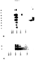
- the purified GalNAcT preparation contains only one polypeptide, with a molecular mass of approximately 70 kDa, detectable with silver staining (Figure 1A).
- a portion of the purified preparation is labeled in vitro with 125 I and separated on SDS-PAGE before and after digestion with peptide N-glycosidase F.
- Figure 1B shows that this treatment results in an approximately 6 kDa shift in the apparent molecular mass of the protein.
- N-terminal sequencing of the purified bovine colostrum GalNAcT is done by automated Edman degradation in an Applied Biosystems Sequencer (Model 470) fitted with an on-line HPLC analyzer (Model 120-A) for phenylthiohydantoins. Quantitation of the latter is afforded by the Nelson Analytical Turbochrom chromatography data system connected in parallel with the recorder to the output from the HPLC system.
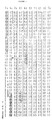
- the 34 amino acid sequence is shown in Figure 2A [SEQ ID NO:1].
- Oligonucleotide primers are synthesized based on the partial N-terminal amino acid sequence of the purified bovine colostrum enzyme with an Applied Biosystems DNA Synthesizer, model 380B.
- the oligonucleotide (oligos A-E) [SEQ ID NOS: 2-6, respectively,] sequence of the primers and probes used in the Polymerase Chain Reaction (hereinafter referred to as PCR) and later in a Southern Blot analysis are shown in Fig. 2A below the GalNAcT amino acid sequence.
- the degeneracy of oligonucleotides A, B and C are 512, 64 and 64, respectively.
- the PCR is carried out in 0.1 ml of solution containing 50 mM KCl, 10 mM Tris-HCL pH 8.3, 1.5 mM MgCl 2 , 0.2 mM each of the four dNTP's, 1 ⁇ M of each oligonucleotide, either 5 ⁇ l of the bovine intestine cDNA library or 10 ng of plasmid or ⁇ DNA and 2.5 units of Taq polymerase.
- the reaction is covered with 0.1 ml of mineral oil and subjected to a temperature step cycle. When degenerate oligonucleotides are used the steps are 94°C (1 min), 37°C (2 min), 72°C (3 min) for a total of 35 cycles.
- oligonucleotides For nondegenerate oligonucleotides the steps are 94°C (1 min), 55°C (2 min), 72°C (3 min) for a total of 25 cycles. Standard DNA manipulations are performed as described in Sambrook, J., Fritsch, E.F., and Maniatis, T. (1989) Molecular Cloning .
- the cDNA encoding the GalNAcT gene is cloned using the following approach. Oligonucleotides A [SEQ ID NO:2] and C [SEQ ID NO:4] are used as opposing primers in a PCR reaction. A bovine small intestine cDNA library cloned into a ⁇ gt10 vector is used as the template for the reaction. On the basis of the amino acid sequence, the predicted size of the amplified PCR product is 93 bp. The products of the PCR reaction are analyzed by Southern blot analysis using oligonucleotide B [SEQ ID NO:3] as a probe ( Figure 2A).
- oligonucleotide primers D-G [SEQ ID NOS: 5-8, respectively] are synthesized. Oligonucleotides D [SEQ ID NO:5] and E [SEQ ID NO:6] are derived from the sequence of the pCR1000-93I insert and F [SEQ ID NO:7] and G [SEQ ID NO:8] are primers that directly flank either side of the EcoRI cloning site of ⁇ gt10 ( Figure 2B).
- PCR reactions are run using the bovine cDNA library as template with oligonucleotides D+F or D+G as primers.
- the resulting PCR products are analyzed by Southern blot analysis using oligonucleotide E [SEQ ID NO:6] as a probe.
- the 621 bp insert contains a 207 amino acid open reading frame with the first 23 amino acids of that open reading frame being a perfect match to amino acids 12-34 of the purified protein ( Figure 2A) [SEQ ID NO:1].
- the 621 bp fragment contains a portion of the GalNAcT gene
- this fragment is labeled with [ ⁇ - 32 P]dATP by nick translation (Goldin et al ., 1981) and is used as a probe to screen the bovine cDNA library.
- the cDNA library (containing 2.5 x 10 6 independent clones) is screened by plaque hybridization using the above labeled DNA fragment as a probe. Seven positive plaques are obtained from the primary screen and each isolate is plaque purified three times. Five of the seven isolates are found to contain inserts of 600 bp or smaller while the two remaining isolates contain inserts of approximately 1600 and 2300 bp.
- the two larger inserts are PCR amplified and cloned (using oligonucleotides F and G as primers) into the TA cloning vector to yield pCR1000-52A (1600 bp insert) and pCR1000-91B (2300 bp insert).
- the size of the ⁇ inserts are analyzed on 1% agarose gels following restriction digest with EcoRI or by PCR using oligonucleotides F and G as primers.
- the inserts in pCR1000-93I, pCR1000-600, pCR1000-91B (2294 bp) and pCR1000-52A (1582 bp) are sequenced by the dideoxy chain termination method (Sanger et al ., 1977) using Sequenase version 2.0 with [ ⁇ - 33 P]dATP. Double stranded DNA sequencing (Ausubel et al . 1987) is done with 20-mer oligonucleotide primers, synthesized according to the sequence of the cDNA insert. The sequencing strategy is shown in Figure 2C. Sequence analysis is performed using the Sequence Analysis software package of the University of Wisconsin Genetics Computer Group (Devereux et al ., 1984).
- the first ATG codon of the sequence obtained from the 91B clone is present at nucleotide 53.
- the sequence of the 52A clone demonstrated that it is a truncated version of the 91B clone in that the sequence of this clone starts at nucleotide 162 and ends at nucleotide 1744 of the larger 91B clone.
- the 52A insert covers nearly all of the open reading frame sequences (missing codons for the first 37 amino acids) found in the 91B clone.
- the nucleotide sequence of the 52A clone is identical to the 91B clone with the exception that nucleotide 358 is a G in the 52A clone instead of an A. This base change is in the wobble position (AG A to AG G ) of codon 102 so it does not alter the arginine at that position.
- the 3'-untranslated region of the 91B clone is 562 bp in length, contains a consensus polyadenylation signal (nucleotides 2176-2182) and a track of 25 A residues at the end of the clone (Fig. 3), indicating that the 91B clone contains all the 3' terminal sequences of the GalNAcT mRNA [SEQ ID NO:10].
- RNA and poly A + RNA are prepared from bovine mammary tissue and from MDBK using the Invitrogen Fastrack kit, following the manufacturers procedure. Two ⁇ g of poly A + RNA are denatured by glyoxylation and Northern blot analysis is performed as previously described (Homa et al ., 1986).
- a human multiple tissue Northern blot (Clontech (Cat # 7760-1)) is prehybridized in 50% formamide, 5 x SSC, 1 x Denhart's, 1% SDS, 100 ⁇ g per ml denatured salmon testes DNA, at 42°C for 2 h and then hybridized overnight at 42°C with the 32 P-labeled 600 bp insert isolated from the pCR1000-600. Filters are washed three times for 15 min in 0.1 X SSC, 0.1% SDS at 55°C. As shown in Figure 5, at least two different sized GalNAcT mRNA's are detected from all the samples. The size of the bovine messages are approximately 4.1 and 3.2 kb, while all the human tissues express messages of 4.8 and 3.9 kb. In addition, a third mRNA of approximately 1.5 kb is detected in the skeletal muscle sample.
- the putative GalNAcT coding region, pCR1000-91B is digested with SstII and HindIII (both enzymes cut only in pCR1000 sequences that flare the insert; Figure 2C) and these sites are blunted using T4 DNA polymerase so that it can be cloned into a baculovirus expression vector.
- BamHI linkers are then ligated onto the blunted ends and the resulting sample is ligated into the BamHI site of the baculovirus expression vector pAC373 (Summers and Smith, 1986).
- the resulting isolates are screened for proper orientation of the GalNAcT open reading frame with respect to the baculovirus polyhedron promoter, to yield pAC373-GalNAcT.
- Cotransfection of Sf9 cells with pAC373-GalNAcT and linearized baculovirus DNA from PharMingen's baculoGold transfection kit is performed using calcium phosphate precipitation (Summers & Smith, 1986).
- the baculovirus DNA provided in the PharMingen transfection kit contains a lethal mutation that can be corrected by homologous recombination with sequences contained in the pAC373 vector. Therefore, following transfection, only recombinant viruses will grow on Sf9 cells.
- GalNAcT 2-1A and GalNAcT 2-1B Transfections are done in duplicate and the resulting virus samples are referred to as GalNAcT 2-1A and GalNAcT 2-1B.
- Cells are harvested 48 hours post infection and lysed in a detergent containing buffer. Following sedimentation of undissolved material, the cleared lysates are assayed for GalNAcT activity. Lysates from uninfected cells or from cells infected with either a baculovirus containing an unrelated gene, CMV-POL (human cytomegalovirus DNA polymerase gene), or two separate baculovirus isolates of the GalNAcT gene, GalNAcT 2-1A and GalNAcT 2-1B, are assayed.
- CMV-POL human cytomegalovirus DNA polymerase gene
- the baculovirus expressed protein is further examined by immunoprecipitation and SDS-PAGE analysis.
- Baculovirus infected cells are labeled from 24 to 48 hours postinfection with [ 35 S]methionine.
- GalNAcT is immunoprecipitated from lysates and culture media of the labeled cells using a chicken polyclonal antibody raised against the purified bovine colostrum enzyme.
- a chicken is injected with 100 ⁇ g purified enzyme axillary, intramuscularly (with Freund's complete adjuvant).
- One month later the chicken is boosted with another 50 ⁇ g antigen subcutaneously (with Freund's incomplete adjuvant); a second booster, 50 ⁇ g enzyme axillary, intra-muscularly, is administered after an additional 21 days.
- Test bleeds are done two weeks after each booster. After the second test bleed (which upon analysis is found to contain anti-GalNAcT antibodies) eggs are collected each day and used as a source for antibodies.
- IgG is isolated from egg yolk as described by Jensenius et al ., 1981.
- Immunoprecipitation of the in vivo 35 S-methionine labeled enzyme is done from crude cell lysates. Infected cells are labeled between 24-48 hours postinfection with 50 ⁇ Ci/ml 35 S-methionine in medium that contains one tenth the normal methionine concentration. Approximately 1.5 X 10 6 labeled, infected cells are dissolved in 670 ⁇ l PBS containing 0.5% Triton X-100, 0.5% taurodeoxycholate, 0.05% SDS, 0.1 TIU/ml of Aprotinin and 10 ⁇ g/ml each of leupeptin, antipain, chymostatin and pepstatin.
- any undissolved debris is sedimented at 10,000 x g for 20 minutes and the supernatant is collected.
- Immunoprecipitation is carried out by the addition of 4 ⁇ l (approximately 20 ⁇ g chicken IgG) of chicken anti GalNacT antibodies; purified IgG isolated from egg yolk is used for all immunoprecipitation experiments.
- the antigen-antibody complexes are isolated by over night adsorption to 22 ⁇ l (volume of sedimented gel) of protein A-Sepharose coated with rabbit anti-chicken IgG antibodies.
- the coated protein A-Sepharose is prepared by incubating 330 ⁇ l sedimented protein A-Sepharose with 2.3 mg rabbit anti-chicken IgG antibodies (an affinity purified IgG fraction) in 1 ml of PBS over night; the coated protein A-Sepharose is washed three times with 1 ml PBS containing 0.5% Triton X-100, 0.5% taurodeoxycholate, 0.05% SDS. Following adsorption of the antigen, the immunosorbent is sedimented by centrifugation and washed extensively essentially as described by Dunphy et al . (1985).
- the washed antigen-antibody-immunosorbent complexes are suspended in 50 ⁇ l SDS-PAGE sample buffer (Laemmli, 1970) and heated for five minutes on a boiling water bath to release the bound antigen. Following sedimentation of the protein A-Sepharose the antigen containing supernatants are aspirated and loaded on SDS-PAGE. SDS-PAGE, and fluorography of the dried gels is done as described previously (Davis et al ., 1986) (Fig. 6).
- the amino acid sequence [SEQ ID NO:9] predicted for the larger clone isolated provides a plausible explanation for the water solubility of the bovine colostrum GalNAcT.
- This enzyme apparently lacks the N-terminal 40 amino acids of the membrane bound molecule, a segment which includes both the cytoplasmic and membrane spanning domains.
- the Kyte-Doolittle hydropathicity plot from the cloned enzyme shows only one sequence segment, residues #9 through 28, with a high membrane spanning probability. It is not clear, at present, if the soluble bovine colostrum enzyme is the result of proteolytic cleavage of a membrane bound molecule or if it represents a bona fide secretory protein.
- Soluble, enzymatically active forms of a ⁇ 1-4 galactosyltransferase and a ⁇ 2-6 sialyltransferase have been reported, both of which appear to be the result of proteolytic cleavage of membrane bound proteins (Paulson and Colley, 1989 and references therein).
- the translation products from the different mRNA species related to both these molecules appears in most tissues to be membrane bound molecules (Joziasse, 1992).
- the larger sizes of the two GalNAcT messages are presumably related to untranslated sequences larger than those recovered in the isolated clones, in the 5' and/or 3' ends of the native molecules.
- Messenger RNA molecules from previously characterized cloned glycosyltransferases frequently contain extensive 5' and 3' untranslated sequences ( e.g. Weinstein et al ., 1987; Larsen et al ., 1989; Russo et al ., 1990; Scocca et al ., 1990; Sarkar et al ., 1991; Nagata et al ., 1992).
- these two proteins are not known at present; they may represent different glycoforms of the enzyme or, perhaps more likely, the lower molecular mass form may be a proteolytic fragment, similar to the enzyme purified from bovine colostrum.
- the latter possibility is supported by two observations: 1), the mass difference between the two molecules is roughly equal to that of the sequence (40 amino acids) missing in bovine colostrum enzyme and 2), while the immunoprecipitates from cell lysates contains predominantly the higher molecular mass form of the enzyme, the culture medium appears to be enriched in the lower mass form. High-speed centrifugation of the culture medium failed to sediment more than approximately 30% of the enzymatic activity (Data not shown).
- the smaller molecular mass of the insect cell produced molecule as compared to the predicted mass of a membrane bound bovine enzyme may be the result of differences in glycosylation of the two molecules.
- Insect cells typically synthesize truncated, non-sialylated N- and O-linked oligosaccharides (e.g. Hsieh and Robbins, 1984; Domingo and Throwbridge, 1988; Kuroda et al ., 1990; Thomsen et al ., 1990; Wathen et al ., 1991; Chen and Bahl, 1991); this results in a reduced molecular mass of insect cell produced glycoproteins on SDS-PAGE. The identity of higher molecular mass bands, approximately 120-180 kDa, on the gel is not clear.
- the sequences coding for the cytoplasmic and membrane spanning domains of the full-length cDNA were replaced with sequences that code for the honeybee melittin signal peptide (Fig. 12).
- the honeybee melittin signal sequence was chosen since the intended expression system for the construct was baculovirus/Sf9 cells. It has been demonstrated that use of an insect derived signal peptide often results in increased secretion (as compared to a signal peptide of heterologous origin) of the recombinant molecule when expressed in Sf9 cells (Tessier et al., 1991).
- the fusion site for the signal peptide was chosen based on the N-terminal sequence of the soluble colostrum enzyme.
- the plasmid pAC373-GalNAcT (Homa et al., 1993) which contains the full length GalNAc-transferase gene under the control of the baculovirus polyhedron promoter was digested with XbaI and BglII, which generated a 150 bp fragment, and with BglII and XhoI, which generated a 9700 bp vector fragment. Both fragments were gel purified.
- the XbaI site used is located 7 amino acids from the N-terminus of the soluble colostrum enzyme, in a portion of the molecule corresponding to what is referred to as the "stem region" in other glycosyltransferases (reviewed by Shaper & Shaper, 1992). Soluble forms of several glycosyltransferases are generated by proteolytic cleavages in this region. Consequently, it is believed to be unimportant for catalytic activity (Joziasse, 1992; Shaper & Shaper, 1992).
- pVT-BAC (Tessier et al, 1991), which contains the coding sequences of the honeybee melittin signal peptide, was digested with SmaI and a 12 bp XbaI linker was ligated onto the SmaI site. The sample was then digested with XbaI and XhoI and the 2100 bp fragment generated by this digest was gel purified. The three gel purified fragments were added to the same tube and ligated.
- the resulting plasmid contains a GalNAc-transferase gene under the control of the baculovirus polyhedron promoter in which the first 47 amino acids (141 nucleotides) have been replaced with 21 amino acids (63 nucleotides) of the honeybee melittin signal peptide plus five (5) amino acids (15 nucleotides) that link the two domains together (Fig. 13) [SEQ ID NO:18].
- GalNAcTs was routinely expressed in one liter batches. Approximately 1X10 9 cells in one liter Grace's serum-free insect medium (Insect Express) were infected with 5 pfu/cell of GalNAcTs-Mel. The infected cells were cultured in shaker flasks for 65 hours before harvest. Expression of the construct GalNAcTs-Mel in Sf9 cells resulted in 130-fold increase in GalNAc-transferase activity in the culture medium, as compared to uninfected cells (Table 2) or cells infected with an unrelated molecule ( ⁇ 6-3). This is more than 35 times the amount recovered in the medium of cells expressing the full length molecule (Homa & Elhammer, unpublished observations). A significant portion (36%) of the total enzymatic activity resulting from expression of the soluble molecule was, however, retained inside the cells; the reason for this is not clear at present.
- Purification of the soluble molecule was accomplished in one step, by chromatography on apomucin-Sepharose.
- the procedure used for the purification of the recombinant enzyme was a modification of the second affinity chromatography step in the purification of the bovine colostrum enzyme (Elhammer & Kornfeld, 1986).
- the affinity column used for purification of the recombinant enzyme was prepared as described (Elhammer & Kornfeld, 1986). Total bed volume was approximately 200 ml; ligand density was approximately 3.5 mg/ml gel. Before each separation run the column was washed with 1000 ml Buffer B and equilibrated with 1000ml Buffer F followed by 420 ml Buffer F containing 0.25 mM UDP. Crude conditioned cell culture medium was fractionated on this column in 1 liter batches as follows.
- the medium was first dialyzed against 25 mM Imidazole, pH 7.2, 100 mM NaCl, 30 mM MnCl 2 with three buffer changes. Following centrifugation at 12,000Xg for 20 minutes to remove precipitated material (no enzyme activity was lost in this step), the dialyzed medium was supplemented with UDP to 1.25 mM and loaded directly on the affinity column; the column run-through was collected in five 200 ml fractions. The column was then washed sequentially with 500 ml buffer F, 250 ml Buffer F containing 0.2% Triton X-100, 250 ml Buffer F and 250 ml Buffer F containing 1.0 M NaCl.
- Dialyzing the enzyme into a buffer containing 300 mM NaCl prior to concentration was an absolute necessity to avoid even higher losses in this step.
- the enzyme isolated from bovine colostrum shows a similar behavior in this regard (Elhammer & Kornfeld, 1986).
- the purified preparation was concentrated by ultrafiltration on a YM-10 membrane at 45 psi pressure.
- the crude medium sample was precipitated as described by Wessel and Flugge (1984) prior to electrophoresis; precipitate corresponding to approximately 250 ⁇ l medium was loaded.
- NH 2 -terminal sequencing of the purified molecule was done as described in Example 2. Interestingly, this purification procedure yielded a homogenous preparation only if expression of the molecule was carried out in serum-free medium.
- the amino acid composition of the purified GalNacTs was determined by automated ion-exchange chromatography on a Beckman amino acid analyzer (Beckman 6300) and was found to correspond with the composition predicted from the nucleic acid sequence shown in Fig. 3 [SEQ ID NO:9]. Samples were hydrolyzed for 24 hours in vacuo at 110°C in 6N HCl. Dried hydrolysates were dissolved in buffer at pH 2.2 (NaS; Beckman) prior to application to the analyzer. Quantitation of the latter was afforded by the Nelson Analytical Turbochrom chromatography data system connected in parallel with the recorder to the output of the dual-channel spectrophotometer.
- NH 2 -terminal sequencing of the purified, concentrated enzyme demonstrated that the melittin leader sequence was cleaved from the molecule at the predicted site (Fig. 12). Furthermore, only one sequence was detected in the preparation, consistent with an essentially pure enzyme preparation. The purity of the enzyme was also investigated on SDS-PAGE. As can be seen in Fig. 14, only one band, with a molecular mass of approximately 61 kDa, could be detected by Coomassie Blue staining. Silver staining also failed to detect more that one protein band on the gel and separation of the purified enzyme on capillary electrophoresis resulted in only one UV absorbing peak eluting at a position consistent with the molecular mass of approximately 61 kDa (Data not shown).
- the efficient production of the cloned molecule using the baculovirus expression system facilitated preparation of GalNAc-transferase in amounts sufficient for detailed biochemical and enzymatic studies to determine the acceptor substrate specificity of GalNAc-transferase from a database of in vivo substrates and from the in vitro glycosylation of proteins and peptides. These studies have been facilitated by the availability of information regarding the presence of glycosylated serine and threonine residues in proteins obtained during protein sequencing. This information is registered in the NBRF protein sequence repository.
- a search of the NBRF protein database yields several hundred definite or probable Thr and Ser O-glycosylation sites. From these, only those with reasonably unambiguous assignments are chosen and all proteoglycans are excluded since they contain primarily glycosaminoglycan chains where the anchoring sugar is xylose and not GalNAc. Also included into the reference set are the O-glycosylation sites identified in some recently analyzed glycoproteins. In the case of proteins listed for several different species, homologous proteins are included only when the glycosylation sites themselves show no homology. The complete reference set consists of the 196 glycosylated peptide segments (shown in Table 1 in Elhammer et al., 1993).
- the glycosylated peptides are listed as enneapeptide ( ennea Greek, nine) segments, with the reactive Ser or Thr in the central position, designated as P0. Accordingly, the amino acid side chains toward the N-terminus are designated as the subsites P1 to P4 and those toward the C-terminus as subsites P1' to P4'. A length of nine residues is chosen as a starting point, with the option that, depending on the results on the selectivity of the subsites, the portion of the peptides subject to analysis may be extended or truncated.
- the sequences show that besides the obvious need for Ser or Thr in P0, no other subsite has an absolute requirement for any given amino acid. This then suggests that specificity of the enzyme may be the result of the cooperation of several subsites, none of them essential, but all of them contributing to catalytic efficiency.
- h RP RP + 1 PS
- PS the abundance of glycosylatable peptides in all proteins
- a Ser or Thr-containing peptide may be predicted to be a substrate for the enzyme if the probability, h, is higher than a certain cutoff value, h c .
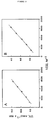
- the probability pattern of these two proteins is shown in Figure 7. It is interesting to note that the calculated probabilities for these two proteins are not distributed uniformly between the two extremes. Rather, a small number of residues are associated with very high probabilities whereas the rest of the sequence indicates uniformly low probabilities. Furthermore, the residues with high probabilities are clustered into one or two distinct segments where the clustering of Ser and Thr residues may perhaps be a necessary but certainly not a sufficient criterion for creating a highly glycosylated protein segment.
- subtilisin BPN' (subsn.aa, Figure 7C) which is produced by a microbial system incapable of O-linked oligosaccharide biosynthesis, contains a number of randomly distributed potential glycosylation sites, while very few nonglycosylated mammalian proteins contain any potential glycosylation sites. It is perhaps more typical to find no potential glycosylation sites at all, as in the case of horse hemoglobin (hbho.aa) or that of bovine cytochrome C (ccpg.aa, Figure 7D).
- this region of the protein consists of a fully exposed segment linking the two homologous domains of the enzyme, exposure of native or mildly denatured rhodanese to GalNAc-transferase should result in glycosylation of the molecule.
- the chimeric protein, CD4PE40 constructed from two domains of the human CD4 protein and three domains of the Pseudomonas exotoxin, shows two prominent potential glycosylation sites, both at regions linking individual domains, see Figure 8B.
- subtilisin could also be extensively glycosylated, if not in the native form then at least after mild denaturation.
- LDL receptor LDL receptor
- Alzheimer precursor protein alz.aa, Figure 8D
- the present method not only correctly identifies these regions of glycosylation but also specifically predicts which Ser and Thr residues may be modified.
- s i,j should depend critically on that property. Attempts to correlate the s i,j distribution with a variety of parameters possibly characterizing the individual amino acids met with little success. Most importantly, no correlation has been found with a parameter predicting ⁇ -turns, although such a correlation has been inferred for xylosyltransferase (M.A. Bourdon et al ., 1987).
- the enzyme would recognize a specific secondary structure of the target enneapeptide and that this secondary structure would be induced not by the cooperative action of several constituent amino acids but by their intrinsic tendency to form a given secondary structure.
- the potential glycosylation sites predicted for proteins in the Kabsch-Sander (Kabsch and Sander, 1983) database is used since no secondary structural information is available at the present time for any O-glycosylated proteins.
- the 50 potential sites with the highest h values are selected. As shown in Table 6, these potential glycosylation sites are very strongly selected against ⁇ -helical conformation.
- the preferred conformations appear to be a random coil, a sharp bend, or a ⁇ -strand from P4 to P0 followed by a turn.
- the enzyme does not require a preformed secondary structure but imposes one upon binding of the substrate.
- the hydration index of the amino acids in the potential glycosylation sites also shown in Table 6, indicates that most pep:ides are reasonably exposed to the aqueous environment.
- reaction products are characterized using alkaline sodium borohydride treatment essentially as described by Carlson (1968).
- Digestion with Patella vulgata ⁇ -N-acetylgalactosaminidase (approximately 1 unit/ml) is done in 25 mM citrate buffer pH 4.0 in a final volume of 30 ⁇ l for 24 hours.
- Released radioactive sugars are separated on descending paper chromatography in pyridine-ethyl acetate-glacial acetic acid-water (5:5:1:3; v:v:v:v).
- Table 7 shows that, as predicted, both bovine rhodanese and, to a lesser extent, the bacterial protein subtilisin do indeed function as acceptors for the enzyme, although neither of them reacts unless reduced and carboxymethylated prior to exposure to the enzyme.
- bovine cytochrome C which contains one Ser and eight Thr residues but no predicted potential sites, is not an acceptor for the enzyme, whether in the native, or in the reduced and carboxymethylated state.
- Myelin basic protein a molecule which previously has been shown to be an efficient acceptor for GalNAc-transferase (Hagopian et al ., 1971) is included as a positive control in this experiment.
- Rhodanese contains two additional predicted acceptor sites, Ser 142 and Ser 145 , (Fig. 8). However, due to the low rates of transfer to serine residues under our standard assay conditions, transfer to these sites should not contribute significantly to the total transfer in the assay.
- the lower rate of transfer to reduced and carboxymethylated rhodanese compared to that of myelin basic protein, may be related to incomplete exposure of the acceptor sites even by the reduction and carboxymethylation procedure, and/or differences in rate constants between the acceptor sequences on the two molecules.
- Myelin basic protein contains one site predicted with high probability and three additional low probability sites. The molecule can reportedly be glycosylated with 1.2 to 1.5 N-acetylgalactosamines per molecule (Cruz and Moscarello, 1983).
- the bacterial protein subtilisin contains four predicted serine sites with probabilities higher than 0.6 (Fig. 8).
- Three of the serines have a high exposure index in the native protein (Kabsch and Sander, 1983), but the three-dimensional structure of the protein indicates that the hydroxyls are located in a restrained environment. Again, this could account for the need for reduction and carboxymethylation for acceptor activity.
- the 35 times slower transfer rate to denatured subtilisin, as compared to myelin basic protein indicates again a slower transfer to serines than to threonines, under the conditions used. Factors such as those discussed for rhodanese may also contribute to the low levels of transfer.
- cytochrome C which does not contain any predicted acceptor site, is completely inactive as an acceptor, whether in the native or in the reduced and carboxymethylated form.
- the ability of GalNAc-transferase to glycosylate a series of synthetic acceptor peptides is shown in Figure 9 and Table 8.
- the t-Boc-amino acids and the PAM resin solid supports are supplied by ABI.
- the completed peptides are removed from the supporting resin, concurrently with the side chain-protecting groups, by a standard HF cleavage procedure using anisole as a cation scavenger (10% v/v).
- the crude peptides are purified by preparative reverse phase chromatography on a C18 Vydac column (2.5 x 30 cm) using a water/acetonitrile gradient, each phase containing 0.1% TFA.
- Each purified peptide is characterized by FAB/MS and shows a single symmetrical peak on analytical HPLC.
- glycosylated amino acids in the peptides PPASTSAPG [SEQ ID NO:14] and PPASSSAPG [SEQ ID NO:15] are identified by sequencing of the reaction products from the corresponding assay.
- Fig. 11 shows that for both glycosylated peptides the majority of the sugar-linked radioactivity is associated with residue #5, the central amino acid, be it threonine, as in PPASTSAPG [SEQ ID NO:14], or serine, as in PPASSSAPG [SEQ ID NO:15].
- the measurable amounts of radioactivity associated with the residues following residue 5 are presumably due to the large load of peptide in the sequencer necessitated by the low specific radioactivity of the sample. Nevertheless, since the radioactivities associated with residues 7 and 8 extrapolate smoothly to that of residue 6, it is most likely that, within our experimental error, residue 6 is not labelled.
- N-acetylgalactosamine to peptide acceptors is assayed by two different assays.
- concentration of UDP-GalNAc is saturating in all assays; a K m of 8 ⁇ M is reported for bovine colostrum GalNAc-transferase (Elhammer and Kornfeld, 1986).
- the reaction mixture contains 50 mM Imidazole, pH 7.2, 10 mM MnCl 2 , 0.5% Triton X-100, 150 ⁇ M UDP-GalNAc, approximately 120,000 cpm of UDP-[1- 3 H]-GalNAc, varying concentrations of acceptor (see individual experiments) and approximately 5 mU of enzyme in a final volume of 40 ⁇ l; standard incubation time is 20 minutes.
- the reaction is terminated by placing the samples in a boiling water bath for 1.5 minutes.
- reaction product glycosylated peptide
- UDP-GalNAc a product that is separated from unreacted UDP-GalNAc by chromatography on Dowex-2 columns (0.5 ml bed volume) equilibrated in water; the run-through fraction (2.5 ml) containing the glycosylated peptide is collected, supplemented with scintillation fluid and counted for radioactivity.
- the assay conditions are as follows: 50 mM Imidazole, pH 7.2, 10 mM MnCl 2 , 0.5% Triton X-100, 150 ⁇ M UDP-GalNAc, approximately 260,000 cpm UDP-[ 3 H]-GalNAc and 3.2 mM acceptor peptide (the concentration of RSPPP is 3.7 mM).
- the assays are incubated for 20 minutes (PPASTSAPG) [SEQ ID NO:14] or 8 hours (PPASSSAPG [SEQ ID NO:15], PPAdSSdSAPG and RSPPP [SEQ ID NO:13]).
- the enzyme is inactivated by placing the samples on a boiling water bath for 1.5 minutes. The samples are then allowed to cool and the reaction products are separated from unreacted UDP-GalNAc and free GalNAc by chromatography on a Biogel P-2 column (1 X 50 cm) equilibrated in 7% isopropanol; thirty 1.3 ml fractions are collected.
- the peptide PPASTSAPG [SEQ ID NO:14] is designed to contain a single Thr, at P0.
- the proline residues at P4, P3 and P3' provide maximum probabilities at those positions; serine residues at P1 and P1' result in good probabilities without much steric constraint.
- the alanine residues at P2 and P2' and the glycine at P4 are indifferent as to the probability of glycosylation but allow for flexibility of the peptide backbone.
- Tables 8 and 9 show that this peptide is the most efficient of the acceptors tested and comparative assays show that its reactivity is very close to that of bovine apomucin (data not shown). Furthermore, the kinetic parameters for the two peptides, determined under our conditions, are quite comparable to those of the purified porcine submaxillary GalNAc-transferase-catalyzed glycosylation of peptides whose structure is derived from sites identified in porcine submaxillary mucin (Wang et al ., 1992).
- the peptide RTPPP [SEQ ID NO:12] derived from the major acceptor sequence in myelin basic protein (Hagopian et al .,1971), has a K m lower than that of PPASTSAPG [SEQ ID NO:14] but also a much lower V max and, hence, its catalytic efficiency is only half of that of PPASTSAPG [SEQ ID NO:14].
- the activities of the two corresponding peptides containing serine instead of threonines are measurable but too low for determining the kinetic parameters under the conditions used.
- bovine colostrum GalNAc-transferase is capable of transferring GalNAc to the serine of these peptides, albeit ⁇ under our in vitro conditions ⁇ approximately 35 times slower than to threonine (Table 9).
- bovine colostrum GalNAc-transferase In contrast to the enzyme recently purified from porcine submaxillary gland (Wang et al ., 1992), however, the bovine colostrum GalNAc-transferase is definitely capable of glycosylating both threonine and serine residues. In this context it should be noted that in experiments reported by O'Connel et al . (1992), bovine colostrum GalNAc-transferase failed to glycosylate serine in a peptide derived from human erythropoietin. This phenomenon may be related to the specific acceptor peptide used, even though a serine in this position is glycosylated in vivo .
- Ser is indeed an in vivo acceptor, for the following reasons: 1) numerous reports identify unambiguously O-glycosylated serine residues in glycoproteins; 2) preliminary experiments indicate that the Ser- and Thr-glycosylating activities have similar pH optima and that both are independent of calcium concentrations in the assays; 3) the affinity matrix for isolation of the enzyme is bovine apomucin immobilized on Sepharose. This ligand contains comparable numbers of serine and threonine residues and should consequently not distinguish between the two activities (compare below); 4) a mere 35-fold difference in rate is quite usual and is, in fact, rather small as a range of specificity for multisubstrate enzymes.
- GalNAc-transferase The specificity of GalNAc-transferase is adequately described by a cumulative specificity model, where the independent contribution of several subsites can produce a range of reactivities toward peptide segments. Additional important features of the acceptor peptide segments are exposure on the surface of the protein substrate and an extended conformation.
- the acceptor amino acid is surrounded preferentially by Ser, Thr, Pro, Ala and Gly residues, but in no specific order. Both serine and threonine are acceptor amino acids, at least for the bovine colostrum enzyme.
- the Km for UDP-GalNAc is approximately 1.7 ⁇ M and the Km:s for the threonine containing acceptor peptide PPASTSAPG [SEQ ID NO:14] and the serine containing acceptor peptide PPDAASAAPLR [SEQ ID NO:17] are approximately 6.5 and 3.6 mM, respectively.
- Transfer by GalNAcTs to another serine containing acceptor peptide PPASSSAPG [SEQ ID NO:15] is approximately 70 times slower than to PPASTSAPG [SEQ ID NO:14] (Data not shown).
- the specific activity of the purified enzyme preparation, using bovine apomucin as acceptor, is approximately 2,160 U/mg protein (Table 4).
- the enzymatic properties of the purified GalNacTs appear to be similar to the those determined for the enzymes purified from bovine colostrum and porcine submaxillary gland (Elhammer and Kornfeld, 1986; Elhammer et al., 1993; Wang et al., 1992; Wang et al., 1993).
- the Km for the acceptor peptide PPASTSAPG [SEQ ID NO:14] is almost identical for the colostrum enzyme and the baculo expressed molecule, 6.0 vs. 6.5 mM.
- the serine containing acceptor peptide PPDAASAAPLR [SEQ ID NO:17] (O'Connel et al.
- GalNAcTs and the bovine colostrum enzyme glycosylate the peptide PPASSSAGP [SEQ ID NO:15] are at least 35 times slower than PPASTSAPG [SEQ ID NO:14] (Elhammer et al., 1993).
- the Km for UDP-GalNAc in assays using GalNAcTs is lower than those determined for the bovine colostrum and the porcine submaxillary gland enzymes, 1.7 ⁇ M vs. 8 ⁇ M and 6 ⁇ M, respectively (Table V; Elhammer & Kornfeld, 1986; Wang et al., 1992). The reason for this is not clear at present.
- the amino acid sequence of the bovine colostrum and the cloned molecules should be identical except for five amino acids in the NH 2 -terminal end of the molecule; the colostrum enzyme sequence also contains an additional two amino acids at the NH 2 -terminus.
- differences in post-translational processing, in particular glycosylation, of the Sf9 produced vs. the bovine molecule may, to some extent, influence the kinetic characteristics of the two molecules.
- the oligosaccharide structures on the insect produced molecule are most likely of high mannose and/or truncated high mannose type, while results from endoglycosidase digestion experiments suggest that the colostrum enzyme contains complex type oligosaccharides (Elhammer & Kornfeld, 1986). It is likely that the N-linked oligosaccharide structures on the porcine enzyme also would be of the types normally synthesized by mammalian cells; peptide: N-glycosidase F digestion experiments suggest that this molecule contains 9 kDa of N-linked oligosaccharides (Wang et al., 1992). Further experiments will however be needed to clarify this question.
- Table 1 Expression of baculovirus isolates of the GalNAc-transferase Construct Specific activity, Units/mg protein Relative rate Uninfected cells 0.16 1 CMW Pol-1 0.13 0.8 GalNAcT 2-1.A 13.9 87 GalNAcT 2-1.B 11.9 74
- Table 2 Expression of baculovirus isolates of soluble GalNAc-transferase Construct Total activity (U a ) Cell lysate Culture medium Uninfected cells 0.15 0.44 ⁇ 6-3 0.12 0.40 GalNAcT 2-1A 16.22 1.61 GalNAcT-Mel 32.15 57.80 a 1 unit equals one mole N-acetylgalactosamine transferred to apomucin per minute, under assay conditions.
- Cells (1X10 6 ) were infected with recombinant virus containing GalNAcTs-Mel (5 pfu/cell). The cells were harvested 65 hours post infection, lysed in a detergent containing buffer and the GalNac-transferase activity was determined in the cell lysates and the corresponding culture media; lysate and culture medium from uninfected cells were assayed as control.
- Surabundant amino acids surrounding the reactive Ser or Thr Surabundance at a given subsite for a given amino acid is expressed as the number of that amino acid found at the site in excess to that expected from random distribution, divided by the S.D. of the expected distribution.
- the excess of surabundant residues is equal to or higher than twice the S.D.
Applications Claiming Priority (3)
| Application Number | Priority Date | Filing Date | Title |
|---|---|---|---|
| US6318693A | 1993-05-14 | 1993-05-14 | |
| EP94915336A EP0698103A1 (fr) | 1993-05-14 | 1994-03-17 | UN ADN CLONE CODANT UNE UDT-GALNAc:POLYPEPTIDE, N-ACETYLGALACTOSAMINYLTRANSFERASE |
| US63186 | 1997-10-20 |
Related Parent Applications (2)
| Application Number | Title | Priority Date | Filing Date |
|---|---|---|---|
| EP94915336.5 Division | 1994-03-17 | ||
| EP94915336A Division EP0698103A1 (fr) | 1993-05-14 | 1994-03-17 | UN ADN CLONE CODANT UNE UDT-GALNAc:POLYPEPTIDE, N-ACETYLGALACTOSAMINYLTRANSFERASE |
Publications (1)
| Publication Number | Publication Date |
|---|---|
| EP0726318A1 true EP0726318A1 (fr) | 1996-08-14 |
Family
ID=22047525
Family Applications (2)
| Application Number | Title | Priority Date | Filing Date |
|---|---|---|---|
| EP94915336A Withdrawn EP0698103A1 (fr) | 1993-05-14 | 1994-03-17 | UN ADN CLONE CODANT UNE UDT-GALNAc:POLYPEPTIDE, N-ACETYLGALACTOSAMINYLTRANSFERASE |
| EP96104017A Withdrawn EP0726318A1 (fr) | 1993-05-14 | 1994-03-17 | Polypeptide accepteur pour la N-acétylgalactosaminyl-transférase |
Family Applications Before (1)
| Application Number | Title | Priority Date | Filing Date |
|---|---|---|---|
| EP94915336A Withdrawn EP0698103A1 (fr) | 1993-05-14 | 1994-03-17 | UN ADN CLONE CODANT UNE UDT-GALNAc:POLYPEPTIDE, N-ACETYLGALACTOSAMINYLTRANSFERASE |
Country Status (6)
| Country | Link |
|---|---|
| US (1) | US6096512A (fr) |
| EP (2) | EP0698103A1 (fr) |
| JP (1) | JPH09501044A (fr) |
| AU (1) | AU6663294A (fr) |
| IL (1) | IL109364A0 (fr) |
| WO (1) | WO1994026906A2 (fr) |
Families Citing this family (61)
| Publication number | Priority date | Publication date | Assignee | Title |
|---|---|---|---|---|
| GB2288401A (en) * | 1994-04-15 | 1995-10-18 | Bay Dev Corp Sa | N-acetylgalactosaminyltransferase |
| US5545553A (en) * | 1994-09-26 | 1996-08-13 | The Rockefeller University | Glycosyltransferases for biosynthesis of oligosaccharides, and genes encoding them |
| EP0754703A4 (fr) * | 1994-11-01 | 1999-11-24 | Kirin Brewery | Sequence peptidique formant une chaine de mucine et technique de modification de la proteine a lier a la chaine de mucine |
| US5861318A (en) * | 1994-11-16 | 1999-01-19 | Pharmacia & Upjohn Company | Scintillation proximity assay for N-acetylgalactosaminyltransferase activity |
| US5871990A (en) * | 1996-05-15 | 1999-02-16 | Clausen; Henrik | UDP-N-acetyl-α-D-galactosamine: polypeptide N-acetylgalactosaminyltransferase, gAlnAc-T3 |
| US8697394B2 (en) * | 2000-06-28 | 2014-04-15 | Glycofi, Inc. | Production of modified glycoproteins having multiple antennary structures |
| US7863020B2 (en) * | 2000-06-28 | 2011-01-04 | Glycofi, Inc. | Production of sialylated N-glycans in lower eukaryotes |
| US7625756B2 (en) * | 2000-06-28 | 2009-12-01 | GycoFi, Inc. | Expression of class 2 mannosidase and class III mannosidase in lower eukaryotic cells |
| EP2322644A1 (fr) * | 2000-06-28 | 2011-05-18 | GlycoFi, Inc. | Procédé de production de glycoprotéines modifiées |
| US7598055B2 (en) * | 2000-06-28 | 2009-10-06 | Glycofi, Inc. | N-acetylglucosaminyltransferase III expression in lower eukaryotes |
| US7449308B2 (en) | 2000-06-28 | 2008-11-11 | Glycofi, Inc. | Combinatorial DNA library for producing modified N-glycans in lower eukaryotes |
| GB2379661A (en) * | 2001-01-30 | 2003-03-19 | Aeomica Inc | Human UDP-GALNAC:Polypeptide N-Acetylgalactosaminyltransferase 10 |
| US7157277B2 (en) | 2001-11-28 | 2007-01-02 | Neose Technologies, Inc. | Factor VIII remodeling and glycoconjugation of Factor VIII |
| US7399613B2 (en) * | 2001-10-10 | 2008-07-15 | Neose Technologies, Inc. | Sialic acid nucleotide sugars |
| US8008252B2 (en) * | 2001-10-10 | 2011-08-30 | Novo Nordisk A/S | Factor VII: remodeling and glycoconjugation of Factor VII |
| US7179617B2 (en) * | 2001-10-10 | 2007-02-20 | Neose Technologies, Inc. | Factor IX: remolding and glycoconjugation of Factor IX |
| US7214660B2 (en) | 2001-10-10 | 2007-05-08 | Neose Technologies, Inc. | Erythropoietin: remodeling and glycoconjugation of erythropoietin |
| US7795210B2 (en) * | 2001-10-10 | 2010-09-14 | Novo Nordisk A/S | Protein remodeling methods and proteins/peptides produced by the methods |
| US7173003B2 (en) | 2001-10-10 | 2007-02-06 | Neose Technologies, Inc. | Granulocyte colony stimulating factor: remodeling and glycoconjugation of G-CSF |
| US7696163B2 (en) | 2001-10-10 | 2010-04-13 | Novo Nordisk A/S | Erythropoietin: remodeling and glycoconjugation of erythropoietin |
| US7473680B2 (en) | 2001-11-28 | 2009-01-06 | Neose Technologies, Inc. | Remodeling and glycoconjugation of peptides |
| JP2003199573A (ja) * | 2001-12-28 | 2003-07-15 | Jgs:Kk | 新規udp−n−アセチル−d−ガラクトサミン:ポリペプチドn−アセチルガラクトサミン転移酵素及びこれをコードする核酸 |
| WO2003074687A1 (fr) | 2002-03-07 | 2003-09-12 | Eidgenössische Technische Hochschule Zürich | Systeme et procede de fabrication de proteines glycosylees de recombinaison dans un hote procaryotique |
| MXPA04012496A (es) | 2002-06-21 | 2005-09-12 | Novo Nordisk Healthcare Ag | Glicoformos del factor vii pegilados. |
| US7332299B2 (en) | 2003-02-20 | 2008-02-19 | Glycofi, Inc. | Endomannosidases in the modification of glycoproteins in eukaryotes |
| CA2519092C (fr) | 2003-03-14 | 2014-08-05 | Neose Technologies, Inc. | Polymeres hydrosolubles ramifies et conjugues de ceux-ci |
| US8791070B2 (en) | 2003-04-09 | 2014-07-29 | Novo Nordisk A/S | Glycopegylated factor IX |
| WO2006127896A2 (fr) | 2005-05-25 | 2006-11-30 | Neose Technologies, Inc. | Facteur ix glycopegyle |
| EP1613261A4 (fr) * | 2003-04-09 | 2011-01-26 | Novo Nordisk As | Formation intracellulaire de conjugues de peptides |
| US20070026485A1 (en) | 2003-04-09 | 2007-02-01 | Neose Technologies, Inc. | Glycopegylation methods and proteins/peptides produced by the methods |
| BRPI0410164A (pt) | 2003-05-09 | 2006-05-16 | Neose Technologies Inc | composições e métodos para preparação de mutantes de glicosilação de hormÈnio do crescimento humano |
| WO2005012484A2 (fr) | 2003-07-25 | 2005-02-10 | Neose Technologies, Inc. | Conjugues anticorps-toxines |
| US20080305992A1 (en) | 2003-11-24 | 2008-12-11 | Neose Technologies, Inc. | Glycopegylated erythropoietin |
| US8633157B2 (en) | 2003-11-24 | 2014-01-21 | Novo Nordisk A/S | Glycopegylated erythropoietin |
| ES2445948T3 (es) * | 2003-11-24 | 2014-03-06 | Ratiopharm Gmbh | Eritropoyetina glicopegilada |
| US7956032B2 (en) | 2003-12-03 | 2011-06-07 | Novo Nordisk A/S | Glycopegylated granulocyte colony stimulating factor |
| US20060040856A1 (en) | 2003-12-03 | 2006-02-23 | Neose Technologies, Inc. | Glycopegylated factor IX |
| US20080318850A1 (en) * | 2003-12-03 | 2008-12-25 | Neose Technologies, Inc. | Glycopegylated Factor Ix |
| ES2560657T3 (es) | 2004-01-08 | 2016-02-22 | Ratiopharm Gmbh | Glicosilación con unión en O de péptidos G-CSF |
| WO2006010143A2 (fr) | 2004-07-13 | 2006-01-26 | Neose Technologies, Inc. | Remodelage de peg ramifie et glycosylation de peptide-1 semblable a glucagon [glp-1] |
| EP1799249A2 (fr) | 2004-09-10 | 2007-06-27 | Neose Technologies, Inc. | Interferon alpha glycopegyle |
| DK2586456T3 (en) | 2004-10-29 | 2016-03-21 | Ratiopharm Gmbh | Conversion and glycopegylation of fibroblast growth factor (FGF) |
| JP4951527B2 (ja) | 2005-01-10 | 2012-06-13 | バイオジェネリックス アーゲー | 糖peg化顆粒球コロニー刺激因子 |
| US20060246544A1 (en) * | 2005-03-30 | 2006-11-02 | Neose Technologies,Inc. | Manufacturing process for the production of peptides grown in insect cell lines |
| US20070154992A1 (en) * | 2005-04-08 | 2007-07-05 | Neose Technologies, Inc. | Compositions and methods for the preparation of protease resistant human growth hormone glycosylation mutants |
| ES2703061T3 (es) | 2005-05-11 | 2019-03-06 | Eth Zuerich | Proteínas N-glicosiladas recombinantes procedentes de células procariotas |
| EP1888098A2 (fr) | 2005-05-25 | 2008-02-20 | Neose Technologies, Inc. | Formulations d'erythropoietine glycopegylees |
| BRPI0614839A2 (pt) * | 2005-08-19 | 2009-05-19 | Neose Technologies Inc | fator vii e fator viia glicopeguilados |
| US20070105755A1 (en) * | 2005-10-26 | 2007-05-10 | Neose Technologies, Inc. | One pot desialylation and glycopegylation of therapeutic peptides |
| US20090048440A1 (en) | 2005-11-03 | 2009-02-19 | Neose Technologies, Inc. | Nucleotide Sugar Purification Using Membranes |
| WO2008011633A2 (fr) | 2006-07-21 | 2008-01-24 | Neose Technologies, Inc. | Glycosylation de peptides par l'intermédiaire de séquences de glycosylation à liaison o |
| US8969532B2 (en) | 2006-10-03 | 2015-03-03 | Novo Nordisk A/S | Methods for the purification of polypeptide conjugates comprising polyalkylene oxide using hydrophobic interaction chromatography |
| US20080207487A1 (en) * | 2006-11-02 | 2008-08-28 | Neose Technologies, Inc. | Manufacturing process for the production of polypeptides expressed in insect cell-lines |
| CA2682897C (fr) | 2007-04-03 | 2016-11-22 | Biogenerix Ag | Methodes de traitement a l'aide d'un facteur g-csf glycopegyle |
| US20090053167A1 (en) * | 2007-05-14 | 2009-02-26 | Neose Technologies, Inc. | C-, S- and N-glycosylation of peptides |
| MX2009013259A (es) | 2007-06-12 | 2010-01-25 | Novo Nordisk As | Proceso mejorado para la produccion de azucares de nucleotidos. |
| US8207112B2 (en) | 2007-08-29 | 2012-06-26 | Biogenerix Ag | Liquid formulation of G-CSF conjugate |
| CA2716187C (fr) | 2008-02-20 | 2020-01-07 | Glycovaxyn Ag | Bioconjugues faits a partir de proteines n-glycosylees recombinees issues de cellules procaryotes |
| PL2257311T3 (pl) | 2008-02-27 | 2014-09-30 | Novo Nordisk As | Koniugaty cząsteczek czynnika VIII |
| AU2010322454B2 (en) | 2009-11-19 | 2016-05-19 | Glaxosmithkline Biologicals S.A. | Biosynthetic system that produces immunogenic polysaccharides in prokaryotic cells |
| EP3281639B1 (fr) | 2010-05-06 | 2020-11-11 | GlaxoSmithKline Biologicals S.A. | Vaccins de bioconjugué de bactéries gram positif capsulaire |
Family Cites Families (10)
| Publication number | Priority date | Publication date | Assignee | Title |
|---|---|---|---|---|
| SE8001748L (sv) * | 1980-03-05 | 1981-09-06 | Kaellenius Gunilla | Kompositioner for terapeutisk eller diagnostisk anvendning jemte forfarande for terapeutik |
| US4521592A (en) * | 1981-10-23 | 1985-06-04 | Svenska Sockerfabriks Ab | Compounds for therapeutic or diagnostic use, a process and intermediates for their preparation |
| US4801583A (en) * | 1982-01-15 | 1989-01-31 | Choay S.A. | Oligosaccharides and their biological applications |
| SE8200751L (sv) * | 1982-02-09 | 1983-08-10 | Olle Larm | Forfarande for kovalent koppling for framstellning av konjugat och hervid erhallna produkter |
| AU561067B2 (en) * | 1982-03-22 | 1987-04-30 | Biocarb Ab | Anti-bacterial composition containing an oligosaccharide |
| US5047335A (en) * | 1988-12-21 | 1991-09-10 | The Regents Of The University Of Calif. | Process for controlling intracellular glycosylation of proteins |
| US5068191A (en) * | 1989-08-31 | 1991-11-26 | The Biomembrane Institute | Purified histo-blood group a glycosyltransferase and antibodies thereto |
| US5032519A (en) * | 1989-10-24 | 1991-07-16 | The Regents Of The Univ. Of California | Method for producing secretable glycosyltransferases and other Golgi processing enzymes |
| US5324663A (en) * | 1990-02-14 | 1994-06-28 | The Regents Of The University Of Michigan | Methods and products for the synthesis of oligosaccharide structures on glycoproteins, glycolipids, or as free molecules, and for the isolation of cloned genetic sequences that determine these structures |
| ATE188999T1 (de) * | 1990-02-14 | 2000-02-15 | Univ Michigan | Verfahren und produkte für die synthese von oligosaccharidstrukturen auf glykoproteinen, glykolipiden oder als freie moleküle |
-
1994
- 1994-03-17 EP EP94915336A patent/EP0698103A1/fr not_active Withdrawn
- 1994-03-17 WO PCT/US1994/002552 patent/WO1994026906A2/fr not_active Application Discontinuation
- 1994-03-17 AU AU66632/94A patent/AU6663294A/en not_active Abandoned
- 1994-03-17 JP JP6525397A patent/JPH09501044A/ja active Pending
- 1994-03-17 EP EP96104017A patent/EP0726318A1/fr not_active Withdrawn
- 1994-04-21 IL IL10936494A patent/IL109364A0/xx unknown
-
1997
- 1997-11-11 US US08/967,506 patent/US6096512A/en not_active Expired - Fee Related
Non-Patent Citations (2)
| Title |
|---|
| ELHAMMER ET AL.: "The specificity of UDP-GalNAc: polypeptide N-actylgalactosaminyltransferase as inferred from a database of in-vivo substrates and from the in-vitro glycosylation of proteins and peptides", JOURNAL OF BIOLOGICAL CHEMISTRY, vol. 268, no. 14, 15 May 1993 (1993-05-15), pages 10029 - 10038, XP002003696 * |
| O' CONNELL ET AL.: "The influence of flanking sequence on the O-glycosylation of threonine in-vitro", JOURNAL OF BIOLOGICAL CHEMISTRY, vol. 267, no. 35, 15 December 1992 (1992-12-15), pages 25010 - 25018, XP002003697 * |
Also Published As
| Publication number | Publication date |
|---|---|
| EP0698103A1 (fr) | 1996-02-28 |
| AU6663294A (en) | 1994-12-12 |
| IL109364A0 (en) | 1994-07-31 |
| WO1994026906A3 (fr) | 1996-06-13 |
| JPH09501044A (ja) | 1997-02-04 |
| US6096512A (en) | 2000-08-01 |
| WO1994026906A2 (fr) | 1994-11-24 |
Similar Documents
| Publication | Publication Date | Title |
|---|---|---|
| EP0726318A1 (fr) | Polypeptide accepteur pour la N-acétylgalactosaminyl-transférase | |
| Homa et al. | Isolation and expression of a cDNA clone encoding a bovine UDP-GalNAc: polypeptide N-acetylgalactosaminyltransferase | |
| Elhammer et al. | The specificity of UDP-GalNAc: polypeptide N-acetylgalactosaminyltransferase as inferred from a database of in vivo substrates and from the in vitro glycosylation of proteins and peptides | |
| Tibell et al. | Expression of human extracellular superoxide dismutase in Chinese hamster ovary cells and characterization of the product. | |
| Rajan et al. | A Cloned Human DNA Restriction Fragment Determines Expression of a GDP-L-fucose: β-D-Galactoside 2-α-L-fucosytransferase in Transfected Cells: Evidence for isolation and transfer of the human h blood group locus | |
| Aeed et al. | Glycosylation of recombinant prorenin in insect cells: the insect cell line Sf9 does not express the mannose 6-phosphate recognition signal | |
| US5858751A (en) | Compositions and methods for producing sialyltransferases | |
| Weinstein et al. | Primary structure of beta-galactoside alpha 2, 6-sialyltransferase. Conversion of membrane-bound enzyme to soluble forms by cleavage of the NH2-terminal signal anchor. | |
| Hendershot et al. | In vivo expression of mammalian BiP ATPase mutants causes disruption of the endoplasmic reticulum. | |
| EP0632831B1 (fr) | Acide nucleique, vecteur d'expression et compositions pour l'identification de sialyltransferases recombinantes | |
| WO1995004816A1 (fr) | Compositions et procedes de production de sialyltransferases | |
| JP2012016354A (ja) | アミド化生成物の調製に有用な酵素を発現させるための細胞株 | |
| US5910570A (en) | Cloned DNA encoding a UDP-GalNAc: polypeptide N-acetylgalactosaminy-ltransferase | |
| CA2114631C (fr) | Sequences de codage de la n-acetylglucosaminyltransferase v | |
| Bies et al. | Characterization of pancreatic ERj3p, ahomolog of yeast DnaJ-like protein Scj1p | |
| WO1997013783A1 (fr) | Polypeptide accepteur pour une enzyme n-acetylgalactosaminyltransferase | |
| US5789558A (en) | Protein prenyltransferase | |
| Woodward et al. | 3-Hydroxy-3-methylglutaryl coenzyme A reductase in the sea urchin embryo is developmentally regulated. | |
| US20040191867A1 (en) | DNA sequences coding for a mammalian glucuronyl C5-epimerase and a process for its production | |
| Homa et al. | Conversion of a Bovine UDP-GalNAc: polypeptide, N-acetylgalactosaminyltransferase, to a Soluble, Secreted Enzyme, and Expression in Sf9 Cells | |
| AU7639294A (en) | Recombinant alpha-galactosidase enzyme and cdna encoding said enzyme | |
| Fred et al. | UDP-Ga1NAc: Polypeptide N-Acetylgalactosaminyltransferase | |
| Bianchi et al. | High-level expression and purification of a human “mini”-hexokinase | |
| Gordon | Protein-protein interactions of human somatic angiotensin-converting enzyme | |
| SIGNAL | High Mannose N-Glycans Processing Glycosidases |
Legal Events
| Date | Code | Title | Description |
|---|---|---|---|
| PUAI | Public reference made under article 153(3) epc to a published international application that has entered the european phase |
Free format text: ORIGINAL CODE: 0009012 |
|
| AC | Divisional application: reference to earlier application |
Ref document number: 698103 Country of ref document: EP |
|
| AK | Designated contracting states |
Kind code of ref document: A1 Designated state(s): AT BE CH DE DK ES FR GB GR IE IT LI LU MC NL PT SE |
|
| AX | Request for extension of the european patent |
Free format text: SI PAYMENT 960322 |
|
| RAX | Requested extension states of the european patent have changed |
Free format text: SI PAYMENT 960322 |
|
| RAP1 | Party data changed (applicant data changed or rights of an application transferred) |
Owner name: PHARMACIA & UPJOHN COMPANY |
|
| 17P | Request for examination filed |
Effective date: 19970207 |
|
| STAA | Information on the status of an ep patent application or granted ep patent |
Free format text: STATUS: THE APPLICATION HAS BEEN WITHDRAWN |
|
| 18W | Application withdrawn |
Withdrawal date: 20001117 |