CROSS REFERENCE TO RELATED APPLICATIONS
This application is a divisional application of U.S. patent application Ser. No. 10/287,994, filed Nov. 5, 2002; which is a continuation of prior International Application No. PCT/US2002/032263, filed Oct. 9, 2002 and published in English under Article 21(2) of the PCT as WO. This application claims the benefit under 35 USC119(e) of U.S. Provisional Patent Application No. 60/407,527, filed Aug. 28, 2002; U.S. Provisional Patent Application No. 60/404,249, filed Aug. 16, 2002; U.S. Provisional Patent Application No. 60/396,594, filed Jul. 17, 2002; U.S. Provisional Patent Application No. 60/391,777, filed Jun. 25, 2002; U.S. Provisional Patent Application No. 60/387,292, filed Jun. 7, 2002; U.S. Provisional Patent Application No. 60/334,301, filed Nov. 28, 2001; U.S. Provisional Patent Application No. 60/334,233, filed Nov. 28, 2001 and is related to U.S. patent application Ser. No. 10/360,770, filed Jan. 6, 2003; U.S. patent application Ser. No. 10/360,779, filed Feb. 19, 2003; U.S. patent application Ser. No. 10/287,994, filed Nov. 5, 2002, the disclosures of which are incorporated herein by reference, in their entirety, for all purposes.
BACKGROUND OF THE INVENTION
Most naturally occurring peptides contain carbohydrate moieties attached to the peptide via specific linkages to a select number of amino acids along the length of the primary peptide chain. Thus, many naturally occurring peptides are termed “glycopeptides.” The variability of the glycosylation pattern on any given peptide has enormous implications for the function of that peptide. For example, the structure of the N-linked glycans on a peptide can impact various characteristics of the peptide, including the protease susceptibility, intracellular trafficking, secretion, tissue targeting, biological half-life and antigenicity of the peptide in a cell or organism. The alteration of one or more of these characteristics greatly affects the efficacy of a peptide in its natural setting, and also affects the efficacy of the peptide as a therapeutic agent in situations where the peptide has been generated for that purpose.
The carbohydrate structure attached to the peptide chain is known as a “glycan” molecule. The specific glycan structure present on a peptide affects the solubility and aggregation characteristics of the peptide, the folding of the primary peptide chain and therefore its functional or enzymatic activity, the resistance of the peptide to proteolytic attack and the control of proteolysis leading to the conversion of inactive forms of the peptide to active forms. Importantly, terminal sialic acid residues present on the glycan molecule affect the length of the half life of the peptide in the mammalian circulatory system. Peptides whose glycans do not contain terminal sialic acid residues are rapidly removed from the circulation by the liver, an event which negates any potential therapeutic benefit of the peptide.
The glycan structures found in naturally occurring glycopeptides are typically divided into two classes, N-linked and O-linked glycans.
Peptides expressed in eukaryotic cells are typically N-glycosylated on asparagine residues at sites in the peptide primary structure containing the sequence asparagine-X-serine/threonine where X can be any amino acid except proline and aspartic acid. The carbohydrate portion of such peptides is known as an N-linked glycan. The early events of N-glycosylation occur in the endoplasmic reticulum (ER) and are identical in mammals, plants, insects and other higher eukaryotes. First, an oligosaccharide chain comprising fourteen sugar residues is constructed on a lipid carrier molecule. As the nascent peptide is translated and translocated into the ER, the entire oligosaccharide chain is transferred to the amide group of the asparagine residue in a reaction catalyzed by a membrane bound glycosyltransferase enzyme. The N-linked glycan is further processed both in the ER and in the Golgi apparatus. The further processing generally entails removal of some of the sugar residues and addition of other sugar residues in reactions catalyzed by glycosylases and glycosyltransferases specific for the sugar residues removed and added.
Typically, the final structures of the N-linked glycans are dependent upon the organism in which the peptide is produced. For example, in general, peptides produced in bacteria are completely unglycosylated. Peptides expressed in insect cells contain high mannose, paunci-mannose N-linked oligosaccharide chains, among others. Peptides produced in mammalian cell culture are usually glycosylated differently depending, e.g., upon the species and cell culture conditions. Even in the same species and under the same conditions, a certain amount of heterogeneity in the glycosyl chain is sometimes encountered. Further, peptides produced in plant cells comprise glycan structures that differ significantly from those produced in animal cells. The dilemma in the art of the production of recombinant peptides, particularly when the peptides are to be used as therapeutic agents, is to be able to generate peptides that are correctly glycosylated, i.e., to be able to generate a peptide having a glycan structure that resembles, or is identical to that present on the naturally occurring form of the peptide. Most peptides produced by recombinant means comprise glycan structures that are different from the naturally occurring glycans.
A variety of methods have been proposed in the art to customize the glycosylation pattern of a peptide including those described in WO 99/22764, WO 98/58964, WO 99/54342 and U.S. Pat. No. 5,047,335, among others. Essentially, many of the enzymes required for the in vitro glycosylation of peptides have been cloned and sequenced. In some instances, these enzymes have been used in vitro to add specific sugars to an incomplete glycan molecule on a peptide. In other instances, cells have been genetically engineered to express a combination of enzymes and desired peptides such that addition of a desired sugar moiety to an expressed peptide occurs within the cell.
Peptides may also be modified by addition of O-linked glycans, also called mucin-type glycans because of their prevalence on mucinous glycopeptide. Unlike N-glycans that are linked to asparagine residues and are formed by en bloc transfer of oligosaccharide from lipid-bound intermediates, O-glycans are linked primarily to serine and threonine residues and are formed by the stepwise addition of sugars from nucleotide sugars (Tanner et al., Biochim. Biophys. Acta. 906:81-91 (1987); and Hounsell et al., Glycoconj. J. 13:19-26 (1996)). Peptide function can be affected by the structure of the O-linked glycans present thereon. For example, the activity of P-selectin ligand is affected by the O-linked glycan structure present thereon. For a review of O-linked glycan structures, see Schachter and Brockhausen, The Biosynthesis of Branched O-Linked Glycans, 1989, Society for Experimental Biology, pp. 1-26 (Great Britain). Other glycosylation patterns are formed by linking glycosylphosphatidylinositol to the carboxyl-terminal carboxyl group of the protein (Takeda et al., Trends Biochem. Sci. 20:367-371 (1995); and Udenfriend et al., Ann. Rev. Biochem. 64:593-591 (1995).
Although various techniques currently exist to modify the N-linked glycans of peptides, there exists in the art the need for a generally applicable method of producing peptides having a desired, i.e., a customized glycosylation pattern. There is a particular need in the art for the customized in vitro glycosylation of peptides, where the resulting peptide can be produced at industrial scale. This and other needs are met by the present invention.
The administration of glycosylated and non-glycosylated peptides for engendering a particular physiological response is well known in the medicinal arts. Among the best known peptides utilized for this purpose is insulin, which is used to treat diabetes. Enzymes have also been used for their therapeutic benefits. A major factor, which has limited the use of therapeutic peptides is the immunogenic nature of most peptides. In a patient, an immunogenic response to an administered peptide can neutralize the peptide and/or lead to the development of an allergic response in the patient. Other deficiencies of therapeutic peptides include suboptimal potency and rapid clearance rates. The problems inherent in peptide therapeutics are recognized in the art, and various methods of eliminating the problems have been investigated. To provide soluble peptide therapeutics, synthetic polymers have been attached to the peptide backbone.
Poly(ethylene glycol) (“PEG”) is an exemplary polymer that has been conjugated to peptides. The use of PEG to derivatize peptide therapeutics has been demonstrated to reduce the immunogenicity of the peptides and prolong the clearance time from the circulation. For example, U.S. Pat. No. 4,179,337 (Davis et al.) concerns non-immunogenic peptides, such as enzymes and peptide hormones coupled to polyethylene glycol (PEG) or polypropylene glycol. Between 10 and 100 moles of polymer are used per mole peptide and at least 15% of the physiological activity is maintained.
WO 93/15189 (Veronese et al.) concerns a method to maintain the activity of polyethylene glycol-modified proteolytic enzymes by linking the proteolytic enzyme to a macromolecularized inhibitor. The conjugates are intended for medical applications.
The principal mode of attachment of PEG, and its derivatives, to peptides is a non-specific bonding through a peptide amino acid residue. For example, U.S. Pat. No. 4,088,538 discloses an enzymatically active polymer-enzyme conjugate of an enzyme covalently bound to PEG. Similarly, U.S. Pat. No. 4,496,689 discloses a covalently attached complex of α-1 protease inhibitor with a polymer such as PEG or methoxypoly(ethylene glycol) (“mPEG”). Abuchowski et al. (J. Biol. Chem. 252: 3578 (1977) discloses the covalent attachment of mPEG to an amine group of bovine serum albumin. U.S. Pat. No. 4,414,147 discloses a method of rendering interferon less hydrophobic by conjugating it to an anhydride of a dicarboxylic acid, such as poly(ethylene succinic anhydride). PCT WO 87/00056 discloses conjugation of PEG and poly(oxyethylated) polyols to such proteins as interferon-β, interleukin-2 and immunotoxins. EP 154,316 discloses and claims chemically modified lymphokines, such as IL-2 containing PEG bonded directly to at least one primary amino group of the lymphokine. U.S. Pat. No. 4,055,635 discloses pharmaceutical compositions of a water-soluble complex of a proteolytic enzyme linked covalently to a polymeric substance such as a polysaccharide.
Another mode of attaching PEG to peptides is through the non-specific oxidation of glycosyl residues on a peptide. The oxidized sugar is utilized as a locus for attaching a PEG moiety to the peptide. For example M'Timkulu (WO 94/05332) discloses the use of a hydrazine- or amino-PEG to add PEG to a glycoprotein. The glycosyl moieties are randomly oxidized to the corresponding aldehydes, which are subsequently coupled to the amino-PEG.
In each of the methods described above, poly(ethylene glycol) is added in a random, non-specific manner to reactive residues on a peptide backbone. For the production of therapeutic peptides, it is clearly desirable to utilize a derivatization strategy that results in the formation of a specifically labeled, readily characterizable, essentially homogeneous product.
Two principal classes of enzymes are used in the synthesis of carbohydrates, glycosyltransferases (e.g., sialyltransferases, oligosaccharyltransferases, N-acetylglucosaminyltransferases), and glycosidases. The glycosidases are further classified as exoglycosidases (e.g., β-mannosidase, β-glucosidase), and endoglycosidases (e.g., Endo-A, Endo-M). Each of these classes of enzymes has been successfully used synthetically to prepare carbohydrates. For a general review, see, Crout et al., Curr. Opin. Chem. Biol. 2: 98-111 (1998).
Glycosyltransferases modify the oligosaccharide structures on peptides. Glycosyltransferases are effective for producing specific products with good stereochemical and regiochemical control. Glycosyltransferases have been used to prepare oligosaccharides and to modify terminal N- and O-linked carbohydrate structures, particularly on peptides produced in mammalian cells. For example, the terminal oligosaccharides of glycopeptides have been completely sialylated and/or fucosylated to provide more consistent sugar structures, which improves glycopeptide pharmacodynamics and a variety of other biological properties. For example, β-1,4-galactosyltransferase is used to synthesize lactosamine, an illustration of the utility of glycosyltransferases in the synthesis of carbohydrates (see, e.g., Wong et al., J. Org. Chem. 47: 5416-5418 (1982)). Moreover, numerous synthetic procedures have made use of α-sialyltransferases to transfer sialic acid from cytidine-5′-monophospho-N-acetylneuraminic acid to the 3-OH or 6-OH of galactose (see, e.g., Kevin et al., Chem. Eur. J. 2: 1359-1362 (1996)). Fucosyltransferases are used in synthetic pathways to transfer a fucose unit from guanosine-5′-diphosphofucose to a specific hydroxyl of a saccharide acceptor. For example, Ichikawa prepared sialyl Lewis-X by a method that involves the fucosylation of sialylated lactosamine with a cloned fucosyltransferase (Ichikawa et al., J. Am. Chem. Soc. 114: 9283-9298 (1992)). For a discussion of recent advances in glycoconjugate synthesis for therapeutic use see, Koeller et al., Nature Biotechnology 18: 835-841 (2000). See also, U.S. Pat. Nos. 5,876,980; 6,030,815; 5,728,554; 5,922,577; and WO/9831826.
Glycosidases can also be used to prepare saccharides. Glycosidases normally catalyze the hydrolysis of a glycosidic bond. However, under appropriate conditions, they can be used to form this linkage. Most glycosidases used for carbohydrate synthesis are exoglycosidases; the glycosyl transfer occurs at the non-reducing terminus of the substrate. The glycosidase binds a glycosyl donor in a glycosyl-enzyme intermediate that is either intercepted by water to yield the hydrolysis product, or by an acceptor, to generate a new glycoside or oligosaccharide. An exemplary pathway using an exoglycosidase is the synthesis of the core trisaccharide of all N-linked glycopeptides, including the β-mannoside linkage, which is formed by the action of β-mannosidase (Singh et al., Chem. Commun. 993-994 (1996)).
In another exemplary application of the use of a glycosidase to form a glycosidic linkage, a mutant glycosidase has been prepared in which the normal nucleophilic amino acid within the active site is changed to a non-nucleophilic amino acid. The mutant enzyme does not hydrolyze glycosidic linkages, but can still form them. Such a mutant glycosidase is used to prepare oligosaccharides using an α-glycosyl fluoride donor and a glycoside acceptor molecule (Withers et al., U.S. Pat. No. 5,716,812).
Although their use is less common than that of the exoglycosidases, endoglycosidases are also utilized to prepare carbohydrates. Methods based on the use of endoglycosidases have the advantage that an oligosaccharide, rather than a monosaccharide, is transferred. Oligosaccharide fragments have been added to substrates using endo-β-N-acetylglucosamines such as endo-F, endo-M (Wang et al., Tetrahedron Lett. 37: 1975-1978); and Haneda et al., Carbohydr. Res. 292: 61-70 (1996)).
In addition to their use in preparing carbohydrates, the enzymes discussed above are applied to the synthesis of glycopeptides as well. The synthesis of a homogenous glycoform of ribonuclease B has been published (Witte K. et al., J. Am. Chem. Soc. 119: 2114-2118 (1997)). The high mannose core of ribonuclease B was cleaved by treating the glycopeptide with endoglycosidase H. The cleavage occurred specifically between the two core GlcNAc residues. The tetrasaccharide sialyl Lewis X was then enzymatically rebuilt on the remaining GlcNAc anchor site on the now homogenous protein by the sequential use of β-1,4-galactosyltransferase, α-2,3-sialyltransferase and α-1,3-fucosyltransferase V. However, while each enzymatically catalyzed step proceeded in excellent yield, such procedures have not been adapted for the generation of glycopeptides on an industrial scale.
Methods combining both chemical and enzymatic synthetic elements are also known in the art. For example, Yamamoto and coworkers (Carbohydr. Res. 305: 415-422 (1998)) reported the chemoenzymatic synthesis of the glycopeptide, glycosylated Peptide T, using an endoglycosidase. The N-acetylglucosaminyl peptide was synthesized by purely chemical means. The peptide was subsequently enzymatically elaborated with the oligosaccharide of human transferrin peptide. The saccharide portion was added to the peptide by treating it with an endo-β-N-acetylglucosaminidase. The resulting glycosylated peptide was highly stable and resistant to proteolysis when compared to the peptide T and N-acetylglucosaminyl peptide T.
The use of glycosyltransferases to modify peptide structure with reporter groups has been explored. For example, Brossmer et al. (U.S. Pat. No. 5,405,753) discloses the formation of a fluorescent-labeled cytidine monophosphate (“CMP”) derivative of sialic acid and the use of the fluorescent glycoside in an assay for sialyl transferase activity and for the fluorescent-labeling of cell surfaces, glycoproteins and peptides. Gross et al. (Analyt. Biochem. 186: 127 (1990)) describe a similar assay. Bean et al. (U.S. Pat. No. 5,432,059) discloses an assay for glycosylation deficiency disorders utilizing reglycosylation of a deficiently glycosylated protein. The deficient protein is reglycosylated with a fluorescent-labeled CMP glycoside. Each of the fluorescent sialic acid derivatives is substituted with the fluorescent moiety at either the 9-position or at the amine that is normally acetylated in sialic acid. The methods using the fluorescent sialic acid derivatives are assays for the presence of glycosyltransferases or for non-glycosylated or improperly glycosylated glycoproteins. The assays are conducted on small amounts of enzyme or glycoprotein in a sample of biological origin. The enzymatic derivatization of a glycosylated or non-glycosylated peptide on a preparative or industrial scale using a modified sialic acid has not been disclosed or suggested in the prior art.
Considerable effort has also been directed towards the modification of cell surfaces by altering glycosyl residues presented by those surfaces. For example, Fukuda and coworkers have developed a method for attaching glycosides of defined structure onto cell surfaces. The method exploits the relaxed substrate specificity of a fucosyltransferase that can transfer fucose and fucose analogs bearing diverse glycosyl substrates (Tsuboi et al., J. Biol. Chem. 271: 27213 (1996)).
Enzymatic methods have also been used to activate glycosyl residues on a glycopeptide towards subsequent chemical elaboration. The glycosyl residues are typically activated using galactose oxidase, which converts a terminal galactose residue to the corresponding aldehyde. The aldehyde is subsequently coupled to an amine-containing modifying group. For example, Casares et al. (Nature Biotech. 19: 142 (2001)) have attached doxorubicin to the oxidized galactose residues of a recombinant MHCII-peptide chimera.
Glycosyl residues have also been modified to contain ketone groups. For example, Mahal and co-workers (Science 276: 1125 (1997)) have prepared N-levulinoyl mannosamine (“ManLev”), which has a ketone functionality at the position normally occupied by the acetyl group in the natural substrate. Cells were treated with the ManLev, thereby incorporating a ketone group onto the cell surface. See, also Saxon et al., Science 287: 2007 (2000); Hang et al., J. Am. Chem. Soc. 123: 1242 (2001); Yarema et al., J. Biol. Chem. 273: 31168 (1998); and Charter et al., Glycobiology 10: 1049 (2000).
The methods of modifying cell surfaces have not been applied in the absence of a cell to modify a glycosylated or non-glycosylated peptide. Further, the methods of cell surface modification are not utilized for the enzymatic incorporation preformed modified glycosyl donor moiety into a peptide. Moreover, none of the cell surface modification methods are practical for producing glycosyl-modified peptides on an industrial scale.
Despite the efforts directed toward the enzymatic elaboration of saccharide structures, there remains still a need for an industrially practical method for the modification of glycosylated and non-glycosylated peptides with modifying groups such as water-soluble polymers, therapeutic moieties, biomolecules and the like. Of particular interest are methods in which the modified peptide has improved properties, which enhance its use as a therapeutic or diagnostic agent. The present invention fulfills these and other needs.
SUMMARY OF THE INVENTION
The invention includes a multitude of methods of remodeling a peptide to have a specific glycan structure attached thereto. Although specific glycan structures are described herein, the invention should not be construed to be limited to any one particular structure. In addition, although specific peptides are described herein, the invention should not be limited by the nature of the peptide described, but rather should encompass any and all suitable peptides and variations thereof.
The description which follows discloses the preferred embodiments of the invention and provides a written description of the claims appended hereto. The invention encompasses any and all variations of these embodiments that are or become apparent following a reading of the present specification.
The invention includes a cell-free, in vitro method of remodeling a peptide having the formula:
AA is a terminal or internal amino acid residue of the peptide;
X1-X2 is a saccharide covalently linked to the AA, wherein
X1 is a first glycosyl residue; and
X2 is a second glycosyl residue covalently linked to X1, wherein X1 and X2 are selected from monosaccharyl and oligosaccharyl residues. The method comprises:
-
- (a) removing X2 or a saccharyl subunit thereof from the peptide, thereby forming a truncated glycan; and
- (b) contacting the truncated glycan with at least one glycosyltransferase and at least one glycosyl donor under conditions suitable to transfer the at least one glycosyl donor to the truncated glycan, thereby remodeling the peptide.
In one aspect, the method further comprises
-
- (c) removing X1, thereby exposing the AA; and
- (d) contacting the AA with at least one glycosyltransferase and at least one glycosyl donor under conditions suitable to transfer the at least one glycosyl donor to the AA, thereby remodeling the peptide.
In another aspect, the method further comprises:
-
- (e) prior to step (b), removing a group added to the saccharide during post-translational modification.
In one embodiment, the group is a member selected from phosphate, sulfate, carboxylate and esters thereof.
In another embodiment, the peptide has the formula:
Z is a member selected from O, S, NH, and a crosslinker.
At least one of the glycosyl donors comprises a modifying group, and the modifying group may be a member selected from the group consisting of a water-soluble polymer, a therapeutic moiety, a detectable label, a reactive linker group, and a targeting moiety. Preferably, the modifying group is a water soluble polymer, and more preferably, the water soluble polymer comprises poly(ethylene glycol). Even more preferably, the poly(ethylene glycol) has a molecular weight distribution that is essentially homodisperse.
In this and several other embodiments, the peptide may be selected from the group consisting of granulocyte colony stimulating factor, interferon-alpha, interferon-beta, Factor VIIa, Factor IX, follicle stimulating hormone, erythropoietin, granulocyte macrophage colony stimulating factor, interferon-gamma, alpha-1-protease inhibitor, beta-glucosidase, tissue plasminogen activator protein, interleukin-2, Factor VIII, chimeric tumor necrosis factor receptor, urokinase, chimeric anti-glycoprotein IIb/IIIa antibody, chimeric anti-HER2 antibody, chimeric anti-respiratory syncytial virus antibody, chimeric anti-CD20 antibody, DNase, chimeric anti-tumor necrosis factor antibody, human insulin, hepatitis B sAg, and human growth hormone.
Also included in the invention is a cell-free in vitro method of remodeling a peptide having the formula:
-
- X3, X4, X5, X6, X7 and X17 are independently selected monosaccharyl or oligosaccharyl residues; and
- a, b, c, d, e, and x are independently selected from the integers 0, 1 and 2, with the proviso that at least one member selected from a, b, c, d, e, and x is 1 or 2. The method comprises:
- (a) removing at least one of X3, X4, X5, X6, X7 or X17, or a saccharyl subunit thereof from the peptide, thereby forming a truncated glycan; and
- (b) contacting the truncated glycan with at least one glycosyltransferase and at least one glycosyl donor under conditions suitable to transfer the at least one glycosyl donor to the truncated glycan, thereby remodeling the peptide.
In one aspect, the removing of step (a) produces a truncated glycan in which a, b, c, e and x are each 0.
In another aspect, X3, X5 and X7 are selected from the group consisting of (mannose)z and (mannose)n—(X8)y
wherein
-
- X8 is a glycosyl moiety selected from mono- and oligo-saccharides;
- y is an integer selected from 0 and 1; and
- z is an integer between 1 and 20, wherein
- when z is 3 or greater, (mannose)z is selected from linear and branched structures.
In yet another aspect, X4 is selected from the group consisting of GlcNAc and xylose. In a further aspect, wherein X3, X5 and X7 are (mannose)u, wherein u is selected from the integers between 1 and 20, and when u is 3 or greater, (mannose)u is selected from linear and branched structures.
At least one of the glycosyl donors comprises a modifying group, and the modifying group may be a member selected from the group consisting of a water-soluble polymer, a therapeutic moiety, a detectable label, a reactive linker group, and a targeting moiety. Preferably, the modifying group is a water soluble polymer, and more preferably, the water soluble polymer comprises poly(ethylene glycol). Even more preferably, the poly(ethylene glycol) has a molecular weight distribution that is essentially homodisperse.
In addition, the peptide may be selected from the group consisting of granulocyte colony stimulating factor, interferon-alpha, interferon-beta, Factor VIIa, Factor IX, follicle stimulating hormone, erythropoietin, granulocyte macrophage colony stimulating factor, interferon-gamma, alpha-1-protease inhibitor, beta-glucosidase, tissue plasminogen activator protein, interleukin-2, Factor VIII, chimeric tumor necrosis factor receptor, urokinase, chimeric anti-glycoprotein IIb/IIIa antibody, chimeric anti-HER2 antibody, chimeric anti-respiratory syncytial virus antibody, chimeric anti-CD20 antibody, DNase, chimeric anti-tumor necrosis factor antibody, human insulin, hepatitis B sAg, and human growth hormone.
Also included is a cell-free in vitro method of remodeling a peptide comprising a glycan having the formula:
-
- r, s, and t are integers independently selected from 0 and 1. The method comprises:
- (a) contacting the peptide with at least one glycosyltransferase and at least one glycosyl donor under conditions suitable to transfer the at least one glycosyl donor to the glycan, thereby remodeling the peptide.
In a preferred embodiment, at least one of the glycosyl donors comprises a modifying group, and the modifying group may be a member selected from the group consisting of a water-soluble polymer, a therapeutic moiety, a detectable label, a reactive linker group, and a targeting moiety. Preferably, the modifying group is a water soluble polymer, and more preferably, the water soluble polymer comprises poly(ethylene glycol). Even more preferably, the poly(ethylene glycol) has a molecular weight distribution that is essentially homodisperse.
Further, the peptide may be selected from the group consisting of granulocyte colony stimulating factor, interferon-alpha, interferon-beta, Factor VIIa, Factor IX, follicle stimulating hormone, erythropoietin, granulocyte macrophage colony stimulating factor, interferon-gamma, alpha-1-protease inhibitor, beta-glucosidase, tissue plasminogen activator protein, interleukin-2, Factor VIII, chimeric tumor necrosis factor receptor, urokinase, chimeric anti-glycoprotein IIb/IIIa antibody, chimeric anti-HER2 antibody, chimeric anti-respiratory syncytial virus antibody, chimeric anti-CD20 antibody, DNase, chimeric anti-tumor necrosis factor antibody, human insulin, hepatitis B sAg, and human growth hormone.
In yet another aspect, the peptide has the formula:
X9 and X10 are independently selected monosaccharyl or oligosaccharyl residues; and
m, n and f are integers selected from 0 and 1.
In another aspect, the peptide has the formula:
-
- X11 and X12 are independently selected glycosyl moieties; and
- r and x are integers independently selected from 0 and 1.
In a preferred embodiment, X11 and X12 are (mannose)q, wherein
q is selected from the integers between 1 and 20, and when q is three or greater, (mannose)q is selected from linear and branched structures.
In another aspect, the peptide has the formula:
-
- X13, X14, and X15 are independently selected glycosyl residues; and
- g, h, i, j, k, and p are independently selected from the integers 0 and 1, with the proviso that at least one of g, h, i, j, k and p is 1.
In one embodiment of this aspect of the invention, X14 and X15 are members independently selected from GlcNAc and Sia; and i and k are independently selected from the integers 0 and 1, with the proviso that at least one of i and k is 1, and if k is 1, g, h, and j are 0.
In another aspect of the invention, the peptide has the formula:
X16 is a member selected from:
-
- s, u and i are independently selected from the integers 0 and 1.
On one embodiment of the invention the removing utilizes a glycosidase.
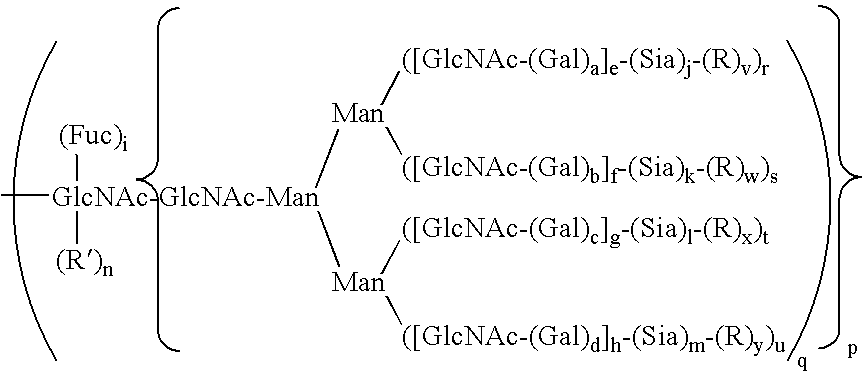
Also included in the invention is a cell-free, in vitro method of remodeling a peptide having the formula:
-
- AA is a terminal or internal amino acid residue of the peptide;
- X1 is a glycosyl residue covalently linked to the AA, selected from monosaccharyl and oligosaccharyl residues; and
- u is an integer selected from 0 and 1. The method comprises: contacting the peptide with at least one glycosyltransferase and at least one glycosyl donor under conditions suitable to transfer the at least one glycosyl donor to the truncated glycan, wherein the glycosyl donor comprises a modifying group, thereby remodeling the peptide.
In a preferred embodiment, at least one of the glycosyl donors comprises a modifying group, and the modifying group may be a member selected from the group consisting of a water-soluble polymer, a therapeutic moiety, a detectable label, a reactive linker group, and a targeting moiety. Preferably, the modifying group is a water soluble polymer, and more preferably, the water soluble polymer comprises poly(ethylene glycol). Even more preferably, the poly(ethylene glycol) has a molecular weight distribution that is essentially homodisperse.
In addition, the peptide may be selected from the group consisting of granulocyte colony stimulating factor, interferon-alpha, interferon-beta, Factor VIIa, Factor IX, follicle stimulating hormone, erythropoietin, granulocyte macrophage colony stimulating factor, interferon-gamma, alpha-1-protease inhibitor, beta-glucosidase, tissue plasminogen activator protein, interleukin-2, Factor VIII, chimeric tumor necrosis factor receptor, urokinase, chimeric anti-glycoprotein IIb/IIIa antibody, chimeric anti-HER2 antibody, chimeric anti-respiratory syncytial virus antibody, chimeric anti-CD20 antibody, DNase, chimeric anti-tumor necrosis factor antibody, human insulin, hepatitis B sAg, and human growth hormone.
The invention additionally includes a covalent conjugate between a peptide and a modifying group that alters a property of the peptide, wherein the modifying group is covalently attached to the peptide at a preselected glycosyl or amino acid residue of the peptide via an intact glycosyl linking group.
In one aspect, the modifying group is a member selected from the group consisting of a water-soluble polymer, a therapeutic moiety, a detectable label, a reactive linker group, and a targeting moiety.
In another aspect, the modifying group and an intact glycosyl linking group precursor are bound as a covalently attached unit to the peptide via the action of an enzyme, the enzyme converting the precursor to the intact glycosyl linking group, thereby forming the conjugate.
The covalent conjugate of the invention comprises:
-
- a first modifying group covalently bound to a first residue of the peptide via a first intact glycosyl linking group, and
- a second glycosyl linking group bound to a second residue of the peptide via a second intact glycosyl linking group.
In one embodiment, the first residue and the second residue are structurally identical. In another embodiment, the first residue and the second residue have different structures. In an additional embodiment, the first residue and the second residue are glycosyl residues. In another embodiment, the first residue and the second residue are amino acid residues.
In yet another embodiment, the peptide is remodeled prior to forming the conjugate. Preferably, peptide is remodeled to introduce an acceptor moiety for the intact glycosyl linking group.
In another embodiment, the modifying group is a water-soluble polymer that may comprises poly(ethylene glycol), which, in another embodiment, may have a molecular weight distribution that is essentially homodisperse.
In yet a further embodiment, the peptide is selected from the group consisting of granulocyte colony stimulating factor, interferon-alpha, interferon-beta, Factor VIIa, Factor IX, follicle stimulating hormone, erythropoietin, granulocyte macrophage colony stimulating factor, interferon-gamma, alpha-1-protease inhibitor, beta-glucosidase, tissue plasminogen activator protein, interleukin-2, Factor VIII, chimeric tumor necrosis factor receptor, urokinase, chimeric anti-glycoprotein IIb/IIIa antibody, chimeric anti-HER2 antibody, chimeric anti-respiratory syncytial virus antibody, chimeric anti-CD20 antibody, DNase, chimeric anti-tumor necrosis factor antibody, human insulin, hepatitis B sAg, and human growth hormone.
In another embodiment, the intact glycosyl linking unit is a member selected from the group consisting of a sialic acid residue, a Gal residue, a GlcNAc residue and a GalNAc residue.
There is also provided in the invention a method of forming a covalent conjugate between a polymer and a glycosylated or non-glycosylated peptide, wherein the polymer is conjugated to the peptide via an intact glycosyl linking group interposed between and covalently linked to both the peptide and the polymer. The method comprises contacting the peptide with a mixture comprising a nucleotide sugar covalently linked to the polymer and a glycosyltransferase for which the nucleotide sugar is a substrate under conditions sufficient to form the conjugate.
In a preferred embodiment, the polymer is a water-soluble polymer. In another preferred embodiment, the glycosyl linking group is covalently attached to a glycosyl residue covalently attached to the peptide, and in another embodiment, the glycosyl linking group is covalently attached to an amino acid residue of the peptide.
In yet a further embodiment, the polymer comprises a member selected from the group consisting of a polyalkylene oxide and a polypeptide. The polyalkylene oxide may be poly(ethylene glycol) in one embodiment of the invention. In another embodiment, the poly(ethylene glycol) has a degree of polymerization of from about 1 to about 20,000, preferably, from about 1 to about 5,000, or also preferably, the polyethylene glycol has a degree of polymerization of from about 1 to about 1,000.
In another embodiment, the glycosyltransferase is selected from the group consisting of sialyltransferase, galactosyltransferase, glucosyltransferase, GalNAc transferase, GlcNAc transferase, fucosyltransferase, and mannosyltransferase. In one embodiment, the glycosyltransferase is recombinantly produced, and in another embodiment, the glycosyltransferase is a recombinant prokaryotic enzyme, or a recombinant eukaryotic enzyme.
In yet a further embodiment, the nucleotide sugar is selected from the group consisting of UDP-glycoside, CMP-glycoside, and GDP-glycoside and is preferably selected from the group consisting of UDP-galactose, UDP-galactosamine, UDP-glucose, UDP-glucosamine, UDP-N-acetylgalactosamine, UDP-N-acetylglucosamine, GDP-mannose, GDP-fucose, CMP-sialic acid, CMP-NeuAc.
In another embodiment, the peptide is a therapeutic agent.
In yet another embodiment, the glycosylated peptide is partially deglycosylated prior to the contacting.
In a further embodiment, the intact glycosyl linking group is a sialic acid residue.
Further, the method may be performed in a cell-free environment.
And, in another embodiment, the covalent conjugate may be isolated, and preferably, the covalent conjugate is isolated by membrane filtration.
There is also provided a method of forming a covalent conjugate between a first glycosylated or non-glycosylated peptide, and a second glycosylated or non-glycosylated peptide cojoined by a linker moiety, wherein
-
- the linker moiety is conjugated to the first peptide via a first intact glycosyl linking group interposed between and covalently linked to both the first peptide and the linker moiety, and
- the linker moiety is conjugated to the second peptide via a second intact glycosyl linking group interposed between and covalently linked to both the second peptide and the linker moiety. The method comprises:
- (a) contacting the first peptide with a derivative of the linker moiety precursor comprising a precursor of the first intact glycosyl linking group and a precursor of the second intact glycosyl linking group;
- (b) contacting the mixture from (a) with a glycosyl transferase for which the precursor of the first glycosyl linking group is a substrate, under conditions sufficient to convert the precursor of the first intact glycosyl linking group into the first intact glycosyl linking group, thereby forming a first conjugate between the linker moiety precursor and the first peptide;
- (c) contacting the first conjugate with the second peptide and a glycosyltransferase for which the precursor of the second intact glycosyl group is a substrate under conditions sufficient to convert the precursor of the second intact glycosyl linking group into the second glycosyl linking group, thereby forming the conjugate between the linker moiety and the first glycosylated or non-glycosylated peptide, and the second glycosylated or non-glycosylated peptide.
In one aspect, the linker moiety comprises a water-soluble polymer, and in one embodiment, the water-soluble polymer comprises poly(ethylene glycol).
There is also provided a method of forming a covalent conjugate between a first glycosylated or non-glycosylated peptide, and a second glycosylated or non-glycosylated peptide cojoined by a linker moiety, wherein
-
- the linker moiety is covalently conjugated to the first peptide, and
- the linker moiety is conjugated to the second peptide via an intact glycosyl linking group interposed between and covalently linked to both the second peptide and the linker moiety. The method comprises:
- (a) contacting the first peptide with an activated derivative of the linker moiety comprising;
- a reactive functional group of reactivity complementary to a residue on the first peptide, and a precursor of the intact glycosyl linking group, under conditions sufficient to form a covalent bond between the reactive functional group and the residue, thereby forming a first conjugate; and
- (b) contacting the first conjugate with the second peptide and a glycosyltransferase for which the precursor of the intact glycosyl linking group is a substrate, under conditions sufficient to convert the precursor of the intact glycosyl linking group into the intact glycosyl linking group, thereby forming the conjugate between the first glycosylated or non-glycosylated peptide, and the second glycosylated or non-glycosylated peptide cojoined by the linker moiety.
In one embodiment the linker moiety comprises a water-soluble polymer, which may be poly(ethylene glycol).
Also provided is a pharmaceutical composition comprising a pharmaceutically acceptable diluent and a covalent conjugate between a polymer and a glycosylated or non-glycosylated peptide, wherein the polymer is conjugated to the peptide via an intact glycosyl linking group interposed between and covalently linked to both the peptide and the polymer.
The invention further includes a composition for forming a conjugate between a peptide and a modified sugar, the composition comprising: an admixture of a modified sugar, a glycosyltransferase, and a peptide acceptor substrate, wherein the modified sugar has covalently attached thereto a member selected from a polymer, a therapeutic moiety and a biomolecule.
The invention also includes peptides remodeled using the methods of the invention and pharmaceutical compositions comprising the remodeled peptides.
Also provided in the invention is a compound having the formula:
-
- MS is a modified sugar comprising a sugar covalently bonded to a modifying group;
- Nu is a nucleoside; and
- b is an integer from 0 to 2.
In one aspect, there is included a compound having the formula:
-
- X, Y, Z, A and B are members independently selected from S, O and NH;
- R21, R22, R23, R24, and R25 members independently selected from H and a polymer;
- R26 is a member selected from H, OH, and a polymer;
- R27 is a member selected from COO— and Na+;
- Nu is a nucleoside; and
- a is an integer from 1 to 3.
The invention further provides a cell-free, in vitro method of remodeling a peptide having the formula:
AA is a terminal or internal amino acid residue of the peptide. The method comprises:
contacting the peptide with at least one glycosyltransferase and at least one glycosyl donor under conditions suitable to transfer the at least one glycosyl donor to the amino acid residue, wherein the glycosyl donor comprises a modifying group, thereby remodeling the peptide.
In each of the embodiments that are discussed below, specific remodeling schemes and peptides are identified solely to emphasize preferred embodiments of the invention.
The invention therefore includes a method of forming a conjugate between a granulocyte colony stimulating factor (G-CSF) peptide and a modifying group, wherein the modifying group is covalently attached to the G-CSF peptide through an intact glycosyl linking group, the G-CSF peptide comprising a glycosyl residue having the formula:
-
- a, b, c, and e are members independently selected from 0 and 1;
- d is 0; and
- R is a modifying group, a mannose or an oligomannose. The method comprises:
- (a) contacting the G-CSF peptide with a glycosyltransferase and a modified glycosyl donor, comprising a glycosyl moiety which is a substrate for the glycosyltransferase covalently bound to the modifying group, under conditions appropriate for the formation of the intact glycosyl linking group.
In one embodiment, the method further comprises:
-
- (b) prior to step (a), contacting the G-CSF peptide with a sialidase under conditions appropriate to remove sialic acid from the G-CSF peptide.
In another embodiment, the method further comprises:
-
- (c) prior to step (a), contacting the G-CSF peptide with a galactosyl transferase and a galactose donor under conditions appropriate to transfer the galactose to the G-CSF peptide.
In yet another embodiment, the method further comprises:
-
- (d) contacting the product from step (a) with a moiety that reacts with the modifying group, thereby forming a conjugate between the intact glycosyl linking group and the moiety.
In another embodiment, the method further comprises:
-
- (e) prior to step (a), contacting the G-CSF peptide with N-acetylgalactosamine transferase and a GalNAc donor under conditions appropriate to transfer GalNAc to the G-CSF peptide.
In a further embodiment, the method further comprises:
-
- (f) prior to step (a), contacting the G-CSF peptide with endo-N-acetylgalactosaminidase operating synthetically and a GalNAc donor under conditions appropriate to transfer GalNAc to the G-CSF peptide.
In yet a further embodiment, the modifying group is a member selected from a polymer, a toxin, a radioisotope, a therapeutic moiety and a glycoconjugate.
In specific embodiments, referring to the G-CSF peptide formula presented above, a, b, c, and e are 0. Alternatively, a and e are members independently selected from 0 and 1; and b, c, and d are 0. Alternatively, a, b, c, d, and e are members independently selected from 0 and 1.
The invention further includes a G-CSF peptide conjugate formed by the above-described methods.
There is also included a method of forming a conjugate between an interferon alpha peptide and a modifying group, wherein the modifying group is covalently attached to the glycopeptide through an intact glycosyl linking group, the glycopeptide comprising a glycosyl residue having a formula selected from:
-
- a, b, c, d, i, n, o, p, q, r, s, t, u, aa, bb, cc, dd, and ee are members independently selected from 0 and 1;
- e, f, g, and h are members independently selected from the integers from 0 to 6;
- j, k, l, and m are members independently selected from the integers from 0 to 20;
- v, w, x, y, and z are 0; and
- R is a modifying group, a mannose or an oligomannose
- R′ is H, a glycosyl residue, a modifying group, or a glycoconjugate.
The method comprises:
-
- (a) contacting the glycopeptide with a member selected from a glycosyltransferase, an endo-acetylgalactosaminidase operating synthetically and a trans-sialidase, and a modified glycosyl donor, comprising a glycosyl moiety which is a substrate for the glycosyltransferase covalently bound to the modifying group, under conditions appropriate for the formation of the intact glycosyl linking group.
In one embodiment, the method further comprises:
- (b) prior to step (a), contacting the glycopeptide with a sialidase under conditions appropriate to remove sialic acid from the glycopeptide.
In another embodiment, the method further comprises:
- (c) contacting the product from step (a) with a moiety that reacts with the modifying group, thereby forming a conjugate between the intact glycosyl linking group and the moiety.
In yet an additional embodiment, the method further comprises:
- (d) prior to step (a) contacting the glycopeptide with a combination of a glycosidase and a sialidase.
In an additional embodiment, the method further comprises:
- (e) prior to step (a), contacting the glycopeptide with an endoglycanase under conditions appropriate to cleave a glycosyl moiety from the glycopeptide.
In yet another embodiment, the method also comprises:
- (f) prior to step (a), contacting the glycopeptide with N-acetylglucosamine transferase and a GlcNAc donor under conditions appropriate to transfer GlcNAc to the glycopeptide.
In addition, the method also comprises:
- (g) prior to step (a), contacting the glycopeptide with a galactosyl transferase and a galactose donor under conditions appropriate to transfer galactose to the product.
Also, the method further comprises:
- (h) prior to step (b), contacting the glycopeptide with endoglycanase under conditions appropriate to cleave a glycosyl moiety from the glycopeptide.
The invention also further comprises:
- (i) prior to step (a), contacting the glycopeptide with a mannosidase under conditions appropriate to remove mannose from the glycopeptide.
In addition, the method further comprises:
- (j) contacting the product of step (a) with a sialyltransferase and a sialic acid donor under conditions appropriate to transfer sialic acid to the product.
In one aspect, the modifying group is a member selected from a polymer, a toxin, a radioisotope, a therapeutic moiety and a glycoconjugate.
According to the invention and with respect to the interferon alpha peptide formula disclosed above, a, b, c, d, aa, and bb are 1; e, f, g, and h are members independently selected from the integers from 1 to 4; i, j, k, l, m, r, s, t, u, and cc are members independently selected from 0 and 1; and n, o, p, q, v, w, x, y, z, dd, and ee are 0. Alternatively, a, b, c, d, f, h, j, k, l, m, n, o, p, q, s, u, v, w, x, y, z, cc, dd, and ee are 0; e, g, i, r, and t are members independently selected from 0 and 1; and aa and bb are 1. Alternatively, a, b, c, d, e, f, g, i, j, k, l, m, r, s, t, and u are members independently selected from 0 and 1; h is a member independently selected from the integers from 1 to 3; dd, v, w, x, and y are 0; and aa and bb are 1. Alternatively, a, b, c, d, f, h, j, k, l, m, s, u, v, w, x, y, and dd are 0; e, g, i, r, and t are members independently selected from 0 and 1; and aa and bb are 1. Alternatively, a, b, c, d, e, f, g, h, i, j, k, l, m, and dd are 0; r, s, t, u, v, w, x, and y are members independently selected from 0 and 1; and aa and bb are 1. Alternatively, a, b, c, d, e, f, g, h, i, r, s, t, and u are members independently selected from 0 and 1; j, k, l, m, v, w, x, y, and dd are 0; and aa and bb are 1. Alternatively, a, b, c, d, e, f, g, i, j, k, l, m, r, s, t, and u are members independently selected from 0 and 1; h is a member independently selected from the integers from 1 to 3; v, w, x, y, and dd are 0; and aa and bb are 1. Alternatively, a, b, c, d, f, h, j, k, l, m, s, u, v, w, x, y, and dd are 0; e, g, i, r, and t are members independently selected from 0 and 1; and aa and bb are 1. Alternatively, n, o, and p are members independently selected from 0 and 1; q is 1; and z, cc, and ee are 0. Alternatively, n and q are members independently selected from 0 and 1; and o, p, z, cc, and ee are 0. Alternatively, n is 0 or 1; q is 1; and o, p, z, cc, and ee are 0. Alternatively, n, o, p, and f are members independently selected from 0 and 1; q is 1; and z and ee are 0. Alternatively, n, o, p, and q are members independently selected from 0 and 1; and z, cc, and ee are 0. Alternatively, n and q are members independently selected from 0 and 1; and o, p, z, cc, and ee are 0. Alternatively, n, o, p, q, z, cc, and ee are 0.
There is also provided an interferon alpha peptide conjugate formed by the disclosed method.
The invention also includes a method of forming a conjugate between an interferon beta peptide and a modifying group, wherein the modifying group is covalently attached to the interferon beta peptide through an intact glycosyl linking group, the interferon beta peptide comprising a glycosyl residue having the formula:
-
- a, b, c, d, i, p, q, r, s, t, and u are members independently selected from 0 and 1;
- e, f, g, and h are members independently selected from the integers between 0 and 6;
- j, k, l, and m are members independently selected from the integers between 0 and 100;
- v, w, x, and y are 0;
- R is a modifying group, mannose or oligomannose; and
- R′ is H or a glycosyl, modifying group or glycoconjugate group. the method comprises:
- (a) contacting the interferon beta peptide with a member selected from a glycosyltransferase and a trans-sialidase and a modified glycosyl donor, comprising a glycosyl moiety which is a substrate for the glycosyltransferase covalently bound to the modifying group, under conditions appropriate for the formation of the intact glycosyl linking group.
In one embodiment, the method further comprises:
-
- (b) prior to step (a), contacting the interferon beta peptide with a sialidase under conditions appropriate to remove sialic acid from the interferon beta peptide.
In another embodiment, the method further comprises:
-
- (c) contacting the product from step (a) with a moiety that reacts with the modifying group, thereby forming a conjugate between the intact glycosyl linking group and the moiety.
In yet another embodiment, the method also further comprises:
-
- (d) prior to step (a) contacting the interferon beta peptide with a combination of a glycosidase and a sialidase.
In an additional embodiment, the method further comprises:
-
- (e) prior to step (a), contacting the interferon beta peptide with an endoglycanase under conditions appropriate to cleave a glycosyl moiety from the interferon beta peptide.
Also, the method further comprises:
-
- (f) prior to step (a), contacting the interferon beta peptide with N-acetylglucosamine transferase and a GlcNAc donor under conditions appropriate to transfer GlcNAc to the interferon beta peptide.
Additionally, the method also further comprises:
-
- (g) prior to step (a), contacting the interferon beta peptide with a galactosyl transferase and a galactose donor under conditions appropriate to transfer galactose to the product.
In yet another embodiment, the method further comprises:
-
- (h) prior to step (b), contacting the interferon beta peptide with endoglycanase under conditions appropriate to cleave a glycosyl moiety from the interferon beta peptide.
In yet a further embodiment, the method further comprises:
-
- (i) prior to step (a), contacting the interferon beta peptide with a mannosidase under conditions appropriate to remove mannose from the interferon beta peptide.
In addition, the method further comprises:
-
- (j) contacting the product of step (a) with a sialyltransferase and a sialic acid donor under conditions appropriate to transfer sialic acid to the product.
In one aspect, the modifying group is a member selected from a polymer, a toxin, a radioisotope, a therapeutic moiety and a glycoconjugate.
In preferred embodiments and referring to the beta interferon peptide formula disclosed above, h is a member independently selected from the integers between 1 and 3; a, b, c, d, e, f, g, i, j, k, l, m, r, s, t, and u are members independently selected from 0 and 1; n, v, w, x, and y are 0; and q, p are 1. Alternatively, a, b, c, d, f, h, j, k, l, m, n, s, u, v, w, x, and y are 0; e, g, i, r, and t are members independently selected from 0 and 1; and q, p are 1. Alternatively, a, b, c, d, e, f, g, h, j, k, l, m, n, r, s, t, u, v, w, x, and y are 0; q, p are 1; and i is independently selected from 0 and 1. Alternatively, a, b, c, d, e, f, g, h, i, j, k, l, m, r, s, t, u, v, w, x, and y are 0; and p, q are 1. Alternatively, a, b, c, d, e, f, g, h, i, j, k, l, m, and n are 0; q, p are 1; and r, s, t, u, v, w, x, and y are members independently selected from 0 and 1. Alternatively, a, b, c, d, e, f, g, h, i, r, s, t, and u are members independently selected from 0 and 1; j, k, l, m, n, v, w, x, and y are 0; and q, p are 1. Alternatively, wherein a, b, c, d, h, j, k, l, m, r, s, t, and u are members independently selected from 0 and 1; e, f, g, are members selected from the integers between 0 and 3; n, v, w, x, and y are 0; and q, p are 1. Alternatively, a, b, c, d, i, j, k, l, m, r, s, t, u, p and q are members independently selected from 0 and 1; e, f, g, and h are 1; and n, v, w, x, and y are 0.
Further included is an interferon beta peptide conjugate formed by the above-described method.
The invention also provides a method of forming a conjugate between a Factor VIIa peptide and a modifying group, wherein the modifying group is covalently attached to the Factor VIIa peptide through an intact glycosyl linking group, the Factor VIIa peptide comprising a glycosyl residue having a formula which is a member selected from:
-
- a, b, c, d, i, o, p, q, r, s, t, and u, are members independently selected from 0 and 1;
- e, f, g, h and n are members independently selected from the integers from 0 to 6;
- j, k, l and m are members independently selected from the integers from 0 to 20;
- v, w, x and y are 0; and
- R is a modifying group, a mannose, an oligomannose, SialylLewisx or SialylLewisa.
The method comprises:
-
- (a) contacting the Factor VIIa peptide with a glycosyltransferase and a modified glycosyl donor, comprising a glycosyl moiety which is a substrate for the glycosyltransferase covalently bound to the modifying group, under conditions appropriate for the formation of the intact glycosyl linking group.
In a preferred embodiment, the method further comprises:
-
- (b) prior to step (a), contacting the Factor VIIa peptide with a sialidase under conditions appropriate to remove sialic acid from the Factor VIIa peptide.
In yet another preferred embodiment, the method comprises:
-
- (c) prior to step (a), contacting the Factor VIIa peptide with a galactosidase under conditions appropriate to remove galactose from the Factor VIIa peptide.
In another embodiment, the method comprises:
-
- (d) prior to step (a), contacting the Factor VIIa peptide with a galactosyl transferase and a galactose donor under conditions appropriate to transfer the galactose to the Factor VIIa peptide.
In an additional embodiment, the method comprises:
-
- (e) contacting the product of step (a) with a sialyltransferase and a sialic acid donor under conditions appropriate to transfer sialic acid to the product.
In one aspect, the modifying group is a member selected from a polymer, a toxin, a radioisotope, a therapeutic moiety and a glycoconjugate.
In preferred embodiments, and referring to the Factor VIIa peptide formula disclosed above, a, b, c, d, e, g, i, j, l, o, p and q members independently selected from 0 and 1; r and t are 1; f, h, k, m, s, u, v, w, x and y are 0; and n is selected from the integers from 0 to 4. Alternatively, a, b, c, d, e, f, g, h, i, j, k l, m, n, o, p, q, r, s, t and u are members independently selected from 0 and 1; v, w, x and y are 0; and n is a member selected from the integers from 0 to 4.
In addition, there is included a Factor VIIa peptide conjugate formed by the method disclosed herein.
The invention additionally provides a method of forming a conjugate between a Factor IX peptide and a modifying group, wherein the modifying group is covalently attached to the Factor IX peptide through an intact glycosyl linking group, the Factor IX peptide comprising a glycosyl residue having a formula which is a member selected from:
-
- a, b, c, d, i, n, o, p, q, r, s, t, u, bb, cc, dd, ee, ff and gg are members independently selected from 0 and 1;
- e, f, g, h and aa are members independently selected from the integers from 0 to 6;
- j, k, l and m are members independently selected from the integers from 0 to 20;
- v, w, x, y and z are 0;
- R is a modifying group, a mannose or an oligomannose. The method comprises
- (a) contacting the Factor IX peptide with a glycosyltransferase and a modified glycosyl donor, comprising a glycosyl moiety which is a substrate for the glycosyltransferase covalently bound to the modifying group, under conditions appropriate for the formation of the intact glycosyl linking group.
In one embodiment, the method further comprises:
-
- (b) prior to step (a), contacting the Factor IX peptide with a sialidase under conditions appropriate to remove sialic acid from the Factor IX peptide.
In another embodiment, the method further comprises: (c) contacting the product formed in step (a) with a sialyltransferase and a sialic acid donor under conditions appropriate to transfer sialic acid to the product.
Additionally, the method comprises:
-
- (d) contacting the product from step (b) with a galactosyltransferase and a galactose donor under conditions appropriate to transfer the galactose to the product.
Moreover, the method comprises:
-
- (e) contacting the product from step (d) with ST3Gal3 and a sialic acid donor under conditions appropriate to transfer sialic acid to the product.
In yet another embodiment, the method further comprises:
-
- (d) contacting the product from step (a) with a moiety that reacts with the modifying group, thereby forming a conjugate between the intact glycosyl linking group and the moiety.
Also included is the fact that the modifying group is a member selected from a polymer, a toxin, a radioisotope, a therapeutic moiety and a glycoconjugate.
In additional embodiments and referring to the Factor IX peptide formula disclosed above, a, b, c, and d are 1; e, f, g and h are members independently selected from the integers from 1 to 4; aa, bb, cc, dd, ee, ff, j, k, l, m, i, n, o, p, q, r, s, t and u are members independently selected from 0 and 1; and v, w, x, y, z and gg are 0. Alternatively, a, b, c, d, n, q are independently selected from 0 and 1; aa, e, f, g and h are members independently selected from the integers from 1 to 4; bb, cc, dd, ee, ff, j, k, l, m, i, o, p, r, s, t and u are members independently selected from 0 and 1; and v, w, x, y, z and gg are 0. Alternatively, a, b, c, d, n, bb, cc, dd and ff are 1; e, f, g, h and aa are members independently selected from the integers from 1 to 4; q, ee, i, j, k, l, m, o, p, r, s, t and u are members independently selected from 0 and 1; and v, w, x, y, z and gg are 0. Alternatively, a, b, c, d and q are 1; e, f, g and h are members independently selected from the integers from 1 to 4; aa, bb, cc, dd, ee, ff, j, k, l, m, i, n, o, p, r, s, t and u are members independently selected from 0 and 1; and v, w, x, y, z and gg are 0. Alternatively, a, b, c, d, q, bb, cc, dd and ff are 1; aa, e, f, g and h are members independently selected from the integers from 1 to 4; ee, i, j, k, l, m, o, p, r, s, t and u are members independently selected from 0 and 1; and v, w, x, y, z and gg are 0.
Also included is a Factor IX peptide conjugate formed by the above disclosed method.
The invention also provides a method of forming a conjugate between a follicle stimulating hormone (FSH) peptide and a modifying group, wherein the modifying group is covalently attached to the FSH peptide through an intact glycosyl linking group, the FSH peptide comprising a glycosyl residue having the formula:
-
- a, b, c, d, i, q, r, s, t, and u are members independently selected from 0 and 1;
- e, f, g, and h are members independently selected from the integers between 0 and 6;
- j, k, l, and m are members independently selected from the integers between 0 and 100;
- v, w, x, and y are 0; and
- R is a modifying group, a mannose or an oligomannose. The method comprises:
- (a) contacting the FSH peptide with a glycosyltransferase and a modified glycosyl donor, comprising a glycosyl moiety which is a substrate for the glycosyltransferase covalently bound to the modifying group, under conditions appropriate for the formation of the intact glycosyl linking group.
In one embodiment, the method comprises:
-
- (b) prior to step (a), contacting the FSH peptide with a sialidase under conditions appropriate to remove sialic acid from the FSH peptide.
In another embodiment, the method comprises:
-
- (c) contacting the product of step (a) with a sialyltransferase and a sialic acid donor under conditions appropriate to transfer sialic acid to the product.
In yet another embodiment, the method comprises:
-
- (d) prior to step (a), contacting the FSH peptide with a galactosidase under conditions appropriate to remove galactose from the FSH peptide.
In an additional embodiment, the method comprises:
-
- (e) prior to step (a) contacting the FSH peptide with a combination of a glycosidase and a sialidase.
In yet a further embodiment, the method comprises:
-
- (f) prior to step (a), contacting the FSH peptide with a galactosyl transferase and a galactose donor under conditions appropriate to transfer the galactose to the FSH peptide.
In another embodiment, the method comprises:
-
- (d) contacting the product from step (a) with a moiety that reacts with the modifying group, thereby forming a conjugate between the intact glycosyl linking group and the moiety.
In a further embodiment, the method comprises:
-
- (e) prior to step (b), contacting the FSH peptide with an endoglycanase under conditions appropriate to cleave a glycosyl moiety from the FSH peptide.
In another embodiment, the method comprises:
-
- (f) prior to step (a), contacting the FSH peptide with N-acetylglucosamine transferase and a GlcNAc donor under conditions appropriate to transfer GlcNAc to the FSH peptide.
In yet another embodiment, the modifying group is a member selected from a polymer, a toxin, a radioisotope, a therapeutic moiety and a glycoconjugate.
In additional preferred embodiments and referring to the FSH peptide formula described above, a, b, c, d, i, j, k, l, m, q, r, s, t, and u are members independently selected from 0 and 1; e, f, g, and h are 1; and v, w, x, and y are 0. Alternatively, a, b, c, d, e, f, g, h, i, j, k, l, m, q, r, s, t, and u are members independently selected from 0 and 1; v, w, x, and y are 0. Alternatively, a, b, c, d, f, h, j, k, l, m, s, u, v, w, x, and y are 0; and e, g, i, q, r, and t are members independently selected from 0 and 1. Alternatively, a, b, c, d, e, f, g, h, j, k, l, and m are 0; i, q, r, s, t, u, v, w, x, and y are independently selected from 0 and 1; p is 1; R (branched or linear) is a member selected from mannose and oligomannose. Alternatively, a, b, c, d, e, f, g, h, j, k, l, m, r, s, t, u, v, w, and y are 0; i is 0 or 1; and q is 1.
Also included is a FSH peptide conjugate formed by the above-described method.
The invention further provides a method of forming a conjugate between an erythropoietin (EPO) peptide and a modifying group, wherein the modifying group is covalently attached to the EPO peptide through an intact glycosyl linking group, the EPO peptide comprising a glycosyl residue having a formula which is a member selected from:
-
- a, b, c, d, i, n, o, p, q, r, s, t, and u are members independently selected from 0 and 1;
- e, f, g, and h are members independently selected from the integers between 0 and 4;
- j, k, l, and m are members independently selected from the integers between 0 and 20;
- v, w, x, y, and z are 0; and
- R is a modifying group, a mannose or an oligomannose. The method comprises:
- (a) contacting the EPO peptide with a glycosyltransferase and a modified glycosyl donor, comprising a glycosyl moiety which is a substrate for the glycosyltransferase covalently bound to the modifying group, under conditions appropriate for the formation of the intact glycosyl linking group.
In one embodiment, the method comprises:
-
- (b) prior to step (a), contacting the EPO peptide with a sialidase under conditions appropriate to remove sialic acid from the EPO peptide.
In another embodiment, the method comprises:
-
- (c) contacting the product of step (a) with a sialyltransferase and a sialic acid donor under conditions appropriate to transfer sialic acid to the product.
In yet another embodiment, the method comprises:
-
- (d) prior to step (a), contacting the EPO peptide with a galactosidase operating synthetically under conditions appropriate to add a galactose to the EPO peptide.
In an additional embodiment, the method comprises:
-
- (e) prior to step (a), contacting the EPO peptide with a galactosyl transferase and a galactose donor under conditions appropriate to transfer the galactose to the EPO peptide.
In a further embodiment, the method comprises:
-
- (f) contacting the product from step (e) with ST3Gal3 and a sialic acid donor under conditions appropriate to transfer sialic acid to the product.
Additionally, the method comprises:
-
- (g) contacting the product from step (a) with a moiety that reacts with the modifying group, thereby forming a conjugate between the intact glycosyl linking group and the moiety.
Also, the method comprises:
-
- (h) prior to step (a), contacting the EPO peptide with N-acetylglucosamine transferase and a GlcNAc donor under conditions appropriate to transfer GlcNAc to the EPO peptide.
In another aspect, the modifying group is a member selected from a polymer, a toxin, a radioisotope, a therapeutic moiety and a glycoconjugate.
In preferred embodiment, and referring to the EPO peptide formula above, a, b, c, d, e, f, g, n, and q are 1; h is a member selected from the integers between 1 and 3; i, j, k, l, m, o, p, r, s, t, and u are members independently selected from 0 and 1; and, v, w, x, y and z are 0. Alternatively, a, b, c, d, f, h, j, k, l, m, q, s, u, v, w, x, y, and z are 0; and e, g, i, r, and t are members independently selected from 0 and 1. Alternatively, a, b, c, d, e, f, g, h, i, j, k, l, m, n, o, p, q, r, s, t, and u are members independently selected from 0 and 1; and v, w, x, y, and z are 0. Alternatively, a, b, c, d, e, f, g, n, and q are 1; h is a member selected from the integers between 1 and 3; i, j, k, l, m, o, p, r, s, t, and u are members independently selected from 0 and 1; and v, w, x, y and z are 0. Alternatively, a, b, c, d, f, h, j, k, l, m, o, p, s, u, v, w, x, y, and z are 0; and e, g, i, n, q, r, and t are independently selected from 0 and 1. Alternatively, a, b, c, d, f, h, j, k, l, m, n, o, p, s, u, v, w, x, y, and z are 0; and e, g, i, q, r, and t are members independently selected from 0 and 1. Alternatively, q is 1; a, b, c, d, e, f, g, h, i, n, r, s, t, and u are members independently selected from 0 and 1; and j, k, l, m, o, p, v, w, x, y, and z are 0.
Also included is an EPO peptide conjugate formed by the above-described method.
The invention further provides a method of forming a conjugate between a granulocyte macrophage colony stimulating factor (GM-CSF) peptide and a modifying group, wherein the modifying group is covalently attached to the GM-CSF peptide through an intact glycosyl linking group, the GM-CSF peptide comprising a glycosyl residue having a formula selected from:
-
- a, b, c, d, i, n, o, p, q, r, s, t, u, aa, bb, and cc are members independently selected from 0 and 1;
- e, f, g, and h are members independently selected from the integers between 0 and 6;
- j, k, l, and m are members independently selected from the integers between 0 and 100;
- v, w, x, and y are 0;
- R is a modifying group, mannose or oligomannose; and
- R′ is H or a glycosyl residue, or a modifying group or a glycoconjugate. The method comprises:
- (a) contacting the GM-CSF peptide with a glycosyltransferase and a modified glycosyl donor, comprising a glycosyl moiety which is a substrate for the glycosyltransferase covalently bound to the modifying group, under conditions appropriate for the formation of the intact glycosyl linking group.
In one embodiment, the method comprises:
-
- (b) prior to step (a), contacting the GM-CSF peptide with a sialidase under conditions appropriate to remove sialic acid from the GM-CSF peptide.
In another embodiment, the method comprises:
-
- (c) contacting the product from step (a) with a moiety that reacts with the modifying group, thereby forming a conjugate between the intact glycosyl linking group and the moiety.
In yet another embodiment, the method comprises:
-
- (d) prior to step (a) contacting the GM-CSF peptide with a combination of a glycosidase and a sialidase.
In an additional embodiment, the method comprises:
-
- (e) prior to step (a), contacting the GM-CSF peptide with an endoglycanase under conditions appropriate to cleave a glycosyl moiety from the GM-CSF peptide.
Also, the method comprises:
-
- (f) prior to step (a), contacting the GM-CSF peptide with N-acetylglucosamine transferase and a GlcNAc donor under conditions appropriate to transfer GlcNAc to the GM-CSF peptide.
Additionally, the method comprises:
-
- (g) prior to step (a) contacting the GM-CSF peptide with a mannosidase under conditions appropriate to cleave a mannose residue from the GM-CSF peptide.
Further, the method comprises:
-
- (h) prior to step (a), contacting the GM-CSF peptide with ST3Gal3 and a sialic acid donor under conditions appropriate to transfer sialic acid to the product.
In one aspect, the modifying group is a member selected from a polymer, a toxin, a radioisotope, a therapeutic moiety and a glycoconjugate.
In additional preferred embodiments and referring to the GM-CSF peptide formula described above, a, b, c, d, i, j, k, l, m, o, p, q, r, s, t, u, and aa are members independently selected from 0 and 1; bb, e, f, g, h, and n are 1; and cc, v, w, x, y, and z are 0. Alternatively, a, b, c, d, i, j, k, l, m, o, p, q, r, s, t, u, and aa are members independently selected from 0 and 1; bb, e, f, g, h, and n are members independently selected from 0 and 1; and cc, v, w, x, y, and z are 0. Alternatively, cc, a, b, c, d, f, h, j, k, l, m, 0, p, s, u, v, w, x, y, and z are 0; and e, g, i, n, q, r, t, and aa are members independently selected from 0 and 1; and bb is 1. Alternatively, a, b, c, d, e, f, g, h, i, j, k, l, m, n, o, p, z and cc are 0; q, r, s, t, u, v, w, x, y, and aa are members independently selected from 0 and 1; bb is 1; and R is mannose or oligomannose. Alternatively, a, b, c, d, e, f, g, h, i, j, k, l, m, o, q, r, s, t, u, aa, and bb are members independently selected from 0 and 1; and n, p, v, w, x, y, z, and cc are 0.
Further included is a GM-CSF peptide conjugate formed by the above-described method.
The invention also includes a method of forming a conjugate between an interferon gamma peptide and a modifying group, wherein the modifying group is covalently attached to the interferon gamma peptide through an intact glycosyl linking group, the interferon gamma peptide comprising a glycosyl residue having the formula:
-
- a, b, c, d, i, n, p, q, r, s, t, and u are members independently selected from 0 and 1;
- e, f, g, and h are members independently selected from the integers between 0 and 6;
- j, k, l, and m are members independently selected from the integers between 0 and 100;
- v, w, x, and y are 0;
- R is a modifying group, mannose or oligomannose; and
- R′ is H or a glycosyl residue, a glycoconjugate, or a modifying group.
The method comprises:
-
- (a) contacting the interferon gamma peptide with a member selected from a glycosyltransferase and a galactosidase operating synthetically and a modified glycosyl donor, comprising a glycosyl moiety which is a substrate for the glycosyltransferase covalently bound to the modifying group, under conditions appropriate for the formation of the intact glycosyl linking group.
In one embodiment, the method comprises:
-
- (b) prior to step (a), contacting the interferon gamma peptide with a sialidase under conditions appropriate to remove sialic acid from the interferon gamma peptide.
In another embodiment, the method comprises:
-
- (c) contacting the product from step (a) with a moiety that reacts with the modifying group, thereby forming a conjugate between the intact glycosyl linking group and the moiety.
In an additional embodiment, the method comprises:
-
- (d) prior to step (a) contacting the interferon gamma peptide with a combination of a glycosidase and a sialidase.
The method also comprises:
-
- (e) prior to step (a), contacting the interferon gamma peptide with an endoglycanase under conditions appropriate to cleave a glycosyl moiety from the interferon gamma peptide.
Additionally, the method comprises:
-
- (f) prior to step (a), contacting the interferon gamma peptide with N-acetylglucosamine transferase and a GlcNAc donor under conditions appropriate to transfer GlcNAc to the interferon gamma peptide.
Also, the method comprises:
-
- (g) prior to step (a), contacting the interferon gamma peptide with a galactosyl transferase and a galactose donor under conditions appropriate to transfer galactose to the product.
In a further embodiment, the method comprises:
-
- (h) contacting the product of step (a) with a sialyltransferase and a sialic acid donor under conditions appropriate to transfer sialic acid to the product.
In another aspect, the modifying group is a member selected from a polymer, a toxin, a radioisotope, a therapeutic moiety and a glycoconjugate.
Additional preferred embodiments include, referring to the interferon gamma interferon peptide formula above, where a, b, c, d, i, j, k, l, m, q, p, r, s, t, and u are members independently selected from 0 and 1; e, f, g, and h are 1; and n, v, w, x, and y are 0. Alternatively, a, b, c, d, i, j, k, l, m, r, s, t, and u are members independently selected from 0 and 1; p, q, e, f, g, and h are 1; and n, v, w, x, and y are 0. Alternatively, a, b, c, d, f, h, j, k, l, m, n, s, u, v, w, x, and y are 0; and e, g, i, q, r, and t are members independently selected from 0 and 1; and p is 1. Alternatively, a, b, c, d, e, f, g, h, i, j, k, l, m, and n are 0; q, r, s, t, u, v, w, x, and y are members independently selected from 0 and 1; and p is 1; and R is mannose or oligomannose. Alternatively, a, b, c, d, i, j, k, l, m, q, r, s, t, and u are members independently selected from 0 and 1; e, f, g, h, and p are 1; and n, v, w, x, and y are 0.
Further included is an interferon gamma peptide conjugate formed by the above-described method.
The invention further includes a method of forming a conjugate between an alpha 1 protease inhibitor (A-1-PI) peptide and a modifying group, wherein the modifying group is covalently attached to the A-1-PI peptide through an intact glycosyl linking group, the A-1-PI peptide comprising a glycosyl residue having the formula:
-
- a, b, c, d, i, n, p, q, r, s, t, and u are members independently selected from 0 and 1;
- e, f, g, and h are members independently selected from the integers between 0 and 6;
- j, k, l, and m are members independently selected from the integers between 0 and 100;
- v, w, x, and y are 0;
- R is a modifying group, mannose and oligomannose; and
- R′ is H or a glycosyl residue, a glycoconjugate, or a modifying group.
The method comprises:
-
- (a) contacting the A-1-PI peptide with a glycosyltransferase and a modified glycosyl donor, comprising a glycosyl moiety which is a substrate for the glycosyltransferase covalently bound to the modifying group, under conditions appropriate for the formation of the intact glycosyl linking group.
In one embodiment, the method comprises:
-
- (b) prior to step (a), contacting the A-1-PI peptide with a sialidase under conditions appropriate to remove sialic acid from the A-1-PI peptide.
In another embodiment, the method comprises:
-
- (c) contacting the product from step (a) with a moiety that reacts with the modifying group, thereby forming a conjugate between the intact glycosyl linking group and the moiety.
The method also comprises:
-
- (d) prior to step (a) contacting the A-1-PI peptide with a combination of a glycosidase and a sialidase.
In addition, the method comprises:
-
- (e) prior to step (a), contacting the A-1-PI peptide with an endoglycanase under conditions appropriate to cleave a glycosyl moiety from the A-1-PI peptide.
In yet another embodiment, the method comprises:
-
- (f) prior to step (a), contacting the A-1-PI peptide with N-acetylglucosamine transferase and a GlcNAc donor under conditions appropriate to transfer GlcNAc to the A-1-PI peptide.
Additionally, the method comprises:
-
- (g) prior to step (a), contacting the A-1-PI peptide with a mannosidase under conditions appropriate to remove mannose from the A-1-PI peptide.
Further, the method comprises:
-
- (h) prior to step (a), contacting the A-1-PI peptide with a member selected from a mannosidase, a xylosidase, a hexosaminidase and combinations thereof under conditions appropriate to remove a glycosyl residue from the A-1-PI peptide.
In one aspect, the modifying group is a member selected from a polymer, a toxin, a radioisotope, a therapeutic moiety and a glycoconjugate.
In other preferred embodiments and referring to the A-1PI peptide formula above, a, b, c, d, i, j, k, l, m, q, r, s, t, and u are members independently selected from 0 and 1; and e, f, g, and h are 1; and n, v, w, x, and y are 0. Alternatively, a, b, c, d, e, f, g, h, i, j, k, l, m, q, r, s, t and u are members independently selected from 0 and 1; and n, v, w, x, and y are 0. Alternatively, a, b, c, d, f, h, j, k, l, m, n, s, u, v, w, x, and y are 0; and e, g, i, q, r, and t are members independently selected from 0 and 1. Alternatively, n, a, b, c, d, e, f, g, h, i, j, k, l, and m are 0; q, r, s, t, u, v, w, x, and y area members independently selected from 0 and 1; and p is 1. Alternatively, a, b, c, d, e, f, g, h, i, j, k, l, m, n, p, and q are 0; r, s, t, u, v, w, x, and y are members independently selected from 0 and 1. Alternatively, a, b, c, d, e, f, g, h, i, j, k, l, m, r, s, t, and u are members independently selected from 0 and 1; p, v, w, x, and y are 0; and n and q are 1.
There is also provided an alpha 1 protease inhibitor peptide conjugate formed by the above-described method.
Also included in the invention is a method of forming a conjugate between a beta glucosidase peptide and a modifying group, wherein the modifying group is covalently attached to the beta glucosidase peptide through an intact glycosyl linking group, the beta glucosidase peptide comprising a glycosyl residue having the formula:
-
- a, b, c, d, i, n, p, q, r, s, t, and u are members independently selected from 0 and 1;
- e, f, g, and h are members independently selected from the integers between 0 and 6;
- j, k, l, and m are members independently selected from the integers between 0 and 100; and
- v, w, x, and y are 0;
- R is a modifying group, a mannose or an oligomannose; and
- R′ is H or a glycosyl residue, a glycoconjugate, or a modifying group.
The method comprises:
-
- (a) contacting the beta glucosidase peptide with a glycosyltransferase and a modified glycosyl donor, comprising a glycosyl moiety which is a substrate for the glycosyltransferase covalently bound to the modifying group, under conditions appropriate for the formation of the intact glycosyl linking group.
In one embodiment, the method comprises:
-
- (b) prior to step (a), contacting the beta glucosidase peptide with a sialidase under conditions appropriate to remove sialic acid from the beta glucosidase peptide.
In another embodiment, the method further comprises:
-
- (c) contacting the product from step (a) with a moiety that reacts with the modifying group, thereby forming a conjugate between the intact glycosyl linking group and the moiety.
In yet another embodiment, the method comprises:
-
- (d) prior to step (a) contacting the beta glucosidase peptide with a combination of a glycosidase and a sialidase.
In an additional embodiment, the method comprises:
-
- (e) prior to step (a), contacting the beta glucosidase peptide with an endoglycanase under conditions appropriate to cleave a glycosyl moiety from the beta glucosidase peptide.
Additionally, the method comprises:
-
- (f) prior to step (a), contacting the beta glucosidase peptide with N-acetylglucosamine transferase and a GlcNAc donor under conditions appropriate to transfer GlcNAc to the beta glucosidase peptide.
Further, the method comprises:
-
- (g) prior to step (a), contacting the beta glucosidase peptide with a galactosyl transferase and a galactose donor under conditions appropriate to transfer galactose to the product.
In another aspect, the modifying group is a member selected from a polymer, a toxin, a radioisotope, a therapeutic moiety and a glycoconjugate.
In preferred embodiments and referring to the beta glucosidase peptide formula described above, a, b, c, d, i, j, k, l, m, q, r, s, t, and u are members independently selected from 0 and 1; p, e, f, g, and h are 1; and n, v, w, x, and y are 0. Alternatively, a, b, c, d, e, f, g, h, i, j, k, l, m, q, r, s, t, and u are members independently selected from 0 and 1; and n, v, w, x, and y are 0. Alternatively, a, b, c, d, f h, j, k, l, m, n, s, u, v, w, x, and y are 0; e, g, i, q, r, and t are members independently selected from 0 and 1; and p is 1. or, n, a, b, c, d, e, f, g, h, i, j, k, l, and m are 0; q, r, s, t, u, v, w, x, and y are members independently selected from 0 and 1; p is 1; and R is mannose or oligomannose.
The invention also includes a beta glucosidase peptide conjugate formed by the above described method.
The invention further provides a method of forming a conjugate between a tissue plasminogen activator (TPA) peptide and a modifying group, wherein the modifying group is covalently attached to the TPA peptide through an intact glycosyl linking group, the TPA peptide having a glycosyl subunit comprising the formula:
-
- a, b, c, d, i, n, o, p, q, r, s, t, u, v, w, x and y are members independently selected from 0 and 1;
- e, f, g, and h are members independently selected from the integers from 0 and 6;
- j, k, l, and m are members independently selected from the integers from 0 and 100;
- R is a modifying group, mannose or oligomannose;
- R′ is H or a glycosyl residue, a glycoconjugate, or a modifying group; and
- R″ is a glycosyl group, a glycoconjugate or a modifying group. The method comprises:
- (a) contacting the TPA peptide with a member selected from a glycosyltransferase and a glycosidase operating synthetically and a modified glycosyl donor, comprising a glycosyl moiety which is a substrate for the glycosyltransferase covalently bound to the modifying group, under conditions appropriate for the formation of the intact glycosyl linking group.
In one embodiment, the method further comprises:
-
- (b) prior to step (a), contacting the TPA peptide with a sialidase under conditions appropriate to remove sialic acid from the TPA peptide.
In another embodiment, the method comprises:
-
- (c) contacting the product of step (a) with a sialyltransferase and a sialic acid donor under conditions appropriate to transfer sialic acid to the product.
In yet another embodiment, the method comprises:
-
- (d) prior to step (a), contacting the TPA peptide with a galactosyl transferase and a galactose donor under conditions appropriate to transfer the galactose to the TPA peptide.
In an additional embodiment, the method comprises:
-
- (e) prior to step (a) contacting the TPA peptide with a combination of a glycosidase and a sialidase.
In yet another embodiment, the method comprises:
-
- (f) contacting the product from step (a) with a moiety that reacts with the modifying group, thereby forming a conjugate between the intact glycosyl linking group and the moiety.
In another embodiment, the method comprises:
-
- (g) prior to step (a), contacting the TPA peptide with N-acetylglucosamine transferase and a GlcNAc donor under conditions appropriate to transfer GlcNAc to the TPA peptide.
In addition, the method comprises:
-
- (h) prior to step (a), contacting the TPA peptide with an endoglycanase under conditions appropriate to cleave a glycosyl moiety from the TPA peptide.
In yet another embodiment, the method comprises:
-
- (i) prior to step (a), contacting the TPA peptide with a member selected from a mannosidase, a xylosidase, a hexosaminidase and combinations thereof under conditions appropriate to remove a glycosyl residue from the TPA peptide.
In one aspect, the modifying group is a member selected from a polymer, a toxin, a radioisotope, a therapeutic moiety and a glycoconjugate.
In preferred embodiments and referring to the TPA peptide formula described above, a, b, c, d are 1; e, f, g and h are members selected from the integers between 1 and 3; i, j, k, l, m, r, s, t, and u are members independently selected from 0 and 1; and n, o, v, w, x, and y are 0. Alternatively, a, b, c, d, f, h, j, k, l, m, n, o, s, u, v, w, x, and y are 0; e, g, i, r, and t are members independently selected from 0 and 1; and q and p are 1. Alternatively, a, b, c, d, e, f, g, h, i, j, k, l, m, p, q, r, s, t, and u are members independently selected from 0 and 1; and n, o, v, w, x, and y are 0. Alternatively, a, b, c, d, e, f, g, and p are 1; h is a member selected from the integers between 1 and 3; j, k, l, m, i, q, r, s, t, and u are members independently selected from 0 and 1; and n, o, v, w, x, and y are 0. Alternatively, a, b, c, d, f, h, j, k, l, m, n, s, u, v, w, x, and y are 0; e, g, i, q, r, and t are members independently selected from 0 and 1; o is 1; and R″ is xylose. Alternatively, a, b, c, d, i, j, k, l, m, q, r, s, t, and u are members independently selected from 0 and 1; e, f, g, and h are 1; and n, o, v, w, x, and y are 0. Alternatively, a, b, c, d, e, f, g, h, j, k, l, m, n, r, s, t, u, v, w, x, and y are 0; i and q are members independently selected from 0 and 1; and p is 1. Alternatively, a, b, c, d, e, f, g, h, j, k, l, m, o, r, s, t, u, v, w, x, and y are 0; i and q are members independently selected from 0 and 1; p is 0; and n is 1.
Also included is a TPA peptide conjugate formed by the above described method.
The invention also provides a method of forming a conjugate between an interleukin 2 (IL-2) peptide and a modifying group, wherein the modifying group is covalently attached to the IL-2 peptide through an intact glycosyl linking group, the IL-2 peptide comprising a glycosyl residue having the formula:
-
- a, b, c, and e are members independently selected from 0 and 1;
- d is 0; and
- R is a modifying group. The method comprises:
- (a) contacting the IL-2 peptide with a glycosyltransferase and a modified glycosyl donor, comprising a glycosyl moiety which is a substrate for the glycosyltransferase covalently bound to the modifying group, under conditions appropriate for the formation of the intact glycosyl linking group.
In one embodiment, the method further comprises:
-
- (b) prior to step (a), contacting the IL-2 peptide with a sialidase under conditions appropriate to remove sialic acid from the IL-2 peptide.
In another embodiment, the method comprises:
-
- (c) prior to step (a), contacting the IL-2 peptide with an endo-N-acetylgalactosaminidase operating synthetically under conditions appropriate to add a GalNAc to the IL-2 peptide.
In yet an additional embodiment, the method comprises:
-
- (d) contacting the product from step (a) with a moiety that reacts with the modifying group, thereby forming a conjugate between the intact glycosyl linking group and the moiety.
Further, the method comprises:
-
- (e) prior to step (a), contacting the IL-2 peptide with N-acetylgalactosamine transferase and a GalNAc donor under conditions appropriate to transfer GalNAc to the IL-2 peptide.
In addition, the method comprises:
-
- (f) prior to step (a) contacting the IL-2 peptide with galactosyltransferase and a galactose donor under conditions appropriate to transfer galactose to the IL-2 peptide.
In one aspect, the modifying group is a member selected from a polymer, a toxin, a radioisotope, a therapeutic moiety and a glycoconjugate.
In preferred embodiments and referring to the IL-2 peptide formula described above, a and e are members independently selected from 0 and 1; and b, c, and d are 0. Alternatively, a, b, c, d, and e are 0.
The invention additionally includes an IL-2 peptide conjugate formed by the above described method.
Also included in the invention is a method of forming a conjugate between a Factor VIII peptide and a modifying group, wherein the modifying group is covalently attached to the glycopeptide through an intact glycosyl linking group, the glycopeptide comprising a glycosyl residue having a formula which is a member selected from:
-
- a, b, c, d, i, n, o, p, q, r, s, t, u, aa, cc, and dd are members independently selected from 0 and 1;
- e, f, g, and h are members independently selected from the integers between 0 and 6;
- j, k, l, and m are members independently selected from the integers between 0 and 20;
- v, w, x, y and z are 0; and
- R is a modifying group, a mannose or an oligomannose;
- R′ is a member selected from H, a glycosyl residue, a modifying group and a glycoconjugate. The method comprises:
- (a) contacting the glycopeptide with a glycosyltransferase and a modified glycosyl donor, comprising a glycosyl moiety which is a substrate for the glycosyltransferase covalently bound to the modifying group, under conditions appropriate for the formation of the intact glycosyl linking group.
In one embodiment, the method comprises:
-
- (b) prior to step (a), contacting the glycopeptide with a sialidase under conditions appropriate to remove sialic acid from the glycopeptide.
In another embodiment, the method comprises:
-
- (c) contacting the product of step (a) with a sialyltransferase and a sialic acid donor under conditions appropriate to transfer sialic acid to the product.
In an additional embodiment, the method comprises:
-
- (d) prior to step (a), contacting the glycopeptide with a galactosyl transferase and a galactose donor under conditions appropriate to transfer the galactose to the glycopeptide.
Also, the method comprises:
-
- (e) contacting the product from step (a) with a moiety that reacts with the modifying group, thereby forming a conjugate between the intact glycosyl linking group and the moiety.
Further, the method comprises:
-
- (f) prior to step (a), contacting the glycopeptide with N-acetylglucosamine transferase and a GlcNAc donor under conditions appropriate to transfer GlcNAc to the glycopeptide.
In addition, the method comprises:
-
- (g) prior to step (a), contacting the glycopeptide with endoglycanase under conditions appropriate to cleave a glycosyl moiety from the glycopeptide.
The method also comprises:
-
- (h) prior to step (a), contacting the glycopeptide with ST3Gal3 and a sialic acid donor under conditions appropriate to transfer sialic acid to the product.
Moreover, the method comprises:
-
- (i) prior to step (a), contacting the glycopeptide with a mannosidase under conditions appropriate to remove mannose from the glycopeptide.
In one aspect, the modifying group is a member selected from a polymer, a toxin, a radioisotope, a therapeutic moiety and a glycoconjugate.
In preferred embodiments and referring to the Factor VIII peptide formula described above, e, f, g, and h are members independently selected from the integers between 1 and 4; a, b, c, d, i, j, k, l, m, n, o, p, q, r, s, t, u, aa, and cc are members independently selected from 0 and 1; and v, w, x, y, z, and dd are 0.
There is also provided a Factor VIII peptide conjugate formed by the above described method.
Further provided in the invention is a method of forming a conjugate between a tumor necrosis factor (TNF) alpha receptor/IgG fusion peptide and a modifying group, wherein the modifying group is covalently attached to the glycopeptide through an intact glycosyl linking group, the glycopeptide comprising a glycosyl residue having the formula:
-
- a, b, c, d, i, j, k, l, m, q, r, s, t, u, w, ww, and z are members independently selected from 0 and 1;
- e, f, g, and h are members independently selected from the integers between 0 and 4;
- n, v, x, and y are 0;
- R is a modifying group, a mannose or an oligomannose; and
- R′ is a member selected from H, a glycosyl residue, a modifying group and a glycoconjugate. The method comprises:
- (a) contacting the glycopeptide with a glycosyltransferase and a modified glycosyl donor, comprising a glycosyl moiety which is a substrate for the glycosyltransferase covalently bound to the modifying group, under conditions appropriate for the formation of the intact glycosyl linking group.
In one embodiment, the method comprises:
-
- (b) prior to step (a), contacting the glycopeptide with a galactosyl transferase and a galactose donor under conditions appropriate to transfer the galactose to the glycopeptide.
In another embodiment, the method comprises:
-
- (c) prior to step (a), contacting the glycopeptide with endoglycanase under conditions appropriate to cleave a glycosyl moiety from the glycopeptide.
In one aspect, the modifying group is a member selected from a polymer, a toxin, a radioisotope, a therapeutic moiety and a glycoconjugate.
In preferred embodiments and referring to the TNF alpha receptor/IgG fusion peptide formula presented above, a, c, i, j, and l are members independently selected from 0 and 1; e, g, q, r, t, and z are 1; and b, d, f, h, j, k, l, m, n, s, u, v, w, x, and y are 0. Alternatively, e, g, i, r, and t are members independently selected from 0 and 1; a, b, c, d, f, h, j, k, l, m, n, s, u, v, w, x, and y are 0; and q and z are 1.
There is also provided a TNF alpha receptor/IgG fusion peptide conjugate formed by the above described method.
The invention also includes a method of forming a conjugate between a urokinase peptide and a modifying group, wherein the modifying group is covalently attached to the urokinase peptide through an intact glycosyl linking group, the urokinase peptide comprising a glycosyl residue having the formula:
-
- a, b, c, d, i, n, p, q, r, s, t, and u are members independently selected from 0 and 1;
- e, f, g, and h are members independently selected from the integers between 0 and 6;
- j, k, l, and m are members independently selected from the integers between 0 and 100;
- v, w, x, and y are 0;
- R is a modifying group, a mannose or an oligomannose; and
- R′ is H or a glycosyl residue, a glycoconjugate, or a modifying group.
The method comprises:
-
- (a) contacting the urokinase peptide with a glycosyltransferase and a modified glycosyl donor, comprising a glycosyl moiety which is a substrate for the glycosyltransferase covalently bound to the modifying group, under conditions appropriate for the formation of the intact glycosyl linking group.
In one embodiment, the method comprises:
-
- (b) prior to step (a), contacting the urokinase peptide with a sialidase under conditions appropriate to remove sialic acid from the urokinase peptide.
In another embodiment, the method comprises:
-
- (c) contacting the product of step (a) with a sialyltransferase and a sialic acid donor under conditions appropriate to transfer sialic acid to the product.
In yet another embodiment, the method comprises:
-
- (d) prior to step (a), contacting the urokinase peptide with a galactosyl transferase and a galactose donor under conditions appropriate to transfer the galactose to the urokinase peptide.
In a further embodiment, the method comprises:
-
- (e) prior to step (a) contacting the urokinase peptide with a combination of a glycosidase and a sialidase.
In yet another embodiment, the method comprises:
-
- (f) contacting the product from step (a) with a moiety that reacts with the modifying group, thereby forming a conjugate between the intact glycosyl linking group and the moiety.
Additionally, the method comprises:
-
- (g) prior to step (a), contacting the urokinase peptide with N-acetylglucosamine transferase and a GlcNAc donor under conditions appropriate to transfer GlcNAc to the urokinase peptide.
Further, the method comprises:
-
- (h) prior to step (a), contacting the urokinase peptide with an endoglycanase under conditions appropriate to cleave a glycosyl moiety from the urokinase peptide.
In one aspect, the modifying group is a member selected from a polymer, a toxin, a radioisotope, a therapeutic moiety and a glycoconjugate.
In preferred embodiments and referring to the urokinase peptide formula described above, a, b, c, d, i, j, k, l, m, q, r, s, t, and u are members independently selected from 0 and 1; e, f, g, and h are 1; v, w, x, and y are 0; and p is 1. Alternatively, a, b, c, d, e, f, g, h, i, j, k, l, m, q, r, s, t, and u are members independently selected from 0 and 1; n, v, w, x, and y are 0; and p is 1. Alternatively, a, b, c, d, f, h, j, k, l, m, n, s, u, v, w, x, and y are 0; and e, g, i, q, r, and t are members independently selected from 0 and 1; and p is 1. Alternatively, a, b, c, d, e, f, g, h, j, k, l, m, n, r, s, t, u, v, w, x and y are 0; i is 0 or 1; and q and p are 1. Alternatively, a, b, c, d, i, j, k, l, m, q, r, s, t, and u are members independently selected from 0 and 1; e, f, g, and h are independently selected from 0, 1, 2, 3 and 4; and n, v, w, x, and y are 0. Alternatively, a, b, c, d, e, f, g, h, i, j, k, l, m, o, r, s, t, u, v, w, x and y are 0; q is 1; and n is 0 or 1.
Also provided is a urokinase peptide conjugate formed by the above described method.
The invention also includes a method of forming a conjugate between an anti-glycoprotein IIb/IIIa monoclonal antibody peptide and a modifying group, wherein the modifying group is covalently attached to the glycopeptide through an intact glycosyl linking group, the glycopeptide comprising a glycosyl residue having a formula which is a member selected from:
-
- a, b, c, d, i, j, k, l, m, r, s, t, u, z, aa, bb, cc, and ee are members independently selected from 0 and 1;
- e, f, g, and h are members independently selected from the integers from 0 and 4;
- n, v, w, x, y, and dd are 0;
- R is a modifying group a mannose or an oligomannose; and
- R′ is a member selected from H, a glycosyl residue, a modifying group and a glycoconjugate. The method comprises:
- (a) contacting the glycopeptide with a glycosyltransferase and a modified glycosyl donor, comprising a glycosyl moiety which is a substrate for the glycosyltransferase covalently bound to the modifying group, under conditions appropriate for the formation of the intact glycosyl linking group.
In one embodiment, the method comprises:
-
- (b) prior to step (a), contacting the glycopeptide with a sialidase under conditions appropriate to remove sialic acid from the glycopeptide.
In another embodiment, the method comprises:
-
- (c) contacting the product of step (a) with a sialyltransferase and a sialic acid donor under conditions appropriate to transfer sialic acid to the product.
In yet another embodiment, the method comprises:
-
- (d) prior to step (a), contacting the glycopeptide with a galactosidase operating synthetically under conditions appropriate to add a galactose to the glycopeptide.
In a further embodiment, the method comprises:
-
- (e) prior to step (a), contacting the glycopeptide with a galactosyl transferase and a galactose donor under conditions appropriate to transfer the galactose to the glycopeptide.
In addition, the method comprises:
-
- (f) contacting the product from step (e) with ST3Gal3 and a sialic acid donor under conditions appropriate to transfer sialic acid to the product.
Further, the method comprises:
-
- (g) contacting the product from step (a) with a moiety that reacts with the modifying group, thereby forming a conjugate between the intact glycosyl linking group and the moiety.
Also, the method comprises:
-
- (h) prior to step (a), contacting the glycopeptide with N-acetylglucosamine transferase and a GlcNAc donor under conditions appropriate to transfer GlcNAc to the glycopeptide.
Moreover, the method comprises:
-
- (i) prior to step (a), contacting the glycopeptide with endoglycanase under conditions appropriate to cleave a glycosyl moiety from the glycopeptide.
In one aspect, the modifying group is a member selected from a polymer, a toxin, a radioisotope, a therapeutic moiety and a glycoconjugate.
In preferred embodiments and referring to the anti-glycoprotein IIb/IIIa monoclonal antibody peptide formula described above, a, b, c, d, e, f, g, h, i, j, k, l, m r, s, t, and u are members independently selected from 0 and 1; n, v, w, x, and y are 0; and z is 1. Alternatively, a, b, c, d, e, f, g, h, j, k, l, m, n, s, t, u, v, w, x, and y are 0; i and r are members independently selected from 0 and 1; and z is 1. Alternatively, a, b, c, d, e, f, g, h, i, j, k, l, m, and n are 0; r, s, t, u, v, w, x, and y are members independently selected from 0 and 1; and z is 1. Alternatively, aa, bb, cc, and ee are members independently selected from 0 and 1; and dd is 0. Alternatively, aa and ee are members independently selected from 0 and 1; and bb, cc, and dd are 0. Alternatively, aa, bb, cc, dd, and ee are 0.
Also provided is an anti-glycoprotein IIb/IIIa monoclonal antibody peptide conjugate formed by the above described method.
There is further provided in the invention a method of forming a conjugate between a chimeric anti-HER2 antibody peptide and a modifying group, wherein the modifying group is covalently attached to the chimeric anti-HER2 antibody peptide through an intact glycosyl linking group, the chimeric anti-HER2 antibody peptide comprising a glycosyl residue having the formula:
-
- a, b, c, d, i, j, k, l, q, r, s, t, u, and z are members independently selected from 0 and 1;
- e, f, g, and h are members independently selected from the integers between 0 and 4;
- n, v, w, x, and y are 0;
- m is 0-20;
- R is a modifying group, a mannose or an oligomannose; and
- R′ is a member selected from hydrogen and a glycosyl residue, and a modifying group. the method comprises:
- (a) contacting the chimeric anti-HER2 antibody peptide with a glycosyltransferase and a modified glycosyl donor, comprising a glycosyl moiety which is a substrate for the glycosyltransferase covalently bound to the modifying group, under conditions appropriate for the formation of the intact glycosyl linking group.
In one embodiment, the method comprises:
-
- (b) prior to step (a), contacting the chimeric anti-HER2 antibody peptide with a galactosyl transferase and a galactose donor under conditions appropriate to transfer the galactose to the chimeric anti-HER2 antibody peptide.
In another embodiment, the method comprises:
-
- (c) prior to step (a), contacting the chimeric anti-HER2 antibody peptide with endoglycanase under conditions appropriate to cleave a glycosyl moiety from the chimeric anti-HER2 antibody peptide.
In one aspect, the modifying group is a member selected from a polymer, a toxin, a radioisotope, a therapeutic moiety and a glycoconjugate.
In preferred embodiments and referring to the anti-HER2 antibody peptide formula described above, a, c, and i are members independently selected from 0 and 1; e, g, r, and t are 1; b, d, f, h, j, k, l, m, n, s, u, v, w, x, and y are 0; and q and z are 1. Alternatively, i is 0 or 1; q and z are 1; and a, b, c, d, e, f, g, h, j, k, l, m, n, r, s, t, u, v, w, x, and y are 0. Alternatively, e, g, i, r, and t are members independently selected from 0 and 1; a, b, c, d, f, h, j, k, l, m, n, s, u, v, w, x, and y are 0; and q and z are 1.
Also provided is an anti-HER2 antibody peptide conjugate formed by the above described method.
The invention further provides method of forming a conjugate between an anti-RSV F peptide and a modifying group, wherein the modifying group is covalently attached to the anti-RSV F peptide through an intact glycosyl linking group, the anti-RSV F peptide comprising a glycosyl residue having the formula:
-
- a, b, c, d, i, j, k, l, m, p, q, r, s, t, u, and z are members independently selected from 0 and 1;
- e, f, g and h are members independently selected from the integers from 0 to 4;
- n, v, w, x and y are 0;
- R is a modifying group, a mannose or an oligomannose; and
- R′ is a member selected from H and a glycosyl residue, a glycoconjugate, and a modifying group. The method comprises:
- (a) contacting the anti-RSV F peptide with a glycosyltransferase and a modified glycosyl donor, comprising a glycosyl moiety which is a substrate for the glycosyltransferase covalently bound to the modifying group, under conditions appropriate for the formation of the intact glycosyl linking group.
In one embodiment, the method comprises:
-
- (b) prior to step (a), contacting the anti-RSV F peptide with a galactosyl transferase and a galactose donor under conditions appropriate to transfer the galactose to the anti-RSV F peptide.
In another embodiment, the method comprises:
-
- (c) prior to step (b), contacting the anti-RSV F peptide with endoglycanase under conditions appropriate to cleave a glycosyl moiety from the anti-RSV F peptide.
In preferred embodiments and referring to the anti-RSV F peptide formula presented above, a, c, e, g and i are members independently selected from 0 and 1; r and t are 1; b, d, f, h, j, k, l, m, n, s, u, v, w, x and y are 0; and z is 1. Alternatively, a, b, c, d, e, f, g, h, j, k, l, m, r, s, t, u, v, w, x, y are 0; i and p are independently selected from 0 or 1; q and z are 1; and n is 0. Alternatively, e, g, i, r and t are members independently selected from 0 and 1; a, b, c, d, f, h, j, k, l, m, n, s, u, v, w, x and y are 0; and q and z are 1.
In one aspect, the modifying group is a member selected from a polymer, a toxin, a radioisotope, a therapeutic moiety and a glycoconjugate.
Also provided is an anti RSV F peptide conjugate formed by the above described method.
Also included in the invention is a method of forming a conjugate between an anti-CD20 antibody peptide and a modifying group, wherein the modifying group is covalently attached to the anti-CD20 antibody peptide through an intact glycosyl linking group, the anti-CD20 antibody peptide having a glycosyl subunit comprising the formula:
-
- a, b, c, d, i, j, k, l, m q, r, s, t, u and z are integers independently selected from 1 and 1;
- e, f, g, and h are independently selected from the integers from 0 to 4;
- n, v, w, x, and y are 0;
- R is a modifying group, a mannose or an oligomannose; and
- R′ is a member selected from H, a glycosyl residue, a glycoconjugate or a modifying group. The method comprises:
- (a) contacting the anti-CD20 antibody peptide with a glycosyltransferase and a modified glycosyl donor, comprising a glycosyl moiety which is a substrate for the glycosyltransferase covalently bound to the modifying group, under conditions appropriate for the formation of the intact glycosyl linking group.
In one embodiment, the method comprises:
-
- (b) prior to step (a), contacting the anti-CD20 antibody peptide with a galactosyltransferase and a galactosyl donor under conditions appropriate for the transfer of the galactosyl donor to the anti-CD20 antibody peptide.
In another embodiment, the method comprises:
-
- (c) prior to step (b), contacting the anti-CD20 antibody peptide with endoglycanase under conditions appropriate to cleave a glycosyl moiety from the anti-CD20 antibody peptide.
In yet another embodiment, the method comprises:
-
- (d) prior to step (a), contacting the anti-CD20 antibody peptide with a mannosidase under conditions appropriate to remove mannose from the anti-CD20 antibody peptide.
In one aspect, the modifying group is a member selected from a polymer, a toxin, a radioisotope, a therapeutic moiety and a glycoconjugate.
In another aspect, the glycosyltransferase is galactosyltransferase and the modified glycosyl donor is a modified galactosyl donor.
In preferred embodiments and referring to the anti-CD20 peptide formula presented above, a, c, e, g and i are members independently selected from 0 and 1; r, t, q and z are 1; and b, d, f, h, j, k, l, m, n, s, u, v, w, x and y are 0. Alternatively, a, c, e, g, i, q, r, and t are members independently selected from 0 and 1; b, d, f, h, j, k, l, m, s, u, v, w, x, y are 0; and z is 1. Alternatively, e, g, i, q, r, and t are members independently selected from 0 and 1; a, b, c, d, f, h, j, k, l, m, n, s, u, v, w, x, and y are 0; and z is 1. Alternatively, i is 0 or 1; q and z are 1; and a, b, c, d, e, f, g, h, j, k, l, m, n, r, s, t, u, v, w, x and y are 0. Alternatively, e, g, i, r, t, v, x and z are members independently selected from 0 and 1; a, b, c, d, f, h, j, k, l, m, n, s, u, w and y are 0; and z is 1. Alternatively, a, b, c, d, e, f, g, h, j, k, l, m, r, s, t, u, v, w, x and y are 0; n and q are 1; and i is 0 or 1.
Also included is an anti-CD20 antibody peptide conjugate formed by the above-described method.
The invention additionally provides a method of forming a conjugate between a recombinant DNase peptide and a modifying group, wherein the modifying group is covalently attached to the recombinant DNase peptide through an intact glycosyl linking group, the recombinant DNase peptide comprising a glycosyl residue having the formula:
-
- a, b, c, d, i, n, p q, r, s, t, and u are members independently selected from 0 and 1;
- e, f, g, and h are members independently selected from the integers between 0 and 6;
- j, k, l, and m are members independently selected from the integers between 0 and 100;
- v, w, x, and y are 0; and
- R is a member selected from polymer, a glycoconjugate, a mannose, an oligomannose and a modifying group. The method comprises:
- (a) contacting the recombinant DNase peptide with a glycosyltransferase and a modified glycosyl donor, comprising a glycosyl moiety which is a substrate for the glycosyltransferase covalently bound to the modifying group, under conditions appropriate for the formation of the intact glycosyl linking group.
In one embodiment, the method comprises:
-
- (b) prior to step (a), contacting the recombinant DNase peptide with a sialidase under conditions appropriate to remove sialic acid from the recombinant DNase peptide.
In another embodiment, the method comprises:
-
- (c) contacting the product of step (a) with a sialyltransferase and a sialic acid donor under conditions appropriate to transfer sialic acid to the product.
In an additional embodiment, the method comprises:
-
- (d) prior to step (a), contacting the recombinant DNase peptide with a galactosyl transferase and a galactose donor under conditions appropriate to transfer the galactose to the recombinant DNase peptide.
In yet a further embodiment, the method comprises:
-
- (e) prior to step (a) contacting the recombinant DNase peptide with a combination of a glycosidase and a sialidase.
In another embodiment, the method comprises:
-
- (f) contacting the product from step (a) with a moiety that reacts with the modifying group, thereby forming a conjugate between the intact glycosyl linking group and the moiety.
The method also comprises:
-
- (g) prior to step (a), contacting the recombinant DNase peptide with N-acetylglucosamine transferase and a GlcNAc donor under conditions appropriate to transfer GlcNAc to the recombinant DNase peptide.
In addition, the method comprises:
-
- (h) prior to step (a), contacting the recombinant DNase peptide with an endoglycanase under conditions appropriate to cleave a glycosyl moiety from the recombinant DNase peptide.
In preferred embodiments and referring to the DNase peptide formula presented above, a, b, c, d, i, j, k, l, m, q, r, s, t, and u are members independently selected from 0 and 1; e, f, g, h and p are 1; and n, v, w, x, and y are 0. Alternatively, a, b, c, d, e, f, g, h, i, j, k, l, m, q, r, s, t, and u are members independently selected from 0 and 1; p is 1; and n, v, w, x, and y are 0. Alternatively, a, b, c, d, f, h, j, k, l, m, s, u, v, w, x, and y are 0; and e, g, i, q, r, and t are members independently selected from 0 and 1; and p is 1. Alternatively, a, b, c, d, e, f, g, h, j, k, l, m, n, r, s, t, u, v, w, x, and y are 0; i is 0 or 1; and p is 1. Alternatively, a, b, c, d, e, f, g, h, j, k, l and m are 0; i, q, r, s, t, u, v, x and y are independently selected from 0 or 1; p is 1; and R is mannose or oligomannose.
Also provided is a recombinant DNase peptide conjugate formed by the above described method.
The invention additionally includes a method of forming a conjugate between an anti-tumor necrosis factor (TNF) alpha peptide and a modifying group, wherein the modifying group is covalently attached to the anti-TNF alpha peptide through an intact glycosyl linking group, the anti-TNF alpha peptide comprising a glycosyl residue having the formula:
-
- a, b, c, d, i, n, o, p, q, r, s, t, u and z are members independently selected from 0 and 1;
- e, f, g, and h are members independently selected from the integers between 0 and 6;
- j, k, l, and m are members independently selected from the integers between 0 and 20;
- n, v, w, x and y are 0; and
- R is a modifying group, a mannose or an oligomannose;
- R′ is a glycoconjugate or a modifying group. The method comprises:
- (a) contacting the anti-TNF alpha peptide with a glycosyltransferase and a modified glycosyl donor, comprising a glycosyl moiety which is a substrate for the glycosyltransferase covalently bound to the modifying group, under conditions appropriate for the formation of the intact glycosyl linking group.
In one embodiment, the method comprises:
-
- (b) prior to step (a), contacting the anti-TNF alpha peptide with a galactosyl transferase and a galactose donor under conditions appropriate to transfer the galactose to the anti-TNF alpha peptide.
In another embodiment, the method comprises:
-
- (c) prior to step (a), contacting the anti-TNF alpha peptide with endoglycanase under conditions appropriate to cleave a glycosyl moiety from the anti-TNF alpha peptide.
In one aspect, the modifying group is a member selected from a polymer, a toxin, a radioisotope, a therapeutic moiety and a glycoconjugate.
In preferred embodiments and referring to the anti-TNF alpha peptide formula presented above, a, b, c, d, e, f, g, h, i, j, k, l, m, o, p, q, r, s, t and u are members independently selected from 0 and 1; n is 1; and v, w, x, y, and z are 0. Alternatively, a, c, e, g and i are members independently selected from 0 and 1; r and t are 1; b, d, f, h, j, k, l, m, n, s, u, v, w, x and y; and q and z are 1.
Also included is an anti-TNF alpha peptide conjugate formed by the above described method.
The invention also provides a method of forming a conjugate between an insulin peptide and a modifying group, wherein the modifying group is covalently attached to the glycopeptide through an intact glycosyl linking group, the glycopeptide comprising a glycosyl residue having a formula which is a member selected from:
-
- a, b, c, d, i, j, k, l, m, r, s, t, u, z, aa, bb, cc, and ee are members independently selected from 0 and 1;
- e, f, g, and h are members independently selected from the integer between 0 and 4;
- dd, n, v, w, x and y are 0;
- R is a modifying group, a mannose or an oligomannose; and
- R′ is a member selected from H, a glycosyl residue, a modifying group and a glycoconjugate. The method comprises:
- (a) contacting the glycopeptide with a glycosyltransferase and a modified glycosyl donor, comprising a glycosyl moiety which is a substrate for the glycosyltransferase covalently bound to the modifying group, under conditions appropriate for the formation of the intact glycosyl linking group.
In one embodiment, the method comprises:
-
- (b) prior to step (a), contacting the glycopeptide with a sialidase under conditions appropriate to remove sialic acid from the glycopeptide.
In another embodiment, the method comprises:
-
- (c) contacting the product of step (a) with a sialyltransferase and a sialic acid donor under conditions appropriate to transfer sialic acid to the product.
In yet another embodiment, the method comprises:
-
- (d) prior to step (a), contacting the glycopeptide with N-acetylglucosamine transferase and a GlcNAc donor under conditions appropriate to transfer GlcNAc to the glycopeptide.
In a further embodiment, the method comprises:
-
- (e) prior to step (a), contacting the glycopeptide with endoglycanase under conditions appropriate to cleave a glycosyl moiety from the glycopeptide.
In one aspect, the modifying group is a member selected from a polymer, a toxin, a radioisotope, a therapeutic moiety and a glycoconjugate.
In preferred embodiments and referring to the insulin peptide formula presented above, a, b, c, d, e, f, g, h, i, j, k, l, m, r, s, t, and u are members independently selected from 0 and 1; n, v, w, x, and y are 0; and z is 1. Alternatively, a, b, c, d, e, f, g, h, j, k, l, m, n, s, t, u, v, w, x, and y are 0; i and r are members independently selected from 0 and 1; and z is 1. Alternatively, a, b, C d, e, f, g, h, i, j, k, l, m, and n are 0; r, s, t, u, v, w, x, and y are members independently selected from 0 and 1; and z is 1. Alternatively, aa, bb, cc, and ee are members independently selected from 0 and 1; and dd is 0. Alternatively, aa and ee are members independently selected from 0 and 1; and bb, cc, and dd are 0. Alternatively, aa, bb, cc, dd, and ee are 0.
The invention further includes an insulin peptide conjugate formed by the above described method.
In addition, there is provided in the invention a method of forming a conjugate between a hepatitis B surface antigen (HBsAg) peptide and a modifying group, wherein the modifying group is covalently attached to the HBsAg peptide through an intact glycosyl linking group, the HBsAg peptide comprising a glycosyl residue having a formula which is a member selected from:
-
- aa, bb, a, b, c, d, i, n, q, r, s, t, and u are members independently selected from 0 and 1;
- e, f, g, and h are members independently selected from the integers between 0 and 6;
- o, p, j, k, l, and m are members independently selected from the integers between 0 and 100;
- cc, v, w, x, and y are 0;
- R is a modifying group, a mannose or an oligomannose; and
- R′ is H or a glycosyl residue, a glycoconjugate, or a modifying group.
The method comprises:
-
- (a) contacting the HBsAg peptide with a glycosyltransferase and a modified glycosyl donor, comprising a glycosyl moiety which is a substrate for the glycosyltransferase covalently bound to the modifying group, under conditions appropriate for the formation of the intact glycosyl linking group.
In one embodiment, the method comprises:
- (b) prior to step (a), contacting the HBsAg peptide with a sialidase under conditions appropriate to remove sialic acid from the HBsAg peptide.
In another embodiment, the method comprises:
- (c) contacting the product of step (a) with a sialyltransferase and a sialic acid donor under conditions appropriate to transfer sialic acid to the product.
In yet another embodiment, the method comprises:
- (d) prior to step (a), contacting the HBsAg peptide with a galactosidase under conditions appropriate to cleave a glycosyl residue from the HBsAg peptide.
The method also comprises:
- (e) prior to step (a), contacting the HBsAg peptide with a galactosyl transferase and a galactose donor under conditions appropriate to transfer the galactose to the HBsAg peptide.
In addition, the method comprises:
- (f) contacting the product of step (d) with ST3Gal3 and a sialic acid donor under conditions appropriate to transfer sialic acid to the product.
Also, the method comprises:
- (g) contacting the product from step (a) with a moiety that reacts with the modifying group, thereby forming a conjugate between the intact glycosyl linking group and the moiety.
Also, the method comprises:
- (h) prior to step (a), contacting the HBsAg peptide with N-acetylglucosamine transferase and a GlcNAc donor under conditions appropriate to transfer GlcNAc to the HBsAg peptide.
In addition, the method comprises:
- (i) prior to step (a), contacting the HBsAg peptide with a mannosidase under conditions appropriate to cleave mannose from the HBsAg peptide.
Also, the method comprises:
- (j) prior to step (a), contacting the HBsAg peptide with endoglycanase under conditions sufficient to cleave a glycosyl group from the HBsAg peptide.
In one aspect, the modifying group is a member selected from a polymer, a toxin, a radioisotope, a therapeutic moiety, an adjuvant and a glycoconjugate.
In preferred embodiments and referring to the HBsAg peptide formula presented above, a, b, c, d, i, j, k, l, m, o, p, q, r, s, t, u, and aa are members independently selected from 0 and 1; bb, e, f, g, h, and n are 1; and cc, v, w, x, y, and z are 0. Alternatively, a, b, c, d, i, j, k, l, m, n, o, p, q, r, s, t, u, and aa are members independently selected from 0 and 1; e, f, g, and h are independently selected from 0, 1, 2, 3, or 4; cc, v, w, x, y, and z are 0; and bb is 1. Alternatively, cc, a, b, c, d, e, f, g, h, i, j, k, l, m, n, o, p, v, w, x, y and z are 0; and q, r, s, t, u, v, w, x, y, and aa are members independently selected from 0 and 1; and bb is 1. Alternatively, a, b, c, d, i, j, k, l, m, o, q, r, s, t, u, and aa are members independently selected from 0 and 1; bb, e, f, g, h, and n are 1; and n, p cc, v, w, x, y, and z are 0. Alternatively, bb, a, b, c, d, e, f, g, h, i, j, k, l, m, o, p, q, r, s, t, u, v, w, x, y, and z are members independently selected from 0 and 1; cc is 1; and n is 0 or 1. Alternatively, a, b, c, d, f, h, j, k, l, m, o, p, s, u, v, w, x, y, z, and cc are 0; bb is 1; e, g, i, n, q, r, t, and aa are members independently selected from 0 and 1. Alternatively, a, b, c, d, e, f, g, h, i, j, k, l, m, n, o, p, z, and cc are 0; q, r, s, t, u, v, w, x, y, and aa are members independently selected from 0 and 1; and bb is 1.
Also included is a HBsAg peptide conjugate formed by the above described method.
The invention further provides a method of forming a conjugate between a human growth hormone (HGH) peptide and a modifying group, wherein the modifying group is covalently attached to the glycopeptide through an intact glycosyl linking group, the glycopeptide comprising a glycosyl residue having a formula which is a member selected from:
-
- a, b, c, d, i, j, k, l, m, r, s, t, u, z, aa, bb, cc, and ee are members independently selected from 0 and 1;
- e, f, g, and h are members independently selected from the integers between 0 and 4;
- n, v, w, x, y, and dd are 0;
- R is a modifying group, a mannose or an oligomannose; and
- R′ is a member selected from H, a glycosyl residue, a modifying group and a glycoconjugate. The method comprises:
- (a) contacting the glycopeptide with a glycosyltransferase and a modified glycosyl donor, comprising a glycosyl moiety which is a substrate for the glycosyltransferase covalently bound to the modifying group, under conditions appropriate for the formation of the intact glycosyl linking group.
In another embodiment, the method comprises:
-
- (b) prior to step (a), contacting the glycopeptide with a sialidase under conditions appropriate to remove sialic acid from the glycopeptide.
In one embodiment, the method comprises:
-
- (c) prior to step (a), contacting the glycopeptide with endoglycanase under conditions appropriate to cleave a glycosyl moiety from the glycopeptide.
In another embodiment, the method comprises:
-
- (c) prior to step (a), contacting the glycopeptide with a galactosyl transferase and a galactose donor under conditions appropriate to transfer the galactose to the glycopeptide.
In yet another embodiment, the method comprises:
-
- (d) contacting the product of step (a) with a sialyltransferase and a sialic acid donor under conditions appropriate to transfer sialic acid to the product.
In a further embodiment, the method comprises:
-
- (d) prior to step (a), contacting the glycopeptide with a galactosidase under conditions appropriate to cleave a glycosyl residue from the glycopeptide.
In preferred embodiments and referring to the HGH peptide formula presented above, a, b, c, d, e, f, g, h, i, j, k, l, m, r, s, t, and u are members independently selected from 0 and 1; n, v, w, x, and y are 0; and z is 1. Alternatively, a, b, c, d, e, f, g, h, j, k, l, m, n, s, t, u, v, w, x, and y are 0; i and r are members independently selected from 0 and 1; and z is 1. Alternatively, a, b, c, d, e, f, g, h, i, j, k, l, m, and n are 0; r, s, t, u, v, w, x and y are members independently selected from 0 and 1; and z is 1. Alternatively, aa and ee are members independently selected from 0 and 1; and bb, cc, and dd are 0. Alternatively, aa, bb, cc, dd, and ee are 0. Alternatively, aa, bb, cc, dd, ee, and n are 0.
Also included is a HGH peptide conjugate formed by the above described method.
BRIEF DESCRIPTION OF THE DRAWINGS
For the purpose of illustrating the invention, there are depicted in the drawings certain embodiments of the invention. However, the invention is not limited to the precise arrangements and instrumentalities of the embodiments depicted in the drawings.
FIG. 1, comprising FIG. 1A to FIG. 1Z and FIG. 1AA to FIG. 1CC, is a list of peptides useful in the methods of the invention.
FIG. 2 is a scheme depicting a trimannosyl core glycan (left side) and the enzymatic process for the generation of a glycan having a bisecting GlcNAc (right side).
FIG. 3 is a scheme depicting an elemental trimannosyl core structure and complex chains in various degrees of completion. The in vitro enzymatic generation of an elemental trimannosyl core structure from a complex carbohydrate glycan structure which does not contain a bisecting GlcNAc residue is shown as is the generation of a glycan structure therefrom which contains a bisecting GlcNAc. Symbols: squares: GlcNAc; light circles: Man; dark circles: Gal; triangles: NeuAc.
FIG. 4 is a scheme for the enzymatic generation of a sialylated glycan structure (right side) beginning with a glycan having a trimannosyl core and a bisecting GlcNAc (left side).
FIG. 5 is a scheme of a typical high mannose containing glycan structure (left side) and the enzymatic process for reduction of this structure to an elemental trimannosyl core structure.
FIG. 6 is a diagram of a fucose and xylose containing N-linked glycan structure typically produced in plant cells.
FIG. 7 is a diagram of a fucose containing N-linked glycan structure typically produced in insect cells.
FIG. 8 is a scheme depicting a variety of pathways for the trimming of a high mannose structure and the synthesis of complex sugar chains therefrom. Symbols: squares: GlcNAc; circles: Man; diamonds: fucose; pentagon: xylose.
FIG. 9 is a scheme depicting in vitro strategies for the synthesis of complex structures from an elemental trimannosyl core structure. Symbols: Dark squares: GlcNAc; light circles: Man; dark circles: Gal; dark triangles: NeuAc; GnT: N-acetyl glucosaminyltransferase; GalT: galactosyltransferase; ST: sialyltransferase.
FIG. 10 is a scheme depicting various complex structures which may be synthesized from an elemental trimannosyl core structure. Symbols: Dark squares: GlcNAc; light circles: Man; dark circles: Gal; dark triangles: NeuAc; dark diamonds: fucose; FT and FucT: fucosyltransferase; GalT: galactosyltransferase; ST: sialyltransferase; Le: Lewis antigen; SLe: sialylated Lewis antigen.
FIG. 11 is an exemplary scheme for preparing O-linked glycopeptides originating with serine or threonine.
FIG. 12 is a series of diagrams depicting the four types of O-glycan structure, termed cores 1 through 4. The core structure is outlined in dotted lines.
FIG. 13, comprising FIG. 13A and FIG. 13B, is a series of schemes showing an exemplary embodiment of the invention in which carbohydrate residues comprising complex carbohydrate structures and/or high mannose high mannose structures are trimmed back to the first generation biantennary structure. A modified sugar bearing a water soluble polymer (WSP) is then conjugated to one or more of the sugar residues exposed by the trimming back process.
FIG. 14 is a scheme similar to that shown in FIG. 2, in which a high mannose structure is “trimmed back” to the mannose from which the biantennary structure branches and a modified sugar bearing a water soluble polymer is then conjugated to one or more of the sugar residues exposed by the trimming back process.
FIG. 15 is a scheme similar to that shown in FIG. 2, in which high mannose is trimmed back to the GlcNAc to which the first mannose is attached, and a modified sugar bearing a water soluble polymer is then conjugated to one or more of the sugar residues exposed by the trimming back process.
FIG. 16 is a scheme similar to that shown in FIG. 2, in which high mannose is trimmed back to the first GlcNAc attached to the Asn of the peptide, following which a water soluble polymer is conjugated to one or more sugar residues which have subsequently added on.
FIG. 17, comprising FIGS. 17A and 17B, is a scheme in which a N-linked carbohydrate is trimmed back and subsequently derivatized with a modified sugar moiety (GlcNAc) bearing a water-soluble polymer.
FIG. 18, comprising FIGS. 18A and 18B, is a scheme in which a N-linked carbohydrate is trimmed back and subsequently derivatized with a sialic acid moiety bearing a water-soluble polymer.
FIG. 19 is a scheme in which a N-linked carbohydrate is trimmed back and subsequently derivatized with one or more sialic acid moieties, and terminated with a sialic acid derivatized with a water-soluble polymer.
FIG. 20 is a scheme in which an O-linked saccharide is “trimmed back” and subsequently conjugated to a modified sugar bearing a water soluble polymer. In the exemplary scheme, the carbohydrate moiety is “trimmed back” to the first generation of the biantennary structure.
FIG. 21 is an exemplary scheme for trimming back the carbohydrate moiety of an O-linked glycopeptide to produce a mannose available for conjugation with a modified sugar having a water-soluble polymer attached thereto.
FIG. 22, comprising FIG. 22A to FIG. 22C, is a series of exemplary schemes. FIG. 22A is a scheme that illustrates addition of a PEGylated sugar, followed by the addition of a non-modified sugar. FIG. 22B is a scheme that illustrates the addition of more that one kind of modified sugar onto on one glycan. FIG. 22C is a scheme that illustrates the addition of different modified sugars onto O-linked glycans and N-linked glycans.
FIG. 23 is a diagram of various methods of improving the therapeutic function of a peptide by glycan remodeling, including conjugation.
FIG. 24 is a set of schemes for glycan remodeling of a therapeutic peptide to treat Gaucher's Disease.
FIG. 25 is a scheme for glycan remodeling to generate glycans having a terminal mannose-6-phosphate moiety.
FIG. 26 is a diagram illustrating the array of glycan structures found on CHO-produced glucocerebrosidase (Cerezyme™) after sialylation.
FIG. 27, comprising FIGS. 27A to 27G, provides exemplary schemes for remodeling glycan structures on granulocyte colony stimulating factor (G-CSF). FIG. 27A is a diagram depicting the G-CSF peptide indicating the amino acid residue to which a glycan binds, and an exemplary glycan formula bound thereto. FIG. 27B to 27G are diagrams of contemplated remodeling steps of the glycan of the peptide in FIG. 27A based on the type of cell the peptide is expressed in and the desired remodeled glycan structure.
FIG. 28, comprising FIGS. 28A to 28AA sets forth exemplary schemes for remodeling glycan structures on interferon-alpha. FIG. 28A is a diagram depicting the interferon-alpha isoform 14c peptide indicating the amino acid residue to which a glycan binds, and an exemplary glycan formula bound thereto. FIG. 28B to 28D are diagrams of contemplated remodeling steps of the glycan of the peptide in FIG. 28A based on the type of cell the peptide is expressed in and the desired remodeled glycan structure. FIG. 28E is a diagram depicting the interferon-alpha isoform 14c peptide indicating the amino acid residue to which a glycan binds, and an exemplary glycan formula bound thereto. FIG. 28F to 28N are diagrams of contemplated remodeling steps of the glycan of the peptide in FIG. 28E based on the type of cell the peptide is expressed in and the desired remodeled glycan structure. FIG. 28O is a diagram depicting the interferon- alpha isoform 2a or 2b peptides indicating the amino acid residue to which a glycan binds, and an exemplary glycan formula bound thereto. FIG. 28P to 28W are diagrams of contemplated remodeling steps of the glycan of the peptide in FIG. 28O based on the type of cell the peptide is expressed in and the desired remodeled glycan structure. FIG. 28X is a diagram depicting the interferon-alpha-mucin fusion peptides indicating the residue(s) which binds to glycans contemplated for remodeling, and exemplary glycan formulas bound thereto. FIG. 28Y to 28AA are diagrams of contemplated remodeling steps of the glycan of the peptides in FIG. 28X based on the type of cell the peptide is expressed in and the desired remodeled glycan structure. FIG. 28BB is a diagram depicting the interferon-alpha-mucin fusion peptides and interferon-alpha peptides indicating the residue(s) which bind to glycans contemplated for remodeling, and formulas for the glycans. FIG. 28CC to 28EE are diagrams of contemplated remodeling steps of the glycan of the peptides in FIG. 28BB based on the type of cell the peptide is expressed in and the desired remodeled glycan structure.
FIG. 29, comprising FIGS. 29A to 29S, sets forth exemplary schemes for remodeling glycan structures on interferon-beta. FIG. 29A is a diagram depicting the interferon-beta peptide indicating the amino acid residue to which a glycan binds, and an exemplary glycan formula bound thereto. FIG. 29B to 29O are diagrams of contemplated remodeling steps of the glycan of the peptide in FIG. 29A based on the type of cell the peptide is expressed in and the desired remodeled glycan structure. FIG. 29P is a diagram depicting the interferon-beta peptide indicating the amino acid residue to which a glycan binds, and an exemplary glycan formula bound thereto. FIG. 29Q to 29S are diagrams of contemplated remodeling steps of the glycan of the peptide in FIG. 29P based on the type of cell the peptide is expressed in and the desired remodeled glycan structure.
FIG. 30, comprising FIGS. 30A to 30D, sets forth exemplary schemes for remodeling glycan structures on Factor VII and Factor VIIa. FIG. 30A is a diagram depicting the Factor-VII and Factor-VIIa peptides A (solid line) and B (dotted line) indicating the residues which bind to glycans contemplated for remodeling, and the formulas for the glycans. FIG. 30B to 30D are diagrams of contemplated remodeling steps of the glycan of the peptide in FIG. 30A based on the type of cell the peptide is expressed in and the desired remodeled glycan structure.
FIG. 31, comprising FIGS. 31A to 31G, sets forth exemplary schemes for remodeling glycan structures on Factor IX. FIG. 31A is a diagram depicting the Factor-IX peptide indicating residues which bind to glycans contemplated for remodeling, and formulas of the glycans. FIG. 31B to 31G are diagrams of contemplated remodeling steps of the glycan of the peptide in FIG. 31A based on the type of cell the peptide is expressed in and the desired remodeled glycan structure.
FIG. 32, comprising FIGS. 32A to 32J, sets forth exemplary schemes for remodeling glycan structures on follicle stimulating hormone (FSH), comprising α and β subunits. FIG. 32A is a diagram depicting the Follicle Stimulating Hormone peptides FSHα and FSHβ indicating the residues which bind to glycans contemplated for remodeling, and exemplary glycan formulas bound thereto. FIG. 32B to 32J are diagrams of contemplated remodeling steps of the glycan of the peptides in FIG. 32A based on the type of cell the peptides are expressed in and the desired remodeled glycan structures.
FIG. 33, comprising FIGS. 33A to 33J, sets forth exemplary schemes for remodeling glycan structures on Erythropoietin (EPO). FIG. 33A is a diagram depicting the EPO peptide indicating the residues which bind to glycans contemplated for remodeling, and formulas for the glycans. FIG. 33B to 33J are diagrams of contemplated remodeling steps of the glycan of the peptide in FIG. 33A based on the type of cell the peptide is expressed in and the desired remodeled glycan structure.
FIG. 34, comprising FIGS. 34A to 34K sets forth exemplary schemes for remodeling glycan structures on Granulocyte-Macrophage Colony Stimulating Factor (GM-CSF). FIG. 34A is a diagram depicting the GM-CSF peptide indicating the residues which bind to glycans contemplated for remodeling, and formulas for the glycans. FIG. 34B to 34G are diagrams of contemplated remodeling steps of the glycan of the peptide in FIG. 34A based on the type of cell the peptide is expressed in and the desired remodeled glycan structure. FIG. 34H is a diagram depicting the GM-CSF peptide indicating the residues which bind to glycans contemplated for remodeling, and formulas for the glycans. FIG. 34I to 34K are diagrams of contemplated remodeling steps of the glycan of the peptide in FIG. 34H based on the type of cell the peptide is expressed in and the desired remodeled glycan structure.
FIG. 35, comprising FIGS. 35A to 35N, sets forth exemplary schemes for remodeling glycan structures on interferon-gamma. FIG. 35A is a diagram depicting an interferon-gamma peptide indicating the residues which bind to glycans contemplated for remodeling, and exemplary glycan formulas bound thereto. FIG. 35B to 35G are diagrams of contemplated remodeling steps of the peptide in FIG. 35A based on the type of cell the peptide is expressed in and the desired remodeled glycan structure. FIG. 35H is a diagram depicting an interferon-gamma peptide indicating the residues which bind to glycans contemplated for remodeling, and exemplary glycan formulas bound thereto. FIG. 35I to 35N are diagrams of contemplated remodeling steps of the peptide in FIG. 35H based on the type of cell the peptide is expressed in and the desired remodeled glycan structure.
FIG. 36, comprising FIGS. 36A to 36O, sets forth exemplary schemes for remodeling glycan structures on α1-antitrypsin (ATT, or α-1 protease inhibitor). FIG. 36A is a diagram depicting an AAT peptide indicating the residues which bind to glycans contemplated for remodeling, and exemplary glycan formulas bound thereto. FIG. 36B to 36G are diagrams of contemplated remodeling steps of the glycan of the peptide in FIG. 36A based on the type of cell the peptide is expressed in and the desired remodeled glycan structure. FIG. 36H is a diagram depicting an AAT peptide indicating the residues which bind to glycans contemplated for remodeling, and exemplary glycan formulas bound thereto. FIG. 36I to 36K are diagrams of contemplated remodeling steps of the peptide in FIG. 36H based on the type of cell the peptide is expressed in and the desired remodeled glycan structure. FIG. 36L is a diagram depicting an AAT peptide indicating the residues which bind to glycans contemplated for remodeling, and exemplary glycan formulas bound thereto. FIG. 36M to 36O are diagrams of contemplated remodeling steps of the peptide in FIG. 36L based on the type of cell the peptide is expressed in and the desired remodeled glycan structure.
FIG. 37, comprising FIGS. 37A to 37K sets forth exemplary schemes for remodeling glycan structures on glucocerebrosidase. FIG. 37A is a diagram depicting the glucocerebrosidase peptide indicating the residues which bind to glycans contemplated for remodeling, and exemplary glycan formulas bound thereto. FIG. 37B to 37G are diagrams of contemplated remodeling steps of the glycan of the peptide in FIG. 37A based on the type of cell the peptide is expressed in and the desired remodeled glycan structure. FIG. 37H is a diagram depicting the glucocerebrosidase peptide indicating the residues which bind to glycans contemplated for remodeling, and exemplary glycan formulas bound thereto. FIG. 37I to 37K are diagrams of contemplated remodeling steps of the glycan of the peptide in FIG. 37H based on the type of cell the peptide is expressed in and the desired remodeled glycan structure.
FIG. 38, comprising FIGS. 38A to 38W, sets forth exemplary schemes for remodeling glycan structures on Tissue-Type Plasminogen Activator (TPA). FIG. 38A is a diagram depicting the TPA peptide indicating the residues which bind to glycans contemplated for remodeling, and formulas for the glycans. FIG. 38B to 38G are diagrams of contemplated remodeling steps of the peptide in FIG. 38A based on the type of cell the peptide is expressed in and the desired remodeled glycan structure. FIG. 38H is a diagram depicting the TPA peptide indicating the residues which bind to glycans contemplated for remodeling, and formulas for the glycans. FIG. 38I to 38K are diagrams of contemplated remodeling steps of the peptide in FIG. 38H based on the type of cell the peptide is expressed in and the desired remodeled glycan structure. FIG. 38L is a diagram depicting a mutant TPA peptide indicating the residues which bind to glycans contemplated for remodeling, and the formula for the glycans. FIG. 38M to 38O are diagrams of contemplated remodeling steps of the peptide in FIG. 38L based on the type of cell the peptide is expressed in and the desired remodeled glycan structure. FIG. 38P is a diagram depicting a mutant TPA peptide indicating the residues which bind to glycans contemplated for remodeling, and formulas for the glycans. FIG. 38Q to 38S are diagrams of contemplated remodeling steps of the peptide in FIG. 38P based on the type of cell the peptide is expressed in and the desired remodeled glycan structure. FIG. 38T is a diagram depicting a mutant TPA peptide indicating the residues which binds to glycans contemplated for remodeling, and formulas for the glycans. FIG. 38U to 38W are diagrams of contemplated remodeling steps of the peptide in FIG. 38T based on the type of cell the peptide is expressed in and the desired remodeled glycan structure.
FIG. 39, comprising FIGS. 39A to 39G, sets forth exemplary schemes for remodeling glycan structures on Interleukin-2 (IL-2). FIG. 39A is a diagram depicting the interleukin-2 peptide indicating the amino acid residue to which a glycan binds, and an exemplary glycan formula bound thereto. FIG. 39B to 39G are diagrams of contemplated remodeling steps of the glycan of the peptide in FIG. 39A based on the type of cell the peptide is expressed in and the desired remodeled glycan structure.
FIG. 40, comprising FIGS. 40A to 40M, sets forth exemplary schemes for remodeling glycan structures on Factor VIII. FIG. 40A are the formulas for the glycans that bind to the N-linked glycosylation sites (A and A′) and to the O-linked sites (B) of the Factor VIII peptides. FIG. 40B to 40F are diagrams of contemplated remodeling steps of the peptides in FIG. 40A based on the type of cell the peptide is expressed in and the desired remodeled glycan structure. FIG. 40G are the formulas for the glycans that bind to the N-linked glycosylation sites (A and A′) and to the O-linked sites (B) of the Factor VIII peptides. FIG. 40H to 40M are diagrams of contemplated remodeling steps of the peptides in FIG. 40G based on the type of cell the peptide is expressed in and the desired remodeled glycan structures.
FIG. 41, comprising FIGS. 41A to 41M, sets forth exemplary schemes for remodeling glycan structures on urokinase. FIG. 41A is a diagram depicting the urokinase peptide indicating a residue which binds to a glycan contemplated for remodeling, and an exemplary glycan formula bound thereto. FIG. 41B to 41G are diagrams of contemplated remodeling steps of the peptide in FIG. 41A based on the type of cell the peptide is expressed in and the desired remodeled glycan structure. FIG. 41H is a diagram depicting the urokinase peptide indicating a residue which binds to a glycan contemplated for remodeling, and an exemplary glycan formula bound thereto. FIG. 41I to 41M are diagrams of contemplated remodeling steps of the peptide in FIG. 41H based on the type of cell the peptide is expressed in and the desired remodeled glycan structure.
FIG. 42, comprising FIGS. 42A to 42K, sets forth exemplary schemes for remodeling glycan structures on human DNase (hDNase). FIG. 42A is a diagram depicting the human DNase peptide indicating the residues which bind to glycans contemplated for remodeling, and exemplary glycan formulas bound thereto. FIG. 42B to 42G are diagrams of contemplated remodeling steps of the peptide in FIG. 42A based on the type of cell the peptide is expressed in and the desired remodeled glycan structure. FIG. 42H is a diagram depicting the human DNase peptide indicating residues which bind to glycans contemplated for remodeling, and exemplary glycan formulas bound thereto. FIG. 42I to 42K are diagrams of contemplated remodeling steps of the peptide in FIG. 42H based on the type of cell the peptide is expressed in and the desired remodeled glycan structure.
FIG. 43, comprising FIGS. 43A to 43L, sets forth exemplary schemes for remodeling glycan structures on insulin. FIG. 43A is a diagram depicting the insulin peptide mutated to contain an N glycosylation site and an exemplary glycan formula bound thereto. FIG. 43B to 43D are diagrams of contemplated remodeling steps of the peptide in Figure INS A based on the type of cell the peptide is expressed in and the desired remodeled glycan structure. FIG. 43E is a diagram depicting insulin-mucin fusion peptides indicating a residue(s) which binds to a glycan contemplated for remodeling, and an exemplary glycan formula bound thereto. FIG. 43F to 43H are diagrams of contemplated remodeling steps of the peptide in FIG. 43E based on the type of cell the peptide is expressed in and the desired remodeled glycan structure. FIG. 43I is a diagram depicting the insulin-mucin fusion peptides and insulin peptides indicating a residue(s) which binds to a glycan contemplated for remodeling, and formulas for the glycan. FIG. 43J to 43L are diagrams of contemplated remodeling steps of the peptide in FIG. 43I based on the type of cell the peptide is expressed in and the desired remodeled glycan structure.
FIG. 44, comprising FIGS. 44A to 44K, sets forth exemplary schemes for remodeling glycan structures on the M-antigen (preS and S) of the Hepatitis B surface protein (HbsAg). FIG. 44A is a diagram depicting the M-antigen peptide indicating the residues which bind to glycans contemplated for remodeling, and formulas for the glycans. FIG. 44B to 44G are diagrams of contemplated remodeling steps of the peptide in FIG. 44A based on the type of cell the peptide is expressed in and the desired remodeled glycan structure. FIG. 44H is a diagram depicting the M-antigen peptide indicating the residues which bind to glycans contemplated for remodeling, and formulas for the glycans. FIG. 44I to 44K are diagrams of contemplated remodeling steps of the peptide in FIG. 44H based on the type of cell the peptide is expressed in and the desired remodeled glycan structure.
FIG. 45, comprising FIGS. 45A to 45K, sets forth exemplary schemes for remodeling glycan structures on human growth hormone, including N, V and variants thereof. FIG. 45A is a diagram depicting the human growth hormone peptide indicating a residue which binds to a glycan contemplated for remodeling, and an exemplary glycan formula bound thereto. FIG. 45B to 45D are diagrams of contemplated remodeling steps of the glycan of the peptide in FIG. 45A based on the type of cell the peptide is expressed in and the desired remodeled glycan structure. FIG. 45E is a diagram depicting the three fusion peptides comprising the human growth hormone peptide and part or all of a mucin peptide, and indicating a residue(s) which binds to a glycan contemplated for remodeling, and exemplary glycan formula(s) bound thereto. FIG. 45F to 45K are diagrams of contemplated remodeling steps of the glycan of the peptides in FIG. 45E based on the type of cell the peptide is expressed in and the desired remodeled glycan structure.
FIG. 46, comprising FIGS. 46A to 46G, sets forth exemplary schemes for remodeling glycan structures on a TNF Receptor-IgG Fc region fusion protein (Enbrel™). FIG. 46A is a diagram depicting a TNF Receptor-IgG Fc region fusion peptide which may be mutated to contain additional N-glycosylation sites indicating the residues which bind to glycans contemplated for remodeling, and formulas for the glycans. The TNF receptor peptide is depicted in bold line, and the IgG Fc regions is depicted in regular line. FIG. 46B to 46G are diagrams of contemplated remodeling steps of the peptide in FIG. 46A based on the type of cell the peptide is expressed in and the desired remodeled glycan structure.
FIG. 47 provides exemplary schemes for remodeling glycan structures on an anti-HER2 monoclonal antibody (Herceptin™). FIG. 47A is a diagram depicting an anti-HER2 monoclonal antibody which has been mutated to contain an N-glycosylation site(s) indicating a residue(s) on the antibody heavy chain which binds to a glycan contemplated for remodeling, and an exemplary glycan formula bound thereto. FIG. 47B to 47D are diagrams of contemplated remodeling steps of the glycan of the peptides in FIG. 47A based on the type of cell the peptide is expressed in and the desired remodeled glycan structure.
FIG. 48, comprising FIGS. 48A to 48D, sets forth exemplary schemes for remodeling glycan structures on a monoclonal antibody to Protein F of Respiratory Syncytial Virus (Synagis™). FIG. 48A is a diagram depicting a monoclonal antibody to Protein F peptide which is mutated to contain an N-glycosylation site(s) indicating a residue(s) which binds to a glycan contemplated for remodeling, and an exemplary glycan formula bound thereto. FIG. 48B to 48D are diagrams of contemplated remodeling steps of the peptide in FIG. 48A based on the type of cell the peptide is expressed in and the desired remodeled glycan structure.
FIG. 49, comprising FIGS. 49A to 49D, sets forth exemplary schemes for remodeling glycan structures on a monoclonal antibody to TNF-α (Remicade™). FIG. 49A is a diagram depicting a monoclonal antibody to TNF-α which has been mutated to contain an N-glycosylation site(s) indicating a residue which binds to a glycan contemplated for remodeling, and an exemplary glycan formula bound thereto. FIG. 49B to 49D are diagrams of contemplated remodeling steps of the peptide in FIG. 49A based on the type of cell the peptide is expressed in and the desired remodeled glycan structure.
FIG. 50, comprising FIGS. 50A to 50D, sets forth exemplary schemes for remodeling glycan structures on a monoclonal antibody to glycoprotein IIb/IIIa (Reopro™). FIG. 50A is a diagram depicting a mutant monoclonal antibody to glycoprotein IIb/IIIa peptides which have been mutated to contain an N-glycosylation site(s) indicating the residue(s) which bind to glycans contemplated for remodeling, and exemplary glycan formulas bound thereto. FIG. 50B to 50D are diagrams of contemplated remodeling steps based on the type of cell the peptide is expressed in and the desired remodeled glycan structure. FIG. 50E is a diagram depicting monoclonal antibody to glycoprotein IIb/IIIa-mucin fusion peptides indicating the residues which bind to glycans contemplated for remodeling, and exemplary glycan formulas bound thereto. FIG. 50F to 50H are diagrams of contemplated remodeling steps based on the type of cell the peptide is expressed in and the desired remodeled glycan structure. FIG. 50I is a diagram depicting monoclonal antibody to glycoprotein IIb/IIIa-mucin fusion peptides and monoclonal antibody to glycoprotein IIb/IIIa peptides indicating the residues which bind to glycans contemplated for remodeling, and exemplary glycan formulas bound thereto. FIG. 50J to 50L are diagrams of contemplated remodeling steps based on the type of cell the peptide is expressed in and the desired remodeled glycan structure.
FIG. 51, comprising FIGS. 51A to 51D, sets forth exemplary schemes for remodeling glycan structures on a monoclonal antibody to CD20 (Rituxan™). FIG. 51A is a diagram depicting monoclonal antibody to CD20 which have been mutated to contain an N-glycosylation site(s) indicating the residue which binds to glycans contemplated for remodeling, and exemplary glycan formulas bound thereto. FIG. 51B to 51D are diagrams of contemplated remodeling steps of the glycan of the peptides in FIG. 51A based on the type of cell the peptide is expressed in and the desired remodeled glycan structure. FIG. 51E is a diagram depicting monoclonal antibody to CD20 which has been mutated to contain an N-glycosylation site(s) indicating the residue(s) which binds to glycans contemplated for remodeling, and exemplary glycan formulas bound thereto. FIG. 51F to 51G are diagrams of contemplated remodeling steps of the glycan of the peptides in FIG. 51E based on the type of cell the peptide is expressed in and the desired remodeled glycan structure.
FIG. 52, comprising FIGS. 52A and 52B, is an exemplary nucleotide and corresponding amino acid sequence of granulocyte colony stimulating factor (G-CSF) (SEQ ID NOS:1 and 2, respectively).
FIG. 53, comprising FIGS. 53A and 53B, is an exemplary nucleotide and corresponding amino acid sequence of interferon alpha (IFN-alpha) (SEQ ID NOS:3 and 4, respectively).
FIG. 54, comprising FIGS. 54A and 54B, is an exemplary nucleotide and corresponding amino acid sequence of interferon beta (IFN-beta) (SEQ ID NOS:5 and 6, respectively).
FIG. 55, comprising FIGS. 55A and 55B, is an exemplary nucleotide and corresponding amino acid sequence of Factor VIIa (SEQ ID NOS:7 and 8, respectively).
FIG. 56, comprising FIGS. 56A and 56B, is an exemplary nucleotide and corresponding amino acid sequence of Factor IX (SEQ ID NOS:9 and 10, respectively).
FIG. 57, comprising FIGS. 57A through 57D, is an exemplary nucleotide and corresponding amino acid sequence of the alpha and beta chains of follicle stimulating hormone (FSH), respectively (SEQ ID NOS:11 through 14, respectively).
FIG. 58, comprising FIGS. 58A and 58B, is an exemplary nucleotide and corresponding amino acid sequence of erythropoietin (EPO) (SEQ ID NOS:15 and 16, respectively).
FIG. 59, comprising FIGS. 59A and 59B, is an exemplary nucleotide and corresponding amino acid sequence of granulocyte-macrophage colony stimulating factor (GM-CSF) (SEQ ID NOS:17 and 18, respectively).
FIG. 60, comprising FIGS. 60A and 60B, is an exemplary nucleotide and corresponding amino acid sequence of interferon gamma (IFN-gamma) (SEQ ID NOS:19 and 20, respectively).
FIG. 61, comprising FIGS. 61A and 61B, is an exemplary nucleotide and corresponding amino acid sequence of α-1-protease inhibitor (A-1-PI, or α-antitrypsin) (SEQ ID NOS:21 and 22, respectively).
FIG. 62, comprising FIGS. 62A-1 to 62A-2, and 62B, is an exemplary nucleotide and corresponding amino acid sequence of glucocerebrosidase (SEQ ID NOS:23 and 24, respectively).
FIG. 63, comprising FIGS. 63A and 63B, is an exemplary nucleotide and corresponding amino acid sequence of tissue-type plasminogen activator (TPA) (SEQ ID NOS:25 and 26, respectively).
FIG. 64, comprising FIGS. 64A and 64B, is an exemplary nucleotide and corresponding amino acid sequence of interleukin-2 (IL-2) (SEQ ID NOS:27 and 28, respectively).
FIG. 65, comprising FIGS. 65A-1 through 65A-4 and FIG. 65B-1 through 65B-4, is an exemplary nucleotide and corresponding amino acid sequence of Factor VIII (SEQ ID NOS:29 and 30, respectively).
FIG. 66, comprising FIGS. 66A and 66B, is an exemplary nucleotide and corresponding amino acid sequence of urokinase (SEQ ID NOS:33 and 34, respectively).
FIG. 67, comprising FIGS. 67A and 67B, is an exemplary nucleotide and corresponding amino acid sequence of human recombinant DNase (hrDNase) (SEQ ID NOS:39 and 40, respectively).
FIG. 68, comprising FIGS. 68A and 68B, is an exemplary nucleotide and corresponding amino acid sequence of a humanized monoclonal antibody to glycoprotein IIb/IIIa (SEQ ID NOS:43 and 44, respectively).
FIG. 69, comprising FIGS. 69A and 69B, is an exemplary nucleotide and corresponding amino acid sequence of S-protein from a Hepatitis B virus (HbsAg) (SEQ ID NOS:45 and 46, respectively).
FIG. 70, comprising FIGS. 70A and 70B, is an exemplary nucleotide and corresponding amino acid sequence of human growth hormone (HGH) (SEQ ID NOS:47 and 48, respectively).
FIG. 71, comprising FIGS. 71A and 71B, is an exemplary nucleotide and corresponding amino acid sequence of the 75 kDa tumor necrosis factor receptor (TNF-R), which comprises a portion of Enbrel™ (tumor necrosis factor receptor (TNF-R)/IgG fusion) (SEQ ID NOS:31 and 32, respectively).
FIG. 72, comprising FIGS. 72A and 72B, is an exemplary amino acid sequence of the light and heavy chains, respectively, of Herceptin™ (monoclonal antibody (MAb) to Her-2, human epidermal growth factor receptor) (SEQ ID NOS:35 and 36, respectively).
FIG. 73, comprising FIGS. 73A and 73B, is an exemplary amino acid sequence the heavy and light chains, respectively, of Synagis™ (MAb to F peptide of Respiratory Syncytial Virus) (SEQ ID NOS:37 and 38, respectively).
FIG. 74, comprising FIGS. 74A and 74B, is an exemplary nucleotide and corresponding amino acid sequence of the non-human variable regions of Remicade™ (MAb to TNFα) (SEQ ID NOS:41 and 42, respectively).
FIG. 75, comprising FIGS. 75A and 75B, is an exemplary nucleotide and corresponding amino acid sequence of the Fc portion of human IgG (SEQ ID NOS:49 and 50, respectively).
FIG. 76 is an exemplary amino acid sequence of the mature variable region light chain of an anti-glycoprotein IIb/IIIa murine antibody (SEQ ID NO:52).
FIG. 77 is an exemplary amino acid sequence of the mature variable region heavy chain of an anti-glycoprotein IIb/IIIa murine antibody (SEQ ID NO:54).
FIG. 78 is an exemplary amino acid sequence of variable region light chain of a human IgG (SEQ ID NO:51).
FIG. 79 is an exemplary amino acid sequence of variable region heavy chain of a human IgG (SEQ ID NO:53).
FIG. 80 is an exemplary amino acid sequence of a light chain of a human IgG (SEQ ID NO:55).
FIG. 81 is an exemplary amino acid sequence of a heavy chain of a human IgG (SEQ ID NOS:56).
FIG. 82, comprising FIGS. 82A and 82B, is an exemplary nucleotide and corresponding amino acid sequence of the mature variable region of the light chain of an anti-CD20 murine antibody (SEQ ID NOS:59 and 60, respectively).
FIG. 83, comprising FIGS. 83A and 83B, is an exemplary nucleotide and corresponding amino acid sequence of the mature variable region of the heavy chain of an anti-CD20 murine antibody (SEQ ID NOS:61 and 62, respectively).
FIG. 84, comprising FIGS. 84A through 84E, is the nucleotide sequence of the tandem chimeric antibody expression vector TCAE 8 (SEQ ID NOS:57).
FIG. 85, comprising FIGS. 85A through 85E, is the nucleotide sequence of the tandem chimeric antibody expression vector TCAE 8 containing the light and heavy variable domains of the anti-CD20 murine antibody (SEQ ID NOS:58).
FIG. 86 is an image of an acrylamide gel depicting the results of FACE analysis of the pre- and post-sialylation of TP10. The BiNA0 species has no sialic acid residues. The BiNA1 species has one sialic acid residue. The BiNA2 species has two sialic acid residues. Bi=biantennary; NA=neuraminic acid.
FIG. 87 is a graph depicting the plasma concentration in μg/ml over time of pre- and post-sialylation TP10 injected into rats.
FIG. 88 is a graph depicting the area under the plasma concentration-time curve (AUC) in μg/hr/ml for pre- and post sialylated TP10.
FIG. 89 is an image of an acrylamide gel depicting the results of FACE analysis of the pre- and post-fucosylation of TP10. The BiNA2F2 species has two neuraminic acid (NA) residues and two fucose residues (F).
FIG. 90 is a graph depicting the in vitro binding of TP20 (sCR1sLeX) glycosylated in vitro (diamonds) and in vivo in Lec11 CHO cells (squares).
FIG. 91 is a graph depicting the analysis by 2-AA HPLC of glycoforms from the GlcNAc-ylation of EPO.
FIG. 92, comprising FIGS. 92A and 92B, is two graphs depicting the MALDI-TOF spectrum of RNaseB (FIG. 92A) and the HPLC profile of the oligosaccharides cleaved from RNaseB by N-Glycanase (FIG. 92B). The majority of N-glycosylation sites of the peptide are modified with high mannose oligosaccharides consisting of 5 to 9 mannose residues.
FIG. 93 is a scheme depicting the conversion of high mannose N-Glycans to hybrid N-Glycans. Enzyme 1 is α1,2-mannosidase, from Trichodoma reesei or Aspergillus saitoi. Enzyme 2 is GnT-I (β-1,2-N-acetyl glucosaminyl transferase I). Enzyme 3 is GalT-I (β1,4-galactosyltransfease 1). Enzyme 4 is α2,3-sialyltransferase or α2,6-sialyltransferase.
FIG. 94, comprising FIGS. 94A and 94B, is two graphs depicting the MALDI-TOF spectrum of RNaseB treated with a recombinant T reesei α1,2-mannosidase (FIG. 94A) and the HPLC profile of the oligosaccharides cleaved by N-Glycanase from the modified RNaseB (FIG. 94B).
FIG. 95 is a graph depicting the MALDI-TOF spectrum of RNaseB treated with a commercially available α1,2-mannosidase purified from A. saitoi (Glyko & CalBioChem).
FIG. 96 is a graph depicting the MALDI-TOF spectrum of modified RNaseB by treating the product shown in FIG. 94 with a recombinant GnT-I (GlcNAc transferase-I).
FIG. 97 is a graph depicting the MALDI-TOF spectrum of modified RNaseB by treating the product shown in FIG. 96 with a recombinant GalT 1 (galactosyltransferase 1).
FIG. 98 is a graph depicting the MALDI-TOF spectrum of modified RNaseB by treating the product shown in FIG. 97 with a recombinant ST3Gal III (α2,3-sialyltransferase III) using CMP-SA as the donor for the transferase.
FIG. 99 is a graph depicting the MALDI-TOF spectrum of modified RNaseB by treating the product shown in FIG. 97 with a recombinant ST3Gal III (α2,3-sialyltransferase III) using CMP-SA-PEG (10 kDa) as the donor for the transferase.
FIG. 100 is a series of schemes depicting the conversion of high mannose N-glycans to complex N-glycans. Enzyme 1 is α1,2-mannosidase from Trichoderma reesei or Aspergillus saitoi. Enzyme 2 is GnT-I. Enzyme 3 is GalT 1. Enzyme 4 is α2,3-sialyltransferase or α2,6-sialyltransferase. Enzyme 5 is α-mannosidase II. Enzyme 6 is α-mannosidase. Enzyme 7 is GnT-II. Enzyme 8 is α1,6-mannosidase. Enzyme 9 is α1,3-mannosidase.
FIG. 101 is a diagram of the linkage catalyzed by N-acetylglucosaminyltransferase I to VI (GnT I-VI). R=GlcNAcβ1,4GlcNAc-Asn-X.
FIG. 102, comprising FIGS. 102A and 102B, are graphs depicting the 2-AA HPLC analysis of two lots of EPO to which N-acetylglucosamine was been added. FIG. 102A depicts the analysis of lot A, and FIG. 102B depicts the analysis of lot B.
FIG. 103 is a graph depicting the 2-AA HPLC analysis of the products the reaction introducing a third glycan branch to EPO with GnT-V.
FIG. 104 is a graph depicting a MALDI-TOF spectrum of the glycans of the EPO preparation after treatment with GnT-I, GnT-II, GnT-III, GnT-IV and GalT1, with appropriate donor groups.
FIG. 105 is an image of an isoelectric focusing (IEF) gel depicting the products of the desialylation reaction of human pituitary FSH. Lanes 1 and 4 are isoelectric focusing (IEF) standards. Lane 2 is native FSH. Lane 3 is desialylated FSH.
FIG. 106 is an image of an SDS-PAGE gel of the products of the reactions to make PEG-sialylation of rFSH. Lanes 1 and 8 are SeeBlue+2 molecular weight standards. Lane 2 is 15 μg of native FSH. Lane 3 is 15 μg of asialo-FSH (AS-FSH). Lane 4 is 15 μg of the products of the reaction of AS-FSH with CMP-SA. Lane 5 is 15 μg of the products of the reaction of AS-FSH with CMP-SA-PEG (1 kDa). Lane 6 is 15 μg of the products of the reaction of AS-FSH with CMP-SA-PEG (5 kDa). Lane 7 is 15 μg of the products of the reaction of AS-FSH with CMP-SA-PEG (10 kDa).
FIG. 107 is an image of an isoelectric focusing gel of the products of the reactions to make PEG-sialylation of FSH. Lanes 1 and 8 are IEF standards. Lane 2 is 15 μg of native FSH. Lane 3 is 15 μg of asialo-FSH (AS-FSH). Lane 4 is 15 μg of the products of the reaction of AS-FSH with CMP-SA. Lane 5 is 15 μg of the products of the reaction of AS-FSH with CMP-SA-PEG (1 kDa). Lane 6 is 15 μg of the products of the reaction of AS-FSH with CMP-SA-PEG (5 kDa). Lane 7 is 15 μg of the products of the reaction of AS-FSH with CMP-SA-PEG (10 kDa).
FIG. 108 is an image of an SDS-PAGE gel of native non-recombinant FSH produced in human pituitary cells. Lanes 1, 2 and 5 are SeeBlue™+2 molecular weight standards. Lanes 3 and 4 are native FSH at 5 μg and 25 μg, respectively.
FIG. 109 is an image of an isoelectric focusing gel (pH 3-7) depicting the products of the asialylation reaction of rFSH. Lanes 1 and 4 are IEF standards. Lane 2 is native rFSH. Lane 3 is asialo-rFSH.
FIG. 110 is an image of an SDS-PAGE gel depicting the results of the PEG-sialylation of asialo-rFSH. Lane 1 is native rFSH. Lane 2 is asialo-FSH. Lane 3 is the products of the reaction of asialo-FSH and CMP-SA. Lanes 4-7 are the products of the reaction between asialoFSH and 0.5 mM CMP-SA-PEG (10 kDa) at 2 hr, 5 hr, 24 hr, and 48 hr, respectively. Lane 8 is the products of the reaction between asialo-FSH and 1.0 mM CMP-SA-PEG (10 kDa) at 48 hr. Lane 9 is the products of the reaction between asialo-FSH and 1.0 mM CMP-SA-PEG (1 kDa) at 48 hr.
FIG. 111 is an image of an isoelectric focusing gel showing the products of PEG-sialylation of asialo-rFSH with a CMP-SA-PEG (1 kDa). Lane 1 is native rFSH. Lane 2 is asialo-rFSH. Lane 3 is the products of the reaction of asialo-rFSH and CMP-SA at 24 hr. Lanes 4-7 are the products of the reaction of asialo-rFSH and 0.5 mM CMP-SA-PEG (1 kDa) at 2 hr, 5 hr, 24 hr, and 48 hr, respectively. Lane 8 is blank. Lanes 9 and 10 are the products of the reaction at 48 hr of asialo-rFSH and CMP-SA-PEG (10 kDa) at 0.5 mM and 1.0 mM, respectively.
FIG. 112 is graph of the pharmacokinetics of rFSH and rFSH-SA-PEG (1 KDa and 10 KDa). This graph illustrates the relationship between the time a rFSH compound is in the blood stream of the rat, and the mean concentration of the rFSH compound in the blood for glycoPEGylated rFSH as compared to non-PEGylated rFSH.
FIG. 113 is a graph of the results of the FSH bioassay using Sertoli cells. This graph illustrates the relationship between the FSH concentration in the Sertoli cell incubation medium and the amount of 17-β estradiol released from the Sertoli cells.
FIG. 114 is an image of an SDS-PAGE gel: standard (Lane 1); native transferrin (Lane 2); asialotransferrin (Lane 3); asialotransferrin and CMP-SA (Lane 4); Lanes 5 and 6, asialotransferrin and CMP-SA-PEG (1 kDa) at 0.5 mM and 5 mM, respectively; Lanes 7 and 8, asialotransferrin and CMP-SA-PEG (5 kDa) at 0.5 mM and 5 mM, respectively; Lanes 9 and 10, asialotransferrin and CMP-SA-PEG (10 kDa) at 0.5 mM and 5 mM, respectively.
FIG. 115 is an image of an IEF gel: native transferrin (Lane 1); asialotransferrin (Lane 2); asialotransferrin and CMP-SA, 24 hr (Lane 3); asialotransferrin and CMP-SA, 96 hr (Lane 4) Lanes 5 and 6, asialotransferrin and CMP-SA-PEG (1 kDa) at 24 hr and 96 hr, respectively; Lanes 7 and 8, asialotransferrin and CMP-SA-PEG (5 kDa) at 24 hr and 96 hr, respectively; Lanes 9 and 10, asialotransferrin and CMP-SA-PEG (10 kDa) at 24 hr and 96 hr, respectively.
FIG. 116 is an image of an isoelectric focusing gel (pH 3-7) of asialo-Factor VIIa. Lane 1 is rFactor VIIa; lanes 2-5 are asialo-Factor VIIa.
FIG. 117 is a graph of a MALDI spectra of Factor VIIa.
FIG. 118 is a graph of a MALDI spectra of Factor VIIa-PEG (1 kDa).
FIG. 119 is a graph depicting a MALDI spectra of Factor VIIa-PEG (10 kDa).
FIG. 120 is an image of an SDS-PAGE gel of PEGylated Factor VIIa. Lane 1 is asialo-Factor VIIa. Lane 2 is the product of the reaction of asialo-Factor VIIa and CMP-SA-PEG (1 kDa) with ST3Gal3 after 48 hr. Lane 3 is the product of the reaction of asialo-Factor VIIa and CMP-SA-PEG (1 kDa) with ST3Gal3 after 48 hr. Lane 4 is the product of the reaction of asialo-Factor VIIa and CMP-SA-PEG (10 kDa) with ST3Gal3 at 96 hr.
FIG. 121 is an image of an IEF gel depicting the pI of the products of the desialylation procedure. Lanes 1 and 5 are IEF standards. Lane 2 is Factor IX protein. Lane 3 is rFactor IX protein. Lane 4 is the desialylation reaction of rFactor IX protein at 20 hr.
FIG. 122 is an image of an SDS-PAGE gel depicting the molecular weight of Factor IX conjugated with either SA-PEG (1 kDa) or SA-PEG (10 kDa) after reaction with CMP-SA-PEG. Lanes 1 and 6 are SeeBlue+2 molecular weight standards. Lane 2 is rF-IX. Lane 3 is desialylated rF-IX. Lane 4 is rFactor IX conjugated to SA-PEG (1 kDa). Lane 5 is rFactor IX conjugated to SA-PEG (10 kDa).
FIG. 123 is an image of an SDS-PAGE gel depicting the reaction products of direct-sialylation of Factor-IX and sialic acid capping of Factor-IX-SA-PEG. Lane 1 is protein standards, lane 2 is blank; lane 3 is rFactor-IX; lane 4 is SA capped rFactor-IX-SA-PEG (10 KDa); lane 5 is rFactor-IX-SA-PEG (10 KDa); lane 6 is ST3Gal1; lane 7 is ST3Gal3; lanes 8, 9, 10 are rFactor-IX-SA-PEG (10 KDa) with no prior sialidase treatment.
FIG. 124 is a graph depicting a MALDI spectrum the glycans of native EPO.
FIG. 125 is an image of an SDS-PAGE gel of the products of the PEGylation reactions using CMP-NAN-PEG (1 KDa), and CMP-NAN-PEG (10 KDa).
FIG. 126 is a graph depicting the results of the in vitro bioassay of PEGylated EPO. Diamonds represent the data from sialylated EPO having no PEG molecules. Squares represent the data obtained using EPO with PEG (1 KDa). Triangles represent the data obtained using EPO with PEG (10 KDa).
DETAILED DESCRIPTION OF THE INVENTION
The present invention includes methods and compositions for the cell free in vitro addition and/or deletion of sugars to or from a peptide molecule in such a manner as to provide a glycopeptide molecule having a specific customized or desired glycosylation pattern, wherein the glycopeptide is produced at an industrial scale. In a preferred embodiment of the invention, the glycopeptide so produced has attached thereto a modified sugar that has been added to the peptide via an enzymatic reaction. A key feature of the invention is to take a peptide produced by any cell type and generate a core glycan structure on the peptide, following which the glycan structure is then remodeled in vitro to generate a glycopeptide having a glycosylation pattern suitable for therapeutic use in a mammal. More specifically, it is possible according to the present invention, to prepare a glycopeptide molecule having a modified sugar molecule or other compound conjugated thereto, such that the conjugated molecule confers a beneficial property on the peptide. According to the present invention, the conjugate molecule is added to the peptide enzymatically because enzyme-based addition of conjugate molecules to peptides has the advantage of regioselectivity and stereoselectivity. It is therefore possible, using the methods and compositions provided herein, to remodel a peptide to confer upon the peptide a desired glycan structure preferably having a modified sugar attached thereto. It is also possible, using the methods and compositions of the invention to generate peptide molecules having desired and or modified glycan structures at an industrial scale, thereby, for the first time, providing the art with a practical solution for the efficient production of improved therapeutic peptides.
DEFINITIONS
Unless defined otherwise, all technical and scientific terms used herein generally have the same meaning as commonly understood by one of ordinary skill in the art to which this invention belongs. Generally, the nomenclature used herein and the laboratory procedures in cell culture, molecular genetics, organic chemistry, and nucleic acid chemistry and hybridization are those well known and commonly employed in the art. Standard techniques are used for nucleic acid and peptide synthesis. The techniques and procedures are generally performed according to conventional methods in the art and various general references (e.g., Sambrook et al., 1989, Molecular Cloning: A Laboratory Manual, 2d ed. Cold Spring Harbor Laboratory Press, Cold Spring Harbor, N.Y.), which are provided throughout this document. The nomenclature used herein and the laboratory procedures used in analytical chemistry and organic syntheses described below are those well known and commonly employed in the art. Standard techniques or modifications thereof, are used for chemical syntheses and chemical analyses.
The articles “a” and “an” are used herein to refer to one or to more than one (i.e. to at least one) of the grammatical object of the article. By way of example, “an element” means one element or more than one element.
The term “antibody,” as used herein, refers to an immunoglobulin molecule which is able to specifically bind to a specific epitope on an antigen. Antibodies can be intact immunoglobulins derived from natural sources or from recombinant sources and can be immunoreactive portions of intact immunoglobulins. Antibodies are typically tetramers of immunoglobulin molecules. The antibodies in the present invention may exist in a variety of forms including, for example, polyclonal antibodies, monoclonal antibodies, Fv, Fab and F(ab)2, as well as single chain antibodies and humanized antibodies (Harlow et al., 1999, Using Antibodies: A Laboratory Manual, Cold Spring Harbor Laboratory Press, NY; Harlow et al., 1989, Antibodies: A Laboratory Manual, Cold Spring Harbor, N.Y.; Houston et al., 1988, Proc. Natl. Acad. Sci. USA 85:5879-5883; Bird et al., 1988, Science 242:423-426).
By the term “synthetic antibody” as used herein, is meant an antibody which is generated using recombinant DNA technology, such as, for example, an antibody expressed by a bacteriophage as described herein. The term should also be construed to mean an antibody which has been generated by the synthesis of a DNA molecule encoding the antibody and which DNA molecule expresses an antibody protein, or an amino acid sequence specifying the antibody, wherein the DNA or amino acid sequence has been obtained using synthetic DNA or amino acid sequence technology which is available and well known in the art.
As used herein, a “functional” biological molecule is a biological molecule in a form in which it exhibits a property by which it is characterized. A functional enzyme, for example, is one which exhibits the characteristic catalytic activity by which the enzyme is characterized.
As used herein, the structure
is the point of connection between an amino acid in the peptide chain and the glycan structure.
“N-linked” oligosaccharides are those oligosaccharides that are linked to a peptide backbone through asparagine, by way of an asparagine-N-acetylglucosamine linkage. N-linked oligosaccharides are also called “N-glycans.” All N-linked oligosaccharides have a common pentasaccharide core of Man3GlcNAc2. They differ in the presence of, and in the number of branches (also called antennae) of peripheral sugars such as N-acetylglucosamine, galactose, N-acetylgalactosamine, fucose and sialic acid. Optionally, this structure may also contain a core fucose molecule and/or a xylose molecule.
An “elemental trimannosyl core structure” refers to a glycan moiety comprising solely a trimannosyl core structure, with no additional sugars attached thereto. When the term “elemental” is not included in the description of the “trimannosyl core structure,” then the glycan comprises the trimannosyl core structure with additional sugars attached thereto. Optionally, this structure may also contain a core fucose molecule and/or a xylose molecule.
The term “elemental trimannosyl core glycopeptide” is used herein to refer to a glycopeptide having glycan structures comprised primarily of an elemental trimannosyl core structure. Optionally, this structure may also contain a core fucose molecule and/or a xylose molecule.
“O-linked” oligosaccharides are those oligosaccharides that are linked to a peptide backbone through threonine or serine.
All oligosaccharides described herein are described with the name or abbreviation for the non-reducing saccharide (i.e., Gal), followed by the configuration of the glycosidic bond (α or β), the ring bond (1 or 2), the ring position of the reducing saccharide involved in the bond (2, 3, 4, 6 or 8), and then the name or abbreviation of the reducing saccharide (i.e., GlcNAc). Each saccharide is preferably a pyranose. For a review of standard glycobiology nomenclature see, Essentials of Glycobiology Varki et al. eds., 1999, CSHL Press.
The term “sialic acid” refers to any member of a family of nine-carbon carboxylated sugars. The most common member of the sialic acid family is N-acetyl-neuraminic acid (2-keto-5-acetamido-3,5-dideoxy-D-glycero-D-galactononulopyranos-1-onic acid (often abbreviated as Neu5Ac, NeuAc, or NANA). A second member of the family is N-glycolylneuraminic acid (Neu5Gc or NeuGc), in which the N-acetyl group of NeuAc is hydroxylated. A third sialic acid family member is 2-keto-3-deoxy-nonulosonic acid (KDN) (Nadano et al. (1986) J. Biol. Chem. 261: 11550-11557; Kanamori et al., J. Biol. Chem. 265: 21811-21819 (1990)). Also included are 9-substituted sialic acids such as a 9-O-C1-C6 acyl-NeuSAc like 9-O-lactyl-NeuSAc or 9-O-acetyl-NeuSAc, 9-deoxy-9-fluoro-Neu5Ac and 9-azido-9-deoxy-NeuSAc. For review of the sialic acid family, see, e.g., Varki, Glycobiology 2: 25-40 (1992); Sialic Acids: Chemistry, Metabolism and Function, R. Schauer, Ed. (Springer-Verlag, New York (1992)). The synthesis and use of sialic acid compounds in a sialylation procedure is disclosed in international application WO 92/16640, published Oct. 1, 1992.
A peptide having “desired glycosylation”, as used herein, is a peptide that comprises one or more oligosaccharide molecules which are required for efficient biological activity of the peptide.
A “disease” is a state of health of an animal wherein the animal cannot maintain homeostasis, and wherein if the disease is not ameliorated then the animal's health continues to deteriorate.
The “area under the curve” or “AUC”, as used herein in the context of administering a peptide drug to a patient, is defined as total area under the curve that describes the concentration of drug in systemic circulation in the patient as a function of time from zero to infinity.
The term “half-life” or “t ½”, as used herein in the context of administering a peptide drug to a patient, is defined as the time required for plasma concentration of a drug in a patient to be reduced by one half. There may be more than one half-life associated with the peptide drug depending on multiple clearance mechanisms, redistribution, and other mechanisms well known in the art. Usually, alpha and beta half-lives are defined such that the alpha phase is associated with redistribution, and the beta phase is associated with clearance. However, with protein drugs that are, for the most part, confined to the bloodstream, there can be at least two clearance half-lives. For some glycosylated peptides, rapid beta phase clearance may be mediated via receptors on macrophages, or endothelial cells that recognize terminal galactose, N-acetylgalactosamine, N-acetylglucosamine, mannose, or fucose. Slower beta phase clearance may occur via renal glomerular filtration for molecules with an effective radius<2 nm (approximately 68 kD) and/or specific or non-specific uptake and metabolism in tissues. GlycoPEGylation may cap terminal sugars (e.g. galactose or N-acetylgalactosamine) and thereby block rapid alpha phase clearance via receptors that recognize these sugars. It may also confer a larger effective radius and thereby decrease the volume of distribution and tissue uptake, thereby prolonging the late beta phase. Thus, the precise impact of glycoPEGylation on alpha phase and beta phase half-lives will vary depending upon the size, state of glycosylation, and other parameters, as is well known in the art. Further explanation of “half-life” is found in Pharmaceutical Biotechnology (1997, D F A Crommelin and R D Sindelar, eds., Harwood Publishers, Amsterdam, pp 101-120).
The term “residence time”, as used herein in the context of administering a peptide drug to a patient, is defined as the average time that drug stays in the body of the patient after dosing.
An “isolated nucleic acid” refers to a nucleic acid segment or fragment which has been separated from sequences which flank it in a naturally occurring state, e.g., a DNA fragment which has been removed from the sequences which are normally adjacent to the fragment, e.g., the sequences adjacent to the fragment in a genome in which it naturally occurs. The term also applies to nucleic acids which have been substantially purified from other components which naturally accompany the nucleic acid, e.g., RNA or DNA or proteins, which naturally accompany it in the cell. The term therefore includes, for example, a recombinant DNA which is incorporated into a vector, into an autonomously replicating plasmid or virus, or into the genomic DNA of a prokaryote or eukaryote, or which exists as a separate molecule (e.g., as a cDNA or a genomic or cDNA fragment produced by PCR or restriction enzyme digestion) independent of other sequences. It also includes a recombinant DNA which is part of a hybrid nucleic acid encoding additional peptide sequence.
A “polynucleotide” means a single strand or parallel and anti-parallel strands of a nucleic acid. Thus, a polynucleotide may be either a single-stranded or a double-stranded nucleic acid.
The term “nucleic acid” typically refers to large polynucleotides. The term “oligonucleotide” typically refers to short polynucleotides, generally no greater than about 50 nucleotides.
Conventional notation is used herein to describe polynucleotide sequences: the left-hand end of a single-stranded polynucleotide sequence is the 5′-end; the left-h and direction of a double-stranded polynucleotide sequence is referred to as the 5′-direction. The direction of 5′ to 3′ addition of nucleotides to nascent RNA transcripts is referred to as the transcription direction. The DNA strand having the same sequence as an mRNA is referred to as the “coding strand”; sequences on the DNA strand which are located 5′ to a reference point on the DNA are referred to as “upstream sequences”; sequences on the DNA strand which are 3′ to a reference point on the DNA are referred to as “downstream sequences.”
“Encoding” refers to the inherent property of specific sequences of nucleotides in a polynucleotide, such as a gene, a cDNA, or an mRNA, to serve as templates for synthesis of other polymers and macromolecules in biological processes having either a defined sequence of nucleotides (i.e., rRNA, tRNA and mRNA) or a defined sequence of amino acids and the biological properties resulting therefrom. Thus, a nucleic acid sequence encodes a protein if transcription and translation of mRNA corresponding to that nucleic acid produces the protein in a cell or other biological system. Both the coding strand, the nucleotide sequence of which is identical to the mRNA sequence and is usually provided in sequence listings, and the non-coding strand, used as the template for transcription of a gene or cDNA, can be referred to as encoding the protein or other product of that nucleic acid or cDNA.
Unless otherwise specified, a “nucleotide sequence encoding an amino acid sequence” includes all nucleotide sequences that are degenerate versions of each other and that encode the same amino acid sequence. Nucleotide sequences that encode proteins and RNA may include introns.
“Homologous” as used herein, refers to the subunit sequence similarity between two polymeric molecules, e.g., between two nucleic acid molecules, e.g., two DNA molecules or two RNA molecules, or between two peptide molecules. When a subunit position in both of the two molecules is occupied by the same monomeric subunit, e.g., if a position in each of two DNA molecules is occupied by adenine, then they are homologous at that position. The homology between two sequences is a direct function of the number of matching or homologous positions, e.g., if half (e.g., five positions in a polymer ten subunits in length) of the positions in two compound sequences are homologous then the two sequences are 50% homologous, if 90% of the positions, e.g., 9 of 10, are matched or homologous, the two sequences share 90% homology. By way of example, the DNA sequences 3′ATTGCC5′ and 3′TATGGC share 50% homology.
As used herein, “homology” is used synonymously with “identity.”
The determination of percent identity between two nucleotide or amino acid sequences can be accomplished using a mathematical algorithm. For example, a mathematical algorithm useful for comparing two sequences is the algorithm of Karlin and Altschul (1990, Proc. Natl. Acad. Sci. USA 87:2264-2268), modified as in Karlin and Altschul (1993, Proc. Natl. Acad. Sci. USA 90:5873-5877). This algorithm is incorporated into the NBLAST and XBLAST programs of Altschul, et al. (1990, J. Mol. Biol. 215:403-410), and can be accessed, for example at the National Center for Biotechnology Information (NCBI) world wide web site having the universal resource locator “http://www.ncbi.nlm.nih.gov/BLAST/”. BLAST nucleotide searches can be performed with the NBLAST program (designated “blastn” at the NCBI web site), using the following parameters: gap penalty=5; gap extension penalty=2; mismatch penalty=3; match reward 1; expectation value 10.0; and word size=11 to obtain nucleotide sequences homologous to a nucleic acid described herein. BLAST protein searches can be performed with the XBLAST program (designated “blastn” at the NCBI web site) or the NCBI “blastp” program, using the following parameters: expectation value 10.0, BLOSUM62 scoring matrix to obtain amino acid sequences homologous to a protein molecule described herein. To obtain gapped alignments for comparison purposes, Gapped BLAST can be utilized as described in Altschul et al. (1997, Nucleic Acids Res. 25:3389-3402). Alternatively, PSI-Blast or PHI-Blast can be used to perform an iterated search which detects distant relationships between molecules (Id.) and relationships between molecules which share a common pattern. When utilizing BLAST, Gapped BLAST, PSI-Blast, and PHI-Blast programs, the default parameters of the respective programs (e.g., XBLAST and NBLAST) can be used. See http://www.ncbi.nlm.nih.gov.
The percent identity between two sequences can be determined using techniques similar to those described above, with or without allowing gaps. In calculating percent identity, typically exact matches are counted.
A “heterologous nucleic acid expression unit” encoding a peptide is defined as a nucleic acid having a coding sequence for a peptide of interest operably linked to one or more expression control sequences such as promoters and/or repressor sequences wherein at least one of the sequences is heterologous, i.e., not normally found in the host cell.
By describing two polynucleotides as “operably linked” is meant that a single-stranded or double-stranded nucleic acid moiety comprises the two polynucleotides arranged within the nucleic acid moiety in such a manner that at least one of the two polynucleotides is able to exert a physiological effect by which it is characterized upon the other. By way of example, a promoter operably linked to the coding region of a nucleic acid is able to promote transcription of the coding region.
As used herein, the term “promoter/regulatory sequence” means a nucleic acid sequence which is required for expression of a gene product operably linked to the promoter/regulator sequence. In some instances, this sequence may be the core promoter sequence and in other instances, this sequence may also include an enhancer sequence and other regulatory elements which are required for expression of the gene product. The promoter/regulatory sequence may, for example, be one which expresses the gene product in a tissue specific manner.
A “constitutive promoter is a promoter which drives expression of a gene to which it is operably linked, in a constant manner in a cell. By way of example, promoters which drive expression of cellular housekeeping genes are considered to be constitutive promoters.
An “inducible” promoter is a nucleotide sequence which, when operably linked with a polynucleotide which encodes or specifies a gene product, causes the gene product to be produced in a living cell substantially only when an inducer which corresponds to the promoter is present in the cell.
A “tissue-specific” promoter is a nucleotide sequence which, when operably linked with a polynucleotide which encodes or specifies a gene product, causes the gene product to be produced in a living cell substantially only if the cell is a cell of the tissue type corresponding to the promoter.
A “vector” is a composition of matter which comprises an isolated nucleic acid and which can be used to deliver the isolated nucleic acid to the interior of a cell. Numerous vectors are known in the art including, but not limited to, linear polynucleotides, polynucleotides associated with ionic or amphiphilic compounds, plasmids, and viruses. Thus, the term “vector” includes an autonomously replicating plasmid or a virus. The term should also be construed to include non-plasmid and non-viral compounds which facilitate transfer of nucleic acid into cells, such as, for example, polylysine compounds, liposomes, and the like. Examples of viral vectors include, but are not limited to, adenoviral vectors, adeno-associated virus vectors, retroviral vectors, and the like.
“Expression vector” refers to a vector comprising a recombinant polynucleotide comprising expression control sequences operatively linked to a nucleotide sequence to be expressed. An expression vector comprises sufficient cis-acting elements for expression; other elements for expression can be supplied by the host cell or in an in vitro expression system. Expression vectors include all those known in the art, such as cosmids, plasmids (e.g., naked or contained in liposomes) and viruses that incorporate the recombinant polynucleotide.
A “genetically engineered” or “recombinant” cell is a cell having one or more modifications to the genetic material of the cell. Such modifications are seen to include, but are not limited to, insertions of genetic material, deletions of genetic material and insertion of genetic material that is extrachromasomal whether such material is stably maintained or not.
A “peptide” is an oligopeptide, polypeptide, peptide, protein or glycoprotein. The use of the term “peptide” herein includes a peptide having a sugar molecule attached thereto when a sugar molecule is attached thereto.
As used herein, “native form” means the form of the peptide when produced by the cells and/or organisms in which it is found in nature. When the peptide is produced by a plurality of cells and/or organisms, the peptide may have a variety of native forms.
“Peptide” refers to a polymer in which the monomers are amino acids and are joined together through amide bonds, alternatively referred to as a peptide. Additionally, unnatural amino acids, for example, β-alanine, phenylglycine and homoarginine are also included. Amino acids that are not nucleic acid-encoded may also be used in the present invention. Furthermore, amino acids that have been modified to include reactive groups, glycosylation sites, polymers, therapeutic moieties, biomolecules and the like may also be used in the invention. All of the amino acids used in the present invention may be either the D- or L-isomer thereof. The L-isomer is generally preferred. In addition, other peptidomimetics are also useful in the present invention. As used herein, “peptide” refers to both glycosylated and unglycosylated peptides. Also included are peptides that are incompletely glycosylated by a system that expresses the peptide. For a general review, see, Spatola, A. F., in CHEMISTRY AND BIOCHEMISTRY OF AMINO ACIDS, PEPTIDES AND PROTEINS, B. Weinstein, eds., Marcel Dekker, New York, p. 267 (1983).
The term “peptide conjugate,” refers to species of the invention in which a peptide is conjugated with a modified sugar as set forth herein.
The term “amino acid” refers to naturally occurring and synthetic amino acids, as well as amino acid analogs and amino acid mimetics that function in a manner similar to the naturally occurring amino acids. Naturally occurring amino acids are those encoded by the genetic code, as well as those amino acids that are later modified, e.g., hydroxyproline, (carboxyglutamate, and O-phosphoserine. Amino acid analogs refers to compounds that have the same basic chemical structure as a naturally occurring amino acid, i.e., an a carbon that is bound to a hydrogen, a carboxyl group, an amino group, and an R group, e.g., homoserine, norleucine, methionine sulfoxide, methionine methyl sulfonium. Such analogs have modified R groups (e.g., norleucine) or modified peptide backbones, but retain the same basic chemical structure as a naturally occurring amino acid. Amino acid mimetics refers to chemical compounds that have a structure that is different from the general chemical structure of an amino acid, but that function in a manner similar to a naturally occurring amino acid.
As used herein, amino acids are represented by the full name thereof, by the three letter code corresponding thereto, or by the one-letter code corresponding thereto, as indicated in the following Table 1:
| TABLE 1 |
| |
| Amino acids, and the three letter and one letter codes. |
| |
Full Name |
Three-Letter Code |
One-Letter Code |
| |
|
| |
Aspartic Acid |
Asp |
D |
| |
Glutamic Acid |
Glu |
E |
| |
Lysine |
Lys |
K |
| |
Arginine |
Arg |
R |
| |
Histidine |
His |
H |
| |
Tyrosine |
Tyr |
Y |
| |
Cysteine |
Cys |
C |
| |
Asparagine |
Asn |
N |
| |
Glutamine |
Gln |
Q |
| |
Serine |
Ser |
S |
| |
Threonine |
Thr |
T |
| |
Glycine |
Gly |
G |
| |
Alanine |
Ala |
A |
| |
Valine |
Val |
V |
| |
Leucine |
Leu |
L |
| |
Isoleucine |
Ile |
I |
| |
Methionine |
Met |
M |
| |
Proline |
Pro |
P |
| |
Phenylalanine |
Phe |
F |
| |
Tryptophan |
Trp |
W |
| |
|
The present invention also provides for analogs of proteins or peptides which comprise a protein as identified above. Analogs may differ from naturally occurring proteins or peptides by conservative amino acid sequence differences or by modifications which do not affect sequence, or by both. For example, conservative amino acid changes may be made, which although they alter the primary sequence of the protein or peptide, do not normally alter its function. Conservative amino acid substitutions typically include substitutions within the following groups:
-
- glycine, alanine;
- valine, isoleucine, leucine;
- aspartic acid, glutamic acid;
- asparagine, glutamine;
- serine, threonine;
- lysine, arginine;
- phenylalanine, tyrosine.
Modifications (which do not normally alter primary sequence) include in vivo, or in vitro, chemical derivatization of peptides, e.g., acetylation, or carboxylation. Also included are modifications of glycosylation, e.g., those made by modifying the glycosylation patterns of a peptide during its synthesis and processing or in further processing steps; e.g., by exposing the peptide to enzymes which affect glycosylation, e.g., mammalian glycosylating or deglycosylating enzymes. Also embraced are sequences which have phosphorylated amino acid residues, e.g., phosphotyrosine, phosphoserine, or phosphothreonine.
It will be appreciated, of course, that the peptides may incorporate amino acid residues which are modified without affecting activity. For example, the termini may be derivatized to include blocking groups, i.e. chemical substituents suitable to protect and/or stabilize the N- and C-termini from “undesirable degradation”, a term meant to encompass any type of enzymatic, chemical or biochemical breakdown of the compound at its termini which is likely to affect the function of the compound, i.e. sequential degradation of the compound at a terminal end thereof.
Blocking groups include protecting groups conventionally used in the art of peptide chemistry which will not adversely affect the in vivo activities of the peptide. For example, suitable N-terminal blocking groups can be introduced by alkylation or acylation of the N-terminus. Examples of suitable N-terminal blocking groups include C1-C5 branched or unbranched alkyl groups, acyl groups such as formyl and acetyl groups, as well as substituted forms thereof, such as the acetamidomethyl (Acm), Fmoc or Boc groups. Desamino analogs of amino acids are also useful N-terminal blocking groups, and can either be coupled to the N-terminus of the peptide or used in place of the N-terminal reside. Suitable C-terminal blocking groups, in which the carboxyl group of the C-terminus is either incorporated or not, include esters, ketones or amides. Ester or ketone-forming alkyl groups, particularly lower alkyl groups such as methyl, ethyl and propyl, and amide-forming amino groups such as primary amines (—NH2), and mono- and di-alkylamino groups such as methylamino, ethylamino, dimethylamino, diethylamino, methylethylamino and the like are examples of C-terminal blocking groups. Descarboxylated amino acid analogues such as agmatine are also useful C-terminal blocking groups and can be either coupled to the peptide's C-terminal residue or used in place of it. Further, it will be appreciated that the free amino and carboxyl groups at the termini can be removed altogether from the peptide to yield desamino and descarboxylated forms thereof without affect on peptide activity.
Other modifications can also be incorporated without adversely affecting the activity and these include, but are not limited to, substitution of one or more of the amino acids in the natural L-isomeric form with amino acids in the D-isomeric form. Thus, the peptide may include one or more D-amino acid resides, or may comprise amino acids which are all in the D-form. Retro-inverso forms of peptides in accordance with the present invention are also contemplated, for example, inverted peptides in which all amino acids are substituted with D-amino acid forms.
Acid addition salts of the present invention are also contemplated as functional equivalents. Thus, a peptide in accordance with the present invention treated with an inorganic acid such as hydrochloric, hydrobromic, sulfuric, nitric, phosphoric, and the like, or an organic acid such as an acetic, propionic, glycolic, pyruvic, oxalic, malic, malonic, succinic, maleic, fumaric, tataric, citric, benzoic, cinnamic, mandelic, methanesulfonic, ethanesulfonic, p-toluenesulfonic, salicyclic and the like, to provide a water soluble salt of the peptide is suitable for use in the invention.
Also included are peptides which have been modified using ordinary molecular biological techniques so as to improve their resistance to proteolytic degradation or to optimize solubility properties or to render them more suitable as a therapeutic agent. Analogs of such peptides include those containing residues other than naturally occurring L-amino acids, e.g., D-amino acids or non-naturally occurring synthetic amino acids. The peptides of the invention are not limited to products of any of the specific exemplary processes listed herein.
As used herein, the term “MALDI” is an abbreviation for Matrix Assisted Laser Desorption Ionization. During ionization, SA-PEG (sialic acid-poly(ethylene glycol)) can be partially eliminated from the N-glycan structure of the glycoprotein.
As used herein, the term “glycosyltransferase,” refers to any enzyme/protein that has the ability to transfer a donor sugar to an acceptor moiety.
As used herein, the term “modified sugar,” refers to a naturally- or non-naturally-occurring carbohydrate that is enzymatically added onto an amino acid or a glycosyl residue of a peptide in a process of the invention. The modified sugar is selected from a number of enzyme substrates including, but not limited to sugar nucleotides (mono-, di-, and tri-phosphates), activated sugars (e.g., glycosyl halides, glycosyl mesylates) and sugars that are neither activated nor nucleotides.
The “modified sugar” is covalently functionalized with a “modifying group.” Useful modifying groups include, but are not limited to, water-soluble polymers, therapeutic moieties, diagnostic moieties, biomolecules and the like. The locus of functionalization with the modifying group is selected such that it does not prevent the “modified sugar” from being added enzymatically to a peptide.
The term “water-soluble” refers to moieties that have some detectable degree of solubility in water. Methods to detect and/or quantify water solubility are well known in the art. Exemplary water-soluble polymers include peptides, saccharides, poly(ethers), poly(amines), poly(carboxylic acids) and the like. Peptides can have mixed sequences or be composed of a single amino acid, e.g. poly(lysine). Similarly, saccharides can be of mixed sequence or composed of a single saccharide subunit, e.g., dextran, amylose, chitosan, and poly(sialic acid). An exemplary poly(ether) is poly(ethylene glycol). Poly(ethylene imine) is an exemplary polyamine, and poly(aspartic) acid is a representative poly(carboxylic acid)
The term, “glycosyl linking group,” as used herein refers to a glycosyl residue to which an agent (e.g., water-soluble polymer, therapeutic moiety, biomolecule) is covalently attached. In the methods of the invention, the “glycosyl linking group” becomes covalently attached to a glycosylated or unglycosylated peptide, thereby linking the agent to an amino acid and/or glycosyl residue on the peptide. A “glycosyl linking group” is generally derived from a “modified sugar” by the enzymatic attachment of the “modified sugar” to an amino acid and/or glycosyl residue of the peptide. An “intact glycosyl linking group” refers to a linking group that is derived from a glycosyl moiety in which the individual saccharide monomer that links the conjugate is not degraded, e.g., oxidized, e.g., by sodium metaperiodate. “Intact glycosyl linking groups” of the invention may be derived from a naturally occurring oligosaccharide by addition of glycosyl unit(s) or removal of one or more glycosyl unit from a parent saccharide structure.
The terms “targeting moiety” and “targeting agent”, as used herein, refer to species that will selectively localize in a particular tissue or region of the body. The localization is mediated by specific recognition of molecular determinants, molecular size of the targeting agent or conjugate, ionic interactions, hydrophobic interactions and the like. Other mechanisms of targeting an agent to a particular tissue or region are known to those of skill in the art.
As used herein, “therapeutic moiety” means any agent useful for therapy including, but not limited to, antibiotics, anti-inflammatory agents, anti-tumor drugs, cytotoxins, and radioactive agents. “Therapeutic moiety” includes prodrugs of bioactive agents, constructs in which more than one therapeutic moiety is bound to a carrier, e.g., multivalent agents. Therapeutic moiety also includes peptides, and constructs that include peptides. Exemplary peptides include those disclosed in FIG. 1 and Tables 5 and 6, herein.
As used herein, “anti-tumor drug” means any agent useful to combat cancer including, but not limited to, cytotoxins and agents such as antimetabolites, alkylating agents, anthracyclines, antibiotics, antimitotic agents, procarbazine, hydroxyurea, asparaginase, corticosteroids, interferons and radioactive agents. Also encompassed within the scope of the term “anti-tumor drug,” are conjugates of peptides with anti-tumor activity, e.g. TNF-α. Conjugates include, but are not limited to those formed between a therapeutic protein and a glycoprotein of the invention. A representative conjugate is that formed between PSGL-1 and TNF-α.
As used herein, “a cytotoxin or cytotoxic agent” means any agent that is detrimental to cells. Examples include taxol, cytochalasin B, gramicidin D, ethidium bromide, emetine, mitomycin, etoposide, tenoposide, vincristine, vinblastine, colchicin, doxorubicin, daunorubicin, dihydroxy anthracinedione, mitoxantrone, mithramycin, actinomycin D, 1-dehydrotestosterone, glucocorticoids, procaine, tetracaine, lidocaine, propranolol, and puromycin and analogs or homologs thereof. Other toxins include, for example, ricin, CC-1065 and analogues, the duocarmycins. Still other toxins include diphtheria toxin, and snake venom (e.g., cobra venom).
As used herein, “a radioactive agent” includes any radioisotope that is effective in diagnosing or destroying a tumor. Examples include, but are not limited to, indium-111, cobalt-60 and technetium. Additionally, naturally occurring radioactive elements such as uranium, radium, and thorium, which typically represent mixtures of radioisotopes, are suitable examples of a radioactive agent. The metal ions are typically chelated with an organic chelating moiety.
Many useful chelating groups, crown ethers, cryptands and the like are known in the art and can be incorporated into the compounds of the invention (e.g. EDTA, DTPA, DOTA, NTA, HDTA, etc. and their phosphonate analogs such as DTPP, EDTP, HDTP, NTP, etc). See, for example, Pitt et al., “The Design of Chelating Agents for the Treatment of Iron Overload,” In, INORGANIC CHEMISTRY IN BIOLOGY AND MEDICINE; Martell, Ed.; American Chemical Society, Washington, D.C., 1980, pp. 279-312; Lindoy, THE CHEMISTRY OF MACROCYCLIC LIGAND COMPLEXES; Cambridge University Press, Cambridge, 1989; Dugas, BIOORGANIC CHEMISTRY; Springer-Verlag, New York, 1989, and references contained therein.
Additionally, a manifold of routes allowing the attachment of chelating agents, crown ethers and cyclodextrins to other molecules is available to those of skill in the art. See, for example, Meares et al., “Properties of In Vivo Chelate-Tagged Proteins and Polypeptides.” In, MODIFICATION OF PROTEINS: FOOD, NUTRITIONAL, AND PHARMACOLOGICAL ASPECTS;” Feeney, et al., Eds., American Chemical Society, Washington, D.C., 1982, pp. 370-387; Kasina et al., Bioconjugate Chem., 9: 108-117 (1998); Song et al., Bioconjugate Chem., 8: 249-255 (1997).
As used herein, “pharmaceutically acceptable carrier” includes any material, which when combined with the conjugate retains the activity of the conjugate activity and is non-reactive with the subject's immune system. Examples include, but are not limited to, any of the standard pharmaceutical carriers such as a phosphate buffered saline solution, water, emulsions such as oil/water emulsion, and various types of wetting agents. Other carriers may also include sterile solutions, tablets including coated tablets and capsules. Typically such carriers contain excipients such as starch, milk, sugar, certain types of clay, gelatin, stearic acid or salts thereof, magnesium or calcium stearate, talc, vegetable fats or oils, gums, glycols, or other known excipients. Such carriers may also include flavor and color additives or other ingredients. Compositions comprising such carriers are formulated by well known conventional methods.
As used herein, “administering” means oral administration, administration as a suppository, topical contact, intravenous, intraperitoneal, intramuscular, intralesional, intranasal or subcutaneous administration, intrathecal administration, or the implantation of a slow-release device e.g., a mini-osmotic pump, to the subject.
The term “isolated” refers to a material that is substantially or essentially free from components, which are used to produce the material. For peptide conjugates of the invention, the term “isolated” refers to material that is substantially or essentially free from components, which normally accompany the material in the mixture used to prepare the peptide conjugate. “Isolated” and “pure” are used interchangeably. Typically, isolated peptide conjugates of the invention have a level of purity preferably expressed as a range. The lower end of the range of purity for the peptide conjugates is about 60%, about 70% or about 80% and the upper end of the range of purity is about 70%, about 80%, about 90% or more than about 90%.
When the peptide conjugates are more than about 90% pure, their purities are also preferably expressed as a range. The lower end of the range of purity is about 90%, about 92%, about 94%, about 96% or about 98%. The upper end of the range of purity is about 92%, about 94%, about 96%, about 98% or about 100% purity.
Purity is determined by any art-recognized method of analysis (e.g., b and intensity on a silver stained gel, polyacrylamide gel electrophoresis, HPLC, or a similar means).
“Essentially each member of the population,” as used herein, describes a characteristic of a population of peptide conjugates of the invention in which a selected percentage of the modified sugars added to a peptide are added to multiple, identical acceptor sites on the peptide. “Essentially each member of the population” speaks to the “homogeneity” of the sites on the peptide conjugated to a modified sugar and refers to conjugates of the invention, which are at least about 80%, preferably at least about 90% and more preferably at least about 95% homogenous.
“Homogeneity,” refers to the structural consistency across a population of acceptor moieties to which the modified sugars are conjugated. Thus, in a peptide conjugate of the invention in which each modified sugar moiety is conjugated to an acceptor site having the same structure as the acceptor site to which every other modified sugar is conjugated, the peptide conjugate is said to be about 100% homogeneous. Homogeneity is typically expressed as a range. The lower end of the range of homogeneity for the peptide conjugates is about 60%, about 70% or about 80% and the upper end of the range of purity is about 70%, about 80%, about 90% or more than about 90%.
When the peptide conjugates are more than or equal to about 90% homogeneous, their homogeneity is also preferably expressed as a range. The lower end of the range of homogeneity is about 90%, about 92%, about 94%, about 96% or about 98%. The upper end of the range of purity is about 92%, about 94%, about 96%, about 98% or about 100% homogeneity. The purity of the peptide conjugates is typically determined by one or more methods known to those of skill in the art, e.g., liquid chromatography-mass spectrometry (LC-MS), matrix assisted laser desorption mass time of flight spectrometry (MALDI-TOF), capillary electrophoresis, and the like.
“Substantially uniform glycoform” or a “substantially uniform glycosylation pattern,” when referring to a glycopeptide species, refers to the percentage of acceptor moieties that are glycosylated by the glycosyltransferase of interest (e.g., fucosyltransferase). For example, in the case of a α1,2 fucosyltransferase, a substantially uniform fucosylation pattern exists if substantially all (as defined below) of the Galβ1,4-GlcNAc-R and sialylated analogues thereof are fucosylated in a peptide conjugate of the invention. It will be understood by one of skill in the art, that the starting material may contain glycosylated acceptor moieties (e.g., fucosylated Galβ1,4-GlcNAc-R moieties). Thus, the calculated percent glycosylation will include acceptor moieties that are glycosylated by the methods of the invention, as well as those acceptor moieties already glycosylated in the starting material.
The term “substantially” in the above definitions of “substantially uniform” generally means at least about 40%, at least about 70%, at least about 80%, or more preferably at least about 90%, and still more preferably at least about 95% of the acceptor moieties for a particular glycosyltransferase are glycosylated.
DESCRIPTION OF THE INVENTION
I. Method to Remodel Glycan Chains
The present invention includes methods and compositions for the in vitro addition and/or deletion of sugars to or from a glycopeptide molecule in such a manner as to provide a peptide molecule having a specific customized or desired glycosylation pattern, preferably including the addition of a modified sugar thereto. A key feature of the invention therefore is to take a peptide produced by any cell type and generate a core glycan structure on the peptide, following which the glycan structure is then remodeled in vitro to generate a peptide having a glycosylation pattern suitable for therapeutic use in a mammal.
The importance of the glycosylation pattern of a peptide is well known in the art as are the limitations of present in vivo methods for the production of properly glycosylated peptides, particularly when these peptides are produced using recombinant DNA methodology. Moreover, until the present invention, it has not been possible to generate glycopeptides having a desired glycan structure thereon, wherein the peptide can be produced at industrial scale.
In the present invention, a peptide produced by a cell is enzymatically treated in vitro by the systematic addition of the appropriate enzymes and substrates therefor, such that sugar moieties that should not be present on the peptide are removed, and sugar moieties, optionally including modified sugars, that should be added to the peptide are added in a manner to provide a glycopeptide having “desired glycosylation”, as defined elsewhere herein.
A. Method to Remodel N-linked Glycans
In one aspect, the present invention takes advantage of the fact that most peptides of commercial or pharmaceutical interest comprise a common five sugar structure referred to herein as the trimannosyl core, which is N-linked to asparagine at the sequence Asn-X-Ser/Thr on a peptide chain. The elemental trimannosyl core consists essentially of two N-acetylglucosamine (GlcNAc) residues and three mannose (Man) residues attached to a peptide, i.e., it comprises these five sugar residues and no additional sugars, except that it may optionally include a fucose residue. The first GlcNAc is attached to the amide group of the asparagine and the second GlcNAc is attached to the first via a β1,4 linkage. A mannose residue is attached to the second GlcNAc via a β1,4 linkage and two mannose residues are attached to this mannose via an α1,3 and an α1,6 linkage respectively. A schematic depiction of a trimannosyl core structure is shown in FIG. 2, left side. While it is the case that glycan structures on most peptides comprise other sugars in addition to the trimannosyl core, the trimannosyl core structure represents an essential feature of N-linked glycans on mammalian peptides.
The present invention includes the generation of a peptide having a trimannosyl core structure as a fundamental element of the structure of the glycan molecules contained thereon. Given the variety of cellular systems used to produce peptides, whether the systems are themselves naturally occurring or whether they involve recombinant DNA methodology, the present invention provides methods whereby a glycan molecule on a peptide produced in any cell type can be reduced to an elemental trimannosyl core structure. Once the elemental trimannosyl core structure has been generated then it is possible using the methods described herein, to generate in vitro, a desired glycan structure on the peptide which confers on the peptide one or more properties that enhances the therapeutic effectiveness of the peptide.
It should be clear from the discussion herein that the term “trimannosyl core” is used to describe the glycan structure shown in FIG. 2, left side. Glycopeptides having a trimannosyl core structure may also have additional sugars added thereto, and for the most part, do have additional structures added thereto irrespective of whether the sugars give rise to a peptide having a desired glycan structure. The term “elemental trimannosyl core structure” is defined elsewhere herein. When the term “elemental” is not included in the description of the “trimannosyl core structure,” then the glycan comprises the trimannosyl core structure with additional sugars attached thereto.
The term “elemental trimannosyl core glycopeptide” is used herein to refer to a glycopeptide having glycan structures comprised primarily of an elemental trimannosyl core structure. However, it may also optionally contain a fucose residue attached thereto. As discussed herein, elemental trimannosyl core glycopeptides are one optimal, and therefore preferred, starting material for the glycan remodeling processes of the invention.
Another optimal starting material for the glycan remodeling process of the invention is a glycan structure having a trimannosyl core wherein one or two additional GlcNAc residues are added to each of the α1,3 and the α1,6 mannose residues (see for example, the structure on the second line of FIG. 3, second structure in from the left of the figure). This structure is referred to herein as “Man3GlcNAc4.” Optionally, this structure may also contain a core fucose molecule. Once the Man3GlcNAc4 structure has been generated then it is possible using the methods described herein, to generate in vitro, a desired glycan structure on the glycopeptide which confers on the glycopeptide one or more properties that enhances the therapeutic effectiveness of the peptide.
In their native form, the N-linked glycopeptides of the invention, and particularly the mammalian and human glycopeptides useful in the present invention, are N-linked glycosylated with a trimannosyl core structure and one or more sugars attached thereto.
The terms “glycopeptide” and “glycopolypeptide” are used synonymously herein to refer to peptide chains having sugar moieties attached thereto. No distinction is made herein to differentiate small glycopolypeptides or glycopeptides from large glycopolypeptides or glycopeptides. Thus, hormone molecules having very few amino acids in their peptide chain (e.g., often as few as three amino acids) and other much larger peptides are included in the general terms “glycopolypeptide” and “glycopeptide,” provided they have sugar moieties attached thereto. However, the use of the term “peptide” does not preclude that peptide from being a glycopeptide.
An example of an N-linked glycopeptide having desired glycosylation is a peptide having an N-linked glycan having a trimannosyl core with at least one GlcNAc residue attached thereto. This residue is added to the trimannosyl core using N-acetyl glucosaminyltransferase I (GnT-I). If a second GlcNAc residue is added, N-acetyl glucosaminyltransferase II (GnT-II) is used. Optionally, additional GlcNAc residues may be added with GnT-IV and/or GnT-V, and a third bisecting GlcNAc residue may be attached to the β1,4 mannose of the trimannosyl core using N-acetyl glucosaminyltransferase III (GnT-III). Optionally, this structure may be extended by treatment with β1,4 galactosyltransferase to add a galactose residue to each non-bisecting GlcNAc, and even further optionally, using α2,3 or α2,6-sialyltransferase enzymes, sialic acid residues may be added to each galactose residue. The addition of a bisecting GlcNAc to the glycan is not required for the subsequent addition of galactose and sialic acid residues; however, with respect to the substrate affinity of the rat and human GnT-III enzymes, the presence of one or more of the galactose residues on the glycan precludes the addition of the bisecting GlcNAc in that the galactose-containing glycan is not a substrate for these forms of GnT-III. Thus, in instances where the presence of the bisecting GlcNAc is desired and these forms of GnT-III are used, it is important should the glycan contain added galactose and/or sialic residues, that they are removed prior to the addition of the bisecting GlcNAc. Other forms of GnT-III may not require this specific order of substrates for their activity.
Examples of glycan structures which represent the various aspects of peptides having “desired glycosylation” are shown in the drawings provided herein. The precise procedures for the in vitro generation of a peptide having “desired glycosylation” are described elsewhere herein. However, the invention should in no way be construed to be limited solely to any one glycan structure disclosed herein. Rather, the invention should be construed to include any and all glycan structures which can be made using the methodology provided herein.
In some cases, an elemental trimannosyl core alone may constitute the desired glycosylation of a peptide. For example, a peptide having only a trimannosyl core has been shown to be involved in Gaucher's disease (Mistry et al., 1966, Lancet 348: 1555-1559; Bijsterbosch et al., 1996, Eur. J. Biochem. 237:344-349).
According to the present invention, the following procedures for the generation of peptides having desired glycosylation become apparent.
a) Beginning with a glycopeptide having one or more glycan molecules which have as a common feature a trimannosyl core structure and at least one or more of a heterogeneous or homogeneous mixture of one or more sugars added thereto, it is possible to increase the proportion of glycopeptides having an elemental trimannosyl core structure as the sole glycan structure or which have Man3GlcNAc4 as the sole glycan structure. This is accomplished in vitro by the systematic addition to the glycopeptide of an appropriate number of enzymes in an appropriate sequence which cleave the heterogeneous or homogeneous mixture of sugars on the glycan structure until it is reduced to an elemental trimannosyl core or Man3GlcNAc4 structure. Specific examples of how this is accomplished will depend on a variety of factors including in large part the type of cell in which the peptide is produced and therefore the degree of complexity of the glycan structure(s) present on the peptide initially produced by the cell. Examples of how a complex glycan structure can be reduced to an elemental trimannosyl core or a Man3GlcNAc4 structure are presented in FIG. 3, described in detail elsewhere herein.
b) It is possible to generate a peptide having an elemental trimannosyl core structure as the sole glycan structure on the peptide by isolating a naturally occurring cell whose glycosylation machinery produces such a peptide. DNA encoding a peptide of choice is then transfected into the cell wherein the DNA is transcribed, translated and glycosylated such that the peptide of choice has an elemental trimannosyl core structure as the sole glycan structure thereon. For example, a cell lacking a functional GnT-I enzyme will produce several types of glycopeptides. In some instances, these will be glycopeptides having no additional sugars attached to the trimannosyl core. However, in other instances, the peptides produced may have two additional mannose residues attached to the trimannosyl core, resulting in a Man5 glycan. This is also a desired starting material for the remodeling process of the present invention. Specific examples of the generation of such glycan structures are described herein.
c) Alternatively, it is possible to genetically engineer a cell to confer upon it a specific glycosylation machinery such that a peptide having an elemental trimannosyl core or Man3GlcNAc4 structure as the sole glycan structure on the peptide is produced. DNA encoding a peptide of choice is then transfected into the cell wherein the DNA is transcribed, translated and glycosylated such that the peptide of choice has an increased number of glycans comprising solely an elemental trimannosyl core structure. For example, certain types of cells that are genetically engineered to lack GnT-I, may produce a glycan having an elemental trimannosyl core structure, or, depending on the cell, may produce a glycan having a trimannosyl core plus two additional mannose residues attached thereto (Man5). When the cell produces a Man5 glycan structure, the cell may be further genetically engineered to express mannosidase 3 which cleaves off the two additional mannose residues to generate the trimannosyl core. Alternatively, the Man5 glycan may be incubated in vitro with mannosidase 3 to have the same effect.
d) It is readily apparent from the discussion in b) and c) that it is not necessary that the cells produce only peptides having elemental trimannosyl core or Man3GlcNAc4 structures attached thereto. Rather, unless the cells described in b) and c) produce peptides having 100% elemental trimannosyl core structures (i.e., having no additional sugars attached thereto) or 100% of Man3GlcNAc4 structures, the cells in fact produce a heterogeneous mixture of peptides having, in combination, elemental trimannosyl core structures, or Man3GlcNAc4 structures, as the sole glycan structure in addition to these structures having additional sugars attached thereto. The proportion of peptides having a trimannosyl core or Man3GlcNAc4 structure having additional sugars attached thereto, as opposed to those having one structure, will vary depending on the cell which produces them. The complexity of the glycans (i.e. which and how many sugars are attached to the trimannosyl core) will also vary depending on the cell which produces them.
e) Once a glycopeptide having an elemental trimannosyl core or a trimannosyl core with one or two GlcNAc residues attached thereto is produced by following a), b) or c) above, according to the present invention, additional sugar molecules are added in vitro to the trimannosyl core structure to generate a peptide having desired glycosylation (i.e., a peptide having an in vitro customized glycan structure).
f) However, when it is the case that a peptide having an elemental trimannosyl core or Man3GlcNAc4 structure with some but not all of the desired sugars attached thereto is produced, then it is only necessary to add any remaining desired sugars without reducing the glycan structure to the elemental trimannosyl core or Man3GlcNAc4 structure. Therefore, in some cases, a peptide having a glycan structure having a trimannosyl core structure with additional sugars attached thereto, will be a suitable substrate for remodeling.
Isolation of an Elemental Trimannosyl Core Glycopeptide
The elemental trimannosyl core or Man3GlcNAc4 glycopeptides of the invention may be isolated and purified, if necessary, using techniques well known in the art of peptide purification. Suitable techniques include chromatographic techniques, isoelectric focusing techniques, ultrafiltration techniques and the like. Using any such techniques, a composition of the invention can be prepared in which the glycopeptides of the invention are isolated from other peptides and from other components normally found within cell culture media. The degree of purification can be, for example, 90% with respect to other peptides or 95%, or even higher, e.g., 98%. See, e.g., Deutscher et al. (ed., 1990, Guide to Peptide Purification, Harcourt Brace Jovanovich, San Diego).
The heterogeneity of N-linked glycans present in the glycopeptides produced by the prior art methodology generally only permits the isolation of a small portion of the target glycopeptides which can be modified to produce desired glycopeptides. In the present methods, large quantities of elemental trimannosyl core glycopeptides and other desired glycopeptides, including Man3GlcNAc4 glycans, can be produced which can then be further modified to generate large quantities of peptides having desired glycosylation.
Specific enrichment of any particular type of glycan bound to a peptide may be accomplished using lectins which have an affinity for the desired glycan. Such techniques are well known in the art of glycobiology.
A key feature of the invention which is described in more detail below, is that once a core glycan structure is generated on any peptide, the glycan structure is then remodeled in vitro to generate a peptide having desired glycosylation that has improved therapeutic use in a mammal. The mammal may be any type of suitable mammal, and is preferably a human.
The various scenarios and the precise methods and compositions for generating peptides with desired glycosylation will become evident from the disclosure which follows.
The ultimate objective of the production of peptides for therapeutic use in mammals is that the peptides should comprise glycan structures that facilitate rather than negate the therapeutic benefit of the peptide. As disclosed throughout the present specification, peptides produced in cells may be treated in vitro with a variety of enzymes which catalyze the cleavage of sugars that should not be present on the glycan and the addition of sugars which should be present on the glycan such that a peptide having desired glycosylation and thus suitable for therapeutic use in mammals is generated. The generation of different glycoforms of peptides in cells is described above. A variety of mechanisms for the generation of peptides having desired glycosylation is now described, where the starting material i.e., the peptide produced by a cell may differ from one cell type to another. As will become apparent from the present disclosure, it is not necessary that the starting material be uniform with respect to its glycan composition. However, it is preferable that the starting material be enriched for certain glycoforms in order that large quantities of end product, i.e., correctly glycosylated peptides are produced.
In a preferred embodiment according to the present invention, the degradation and synthesis events that result in a peptide having desired glycosylation involve at some point, the generation of an elemental trimannosyl core structure or a Man3GlcNAc4 structure on the peptide.
The present invention also provides means of adding one or more selected glycosyl residues to a peptide, after which a modified sugar is conjugated to at least one of the selected glycosyl residues of the peptide. The present embodiment is useful, for example, when it is desired to conjugate the modified sugar to a selected glycosyl residue that is either not present on a peptide or is not present in a desired amount. Thus, prior to coupling a modified sugar to a peptide, the selected glycosyl residue is conjugated to the peptide by enzymatic or chemical coupling. In another embodiment, the glycosylation pattern of a peptide is altered prior to the conjugation of the modified sugar by the removal of a carbohydrate residue from the peptide. See for example WO 98/31826.
Addition or removal of any carbohydrate moieties present on the peptide is accomplished either chemically or enzymatically. Chemical deglycosylation is preferably brought about by exposure of the peptide variant to the compound trifluoromethanesulfonic acid, or an equivalent compound. This treatment results in the cleavage of most or all sugars except the linking sugar (N-acetylglucosamine or N-acetylgalactosamine), while leaving the peptide intact. Chemical deglycosylation is described by Hakimuddin et al., 1987, Arch. Biochem. Biophys. 259: 52 and by Edge et al., 1981, Anal. Biochem. 118: 131. Enzymatic cleavage of carbohydrate moieties on peptide variants can be achieved by the use of a variety of endo- and exo-glycosidases as described by Thotakura et al., 1987, Meth. Enzymol. 138: 350.
Chemical addition of glycosyl moieties is carried out by any art-recognized method. Enzymatic addition of sugar moieties is preferably achieved using a modification of the methods set forth herein, substituting native glycosyl units for the modified sugars used in the invention. Other methods of adding sugar moieties are disclosed in U.S. Pat. Nos. 5,876,980, 6,030,815, 5,728,554, and 5,922,577.
Exemplary attachment points for selected glycosyl residue include, but are not limited to: (a) sites for N- and O-glycosylation; (b) terminal glycosyl moieties that are acceptors for a glycosyltransferase; (c) arginine, asparagine and histidine; (d) free carboxyl groups; (e) free sulfhydryl groups such as those of cysteine; (f) free hydroxyl groups such as those of serine, threonine, or hydroxyproline; (g) aromatic residues such as those of phenylalanine, tyrosine, or tryptophan; or (h) the amide group of glutamine. Exemplary methods of use in the present invention are described in WO 87/05330 published Sep. 11, 1987, and in Aplin and Wriston, CRC Crit. Rev. Biochem., pp. 259-306 (1981).
Dealing specifically with the examples shown in several of the figures provided herein, a description of the sequence of in vitro enzymatic reactions for the production of desired glycan structures on peptides is now presented. The precise reaction conditions for each of the enzymatic conversions disclosed below are well known to those skilled in the art of glycobiology and are therefore not repeated here. For a review of the reaction conditions for these types of reactions, see Sadler et al., 1982, Methods in Enzymology 83:458-514 and references cited therein.
In FIG. 2 there is shown the structure of an elemental trimannosyl core glycan on the left side. It is possible to convert this structure to a complete glycan structure having a bisecting GlcNAc by incubating the elemental trimannosyl core structure in the presence of GnT-I, followed by GnT-II, and further followed by GnT-III, and a sugar donor comprising UDP-GlcNAc, wherein GlcNAc is sequentially added to the elemental trimannosyl core structure to generate a trimannosyl core having a bisecting GlcNAc.
In FIG. 4 there is shown the conversion of a bisecting GlcNAc containing trimannosyl core glycan to a complex glycan structure comprising galactose and N-acetyl neuraminic acid. The bisecting GlcNAc containing trimannosyl core glycan is first incubated with galactosyltransferase and UDP-Gal as a donor molecule, wherein two galactose residues are added to the peripheral GlcNAc residues on the molecule. The enzyme NeuAc-transferase is then used to add two NeuAc residues one to each of the galactose residues.
In FIG. 5 there is shown the conversion of a high mannose glycan structure to an elemental trimannosyl core glycan. The high mannose glycan (Man9) is incubated sequentially in the presence of the mannosidase 1 to generate a Man5 structure and then in the presence of mannosidase 3, wherein all but three mannose residues are removed from the glycan. Alternatively, incubation of the Man9 structure may be trimmed back to the trimannosyl core structure solely by incubation in the presence of mannosidase 3. According to the schemes presented in FIGS. 2 and 4 above, conversion of this elemental trimannosyl core glycan to a complex glycan molecule is then possible.
In FIG. 6 there is shown a typical complex N-linked glycan structure produced in plant cells. It is important to note that when plant cells are deficient in GnT-I enzymatic activity, xylose and fucose cannot be added to the glycan. Thus, the use of GnT-I knock-out cells provides a particular advantage in the present invention in that these cells produce peptides having an elemental trimannosyl core onto which additional sugars can be added without performing any “trimming back” reactions. Similarly, in instances where the structure produced in a plant cell may be of the Man5 variety of glycan, if GnT-I is absent in these cells, xylose and fucose cannot be added to the structure. In this case, the Man5 structure may be trimmed back to an elemental trimannosyl core (Man3) using mannosidase 3. According to the methods provided herein, it is now possible to add desired sugar moieties to the trimannosyl core to generate a desired glycan structure.
In FIG. 7 there is shown a typical complex N-linked glycan structure produced in insect cells. As is evident, additional sugars, such as, for example, fucose may also be present. Further although not shown here, insect cells may produce high mannose glycans having as many as nine mannose residues and may have additional sugars attached thereto. It is also the case in insect cells that GnT-I knock out cells prevent the addition of fucose residues to the glycan. Thus, production of a peptide in insect cells is preferably accomplished in a GnT-I knock out cell. The glycan thus produced may then be trimmed back in vitro if necessary using any of the methods and schemes described herein, and additional sugars may be added in vitro thereto also using the methods and schemes provided herein.
In FIG. 3 there is shown glycan structures in various stages of completion. Specifically, the in vitro enzymatic generation of an elemental trimannosyl core structure from a complex carbohydrate glycan structure which does not contain a bisecting GlcNAc residue is shown. Also shown is the generation of a glycan structure therefrom which contains a bisecting GlcNAc. Several intermediate glycan structures which can be produced are shown. These structures can be produced by cells, or can be produced in the in vitro trimming back reactions described herein. Sugar moieties may be added in vitro to the elemental trimannosyl core structure, or to any suitable intermediate structure in order that a desired glycan is produced.
In FIG. 8 there is shown a series of possible in vitro reactions which can be performed to trim back and add onto glycans beginning with a high mannose structure. For example, a Man9 glycan may be trimmed using mannosidase 1 to generate a Man5 glycan, or it may be trimmed to a trimannosyl core using mannosidase 3 or one or more microbial mannosidases. GnT-I and or GnT-II may then be used to transfer additional GlcNAc residues onto the glycan. Further, there is shown the situation which would not occur when the glycan molecule is produced in a cell that does not have GnT-I (see shaded box). For example, fucose and xylose may be added to a glycan only when GnT-I is active and facilitates the transfer of a GlcNAc to the molecule.
FIG. 9 depicts well know strategies for the synthesis of biantennary, triantennary and even tetraantennary glycan structures beginning with the trimannosyl core structure. According to the methods of the invention, it is possible to synthesize each of these structures in vitro using the appropriate enzymes and reaction conditions well known in the art of glycobiology.
In FIG. 10 there is shown a scheme for the synthesis of yet more complex carbohydrate structures beginning with a trimannosyl core structure. For example, a scheme for the in vitro production of Lewis x and Lewis a antigen structures, which may or may not be sialylated is shown. Such structures when present on a peptide may confer on the peptide immunological advantages for upregulating or downregulating the immune response. In addition, such structures are useful for targeting the peptide to specific cells, in that these types of structures are involved in binding to cell adhesion peptides and the like.
FIG. 11 is an exemplary scheme for preparing an array of O-linked peptides originating with serine or threonine.
FIG. 12 is a series of diagrams depicting the four types of O-linked glycan structure termed cores 1 through 4. The core structure is outlined in dotted lines. Sugars which may also be included in this structure include sialic acid residues added to the galactose residues, and fucose residues added to the GlcNAc residues.
Thus, in preferred embodiments, the present invention provides a method of making an N-linked glycosylated glycopeptide by providing an isolated and purified glycopeptide to which is attached an elemental trimannosyl core or a Man3GlcNAc4 structure, contacting the glycopeptide with a glycosyltransferase enzyme and a donor molecule having a glycosyl moiety under conditions suitable to transfer the glycosyl moiety to the glycopeptide. Customization of a trimannosyl core glycopeptide or Man3GlcNAc4 glycopeptide to produce a peptide having a desired glycosylation pattern is then accomplished by the sequential addition of the desired sugar moieties, using techniques well known in the art.
Determination of Glycan Primary Structure
When an N-linked glycopeptide is produced by a cell, as noted elsewhere herein, it may comprise a heterogeneous mixture of glycan structures which must be reduced to a common, generally elemental trimannosyl core or Man3GlcNAc4 structure, prior to adding other sugar moieties thereto. In order to determine exactly which sugars should be removed from any particular glycan structure, it is sometimes necessary that the primary glycan structure be identified. Techniques for the determination of glycan primary structure are well know in the art and are described in detail, for example, in Montreuil, “Structure and Biosynthesis of Glycopeptides” In Polysaccharides in Medicinal Applications, pp. 273-327, 1996, Eds. Severian Damitriu, Marcel Dekker, NY. It is therefore a simple matter for one skilled in the art of glycobiology to isolate a population of peptides produced by a cell and determine the structure(s) of the glycans attached thereto. For example, efficient methods are available for (i) the splitting of glycosidic bonds either by chemical cleavage such as hydrolysis, acetolysis, hydrazinolysis, or by nitrous deamination; (ii) complete methylation followed by hydrolysis or methanolysis and by gas-liquid chromatography and mass spectroscopy of the partially methylated monosaccharides; and (iii) the definition of anomeric linkages between monosaccharides using exoglycosidases, which also provide insight into the primary glycan structure by sequential degradation. In particular, the techniques of mass spectroscopy and nuclear magnetic resonance (NMR) spectrometry, especially high field NMR have been successfully used to determine glycan primary structure.
Kits and equipment for carbohydrate analysis are also commercially available. Fluorophore Assisted Carbohydrate Electrophoresis (FACE®) is available from Glyko, Inc. (Novato, Calif.). In FACE analysis, glycoconjugates are released from the peptide with either Endo H or N-glycanase (PNGase F) for N-linked glycans, or hydrazine for Ser/Thr linked glycans. The glycan is then labeled at the reducing end with a fluorophore in a non-structure discriminating manner. The fluorophore labeled glycans are then separated in polyacrylamide gels based on the charge/mass ratio of the saccharide as well as the hydrodynamic volume. Images are taken of the gel under UV light and the composition of the glycans are determined by the migration distance as compared with the standards. Oligosaccharides can be sequenced in this manner by analyzing migration shifts due to the sequential removal of saccharides by exoglycosidase digestion.
Exemplary Embodiment
The remodeling of N-linked glycosylation is best illustrated with reference to Formula 1:
where X3, X4, X5, X6, X7 and X17 are (independently selected) monosaccharide or oligosaccharide residues; and
a, b, c, d, e and x are (independently selected) 0, 1 or 2, with the proviso that at least one member selected from a, b, c, d, e and x are 1 or 2.
Formula 1 describes glycan structure comprising the tri-mannosyl core, which is preferably covalently linked to an asparagine residue on a peptide backbone. Preferred expression systems will express and secrete exogenous peptides with N-linked glycans comprising the tri-mannosyl core. Using the remodeling method of the invention, the glycan structures on these peptides can be conveniently remodeled to any glycan structure desired. Exemplary reaction conditions are found throughout the examples and in the literature.
In preferred embodiments, the glycan structures are remodeled so that the structure described in Formula 1 has specific determinates. The structure of the glycan can be chosen to enhance the biological activity of the peptide, give the peptide a new biological activity, remove the biological activity of peptide, or better approximate the glycosylation pattern of the native peptide, among others.
In the first preferred embodiment, the peptide N-linked glycans are remodeled to better approximate the glycosylation pattern of native human proteins. In this embodiment, the glycan structure described in Formula 1 is remodeled to have the following moieties:
X3 and X5=|-GlcNAc-Gal-SA;
a and c=1;
d=0 or 1;
b, e and x=0.
This embodiment is particularly advantageous for human peptides expressed in heterologous cellular expression systems. By remodeling the N-linked glycan structures to this configuration, the peptide can be made less immunogenic in a human patient, and/or more stable, among others.
In the second preferred embodiment, the peptide N-linked glycans are remodeled to have a bisecting GlcNAc residue on the tri-mannosyl core. In this embodiment, the glycan structure described in Formula 1 is remodeled to have the following moieties:
X3 and X5 are |-GlcNAc-Gal-SA;
a and c=1;
X4 is GlcNAc;
b=1;
d=0 or 1;
e and x=0.
This embodiment is particularly advantageous for recombinant antibody molecules expressed in heterologous cellular systems. When the antibody molecule includes a Fc-mediated cellular cytotoxicity, it is known that the presence of bisected oligosaccharides linked the Fc domain dramatically increased antibody-dependent cellular cytotoxicity.
In a third preferred embodiment, the peptide N-linked glycans are remodeled to have a sialylated Lewis X moiety. In this embodiment, the glycan structure described in Formula 1 is remodeled to have the following moieties:
X3 and X5 are
a, c, d=1;
b, e and x=0;
X6=fucose.
This embodiment is particularly advantageous when the peptide which is being remodeling is intended to be targeted to selectin molecules and cells exhibiting the same.
In a fourth preferred embodiment, the peptide N-linked glycans are remodeled to have a conjugated moiety. The conjugated moiety may be a PEG molecule, another peptide, a small molecule such as a drug, among others. In this embodiment, the glycan structure described in Formula 1 is remodeled to have the following moieties:
X3 and X5 are |-GlcNAc-Gal-SA-R;
a and c=1 or 2;
d=0 or 1;
b, d, e and x=0;
where R=conjugate group.
The conjugated moiety may be a PEG molecule, another peptide, a small molecule such as a drug, among others. This embodiment therefore is useful for conjugating the peptide to PEG molecules that will slow the clearance of the peptide from the patient's bloodstream, to peptides that will target both peptides to a specific tissue or cell, or to another peptide of complementary therapeutic use.
It will be clear to one of skill in the art that the invention is not limited to the preferred glycan molecules described above. The preferred embodiments are only a few of the many useful glycan molecules that can be made by the remodeling method of the invention. Those skilled in the art will know how to design other useful glycans.
In the first exemplary embodiments, the peptide is expressed in a CHO (Chinese hamster ovarian cell line) according to methods well known in the art. When a peptide with N-linked glycan consensus sites is expressed and secreted from CHO cells, the N-linked glycans will have the structures depicted in top row of FIG. 3. While all of these structures may be present, by far the most common structures are the two at the right side. In the terms of Formula 1,
X3 and X5 are |-GlcNAc-Gal-(SA);
a and c=1;
b, d, e and x=0.
Therefore, in one exemplary embodiment, the N-linked glycans of peptides expressed in CHO cells are remodeled to the preferred humanized glycan by contacting the peptides with a glycosyltransferase that is specific for a galactose acceptor molecule and a sialic acid donor molecule. This process is illustrated in FIG. 3 and Example 2. In another exemplary embodiment, the N-linked glycans of a peptide expressed and secreted from CHO cells are remodeled to be the preferred PEGylated structures. The peptide is first contacted with a glycosidase specific for sialic acid to remove the terminal SA moiety, and then contacted with a glycosyltransferase specific for a galactose acceptor moiety and an sialic acid acceptor moiety, in the presence of PEG-sialic acid-nucleotide donor molecules. Optionally, the peptide may then be contacted with a glycosyltransferase specific for a galactose acceptor moiety and an sialic acid acceptor moiety, in the presence of sialic acid-nucleotide donor molecules to ensure complete the SA capping of all of the glycan molecules.
In other exemplary embodiments, the peptide is expressed in insect cells, such the SF-9 cell line, according to methods well known in the art. When a peptide with N-linked glycan consensus sites is expressed and secreted from SF-9 cells, the N-linked glycans will often have the structures depicted in top row of FIG. 7. In the terms of Formula 1:
X3 and X5 are |-GlcNAc;
a and c=0 or 1;
b=0;
X6 is fucose,
d=0, 1 or 2; and
e and x=0.
The trimannose core is present in the vast majority of the N-linked glycans made by insect cells, and sometimes an antennary GlcNAc and/or fucose residue(s) are also present. In one exemplary embodiment, the N-linked glycans of a peptide expressed and secreted from insect cells is remodeled to the preferred humanized glycan by first contacting the glycans with a glycosidase specific to fucose molecules, then contacting the glycans with a glycosyltransferases specific to the mannose acceptor molecule on each antennary of the trimannose core, a GlcNAc donor molecule in the presence of nucleotide-GlcNAc molecules; then contacting the glycans with a glycosyltransferase specific to a GlcNAc acceptor molecule, a Gal donor molecule in the presence of nucleotide-Gal molecules; and then contacting the glycans with a glycosyltransferase specific to a galactose acceptor molecule, a sialic acid donor molecule in the presence of nucleotide-SA molecules. One of skill in the art will appreciate that the fucose molecules, if any, can be removed at any time during the procedure. In another exemplary embodiment, the humanized glycan of the previous example is remodeled further to the sialylated Lewis X glycan by contacting the glycan further with a glycosyltransferase specific to a GlcNAc acceptor molecule, a fucose donor molecule in the presence of nucleotide-fucose molecules. This process is illustrated in FIG. 10 and Example 3.
In yet other exemplary embodiments, the peptide is expressed in yeast, such as Saccharomyces cerevisiae, according to methods well known in the art. When a peptide with N-linked glycan consensus sites is expressed and secreted from S. cerevisiae cells, the N-linked glycans will have the structures depicted at the left in FIG. 5. The N-linked glycans will always have the trimannosyl core, which will often be elaborated with mannose or related polysaccharides of up to 1000 residues. In the terms of Formula 1:
X3 and X5=|-Man-Man-(Man)0-1000;
a and c=1 or 2;
b, d, e and x=0.
In one exemplary embodiment, the N-linked glycans of a peptide expressed and secreted from yeast cells are remodeled to the elemental trimannose core by first contacting the glycans with a glycosidase specific to α2 mannose molecules, then contacting the glycans with a glycosidase specific to α6 mannose molecules. This process is illustrated in FIG. 5 and Example 6. In another exemplary embodiment, the N-linked glycans are further remodeled to make a glycan suitable for an recombinant antibody with Fc-mediated cellular toxicity function by contacting the elemental trimannose core glycans with a glycosyltransferase specific to the mannose acceptor molecule on each antennary of the trimannose core, a GlcNAc donor molecule in the presence of nucleotide-GlcNAc molecules; then contacting the glycans with a glycosyltransferase specific to the mannose acceptor molecule in the middle of the trimannose core, a GlcNAc donor molecule in the presence of nucleotide-GlcNAc molecules; then contacting the glycans with a glycosyltransferase specific to a GlcNAc acceptor molecule, a Gal donor molecule in the presence of nucleotide-Gal molecules; and then contacting the glycans with a glycosyltransferase specific to a galactose acceptor molecule, a sialic acid donor molecule in the presence of nucleotide-SA molecules. This process is illustrated in FIGS. 2, 3 and 4.
In another exemplary embodiment, the peptide is expressed in bacterial cells, in particular E. coli cells, according to methods well known in the art. When a peptide with N-linked glycans consensus sites is expressed in E. coli cells, the N-linked consensus sites will not be glycosylated. In an exemplary embodiment, a humanized glycan molecule is built out from the peptide backbone by contacting the peptides with a glycosyltransferase specific for a N-linked consensus site and a GlcNAc donor molecule in the presence of nucleotide-GlcNAc; and further sequentially contacting the growing glycans with glycosyltransferases specific for the acceptor and donor moieties in the present of the required donor moiety until the desired glycan structure is completed. When a peptide with N-linked glycans is expressed in a eukaryotic cells but without the proper leader sequences that direct the nascent peptide to the golgi apparatus, the mature peptide is likely not to be glycosylated. In this case as well the peptide may be given N-linked glycosylation by building out from the peptide N-linked consensus site as aforementioned. When a protein is chemically modified with a sugar moiety, it can be built out as aforementioned.
These examples are meant to illustrate the invention, and not to limit it. One of skill in the art will appreciate that the steps taken in each example may in some circumstances be able to be performed in a different order to get the same result. One of skill in the art will also understand that a different set of steps may also produce the same resulting glycan. The preferred remodeled glycan is by no means specific to the expression system that the peptide is expressed in. The remodeled glycans are only illustrative and one of skill in the art will know how to take the principles from these examples and apply them to peptides produced in different expression systems to make glycans not specifically described herein.
B. Method to Remodel O-linked Glycans
O-glycosylation is characterized by the attachment of a variety of monosaccharides in an O-glycosidic linkage to hydroxy amino acids. O-glycosylation is a widespread post-translational modification in the animal and plant kingdoms. The structural complexity of glycans O-linked to proteins vastly exceeds that of N-linked glycans. Serine or threonine residues of a newly translated peptide become modified by virtue of a peptidyl GalNAc transferase in the cis to trans compartments of the Golgi. The site of O-glycosylation is determined not only by the sequence specificity of the glycosyltransferase, but also epigenetic regulation mediated by competition between different substrate sites and competition with other glycosyltransferases responsible for forming the glycan.
The O-linked glycan has been arbitrarily defined as having three regions: the core, the backbone region and the peripheral region. The “core” region of an O-linked glycan is the inner most two or three sugars of the glycan chain proximal to the peptide. The backbone region mainly contributes to the length of the glycan chain formed by uniform elongation. The peripheral region exhibits a high degree of structural complexity. The structural complexity of the O-linked glycans begins with the core structure. In most cases, the first sugar residue added at the O-linked glycan consensus site is GalNAc; however the sugar may also be GlcNAc, glucose, mannose, galactose or fucose, among others. FIG. 11 is a diagram of some of the known O-linked glycan core structures and the enzymes responsible for their in vivo synthesis.
In mammalian cells, at least eight different O-linked core structures are found, all based on a core-α-GalNAc residue. The four core structures depicted in FIG. 12 are the most common. Core 1 and core 2 are the most abundant structures in mammalian cells, and core 3 and core 4 are found in more restricted, organ-characteristic expression systems. O-linked glycans are reviewed in Montreuil, Structure and Synthesis of Glycopeptides, In Polysaccharides in Medicinal Applications, pp. 273-327, 1996, Eds. Severian Damitriu, Marcel Dekker, NY, and in Schachter and Brockhausen, The Biosynthesis of Branched O-Linked Glycans, 1989, Society for Experimental Biology, pp. 1-26 (Great Britain).
It will be apparent from the present disclosure that the glycan structure of O-glycosylated peptides can be remodeled using similar techniques to those described for N-linked glycans. O-glycans differ from N-glycans in that they are linked to a serine or threonine residue rather than an asparagine residue. As described herein with respect to N-glycan remodeling, hydrolytic enzymes can be used to cleave unwanted sugar moieties in an O-linked glycan and additional desired sugars can then be added thereto, to build a customized O-glycan structure on the peptide (See FIGS. 11 and 12).
The initial step in O-glycosylation in mammalian cells is the attachment of N-acetylgalactosamine (GalNAc) using any of a family of at least eleven known α-N-acetylgalactosaminyltransferases, each of which has a restricted acceptor peptide specificity. Generally, the acceptor peptide recognized by each enzyme constitutes a sequence of at least ten amino acids. Peptides that contain the amino acid sequence recognized by one particular GalNAc-transferase become O-glycosylated at the acceptor site if they are expressed in a cell expressing the enzyme and if they are appropriately localized to the Golgi apparatus where UDP-GalNAc is also present.
However, in the case of recombinant proteins, the initial attachment of the GalNAc may not take place. The α-N-acetylgalactosaminyltransferase enzyme native to the expressing cell may have a consensus sequence specificity which differs from that of the recombinant peptide being expressed.
The desired recombinant peptide may be expressed in a bacterial cell, such as E. coli, that does not synthesize glycan chains. In these cases, it is advantageous to add the initial GalNAc moiety in vitro. The GalNAc moiety can be introduced in vitro onto the peptide once the recombinant peptide has been recovered in a soluble form, by contacting the peptide with the appropriate GalNAc transferase in the presence of UDP-GalNAc.
In one embodiment, an additional sequence of amino acids that constitute an effective acceptor for transfer of an O-linked sugar may be present. Such an amino acid sequence is encoded by a DNA sequence fused in frame to the coding sequence of the peptide, or alternatively, may be introduced by chemical means. The peptide may be otherwise lacking glycan chains. Alternately, the peptide may have N- and/or O-linked glycan chains but require an additional glycosylation site, for example, when an additional glycan substituent is desired.
In an exemplary embodiment, the amino acid sequence PTTTK-COOH, which is the natural GalNAc acceptor sequence in the human mucin MUC-1, is added as a fusion tag. The fusion protein is then expressed in E. coli and purified. The peptide is then contacted with recombinant human GalNAc-transferases T3 or T6 in the presence of UDP-GalNAc to transfer a GalNAc residue onto the peptide in vitro.
This glycan chain on the peptide may then be further elongated using the methods described in reference to the N-linked or O-linked glycans herein. Alternatively, the GalNAc transferase reaction can be carried out in the presence of UDP-GalNAc to which PEG is covalently substituted in the 0-3, 4, or 6 positions or the N-2 position. Glycoconjugation is described in detail elswhere herein. Any antigenicity introduced into the peptide by the new peptide sequence can be conveniently masked by PEGylation of the associated glycan. The acceptor site fusion technique can be used to introduce not only a PEG moiety, but to introduce other glycan and non-glycan moieties, including, but not limited to, toxins, anti-infectives, cytotoxic agents, chelators for radionucleotides, and glycans with other functionalities, such as tissue targeting.
Exemplary Embodiments
The remodeling of O-linked glycosylation is best illustrated with reference to Formula 2:
Formula 2 describes a glycan structure comprising a GalNAc which is covalently linked preferably to a serine or threonine residue on a peptide backbone. While this structure is used to illustrate the most common forms of O-linked glycans, it should not be construed to limit the invention solely to these O-linked glycans. Other forms of O-linked glycans are illustrated in FIG. 11. Preferred expression systems useful in the present invention express and secrete exogenous peptides having O-linked glycans comprising the GalNAc residue. Using the remodeling methods of the invention, the glycan structures on these peptides can be conveniently remodeled to generate any desired glycan structure. One of skill in the art will appreciate that O-linked glycans can be remodeled using the same principles, enzymes and reaction conditions as those available in the art once armed with the present disclosure. Exemplary reaction conditions are found throughout the Examples.
In preferred embodiments, the glycan structures are remodeled so that the structure described in Formula 2 has specific moieties. The structure of the glycan may be chosen to enhance the biological activity of the peptide, confer upon the peptide a new biological activity, remove or alter a biological activity of peptide, or better approximate the glycosylation pattern of the native peptide, among others.
In the first preferred embodiment, the peptide O-linked glycans are remodeled to better approximate the glycosylation pattern of native human proteins. In this embodiment, the glycan structure described in Formula 2 is remodeled to have the following moieties:
X2 is 1-SA; or 1-SA-SA;
f and n=0 or 1;
X10 is SA;
m=0.
This embodiment is particularly advantageous for human peptides expressed in heterologous cellular expression systems. By remodeling the O-linked glycan structures to have this configuration, the peptide can be rendered less immunogenic in a human patient and/or more stable.
In the another preferred embodiment, the peptide O-linked glycans are remodeled to display a sialylated Lewis X antigen. In this embodiment, the glycan structure described in Formula 2 is remodeled to have the following moieties:
X2 is |-SA;
X10 is Fuc or |-GlcNAc(Fuc)-Gal-SA;
f and n=1;
m=0.
This embodiment is particularly advantageous when the peptide which is being remodeled is most effective when targeted to a selectin molecule and cells exhibiting the same.
In a yet another preferred embodiment, the peptide O-linked glycans are remodeled to contain a conjugated moiety. The conjugated moiety may be a PEG molecule, another peptide, a small molecule such as a drug, among others. In this embodiment, the glycan structure described in Formula 2 is remodeled to have the following moieties:
X2 is |-SA-R;
f=1;
n and m=0;
where R is the conjugate group.
This embodiment is useful for conjugating the peptide to PEG molecules that will slow the clearance of the peptide from the patient's bloodstream, to peptides that will target both peptides to a specific tissue or cell or to another peptide of complementary therapeutic use.
It will be clear to one of skill in the art that the invention is not limited to the preferred glycan molecules described above. The preferred embodiments are only a few of the many useful glycan molecules that can be made using the remodeling methods of the invention. Those skilled in the art will know how to design other useful glycans once armed with the present invention.
In the first exemplary embodiment, the peptide is expressed in a CHO (Chinese hamster cell line) according to methods well known in the art. When a peptide with O-linked glycan consensus sites is expressed and secreted from CHO cells, the majority of the O-linked glycans will often have the structure, in the terms of Formula 2,
X2=|-SA;
f=1;
m and n=0.
Therefore, most of the glycans in CHO cells do not require remodeling in order to be acceptable for use in a human patient. In an exemplary embodiment, the O-linked glycans of a peptide expressed and secreted from a CHO cell are remodeled to contain a sialylated Lewis X structure by contacting the glycans with a glycosyltransferase specific for the GalNAc acceptor moiety and the fucose donor moiety in the presence of nucleotide-fucose. This process is illustrated on N-linked glycans in FIG. 10 and Example 3.
In other exemplary embodiments, the peptide is expressed in insect cells such as sf9 according to methods well known in the art. When a peptide having O-linked glycan consensus sites is expressed and secreted from most sf9 cells, the majority of the O-linked glycans have the structure, in the terms of Formula 2:
X2=H;
f=0 or 1;
n and m=0.
See, for example, Marchal et al., (2001, Biol. Chem. 382:151-159). In one exemplary embodiment, the O-linked glycan on a peptide expressed in an insect cell is remodeled to a humanized glycan by contacting the glycans with a glycosyltransferase specific for a GalNAc acceptor molecule and a galactose donor molecule in the presence of nucleotide-Gal; and then contacting the glycans with a glycosyltransferase specific for a Gal acceptor molecule and a SA donor molecule in the presence of nucleotide-SA. In another exemplary embodiment, the O-linked glycans are remodeled further from the humanized form to the sialylated Lewis X form by further contacting the glycans with a glycosyltransferase specific for a GalNAc acceptor molecule and a fucose donor molecule in the presence of nucleotide-fucose.
In yet another exemplary embodiment, the peptide is expressed in fungal cells, in particular S. cerevisiae cells, according to methods well known in the art. When a peptide with O-linked glycans consensus sites is expressed and secreted from S. cerevisiae cells, the majority of the O-linked glycans have the structure:
|-AA-Man-Man1-2.
See Gemmill and Trimble (1999, Biochim. Biophys. Acta 1426:227-237). In order to remodel these O-linked glycans for use in human, it is preferable that the glycan be cleaved at the amino acid level and rebuilt from there.
In an exemplary embodiment, the glycan is the O-linked glycan on a peptide expressed in a fungal cell and is remodeled to a humanized glycan by contacting the glycan with an endoglycosylase specific for an amino acid-GalNAc bond; and then contacting the glycan with a glycosyltransferase specific for a O-linked consensus site and a GalNAc donor molecule in the presence of nucleotide-GalNAc; contacting the glycan with a glycosyltransferase specific for a GalNAc acceptor molecule and a galactose donor molecule in the presence of nucleotide-Gal; and then contacting the glycans with a glycosyltransferase specific for a Gal acceptor molecule and a SA donor molecule in the presence of nucleotide-SA.
Alternately, in another exemplary embodiment, the glycan is the O-linked glycan on a peptide expressed in a fungal cell and is remodeled to a humanized glycan by contacting the glycan with an protein O-mannose β-1,2-N-acetylglucosaminyltransferase (POMGnTI) in the presence of GlcNAc-nucleotide; then contacting the glycan with an galactosyltransferase in the presence of nucleotide-Gal; and then contracting the glycan with an sialyltransferase in the presence of nucleotide-SA.
In another exemplary embodiment, the peptide is expressed in bacterial cells, in particular E. coli cells, according to methods well known in the art. When a peptide with an O-linked glycan consensus site is expressed in E. coli cells, the O-linked consensus site will not be glycosylated. In this case, the desired glycan molecule must be built out from the peptide backbone in a manner similar to that describe for S. cerevisiae expression above. Further, when a peptide having an O-linked glycan is expressed in a eukaryotic cell without the proper leader sequences to direct the nascent peptide to the golgi apparatus, the mature peptide is likely not to be glycosylated. In this case as well, an O-linked glycosyl structure may be added to the peptide by building out the glycan directly from the peptide O-linked consensus site. Further, when a protein is chemically modified with a sugar moiety, it can also be remodeled as described herein.
These examples are meant to illustrate the invention, and not to limit it in any way. One of skill in the art will appreciate that the steps taken in each example may in some circumstances be performed in a different order to achieve the same result. One of skill in the art will also understand that a different set of steps may also produce the same resulting glycan. Futher, the preferred remodeled glycan is by no means specific to the expression system that the peptide is expressed in. The remodeled glycans are only illustrative and one of skill in the art will know how to take the principles from these examples and apply them to peptides produced in different expression systems to generate glycans not specifically described herein.
C. Glycoconjugation, in General
The invention provides methods of preparing a conjugate of a glycosylated or an unglycosylated peptide. The conjugates of the invention are formed between peptides and diverse species such as water-soluble polymers, therapeutic moieties, diagnostic moieties, targeting moieties and the like. Also provided are conjugates that include two or more peptides linked together through a linker arm, i.e., multifunctional conjugates. The multi-functional conjugates of the invention can include two or more copies of the same peptide or a collection of diverse peptides with different structures, and/or properties.
The conjugates of the invention are formed by the enzymatic attachment of a modified sugar to the glycosylated or unglycosylated peptide. The modified sugar, when interposed between the peptide and the modifying group on the sugar becomes what is referred to herein as “an intact glycosyl linking group.” Using the exquisite selectivity of enzymes, such as glycosyltransferases, the present method provides peptides that bear a desired group at one or more specific locations. Thus, according to the present invention, a modified sugar is attached directly to a selected locus on the peptide chain or, alternatively, the modified sugar is appended onto a carbohydrate moiety of a peptide. Peptides in which modified sugars are bound to both a peptide carbohydrate and directly to an amino acid residue of the peptide backbone are also within the scope of the present invention.
In contrast to known chemical and enzymatic peptide elaboration strategies, the methods of the invention make it possible to assemble peptides and glycopeptides that have a substantially homogeneous derivatization pattern; the enzymes used in the invention are generally selective for a particular amino acid residue or combination of amino acid residues of the peptide. The methods are also practical for large-scale production of modified peptides and glycopeptides. Thus, the methods of the invention provide a practical means for large-scale preparation of peptides having preselected substantially uniform derivatization patterns. The methods are particularly well suited for modification of therapeutic peptides, including but not limited to, peptides that are incompletely glycosylated during production in cell culture cells (e.g., mammalian cells, insect cells, plant cells, fungal cells, yeast cells, or prokaryotic cells) or transgenic plants or animals.
The methods of the invention also provide conjugates of glycosylated and unglycosylated peptides with increased therapeutic half-life due to, for example, reduced clearance rate, or reduced rate of uptake by the immune or reticuloendothelial system (RES). Moreover, the methods of the invention provide a means for masking antigenic determinants on peptides, thus reducing or eliminating a host immune response against the peptide. Selective attachment of targeting agents can also be used to target a peptide to a particular tissue or cell surface receptor that is specific for the particular targeting agent. Moreover, there is provided a class of peptides that are specifically modified with a therapeutic moiety.
1. The Conjugates
In a first aspect, the present invention provides a conjugate between a peptide and a selected moiety. The link between the peptide and the selected moiety includes an intact glycosyl linking group interposed between the peptide and the selected moiety. As discussed herein, the selected moiety is essentially any species that can be attached to a saccharide unit, resulting in a “modified sugar” that is recognized by an appropriate transferase enzyme, which appends the modified sugar onto the peptide. The saccharide component of the modified sugar, when interposed between the peptide and a selected moiety, becomes an “intact glycosyl linking group.” The glycosyl linking group is formed from any mono- or oligo-saccharide that, after modification with a selected moiety, is a substrate for an appropriate transferase.
The conjugates of the invention will typically correspond to the general structure:
in which the symbols a, b, c, d and s represent a positive, non-zero integer; and t is either 0 or a positive integer. The “agent” is a therapeutic agent, a bioactive agent, a detectable label, water-soluble moiety or the like. The “agent” can be a peptide, e.g., enzyme, antibody, antigen, etc. The linker can be any of a wide array of linking groups, infra. Alternatively, the linker may be a single bond or a “zero order linker.” The identity of the peptide is without limitation. Exemplary peptides are provided in FIG. 1.
In an exemplary embodiment, the selected moiety is a water-soluble polymer. The water-soluble polymer is covalently attached to the peptide via an intact glycosyl linking group. The glycosyl linking group is covalently attached to either an amino acid residue or a glycosyl residue of the peptide. Alternatively, the glycosyl linking group is attached to one or more glycosyl units of a glycopeptide. The invention also provides conjugates in which the glycosyl linking group is attached to both an amino acid residue and a glycosyl residue.
In addition to providing conjugates that are formed through an enzymatically added intact glycosyl linking group, the present invention provides conjugates that are highly homogenous in their substitution patterns. Using the methods of the invention, it is possible to form peptide conjugates in which essentially all of the modified sugar moieties across a population of conjugates of the invention are attached to multiple copies of a structurally identical amino acid or glycosyl residue. Thus, in a second aspect, the invention provides a peptide conjugate having a population of water-soluble polymer moieties, which are covalently bound to the peptide through an intact glycosyl linking group. In a preferred conjugate of the invention, essentially each member of the population is bound via the glycosyl linking group to a glycosyl residue of the peptide, and each glycosyl residue of the peptide to which the glycosyl linking group is attached has the same structure.
Also provided is a peptide conjugate having a population of water-soluble polymer moieties covalently bound thereto through an intact glycosyl linking group. In a preferred embodiment, essentially every member of the population of water soluble polymer moieties is bound to an amino acid residue of the peptide via an intact glycosyl linking group, and each amino acid residue having an intact glycosyl linking group attached thereto has the same structure.
The present invention also provides conjugates analogous to those described above in which the peptide is conjugated to a therapeutic moiety, diagnostic moiety, targeting moiety, toxin moiety or the like via an intact glycosyl linking group. Each of the above-recited moieties can be a small molecule, natural polymer (e.g., peptide) or synthetic polymer.
In an exemplary embodiment, interleukin-2 (IL-2) is conjugated to transferrin via a bifunctional linker that includes an intact glycosyl linking group at each terminus of the PEG moiety (Scheme 1). For example, one terminus of the PEG linker is functionalized with an intact sialic acid linker that is attached to transferrin and the other is functionalized with an intact GalNAc linker that is attached to IL-2.
In another exemplary embodiment, EPO is conjugated to transferrin. In another exemplary embodiment, EPO is conjugated to glial derived neurotropic growth factor (GDNF). In these embodiments, each conjugation is accomplished via a bifunctional linker that includes an intact glycosyl linking group at each terminus of the PEG moiety, as aforementioned. Transferrin transfers the protein across the blood brain barrier.
As set forth in the Figures appended hereto, the conjugates of the invention can include intact glycosyl linking groups that are mono- or multi-valent (e.g., antennary structures), see, FIGS. 13-21. The conjugates of the invention also include glycosyl linking groups that are O-linked glycans originating from serine or threonine (FIG. 10). Thus, conjugates of the invention include both species in which a selected moiety is attached to a peptide via a monovalent glycosyl linking group. Also included within the invention are conjugates in which more than one selected moiety is attached to a peptide via a multivalent linking group. One or more proteins can be conjugated together to take advantage of their biophysical and biological properties.
In a still further embodiment, the invention provides conjugates that localize selectively in a particular tissue due to the presence of a targeting agent as a component of the conjugate. In an exemplary embodiment, the targeting agent is a protein. Exemplary proteins include transferrin (brain, blood pool), human serum (HS)-glycoprotein (bone, brain, blood pool), antibodies (brain, tissue with antibody-specific antigen, blood pool), coagulation Factors V-XII (damaged tissue, clots, cancer, blood pool), serum proteins, e.g., α-acid glycoprotein, fetuin, α-fetal protein (brain, blood pool), β2-glycoprotein (liver, atherosclerosis plaques, brain, blood pool), G-CSF, GM-CSF, M-CSF, and EPO (immune stimulation, cancers, blood pool, red blood cell overproduction, neuroprotection), and albumin (increase in half-life).
In addition to the conjugates discussed above, the present invention provides methods for preparing these and other conjugates. Thus, in a further aspect, the invention provides a method of forming a covalent conjugate between a selected moiety and a peptide. Additionally, the invention provides methods for targeting conjugates of the invention to a particular tissue or region of the body.
In exemplary embodiments, the conjugate is formed between a water-soluble polymer, a therapeutic moiety, targeting moiety or a biomolecule, and a glycosylated or non-glycosylated peptide. The polymer, therapeutic moiety or biomolecule is conjugated to the peptide via an intact glycosyl linking group, which is interposed between, and covalently linked to both the peptide and the modifying group (e.g., water-soluble polymer). The method includes contacting the peptide with a mixture containing a modified sugar and a glycosyltransferase for which the modified sugar is a substrate. The reaction is conducted under conditions sufficient to form a covalent bond between the modified sugar and the peptide. The sugar moiety of the modified sugar is preferably selected from nucleotide sugars, activated sugars and sugars, which are neither nucleotides nor activated.
In one embodiment, the invention provides a method for linking two or more peptides through a linking group. The linking group is of any useful structure and may be selected from straight-chain and branched chain structures. Preferably, each terminus of the linker, which is attached to a peptide, includes a modified sugar (i.e., a nascent intact glycosyl linking group).
In an exemplary method of the invention, two peptides are linked together via a linker moiety that includes a PEG linker. The construct conforms to the general structure set forth in the cartoon above. As described herein, the construct of the invention includes two intact glycosyl linking groups (i.e., s+t=1). The focus on a PEG linker that includes two glycosyl groups is for purposes of clarity and should not be interpreted as limiting the identity of linker arms of use in this embodiment of the invention.
Thus, a PEG moiety is functionalized at a first terminus with a first glycosyl unit and at a second terminus with a second glycosyl unit. The first and second glycosyl units are preferably substrates for different transferases, allowing orthogonal attachment of the first and second peptides to the first and second glycosyl units, respectively. In practice, the (glycosyl)1-PEG-(glycosyl)2 linker is contacted with the first peptide and a first transferase for which the first glycosyl unit is a substrate, thereby forming (peptide)1-(glycosyl)1-PEG-(glycosyl)2. The first transferase and/or unreacted peptide is then optionally removed from the reaction mixture. The second peptide and a second transferase for which the second glycosyl unit is a substrate are added to the (peptide)1-(glycosyl)1-PEG-(glycosyl)2 conjugate, forming (peptide)1-(glycosyl)1-PEG-(glycosyl)2-(peptide)2. Those of skill in the art will appreciate that the method outlined above is also applicable to forming conjugates between more than two peptides by, for example, the use of a branched PEG, dendrimer, poly(amino acid), polysaccharide or the like.
As noted previously, in an exemplary embodiment, interleukin-2 (IL-2) is conjugated to transferrin via a bifunctional linker that includes an intact glycosyl linking group at each terminus of the PEG moiety (Scheme 1). The IL-2 conjugate has an in vivo half-life that is increased over that of IL-2 alone by virtue of the greater molecular size of the conjugate. Moreover, the conjugation of IL-2 to transferrin serves to selectively target the conjugate to the brain. For example, one terminus of the PEG linker is functionalized with a CMP-sialic acid and the other is functionalized with an UDP-GalNAc. The linker is combined with IL-2 in the presence of a GalNAc transferase, resulting in the attachment of the GalNAc of the linker arm to a serine and/or threonine residue on the IL-2.
In another exemplary embodiment, transferrin is conjugated to a nucleic acid for use in gene therapy.
The processes described above can be carried through as many cycles as desired, and is not limited to forming a conjugate between two peptides with a single linker. Moreover, those of skill in the art will appreciate that the reactions functionalizing the intact glycosyl linking groups at the termini of the PEG (or other) linker with the peptide can occur simultaneously in the same reaction vessel, or they can be carried out in a step-wise fashion. When the reactions are carried out in a step-wise manner, the conjugate produced at each step is optionally purified from one or more reaction components (e.g., enzymes, peptides).
A still further exemplary embodiment is set forth in Scheme 2. Scheme 2 shows a method of preparing a conjugate that targets a selected protein, e.g., EPO, to bone and increases the circulatory half-life of the selected protein.
The use of reactive derivatives of PEG (or other linkers) to attach one or more peptide moieties to the linker is within the scope of the present invention. The invention is not limited by the identity of the reactive PEG analogue. Many activated derivatives of poly(ethylene glycol) are available commercially and in the literature. It is well within the abilities of one of skill to choose, and synthesize if necessary, an appropriate activated PEG derivative with which to prepare a substrate useful in the present invention. See, Abuchowski et al. Cancer Biochem. Biophys., 7: 175-186 (1984); Abuchowski et al., J. Biol. Chem., 252: 3582-3586 (1977); Jackson et al., Anal. Biochem., 165: 114-127 (1987); Koide et al., Biochem Biophys. Res. Commun., 111: 659-667 (1983)), tresylate (Nilsson et al., Methods Enzymol., 104: 56-69 (1984); Delgado et al., Biotechnol. Appl. Biochem., 12: 119-128 (1990)); N-hydroxysuccinimide derived active esters (Buckmann et al., Makromol. Chem., 182: 1379-1384 (1981); Joppich et al., Makromol. Chem., 180: 1381-1384 (1979); Abuchowski et al., Cancer Biochem. Biophys., 7: 175-186 (1984); Katre et al. Proc. Natl. Acad. Sci. U.S.A., 84: 1487-1491 (1987); Kitamura et al., Cancer Res., 51: 4310-4315 (1991); Boccu et al., Z. Naturforsch., 38C: 94-99 (1983), carbonates (Zalipsky et al., POLY(ETHYLENE GLYCOL) CHEMISTRY: BIOTECHNICAL AND BIOMEDICAL APPLICATIONS, Harris, Ed., Plenum Press, New York, 1992, pp. 347-370; Zalipsky et al., Biotechnol. Appl. Biochem., 15: 100-114 (1992); Veronese et al., Appl. Biochem. Biotech., 11: 141-152 (1985)), imidazolyl formates (Beauchamp et al., Anal. Biochem., 131: 25-33 (1983); Berger et al., Blood, 71: 1641-1647 (1988)), 4-dithiopyridines (Woghiren et al., Bioconjugate Chem., 4: 314-318 (1993)), isocyanates (Byun et al., ASAIO Journal, M649-M-653 (1992)) and epoxides (U.S. Pat. No. 4,806,595, issued to Noishiki et al., (1989). Other linking groups include the urethane linkage between amino groups and activated PEG. See, Veronese, et al., Appl. Biochem. Biotechnol., 11: 141-152 (1985).
In another exemplary embodiment in which a reactive PEG derivative is utilized, the invention provides a method for extending the blood-circulation half-life of a selected peptide, in essence targeting the peptide to the blood pool, by conjugating the peptide to a synthetic or natural polymer of a size sufficient to retard the filtration of the protein by the glomerulus (e.g., albumin). This embodiment of the invention is illustrated in Scheme 3 in which erythropoietin (EPO) is conjugated to albumin via a PEG linker using a combination of chemical and enzymatic modification.
Thus, as shown in Scheme 3, an amino acid residue of albumin is modified with a reactive PEG derivative, such as X-PEG-(CMP-sialic acid), in which X is an activating group (e.g., active ester, isothiocyanate, etc). The PEG derivative and EPO are combined and contacted with a transferase for which CMP-sialic acid is a substrate. In a further illustrative embodiment, an ε-amine of lysine is reacted with the N-hydroxysuccinimide ester of the PEG-linker to form the albumin conjugate. The CMP-sialic acid of the linker is enzymatically conjugated to an appropriate residue on EPO, e.g., Gal, thereby forming the conjugate. Those of skill will appreciate that the above-described method is not limited to the reaction partners set forth. Moreover, the method can be practiced to form conjugates that include more than two protein moieties by, for example, utilizing a branched linker having more than two termini.
2. Modified Sugars
Modified glycosyl donor species (“modified sugars”) are preferably selected from modified sugar nucleotides, activated modified sugars and modified sugars that are simple saccharides that are neither nucleotides nor activated. Any desired carbohydrate structure can be added to a peptide using the methods of the invention. Typically, the structure will be a monosaccharide, but the present invention is not limited to the use of modified monosaccharide sugars; oligosaccharides and polysaccharides are useful as well.
The modifying group is attached to a sugar moiety by enzymatic means, chemical means or a combination thereof, thereby producing a modified sugar. The sugars are substituted at any position that allows for the attachment of the modifying moiety, yet which still allows the sugar to function as a substrate for the enzyme used to ligate the modified sugar to the peptide. In a preferred embodiment, when sialic acid is the sugar, the sialic acid is substituted with the modifying group at either the 9-position on the pyruvyl side chain or at the 5-position on the amine moiety that is normally acetylated in sialic acid.
In certain embodiments of the present invention, a modified sugar nucleotide is utilized to add the modified sugar to the peptide. Exemplary sugar nucleotides that are used in the present invention in their modified form include nucleotide mono-, di- or triphosphates or analogs thereof. In a preferred embodiment, the modified sugar nucleotide is selected from a UDP-glycoside, CMP-glycoside, or a GDP-glycoside. Even more preferably, the modified sugar nucleotide is selected from an UDP-galactose, UDP-galactosamine, UDP-glucose, UDP-glucosamine, GDP-mannose, GDP-fucose, CMP-sialic acid, or CMP-NeuAc. N-acetylamine derivatives of the sugar nucleotides are also of use in the method of the invention.
The invention also provides methods for synthesizing a modified peptide using a modified sugar, e.g., modified-galactose, -fucose, and -sialic acid. When a modified sialic acid is used, either a sialyltransferase or a trans-sialidase (for α2,3-linked sialic acid only) can be used in these methods.
In other embodiments, the modified sugar is an activated sugar. Activated modified sugars, which are useful in the present invention are typically glycosides which have been synthetically altered to include an activated leaving group. As used herein, the term “activated leaving group” refers to those moieties, which are easily displaced in enzyme-regulated nucleophilic substitution reactions. Many activated sugars are known in the art. See, for example, Vocadlo et al., In CARBOHYDRATE CHEMISTRY AND BIOLOGY, Vol. 2, Ernst et al. Ed., Wiley-VCH Verlag: Weinheim, Germany, 2000; Kodama et al., Tetrahedron Lett. 34: 6419 (1993); Lougheed, et al., J. Biol. Chem. 274: 37717 (1999)).
Examples of activating groups (leaving groups) include fluoro, chloro, bromo, tosylate ester, mesylate ester, triflate ester and the like. Preferred activated leaving groups, for use in the present invention, are those that do not significantly sterically encumber the enzymatic transfer of the glycoside to the acceptor. Accordingly, preferred embodiments of activated glycoside derivatives include glycosyl fluorides and glycosyl mesylates, with glycosyl fluorides being particularly preferred. Among the glycosyl fluorides, α-galactosyl fluoride, α-mannosyl fluoride, α-glucosyl fluoride, α-fucosyl fluoride, α-xylosyl fluoride, α-sialyl fluoride, α-N-acetylglucosaminyl fluoride, α-N-acetylgalactosaminyl fluoride, β-galactosyl fluoride, β-mannosyl fluoride, β-glucosyl fluoride, β-fucosyl fluoride, β-xylosyl fluoride, β-sialyl fluoride, β-N-acetylglucosaminyl fluoride and β-N-acetylgalactosaminyl fluoride are most preferred.
By way of illustration, glycosyl fluorides can be prepared from the free sugar by first acetylating the sugar and then treating it with HF/pyridine. This generates the thermodynamically most stable anomer of the protected (acetylated) glycosyl fluoride (i.e., the α-glycosyl fluoride). If the less stable anomer (i.e., the β-glycosyl fluoride) is desired, it can be prepared by converting the peracetylated sugar with HBr/HOAc or with HCl to generate the anomeric bromide or chloride. This intermediate is reacted with a fluoride salt such as silver fluoride to generate the glycosyl fluoride. Acetylated glycosyl fluorides may be deprotected by reaction with mild (catalytic) base in methanol (e.g. NaOMe/MeOH). In addition, many glycosyl fluorides are commercially available.
Other activated glycosyl derivatives can be prepared using conventional methods known to those of skill in the art. For example, glycosyl mesylates can be prepared by treatment of the fully benzylated hemiacetal form of the sugar with mesyl chloride, followed by catalytic hydrogenation to remove the benzyl groups.
In a further exemplary embodiment, the modified sugar is an oligosaccharide having an antennary structure. In a preferred embodiment, one or more of the termini of the antennae bear the modifying moiety. When more than one modifying moiety is attached to an oligosaccharide having an antennary structure, the oligosaccharide is useful to “amplify” the modifying moiety; each oligosaccharide unit conjugated to the peptide attaches multiple copies of the modifying group to the peptide. The general structure of a typical chelate of the invention as set forth in the drawing above, encompasses multivalent species resulting from preparing a conjugate of the invention utilizing an antennary structure. Many antennary saccharide structures are known in the art, and the present method can be practiced with them without limitation.
Exemplary modifying groups are discussed below. The modifying groups can be selected for one or more desirable property. Exemplary properties include, but are not limited to, enhanced pharmacokinetics, enhanced pharmacodynamics, improved biodistribution, providing a polyvalent species, improved water solubility, enhanced or diminished lipophilicity, and tissue targeting.
D. Peptide Conjugates
a) Water-Soluble Polymers
The hydrophilicity of a selected peptide is enhanced by conjugation with polar molecules such as amine-, ester-, hydroxyl- and polyhydroxyl-containing molecules. Representative examples include, but are not limited to, polylysine, polyethyleneimine, poly(ethylene glycol) and poly(propyleneglycol). Preferred water-soluble polymers are essentially non-fluorescent, or emit such a minimal amount of fluorescence that they are inappropriate for use as a fluorescent marker in an assay. Polymers that are not naturally occurring sugars may be used. In addition, the use of an otherwise naturally occurring sugar that is modified by covalent attachment of another entity (e.g., poly(ethylene glycol), poly(propylene glycol), poly(aspartate), biomolecule, therapeutic moiety, diagnostic moiety, etc.) is also contemplated. In another exemplary embodiment, a therapeutic sugar moiety is conjugated to a linker arm and the sugar-linker arm is subsequently conjugated to a peptide via a method of the invention.
Methods and chemistry for activation of water-soluble polymers and saccharides as well as methods for conjugating saccharides and polymers to various species are described in the literature. Commonly used methods for activation of polymers include activation of functional groups with cyanogen bromide, periodate, glutaraldehyde, biepoxides, epichlorohydrin, divinylsulfone, carbodiimide, sulfonyl halides, trichlorotriazine, etc. (see, R. F. Taylor, (1991), PROTEIN IMMOBILISATION. FUNDAMENTALS AND APPLICATIONS, Marcel Dekker, N.Y.; S. S. Wong, (1992), CHEMISTRY OF PROTEIN CONJUGATION AND CROSSLINKING, CRC Press, Boca Raton; G. T. Hermanson et al., (1993), IMMOBILIZED AFFINITY LIGAND TECHNIQUES, Academic Press, N.Y.; Dunn, R. L., et al., Eds. POLYMERIC DRUGS AND DRUG DELIVERY SYSTEMS, ACS Symposium Series Vol. 469, American Chemical Society, Washington, D.C. 1991).
Routes for preparing reactive PEG molecules and forming conjugates using the reactive molecules are known in the art. For example, U.S. Pat. No. 5,672,662 discloses a water soluble and isolatable conjugate of an active ester of a polymer acid selected from linear or branched poly(alkylene oxides), poly(oxyethylated polyols), poly(olefinic alcohols), and poly(acrylomorpholine), wherein the polymer has about 44 or more recurring units.
U.S. Pat. No. 6,376,604 sets forth a method for preparing a water-soluble 1-benzotriazolylcarbonate ester of a water-soluble and non-peptidic polymer by reacting a terminal hydroxyl of the polymer with di(1-benzotriazoyl)carbonate in an organic solvent. The active ester is used to form conjugates with a biologically active agent such as a protein or peptide.
WO 99/45964 describes a conjugate comprising a biologically active agent and an activated water soluble polymer comprising a polymer backbone having at least one terminus linked to the polymer backbone through a stable linkage, wherein at least one terminus comprises a branching moiety having proximal reactive groups linked to the branching moiety, in which the biologically active agent is linked to at least one of the proximal reactive groups. Other branched poly(ethylene glycols) are described in WO 96/21469, U.S. Pat. No. 5,932,462 describes a conjugate formed with a branched PEG molecule that includes a branched terminus that includes reactive functional groups. The free reactive groups are available to react with a biologically active species, such as a protein or peptide, forming conjugates between the poly(ethylene glycol) and the biologically active species. U.S. Pat. No. 5,446,090 describes a bifunctional PEG linker and its use in forming conjugates having a peptide at each of the PEG linker termini.
Conjugates that include degradable PEG linkages are described in WO 99/34833; and WO 99/14259, as well as in U.S. Pat. No. 6,348,558. Such degradable linkages are applicable in the present invention.
Although both reactive PEG derivatives and conjugates formed using the derivatives are known in the art, until the present invention, it was not recognized that a conjugate could be formed between PEG (or other polymer) and another species, such as a peptide or glycopeptide, through an intact glycosyl linking group.
Many water-soluble polymers are known to those of skill in the art and are useful in practicing the present invention. The term water-soluble polymer encompasses species such as saccharides (e.g., dextran, amylose, hyalouronic acid, poly(sialic acid), heparans, heparins, etc.); poly (amino acids); nucleic acids; synthetic polymers (e.g., poly(acrylic acid), poly(ethers), e.g., poly(ethylene glycol); peptides, proteins, and the like. The present invention may be practiced with any water-soluble polymer with the sole limitation that the polymer must include a point at which the remainder of the conjugate can be attached.
Methods for activation of polymers can also be found in WO 94/17039, U.S. Pat. No. 5,324,844, WO 94/18247, WO 94/04193, U.S. Pat. No. 5,219,564, U.S. Pat. No. 5,122,614, WO 90/13540, U.S. Pat. No. 5,281,698, and more WO 93/15189, and for conjugation between activated polymers and peptides, e.g. Coagulation Factor VIII (WO 94/15625), hemoglobin (WO 94/09027), oxygen carrying molecule (U.S. Pat. No. 4,412,989), ribonuclease and superoxide dismutase (Veronese at al., App. Biochem. Biotech. 11: 141-45 (1985)).
Preferred water-soluble polymers are those in which a substantial proportion of the polymer molecules in a sample of the polymer are of approximately the same molecular weight; such polymers are “homodisperse.”
The present invention is further illustrated by reference to a poly(ethylene glycol) conjugate. Several reviews and monographs on the functionalization and conjugation of PEG are available. See, for example, Harris, Macronol. Chem. Phys. C25: 325-373 (1985); Scouten, Methods in Enzymology 135: 30-65 (1987); Wong et al., Enzyme Microb. Technol. 14: 866-874 (1992); Delgado et al., Critical Reviews in Therapeutic Drug Carrier Systems 9: 249-304 (1992); Zalipsky, Bioconjugate Chem. 6: 150-165 (1995); and Bhadra, et al., Pharmazie, 57:5-29 (2002).
Poly(ethylene glycol) molecules suitable for use in the invention include, but are not limited to, those described by the following Formula 3:
- R=H, alkyl, benzyl, aryl, acetal, OHC—, H2N—CH2CH2—, HS—CH2CH2—,
-sugar-nucleotide, protein, methyl, ethyl;
- X, Y, W, U (independently selected)=O, S, NH, N—R′;
- R′, R′″ (independently selected)=alkyl, benzyl, aryl, alkyl aryl, pyridyl, substituted aryl, arylalkyl, acylaryl;
- n=1 to 2000;
- m, q, p (independently selected)=0 to 20
- o=0 to 20;
- Z=HO, NH2, halogen, S—R′″, activated esters,
-sugar-nucleotide, protein, imidazole, HOBT, tetrazole, halide; and
- V=HO, NH2, halogen, S—R′″, activated esters, activated amides, -sugar-nucleotide, protein.
In preferred embodiments, the poly(ethylene glycol) molecule is selected from the following:
The poly(ethylene glycol) useful in forming the conjugate of the invention is either linear or branched. Branched poly(ethylene glycol) molecules suitable for use in the invention include, but are not limited to, those described by the following Formula:
- R′, R″, R′″ (independently selected)=H, alkyl, benzyl, aryl, acetal, OHC—, H2N—CH2CH2—, HS—CH2CH2—, —(CH2)qCY-Z, -sugar-nucleotide, protein, methyl, ethyl, heteroaryl, acylalkyl, acylaryl, acylalkylaryl;
- X, Y, W, A, B (independently selected)=O, S, NH, N—R′, (CH2)1;
- n, p (independently selected)=1 to 2000;
- m, q, o (independently selected)=0 to 20;
- Z=HO, NH2, halogen, S—R′″, activated esters,
-sugar-nucleotide, protein;
- V=HO, NH2, halogen, S—R′″, activated esters, activated amides,
- -sugar-nucleotide, protein.
The in vivo half-life, area under the curve, and/or residence time of therapeutic peptides can also be enhanced with water-soluble polymers such as polyethylene glycol (PEG) and polypropylene glycol (PPG). For example, chemical modification of proteins with PEG (PEGylation) increases their molecular size and decreases their surface- and functional group-accessibility, each of which are dependent on the size of the PEG attached to the protein. This results in an improvement of plasma half-lives and in proteolytic-stability, and a decrease in immunogenicity and hepatic uptake (Chaffee et al. J. Clin. Invest. 89: 1643-1651 (1992); Pyatak et al. Res. Commun. Chem. Pathol Pharmacol. 29: 113-127 (1980)). PEGylation of interleukin-2 has been reported to increase its antitumor potency in vivo (Katre et al. Proc. Natl. Acad. Sci. USA. 84: 1487-1491 (1987)) and PEGylation of a F(ab′)2 derived from the monoclonal antibody A7 has improved its tumor localization (Kitamura et al. Biochem. Biophys. Res. Commun. 28: 1387-1394 (1990)).
In one preferred embodiment, the in vivo half-life of a peptide derivatized with a water-soluble polymer by a method of the invention is increased relevant to the in vivo half-life of the non-derivatized peptide. In another preferred embodiment, the area under the curve of a peptide derivatized with a water-soluble polymer using a method of the invention is increased relevant to the area under the curve of the non-derivatized peptide. In another preferred embodiment, the residence time of a peptide derivatized with a water-soluble polymer using a method of the invention is increased relevant to the residence time of the non-derivatized peptide. Techniques to determine the in vivo half-life, the area under the curve and the residence time are well known in the art. Descriptions of such techniques can be found in J. G. Wagner, 1993, Pharmacokinetics for the Pharmaceutical Scientist, Technomic Publishing Company, Inc. Lancaster Pa.
The increase in peptide in vivo half-life is best expressed as a range of percent increase in this quantity. The lower end of the range of percent increase is about 40%, about 60%, about 80%, about 100%, about 150% or about 200%. The upper end of the range is about 60%, about 80%, about 100%, about 150%, or more than about 250%.
In an exemplary embodiment, the present invention provides a PEGylated follicle stimulating hormone (Examples 9 and 10). In a further exemplary embodiment, the invention provides a PEGylated transferrin (Example 13).
Other exemplary water-soluble polymers of use in the invention include, but are not limited to linear or branched poly(alkylene oxides), poly(oxyethylated polyols), poly(olefinic alcohols), and poly(acrylomorpholine), dextran, starch, poly(amino acids), etc.
b) Water-insoluble Polymers
The conjugates of the invention may also include one or more water-insoluble polymers. This embodiment of the invention is illustrated by the use of the conjugate as a vehicle with which to deliver a therapeutic peptide in a controlled manner. Polymeric drug delivery systems are known in the art. See, for example, Dunn et al., Eds. POLYMERIC DRUGS AND DRUG DELIVERY SYSTEMS, ACS Symposium Series Vol. 469, American Chemical Society, Washington, D.C. 1991. Those of skill in the art will appreciate that substantially any known drug delivery system is applicable to the conjugates of the present invention.
Representative water-insoluble polymers include, but are not limited to, polyphosphazines, poly(vinyl alcohols), polyamides, polycarbonates, polyalkylenes, polyacrylamides, polyalkylene glycols, polyalkylene oxides, polyalkylene terephthalates, polyvinyl ethers, polyvinyl esters, polyvinyl halides, polyvinylpyrrolidone, polyglycolides, polysiloxanes, polyurethanes, poly(methyl methacrylate), poly(ethyl methacrylate), poly(butyl methacrylate), poly(isobutyl methacrylate), poly(hexyl methacrylate), poly(isodecyl methacrylate), poly(lauryl methacrylate), poly(phenyl methacrylate), poly(methyl acrylate), poly(isopropyl acrylate), poly(isobutyl acrylate), poly(octadecyl acrylate) polyethylene, polypropylene, poly(ethylene glycol), poly(ethylene oxide), poly (ethylene terephthalate), poly(vinyl acetate), polyvinyl chloride, polystyrene, polyvinyl pyrrolidone, pluronics and polyvinylphenol and copolymers thereof.
Synthetically modified natural polymers of use in conjugates of the invention include, but are not limited to, alkyl celluloses, hydroxyalkyl celluloses, cellulose ethers, cellulose esters, and nitrocelluloses. Particularly preferred members of the broad classes of synthetically modified natural polymers include, but are not limited to, methyl cellulose, ethyl cellulose, hydroxypropyl cellulose, hydroxypropyl methyl cellulose, hydroxybutyl methyl cellulose, cellulose acetate, cellulose propionate, cellulose acetate butyrate, cellulose acetate phthalate, carboxymethyl cellulose, cellulose triacetate, cellulose sulfate sodium salt, and polymers of acrylic and methacrylic esters and alginic acid.
These and the other polymers discussed herein can be readily obtained from commercial sources such as Sigma Chemical Co. (St. Louis, Mo.), Polysciences (Warrenton, Pa.), Aldrich (Milwaukee, Wis.), Fluka (Ronkonkoma, N.Y.), and BioRad (Richmond, Calif.), or else synthesized from monomers obtained from these suppliers using standard techniques.
Representative biodegradable polymers of use in the conjugates of the invention include, but are not limited to, polylactides, polyglycolides and copolymers thereof, poly(ethylene terephthalate), poly(butyric acid), poly(valeric acid), poly(lactide-co-caprolactone), poly(lactide-co-glycolide), polyanhydrides, polyorthoesters, blends and copolymers thereof. Of particular use are compositions that form gels, such as those including collagen, pluronics and the like.
The polymers of use in the invention include “hybrid’ polymers that include water-insoluble materials having within at least a portion of their structure, a bioresorbable molecule. An example of such a polymer is one that includes a water-insoluble copolymer, which has a bioresorbable region, a hydrophilic region and a plurality of crosslinkable functional groups per polymer chain.
For purposes of the present invention, “water-insoluble materials” includes materials that are substantially insoluble in water or water-containing environments. Thus, although certain regions or segments of the copolymer may be hydrophilic or even water-soluble, the polymer molecule, as a whole, does not to any substantial measure dissolve in water.
For purposes of the present invention, the term “bioresorbable molecule” includes a region that is capable of being metabolized or broken down and resorbed and/or eliminated through normal excretory routes by the body. Such metabolites or break down products are preferably substantially non-toxic to the body.
The bioresorbable region may be either hydrophobic or hydrophilic, so long as the copolymer composition as a whole is not rendered water-soluble. Thus, the bioresorbable region is selected based on the preference that the polymer, as a whole, remains water-insoluble. Accordingly, the relative properties, i.e., the kinds of functional groups contained by, and the relative proportions of the bioresorbable region, and the hydrophilic region are selected to ensure that useful bioresorbable compositions remain water-insoluble.
Exemplary resorbable polymers include, for example, synthetically produced resorbable block copolymers of poly(α-hydroxy-carboxylic acid)/poly(oxyalkylene, (see, Cohn et al., U.S. Pat. No. 4,826,945). These copolymers are not crosslinked and are water-soluble so that the body can excrete the degraded block copolymer compositions. See, Younes et al., J. Biomed. Mater. Res. 21: 1301-1316 (1987); and Cohn et al., J. Biomed. Mater. Res. 22: 993-1009 (1988).
Presently preferred bioresorbable polymers include one or more components selected from poly(esters), poly(hydroxy acids), poly(lactones), poly(amides), poly(ester-amides), poly (amino acids), poly(anhydrides), poly(orthoesters), poly(carbonates), poly(phosphazines), poly(phosphoesters), poly(thioesters), polysaccharides and mixtures thereof. More preferably still, the biosresorbable polymer includes a poly(hydroxy) acid component. Of the poly(hydroxy) acids, polylactic acid, polyglycolic acid, polycaproic acid, polybutyric acid, polyvaleric acid and copolymers and mixtures thereof are preferred.
In addition to forming fragments that are absorbed in vivo (“bioresorbed”), preferred polymeric coatings for use in the methods of the invention can also form an excretable and/or metabolizable fragment.
Higher order copolymers can also be used in the present invention. For example, Casey et al., U.S. Pat. No. 4,438,253, which issued on Mar. 20, 1984, discloses tri-block copolymers produced from the transesterification of poly(glycolic acid) and an hydroxyl-ended poly(alkylene glycol). Such compositions are disclosed for use as resorbable monofilament sutures. The flexibility of such compositions is controlled by the incorporation of an aromatic orthocarbonate, such as tetra-p-tolyl orthocarbonate into the copolymer structure.
Other coatings based on lactic and/or glycolic acids can also be utilized. For example, Spinu, U.S. Pat. No. 5,202,413, which issued on Apr. 13, 1993, discloses biodegradable multi-block copolymers having sequentially ordered blocks of polylactide and/or polyglycolide produced by ring-opening polymerization of lactide and/or glycolide onto either an oligomeric diol or a diamine residue followed by chain extension with a di-functional compound, such as, a diisocyanate, diacylchloride or dichlorosilane.
Bioresorbable regions of coatings useful in the present invention can be designed to be hydrolytically and/or enzymatically cleavable. For purposes of the present invention, “hydrolytically cleavable” refers to the susceptibility of the copolymer, especially the bioresorbable region, to hydrolysis in water or a water-containing environment. Similarly, “enzymatically cleavable” as used herein refers to the susceptibility of the copolymer, especially the bioresorbable region, to cleavage by endogenous or exogenous enzymes.
When placed within the body, the hydrophilic region can be processed into excretable and/or metabolizable fragments. Thus, the hydrophilic region can include, for example, polyethers, polyalkylene oxides, polyols, poly(vinyl pyrrolidine), poly(vinyl alcohol), poly(alkyl oxazolines), polysaccharides, carbohydrates, peptides, proteins and copolymers and mixtures thereof. Furthermore, the hydrophilic region can also be, for example, a poly(alkylene) oxide. Such poly(alkylene) oxides can include, for example, poly(ethylene) oxide, poly(propylene) oxide and mixtures and copolymers thereof.
Polymers that are components of hydrogels are also useful in the present invention. Hydrogels are polymeric materials that are capable of absorbing relatively large quantities of water. Examples of hydrogel forming compounds include, but are not limited to, polyacrylic acids, sodium carboxymethylcellulose, polyvinyl alcohol, polyvinyl pyrrolidine, gelatin, carrageenan and other polysaccharides, hydroxyethylenemethacrylic acid (HEMA), as well as derivatives thereof, and the like. Hydrogels can be produced that are stable, biodegradable and bioresorbable. Moreover, hydrogel compositions can include subunits that exhibit one or more of these properties.
Bio-compatible hydrogel compositions whose integrity can be controlled through crosslinking are known and are presently preferred for use in the methods of the invention. For example, Hubbell et al., U.S. Pat. No. 5,410,016, which issued on Apr. 25, 1995 and U.S. Pat. No. 5,529,914, which issued on Jun. 25, 1996, disclose water-soluble systems, which are crosslinked block copolymers having a water-soluble central block segment sandwiched between two hydrolytically labile extensions. Such copolymers are further end-capped with photopolymerizable acrylate functionalities. When crosslinked, these systems become hydrogels. The water soluble central block of such copolymers can include poly(ethylene glycol); whereas, the hydrolytically labile extensions can be a poly(α-hydroxy acid), such as polyglycolic acid or polylactic acid. See, Sawhney et al., Macromolecules 26: 581-587 (1993).
In another preferred embodiment, the gel is a thermoreversible gel. Thermoreversible gels including components, such as pluronics, collagen, gelatin, hyalouronic acid, polysaccharides, polyurethane hydrogel, polyurethane-urea hydrogel and combinations thereof are presently preferred.
In yet another exemplary embodiment, the conjugate of the invention includes a component of a liposome. Liposomes can be prepared according to methods known to those skilled in the art, for example, as described in Eppstein et al., U.S. Pat. No. 4,522,811, which issued on Jun. 11, 1985. For example, liposome formulations may be prepared by dissolving appropriate lipid(s) (such as stearoyl phosphatidyl ethanolamine, stearoyl phosphatidyl choline, arachadoyl phosphatidyl choline, and cholesterol) in an inorganic solvent that is then evaporated, leaving behind a thin film of dried lipid on the surface of the container. An aqueous solution of the active compound or its pharmaceutically acceptable salt is then introduced into the container. The container is then swirled by h and to free lipid material from the sides of the container and to disperse lipid aggregates, thereby forming the liposomal suspension.
The above-recited microparticles and methods of preparing the microparticles are offered by way of example and they are not intended to define the scope of microparticles of use in the present invention. It will be apparent to those of skill in the art that an array of microparticles, fabricated by different methods, are of use in the present invention.
c) Biomolecules
In another preferred embodiment, the modified sugar bears a biomolecule. In still further preferred embodiments, the biomolecule is a functional protein, enzyme, antigen, antibody, peptide, nucleic acid (e.g., single nucleotides or nucleosides, oligonucleotides, polynucleotides and single- and higher-stranded nucleic acids), lectin, receptor or a combination thereof.
Some preferred biomolecules are essentially non-fluorescent, or emit such a minimal amount of fluorescence that they are inappropriate for use as a fluorescent marker in an assay. Other biomolecules may be fluorescent. The use of an otherwise naturally occurring sugar that is modified by covalent attachment of another entity (e.g., PEG, biomolecule, therapeutic moiety, diagnostic moiety, etc.) is appropriate. In an exemplary embodiment, a sugar moiety, which is a biomolecule, is conjugated to a linker arm and the sugar-linker arm cassette is subsequently conjugated to a peptide via a method of the invention.
Biomolecules useful in practicing the present invention can be derived from any source. The biomolecules can be isolated from natural sources or they can be produced by synthetic methods. Peptides can be natural peptides or mutated peptides. Mutations can be effected by chemical mutagenesis, site-directed mutagenesis or other means of inducing mutations known to those of skill in the art. Peptides useful in practicing the instant invention include, for example, enzymes, antigens, antibodies and receptors. Antibodies can be either polyclonal or monoclonal; either intact or fragments. The peptides are optionally the products of a program of directed evolution.
Both naturally derived and synthetic peptides and nucleic acids are of use in conjunction with the present invention; these molecules can be attached to a sugar residue component or a crosslinking agent by any available reactive group. For example, peptides can be attached through a reactive amine, carboxyl, sulfhydryl, or hydroxyl group. The reactive group can reside at a peptide terminus or at a site internal to the peptide chain. Nucleic acids can be attached through a reactive group on a base (e.g., exocyclic amine) or an available hydroxyl group on a sugar moiety (e.g., 3′- or 5′-hydroxyl). The peptide and nucleic acid chains can be further derivatized at one or more sites to allow for the attachment of appropriate reactive groups onto the chain. See, Chrisey et al. Nucleic Acids Res. 24: 3031-3039 (1996).
In a further preferred embodiment, the biomolecule is selected to direct the peptide modified by the methods of the invention to a specific tissue, thereby enhancing the delivery of the peptide to that tissue relative to the amount of underivatized peptide that is delivered to the tissue. In a still further preferred embodiment, the amount of derivatized peptide delivered to a specific tissue within a selected time period is enhanced by derivatization by at least about 20%, more preferably, at least about 40%, and more preferably still, at least about 100%. Presently, preferred biomolecules for targeting applications include antibodies, hormones and ligands for cell-surface receptors.
In a presently preferred embodiment, the modifying group is a protein. In an exemplary embodiment, the protein is an interferon. The interferons are antiviral glycoproteins that, in humans, are secreted by human primary fibroblasts after induction with virus or double-stranded RNA. Interferons are of interest as therapeutics, e.g., antivirals and treatment of multiple sclerosis. For references discussing interferon-β, see, e.g., Yu, et al., J. Neuroimmunol., 64(1):91-100 (1996); Schmidt, J., J. Neurosci. Res., 65(1):59-67 (2001); Wender, et al., Folia Neuropathol., 39(2):91-93 (2001); Martin, et al., Springer Semin. Immunopathol., 18(1):1-24 (1996); Takane, et al., J. Pharmacol. Exp. Ther., 294(2):746-752 (2000); Sburlati, et al., Biotechnol. Prog., 14:189-192 (1998); Dodd, et al., Biochimica et Biophysica Acta, 787:183-187 (1984); Edelbaum, et al., J. Interferon Res., 12:449-453 (1992); Conradt, et al., J. Biol. Chem., 262(30):14600-14605 (1987); Civas, et al., Eur. J. Biochem., 173:311-316 (1988); Demolder, et al., J. Biotechnol., 32:179-189 (1994); Sedmak, et al., J. Interferon Res., 9(Suppl 1):S61-S65 (1989); Kagawa, et al., J. Biol. Chem., 263(33):17508-17515 (1988); Hershenson, et al., U.S. Pat. No. 4,894,330; Jayaram, et al., J. Interferon Res., 3(2):177-180 (1983); Menge, et al., Develop. Biol. Standard., 66:391-401 (1987); Vonk, et al., .J Interferon Res., 3(2):169-175 (1983); and Adolf, et al., J. Interferon Res., 10:255-267 (1990). For references relevant to interferon-α, see, Asano, et al., Eur. J. Cancer, 27(Suppl 4):S21-S25 (1991); Nagy, et al., Anticancer Research, 8(3):467-470 (1988); Dron, et al., J. Biol. Regul. Homeost. Agents, 3(1):13-19 (1989); Habib, et al., Am. Surg, 67(3):257-260 (3/2001); and Sugyiarna, et al., Eur. J. Biochem., 217:921-927 (1993).
In an exemplary interferon conjugate, interferon β is conjugated to a second peptide via a linker arm. The linker arm includes an intact glycosyl linking group through which it is attached to the second peptide via a method of the invention. The linker arm also optionally includes a second intact glycosyl linking group, through which it is attached to the interferon.
In another exemplary embodiment, the invention provides a conjugate of follicle stimulating hormone (FSH). FSH is a glycoprotein hormone. See, for example, Saneyoshi, et al., Biol. Reprod., 65:1686-1690 (2001); Hakola, et al., J. Endocrinol., 158:441-448 (1998); Stanton, et al., Mol. Cell. Endocrinol., 125:133-141 (1996); Walton, et al., J. Clin. Endocrinol. Metab., 86(8):3675-3685 (08/2001); Ulloa-Aguirre, et al., Endocrine, 11(3):205-215 (12/1999); Castro-Fernández, et al.I, J. Clin. Endocrinol. Matab., 85(12):4603-4610 (2000); Prevost, Rebecca R., Pharmacotherapy, 18(5):1001-1010 (1998); Linskens, et al., The FASEB Journal, 13:639-645 (04/1999); Butnev, et al., Biol. Reprod., 58:458-469 (1998); Muyan, et al., Mol. Endo., 12(5):766-772 (1998); Min, et al., Endo. J, 43(5):585-593 (1996); Boime, et al., Recent Progress in Hormone Research, 34:271-289 (1999); and Rafferty, et al., J. Endo., 145:527-533 (1995). The FSH conjugate can be formed in a manner similar to that described for interferon.
In yet another exemplary embodiment, the conjugate includes erythropoietin (EPO). EPO is known to mediate response to hypoxia and to stimulate the production of red blood cells. For pertinent references, see, Cerami, et al., Seminars in Oncology, 28(2)(Suppl 8):66-70 (04/2001). An exemplary EPO conjugate is formed analogously to the conjugate of interferon.
In a further exemplary embodiment, the invention provides a conjugate of human granulocyte colony stimulating factor (G-CSF). G-CSF is a glycoprotein that stimulates proliferation, differentiation and activation of neutropoietic progenitor cells into functionally mature neutrophils. Injected G-CSF is known to be rapidly cleared from the body. See, for example, Nohynek, et al., Cancer Chemother. Pharmacol., 39:259-266 (1997); Lord, et al., Clinical Cancer Research, 7(7):2085-2090 (07/2001); Rotondaro, et al., Molecular Biotechnology, 11(2):117-128 (1999); and Bönig, et al., Bone Marrow Transplantation, 28:259-264 (2001). An exemplary conjugate of G-CSF is prepared as discussed above for the conjugate of the interferons. One of skill in the art will appreciate that many other proteins may be conjugated to interferon using the methods and compositions of the invention, including but not limited to, the peptides listed in Table 6 (presented elsewhere herein) and FIG. 1, and in FIGS. 27-51, where individual modification schemes are presented.
In still a further exemplary embodiment, there is provided a conjugate with biotin. Thus, for example, a selectively biotinylated peptide is elaborated by the attachment of an avidin or streptavidin moiety bearing one or more modifying groups.
In a further preferred embodiment, the biomolecule is selected to direct the peptide modified by the methods of the invention to a specific intracellular compartment, thereby enhancing the delivery of the peptide to that intracellular compartment relative to the amount of underivatized peptide that is delivered to the tissue. In a still further preferred embodiment, the amount of derivatized peptide delivered to a specific intracellular compartment within a selected time period is enhanced by derivatization by at least about 20%, more preferably, at least about 40%, and more preferably still, at least about 100%. In another particularly preferred embodiment, the biomolecule is linked to the peptide by a cleavable linker that can hydrolyze once internalized. Presently, preferred biomolecules for intracellular targeting applications include transferrin, lactotransferrin (lactoferrin), melanotransferrin (β97), ceruloplasmin, and divalent cation transporter. Contemplated linkages include, but are not limited to, protein-sugar-linker-sugar-protein, protein-sugar-linker-protein and multivalent forms thereof, and protein-sugar-linker-drug where the drug includes small molecules, peptides, lipids, among others.
Site-specific and target-oriented delivery of therapeutic agents is desirable for the purpose of treating a wide variety of human diseases, such as different types of malignancies and certain neurological disorders. Such procedures are accompanied by fewer side effects and a higher efficiacy of drug. Various principles have been relied on in designing these delivery systems. For a review, see Garnett, Advanced Drug Delivery Reviews 53:171-216 (2001).
One important consideration in designing a drug delivery system to target tissues specifically. The discovery of tumor surface antigens has made it possible to develop therapeutic approaches where tumor cells displaying definable surface antigens are specifically targeted and killed. There are three main classes of therapeutic monoclonal antibodies (MAb) that have demonstrated effectiveness in human clinical trials in treating malignancies: (1) unconjugated MAb, which either directly induces growth inhibition and/or apoptosis, or indirectly activates host defense mechanisms to mediate antitumor cytotoxicity; (2) drug-conjugated MAb, which preferentially delivers a potent cytotoxic toxin to the tumor cells and therefore minimizes the systemic cytotoxicity commonly associated with conventional chemotherapy; and (3) radioisotope-conjugated MAb, which delivers a sterilizing dose of radiation to the tumor. See review by Reff et al., Cancer Control 9:152-166 (2002).
In order to arm MAbs with the power to kill malignant cells, the MAbs can be connected to a toxin, which may be obtained from a plant, bacterial, or fungal source, to form chimeric proteins called immunotoxins. Frequently used plant toxins are divided into two classes: (1) holotoxins (or class II ribosome inactivating proteins), such as ricin, abrin, mistletoe lectin, and modeccin, and (2) hemitoxins (class I ribosome inactivating proteins), such as pokeweed antiviral protein (PAP), saporin, Bryodin 1, bouganin, and gelonin. Commonly used bacterial toxins include diphtheria toxin (DT) and Pseudomonas exotoxin (PE). Kreitman, Current Pharmaceutical Biotechnology 2:313-325 (2001).
Conventional immunotoxins contain an MAb chemically conjugated to a toxin that is mutated or chemically modified to minimized binding to normal cells. Examples include anti-B4-blocked ricin, targeting CD5; and RFB4-deglycosylated ricin A chain, targeting CD22. Recombinant immunotoxins developed more recently are chimeric proteins consisting of the variable region of an antibody directed against a tumor antigen fused to a protein toxin using recombinant DNA technology. The toxin is also frequently genetically modified to remove normal tissue binding sites but retain its cytotoxicity. A large number of differentiation antigens, overexpressed receptors, or cancer-specific antigens have been identified as targets for immunotoxins, e.g., CD19, CD22, CD20, IL-2 receptor (CD25), CD33, IL-4 receptor, EGF receptor and its mutants, ErB2, Lewis carbohydrate, mesothelin, transferrin receptor, GM-CSF receptor, Ras, Bcr-Abl, and c-Kit, for the treatment of a variety of malignancies including hematopoietic cancers, glioma, and breast, colon, ovarian, bladder, and gastrointestinal cancers. See e.g., Brinkmann et al., Expert Opin. Biol. Ther. 1:693-702 (2001); Perentesis and Sievers, Hematology/Oncology Clinics of North America 15:677-701 (2001).
MAbs conjugated with radioisotope are used as another means of treating human malignancies, particularly hematopoietic malignancies, with a high level of specificity and effectiveness. The most commonly used isotopes for therapy are the high-energy-emitters, such as 131I and 90Y. Recently, 213Bi-labeled anti-CD33 humanized MAb has also been tested in phase I human clinical trials. Reff et al., supra.
A number of MAbs have been used for therapeutic purposes. For example, the use of rituximab (Rituxan™), a recombinant chimeric anti-CD20 MAb, for treating certain hematopoietic malignancies was approved by the FDA in 1997. Other MAbs that have since been approved for therapeutic uses in treating human cancers include: alemtuzumab (Campath-1H™), a humanized rat antibody against CD52; and gemtuzumab ozogamicin (Mylotarg™), a calicheamicin-conjugated humanized mouse antCD33 MAb. The FDA is also currently examining the safety and efficacy of several other MAbs for the purpose of site-specific delivery of cytotoxic agents or radiation, e.g., radiolabeled Zevalin™ and Bexxar™. Reff et al., supra.
A second important consideration in designing a drug delivery system is the accessibility of a target tissue to a therapeutic agent. This is an issue of particular concern in the case of treating a disease of the central nervous system (CNS), where the blood-brain barrier prevents the diffusion of macromolecules. Several approaches have been developed to bypass the blood-brain barrier for effective delivery of therapeutic agents to the CNS.
The understanding of iron transport mechanism from plasma to brain provides a useful tool in bypassing the blood-brain barrier (BBB). Iron, transported in plasma by transferrin, is an essential component of virtually all types of cells. The brain needs iron for metabolic processes and receives iron through transferrin receptors located on brain capillary endothelial cells via receptor-mediated transcytosis and endocytosis. Moos and Morgan, Cellular and Molecular Neurobiology 20:77-95 (2000). Delivery systems based on transferrin-transferrin receptor interaction have been established for the efficient delivery of peptides, proteins, and liposomes into the brain. For example, peptides can be coupled with a Mab directed against the transferrin receptor to achieve greater uptake by the brain, Moos and Morgan, Supra. Similarly, when coupled with an MAb directed against the transferrin receptor, the transportation of basic fibroblast growth factor (bFGF) across the blood-brain barrier is enhanced. Song et al., The Journal of Pharmacology and Experimental Therapeutics 301:605-610 (2002); Wu et al., Journal of Drug Targeting 10:239-245 (2002). In addition, a liposomal delivery system for effective transport of the chemotherapy drug, doxorubicin, into C6 glioma has been reported, where transferrin was attached to the distal ends of liposomal PEG chains. Eavarone et al., J. Biomed. Mater. Res. 51:10-14 (2000). A number of US patents also relate to delivery methods bypassing the blood-brain barrier based on transferrin-transferrin receptor interaction. See e.g., U.S. Pat. Nos. 5,154,924; 5,182,107; 5,527,527; 5,833,988; 6,015,555.
There are other suitable conjugation partners for a pharmaceutical agent to bypass the blood-brain barrier. For example, U.S. Pat. Nos. 5,672,683, 5,977,307 and WO 95/02421 relate to a method of delivering a neuropharmaceutical agent across the blood-brain barrier, where the agent is administered in the form of a fusion protein with a ligand that is reactive with a brain capillary endothelial cell receptor; WO 99/00150 describes a drug delivery system in which the transportation of a drug across the blood-brain barrier is facilitated by conjugation with an MAb directed against human insulin receptor; WO 89/10134 describes a chimeric peptide, which includes a peptide capable of crossing the blood brain barrier at a relatively high rate and a hydrophilic neuropeptide incapable of transcytosis, as a means of introducing hydrophilic neuropeptides into the brain; WO 01/60411 A1 provides a pharmaceutical composition that can easily transport a pharmaceutically active ingredient into the brain. The active ingredient is bound to a hibernation-specific protein that is used as a conjugate, and administered with a thyroid hormone or a substance promoting thyroid hormone production. In addition, an alternative route of drug delivery for bypassing the blood-brain barrier has been explored. For instance, intranasal delivery of therapeutic agents without the need for conjugation has been shown to be a promising alternative delivery method (Frey, 2002, Drug Delivery Technology, 2(5):46-49).
In addition to facilitating the transportation of drugs across the blood-brain barrier, transferrin-transferrin receptor interaction is also useful for specific targeting of certain tumor cells, as many tumor cells overexpress transferrin receptor on their surface. This strategy has been used for delivering bioactive macromolecules into K562 cells via a transferrin conjugate (Wellhoner et al., The Journal of Biological Chemistry 266:4309-4314 (1991)), and for delivering insulin into enterocyte-like Caco-2 cells via a transferrin conjugate (Shah and Shen, Journal of Pharmaceutical Sciences 85:1306-1311 (1996)).
Furthermore, as more becomes known about the functions of various iron transport proteins, such as lactotransferrin receptor, melanotransferrin, ceruloplasmin, and Divalent Cation Transporter and their expression pattern, some of the proteins involved in iron transport mechanism (e.g., melanotransferrin), or their fragments, have been found to be similarly effective in assisting therapeutic agents transport across the blood-brain barrier or targeting specific tissues (WO 02/13843 A2, WO 02/13873 A2). For a review on the use of transferrin and related proteins involved in iron uptake as conjugates in drug delivery, see Li and Qian, Medical Research Reviews 22:225-250 (2002).
The concept of tissue-specific delivery of therapeutic agents goes beyond the interaction between transferrin and transferrin receptor or their related proteins. For example, a bone-specific delivery system has been described in which proteins are conjugated with a bone-seeking aminobisphosphate for improved delivery of proteins to mineralized tissue. Uludag and Yang, Biotechnol. Prog. 18:604-611 (2002). For a review on this topic, see Vyas et al., Critical Reviews in Therapeutic Drug Carrier System 18:1-76 (2001).
A variety of linkers may be used in the process of generating bioconjugates for the purpose of specific delivery of therapeutic agents. Suitable linkers include homo- and heterobifunctional cross-linking reagents, which may be cleavable by, e.g., acid-catalyzed dissociation, or non-cleavable (see, e.g., Srinivasachar and Neville, Biochemistry 28:2501-2509 (1989); Wellhoner et al., The Journal of Biological Chemistry 266:4309-4314 (1991)). Interaction between many known binding partners, such as biotin and avidin/streptavidin, can also be used as a means to join a therapeutic agent and a conjugate partner that ensures the specific and effective delivery of the therapeutic agent. Using the methods of the invention, proteins may be used to deliver molecules to intracellular compartments as conjugates. Proteins, peptides, hormones, cytokines, small molecules or the like that bind to specific cell surface receptors that are internalized after ligand binding may be used for intracellular targeting of conjugated therapeutic compounds. Typically, the receptor-ligand complex is internalized into intracellular vesicles that are delivered to specific cell compartments, including, but not limited to, the nucleus, mitochondria, golgi, ER, lysosome, and endosome, depending on the intracellular location targeted by the receptor. By conjugating the receptor ligand with the desired molecule, the drug will be carried with the receptor-ligand complex and be delivered to the intracellular compartments normally targeted by the receptor. The drug can therefore be delivered to a specific intracellular location in the cell where it is needed to treat a disease.
Many proteins may be used to target therapeutic agents to specific tissues and organs. Targeting proteins include, but are not limited to, growth factors (EPO, HGH, EGF, nerve growth factor, FGF, among others), cytokines (GM-CSF, G-CSF, the interferon family, interleukins, among others), hormones (FSH, LH, the steroid families, estrogen, corticosteroids, insulin, among others), serum proteins (albumin, lipoproteins, fetoprotein, human serum proteins, antibodies and fragments of antibodies, among others), and vitamins (folate, vitamin C, vitamin A, among others). Targeting agents are available that are specific for receptors on most cells types.
Contemplated linkage configurations include, but are not limited to, protein-sugar-linker-sugar-protein and multivalent forms thereof, protein-sugar-linker-protein and multivalent forms thereof, protein-sugar-linker-therapeutic agent, where the therapeutic agent includes, but are not limited to, small molecules, peptides and lipids. In some embodiments, a hydrolysable linker is used that can be hydrolyzed once internalized. An acid labile linker can be used to advantage where the protein conjugate is internalized into the endosomes or lysosomes which have an acidic pH. Once internalized into the endosome or lysosome, the linker is hydrolyzed and the therapeutic agent is released from the targeting agent.
In an exemplary embodiment, transferrin is conjugated via a linker to an enzyme desired to be targeted to a cell that presents transferrin receptors in a patient. The patient could, for example, require enzyme replacement therapy for that particular enzyme. In particularly preferred embodiments, the enzyme is one that is lacking in a patient with a lysosomal storage disease (see Table 4). Once in circulation, the transferrin-enzyme conjugate binds to transferrin receptors and is internalized in early endosomes (Xing et al., 1998, Biochem. J. 336:667; Li et al., 2002, Trends in Pharmcol. Sci. 23:206; Suhaila et al., 1998, J. Biol. Chem. 273:14355). Other contemplated targeting agents that are related to transferrin include, but are not limited to, lactotransferrin (lactoferrin), melanotransferrin (p97), ceruloplasmin, and divalent cation transporter.
In another exemplary embodiment, tranferrin-dystrophin conjugates would enter endosomes by the transferrin pathway. Once there, the dystrophin is released due to a hydrolysable linker which can then be taken to the intracellular compartment where it is required. This embodiment may be used to treat a patient with muscular dystrophy by supplementing a genetically defective dystrophin gene and/or protein with the functional dystrophin peptide connected to the transferrin.
E. Therapeutic Moieties
In another preferred embodiment, the modified sugar includes a therapeutic moiety. Those of skill in the art will appreciate that there is overlap between the category of therapeutic moieties and biomolecules; many biomolecules have therapeutic properties or potential.
The therapeutic moieties can be agents already accepted for clinical use or they can be drugs whose use is experimental, or whose activity or mechanism of action is under investigation. The therapeutic moieties can have a proven action in a given disease state or can be only hypothesized to show desirable action in a given disease state. In a preferred embodiment, the therapeutic moieties are compounds, which are being screened for their ability to interact with a tissue of choice. Therapeutic moieties, which are useful in practicing the instant invention include drugs from a broad range of drug classes having a variety of pharmacological activities. In some embodiments, it is preferred to use therapeutic moieties that are not sugars. An exception to this preference is the use of a sugar that is modified by covalent attachment of another entity, such as a PEG, biomolecule, therapeutic moiety, diagnostic moiety and the like. In another exemplary embodiment, a therapeutic sugar moiety is conjugated to a linker arm and the sugar-linker arm cassette is subsequently conjugated to a peptide via a method of the invention.
Methods of conjugating therapeutic and diagnostic agents to various other species are well known to those of skill in the art. See, for example Hermanson, BIOCONJUGATE TECHNIQUES, Academic Press, San Diego, 1996; and Dunn et al., Eds. POLYMERIC DRUGS AND DRUG DELIVERY SYSTEMS, ACS Symposium Series Vol. 469, American Chemical Society, Washington, D.C. 1991.
In an exemplary embodiment, the therapeutic moiety is attached to the modified sugar via a linkage that is cleaved under selected conditions. Exemplary conditions include, but are not limited to, a selected pH (e.g., stomach, intestine, endocytotic vacuole), the presence of an active enzyme (e.g., esterase, protease, reductase, oxidase), light, heat and the like. Many cleavable groups are known in the art. See, for example, Jung et al., Biochem. Biophys. Acta, 761: 152-162 (1983); Joshi et al., J. Biol. Chem., 265: 14518-14525 (1990); Zarling et al., J. Immunol., 124: 913-920 (1980); Bouizar et al., Eur. J. Biochem., 155: 141-147 (1986); Park et al., J. Biol. Chem., 261: 205-210 (1986); Browning et al., J. Immunol., 143: 1859-1867 (1989).
Classes of useful therapeutic moieties include, for example, non-steroidal anti-inflammatory drugs (NSAIDS). The NSAIDS can, for example, be selected from the following categories: (e.g., propionic acid derivatives, acetic acid derivatives, fenamic acid derivatives, biphenylcarboxylic acid derivatives and oxicams); steroidal anti-inflammatory drugs including hydrocortisone and the like; adjuvants; antihistaminic drugs (e.g., chlorpheniramine, triprolidine); antitussive drugs (e.g., dextromethorphan, codeine, caramiphen and carbetapentane); antipruritic drugs (e.g., methdilazine and trimeprazine); anticholinergic drugs (e.g., scopolamine, atropine, homatropine, levodopa); anti-emetic and antinauseant drugs (e.g., cyclizine, meclizine, chlorpromazine, buclizine); anorexic drugs (e.g., benzphetamine, phentermine, chlorphentermine, fenfluramine); central stimulant drugs (e.g., amphetamine, methamphetamine, dextroamphetamine and methylphenidate); antiarrhythmic drugs (e.g., propanolol, procainamide, disopyramide, quinidine, encainide); β--adrenergic blocker drugs (e.g., metoprolol, acebutolol, betaxolol, labetalol and timolol); cardiotonic drugs (e.g., milrinone, amrinone and dobutamine); antihypertensive drugs (e.g., enalapril, clonidine, hydralazine, minoxidil, guanadrel, guanethidine); diuretic drugs (e.g., amiloride and hydrochlorothiazide); vasodilator drugs (e.g., diltiazem, amiodarone, isoxsuprine, nylidrin, tolazoline and verapamil); vasoconstrictor drugs (e.g., dihydroergotamine, ergotamine and methylsergide); antiulcer drugs (e.g., ranitidine and cimetidine); anesthetic drugs (e.g., lidocaine, bupivacaine, chloroprocaine, dibucaine); antidepressant drugs (e.g., imipramine, desipramine, amitryptiline, nortryptiline); tranquilizer and sedative drugs (e.g., chlordiazepoxide, benacytyzine, benzquinamide, flurazepam, hydroxyzine, loxapine and promazine); antipsychotic drugs (e.g., chlorprothixene, fluphenazine, haloperidol, molindone, thioridazine and trifluoperazine); antimicrobial drugs (antibacterial, antifungal, antiprotozoal and antiviral drugs).
Classes of useful therapeutic moieties include adjuvants. The adjuvants can, for example, be selected from keyhole lymphet hemocyanin conjugates, monophosphoryl lipid A, mycoplasma-derived lipopeptide MALP-2, cholera toxin B subunit, Escherichia coli heat-labile toxin, universal T helper epitope from tetanus toxoid, interleukin-12, CpG oligodeoxynucleotides, dimethyldioctadecylammonium bromide, cyclodextrin, squalene, aluminum salts, meningococcal outer membrane vesicle (OMV), montanide ISA, TiterMax™ (available from Sigma, St. Louis Mo.), nitrocellulose absorption, immune-stimulating complexes such as Quil A, Gerbu™ adjuvant (Gerbu Biotechnik, Kirchwald, Germany), threonyl muramyl dipeptide, thymosin alpha, bupivacaine, GM-CSF, Incomplete Freund's Adjuvant, MTP-PE/MF59 (Ciba/Geigy, Basel, Switzerland), polyphosphazene, saponin derived from the soapbark tree Quillaja saponaria, and Syntex adjuvant formulation (Biocine, Emeryville, Calif.), among others well known to those in the art.
Antimicrobial drugs which are preferred for incorporation into the present composition include, for example, pharmaceutically acceptable salts of β-lactam drugs, quinolone drugs, ciprofloxacin, norfloxacin, tetracycline, erythromycin, amikacin, triclosan, doxycycline, capreomycin, chlorhexidine, chlortetracycline, oxytetracycline, clindamycin, ethambutol, hexamidine isothionate, metronidazole, pentamidine, gentamycin, kanamycin, lineomycin, methacycline, methenamine, minocycline, neomycin, netilmycin, paromomycin, streptomycin, tobramycin, miconazole and amantadine.
Other drug moieties of use in practicing the present invention include antineoplastic drugs (e.g., antiandrogens (e.g., leuprolide or flutamide), cytocidal agents (e.g., adriamycin, doxorubicin, taxol, cyclophosphamide, busulfan, cisplatin, α-2-interferon) anti-estrogens (e.g., tamoxifen), antimetabolites (e.g., fluorouracil, methotrexate, mercaptopurine, thioguanine). Also included within this class are radioisotope-based agents for both diagnosis and therapy, and conjugated toxins, such as ricin, geldanamycin, mytansin, CC-1065, C-1027, the duocarmycins, calicheamycin and related structures and analogues thereof.
The therapeutic moiety can also be a hormone (e.g., medroxyprogesterone, estradiol, leuprolide, megestrol, octreotide or somatostatin); muscle relaxant drugs (e.g., cinnamedrine, cyclobenzaprine, flavoxate, orphenadrine, papaverine, mebeverine, idaverine, ritodrine, diphenoxylate, dantrolene and azumolen); antispasmodic drugs; bone-active drugs (e.g., diphosphonate and phosphonoalkylphosphinate drug compounds); endocrine modulating drugs (e.g., contraceptives (e.g., ethinodiol, ethinyl estradiol, norethindrone, mestranol, desogestrel, medroxyprogesterone), modulators of diabetes (e.g., glyburide or chlorpropamide), anabolics, such as testolactone or stanozolol, androgens (e.g., methyltestosterone, testosterone or fluoxymesterone), antidiuretics (e.g., desmopressin) and calcitonins).
Also of use in the present invention are estrogens (e.g., diethylstilbesterol), glucocorticoids (e.g., triamcinolone, betamethasone, etc.) and progesterones, such as norethindrone, ethynodiol, norethindrone, levonorgestrel; thyroid agents (e.g., liothyronine or levothyroxine) or anti-thyroid agents (e.g., methimazole); antihyperprolactinemic drugs (e.g., cabergoline); hormone suppressors (e.g., danazol or goserelin), oxytocics (e.g., methylergonovine or oxytocin) and prostaglandins, such as mioprostol, alprostadil or dinoprostone, can also be employed.
Other useful modifying groups include immunomodulating drugs (e.g., antihistamines, mast cell stabilizers, such as lodoxamide and/or cromolyn, steroids (e.g., triamcinolone, beclomethazone, cortisone, dexamethasone, prednisolone, methylprednisolone, beclomethasone, or clobetasol), histamine H2 antagonists (e.g., famotidine, cimetidine, ranitidine), immunosuppressants (e.g., azathioprine, cyclosporin), etc. Groups with anti-inflammatory activity, such as sulindac, etodolac, ketoprofen and ketorolac, are also of use. Other drugs of use in conjunction with the present invention will be apparent to those of skill in the art.
F. Preparation of Modified Sugars
Modified sugars useful in forming the conjugates of the invention are discussed herein. The discussion focuses on preparing a sugar modified with a water-soluble polymer for clarity of illustration. In particular, the discussion focuses on the preparation of modified sugars that include a poly(ethylene glycol) moiety. Those of skill will appreciate that the methods set forth herein are broadly applicable to the preparation of modified sugars, therefore, the discussion should not be interpreted as limiting the scope of the invention.
In general, the sugar moiety and the modifying group are linked together through the use of reactive groups, which are typically transformed by the linking process into a new organic functional group or unreactive species. The sugar reactive functional group(s), is located at any position on the sugar moiety. Reactive groups and classes of reactions useful in practicing the present invention are generally those that are well known in the art of bioconjugate chemistry. Currently favored classes of reactions available with reactive sugar moieties are those, which proceed under relatively mild conditions. These include, but are not limited to nucleophilic substitutions (e.g., reactions of amines and alcohols with acyl halides, active esters), electrophilic substitutions (e.g., enamine reactions) and additions to carbon-carbon and carbon-heteroatom multiple bonds (e.g., Michael reaction, Diels-Alder addition). These and other useful reactions are discussed in, for example, Smith and March, ADVANCED ORGANIC CHEMISTRY, 5th Ed., John Wiley & Sons, New York, 2001; Hermanson, BIOCONJUGATE TECHNIQUES, Academic Press, San Diego, 1996; and Feeney et al., MODIFICATION OF PROTEINS; Advances in Chemistry Series, Vol. 198, American Chemical Society, Washington, D.C., 1982.
Useful reactive functional groups pendent from a sugar nucleus or modifying group include, but are not limited to:
(a) carboxyl groups and various derivatives thereof including, but not limited to, N-hydroxysuccinimide esters, N-hydroxybenzotriazole esters, acid halides, acyl imidazoles, thioesters, p-nitrophenyl esters, alkyl, alkenyl, alkynyl and aromatic esters;
(b) hydroxyl groups, which can be converted to, e.g., esters, ethers, aldehydes, etc.
(c) haloalkyl groups, wherein the halide can be later displaced with a nucleophilic group such as, for example, an amine, a carboxylate anion, thiol anion, carbanion, or an alkoxide ion, thereby resulting in the covalent attachment of a new group at the functional group of the halogen atom;
(d) dienophile groups, which are capable of participating in Diels-Alder reactions such as, for example, maleimido groups;
(e) aldehyde or ketone groups, such that subsequent derivatization is possible via formation of carbonyl derivatives such as, for example, imines, hydrazones, semicarbazones or oximes, or via such mechanisms as Grignard addition or alkyllithium addition;
(f) sulfonyl halide groups for subsequent reaction with amines, for example, to form sulfonamides;
(g) thiol groups, which can be, for example, converted to disulfides or reacted with alkyl and acyl halides;
(h) amine or sulfhydryl groups, which can be, for example, acylated, alkylated or oxidized;
(i) alkenes, which can undergo, for example, cycloadditions, acylation, Michael addition, etc; and
(j) epoxides, which can react with, for example, amines and hydroxyl compounds.
The reactive functional groups can be chosen such that they do not participate in, or interfere with, the reactions necessary to assemble the reactive sugar nucleus or modifying group. Alternatively, a reactive functional group can be protected from participating in the reaction by the presence of a protecting group. Those of skill in the art understand how to protect a particular functional group such that it does not interfere with a chosen set of reaction conditions. For examples of useful protecting groups, see, for example, Greene et al., PROTECTIVE GROUPS IN ORGANIC SYNTHESIS, John Wiley & Sons, New York, 1991.
In the discussion that follows, a number of specific examples of modified sugars that are useful in practicing the present invention are set forth. In the exemplary embodiments, a sialic acid derivative is utilized as the sugar nucleus to which the modifying group is attached. The focus of the discussion on sialic acid derivatives is for clarity of illustration only and should not be construed to limit the scope of the invention. Those of skill in the art will appreciate that a variety of other sugar moieties can be activated and derivatized in a manner analogous to that set forth using sialic acid as an example. For example, numerous methods are available for modifying galactose, glucose, N-acetylgalactosamine and fucose to name a few sugar substrates, which are readily modified by art recognized methods. See, for example, Elhalabi et al., Curr. Med. Chem. 6: 93 (1999); and Schafer et al., J. Org. Chem. 65: 24 (2000).
In an exemplary embodiment, the peptide that is modified by a method of the invention is a peptide that is produced in mammalian cells (e.g., CHO cells) or in a transgenic animal and thus, contains N- and/or O-linked oligosaccharide chains, which are incompletely sialylated. The oligosaccharide chains of the glycopeptide lacking a sialic acid and containing a terminal galactose residue can be PEGylated, PPGylated or otherwise modified with a modified sialic acid.
In Scheme 4, the mannosamine glycoside 1, is treated with the active ester of a protected amino acid (e.g., glycine) derivative, converting the sugar amine residue into the corresponding protected amino acid amide adduct. The adduct is treated with an aldolase to form the sialic acid 2. Compound 2 is converted to the corresponding CMP derivative by the action of CMP-SA synthetase, followed by catalytic hydrogenation of the CMP derivative to produce compound 3. The amine introduced via formation of the glycine adduct is utilized as a locus of PEG or PPG attachment by reacting compound 3 with an activated PEG or PPG derivative (e.g., PEG-C(O)NHS, PPG-C(O)NHS), producing 4 or 5, respectively.
Table 2 sets forth representative examples of sugar monophosphates that are derivatized with a PEG or PPG moiety. Certain of the compounds of Table 2 are prepared by the method of Scheme 1. Other derivatives are prepared by art-recognized methods. See, for example, Keppler et al., Glycobiology 11: IR (2001); and Charter et al., Glycobiology 10: 1049 (2000)). Other amine reactive PEG and PPG analogues are commercially available, or they can be prepared by methods readily accessible to those of skill in the art.
| TABLE 2 |
| |
| Examples of sugar monophosphates that are derivatized with a |
| PEG or PPG moiety |
| |
|
|
| |
|
|
| |
|
|
| |
|
|
| |
|
|
| |
|
|
| |
|
|
| |
|
|
| |
|
|
| |
|
|
| |
The modified sugar phosphates of use in practicing the present invention can be substituted in other positions as well as those set forth above. Presently preferred substitutions of sialic acid are set forth in Formula 5.
Formula 5:
in which X is a linking group, which is preferably selected from —O—, —N(H)—, —S, CH2—, and N(R)2, in which each R is a member independently selected from R1-R5. The symbols Y, Z, A and B each represent a group that is selected from the group set forth above for the identity of X. X, Y, Z, A and B are each independently selected and, therefore, they can be the same or different. The symbols R1, R2, R3, R4 and R5 represent H, polymers, a water-soluble polymer, therapeutic moiety, biomolecule or other moiety. The symbol R6 represents H, OH, or a polymer. Alternatively, these symbols represent a linker that is bound to a polymer, water-soluble polymer, therapeutic moiety, biomolecule or other moiety.
In another exemplary embodiment, a mannosamine is simultaneously acylated and activated for a nucleophilic substitution by the use of chloroacetic anhydride as set forth in Scheme 5.
The resulting chloro-derivatized glycan is contacted with pyruvate in the presence of an aldolase, forming a chloro-derivatized sialic acid. The corresponding nucleotide sugar is prepared by contacted the sialic acid derivative with an appropriate nucleotide triphosphates and a synthetase. The chloro group on the sialic acid moiety is then displaced with a nucleophilic PEG derivative, such as thio-PEG.
In a further exemplary embodiment, as shown is Scheme 6, a mannosamine is acylated with a bis-HOPT dicarboxylate, producing the corresponding amido-alkyl-carboxylic acid, which is subsequently converted to a sialic acid derivative. The sialic acid derivative is converted to a nucleotide sugar, and the carboxylic acid is activated and reacted with a nucleophilic PEG derivative, such as amino-PEG.
In another exemplary embodiment, set forth in Scheme 7, amine- and carboxyl-protected neuraminic acid is activated by converting the primary hydroxyl group to the corresponding p-toluenesulfonate ester, and the methyl ester is cleaved. The activated neuraminic acid is converted to the corresponding nucleotide sugar, and the activating group is displaced by a nucleophilic PEG species, such as thio-PEG.
In yet a further exemplary embodiment, as set forth in Scheme 8, the primary hydroxyl moiety of an amine- and carboxyl-protected neuraminic acid derivative is alkylated using an electrophilic PEG, such as chloro-PEG. The methyl ester is subsequently cleaved and the PEG-sugar is converted to a nucleotide sugar.
Glycans other than sialic acid can be derivatized with PEG using the methods set forth herein. The derivatized glycans, themselves, are also within the scope of the invention. Thus, Scheme 9 provides an exemplary synthetic route to a PEGylated galactose nucleotide sugar. The primary hydroxyl group of galactose is activated as the corresponding toluenesulfonate ester, which is subsequently converted to a nucleotide sugar.
Scheme 10 sets forth an exemplary route for preparing a galactose-PEG derivative that is based upon a galactose-6-amine moiety. Thus, galactosamine is converted to a nucleotide sugar, and the amine moiety of galactosamine is functionalized with an active PEG derivative.
Scheme 11 provides another exemplary route to galactose derivatives. The starting point for Scheme 11 is galactose-2-amine, which is converted to a nucleotide sugar. The amine moiety of the nucleotide sugar is the locus for attaching a PEG derivative, such as Methoxy-PEG (mPEG) carboxylic acid.
Exemplary moieties attached to the conjugates disclosed herein include, but are not limited to, PEG derivatives (e.g., acyl-PEG, acyl-alkyl-PEG, alkyl-acyl-PEG carbamoyl-PEG, aryl-PEG, alkyl-PEG), PPG derivatives (e.g., acyl-PPG, acyl-alkyl-PPG, alkyl-acyl-PPG carbamoyl-PPG, aryl-PPG), polyapartic acid, polyglutamate, polylysine, therapeutic moieties, diagnostic moieties, mannose-6-phosphate, heparin, heparan, SLex, mannose, mannose-6-phosphate, Sialyl Lewis X, FGF, VFGF, proteins (e.g., transferrin), chondroitin, keratan, dermatan, dextran, modified dextran, amylose, bisphosphate, poly-SA, hyaluronic acid, keritan, albumin, integrins, antennary oligosaccharides, peptides and the like. Methods of conjugating the various modifying groups to a saccharide moiety are readily accessible to those of skill in the art (POLY (ETHYLENE GLYCOL CHEMISTRY: BIOTECHNICAL AND BIOMEDICAL APPLICATIONS, J. Milton Harris, Ed., Plenum Pub. Corp., 1992; POLY (ETHYLENE GLYCOL) CHEMICAL AND BIOLOGICAL APPLICATIONS, J. Milton Harris, Ed., ACS Symposium Series No. 680, American Chemical Society, 1997; Hermanson, BIOCONJUGATE TECHNIQUES, Academic Press, San Diego, 1996; and Dunn et al., Eds. POLYMERIC DRUGS AND DRUG DELIVERY SYSTEMS, ACS Symposium Series Vol. 469, American Chemical Society, Washington, D.C. 1991).
Purification of Sugars, Nucleotide Sugars and Derivatives
The nucleotide sugars and derivatives produced by the above processes can be used without purification. However, it is usually preferred to recover the product. Standard, well-known techniques for recovery of glycosylated saccharides such as thin or thick layer chromatography, column chromatography, ion exchange chromatography, or membrane filtration can be used. It is preferred to use membrane filtration, more preferably utilizing a reverse osmotic membrane, or one or more column chromatographic techniques for the recovery as is discussed hereinafter and in the literature cited herein. For instance, membrane filtration wherein the membranes have molecular weight cutoff of about 3000 to about 10,000 can be used to remove proteins for reagents having a molecular weight of less than 10,000 Da. Membrane filtration or reverse osmosis can then be used to remove salts and/or purify the product saccharides (see, e.g., WO 98/15581). Nanofilter membranes are a class of reverse osmosis membranes that pass monovalent salts but retain polyvalent salts and uncharged solutes larger than about 100 to about 2,000 Daltons, depending upon the membrane used. Thus, in a typical application, saccharides prepared by the methods of the present invention will be retained in the membrane and contaminating salts will pass through.
G. Cross-linking Groups
Preparation of the modified sugar for use in the methods of the present invention includes attachment of a modifying group to a sugar residue and forming a stable adduct, which is a substrate for a glycosyltransferase. Thus, it is often preferred to use a cross-linking agent to conjugate the modifying group and the sugar. Exemplary bifunctional compounds which can be used for attaching modifying groups to carbohydrate moieties include, but are not limited to, bifunctional poly(ethylene glycols), polyamides, polyethers, polyesters and the like. General approaches for linking carbohydrates to other molecules are known in the literature. See, for example, Lee et al., Biochemistry 28: 1856 (1989); Bhatia et al., Anal. Biochem. 178: 408 (1989); Janda et al., J. Am. Chem. Soc. 112: 8886 (1990) and Bednarski et al., WO 92/18135. In the discussion that follows, the reactive groups are treated as benign on the sugar moiety of the nascent modified sugar. The focus of the discussion is for clarity of illustration. Those of skill in the art will appreciate that the discussion is relevant to reactive groups on the modifying group as well.
An exemplary strategy involves incorporation of a protected sulfhydryl onto the sugar using the heterobifunctional crosslinker SPDP (n-succinimidyl-3-(2-pyridyldithio)propionate and then deprotecting the sulfhydryl for formation of a disulfide bond with another sulfhydryl on the modifying group.
If SPDP detrimentally affects the ability of the modified sugar to act as a glycosyltransferase substrate, one of an array of other crosslinkers such as 2-iminothiolane or N-succinimidyl S-acetylthioacetate (SATA) is used to form a disulfide bond. 2-iminothiolane reacts with primary amines, instantly incorporating an unprotected sulfhydryl onto the amine-containing molecule. SATA also reacts with primary amines, but incorporates a protected sulfhydryl, which is later deacetylated using hydroxylamine to produce a free sulfhydryl. In each case, the incorporated sulfhydryl is free to react with other sulfhydryls or protected sulfhydryl, like SPDP, forming the required disulfide bond.
The above-described strategy is exemplary, and not limiting, of linkers of use in the invention. Other crosslinkers are available that can be used in different strategies for crosslinking the modifying group to the peptide. For example, TPCH(S-(2-thiopyridyl)-L-cysteine hydrazide and TPMPH ((S-(2-thiopyridyl) mercapto-propionohydrazide) react with carbohydrate moieties that have been previously oxidized by mild periodate treatment, thus forming a hydrazone bond between the hydrazide portion of the crosslinker and the periodate generated aldehydes. TPCH and TPMPH introduce 2-pyridylthione protected sulfhydryl group onto the sugar, which can be deprotected with DTT and then subsequently used for conjugation, such as forming disulfide bonds between components.
If disulfide bonding is found unsuitable for producing stable modified sugars, other crosslinkers may be used that incorporate more stable bonds between components. The heterobifunctional crosslinkers GMBS (N-gama-malimidobutyryloxy)succinimide) and SMCC (succinimidyl 4-(N-maleimido-methyl)cyclohexane) react with primary amines, thus introducing a maleimide group onto the component. The maleimide group can subsequently react with sulfhydryls on the other component, which can be introduced by previously mentioned crosslinkers, thus forming a stable thioether bond between the components. If steric hindrance between components interferes with either component's activity or the ability of the modified sugar to act as a glycosyltransferase substrate, crosslinkers can be used which introduce long spacer arms between components and include derivatives of some of the previously mentioned crosslinkers (i.e., SPDP). Thus, there is an abundance of suitable crosslinkers, which are useful; each of which is selected depending on the effects it has on optimal peptide conjugate and modified sugar production.
A variety of reagents are used to modify the components of the modified sugar with intramolecular chemical crosslinks (for reviews of crosslinking reagents and crosslinking procedures see: Wold, F., Meth. Enzymol. 25: 623-651, 1972; Weetall, H. H., and Cooney, D. A., In: ENZYMES AS DRUGS. (Holcenberg, and Roberts, eds.) pp. 395-442, Wiley, New York, 1981; Ji, T. H., Meth. Enzymol. 91: 580-609, 1983; Mattson et al., Mol. Biol. Rep. 17: 167-183, 1993, all of which are incorporated herein by reference). Preferred crosslinking reagents are derived from various zero-length, homo-bifunctional, and hetero-bifunctional crosslinking reagents. Zero-length crosslinking reagents include direct conjugation of two intrinsic chemical groups with no introduction of extrinsic material. Agents that catalyze formation of a disulfide bond belong to this category. Another example is reagents that induce condensation of a carboxyl and a primary amino group to form an amide bond such as carbodiimides, ethylchloroformate, Woodward's reagent K (2-ethyl-5-phenylisoxazolium-3′-sulfonate), and carbonyldiimidazole. In addition to these chemical reagents, the enzyme transglutaminase (glutamyl-peptide γ-glutamyltransferase; EC 2.3.2.13) may be used as zero-length crosslinking reagent. This enzyme catalyzes acyl transfer reactions at carboxamide groups of protein-bound glutaminyl residues, usually with a primary amino group as substrate. Preferred homo- and hetero-bifunctional reagents contain two identical or two dissimilar sites, respectively, which may be reactive for amino, sulfhydryl, guanidino, indole, or nonspecific groups.
2. Preferred Specific Sites in Crosslinking Reagents
In one preferred embodiment, the sites on the cross-linker are amino-reactive groups. Useful non-limiting examples of amino-reactive groups include N-hydroxysuccinimide (NHS) esters, imidoesters, isocyanates, acylhalides, arylazides, p-nitrophenyl esters, aldehydes, and sulfonyl chlorides.
NHS esters react preferentially with the primary (including aromatic) amino groups of a modified sugar component. The imidazole groups of histidines are known to compete with primary amines for reaction, but the reaction products are unstable and readily hydrolyzed. The reaction involves the nucleophilic attack of an amine on the acid carboxyl of an NHS ester to form an amide, releasing the N-hydroxysuccinimide. Thus, the positive charge of the original amino group is lost.
Imidoesters are the most specific acylating reagents for reaction with the amine groups of the modified sugar components. At a pH between 7 and 10, imidoesters react only with primary amines. Primary amines attack imidates nucleophilically to produce an intermediate that breaks down to amidine at high pH or to a new imidate at low pH. The new imidate can react with another primary amine, thus crosslinking two amino groups, a case of a putatively monofunctional imidate reacting bifunctionally. The principal product of reaction with primary amines is an amidine that is a stronger base than the original amine. The positive charge of the original amino group is therefore retained.
Isocyanates (and isothiocyanates) react with the primary amines of the modified sugar components to form stable bonds. Their reactions with sulfhydryl, imidazole, and tyrosyl groups give relatively unstable products.
Acylazides are also used as amino-specific reagents in which nucleophilic amines of the affinity component attack acidic carboxyl groups under slightly alkaline conditions, e.g. pH 8.5.
Arylhalides such as 1,5-difluoro-2,4-dinitrobenzene react preferentially with the amino groups and tyrosine phenolic groups of modified sugar components, but also with sulfhydryl and imidazole groups.
p-Nitrophenyl esters of mono- and dicarboxylic acids are also useful amino-reactive groups. Although the reagent specificity is not very high, α- and ε-amino groups appear to react most rapidly.
Aldehydes such as glutaraldehyde react with primary amines of modified sugar. Although unstable Schiff bases are formed upon reaction of the amino groups with the aldehydes of the aldehydes, glutaraldehyde is capable of modifying the modified sugar with stable crosslinks. At pH 6-8, the pH of typical crosslinking conditions, the cyclic polymers undergo a dehydration to form α-β unsaturated aldehyde polymers. Schiff bases, however, are stable, when conjugated to another double bond. The resonant interaction of both double bonds prevents hydrolysis of the Schiff linkage. Furthermore, amines at high local concentrations can attack the ethylenic double bond to form a stable Michael addition product.
Aromatic sulfonyl chlorides react with a variety of sites of the modified sugar components, but reaction with the amino groups is the most important, resulting in a stable sulfonamide linkage.
-
- b. Sulfhydryl-Reactive Groups
In another preferred embodiment, the sites are sulfhydryl-reactive groups. Useful, non-limiting examples of sulfhydryl-reactive groups include maleimides, alkyl halides, pyridyl disulfides, and thiophthalimides.
Maleimides react preferentially with the sulfhydryl group of the modified sugar components to form stable thioether bonds. They also react at a much slower rate with primary amino groups and the imidazole groups of histidines. However, at pH 7 the maleimide group can be considered a sulfhydryl-specific group, since at this pH the reaction rate of simple thiols is 1000-fold greater than that of the corresponding amine.
Alkyl halides react with sulfhydryl groups, sulfides, imidazoles, and amino groups. At neutral to slightly alkaline pH, however, alkyl halides react primarily with sulfhydryl groups to form stable thioether bonds. At higher pH, reaction with amino groups is favored.
Pyridyl disulfides react with free sulfhydryls via disulfide exchange to give mixed disulfides. As a result, pyridyl disulfides are the most specific sulfhydryl-reactive groups.
Thiophthalimides react with free sulfhydryl groups to form disulfides.
-
- c. Carboxyl-Reactive Residue
In another embodiment, carbodiimides soluble in both water and organic solvent, are used as carboxyl-reactive reagents. These compounds react with free carboxyl groups forming a pseudourea that can then coupled to available amines yielding an amide linkage. Procedures to modify a carboxyl group with carbodiimide is well know in the art (see, Yamada et al., Biochemistry 20:4836-4842, 1981).
3. Preferred Nonspecific Sites in Crosslinking Reagents
In addition to the use of site-specific reactive moieties, the present invention contemplates the use of non-specific reactive groups to link the sugar to the modifying group.
Exemplary non-specific cross-linkers include photoactivatable groups, completely inert in the dark, which are converted to reactive species upon absorption of a photon of appropriate energy. In one preferred embodiment, photoactivatable groups are selected from precursors of nitrenes generated upon heating or photolysis of azides. Electron-deficient nitrenes are extremely reactive and can react with a variety of chemical bonds including N—H, O—H, C—H, and C═C. Although three types of azides (aryl, alkyl, and acyl derivatives) may be employed, arylazides are presently preferred. The reactivity of arylazides upon photolysis is better with N—H and O—H than C—H bonds. Electron-deficient arylnitrenes rapidly ring-expand to form dehydroazepines, which tend to react with nucleophiles, rather than form C—H insertion products. The reactivity of arylazides can be increased by the presence of electron-withdrawing substituents such as nitro or hydroxyl groups in the ring. Such substituents push the absorption maximum of arylazides to longer wavelength. Unsubstituted arylazides have an absorption maximum in the range of 260-280 nm, while hydroxy and nitroarylazides absorb significant light beyond 305 nm. Therefore, hydroxy and nitroarylazides are most preferable since they allow to employ less harmful photolysis conditions for the affinity component than unsubstituted arylazides.
In another preferred embodiment, photoactivatable groups are selected from fluorinated arylazides. The photolysis products of fluorinated arylazides are arylnitrenes, all of which undergo the characteristic reactions of this group, including C—H bond insertion, with high efficiency (Keana et al., J. Org. Chem. 55: 3640-3647, 1990).
In another embodiment, photoactivatable groups are selected from benzophenone residues. Benzophenone reagents generally give higher crosslinking yields than arylazide reagents.
In another embodiment, photoactivatable groups are selected from diazo compounds, which form an electron-deficient carbene upon photolysis. These carbenes undergo a variety of reactions including insertion into C—H bonds, addition to double bonds (including aromatic systems), hydrogen attraction and coordination to nucleophilic centers to give carbon ions.
In still another embodiment, photoactivatable groups are selected from diazopyruvates. For example, the p-nitrophenyl ester of p-nitrophenyl diazopyruvate reacts with aliphatic amines to give diazopyruvic acid amides that undergo ultraviolet photolysis to form aldehydes. The photolyzed diazopyruvate-modified affinity component will react like formaldehyde or glutaraldehyde forming crosslinks.
4. Homobifunctional Reagents
-
- a. Homobifunctional crosslinkers reactive with primary amines
Synthesis, properties, and applications of amine-reactive cross-linkers are commercially described in the literature (for reviews of crosslinking procedures and reagents, see above). Many reagents are available (e.g., Pierce Chemical Company, Rockford, Ill.; Sigma Chemical Company, St. Louis, Mo.; Molecular Probes, Inc., Eugene, Oreg.).
Preferred, non-limiting examples of homobifunctional NHS esters include disuccinimidyl glutarate (DSG), disuccinimidyl suberate (DSS), bis(sulfosuccinimidyl) suberate (BS), disuccinimidyl tartarate (DST), disulfosuccinimidyl tartarate (sulfo-DST), bis-2-(succinimidooxycarbonyloxy)ethylsulfone (BSOCOES), bis-2-(sulfosuccinimidooxycarbonyloxy)ethylsulfone (sulfo-BSOCOES), ethylene glycolbis(succinimidylsuccinate) (EGS), ethylene glycolbis(sulfosuccinimidylsuccinate) (sulfo-EGS), dithiobis(succinimidyl-propionate (DSP), and dithiobis(sulfosuccinimidylpropionate (sulfo-DSP). Preferred, non-limiting examples of homobifunctional imidoesters include dimethyl malonimidate (DMM), dimethyl succinimidate (DMSC), dimethyl adipimidate (DMA), dimethyl pimelimidate (DMP), dimethyl suberimidate (DMS), dimethyl-3,3′-oxydipropionimidate (DODP), dimethyl-3,3′-(methylenedioxy)dipropionimidate (DMDP), dimethyl-,3′-(dimethylenedioxy)dipropionimidate (DDDP), dimethyl-3,3′-(tetramethylenedioxy)-dipropionimidate (DTDP), and dimethyl-3,3′-dithiobispropionimidate (DTBP).
Preferred, non-limiting examples of homobifunctional isothiocyanates include: p-phenylenediisothiocyanate (DITC), and 4,4′-diisothiocyano-2,2′-disulfonic acid stilbene (DIDS).
Preferred, non-limiting examples of homobifunctional isocyanates include xylene-diisocyanate, toluene-2,4-diisocyanate, toluene-2-isocyanate-4-isothiocyanate, 3-methoxydiphenylmethane-4,4′-diisocyanate, 2,2′-dicarboxy-4,4′-azophenyldiisocyanate, and hexamethylenediisocyanate.
Preferred, non-limiting examples of homobifunctional arylhalides include 1,5-difluoro-2,4-dinitrobenzene (DFDNB), and 4,4′-difluoro-3,3′-dinitrophenyl-sulfone.
Preferred, non-limiting examples of homobifunctional aliphatic aldehyde reagents include glyoxal, malondialdehyde, and glutaraldehyde.
Preferred, non-limiting examples of homobifunctional acylating reagents include nitrophenyl esters of dicarboxylic acids.
Preferred, non-limiting examples of homobifunctional aromatic sulfonyl chlorides include phenol-2,4-disulfonyl chloride, and α-naphthol-2,4-disulfonyl chloride.
Preferred, non-limiting examples of additional amino-reactive homobifunctional reagents include erythritolbiscarbonate which reacts with amines to give biscarbamates.
-
- b. Homobifunctional Crosslinkers Reactive with Free Sulfhydryl Groups
Synthesis, properties, and applications of such reagents are described in the literature (for reviews of crosslinking procedures and reagents, see above). Many of the reagents are commercially available (e.g., Pierce Chemical Company, Rockford, Ill.; Sigma Chemical Company, St. Louis, Mo.; Molecular Probes, Inc., Eugene, Oreg.).
Preferred, non-limiting examples of homobifunctional maleimides include bismaleimidohexane (BMH), N,N′-(1,3-phenylene) bismaleimide, N,N′-(1,2-phenylene)bismaleimide, azophenyldimaleimide, and bis(N-maleimidomethyl)ether.
Preferred, non-limiting examples of homobifunctional pyridyl disulfides include 1,4-di-3′-(2′-pyridyldithio)propionamidobutane (DPDPB).
Preferred, non-limiting examples of homobifunctional alkyl halides include 2,2′-dicarboxy-4,4′-diiodoacetamidoazobenzene, α,α′-diiodo-p-xylenesulfonic acid, α,α′-dibromo-p-xylenesulfonic acid, N,N′-bis(b-bromoethyl)benzylamine, N,N′-di(bromoacetyl)phenylthydrazine, and 1,2-di(bromoacetyl)amino-3-phenylpropane.
-
- c. Homobifunctional Photoactivatable Crosslinkers
Synthesis, properties, and applications of such reagents are described in the literature (for reviews of crosslinking procedures and reagents, see above). Some of the reagents are commercially available (e.g., Pierce Chemical Company, Rockford, Ill.; Sigma Chemical Company, St. Louis, Mo.; Molecular Probes, Inc., Eugene, Oreg.).
Preferred, non-limiting examples of homobifunctional photoactivatable crosslinker include bis-β-(4-azidosalicylamido)ethyldisulfide (BASED), di-N-(2-nitro-4-azidophenyl)-cystamine-S,S-dioxide (DNCO), and 4,4′-dithiobisphenylazide.
5. HeteroBifunctional Reagents
-
- a. Amino-Reactive HeteroBifunctional Reagents with a Pyridyl Disulfide Moiety
Synthesis, properties, and applications of such reagents are described in the literature (for reviews of crosslinking procedures and reagents, see above). Many of the reagents are commercially available (e.g., Pierce Chemical Company, Rockford, Ill.; Sigma Chemical Company, St. Louis, Mo.; Molecular Probes, Inc., Eugene, Oreg.).
Preferred, non-limiting examples of hetero-bifunctional reagents with a pyridyl disulfide moiety and an amino-reactive NHS ester include N-succinimidyl-3-(2-pyridyldithio)propionate (SPDP), succinimidyl 6-3-(2-pyridyldithio)propionamidohexanoate (LC-SPDP), sulfosuccinimidyl 6-3-(2-pyridyldithio)propionamidohexanoate (sulfo-LCSPDP), 4-succinimidyloxycarbonyl-α-methyl-α-(2-pyridyldithio)toluene (SMPT), and sulfosuccinimidyl 6-α-methyl-α-(2-pyridyldithio)toluamidohexanoate (sulfo-LC-SMPT).
-
- b. Amino-Reactive HeteroBifunctional Reagents with a Maleimide Moiety
Synthesis, properties, and applications of such reagents are described in the literature. Preferred, non-limiting examples of hetero-bifunctional reagents with a maleimide moiety and an amino-reactive NHS ester include succinimidyl maleimidylacetate (AMAS), succinimidyl 3-maleimidylpropionate (BMPS), N-γ-maleimidobutyryloxysuccinimide ester (GMBS)N-γ-maleimidobutyryloxysulfo succinimide ester (sulfo-GMBS) succinimidyl 6-maleimidylhexanoate (EMCS), succinimidyl 3-maleimidylbenzoate (SMB), m-maleimidobenzoyl-N-hydroxysuccinimide ester (MBS), m-maleimidobenzoyl-N-hydroxysulfosuccinimide ester (sulfo-MBS), succinimidyl 4-(N-maleimidomethyl)-cyclohexane-1-carboxylate (SMCC), sulfosuccinimidyl 4-(N-maleimidomethyl)cyclohexane-1-carboxylate (sulfo-SMCC), succinimidyl 4-(p-maleimidophenyl)butyrate (SMPB), and sulfosuccinimidyl 4-(p-maleimidophenyl)butyrate (sulfo-SMPB).
-
- c. Amino-Reactive HeteroBifunctional Reagents with an Alkyl Halide Moiety
Synthesis, properties, and applications of such reagents are described in the literature. Preferred, non-limiting examples of hetero-bifunctional reagents with an alkyl halide moiety and an amino-reactive NHS ester include N-succinimidyl-(4-iodoacetyl)aminobenzoate (SIAB), sulfosuccinimidyl-(4-iodoacetyl)aminobenzoate (sulfo-SIAB), succinimidyl-6-(iodoacetyl)aminohexanoate (SIAX), succinimidyl-6-(6-((iodoacetyl)-amino)hexanoylamino)hexanoate (SIAXX), succinimidyl-6-(((4-(iodoacetyl)-amino)-methyl)-cyclohexane-1-carbonyl)aminohexanoate (SIACX), and succinimidyl-4((iodoacetyl)-amino)methylcyclohexane-1-carboxylate (SIAC).
A preferred example of a hetero-bifunctional reagent with an amino-reactive NHS ester and an alkyl dihalide moiety is N-hydroxysuccinimidyl 2,3-dibromopropionate (SDBP). SDBP introduces intramolecular crosslinks to the affinity component by conjugating its amino groups. The reactivity of the dibromopropionyl moiety towards primary amine groups is controlled by the reaction temperature (McKenzie et al., Protein Chem. 7: 581-592 (1988)).
Preferred, non-limiting examples of hetero-bifunctional reagents with an alkyl halide moiety and an amino-reactive p-nitrophenyl ester moiety include p-nitrophenyl iodoacetate (NPIA).
Other cross-linking agents are known to those of skill in the art. See, for example, Pomato et al., U.S. Pat. No. 5,965,106. It is within the abilities of one of skill in the art to choose an appropriate cross-linking agent for a particular application.
-
- d. Cleavable Linker Groups
In yet a further embodiment, the linker group is provided with a group that can be cleaved to release the modifying group from the sugar residue. Many cleavable groups are known in the art. See, for example, Jung et al., Biochem. Biophys. Acta 761: 152-162 (1983); Joshi et al., J. Biol. Chem. 265: 14518-14525 (1990); Zarling et al., J. Immunol. 124: 913-920 (1980); Bouizar et al., Eur. J. Biochem. 155: 141-147 (1986); Park et al., J. Biol. Chem. 261: 205-210 (1986); Browning et al., J. Immunol. 143: 1859-1867 (1989). Moreover a broad range of cleavable, bifunctional (both homo- and hetero-bifunctional) linker groups is commercially available from suppliers such as Pierce.
Exemplary cleavable moieties can be cleaved using light, heat or reagents such as thiols, hydroxylamine, bases, periodate and the like. Moreover, certain preferred groups are cleaved in vivo in response to being endocytosed (e.g., cis-aconityl; see, Shen et al., Biochem. Biophys. Res. Commun. 102: 1048 (1991)). Preferred cleavable groups comprise a cleavable moiety which is a member selected from the group consisting of disulfide, ester, imide, carbonate, nitrobenzyl, phenacyl and benzoin groups.
-
- e. Conjugation of Modified Sugars to Peptides
The modified sugars are conjugated to a glycosylated or non-glycosylated peptide using an appropriate enzyme to mediate the conjugation. Preferably, the concentrations of the modified donor sugar(s), enzyme(s) and acceptor peptide(s) are selected such that glycosylation proceeds until the acceptor is consumed. The considerations discussed below, while set forth in the context of a sialyltransferase, are generally applicable to other glycosyltransferase reactions.
A number of methods of using glycosyltransferases to synthesize desired oligosaccharide structures are known and are generally applicable to the instant invention. Exemplary methods are described, for instance, WO 96/32491, Ito et al., Pure Appl. Chem. 65: 753 (1993), and U.S. Pat. Nos. 5,352,670, 5,374,541, and 5,545,553.
The present invention is practiced using a single glycosyltransferase or a combination of glycosyltransferases. For example, one can use a combination of a sialyltransferase and a galactosyltransferase. In those embodiments using more than one enzyme, the enzymes and substrates are preferably combined in an initial reaction mixture, or the enzymes and reagents for a second enzymatic reaction are added to the reaction medium once the first enzymatic reaction is complete or nearly complete. By conducting two enzymatic reactions in sequence in a single vessel, overall yields are improved over procedures in which an intermediate species is isolated. Moreover, cleanup and disposal of extra solvents and by-products is reduced.
In a preferred embodiment, each of the first and second enzyme is a glycosyltransferase. In another preferred embodiment, one enzyme is an endoglycosidase. In another preferred embodiment, one enzyme is an exoglycosidase. In an additional preferred embodiment, more than two enzymes are used to assemble the modified glycoprotein of the invention. The enzymes are used to alter a saccharide structure on the peptide at any point either before or after the addition of the modified sugar to the peptide.
In another embodiment, at least two of the enzymes are glycosyltransferases and the last sugar added to the saccharide structure of the peptide is a non-modified sugar. Instead, the modified sugar is internal to the glycan structure and therefore need not be the ultimate sugar on the glycan. In an exemplary embodiment, galactosyltransferase may catalyze the transfer of Gal-PEG from UDP-Gal-PEG onto the glycan, followed by incubation in the presence of ST3Gal3 and CMP-SA, which serves to add a “capping” unmodified sialic acid onto the glycan (FIG. 22A).
In another embodiment, at least two of the enzymes used are glycosyltransferases, and at least two modified sugars are added to the glycan structures on the peptide. In this manner, two or more different glycoconjugates may be added to one or more glycans on a peptide. This process generates glycan structures having two or more functionally different modified sugars. In an exemplary embodiment, incubation of the peptide with GnT-I, II and UDP-GlcNAc-PEG serves to add a GlcNAc-PEG molecule to the glycan; incubation with galactosyltransferase and UDP-Gal then serves to add a Gal residue thereto; and, incubation with ST3Gal3 and CMP-SA-Man-6-Phosphate serves to add a SA-mannose-6-phosphate molecule to the glycan. This series of reactions results in a glycan chain having the functional characteristics of a PEGylated glycan as well as mannose-6-phosphate targeting activity (FIG. 22B).
In another embodiment, at least two of the enzymes used in the reaction are glycosyltransferases, and again, different modified sugars are added to N-linked and O-linked glycans on the peptide. This embodiment is useful when two different modified sugars are to be added to the glycans of a peptide, but when it is important to spatially separate the modified sugars on the peptide from each other. For example, if the modified sugars comprise bulky molecules, including but not limited to, PEG and other molecules such as a linker molecule, this method may be preferable. The modified sugars may be added simultaneously to the glycan structures on a peptide, or they may be added sequentially. In an exemplary embodiment, incubation with ST3Gal3 and CMP-SA-PEG serves to add sialic acid-PEG to the N-linked glycans, while incubation with ST3Gal1 and CMP-SA-bis Phosphonate serves to add sialylic acid-Bis Phosphonate to the O-linked glycans (FIG. 22C).
In another embodiment, the method makes use of one or more exo- or endoglycosidase. The glycosidase is typically a mutant, which is engineered to form glycosyl bonds rather than rupture them. The mutant glycanase, sometimes called a glycosynthase, typically includes a substitution of an amino acid residue for an active site acidic amino acid residue. For example, when the endoglycanase is endo-H, the substituted active site residues will typically be Asp at position 130, Glu at position 132 or a combination thereof. The amino acids are generally replaced with serine, alanine, asparagine, or glutamine. Exoglycosidases such as transialylidase are also useful.
The mutant enzyme catalyzes the reaction, usually by a synthesis step that is analogous to the reverse reaction of the endoglycanase hydrolysis step. In these embodiments, the glycosyl donor molecule (e.g., a desired oligo- or mono-saccharide structure) contains a leaving group and the reaction proceeds with the addition of the donor molecule to a GlcNAc residue on the protein. For example, the leaving group can be a halogen, such as fluoride. In other embodiments, the leaving group is a Asn, or a Asn-peptide moiety. In yet further embodiments, the GlcNAc residue on the glycosyl donor molecule is modified. For example, the GlcNAc residue may comprise a 1,2 oxazoline moiety.
In a preferred embodiment, each of the enzymes utilized to produce a conjugate of the invention are present in a catalytic amount. The catalytic amount of a particular enzyme varies according to the concentration of that enzyme's substrate as well as to reaction conditions such as temperature, time and pH value. Means for determining the catalytic amount for a given enzyme under preselected substrate concentrations and reaction conditions are well known to those of skill in the art.
The temperature at which an above-described process is carried out can range from just above freezing to the temperature at which the most sensitive enzyme denatures. Preferred temperature ranges are about 0° C. to about 55° C., and more preferably about 30° C. to about 37° C. In another exemplary embodiment, one or more components of the present method are conducted at an elevated temperature using a thermophilic enzyme.
The reaction mixture is maintained for a period of time sufficient for the acceptor to be glycosylated, thereby forming the desired conjugate. Some of the conjugate can often be detected after a few hours, with recoverable amounts usually being obtained within 24 hours or less. Those of skill in the art understand that the rate of reaction is dependent on a number of variable factors (e.g, enzyme concentration, donor concentration, acceptor concentration, temperature, solvent volume), which are optimized for a selected system.
The present invention also provides for the industrial-scale production of modified peptides. As used herein, an industrial scale generally produces at least one gram of finished, purified conjugate.
In the discussion that follows, the invention is exemplified by the conjugation of modified sialic acid moieties to a glycosylated peptide. The exemplary modified sialic acid is labeled with PEG. The focus of the following discussion on the use of PEG-modified sialic acid and glycosylated peptides is for clarity of illustration and is not intended to imply that the invention is limited to the conjugation of these two partners. One of skill understands that the discussion is generally applicable to the additions of modified glycosyl moieties other than sialic acid. Moreover, the discussion is equally applicable to the modification of a glycosyl unit with agents other than PEG including other water-soluble polymers, therapeutic moieties, and biomolecules.
An enzymatic approach can be used for the selective introduction of PEGylated or PPGylated carbohydrates onto a peptide or glycopeptide. The method utilizes modified sugars containing PEG, PPG, or a masked reactive functional group, and is combined with the appropriate glycosyltransferase or glycosynthase. By selecting the glycosyltransferase that will make the desired carbohydrate linkage and utilizing the modified sugar as the donor substrate, the PEG or PPG can be introduced directly onto the peptide backbone, onto existing sugar residues of a glycopeptide or onto sugar residues that have been added to a peptide.
An acceptor for the sialyltransferase is present on the peptide to be modified by the methods of the present invention either as a naturally occurring structure or one placed there recombinantly, enzymatically or chemically. Suitable acceptors, include, for example, galactosyl acceptors such as Galβ1,4GlcNAc, Galβ1,4GalNAc, Galβ1,3GalNAc, lacto-N-tetraose, Galβ1,3GlcNAc, Galβ1,3Ara, Galβ1,6GlcNAc, Galβ1,4Glc (lactose), and other acceptors known to those of skill in the art (see, e.g., Paulson et al., J. Biol. Chem. 253: 5617-5624 (1978)).
In one embodiment, an acceptor for the sialyltransferase is present on the peptide to be modified upon in vivo synthesis of the peptide. Such peptides can be sialylated using the claimed methods without prior modification of the glycosylation pattern of the peptide. Alternatively, the methods of the invention can be used to sialylate a peptide that does not include a suitable acceptor; one first modifies the peptide to include an acceptor by methods known to those of skill in the art. In an exemplary embodiment, a GalNAc residue is added by the action of a GalNAc transferase.
In an exemplary embodiment, the galactosyl acceptor is assembled by attaching a galactose residue to an appropriate acceptor linked to the peptide, e.g., a GlcNAc. The method includes incubating the peptide to be modified with a reaction mixture that contains a suitable amount of a galactosyltransferase (e.g., galβ1,3 or galβ1,4), and a suitable galactosyl donor (e.g., UDP-galactose). The reaction is allowed to proceed substantially to completion or, alternatively, the reaction is terminated when a preselected amount of the galactose residue is added. Other methods of assembling a selected saccharide acceptor will be apparent to those of skill in the art.
In yet another embodiment, peptide-linked oligosaccharides are first “trimmed,” either in whole or in part, to expose either an acceptor for the sialyltransferase or a moiety to which one or more appropriate residues can be added to obtain a suitable acceptor. Enzymes such as glycosyltransferases and endoglycosidases (see, for example U.S. Pat. No. 5,716,812) are useful for the attaching and trimming reactions. A detailed discussion of “trimming” and remodeling N-linked and O-linked glycans is provided elsewhere herein.
In the discussion that follows, the method of the invention is exemplified by the use of modified sugars having a water-soluble polymer attached thereto. The focus of the discussion is for clarity of illustration. Those of skill will appreciate that the discussion is equally relevant to those embodiments in which the modified sugar bears a therapeutic moiety, biomolecule or the like.
An exemplary embodiment of the invention in which a carbohydrate residue is “trimmed” prior to the addition of the modified sugar is set forth in FIG. 13, which sets forth a scheme in which high mannose is trimmed back to the first generation biantennary structure. A modified sugar bearing a water-soluble polymer is conjugated to one or more of the sugar residues exposed by the “trimming back.” In one example, a water-soluble polymer is added via a GlcNAc moiety conjugated to the water-soluble polymer. The modified GlcNAc is attached to one or both of the terminal mannose residues of the biantennary structure. Alternatively, an unmodified GlcNAc can be added to one or both of the termini of the branched species.
In another exemplary embodiment, a water-soluble polymer is added to one or both of the terminal mannose residues of the biantennary structure via a modified sugar having a galactose residue, which is conjugated to a GlcNAc residue added onto the terminal mannose residues. Alternatively, an unmodified Gal can be added to one or both terminal GlcNAc residues.
In yet a further example, a water-soluble polymer is added onto a Gal residue using a modified sialic acid.
Another exemplary embodiment is set forth in FIG. 14, which displays a scheme similar to that shown in FIG. 13, in which the high mannose structure is “trimmed back” to the mannose from which the biantennary structure branches. In one example, a water-soluble polymer is added via a GlcNAc modified with the polymer. Alternatively, an unmodified GlcNAc is added to the mannose, followed by a Gal with an attached water-soluble polymer. In yet another embodiment, unmodified GlcNAc and Gal residues are sequentially added to the mannose, followed by a sialic acid moiety modified with a water-soluble polymer.
FIG. 15 sets forth a further exemplary embodiment using a scheme similar to that shown in FIG. 13, in which high mannose is “trimmed back” to the GlcNAc to which the first mannose is attached. The GlcNAc is conjugated to a Gal residue bearing a water-soluble polymer. Alternatively, an unmodified Gal is added to the GlcNAc, followed by the addition of a sialic acid modified with a water-soluble sugar. In yet a further example, the terminal GlcNAc is conjugated with Gal and the GlcNAc is subsequently fucosylated with a modified fucose bearing a water-soluble polymer.
FIG. 16 is a scheme similar to that shown in FIG. 13, in which high mannose is trimmed back to the first GlcNAc attached to the Asn of the peptide. In one example, the GlcNAc of the GlcNAc-(Fuc)a residue is conjugated with a GlcNAc bearing a water soluble polymer. In another example, the GlcNAc of the GlcNAc-(Fuc)a residue is modified with Gal, which bears a water soluble polymer. In a still further embodiment, the GlcNAc is modified with Gal, followed by conjugation to the Gal of a sialic acid modified with a water-soluble polymer.
Other exemplary embodiments are set forth in FIGS. 17-21. An illustration of the array of reaction types with which the present invention may be practiced is provided in each of the aforementioned figures.
The Examples set forth above provide an illustration of the power of the methods set forth herein. Using the methods of the invention, it is possible to “trim back” and build up a carbohydrate residue of substantially any desired structure. The modified sugar can be added to the termini of the carbohydrate moiety as set forth above, or it can be intermediate between the peptide core and the terminus of the carbohydrate.
In an exemplary embodiment, an existing sialic acid is removed from a glycopeptide using a sialidase, thereby unmasking all or most of the underlying galactosyl residues. Alternatively, a peptide or glycopeptide is labeled with galactose residues, or an oligosaccharide residue that terminates in a galactose unit. Following the exposure of or addition of the galactose residues, an appropriate sialyltransferase is used to add a modified sialic acid. The approach is summarized in Scheme 12.
In yet a further approach, summarized in Scheme 13, a masked reactive functionality is present on the sialic acid. The masked reactive group is preferably unaffected by the conditions used to attach the modified sialic acid to the peptide. After the covalent attachment of the modified sialic acid to the peptide, the mask is removed and the peptide is conjugated with an agent such as PEG, PPG, a therapeutic moiety, biomolecule or other agent. The agent is conjugated to the peptide in a specific manner by its reaction with the unmasked reactive group on the modified sugar residue.
Any modified sugar can be used with its appropriate glycosyltransferase, depending on the terminal sugars of the oligosaccharide side chains of the glycopeptide (Table 3). As discussed above, the terminal sugar of the glycopeptide required for introduction of the PEGylated or PPGylated structure can be introduced naturally during expression or it can be produced post expression using the appropriate glycosidase(s), glycosyltransferase(s) or mix of glycosidase(s) and glycosyltransferase(s).
In a further exemplary embodiment, UDP-galactose-PEG is reacted with bovine milk β1,4-galactosyltransferase, thereby transferring the modified galactose to the appropriate terminal N-acetylglucosamine structure. The terminal GlcNAc residues on the glycopeptide may be produced during expression, as may occur in such expression systems as mammalian, insect, plant or fungus, but also can be produced by treating the glycopeptide with a sialidase and/or glycosidase and/or glycosyltransferase, as required.
In another exemplary embodiment, a GlcNAc transferase, such as GnTI-V, is utilized to transfer PEGylated-GlcNc to a mannose residue on a glycopeptide. In a still further exemplary embodiment, the N- and/or O-linked glycan structures are enzymatically removed from a glycopeptide to expose an amino acid or a terminal glycosyl residue that is subsequently conjugated with the modified sugar. For example, an endoglycanase is used to remove the N-linked structures of a glycopeptide to expose a terminal GlcNAc as a GlcNAc-linked-Asn on the glycopeptide. UDP-Gal-PEG and the appropriate galactosyltransferase is used to introduce the PEG- or PPG-galactose functionality onto the exposed GlcNAc.
In an alternative embodiment, the modified sugar is added directly to the peptide backbone using a glycosyltransferase known to transfer sugar residues to the peptide backbone. This exemplary embodiment is set forth in Scheme 14. Exemplary glycosyltransferases useful in practicing the present invention include, but are not limited to, GalNAc transferases (GalNAc T1-14), GlcNAc transferases, fucosyltransferases, glucosyltransferases, xylosyltransferases, mannosyltransferases and the like. Use of this approach allows the direct addition of modified sugars onto peptides that lack any carbohydrates or, alternatively, onto existing glycopeptides. In both cases, the addition of the modified sugar occurs at specific positions on the peptide backbone as defined by the substrate specificity of the glycosyltransferase and not in a random manner as occurs during modification of a protein's peptide backbone using chemical methods. An array of agents can be introduced into proteins or glycopeptides that lack the glycosyltransferase substrate peptide sequence by engineering the appropriate amino acid sequence into the peptide chain.
In each of the exemplary embodiments set forth above, one or more additional chemical or enzymatic modification steps can be utilized following the conjugation of the modified sugar to the peptide. In an exemplary embodiment, an enzyme (e.g., fucosyltransferase) is used to append a glycosyl unit (e.g., fucose) onto the terminal modified sugar attached to the peptide. In another example, an enzymatic reaction is utilized to “cap” sites to which the modified sugar failed to conjugate. Alternatively, a chemical reaction is utilized to alter the structure of the conjugated modified sugar. For example, the conjugated modified sugar is reacted with agents that stabilize or destabilize its linkage with the peptide component to which the modified sugar is attached. In another example, a component of the modified sugar is deprotected following its conjugation to the peptide. One of skill will appreciate that there is an array of enzymatic and chemical procedures that are useful in the methods of the invention at a stage after the modified sugar is conjugated to the peptide. Further elaboration of the modified sugar-peptide conjugate is within the scope of the invention.
Peptide Targeting With Mannose-6-Phosphate
In an exemplary embodiment the peptide is derivatized with at least one mannose-6-phosphate moiety. The mannose-6-phosphate moiety targets the peptide to a lysosome of a cell, and is useful, for example, to target therapeutic proteins to lysosomes for therapy of lysosomal storage diseases.
Lysosomal storage diseases are a group of over 40 disorders which are the result of defects in genes encoding enzymes that break down glycolipid or polysaccharide waste products within the lysosomes of cells. The enzymatic products, e.g., sugars and lipids, are then recycled into new products. Each of these disorders results from an inherited autosomal or X-linked recessive trait which affects the levels of enzymes in the lysosome. Generally, there is no biological or functional activity of the affected enzymes in the cells and tissues of affected individuals. Table 4 provides a list of representative storage diseases and the enzymatic defect associated with the diseases. In such diseases the deficiency in enzyme function creates a progressive systemic deposition of lipid or carbohydrate substrate in lysosomes in cells in the body, eventually causing loss of organ function and death. The genetic etiology, clinical manifestations, molecular biology and possibility of the lysosomal storage diseases are detailed in Scriver et al., eds., THE METABOLIC AND MOLECULAR BASIS OF INHERITED DISEASE, 7.sup.th Ed., Vol. II, McGraw Hill, (1995).
| TABLE 4 |
| |
| Lysosomal storage diseases and associated enzymatic defects |
| Disease |
Enzymatic Defect |
| |
| Pompe disease |
acid α-glucosidase (acid maltase) |
| MPSI* (Hurler disease) |
α-L-iduronidase |
| MPSII (Hunter disease) |
iduronate sulfatase |
| MPSIII (Sanfilippo) |
heparan N-sulfatase |
| MPS IV (Morquio A) |
galactose-6-sulfatase |
| MPS IV (Morquio B) |
acid β-galactosidase |
| MPS VII (Sly disease) |
β-glucoronidase |
| I-cell disease |
N-acetylglucosamine-l- |
| |
phosphotransferase |
| Schindler disease |
α-N-acetylgalactosaminidase |
| |
(α-galactosidase B) |
| Wolman disease |
acid lipase |
| Cholesterol ester storage disease |
acid lipase |
| Farber disease |
lysosomal acid ceramidase |
| Niemann-Pick disease |
acid sphingomyelinase |
| Gaucher disease |
glucocerebrosidase |
| Krabbe disease |
galactosylceramidase |
| Fabry disease |
α-galactosidase A |
| GM1 gangliosidosis |
acid β-galactosidase |
| Galactosialidosis |
β-galactosidase and neuraminidase |
| Tay-Sach's disease |
hexosaminidase A |
| Sandhoff disease |
hexosaminidase A and B |
| |
| *MPS = mucopolysaccaridosis |
De Duve first suggested that replacement of the missing lysosomal enzyme with exogenous biologically active enzyme might be a viable approach to treatment of lysosomal storage diseases (De Duve, Fed. Proc. 23: 1045 (1964). Since that time, various studies have suggested that enzyme replacement therapy may be beneficial for treating various lysosomal storage diseases. The best success has been shown with individuals with type I Gaucher disease, who have been treated with exogenous enzyme (β-glucocerebrosidase), prepared from placenta (Ceredase™) or, more recently, recombinantly (Cerezyme™). It has been suggested that enzyme replacement may also be beneficial for treating Fabry's disease, as well as other lysosomal storage diseases. See, for example, Dawson et al., Ped. Res. 7(8): 684-690 (1973) (in vitro) and Mapes et al., Science 169: 987 (1970) (in vivo). Clinical trials of enzyme replacement therapy have been reported for Fabry patients using infusions of normal plasma (Mapes et al., Science 169: 987-989 (1970)), α-galactosidase A purified from placenta (Brady et al., N. Eng. J. Med. 279: 1163 (1973)); or α-galactosidase A purified from spleen or plasma (Desnick et al., Proc. Natl. Acad. Sci., USA 76: 5326-5330 (1979)) and have demonstrated the biochemical effectiveness of direct enzyme replacement for Fabry disease. These studies indicate the potential for eliminating, or significantly reducing, the pathological glycolipid storage by repeated enzyme replacement. For example, in one study (Desnick et al., supra), intravenous injection of purified enzyme resulted in a transient reduction in the plasma levels of the stored lipid substrate, globotriasylceramide.
Accordingly, there exists a need in the art for methods for providing sufficient quantities of biologically active lysosomal enzymes, such as human α-galactosidase A, to deficient cells. Recently, recombinant approaches have attempted to address these needs, see, e.g., U.S. Pat. Nos. 5,658,567; 5,580,757; Bishop et al., Proc. Natl. Acad. Sci., USA. 83: 4859-4863 (1986); Medin et al., Proc. Natl. Acad. Sci., USA. 93: 7917-7922 (1996); Novo, F. J., Gene Therapy. 4: 488-492 (1997); Ohshima et al., Proc. Natl. Acad. Sci., USA. 94: 2540-2544 (1997); and Sugimoto et al., Human Gene Therapy 6: 905-915, (1995). Through the mannose-6-phosphate mediated targeting of therapeutic peptides to lysosomes, the present invention provides compositions and methods for delivering sufficient quantities of biologically active lysosomal peptides to deficient cells.
Thus, in an exemplary embodiment, the present invention provides a peptide according to Table 6 that is derivatized with mannose-6-phosphate (FIG. 23 and FIG. 24). The peptide may be recombinantly or chemically prepared. Moreover, the peptide can be the full, natural sequence, or it may be modified by, for example, truncation, extension, or it may include substitutions or deletions. Exemplary proteins that are remodeled using a method of the present invention include glucocerebrosidase, β-glucosidase, α-galactosidase A, acid-α-glucosidase (acid maltase). Representative modified peptides that are in clinical use include, but are not limited to, Ceredase™, Cerezyme™, and Fabryzyme™. A glycosyl group on modified and clinically relevant peptides may also be altered utilizing a method of the invention. The mannose-6-phosphate is attached to the peptide via a glycosyl linking group. In an exemplary embodiment, the glycosyl linking group is derived from sialic acid. Exemplary sialic acid-derived glycosyl linking groups are set forth in Table 2, in which one or more of the “R” moieties is mannose-6-phosphate or a spacer group having one or more mannose-6-phosphate moieties attached thereto. The modified sialic acid moiety is preferably the terminal residue of an oligosaccharide bound to the surface of the peptide (FIG. 25)
In addition to the mannose-6-phosphate, the peptides of the invention may be further derivatized with a moiety such as a water-soluble polymer, a therapeutic moiety, or an additional targeting moiety. Methods for attaching these and other groups are set forth herein. In an exemplary embodiment, the group other than mannose-6-phosphate is attached to the peptide via a derivatized sialic acid derivative according to Table 2, in which one or more of the “R” moieties is a group other than mannose-6-phosphate.
In an exemplary embodiment, a sialic acid moiety modified with a Cbz-protected glycine-based linker arm is prepared. The corresponding nucleotide sugar is prepared and the Cbz group is removed by catalytic hydrogenation. The resulting nucleotide sugar has an available, reactive amine that is contacted with an activated mannose-6-phosphate derivative, providing a mannose-6-phosphate derivatized nucleotide sugar that is useful in practicing the methods of the invention.
As shown in the scheme below (scheme 15), an exemplary activated mannose-6-phosphate derivative is formed by converting 2-bromo-benzyl-protected phosphotriester into the corresponding triflate, in situ, and reacting the triflate with a linker having a reactive oxygen-containing moiety, forming an ether linkage between the sugar and the linker. The benzyl protecting groups are removed by catalytic hydrogenation, and the methyl ester of the linker is hydrolyzed, providing the corresponding carboxylic acid. The carboxylic acid is activated by any method known in the art. An exemplary activation procedure relies upon the conversion of the carboxylic acid to the N-hydroxysuccinimide ester.
In another exemplary embodiment, as shown in the scheme below (scheme 16), a N-acetylated sialic acid is converted to an amine by manipulation of the pyruvyl moiety. Thus, the primary hydroxyl is converted to a sulfonate ester and reacted with sodium azide. The azide is catalytically reduced to the corresponding amine. The sugar is subsequently converted to its nucleotide analogue and coupled, through the amine group, to the linker arm-derivatized mannose-6-phosphate prepared as discussed above.
Peptides useful to treat lysosomal storage disease can be derivatized with other targeting moieties including, but not limited to, transferrin (to deliver the peptide across the blood-brain barrier, and to endosomes), carnitine (to deliver the peptide to muscle cells), and phosphonates, e.g, bisphosphonate (to target the peptide to bone and other calciferous tissues). The targeting moiety and therapeutic peptide are conjugated by any method discussed herein or otherwise known in the art. In an exemplary embodiment, the targeting agent and the therapeutic peptide are coupled via a linker moiety. In this embodiment, at least one of the therapeutic peptide or the targeting agent is coupled to the linker moiety via an intact glycosyl linking group according to a method of the invention. In an exemplary embodiment, the linker moiety includes a poly(ether) such as poly(ethylene glycol). In another exemplary embodiment, the linker moiety includes at least one bond that is degraded in vivo, releasing the therapeutic peptide from the targeting agent, following delivery of the conjugate to the targeted tissue or region of the body.
In yet another exemplary embodiment, the in vivo distribution of the therapeutic moiety is altered via altering a glycoform on the therapeutic moiety without conjugating the therapeutic peptide to a targeting moiety. For example, the therapeutic peptide can be shunted away from uptake by the reticuloendothelial system by capping a terminal galactose moiety of a glycosyl group with sialic acid (or a derivative thereof) (FIGS. 23 and 26). Sialylation to cover terminal Gal avoids uptake of the peptide by hepatic asialoglycoprotein (ASGP) receptors, and may extend the half life of the peptide as compared with peptides having only complex glycan chains, in the absence of sialylation.
II. Peptide/Glycopeptides of the Invention
In one embodiment, the present invention provides a composition comprising multiple copies of a single peptide having an elemental trimannosyl core as the primary glycan structure attached thereto. In preferred embodiments, the peptide may be a therapeutic molecule. The natural form of the peptide may comprise complex N-linked glycans or may be a high mannose glycan. The peptide may be a mammalian peptide, and is preferably a human peptide. In some embodiments the peptide is selected from the group consisting of an immunoglobulin, erythropoietin, tissue-type activator peptide, and others (See FIG. 1).
Exemplary peptides whose glycans can be remodeled using the methods of the invention are set forth in FIG. 1.
| TABLE 5 |
| |
| Preferred peptides for glycan remodeling |
| |
| |
| |
Hormones and Growth Factors |
| |
G-CSF |
| |
GM-CSF |
| |
TPO |
| |
EPO |
| |
EPO variants |
| |
FSH |
| |
HGH |
| |
insulin |
| |
alpha-TNF |
| |
Leptin |
| |
Enzymes and Inhibitors |
| |
TPA |
| |
TPA variants |
| |
Urokinase |
| |
Factors VII, VIII, IX, X |
| |
DNase |
| |
Glucocerebrosidase |
| |
Hirudin |
| |
α1 antitrypsin (α1 protease inhibitor) |
| |
Antithrombin III |
| |
Acid α-glucosidase (acid maltase) |
| |
α galactosidase A |
| |
α-L-iduronidase |
| |
Urokinase |
| |
Cytokines and Chimeric Cytokines |
| |
Interleukin-1 (IL-1), 1B, 2, 3, 4 |
| |
Interferon-alpha (IFN-alpha) |
| |
IFN-alpha-2b |
| |
IFN-beta |
| |
IFN-gamma |
| |
Chimeric diphtheria toxin-IL-2 |
| |
Receptors and Chimeric Receptors |
| |
CD4 |
| |
Tumor Necrosis Factor receptor (TNF-R) |
| |
TNF-R: IgG Fc fusion |
| |
Alpha-CD20 |
| |
PSGL-1 |
| |
Complement |
| |
GlyCAM or its chimera |
| |
N-CAM or its chimera |
| |
Monoclonal Antibodies (Immunoglobulins) |
| |
MAb-anti-RSV |
| |
MAb-anti-IL-2 receptor |
| |
MAb-anti-CEA |
| |
MAb-anti-glycoprotein IIb/IIIa |
| |
MAb-anti-EGF |
| |
MAb-anti-Her2 |
| |
MAb-CD20 |
| |
MAb-alpha-CD3 |
| |
MAb-TNFα |
| |
MAb-CD4 |
| |
MAb-PSGL-1 |
| |
Mab-anti F protein of Respiratory Syncytial Virus |
| |
Cells |
| |
Red blood cells |
| |
White blood cells (e.g., T cells, B cells, dendritic cells, |
| |
macrophages, NK cells, neutrophils, monocytes and the like) |
| |
Stem cells |
| |
Others |
| |
Hepatits B surface antigen (HbsAg) |
| |
|
| TABLE 6 |
| |
| Most preferred peptides for glycan remodeling |
| |
| |
| |
Granulocyte colony |
| |
stimulating factor (G-CSF) |
| |
Interferon α |
| |
Interferon β |
| |
Factor VII clotting factor |
| |
Factor IX clotting factor |
| |
Follicle Stimulating Hormone (FSH) |
| |
Erythropoietin (EPO) |
| |
Granulocyte-macrophage colony |
| |
stimulating factor (GM-CSF) |
| |
Interferon γ |
| |
α1 protease inhibitor (α1 antitrypsin) |
| |
Tissue-type plasminogen activator (TPA) |
| |
Glucocerebrosidase (Cerezyme ™) |
| |
Interleukin-2 (IL-2) |
| |
Factor VIII |
| |
hrDNase |
| |
Insulin |
| |
Hepatitis B surface protein (HbsAg) |
| |
Human Growth Hormone (HGH) |
| |
Urokinase |
| |
TNF receptor-IgG Fc fusion (Enbrel ™) |
| |
MAb-Her-2 (Herceptin ™) |
| |
MAb-F protein of Respiratory |
| |
Syncytial Virus (Synagis ™) |
| |
MAb-CD20 (Rituxan ™) |
| |
MAb-TNFα (Remicade ™) |
| |
MAb-Glycoprotein IIb/IIIa (Reopro ™) |
| |
|
A more detailed list of peptides useful in the invention and their source is provided in FIG. 1.
Other exemplary peptides that are modified by the methods of the invention include members of the immunoglobulin family (e.g., antibodies, MHC molecules, T cell receptors, and the like), intercellular receptors (e.g., integrins, receptors for hormones or growth factors and the like) lectins, and cytokines (e.g., interleukins). Additional examples include tissue-type plasminogen activator (TPA), renin, clotting factors such as Factor VIII and Factor IX, bombesin, thrombin, hematopoietic growth factor, colony stimulating factors, viral antigens, complement peptides, α1-antitrypsin, erythropoietin, P-selectin glycopeptide ligand-1 (PSGL-1), granulocyte-macrophage colony stimulating factor, anti-thrombin III, interleukins, interferons, peptides A and C, fibrinogen, herceptin™, leptin, glycosidases, among many others. This list of peptides is exemplary and should not be considered to be exclusive. Rather, as is apparent from the disclosure provided herein, the methods of the invention are applicable to any peptide in which a desired glycan structure can be fashioned.
The methods of the invention are also useful for modifying chimeric peptides, including, but not limited to, chimeric peptides that include a moiety derived from an immunoglobulin, such as IgG.
Peptides modified by the methods of the invention can be synthetic or wild-type peptides or they can be mutated peptides, produced by methods known in the art, such as site-directed mutagenesis. Glycosylation of peptides is typically either N-linked or O-linked. An exemplary N-linkage is the attachment of the modified sugar to the side chain of an asparagine residue. The tripeptide sequences asparagine-X-serine and asparagine-X-threonine, where X is any amino acid except proline, are the recognition sequences for enzymatic attachment of a carbohydrate moiety to the asparagine side chain. Thus, the presence of either of these tripeptide sequences in a peptide creates a potential glycosylation site. As described elsewhere herein, O-linked glycosylation refers to the attachment of one sugar (e.g., N-acetylgalactosamine, galactose, mannose, GlcNAc, glucose, fucose or xylose) to a hydroxy side chain of a hydroxyamino acid, preferably serine or threonine, although 5-hydroxyproline or 5-hydroxylysine may also be used.
Several exemplary embodiments of the invention are discussed below. While several of these embodiments use peptides having names carried by trandemarks, and other specific peptides as the exemplary peptide, these examples are not confined to any specific peptide. The following exemplary embodiments are contemplated to include all peptide equivalents and variants of any peptide. Such variants include, but are not limited to, adding and deleting N-linked and O-linked glycosylation sites, and fusion proteins with added glycosylation sites. One of skill in the art will appreciate that the following embodiments and the basic methods disclosed therein can be applied to many peptides with equal success.
In one exemplary embodiment, the present invention provides methods for modifying Granulocyte Colony Stimulating Factor (G-CSF). FIGS. 27A to 27G set forth some examples of how this is accomplished using the methodology disclosed herein. In FIG. 27B, a G-CSF peptide that is expressed in a mammalian cell system is trimmed back using a sialidase. The residues thus exposed are modified by the addition of a sialic acid-poly(ethylene glycol) moiety (PEG moiety), using an appropriate donor therefor and ST3Gal1. FIG. 27C sets forth an exemplary scheme for modifying a G-CSF peptide that is expressed in an insect cell. The peptide is modified by adding a galactose moiety using an appropriate donor thereof and a galactosyltransferase. The galactose residues are functionalized with PEG via a sialic acid-PEG derivative, through the action of ST3Gal1. In FIG. 27D, bacterially expressed G-CSF is contacted with an N-acetylgalactosamine donor and N-acetylgalactosamine transferase. The peptide is functionalized with PEG, using a PEGylated sialic acid donor and a sialyltransferase. In FIG. 27E, mammalian cell expressed G-CSF is contacted with a sialic acid donor that is modified with levulinic acid, adding a reactive ketone to the sialic acid donor. After addition to a glycosyl residue on the glycan on the peptide, the ketone is derivatized with a moiety such as a hydrazine- or amine-PEG. In FIG. 27F, bacterially expressed G-CSF is remodeled by contacting the peptide with an endo-GalNAc enzyme under conditions where it functions in a synthetic, rather than a hydrolytic manner, thereby adding a PEG-Gal-GalNAc molecule from an activated derivative thereof. FIG. 27G provides another route for remodeling bacterially expressed G-CSF. The polypeptide is derivatized with a PEGylated N-acetylgalactosamine residue by contacting the polypeptide with an N-acetylgalactosamine transferase and an appropriate donor of PEGylated N-acetylgalactosamine.
In another exemplary embodiment, the invention provides methods for modifying Interferon α-14C (IFNα14C), as shown in FIGS. 28A to 28N. The various forms of IFNα are disclosed elsewhere herein. In FIG. 28B, IFNα14C expressed in mammalian cells is first treated with sialidase to trim back the sialic acid units thereon, and then the molecule is PEGylated using ST3Gal3 and a PEGylated sialic acid donor. In FIG. 28C, N-acetylglucosamine is first added to IFNα14C which has been expressed in insect or fungal cells, where the reaction is conducted via the action of GnT-I and/or II using an N-acetylglucosamine donor. The polypeptide is then PEGylated using a galactosyltransferase and a donor of PEG-galactose. In FIG. 28D, IFNα14C expressed in yeast is first treated with Endo-H to trim back the glycosyl units thereon. The molecules is galactosylated using a galactosyltransferase and a galactose donor, and it is then PEGylated using ST3Gal3 and a donor of PEG-sialic acid. In FIG. 28F, IFNα14C produced by mammalian cells is modified to inched a PEG moiety using ST3Gal3 and a donor of PEG-sialic acid. In FIG. 28G, IFNα14C expressed in insect of fungal cells first has N-acetylglucosamine added using one or more of GnT I, II, IV, and V, and an N-acetylglucosamine donor. The protein is subsequently galactosylated using an appropriate donor and a galactosyltransferase. Then, IFNα14C is PEGylated using ST3Gal3 and a donor of PEG-sialic acid. In FIG. 28H, yeast produced IFNα14C is first treated with mannosidases to trim back the mannosyl groups. N-acetylglucosamine is then added using a donor of N-acetylglucosamine and one or more of GnT I, II, IV, and V. IFNα14C is further galactosylated using an appropriate donor and a galactosyltransferase. Then, the polypeptide is PEGylated using ST3Gal3 and a donor of PEG-sialic acid. In FIG. 28I, NSO cell expressed IFNα14C is modified by capping appropriate terminal residues with a sialic acid donor that is modified with levulinic acid, thereby adding a reactive ketone to the sialic acid donor. After addition to a glycosyl residue of the peptide, the ketone is derivatized with a moiety such as a hydrazine- or amine-PEG. In FIG. 28J, IFNα14C expressed by mammalian cells is PEGylated using a donor of PEG-sialic acid and α2,8-sialyltransferase. In FIG. 28K, IFNα14C produced by mammalian cells is first treated with sialidase to trim back the terminal sialic acid residues, and then the molecule is PEGylated using trans-sialidase and PEGylated sialic acid-lactose complex. In FIG. 28L, IFNα14C expressed in a mammalian system is sialylated using a donor of sialic acid and α2,8-sialyltransferase. In FIG. 28M, IFNα14C expressed in insect or fungal cells first has N-acetylglucosamine added using an appropriate donor and GnT I and/or II. The molecule is then contacted with a galactosyltransferase and a galactose donor that is derivatized with a reactive sialic acid via a linker, so that the polypeptide is attached to the reactive sialic acid via the linker and the galactose residue. The polypeptide is then contacted with ST3Gal3 and transferrin, and thus becomes connected with transferrin via the sialic acid residue. In FIG. 28N, IFNα14C expressed in either insect or fungal cells is first treated with endoglycanase to trim back the glycosyl groups, and is then contacted with a galactosyltransferase and a galactose donor that is derivatized with a reactive sialic acid via a linker, so that the polypeptide is attached to the reactive sialic acid via the linker and the galactose residue. The molecule is then contacted with ST3Gal3 and transferrin, and thus becomes connected with transferrin via the sialic acid residue.
In another exemplary embodiment, the invention provides methods for modifying Interferon α-2a or 2b (IFNα), as shown in FIGS. 28O to 28EE. In FIG. 28P, IFNα produced in mammalian cells is first treated with sialidase to trim back the glycosyl units, and is then PEGylated using ST3Gal3 and a PEGylated sialic acid donor. In FIG. 28Q, IFNα expressed in insect cells is first galactosylated using an appropriate donor and a galactosyltransferase, and is then PEGylated using ST3Gal1 and a PEGylated sialic acid donor. FIG. 28R offers another method for remodeling IFNα expressed in bacteria: PEGylated N-acetylgalactosamine is added to the protein using an appropriate donor and N-acetylgalactosamine transferase. In FIG. 28S, IFNα expressed in mammalian cells is modified by capping appropriate terminal residues with a sialic acid donor that is modified with levulinic acid, adding a reactive ketone to the sialic acid donor. After addition to a glycosyl residue of the peptide, the ketone is derivatized with a moiety such as a hydrazine- or amine-PEG. In FIG. 28T, IFNα expressed in bacteria is PEGylated using a modified enzyme Endo-N-acetylgalactosamidase, which functions in a synthetic instead of a hydrolytic manner, and using a N-acetylgalactosamine donor derivatized with a PEG moiety. In FIG. 28U, N-acetylgalactosamine is first added IFNα using an appropriate donor and N-acetylgalactosamine transferase, and then is PEGylated using a sialyltransferase and a PEGylated sialic acid donor. In FIG. 28V, IFNα expressed in a mammalian system is first treated with sialidase to trim back the sialic acid residues, and is then PEGylated using a suitable donor and ST3Gal1 and/or ST3Gal3. In FIG. 28W, IFNα expressed in mammalian cells is first treated with sialidase to trim back the sialic acid residues. The polypeptide is then contacted with ST3 Gal1 and two reactive sialic acid residues that are connect via a linker, so that the polypeptide is attached to one reactive sialic acid via the linker and the second sialic acid residue. The polypeptide is subsequently contacted with ST3Gal3 and transferrin, and thus becomes connected with transferrin via the sialic acid residue. In FIG. 28Y, IFNα expressed in mammalian cells is first treated with sialidase to trim back the sialic acid residues, and is then PEGylated using ST3Gal1 and a donor of PEG-sialic acid. In FIG. 28Z, IFNα produced by insect cells is PEGylated using a galactosyltransferase and a donor of PEGylated galactose. In FIG. 28AA, bacterially expressed IFNα first has N-acetylgalactosamine added using a suitable donor and N-acetylgalactosamine transferase. The protein is then PEGylated using a sialyltransferase and a donor of PEG-sialic acid. In FIG. 28CC, IFNα expressed in bacteria is modified in another procedure: PEGylated N-acetylgalactosamine is added to the protein by N-acetylgalactosamine transferase using a donor of PEGylated N-acetylgalactosamine. In FIG. 28DD, IFNα expressed in bacteria is remodeled in yet another scheme. The polypeptide is first contacted with N-acetylgalactosamine transferase and a donor of N-acetylgalactosamine that is derivatized with a reactive sialic acid via a linker, so that IFNα is attached to the reactive sialic acid via the linker and the N-acetylgalactosamine. IFNα is then contacted with ST3Gal3 and asialo-transferrin so that it becomes connected with transferrin via the sialic acid residue. Then, IFNα is capped with sialic acid residues using ST3Gal3 and a sialic acid donor. An additional method for modifying bacterially expressed IFNα is disclosed in FIG. 28EE, where IFNα is first exposed to NHS-CO-linker-SA-CMP and is then connected to a reactive sialic acid via the linker. It is subsequently conjugated with transferrin using ST3Gal3 and transferrin.
In another exemplary embodiment, the invention provides methods for modifying Interferon β (IFN-β), as shown in FIGS. 29A to 29S. In FIG. 29B, IFN-β expressed in a mammalian system is first treated with sialidase to trim back the terminal sialic acid residues. The protein is then PEGylated using ST3Gal3 and a donor of PEGylated sialic acid. FIG. 29C is a scheme for modifying IFN-β produced by insect cells. First, N-acetylglucosamine is added to IFN-β using an appropriate donor and GnT-i and/or -II. The protein is then galactosylated using a galactose donor and a galactosyltransferase. Finally, IFN-β is PEGylated using ST3Gal3 and a donor of PEG-sialic acid. In FIG. 29D, IFN-β expressed in yeast is first treated with Endo-H to trim back its glycosyl chains, and is then galactosylated using a galactose donor and a galactosyltransferase, and is then PEGylated using ST3Gal3 and a donor of PEGylated sialic acid. In FIG. 29E, IFN-β produced by mammalian cells is modified by PEGylation using ST3Gal3 and a donor of sialic acid already derivatized with a PEG moiety. In FIG. 29F, IFN-β expressed in insect cells first has N-acetylglucosamine added by one or more of GnT I, II, IV, and V using a N-acetylglucosamine donor, and then is galactosylated using a galactose donor and a galactosyltransferase, and is then PEGylated using ST3Gal3 and a donor of PEG-sialic acid. In FIG. 29G, IFN-β expressed in yeast is first treated with mannosidases to trim back the mannosyl units, then has N-acetylglucosamine added using a N-acetylglucosamine donor and one or more of GnT I, II, IV, and V. The protein is further galactosylated using a galactose donor and a galactosyltransferase, and then PEGylated using ST3Gal3 and a PEG-sialic acid donor. In FIG. 29H, mammalian cell expressed IFN-β is modified by capping appropriate terminal residues with a sialic acid donor that is modified with levulinic acid, adding a reactive ketone to the sialic acid donor. After addition to a glycosyl residue of the peptide, the ketone is derivatized with a moiety such as a hydrazine- or amine-PEG. In FIG. 29I, IFN-β expressed in a mammalian system is PEGylated using a donor of PEG-sialic acid and α 2,8-sialyltransferase. In FIG. 29J, IFN-β expressed by mammalian cells is first treated with sialidase to trim back its terminal sialic acid residues, and then PEGylated using trans-sialidase and a donor of PEGylated sialic acid. In FIG. 29K, IFN-β expressed in mammalian cells is first treated with sialidase to trim back terminal sialic acid residues, then PEGylated using ST3Gal3 and a donor of PEG-sialic acid, and then sialylated using ST3Gal3 and a sialic acid donor. In FIG. 29L, IFN-β expressed in mammalian cells is first treated with sialidase and galactosidase to trim back the glycosyl chains, then galactosylated using a galactose donor and an α-galactosyltransferase, and then PEGylated using ST3Gal3 or a sialyltransferase and a donor of PEG-sialic acid. In FIG. 29M, IFN-β expressed in mammalian cells is first treated with sialidase to trim back the glycosyl units. It is then PEGylated using ST3Gal3 and a donor of PEG-sialic acid, and is then sialylated using ST3Gal3 and a sialic acid donor. In FIG. 29N, IFN-β expressed in mammalian cells is modified by capping appropriate terminal residues with a sialic acid donor that is modified with levulinic acid, adding a reactive ketone to the sialic acid donor. After addition to a glycosyl residue of the peptide, the ketone is derivatized with a moiety such as a hydrazine- or amine-PEG. In FIG. 29O, IFN-β expressed in mammalian cells is sialylated using a sialic acid donor and α2,8-sialyltransferase. In FIG. 29Q, IFN-β produced by insect cells first has N-acetylglucosamine added using a donor of N-acetylglucosamine and one or more of GnT I, II, IV, and V, and is further PEGylated using a donor of PEG-galactose and a galactosyltransferase. In FIG. 29R, IFN-β expressed in yeast is first treated with endoglycanase to trim back the glycosyl groups, then galactosylated using a galactose donor and a galactosyltransferase, and then PEGylated using ST3Gal3 and a donor of PEG-sialic acid. In FIG. 29S, IFN-β expressed in a mammalian system is first contacted with ST3Gal3 and two reactive sialic acid residues connected via a linker, so that the polypeptide is attached to one reactive sialic acid via the linker and the second sialic acid residue. The polypeptide is then contacted with ST3Gal3 and desialylated transferrin, and thus becomes connected with transferrin via the sialic acid residue. Then, IFN-β is further sialylated using a sialic acid donor and ST3Gal3.
In another exemplary embodiment, the invention provides methods for modifying Factor VII or VIIa, as shown in FIGS. 30A to 30D. In FIG. 30B, Factor VII or VIIa produced by a mammalian system is first treated with sialidase to trim back the terminal sialic acid residues, and then PEGylated using ST3Gal3 and a donor of PEGylated sialic acid. FIG. 30C, Factor VII or VIIa expressed by mammalian cells is first treated with sialidase to trim back the terminal sialic acid residues, and then PEGylated using ST3Gal3 and a donor of PEGylated sialic acid. Further, the polypeptide is sialylated with ST3Gal3 and a sialic acid donor. FIG. 30D offers another modification scheme for Factor VII or VIIa produced by mammalian cells: the polypeptide is first treated with sialidase and galactosidase to trim back its sialic acid and galactose residues, then galactosylated using a galactosyltransferase and a galactose donor, and then PEGylated using ST3Gal3 and a donor of PEGylated sialic acid.
In another exemplary embodiment, the invention provides methods for modifying Factor IX, some examples of which are included in FIGS. 31A to 31G. In FIG. 31B, Factor IX produced by mammalian cells is first treated with sialidase to trim back the terminal sialic acid residues, and is then PEGylated with ST3Gal3 using a PEG-sialic acid donor. In FIG. 31C, Factor IX expressed by mammalian cells is first treated with sialidase to trim back the terminal sialic acid residues, it is then PEGylated using ST3Gal3 and a PEG-sialic acid donor, and further sialylated using ST3Gal1 and a sialic acid donor. Another scheme for remodeling mammalian cell produced Factor IX can be found in FIG. 31D. The polypeptide is first treated with sialidase to trim back the terminal sialic acid residues, then galactosylated using a galactose donor and a galactosyltransferase, further sialylated using a sialic acid donor and ST3Gal3, and then PEGylated using a donor of PEGylated sialic acid and ST3Gal1. In FIG. 31E, Factor IX that is expressed in a mammalian system is PEGylated through the process of sialylation catalyzed by ST3Gal3 using a donor of PEG-sialic acid. In FIG. 31F, Factor IX expressed in mammalian cells is modified by capping appropriate terminal residues with a sialic acid donor that is modified with levulinic acid, adding a reactive ketone to the sialic acid donor. After addition to a glycosyl residue of the peptide, the ketone is derivatized with a moiety such as a hydrazine- or amine-PEG. FIG. 31G provides an additional method of modifying Factor IX. The polypeptide, produced by mammalian cells, is PEGylated using a donor of PEG-sialic acid and α2,8-sialyltransferase.
In another exemplary embodiment, the invention provides methods for modification of Follicle Stimulating Hormone (FSH). FIGS. 32A to 32J present some examples. In FIG. 32B, FSH is expressed in a mammalian system and modified by treatment of sialidase to trim back terminal sialic acid residues, followed by PEGylation using ST3Gal3 and a donor of PEG-sialic acid. In FIG. 32C, FSH expressed in mammalian cells is first treated with sialidase to trim back terminal sialic acid residues, then PEGylated using ST3Gal3 and a donor of PEG-sialic acid, and then sialylated using ST3Gal3 and a sialic acid donor. FIG. 32D provides a scheme for modifying FSH expressed in a mammalian system. The polypeptide is treated with sialidase and galactosidase to trim back its sialic acid and galactose residues, then galactosylated using a galactose donor and a galactosyltransferase, and then PEGylated using ST3Gal3 and a donor of PEG-sialic acid. In FIG. 32E, FSH expressed in mammalian cells is modified in the following procedure: FSH is first treated with sialidase to trim back the sialic acid residues, then PEGylated using ST3Gal3 and a donor of PEG-sialic acid, and is then sialylated using ST3Gal3 and a sialic acid donor. FIG. 32F offers another example of modifying FSH produced by mammalian cells: The polypeptide is modified by capping appropriate terminal residues with a sialic acid donor that is modified with levulinic acid, adding a reactive ketone to the sialic acid donor. After addition to a glycosyl residue of the peptide, the ketone is derivatized with a moiety such as a hydrazine- or amine-PEG. In FIG. 32G, FSH expressed in a mammalian system is modified in another procedure: the polypeptide is remodeled with addition of sialic acid using a sialic acid donor and an α2,8-sialyltransferase. In FIG. 32H, FSH is expressed in insect cells and modified in the following procedure: N-acetylglucosamine is first added to FSH using an appropriate N-acetylglucosamine donor and one or more of GnT I, II, IV, and V; FSH is then PEGylated using a donor of PEG-galactose and a galactosyltransferase. FIG. 32I depicts a scheme of modifying FSH produced by yeast. According to this scheme, FSH is first treated with endoglycanase to trim back the glycosyl groups, galactosylated using a galactose donor and a galactosyltransferase, and is then PEGylated with ST3Gal3 and a donor of PEG-sialic acid. In FIG. 32J, FSH expressed by mammalian cells is first contacted with ST3Gal3 and two reactive sialic acid residues via a linker, so that the polypeptide is attached to a reactive sialic acid via the linker and a second sialic acid residue. The polypeptide is then contacted with ST3Gal1 and desialylated chorionic gonadotrophin (CG) produced in CHO, and thus becomes connected with CG via the second sialic acid residue. Then, FSH is sialylated using a sialic acid donor and ST3Gal3 and/or ST3Gal1.
In another exemplary embodiment, the invention provides methods for modifying erythropoietin (EPO), FIGS. 33A to 33J set forth some examples which are relevant to the remodeling of both wild-type and mutant EPO peptides. In FIG. 33B, EPO expressed in various mammalian systems is remodeled by contacting the expressed protein with a sialidase to remove terminal sialic acid residues. The resulting peptide is contacted with a sialyltransferase and a CMP-sialic acid that is derivatized with a PEG moiety. In FIG. 33C, EPO that is expressed in insect cells is remodeled with N-acetylglucosamine, using GnT I and/or GnT II. Galactose is then added to the peptide, using galactosyltransferase. PEG group is added to the remodeled peptide by contacting it with a sialyltransferase and a CMP-sialic acid that is derivatized with a PEG moiety. In FIG. 33D, EPO that is expressed in a mammalian cell system is remodeled by removing terminal sialic acid moieties via the action of a sialidase. Galactose is added to the newly exposed termini, using a galactosyltransferase and a galactose donor. The terminal galactose residues of the N-linked glycosyl units are “capped” with sialic acid, using ST3Gal3 and a sialic acid donor. The terminal galactose residues are functionalized with a sialic acid bearing a PEG moiety, using an appropriate sialic acid donor and ST3Gal1. In FIG. 33E, EPO that is expressed in a mammalian cell system is remodeled by functionalizing the N-linked glycosyl residues with a PEG-derivatized sialic acid moiety. The peptide is contacted with ST3Gal3 and an appropriately modified sialic acid donor. In FIG. 33F, EPO that is expressed in an insect cell system is remodeled by adding one or more terminal N-acetylglucosamine residues by contacting the peptide with a N-acetylglucosamine donor and of one or more of GnTI, GnTII, and GnTV. The peptide is then PEGylated by contacting it with a PEGylated galactose donor and a galactosyltransferase. In FIG. 33G, EPO that is expressed in an insect cell system is remodeled by the addition of terminal N-acetylglucosamine residues, using an appropriate N-acetylglucosamine donor and one or more of GnTI, GnTII, and GnTV. A galactosidase that is made to operate in a synthetic, rather than a hydrolytic manner is utilized to add an activated PEGylated galactose donor to the N-acetylglucosamine residues. In FIG. 33H, a mutant EPO expressed in mammalian cells is remodeled by capping appropriate terminal residues with a sialic acid donor that is modified with levulinic acid, adding a reactive ketone to the sialic acid donor. After addition to a glycosyl residue of the peptide, the ketone is derivatized with a moiety such as a hydrazine- or amine-PEG. FIG. 33I sets forth an exemplary remodeling pathway for a mutant EPO that is expressed in a mammalian cell system. PEG is added to the glycosyl residue using a PEG-modified sialic acid and an α2,8-sialyltransferase. FIG. 33J sets forth another exemplary remodeling pathway for a mutant EPO that is expressed in a mammalian cell system. The sialic acid is added to the glycosyl residue with a sialic acid donor and an α2,8-sialyltransferase.
In another exemplary embodiment, the invention provides methods for modifying granulocyte-macrophage colony-stimulating factor (GM-CSF), as shown in FIGS. 34A to 34K. In FIG. 34B, GM-CSF expressed in mammalian cells is first treated with sialidase to trim back the sialic acid residues, and then PEGylated using ST3Gal3 and a donor of PEG-sialic acid. In FIG. 34C, GM-CSF expressed in mammalian cells is first treated with sialidase to trim back the sialic acid residues, then PEGylated using ST3Gal3 and a donor of PEG-sialic acid, and then is further sialylated using a sialic acid donor and ST3Gal1 and/or ST3Gal3. In FIG. 34D, GM-CSF expressed in NSO cells is first treated with sialidase and α-galactosidase to trim back the glycosyl groups, then sialylated using a sialic acid donor and ST3Gal3, and is then PEGylated using ST3Gal1 and a donor of PEG-sialic acid. In FIG. 34E, GM-CSF expressed in mammalian cells is first treated with sialidase to trim back sialic acid residues, then PEGylated using ST3Gal3 and a donor of PEG-sialic acid, and then is further sialylated using ST3Gal3 and a sialic acid donor. In FIG. 34F, GM-CSF expressed in mammalian cells is modified by capping appropriate terminal residues with a sialic acid donor that is modified with levulinic acid, adding a reactive ketone to the sialic acid donor. After addition to a glycosyl residue of the peptide, the ketone is derivatized with a moiety such as a hydrazine- or amine-PEG. In FIG. 34G, GM-CSF expressed in mammalian cells is sialylated using a sialic acid donor and α2,8-sialyltransferase. In FIG. 34I, GM-CSF expressed in insect cells is modified by addition of N-acetylglucosamine using a suitable donor and one or more of GnT I, II, IV, and V, followed by addition of PEGylated galactose using a suitable donor and a galactosyltransferase. In FIG. 34J, yeast expressed GM-CSF is first treated with endoglycanase and/or mannosidase to trim back the glycosyl units, and subsequently PEGylated using a galactosyltransferase and a donor of PEG-galactose. In FIG. 34K, GM-CSF expressed in mammalian cells is first treated with sialidase to trim back sialic acid residues, and is subsequently sialylated using ST3Gal3 and a sialic acid donor. The polypeptide is then contacted with ST3Gal1 and two reactive sialic acid residues connected via a linker, so that the polypeptide is attached to one reactive sialic acid via the linker and second sialic acid residue. The polypeptide is further contacted with ST3Gal3 and transferrin, and thus becomes connected with transferrin.
In another exemplary embodiment, the invention provides methods for modification of Interferon gamma (IFNγ). FIGS. 35A to 35N contain some examples. In FIG. 35B, IFNγ expressed in a variety of mammalian cells is first treated with sialidase to trim back terminal sialic acid residues, and is subsequently PEGylated using ST3Gal3 and a donor of PEG-sialic acid. In FIG. 35C, IFNγ expressed in a mammalian system is first treated with sialidase to trim back terminal sialic acid residues. The polypeptide is then PEGylated using ST3Gal3 and a donor of PEG-sialic acid, and is further sialylated with ST3Gal3 and a donor of sialic acid. In FIG. 35D, mammalian cell expressed IFNγ is first treated with sialidase and α-galactosidase to trim back sialic acid and galactose residues. The polypeptide is then galactosylated using a galactose donor and a galactosyltransferase. Then, IFNγ is PEGylated using a donor of PEG-sialic acid and ST3Gal3. In FIG. 35E, IFNγ that is expressed in a mammalian system is first treated with sialidase to trim back terminal sialic acid residues. The polypeptide is then PEGylated using ST3Gal3 and a donor of PEG-sialic acid, and is further sialylated with ST3Gal3 and a sialic acid donor. FIG. 35F describes another method for modifying IFNγ expressed in a mammalian system. The protein is modified by capping appropriate terminal residues with a sialic acid donor that is modified with levulinic acid, adding a reactive ketone to the sialic acid donor. After addition to a glycosyl residue of the peptide, the ketone is derivatized with a moiety such as a hydrazine- or amine-PEG. In FIG. 35G, IFNγ expressed in mammalian cells is remodeled by addition of sialic acid using a sialic acid donor and an α2,8-sialyltransferase. In FIG. 35I, IFNγ expressed in insect or fungal cells is modified by addition of N-acetylglucosamine using an appropriate donor and one or more of GnT I, II, IV, and V. The protein is further modified by addition of PEG moieties using a donor of PEGylated galactose and a galactosyltransferase. FIG. 35J offers a method for modifying IFNγ expressed in yeast. The polypeptide is first treated with endoglycanase to trim back the saccharide chains, and then galactosylated using a galactose donor and a galactosyltransferase. Then, IFNγ is PEGylated using a donor of PEGylated sialic acid and ST3Gal3. In FIG. 35K, IFNγ produced by mammalian cells is modified as follows: the polypeptide is first contacted with ST3Gal3 and a donor of sialic acid that is derivatized with a reactive galactose via a linker, so that the polypeptide is attached to the reactive galactose via the linker and sialic acid residue. The polypeptide is then contacted with a galactosyltransferase and transferrin pre-treated with endoglycanase, and thus becomes connected with transferrin via the galactose residue. In the scheme illustrated by FIG. 35L, IFNγ, which is expressed in a mammalian system, is modified via the action of ST3Gal3: PEGylated sialic acid is transferred from a suitable donor to IFNγ. FIG. 35M is an example of modifying IFNγ expressed in insect or fungal cells, where PEGylation of the polypeptide is achieved by transferring PEGylated N-acetylglucosamine from a donor to IFNγ using GnT I and/or II. In FIG. 35N, IFNγ expressed in a mammalian system is remodeled with addition of PEGylated sialic acid using a suitable donor and an α2,8-sialyltransferase.
In another exemplary embodiment, the invention provides methods for modifying α1 anti-trypsin (α1-protease inhibitor). Some such examples can be found in FIGS. 36A to 36O. In FIG. 36B, α1 anti-trypsin expressed in a variety of mammalian cells is first treated with sialidase to trim back sialic acid residues. PEGylated sialic acid residues are then added using an appropriate donor, such as CMP-SA-PEG, and a sialyltransferase, such as ST3Gal3. FIG. 36C demonstrates another scheme of α1 anti-trypsin modification. α1 anti-trypsin expressed in a mammalian system is first treated with sialidase to trim back sialic acid residues. Sialic acid residues derivatized with PEG are then added using an appropriate donor and a sialyltransferase, such as ST3Gal3. Subsequently, the molecule is further modified by the addition of sialic acid residues using a sialic acid donor and ST3Gal3. In FIG. 36D, mammalian cell expressed α1 anti-trypsin is first treated with sialidase and α-galactosidase to trim back terminal sialic acid and α-linkage galactose residues. The polypeptide is then galactosylated using galactosyltransferase and a suitable galactose donor. Further, sialic acid derivatized with PEG is added by the action of ST3Gal3 using a PEGylated sialic acid donor. In FIG. 36E, α1 anti-trypsin expressed in a mammalian system first has the terminal sialic acid residues trimmed back using sialidase. PEG is then added to N-linked glycosyl residues via the action of ST3Gal3, which mediates the transfer of PEGylated sialic acid from a donor, such as CMP-SA-PEG, to α1 anti-trypsin. More sialic acid residues are subsequently attached using a sialic acid donor and ST3Gal3. FIG. 36F illustrates another process through which α1 anti-trypsin is remodeled. α1 anti-trypsin expressed in mammalian cells is modified by capping appropriate terminal residues with a sialic acid donor that is modified with levulinic acid, adding a reactive ketone to the sialic acid donor. After addition to a glycosyl residue of the peptide, the ketone is derivatized with a moiety such as a hydrazine- or amine-PEG. In FIG. 36G, yet another method of α1 anti-trypsin modification is disclosed. α1 anti-trypsin obtained from a mammalian expression system is remodeled with addition of sialic acid using a sialic acid donor and an α2,8-sialyltransferase. In FIG. 36I, α1 anti-trypsin is expressed in insect or yeast cells, and remodeled by the addition of terminal N-acetylglucosamine residues by way of contacting the polypeptide with UDP-N-acetylglucosamine and one or more of GnT I, II, IV, or V. Then, the polypeptide is modified with PEG moieties using a donor of PEGylated galactose and a galactosyltransferase. In FIG. 36J, α1 anti-trypsin expressed in yeast cells is treated first with endoglycanase to trim back glycosyl chains. It is then galactosylated with a galactosyltransferase and a galactose donor. Then, the polypeptide is PEGylated using ST3Gal3 and a donor of PEG-sialic acid. In FIG. 36K, α1 anti-trypsin is expressed in a mammalian system. The polypeptide is first contacted with ST3Gal3 and a donor of sialic acid that is derivatized with a reactive galactose via a linker, so that the polypeptide is attached to the reactive galactose via the linker and sialic acid residue. The polypeptide is then contacted with a galactosyltransferase and transferrin pre-treated with endoglycanase, and thus becomes connected with transferrin via the galactose residue. In FIG. 36M, α1 anti-trypsin expressed in yeast is first treated with endoglycanase to trim back its glycosyl groups. The protein is then PEGylated using a galactosyltransferase and a donor of galactose with a PEG moiety. In FIG. 36N, α1 anti-trypsin expressed in plant cells is treated with hexosaminidase, mannosidase, and xylosidase to trim back its glycosyl chains, and subsequently modified with N-acetylglucosamine derivatized with a PEG moiety, using N-acetylglucosamine transferase and a suitable donor. In FIG. 36O, α1 anti-trypsin expressed in mammalian cells is modified by adding PEGylated sialic acid residues using ST3Gal3 and a donor of sialic acid derivatized with PEG.
In another exemplary embodiment, the invention provides methods for modifying glucocerebrosidase (β-glucosidase, Cerezyme™ or Ceredase™), as shown in FIGS. 37A to 37K. In FIG. 37B, Cerezyme™ expressed in a mammalian system is first treated with sialidase to trim back terminal sialic acid residues, and is then PEGylated using ST3Gal3 and a donor of PEG-sialic acid. In FIG. 37C, Cerezyme™ expressed in mammalian cells is first treated with sialidase to trim back the sialic acid residues, then has mannose-6-phosphate group attached using ST3Gal3 and a reactive sialic acid derivatized with mannose-6-phosphate, and then is sialylated using ST3Gal3 and a sialic acid donor. In FIG. 37D, NSO cell expressed Cerezyme™ is first treated with sialidase and galactosidase to trim back the glycosyl groups, and is then galactosylated using a galactose donor and an α-galactosyltransferase. Then, mannose-6-phosphate moiety is added to the molecule using ST3Gal3 and a reactive sialic acid derivatized with mannose-6-phosphate. In FIG. 37E, Cerezyme™ expressed in mammalian cells is first treated with sialidase to trim back the sialic acid residues, it is then PEGylated using ST3Gal3 and a donor of PEG-sialic acid, and is then sialylated using ST3Gal3 and a sialic acid donor. In FIG. 37F, Cerezyme™ expressed in mammalian cells is modified by capping appropriate terminal residues with a sialic acid donor that is modified with levulinic acid, adding a reactive ketone to the sialic acid donor. After addition to a glycosyl residue of the peptide, the ketone is derivatized with a moiety such as one or more mannose-6-phosphate groups. In FIG. 37G, Cerezyme™ expressed in mammalian cells is sialylated using a sialic acid donor and α2,8-sialyltransferase. In FIG. 37I, Cerezyme™ expressed in insect cells first has N-acetylglucosamine added using a suitable donor and one or more of GnT I, II, IV, and V, and then is PEGylated using a galactosyltransferase and a donor of PEG-galactose. In FIG. 37J, Cerezyme™ expressed in yeast is first treated with endoglycanase to trim back the glycosyl groups, then galactosylated using a galactose donor and a galactosyltransferase, and then PEGylated using ST3Gal3 and a donor of PEG-sialic acid. In FIG. 37K, Cerezyme™ expressed in mammalian cells is first contacted with ST3Gal3 and two reactive sialic acid residues connected via a linker, so that the polypeptide is attached to one reactive sialic acid via the linker and the second sialic acid residue. The polypeptide is then contacted with ST3Gal3 and desialylated transferrin, and thus becomes connected with transferrin. Then, the polypeptide is sialylated using a sialic acid donor and ST3Gal3.
In another exemplary embodiment, the invention provides methods for modifying Tissue-Type Plasminogen Activator (TPA) and its mutant. Several specific modification schemes are presented in FIGS. 38A to 38W. FIG. 38B illustrates one modification procedure: after TPA is expressed by mammalian cells, it is treated with one or more of mannosidase(s) and sialidase to trim back mannosyl and/or sialic acid residues. Terminal N-acetylglucosamine is then added by contacting the polypeptide with a suitable donor of N-acetylglucosamine and one or more of GnT I, II, IV, and V. TPA is further galactosylated using a galactose donor and a galactosyltransferase. Then, PEG is attached to the molecule by way of sialylation catalyzed by ST3Gal3 and using a donor of sialic acid derivatized with a PEG moiety. In FIG. 38C, TPA is expressed in insect or fungal cells. The modification includes the steps of addition of N-acetylglucosamine using an appropriate donor of N-acetylglucosamine and GnT I and/or II; galactosylation using a galactose donor and a galactosyltransferase; and attachment of PEG by way of sialylation using ST3Gal3 and a donor of sialic acid derivatized with PEG. In FIG. 38D, TPA is expressed in yeast and subsequently treated with endoglycanase to trim back the saccharide chains. The polypeptide is further PEGylated via the action of a galactosyltransferase, which catalyzes the transfer of a PEG-galactose from a donor to TPA. In FIG. 38E, TPA is expressed in insect or yeast cells. The polypeptide is then treated with α- and β-mannosidases to trim back terminal mannosyl residues. Further, PEG moieties are attached to the molecule via transfer of PEG-galactose from a suitable donor to TPA, which is mediated by a galactosyltransferase. FIG. 38F provides a different method for modification of TPA obtained from an insect or yeast system: the polypeptide is remodeled by addition of N-acetylglucosamine using a donor of N-acetylglucosamine and GnT I and/or II, followed by PEGylation using a galactosyltransferase and a donor of PEGylated galactose. FIG. 38G offers another scheme for remodeling TPA expressed in insect or yeast cells. Terminal N-acetylglucosamine is added using a donor of N-acetylglucosamine and GnT I and/or II. A galactosidase that is modified to operate in a synthetic, rather than a hydrolytic manner, is utilized to add PEGylated galactose from a proper donor to the N-acetylglucosamine residues. In FIG. 38I, TPA expressed in a mammalian system is first treated with sialidase and galactosidase to trim back sialic acid and galactose residues. The polypeptide is further modified by capping appropriate terminal residues with a sialic acid donor that is modified with levulinic acid, adding a reactive ketone to the sialic acid donor. After addition to a glycosyl residue of the peptide, the ketone is derivatized with a moiety such as a hydrazine- or amine-PEG. In FIG. 38J, TPA, which is expressed in a mammalian system, is remodeled following this scheme: first, the polypeptide is treated with α- and β-mannosidases to trim back the terminal mannosyl residues; sialic acid residues are then attached to terminal galactosyl residues using a sialic acid donor and ST3Gal3; further, TPA is PEGylated via the transfer of PEGylated galactose from a donor to a N-acetylglucosaminyl residue catalyzed by a galactosyltransferase. In FIG. 38K, TPA is expressed in a plant system. The modification procedure in this example is as follows: TPA first treated with hexosaminidase, mannosidase, and xylosidase to trim back its glycosyl groups; PEGylated N-acetylglucosamine is then added to TPA using a proper donor and N-acetylglucosamine transferase. In FIG. 38M, a TPA mutant (TNK TPA), expressed in mammalian cells, is remodeled. Terminal sialic acid residues are first trimmed back using sialidase; ST3Gal3 is then used to transfer PEGylated sialic acid from a donor to TNK TPA, such that the polypeptide is PEGylated. In FIG. 38N, TNK TPA expressed in a mammalian system is first treated with sialidase to trim back terminal sialic acid residues. The protein is then PEGylated using CMP-SA-PEG as a donor and ST3Gal3, and further sialylated using a sialic acid donor and ST3Gal3. In FIG. 38O, NSO cell expressed TNK TPA is first treated with sialidase and α-galactosidase to trim back terminal sialic acid and galactose residues. TNK TPA is then galactosylated using a galactose donor and a galactosyltransferase. The last step in this remodeling scheme is transfer of sialic acid derivatized with PEG moiety from a donor to TNK TPA using sialyltransferase or ST3Gal3. In FIG. 38Q, TNK TPA is expressed in a mammalian system and is first treated with sialidase to trim back terminal sialic acid residues. The protein is then PEGylated using ST3Gal3 and a donor of PEGylated sialic acid. Then, the protein is sialylated using a sialic acid donor and ST3Gal3. In FIG. 38R, TNK TPA expressed in a mammalian system is modified by capping appropriate terminal residues with a sialic acid donor that is modified with levulinic acid, adding a reactive ketone to the sialic acid donor. After addition to a glycosyl residue of the peptide, the ketone is derivatized with a moiety such as a hydrazine- or amine-PEG. In FIG. 38S, TNK TPA expressed in mammalian cells is modified via a different method: the polypeptide is remodeled with addition of sialic acid using a sialic acid donor and α2,8-sialyltransferase. In FIG. 38U, TNK TPA expressed in insect cells is remodeled by addition of N-acetylglucosamine using an appropriate donor and one or more of GnT I, II, IV, and V. The protein is further modified by addition of PEG moieties using a donor of PEGylated galactose and a galactosyltransferase. In FIG. 38V, TNK TPA is expressed in yeast. The polypeptide is first treated with endoglycanase to trim back its glycosyl chains and then PEGylated using a galactose donor derivatized with PEG and a galactosyltransferase. In FIG. 38W, TNK TPA is produced in a mammalian system. The polypeptide is first contacted with ST3Gal3 and a donor of sialic acid that is derivatized with a reactive galactose via a linker, so that the polypeptide is attached to the reactive galactose via the linker and sialic acid residue. The polypeptide is then contacted with a galactosyltransferase and anti-TNF IG chimera produced in CHO, and thus becomes connected with the chimera via the galactose residue.
In another exemplary embodiment, the invention provides methods for modifying Interleukin-2 (IL-2). FIGS. 39A to 39G provide some examples. FIG. 39B provides a two-step modification scheme: IL-2 produced by mammalian cells is first treated with sialidase to trim back its terminal sialic acid residues, and is then PEGylated using ST3Gal3 and a donor of PEGylated sialic acid. In FIG. 39C, insect cell expressed IL-2 is modified first by galactosylation using a galactose donor and a galactosyltransferase. Subsequently, IL-2 is PEGylated using ST3Gal3 and a donor of PEGylated sialic acid. In FIG. 39D, IL-2 expressed in bacteria is modified with N-acetylgalactosamine using a proper donor and N-acetylgalactosamine transferase, followed by a step of PEGylation with a PEG-sialic acid donor and a sialyltransferase. FIG. 39E offers another scheme of modifying IL-2 produced by a mammalian system. The polypeptide is modified by capping appropriate terminal residues with a sialic acid donor that is modified with levulinic acid, adding a reactive ketone to the sialic acid donor. After addition to a glycosyl residue of the peptide, the ketone is derivatized with a moiety such as a hydrazine- or amine-PEG. FIG. 39F illustrates an example of remodeling IL-2 expressed by E. coli. The polypeptide is PEGylated using a reactive N-acetylgalactosamine complex derivatized with a PEG group and an enzyme that is modified so that it functions as a synthetic enzyme rather than a hydrolytic one. In FIG. 39G, IL-2 expressed by bacteria is modified by addition of PEGylated N-acetylgalactosamine using a proper donor and N-acetylgalactosamine transferase.
In another exemplary embodiment, the invention provides methods for modifying Factor VIII, as shown in FIGS. 40A to 40M. In FIG. 40B, Factor VIII expressed in mammalian cells is first treated with sialidase to trim back the sialic acid residues, and is then PEGylated using ST3Gal3 and a donor of PEG-sialic acid. In FIG. 40C, Factor VIII expressed in mammalian cells is first treated with sialidase to trim back the sialic acid residues, then PEGylated using ST3Gal3 and a proper donor, and is then further sialylated using ST3Gal1 and a sialic acid donor.
In FIG. 40E, mammalian cell produced Factor VIII is modified by the single step of PEGylation, using ST3Gal3 and a donor of PEGylated sialic acid. FIG. 40F offers another example of modification of Factor VIII that is expressed by mammalian cells. The protein is PEGylated using ST3Gal 1 and a donor of PEGylated sialic acid. In FIG. 40G, mammalian cell expressed Factor VIII is remodeled following another scheme: it is PEGylated using α2,8-sialyltransferase and a donor of PEG-sialic acid. In FIG. 40I, Factor VIII produce by mammalian cells is modified by capping appropriate terminal residues with a sialic acid donor that is modified with levulinic acid, adding a reactive ketone to the sialic acid donor. After addition to a glycosyl residue of the peptide, the ketone is derivatized with a moiety such as a hydrazine- or amine-PEG. In FIG. 40J, Factor VIII expressed by mammalian cells is first treated with Endo-H to trim back glycosyl groups. It is then PEGylated using a galactosyltransferase and a donor of PEG-galactose. In FIG. 40K, Factor VIII expressed in a mammalian system is first sialylated using ST3Gal3 and a sialic acid donor, then treated with Endo-H to trim back the glycosyl groups, and then PEGylated with a galactosyltransferase and a donor of PEG-galactose. In FIG. 40L, Factor VIII expressed in a mammalian system is first treated with mannosidases to trim back terminal mannosyl residues, then has N-acetylglucosamine group added using a suitable donor and GnT I and/or II, and then is PEGylated using a galactosyltransferase and a donor of PEG-galactose. In FIG. 40M, Factor VIII expressed in mammalian cells is first treated with mannosidases to trim back mannosyl units, then has N-acetylglucosamine group added using N-acetylglucosamine transferase and a suitable donor. It is further galactosylated using a galactosyltransferase and a galactose donor, and then sialylated using ST3Gal3 and a sialic acid donor.
In another exemplary embodiment, the invention provides methods for modifying urokinase, as shown in FIGS. 41A to 41M. In FIG. 41B, urokinase expressed in mammalian cells is first treated with sialidase to trim back sialic acid residues, and is then PEGylated using ST3Gal3 and a donor of PEGylated sialic acid. In FIG. 41C, urokinase expressed in mammalian cells is first treated with sialidase to trim back sialic acid residues, then PEGylated using ST3Gal3 and a donor of PEGylated sialic acid, and then sialylated using ST3Gal3 and a sialic acid donor. In FIG. 41D, urokinase expressed in a mammalian system is first treated with sialidase and galactosidase to trim back glycosyl chains, then galactosylated using a galactose donor and an α-galactosyltransferase, and then PEGylated using ST3Gal3 or sialyltransferase and a donor of PEG-sialic acid. In FIG. 41E, urokinase expressed in mammalian cells is first treated with sialidase to trim back sialic acid residues, then PEGylated using ST3Gal3 and a donor of PEG-sialic acid, and then further sialylated using ST3Gal3 and a sialic acid donor. In FIG. 41F, urokinase expressed in mammalian cells is modified by capping appropriate terminal residues with a sialic acid donor that is modified with levulinic acid, adding a reactive ketone to the sialic acid donor. After addition to a glycosyl residue of the peptide, the ketone is derivatized with a moiety such as a hydrazine- or amine-PEG. In FIG. 41G, urokinase expressed in mammalian cells is sialylated using a sialic acid donor and α2,8-sialyltransferase. In FIG. 41I, urokinase expressed in insect cells is modified in the following steps: first, N-acetylglucosamine is added to the polypeptide using a suitable donor of N-acetylglucosamine and one or more of GnT I, II, IV, and V; then PEGylated galactose is added, using a galactosyltransferase and a donor of PEG-galactose. In FIG. 41J, urokinase expressed in yeast is first treated with endoglycanase to trim back glycosyl groups, then galactosylated using a galactose donor and a galactosyltransferase, and then PEGylated using ST3Gal3 and a donor of PEG-sialic acid. In FIG. 41K, urokinase expressed in mammalian cells is first contacted with ST3Gal3 and two reactive sialic acid residues that are connected via a linker, so that the polypeptide is attached to one reactive sialic acid via the linker and second sialic acid residue. The polypeptide is then contacted with ST3Gal1 and desialylated urokinase produced in mammalian cells, and thus becomes connected with a second molecule of urokinase. Then, the whole molecule is further sialylated using a sialic donor and ST3Gal1 and/or ST3Gal3. In FIG. 41L, isolated urokinase is first treated with sulfohydrolase to remove sulfate groups, and is then PEGylated using a sialyltransferase and a donor of PEG-sialic acid. In FIG. 41M, isolated urokinase is first treated with sulfohydrolase and hexosaminidase to remove sulfate groups and hexosamine groups, and then PEGylated using a galactosyltransferase and a donor of PEG-galactose.
In another exemplary embodiment, the invention provides methods for modifying DNase I, as shown in FIGS. 42A to 42K. In FIG. 42B, DNase I is expressed in a mammalian system and modified in the following steps: first, the protein is treated with sialidase to trim back the sialic acid residues; then the protein is PEGylated with ST3Gal3 using a donor of PEG-sialic acid. In FIG. 42C, DNase I expressed in mammalian cells is first treated with sialidase to trim back the sialic acid residues, then PEGylated with ST3Gal3 using a PEG-sialic acid donor, and is then sialylated using ST3Gal3 and a sialic acid donor. In FIG. 42D, DNase I expressed in a mammalian system is first exposed to sialidase and galactosidase to trim back the glycosyl groups, then galactosylated using a galactose donor and an α-galactosyltransferase, and then PEGylated using ST3Gal3 or sialyltransferase and a donor of PEG-sialic acid. In FIG. 42E, DNase I expressed in mammalian cells is first treated with sialidase to trim back the sialic acid residues, then PEGylated using ST3Gal3 and a PEG-sialic acid donor, and then sialylated with ST3Gal3 using a sialic acid donor. In FIG. 42F, DNase I expressed in mammalian cells is modified by capping appropriate terminal residues with a sialic acid donor that is modified with levulinic acid, adding a reactive ketone to the sialic acid donor. After addition to a glycosyl residue of the peptide, the ketone is derivatized with a moiety such as a hydrazine- or amine-PEG. In FIG. 42G, DNase I expressed in mammalian cells is sialylated using a sialic acid donor and α2,8-sialyltransferase. In FIG. 42I, DNase I expressed in insect cells first has N-acetylglucosamine added using a suitable donor and one or more of GnT I, II, IV, and V. The protein is then PEGylated using a galactosyltransferase and a donor of PEG-galactose. In FIG. 42J, DNase I expressed in yeast is first treated with endoglycanase to trim back the glycosyl units, then galactosylated using a galactose donor and a galactosyltransferase, and then PEGylated using ST3Gal3 and a donor of PEG-sialic acid. In FIG. 42K, DNase I expressed in mammalian cells is first contacted with ST3Gal3 and two reactive sialic acid residues connected via a linker, so that the polypeptide is attached to one reactive sialic acid via the linker and the second sialic acid residue. The polypeptide is then contacted with ST3Gal1 and desialylated α-1-protease inhibitor, and thus becomes connected with the inhibitor via the sialic acid residue. Then, the polypeptide is further sialylated using a suitable donor and ST3Gal1 and/or ST3Gal3.
In another exemplary embodiment, the invention provides methods for modifying insulin that is mutated to contain N glycosylation site, as shown in FIGS. 43A to 43L. In FIG. 43B, insulin expressed in a mammalian system is first treated with sialidase to trim back the sialic acid residues, and then PEGylated using ST3Gal3 and a PEG-sialic acid donor. In FIG. 43C, insulin expressed in insect cells is modified by addition of PEGylated N-acetylglucosamine using an appropriate donor and GnT I and/or II. In FIG. 43D, insulin expressed in yeast is first treated with Endo-H to trim back the glycosyl groups, and then PEGylated using a galactosyltransferase and a donor of PEG-galactose. In FIG. 43F, insulin expressed in mammalian cells is first treated with sialidase to trim back the sialic acid residues and then PEGylated using ST3Gal1 and a donor of PEG-sialic acid. In FIG. 43G, insulin expressed in insect cells is modified by means of addition of PEGylated galactose using a suitable donor and a galactosyltransferase. In FIG. 43H, insulin expressed in bacteria first has N-acetylgalactosamine added using a proper donor and N-acetylgalactosamine transferase. The polypeptide is then PEGylated using a sialyltransferase and a donor of PEG-sialic acid. In FIG. 43J, insulin expressed in bacteria is modified through a different method: PEGylated N-acetylgalactosamine is added to the protein using a suitable donor and N-acetylgalactosamine transferase. In FIG. 43K, insulin expressed in bacteria is modified following another scheme: the polypeptide is first contacted with N-acetylgalactosamine transferase and a reactive N-acetylgalactosamine that is derivatized with a reactive sialic acid via a linker, so that the polypeptide is attached to the reactive sialic acid via the linker and N-acetylgalactosamine. The polypeptide is then contacted with ST3Gal3 and asialo-transferrin, and therefore becomes connected with transferrin. Then, the polypeptide is sialylated using ST3Gal3 and a sialic acid donor. In FIG. 43L, insulin expressed in bacteria is modified using yet another method: the polypeptide is first exposed to NHS-CO-linker-SA-CMP and becomes connected to the reactive sialic acid residue via the linker. The polypeptide is then conjugated to transferrin using ST3Gal3 and asialo-transferrin. Then, the polypeptide is further sialylated using ST3Gal3 and a sialic acid donor.
In another exemplary embodiment, the invention provides methods for modifying Hepatitis B antigen (M antigen-preS2 and S), as shown in FIGS. 44A to 44K. In FIG. 44B, M-antigen is expressed in a mammalian system and modified by initial treatment of sialidase to trim back the sialic acid residues and subsequent conjugation with lipid A, using ST3Gal3 and a reactive sialic acid linked to lipid A via a linker. In FIG. 44C, M-antigen expressed in mammalian cells is first treated with sialidase to trim back the terminal sialic acid residues, then conjugated with tetanus toxin via a linker using ST3Gal1 and a reactive sialic acid residue linked to the toxin via the linker, and then sialylated using ST3Gal3 and a sialic acid donor. In FIG. 44D, M-antigen expressed in a mammalian system is first treated with a galactosidase to trim back galactosyl residues, and then sialylated using ST3Gal3 and a sialic acid donor. The polypeptide then has sialic acid derivatized with KLH added using ST3Gal1 and a suitable donor. In FIG. 44E, yeast expressed M-antigen is first treated with a mannosidase to trim back the mannosyl residues, and then conjugated to a diphtheria toxin using GnT I and a donor of N-acetylglucosamine linked to the diphtheria toxin. In FIG. 44F, mammalian cell expressed M-antigen is modified by capping appropriate terminal residues with a sialic acid donor that is modified with levulinic acid, adding a reactive ketone to the sialic acid donor. After addition to a glycosyl residue of the peptide, the ketone is derivatized with a moiety such as a hydrazine- or amine-PEG. In FIG. 44G, M-antigen obtained from a mammalian system is remodeled by sialylation using a sialic acid donor and poly α2,8-sialyltransferase. In FIG. 44I, M-antigen expressed in insect cells is conjugated to a Neisseria protein by using GnT II and a suitable donor of N-acetylglucosamine linked to the Neisseria protein. In FIG. 44J, yeast expressed M-antigen is first treated with endoglycanase to trim back its glycosyl chains, and then conjugated to a Neisseria protein using a galactosyltransferase and a proper donor of galactose linked to the Neisseria protein. FIG. 44K is another example of modification of M-antigen expressed in yeast. The polypeptide is first treated with mannosidases to trim back terminal mannosyl residues, and then has N-acetylglucosamine added using GnT I and/or II. Subsequently, the polypeptide is galactosylated using a galactose donor and a galactosyltransferase, and then capped with sialic acid residues using a sialyltransferase and a sialic acid donor.
In another exemplary embodiment, the invention provides methods for modifying human growth hormone (N, V, and variants thereof), as shown in FIGS. 45A to 45K. In FIG. 45B, human growth hormone either mutated to contain a N-linked site, or a naturally occurring isoform that has an N-linked side (i.e., the placental enzyme) produced by mammalian cells is first treated with sialidase to trim back terminal sialic acid residues and subsequently PEGylated with ST3Gal3 and using a donor of PEGylated sialic acid. In FIG. 45C, human growth hormone expressed in insect cells is modified by addition of PEGylated N-acetylglucosamine using GnT I and/or II and a proper donor of PEGylated N-acetylglucosamine. In FIG. 45D, human growth hormone is expressed in yeast, treated with Endo-H to trim back glycosyl groups, and further PEGylated with a galactosyltransferase using a donor of PEGylated galactose. In FIG. 45F, human growth hormone-mucin fusion protein expressed in a mammalian system is modified by initial treatment of sialidase to trim back sialic acid residues and subsequent PEGylation using a donor of PEG-sialic acid and ST3Gal1. In FIG. 45G, human growth hormone-mucin fusion protein expressed in insect cells is remodeled by PEGylation with a galactosyltransferase and using a donor of PEGylated galactose. In FIG. 45H, human growth hormone-mucin fusion protein is produced in bacteria. N-acetylgalactosamine is first added to the fusion protein by the action of N-acetylgalactosamine transferase using a donor of N-acetylgalactosamine, followed by PEGylation of the fusion protein using a donor of PEG-sialic acid and a sialyltransferase. FIG. 45I describes another scheme of modifying bacterially expressed human growth hormone-mucin fusion protein: the fusion protein is PEGylated through the action of N-acetylgalactosamine transferase using a donor of PEGylated N-acetylgalactosamine. FIG. 45J provides a further remodeling scheme for human growth hormone-mucin fusion protein. The fusion protein is first contacted with N-acetylgalactosamine transferase and a donor of N-acetylgalactosamine that is derivatized with a reactive sialic acid via a linker, so that the fusion protein is attached to the reactive sialic acid via the linker and N-acetylgalactosamine. The fusion protein is then contacted with a sialyltransferase and asialo-transferrin, and thus becomes connected with transferrin via the sialic acid residue. Then, the fusion protein is capped with sialic acid residues using ST3Gal3 and a sialic acid donor. In FIG. 45K, yet another scheme is given for modification of human growth hormone (N) produced in bacteria. The polypeptide is first contacted with NHS-CO-linker-SA-CMP and becomes coupled with the reactive sialic acid through the linker. The polypeptide is then contacted with ST3Gal3 and asialo-transferrin and becomes linked to transferrin via the sialic acid residue. Then, the polypeptide is sialylated using ST3Gal3 and a sialic acid donor.
In another exemplary embodiment, the invention provides methods for remodeling TNF receptor IgG fusion protein (TNFR-IgG, or Enbrel™), as shown in FIGS. 46A to G. FIG. 46B illustrates a modification procedure in which TNFR-IgG, expressed in a mammalian system is first sialylated with a sialic acid donor and a sialyltransferase, ST3 Gal1; the fusion protein is then galactosylated with a galactose donor and a galactosyltransferase; then, the fusion protein is PEGylated via the action of ST3Gal3 and a donor of sialic acid derivatized with PEG. In FIG. 46C, TNFR-IgG expressed in mammalian cells is initially treated with sialidase to trim back sialic acid residues. PEG moieties are subsequently attached to TNFR-IgG by way of transferring PEGylated sialic acid from a donor to the fusion protein in a reaction catalyzed by ST3Gal1. In FIG. 46D, TNFR-IgG is expressed in a mammalian system and modified by addition of PEG through the galactosylation process, which is mediated by a galactosyltransferase using a PEG-galactose donor. In FIG. 46E, TNFR-IgG is expressed in a mammalian system. The first step in remodeling of the fusion protein is adding O-linked sialic acid residues using a sialic acid donor and a sialyltransferase, ST3Gal1. Subsequently, PEGylated galactose is added to the fusion protein using a galactosyltransferase and a suitable donor of galactose with PEG moiety. In FIG. 46F, TNFR-IgG expressed in mammalian cells is modified first by capping appropriate terminal residues with a sialic acid donor that is modified with levulinic acid, adding a reactive ketone to the sialic acid donor. After addition to a glycosyl residue of the fusion protein, the ketone is derivatized with a moiety such as a hydrazine- or amine-PEG. In FIG. 46G, TNFR-IgG expressed in mammalian cells is remodeled by 2,8-sialyltransferase, which catalyzes the reaction in which PEGylated sialic acid is transferred to the fusion protein from a donor of sialic acid with PEG moiety.
In another exemplary embodiment, the invention provides methods for generating Herceptin™ conjugates, as shown in FIGS. 47A to 47D. In FIG. 47B, Herceptin™ is expressed in a mammalian system and is first galactosylated using a galactose donor and a galactosyltransferase. Herceptin™ is then conjugated with a toxin via a sialic acid through the action of ST3Gal3 using a reactive sialic acid-toxin complex. In FIG. 47C, Herceptin™ produced in either mammalian cells or fungi is conjugated to a toxin through the process of galactosylation, using a galactosyltransferase and a reactive galactose-toxin complex. FIG. 47D contains another scheme of making Herceptin™ conjugates: Herceptin™ produced in fungi is first treated with Endo-H to trim back glycosyl groups, then galactosylated using a galactose donor and a galactosyltransferase, and then conjugated with a radioisotope by way of sialylation, by using ST3Gal3 and a reactive sialic acid-radioisotope complex.
In another exemplary embodiment, the invention provides methods for making Synagis™ conjugates, as shown in FIGS. 48A to 48D. In FIG. 48B, Synagis™ expressed in mammalian cells is first galactosylated using a galactose donor and a galactosyltransferase, and then PEGylated using ST3Gal3 and a donor of PEG-sialic acid. In FIG. 48C, Synagis™ expressed in mammalian or fungal cells is PEGylated using a galactosyltransferase and a donor of PEG-galactose. In FIG. 48D, Synagis™ expressed in first treated with Endo-H to trim back the glycosyl groups, then galactosylated using a galactose donor and a galactosyltransferase, and is then PEGylated using ST3Gal3 and a donor of PEG-sialic acid.
In another exemplary embodiment, the invention provides methods for generating Remicade™ conjugates, as shown in FIGS. 49A to 49D. In FIG. 49B, Remicade™ expressed in a mammalian system is first galactosylated using a galactose donor and a galactosyltransferase, and then PEGylated using ST3Gal3 and a donor of PEG-sialic acid. In FIG. 49C, Remicade™ expressed in a mammalian system is modified by addition of PEGylated galactose using a suitable donor and a galactosyltransferase. In FIG. 49D, Remicade™ expressed in fungi is first treated with Endo-H to trim back the glycosyl chains, then galactosylated using a galactose donor and a galactosyltransferase, and then conjugated to a radioisotope using ST3Gal3 and a reactive sialic acid derivatized with the radioisotope.
In another exemplary embodiment, the invention provides methods for modifying Reopro, which is mutated to containan N glycosylation site. FIGS. 50A to 50L contain such examples. In FIG. 50B, Reopro expressed in a mammalian system is first treated with sialidase to trim back the sialic acid residues, and the PEGylated using ST3Gal3 and a donor of PEG-sialic acid. In FIG. 50C, Reopro expressed in insect cells is modified by addition of PEGylated N-acetylglucosamine using an appropriate donor and GnT I and/or II. In FIG. 50D, Reopro expressed in yeast is first treated with Endo-H to trim back the glycosyl groups. Subsequently, the protein is PEGylated using a galactosyltransferase and a donor of PEG-galactose. In FIG. 50F, Reopro expressed in mammalian cells is first treated with sialidase to trim back the sialic acid residues and then PEGylated with ST3Gal1 using a donor of PEGylated sialic acid. In FIG. 50G, Reopro expressed in insect cells is modified by PEGylation using a galactosyltransferase and a donor of PEG-galactose. In FIG. 50H, Reopro expressed in bacterial first has N-acetylgalactosamine added using N-acetylgalactosamine transferase and a suitable donor. The protein is then PEGylated using a sialyltransferase and a donor of PEG-sialic acid. In FIG. 50J, Reopro expressed in bacteria is modified in a different scheme: it is PEGylated via the action of N-acetylgalactosamine transferase, using a donor of PEGylated N-acetylgalactosamine. In FIG. 50K, bacterially expressed Reopro is modified in yet another method: first, the polypeptide is contacted with N-acetylgalactosamine transferase and a donor of N-acetylgalactosamine that is derivatized with a reactive sialic acid via a linker, so that the polypeptide is attached to the reactive sialic acid via the linker and N-acetylgalactosamine. The polypeptide is then contacted with ST3Gal3 and asialo-transferrin and thus becomes connected with transferrin via the sialic acid residue. Then, the polypeptide is capped with sialic acid residues using a proper donor and ST3Gal3. FIG. 50L offers an additional scheme of modifying bacterially expressed Reopro. The polypeptide is first exposed to NHS-CO-linker-SA-CMP and becomes connected with the reactive sialic acid through the linker. The polypeptide is then contacted with ST3Gal3 and asialo-transferrin and thus becomes connected with transferrin via the sialic acid residue. Then, the polypeptide is capped with sialic acid residues using a proper donor and ST3Gal3.
In another exemplary embodiment, the invention provides methods for producing Rituxan™ conjugates. FIGS. 51A to 51G presents some examples. In FIG. 51B, Rituxan™ expressed in various mammalian systems is first galactosylated using a proper galactose donor and a galactosyltransferase. The peptide is then functionalized with a sialic acid derivatized with a toxin moiety, using a sialic acid donor and ST3Gal3. In FIG. 51C, Rituxan™ expressed in mammalian cells or fungal cells is galactosylated using a galactosyltransferase and a galactose donor, which provides the peptide galactose containing a drug moiety. FIG. 51D provides another example of remodeling Rituxan™ expressed in a fungal system. The polypeptide's glycosyl groups are first trimmed back using Endo-H. Galactose is then added using a galactosyltransferase and a galactose donor. Subsequently, a radioisotope is conjugated to the molecule through a radioisotope-complexed sialic acid donor and a sialyltransferase, ST3Gal3. In FIG. 51F, Rituxan™ is expressed in a mammalian system and first galactosylated using a galactosyltransferase and a proper galactose donor; sialic acid with a PEG moiety is then attached to the molecule using ST3Gal3 and a PEGylated sialic acid donor. As shown in FIG. 51G, Rituxan™ expressed in fungi, yeast, or mammalian cells can also be modified in the following process: first, the polypeptide is treated with α- and β-mannosidases to remove terminal mannosyl residues; GlcNAc is then attached to the molecule using GnT-I, II and a GlcNAc donor, radioisotope is then attached by way of galactosylation using a galactosyltransferase and a donor of galactose that is coupled to a chelating moiety capable of binding a radioisotope.
A. Creation or Elimination of N-linked Glycosylation Sites
The present invention contemplates the use of peptides in which the site of the glycan chain(s) on the peptide have been altered from that of the native peptide. Typically, N-linked glycan chains are linked to the primary peptide structure at asparagine residues where the asparagine residue is within an amino acid sequence that is recognized by a membrane-bound glycosyltransferase in the endoplasmic reticulum (ER). Typically, the recognition site on the primary peptide structure is the sequence asparagine-X-serine/threonine where X can be any amino acid except proline and aspartic acid. While this recognition site is typical, the invention further encompasses peptides that have N-linked glycan chains at other recognition sites where the N-linked chains are added using natural or recombinant glycosyltransferases.
Since the recognition site for N-linked glycosylation of a peptide is known, it is within the skill of persons in the art to create mutated primary peptide sequences wherein a native N-linked glycosylation recognition site is removed, or alternatively or in addition, one or more additional N-glycosylation recognition sites are created. Most simply, an asparagine residue can be removed from the primary sequence of the peptide thereby removing the attachment site for a glycan, thus removing one glycan from the mature peptide. For example, a native recognition site with the sequence of asparagine-serine-serine can be genetically engineered to have the sequence leucine-serine-serine, thus eliminating a N-linked glycosylation site at this position.
Further, an N-linked glycosylation site can be removed by altering the residues in the recognition site so that even though the asparagine residue is present, one or more of the additional recognition residues are absent. For example, a native sequence of asparagine-serine-serine can be mutated to asparagine-serine-lysine, thus eliminating an N-glycosylation site at that position. In the case of N-linked glycosylation sites comprising residues other than the typical recognition sites described above, the skilled artisan can determine the sequence and residues required for recognition by the appropriate glycosyltransferase, and then mutate at least one residue so the appropriate glycosyltransferase no longer recognizes that site. In other words, it is well within the skill of the artisan to manipulate the primary sequence of a peptide such that glycosylation sites are either created or are removed, or both, thereby generating a peptide having an altered glycosylation pattern. The invention should therefore not be construed to be limited to any primary peptide sequence provided herein as the sole sequence for glycan remodeling, but rather should be construed to include any and all peptide sequences suitable for glycan remodeling.
To create a mutant peptide, the nucleic acid sequence encoding the primary sequence of the peptide is altered so that native codons encoding native amino acid residues are mutated to generate a codon encoding another amino acid residue. Techniques for altering nucleic acid sequence are common in the art and are described for example in any well-known molecular biology manual.
In addition, the nucleic acid encoding a primary peptide structure can be synthesized in vitro, using standard techniques. For example, a nucleic acid molecule can be synthesized in a “gene machine” using protocols such as the phosphoramidite method. If chemically-synthesized double stranded DNA is required for an application such as the synthesis of a nucleic acid or a fragment thereof, then each complementary strand is synthesized separately. The production of short nucleic acids (60 to 80 base pairs) is technically straightforward and can be accomplished by synthesizing the complementary strands and then annealing them. For the production of longer nucleic acids (>300 base pairs), special strategies may be required, because the coupling efficiency of each cycle during chemical DNA synthesis is seldom 100%. To overcome this problem, synthetic genes (double-stranded) are assembled in modular form from single-stranded fragments that are from 20 to 100 nucleotides in length. For reviews on polynucleotide synthesis, see, for example, Glick and Pasternak (Molecular Biotechnology, Principles and Applications of Recombinant DNA, 1994, ASM Press), Itakura et al. (1984, Annu. Rev. Biochem. 53:323), and Climie et al. (1990, Proc. Nat'l Acad. Sci. USA 87:633).
Additionally, changes in the nucleic acid sequence encoding the peptide can be made by site-directed mutagenesis. As will be appreciated, this technique typically employs a phage vector which exists in both a single stranded and double stranded form. Typical vectors useful in site-directed mutagenesis include vectors such as the M13 phage. These phage are readily available and their use is generally well known to those skilled in the art. Double stranded plasmids are also routinely employed in site-directed mutagenesis which eliminates the step of transferring the nucleic acid of interest from a plasmid to a phage.
In general, site-directed mutagenesis is performed by first obtaining a single-stranded vector or melting the two strands of a double stranded vector which includes within its sequence a DNA sequence which encodes the desired peptide. An oligonucleotide primer bearing the desired mutated sequence is prepared generally synthetically. This primer is then annealed with the single-stranded vector, and subjected to DNA polymerizing enzymes such as E. coli polymerase I Klenow fragment, in order to complete the synthesis of the mutation-bearing strand. Thus, a heteroduplex is formed wherein one strand encodes the original non-mutated sequence and the second strand bears the desired mutation. This heteroduplex vector is then used to transform or transfect appropriate cells, such as E. coli cells, and clones are selected which include recombinant vectors bearing the mutated sequence arrangement. A genetic selection scheme was devised by Kunkel et al. (1987, Kunkel et al., Methods Enzymol. 154:367-382) to enrich for clones incorporating the mutagenic oligonucleotide. Alternatively, the use of PCR™ with commercially available thermostable enzymes such as Taq polymerase may be used to incorporate a mutagenic oligonucleotide primer into an amplified DNA fragment that can then be cloned into an appropriate cloning or expression vector. The PCR™-mediated mutagenesis procedures of Tomic et al. (1990, Nucl. Acids Res., 12:1656) and Upender et al. (1995, Biotechniques, 18:29-31) provide two examples of such protocols. A PCR™ employing a thermostable ligase in addition to a thermostable polymerase may also be used to incorporate a phosphorylated mutagenic oligonucleotide into an amplified DNA fragment that may then be cloned into an appropriate cloning or expression vector. The mutagenesis procedure described by Michael (1994, Biotechniques 16:410-412) provides an example of one such protocol.
Not all Asn-X-Ser/Thr sequences are N-glycosylated suggesting the context in which the motif is presented is important. In another approach, libraries of mutant peptides having novel N-linked consensus sites are created in order to identify novel N-linked sites that are glycosylated in vivo and are beneficial to the activity, stability or other characteristics of the peptide.
As noted previously, the consensus sequence for the addition of N-linked glycan chains in glycoproteins is Asn-X-Ser/Thr where X can be any amino acid. The nucleotide sequence encoding the amino acid two positions to the carboxyl terminal side of the Asn may be mutated to encode a Ser and/or Thr residue using standard procedures known to those of ordinary skill in the art. As stated above not all Asn-X-Ser/Thr sites are modified by the addition of glycans. Therefore, each recombinant mutated glycoprotein must be expressed in a fungal, yeast or animal or mammalian expression system and analyzed for the addition of an N-linked glycan chain. The techniques for the characterization of glycosylation sites are well known to one skilled in the art. Further, the biological function of the mutated recombinant glycoprotein can be determined using assays standard for the particular protein being examined. Thus, it becomes a simple matter to manipulate the primary sequence of a peptide and identify novel glycosylation sites contained therein, and further determine the effect of the novel site on the biological activity of the peptide.
In an alternative embodiment, the nucleotide sequence encoding the amino acid two positions to the amino terminal side of Ser/Thr residues may be mutated to encode an Asn using standard procedures known to those of ordinary skill in the art. The procedures to determine whether a novel glycosylation site has been created and the effect of this site on the biological activity of the peptide are described above.
B. Creation or Elimination of O-Linked Glycosylation Sites
The addition of an O-linked glycosylation site to a peptide is conveniently accomplished by altering the primary amino acid sequence of the peptide such that it contains one or more additional O-linked glycosylation sites compared with the beginning primary amino acid sequence of the peptide. The addition of an O-linked glycosylation site to the peptide may also be accomplished by incorporation of one or more amino acid species into the peptide which comprises an —OH group, preferably serine or threonine residues, within the sequence of the peptide, such that the OH group is accessible and available for O-linked glycosylation. Similar to the discussion of alteration of N-linked glycosylation sites in a peptide, the primary amino acid sequence of the peptide is preferably altered at the nucleotide level. Specific nucleotides in the DNA sequence encoding the peptide may be altered such that a desired amino acid is encoded by the sequence. Mutation(s) in DNA are preferably made using methods known in the art, such as the techniques of phosphoramidite method DNA synthesis and site-directed mutagenesis described above.
Alternatively, the nucleotide sequence encoding a putative site for O-linked glycan addition can be added to the DNA molecule in one or several copies to either 5′ or the 3′ end of the molecule. The altered DNA sequence is then expressed in any one of a fungal, yeast, or animal or mammalian expression system and analyzed for the addition of the sequence to the peptide and whether or not this sequence is a functional O-linked glycosylation site. Briefly, a synthetic peptide acceptor sequence is introduced at either the 5′ or 3′ end of the nucleotide molecule. In principle, the addition of this type of sequence is less disruptive to the resulting glycoprotein when expressed in a suitable expression system. The altered DNA is then expressed in CHO cells or other suitable expression system and the proteins expressed thereby are examined for the presence of an O-linked glycosylation site. In addition, the presence or absence of glycan chains can be determined.
In yet another approach, advantageous sites for new O-linked sites may be found in a peptide by creating libraries of the peptide containing various new O-linked sites. For example, the consensus amino acid sequence for N-acetylgalactosamine addition by an N-acetylgalactosaminyltransferase depends on the specific transferase used. The amino acid sequence of a peptide may be scanned to identify contiguous groups of amino acids that can be mutated to generate potential sites for addition of O-linked glycan chains. These mutations can be generated using standard procedures known to those of ordinary skill in the art as described previously. In order to determine if any discovered glycosylation site is actually glycosylated, each recombinant mutated peptide is then expressed in a suitable expression system and is subsequently analyzed for the addition of the site and/or the presence of an O-linked glycan chain.
C. Chemical Synthesis of Peptides
While the primary structure of peptides useful in the invention can be generated most efficiently in a cell-based expression system, it is within the scope of the present invention that the peptides may be generated synthetically. Chemical synthesis of peptides is well known in the art and include, without limitation, stepwise solid phase synthesis, and fragment condensation either in solution or on solid phase. A classic stepwise solid phase synthesis of involves covalently linking an amino acid corresponding to the carboxy-terminal amino acid of the desired peptide chain to a solid support and extending the peptide chain toward the amino end by stepwise coupling of activated amino acid derivatives having activated carboxyl groups. After completion of the assembly of the fully protected solid phase bound peptide chain, the peptide-solid phase covalent attachment is cleaved by suitable chemistry and the protecting groups are removed to yield the product peptide. See, R. Merrifield, Solid Phase Peptide Synthesis: The Synthesis of a Tetrapeptide, J. Am. Chem. Soc., 85:2149-2154 (1963). The longer the peptide chain, the more challenging it is to obtain high-purity well-defined products. Due to the production of complex mixtures, the stepwise solid phase synthesis approach has size limitations. In general, well-defined peptides of 100 contiguous amino acid residues or more are not routinely prepared via stepwise solid phase synthesis.
The segment condensation method involves preparation of several peptide segments by the solid phase stepwise method, followed by cleavage from the solid phase and purification of these maximally protected segments. The protected segments are condensed one-by-one to the first segment, which is bound to the solid phase.
The peptides useful in the present invention may be synthesized by exclusive solid phase synthesis, partial solid phase methods, fragment condensation or classical solution synthesis. These synthesis methods are well-known to those of skill in the art (see, for example, Merrifield, J. Am. Chem. Soc. 85:2149 (1963), Stewart et al., “Solid Phase Peptide Synthesis” (2nd Edition), (Pierce Chemical Co. 1984), Bayer and Rapp, Chem. Pept. Prot. 3:3 (1986), Atherton et al., Solid Phase Peptide Synthesis: A Practical Approach (IRL Press 1989), Fields and Colowick, “Solid-Phase Peptide Synthesis,” Methods in Enzymology Volume 289 (Academic Press 1997), and Lloyd-Williams et al., Chemical Approaches to the Synthesis of Peptides and Peptides (CRC Press, Inc. 1997)). Variations in total chemical synthesis strategies, such as “native chemical ligation” and “expressed peptide ligation” are also standard (see, for example, Dawson et al., Science 266:776 (1994), Hackeng et al., Proc. Nat'l Acad. Sci. USA 94:7845 (1997), Dawson, Methods Enzymol. 287: 34 (1997), Muir et al, Proc. Nat'l Acad. Sci. USA 95:6705 (1998), and Severinov and Muir, J. Biol. Chem. 273:16205 (1998)). Also useful are the solid phase peptide synthesis methods developed by Gryphon Sciences, South San Francisco, Calif. See, U.S. Pat. Nos. 6,326,468, 6,217,873, 6,174,530, and 6,001,364, all of which are incorporated in their entirety by reference herein.
D. Post-translational Modifications
It will be appreciated to one of ordinary skill in the art that peptides may undergo post-translational modification besides the addition of N-linked and/or O-linked glycans thereto. It is contemplated that peptides having post-translational modifications other than glycosylation can be used as peptides in the invention, as long as the desired biological activity or function of the peptide is maintained or improved. Such post-translational modifications may be natural modifications usually carried out in vivo, or engineered modifications of the peptide carried out in vitro. Contemplated known modifications include, but are not limited to, acetylation, acylation, ADP-ribosylation, amidation, covalent attachment of flavin, covalent attachment of a heme moiety, covalent attachment of a nucleotide or nucleotide derivative, covalent attachment of a lipid or lipid derivative, covalent attachment of phosphotidylinositol, cross-linking, cyclization, disulfide bond formation, demethylation, formation of covalent crosslinks, formation of cysteine, formation of pyroglutamate, formylation, gamma carboxylation, glycosylation, GPI anchor formation, hydroxylation, iodination, methylation, myristoylation, oxidation, proteolytic processing, phosphorylation, prenylation, racemization, selenoylation, sulfation, transfer-RNA mediated addition of amino acids to peptides such as arginylation, and ubiquitination. Enzymes that may be used to carry out many of these modifications are well known in the art, and available commercially from companies such as Boehringer Mannheim (Indianapolis, Ind.) and Sigma Chemical Company (St. Louis, Mo.), among others.
Such modifications are well known to those of skill in the art and have been described in great detail in the scientific literature. Several particularly common modifications, glycosylation, lipid attachment, sulfation, gamma-carboxylation of glutamic acid residues, hydroxylation and ADP-ribosylation, for instance, are described in most basic texts, such as Peptides—Structure and Molecular Properties, 2nd Ed., T. E. Creighton, W. H. Freeman and Company, New York (1993). Many detailed reviews are available on this subject, such as by Wold, F., Post-translational Covalent Modification of Peptides, B. C. Johnson, Ed., Academic Press, New York 1-12 (1983); Seifter et al. (Meth. Enzymol. 182: 626-646 (1990)) and Rattan et al. (Ann. N.Y. Acad. Sci. 663:48-62 (1992)).
Covalent modifications of a peptide may also be introduced into the molecule in vitro by reacting targeted amino-acid residues of the peptide with an organic derivatizing agent that is capable of reacting with selected side chains or terminal amino-acid residues. Most commonly derivatized residues are cysteinyl, histidyl, lysinyl, arginyl, tyrosyl, glutaminyl, asparaginyl and amino terminal residues. Hydroxylation of proline and lysine, phosphorylation of hydroxyl groups of seryl and threonyl residues, methylation of the alpha-amino groups of lysine, histidine, and histidine side chains, acetylation of the N-terminal amine and amidation of the C-terminal carboxylic groups. Such derivatized moieties may improve the solubility, absorption, biological half life and the like. The moieties may also eliminate or attenuate any undesirable side effect of the peptide and the like.
In addition, derivatization with bifunctional agents is useful for cross-linking the peptide to water insoluble support matrices or to other macromolecular carriers. Commonly used cross-linking agents include glutaraldehyde, N-hydroxysuccinimide esters, homobifunctional imidoesters, 1,1-bis(-diazoloacetyl)-2-phenylethane, and bifunctional maleimides. Derivatizing agents such as methyl-3-[9p-azidophenyl)]dithiopropioimidate yield photoactivatable intermediates that are capable of forming crosslinks in the presence of light. Alternatively, reactive water-insoluble matrices such as cyanogen bromide activated carbohydrates and the reactive substrates described in U.S. Pat. Nos. 3,969,287 and 3,691,016 may be employed for peptide immobilization.
E. Fusion Peptides/Peptides
Peptides useful in the present invention may comprise fusion peptides. Fusion peptides are particularly advantageous where biological and/or functional characteristics of two peptides are desired to be combined in one peptide molecule. Such fusion peptides can present combinations of biological activity and function that are not found in nature to create novel and useful molecules of therapeutic and industrial applications. Biological activities of interest include, but are not limited to, enzymatic activity, receptor and/or ligand activity, immunogenic motifs, and structural domains.
Such fusion peptides are well known in the art, and the methods of creation will be well-known to those in the art. For example, a human α-interferon-human fusion peptide has been made wherein the resulting peptide has the therapeutic benefits of α-interferon combined with the long circulating life of albumin, thereby creating a therapeutic composition that allows reduced dosing frequency and potentially reduced side effects in patients. See, Albuferon™ from Human Genome Sciences, Inc. and U.S. Pat. No. 5,766,883. Other fusion peptides include antibody molecules that are described elsewhere herein.
F. Generation of Smaller “Biologically Active” Molecules
The peptides used in the invention may be variants of native peptides, wherein a fragment of the native peptide is used in place of the full length native peptide. In addition, pre-pro-, and pre-peptides are contemplated. Variant peptides may be smaller in size that the native peptide, and may comprise one or more domains of a larger peptide. Selection of specific peptide domains can be advantageous when the biological activity of certain domains in the peptide is desired, but the biological activity of other domains in the peptide is not desired. Also included are truncations of the peptide and internal deletions which may enhance the desired therapeutic effect of the peptide. Any such forms of a peptide is contemplated to be useful in the present invention provided that the desired biological activity of the peptide is preserved.
Shorter versions of peptides may have unique advantages not found in the native peptide. In the case of human albumin, it has been found that a truncated form comprising as little as 63% of the native albumin peptide is advantageous as a plasma volume expander. The truncated albumin peptide is considered to be better than the native peptide for this therapeutic purpose because an individual peptide dose of only one-half to two-thirds that of natural-human serum albumin, or recombinant human serum albumin is required for the equivalent colloid osmotic effect. See U.S. Pat. No. 5,380,712, the entirety of which is incorporated by reference herein.
Smaller “biologically active” peptides have also been found to have enhanced therapeutic activity as compared to the native peptide. The therapeutic potential of IL-2 is limited by various side effects dominated by the vascular leak syndrome. A shorter chemically synthesized version of the peptide consisting of residues 1-30 corresponding to the entire α-helix was found to fold properly and contain the natural IL-2 biological activity with out the attending side effects.
G. Generation of Novel Peptides
The peptide of the invention may be a derived from a primary sequence of a native peptide, or may be engineered using any of the many means known to those of skill in the art. Such engineered peptides can be designed and/or selected because of enhanced or novel properties as compared with the native peptide. For example, peptides may be engineered to have increased enzyme reaction rates, increased or decreased binding affinity to a substrate or ligand, increased or decreased binding affinity to a receptor, altered specificity for a substrate, ligand, receptor or other binding partner, increased or decreased stability in vitro and/or in vivo, or increased or decreased immunogenicity in an animal.
H. Mutations
1. Rational Design Mutation
The peptides useful in the methods of the invention may be mutated to enhance a desired biological activity or function, to diminish an undesirable property of the peptide, and/or to add novel activities or functions to the peptide. “Rational peptide design” may be used to generate such altered peptides. Once the amino acid sequence and structure of the peptide is known and a desired mutation planned, the mutations can be made most conveniently to the corresponding nucleic acid codon which encodes the amino acid residue that is desired to be mutated. One of skill in the art can easily determine how the nucleic acid sequence should be altered based on the universal genetic code, and knowledge of codon preferences in the expression system of choice. A mutation in a codon may be made to change the amino acid residue that will be polymerized into the peptide during translation. Alternatively, a codon may be mutated so that the corresponding encoded amino acid residue is the same, but the codon choice is better suited to the desired peptide expression system. For example, cys-residues may be replaced with other amino acids to remove disulfide bonds from the mature peptide, catalytic domains may be mutated to alter biological activity, and in general, isoforms of the peptide can be engineered. Such mutations can be point mutations, deletions, insertions and truncations, among others.
Techniques to mutate specific amino acids in a peptide are well known in the art. The technique of site-directed mutagenesis, discussed above, is well suited for the directed mutation of codons. The oligonucleotide-mediated mutagenesis method is also discussed in detail in Sambrook et al. (2001, Molecular Cloning: A Laboratory Manual, Cold Spring Harbor Laboratory, New York, starting at page 15.51). Systematic deletions, insertions and truncations can be made using linker insertion mutagenesis, digestion with nuclease Bal31, and linker-scanning mutagenesis, among other method well known to those in the art (Sambrook et al., 2001, Molecular Cloning: A Laboratory Manual, Cold Spring Harbor Laboratory, New York).
Rational peptide design has been successfully used to increase the stability of enzymes with respect to thermoinactivation and oxidation. For example, the stability of an enzyme was improved by removal of asparagine residues in α-amylase (Declerck et al., 2000, J. Mol. Biol. 301:1041-1057), the introduction of more rigid structural elements such as proline into α-amylase (Igarashi et al., 1999, Biosci. Biotechnol. Biochem. 63:1535-1540) and D-xylose isomerase (Zhu et al., 1999, Peptide Eng. 12:635-638). Further, the introduction of additional hydrophobic contacts stabilized 3-isopropylmalate dehydrogenase (Akanuma et al., 1999, Eur. J. Biochem. 260:499-504) and formate dehydrogenase obtained from Pseudomonas sp. (Rojkova et al., 1999, FEBS Lett. 445:183-188). The mechanisms behind the stabilizing effect of these mutations is generally applicable to many peptides. These and similar mutations are contemplated to be useful with respect to the peptides remodeled in the methods of the present invention.
2. Random Mutagenesis Techniques
Novel peptides useful in the methods of the invention may be generated using techniques that introduce random mutations in the coding sequence of the nucleic acid. The nucleic acid is then expressed in a desired expression system, and the resulting peptide is assessed for properties of interest. Techniques to introduce random mutations into DNA sequences are well known in the art, and include PCR mutagenesis, saturation mutagenesis, and degenerate oligonucleotide approaches. See Sambrook and Russell (2001, Molecular Cloning, A Laboratory Approach, Cold Spring Harbor Press, Cold Spring Harbor, N.Y.) and Ausubel et al. (2002, Current Protocols in Molecular Biology, John Wiley & Sons, NY).
In PCR mutagenesis, reduced Taq polymerase fidelity is used to introduce random mutations into a cloned fragment of DNA (Leung et al., 1989, Technique 1:11-15). This is a very powerful and relatively rapid method of introducing random mutations into a DNA sequence. The DNA region to be mutagenized is amplified using the polymerase chain reaction (PCR) under conditions that reduce the fidelity of DNA synthesis by Taq DNA polymerase, e.g., by using an altered dGTP/dATP ratio and by adding Mn2+ to the PCR reaction. The pool of amplified DNA fragments are inserted into appropriate cloning vectors to provide random mutant libraries.
Saturation mutagenesis allows for the rapid introduction of a large number of single base substitutions into cloned DNA fragments (Mayers et al., 1985, Science 229:242). This technique includes generation of mutations, e.g., by chemical treatment or irradiation of single-stranded DNA in vitro, and synthesis of a complementary DNA strand. The mutation frequency can be modulated by modulating the severity of the treatment, and essentially all possible base substitutions can be obtained. Because this procedure does not involve a genetic selection for mutant fragments, both neutral substitutions as well as those that alter function, are obtained. The distribution of point mutations is not biased toward conserved sequence elements.
A library of nucleic acid homologs can also be generated from a set of degenerate oligonucleotide sequences. Chemical synthesis of a degenerate oligonucleotide sequences can be carried out in an automatic DNA synthesizer, and the synthetic genes may then be ligated into an appropriate expression vector. The synthesis of degenerate oligonucleotides is known in the art (see for example, Narang, S A (1983) Tetrahedron 39:3; Itakura et al. (1981) Recombinant DNA, Proc 3rd Cleveland Sympos. Macromolecules, ed. AG Walton, Amsterdam: Elsevier pp. 273-289; Itakura et al. (1984) Annu. Rev. Biochem. 53:323; Itakura et al. (1984) Science 198:1056; Ike et al. (1983) Nucleic Acid Res. 11:477. Such techniques have been employed in the directed evolution of other peptides (see, for example, Scott et al. (1990) Science 249:386-390; Roberts et al. (1992) PNAS 89:2429-2433; Devlin et al. (1990) Science 249: 404-406; Cwirla et al. (1990) PNAS 87: 6378-6382; as well as U.S. Pat. Nos. 5,223,409, 5,198,346, and 5,096,815).
a. Directed Evolution
Peptides useful in the methods of the invention may also be generated using “directed evolution” techniques. In contrast to site directed mutagenesis techniques where knowledge of the structure of the peptide is required, there now exist strategies to generate libraries of mutations from which to obtain peptides with improved properties without knowledge of the structural features of the peptide. These strategies are generally known as “directed evolution” technologies and are different from traditional random mutagenesis procedures in that they involve subjecting the nucleic acid sequence encoding the peptide of interest to recursive rounds of mutation, screening and amplification.
In some “directed evolution” techniques, the diversity in the nucleic acids obtained is generated by mutation methods that randomly create point mutations in the nucleic acid sequence. The point mutation techniques include, but are not limited to, “error-prone PCR™” (Caldwell and Joyce, 1994; PCR Methods Appl. 2: 28-33; and Ke and Madison, 1997, Nucleic Acids Res. 25: 3371-3372), repeated oligonucleotide-directed mutagenesis (Reidhaar-Olson et al., 1991, Methods Enzymol. 208:564-586), and any of the aforementioned methods of random mutagenesis.
Another method of creating diversity upon which directed evolution can act is the use of mutator genes. The nucleic acid of interest is cultured in a mutator cell strain the genome of which typically encodes defective DNA repair genes (U.S. Pat. No. 6,365,410; Selifonova et al., 2001, Appl. Environ. Microbiol. 67:3645-3649; Long-McGie et al., 2000, Biotech. Bioeng. 68:121-125; see, Genencor International Inc, Palo Alto Calif.).
Achieving diversity using directed evolution techniques may also be accomplished using saturation mutagenesis along with degenerate primers (Gene Site Saturation Mutagenesis™, Diversa Corp., San Diego, Calif.). In this type of saturation mutagenesis, degenerate primers designed to cover the length of the nucleic acid sequence to be diversified are used to prime the polymerase in PCR reactions. In this manner, each codon of a coding sequence for an amino acid may be mutated to encode each of the remaining common nineteen amino acids. This technique may also be used to introduce mutations, deletions and insertions to specific regions of a nucleic acid coding sequence while leaving the rest of the nucleic acid molecule untouched. Procedures for the gene saturation technique are well known in the art, and can be found in U.S. Pat. No. 6,171,820.
b. DNA Shuffling
Novel peptides useful in the methods of the invention may also be generated using the techniques of gene-shuffling, motif-shuffling, exon-shuffling, and/or codon-shuffling (collectively referred to as “DNA shuffling”). DNA shuffling techniques are may be employed to modulate the activities of peptides useful in the invention and may be used to generate peptides having altered activity. See, generally, U.S. Pat. Nos. 5,605,793; 5,811,238; 5,830,721; 5,834,252; and 5,837,458, and Stemmer et al. (1994, Nature 370(6488):389-391); Crameri et al. (1998, Nature 391 (6664):288-291); Zhang et al. (1997, Proc. Natl. Acad. Sci. USA 94(9):4504-4509); Stemmer et al. (1994, Proc. Natl. Acad. Sci USA 91(22):10747-10751), Patten et al. (1997, Curr. Opinion Biotechnol. 8:724-33); Harayama, (1998, Trends Biotechnol. 16(2):76-82); Hansson, et al., (1999, J. Mol. Biol. 287:265-76); and Lorenzo and Blasco (1998, Biotechniques 24(2):308-13) (each of these patents are hereby incorporated by reference in its entirety).
DNA shuffling involves the assembly of two or more DNA segments by homologous or site-specific recombination to generate variation in the polynucleotide sequence. DNA shuffling has been used to generate novel variations of human immunodeficiency virus type 1 proteins (Pekrun et al., 2002, J. Virol. 76(6):2924-35), triazine hydrolases (Raillard et al. 2001, Chem Biol 8(9):891-898), murine leukemia virus (MLV) proteins (Powell et al. 2000, Nat Biotechnol 18(12):1279-1282), and indoleglycerol phosphate synthase (Merz et al. 2000, Biochemistry 39(5):880-889).
The technique of DNA shuffling was developed to generate biomolecular diversity by mimicking natural recombination by allowing in vitro homologous recombination of DNA (Stemmler, 1994, Nature 370: 389-391; and Stemmler, 1994, PNAS 91: 10747-10751). Generally, in this method a population of related genes is fragmented and subjected to recursive cycles of denaturation, rehybridization, followed by the extension of the 5′ overhangs by Taq polymerase. With each cycle, the length of the fragments increases, and DNA recombination occurs when fragments originating from different genes hybridize to each other. The initial fragmentation of the DNA is usually accomplished by nuclease digestion, typically using DNase (see Stemmler references, above), but may also be accomplished by interrupted PCR synthesis (U.S. Pat. No. 5,965,408, incorporated herein by reference in its entirety; see, Diversa Corp., San Diego, Calif.). DNA shuffling methods have advantages over random point mutation methods in that direct recombination of beneficial mutations generated by each round of shuffling is achieved and there is therefore a self selection for improved phenotypes of peptides.
The techniques of DNA shuffling are well known to those in art. Detailed explanations of such technology is found in Stemmler, 1994, Nature 370: 389-391 and Stemmler, 1994, PNAS 91: 10747-10751. The DNA shuffling technique is also described in U.S. Pat. Nos. 6,180,406, 6,165,793, 6,132,970, 6,117,679, 6,096,548, 5,837,458, 5,834,252, 5,830,721, 5,811,238, and 5,605,793 (all of which are incorporated by reference herein in their entirety).
The art also provides even more recent modifications of the basic technique of DNA shuffling. In one example, exon shuffling, exons or combinations of exons that encode specific domains of peptides are amplified using chimeric oligonucleotides. The amplified molecules are then recombined by self-priming PCR assembly (Kolkman and Stemmler, 2001, Nat. Biotech. 19:423-428). In another example, using the technique of random chimeragenesis on transient templates (RACHITT) library construction, single stranded parental DNA fragments are annealed onto a full-length single-stranded template (Coco et al., 2001, Nat. Biotechnol. 19:354-359). In yet another example, staggered extension process (StEP), thermocycling with very abbreviated annealing/extension cycles is employed to repeatedly interrupt DNA polymerization from flanking primers (Zhao et al., 1998, Nat. Biotechnol. 16: 258-261). In the technique known as CLERY, in vitro family shuffling is combined with in vivo homologous recombination in yeast (Abecassis et al., 2000, Nucleic Acids Res. 28:E88;). To maximize intergenic recombination, single stranded DNA from complementary strands of each of the nucleic acids are digested with DNase and annealed (Kikuchi et al., 2000, Gene 243:133-137). The blunt ends of two truncated nucleic acids of variable lengths that are linked by a cleavable sequence are then ligated to generate gene fusion without homologous recombination (Sieber et al., 2001, Nat Biotechnol. 19:456-460; Lutz et al., 2001, Nucleic Acids Res. 29:E16; Ostermeier et al., 1999, Nat. Biotechnol. 17:1205-1209; Lutz and Benkovic, 2000, Curr. Opin. Biotechnol. 11:319-324). Recombination between nucleic acids with little sequence homology in common has also been enhanced using exonuclease-mediated blunt-ending of DNA fragments and ligating the fragments together to recombine them (U.S. Pat. No. 6,361,974, incorporated herein by reference in its entirety). The invention contemplates the use of each and every variation described above as a means of enhancing the biological properties of any of the peptides and/or enzymes useful in the methods of the invention.
In addition to published protocols detailing directed evolution and gene shuffling techniques, commercial services are now available that will undertake the gene shuffling and selection procedures on peptides of choice. Maxygen (Redwood City, Calif.) offers commercial services to generate custom DNA shuffled libraries. In addition, this company will perform customized directed evolution procedures including gene shuffling and selection on a peptide family of choice.
Optigenix, Inc. (Newark, Del.) offers the related service of plasmid shuffling. Optigenix uses families of genes to obtain mutants therein having new properties. The nucleic acid of interest is cloned into a plasmid in an Aspergillus expression system. The DNA of the related family is then introduced into the expression system and recombination in conserved regions of the family occurs in the host. Resulting mutant DNAs are then expressed and the peptide produced therefrom are screened for the presence of desired properties and the absence of undesired properties.
c. Screening Procedures
Following each recursive round of “evolution,” the desired peptides expressed by mutated genes are screened for characteristics of interest. The “candidate” genes are then amplified and pooled for the next round of DNA shuffling. The screening procedure used is highly dependant on the peptide that is being “evolved” and the characteristic of interest. Characteristics such as peptide stability, biological activity, antigenicity, among others can be selected using procedures that are well known in the art. Individual assays for the biological activity of preferred peptides useful in the methods of the invention are described elsewhere herein.
d. Combinations of Techniques
It will be appreciated by the skilled artisan that the above techniques of mutation and selection can be combined with each other and with additional procedures to generate the best possible peptide molecule useful in the methods of the invention. Thus, the invention is not limited to any one method for the generation of peptides, and should be construed to encompass any and all of the methodology described herein. For example, a procedure for introducing point mutations into a nucleic acid sequence may be performed initially, followed by recursive rounds of DNA shuffling, selection and amplification. The initial introduction of point mutations may be used to introduce diversity into a gene population where it is lacking, and the following round of DNA shuffling and screening will select and recombine advantageous point mutations.
III. Glycosidases and Glycotransferases
A. Glycosidases
Glycosidases are glycosyltransferases that use water as an acceptor molecule, and as such, are typically glycoside-hydrolytic enzymes. Glycosidases can be used for the formation of glycosidic bonds in vitro by controlling the thermodynamics or kinetics of the reaction mixture. Even with modified reaction conditions, though, glycosidase reactions can be difficult to work with, and glycosidases tend to give low synthetic yields as a result of the reversible transglycosylase reaction and the competing hydrolytic reaction.
A glycosidase can function by retaining the stereochemistry at the bond being broken during hydrolysis or by inverting the stereochemistry at the bond being broken during hydrolysis, classifying the glycosidase as either a “retaining” glycosidase or an “inverting” glycosidase, respectively. Retaining glycosidases have two critical carboxylic acid moieties present in the active site, with one carboxylate acting as an acid/base catalyst and the other as a nucleophile, whereas with the inverting glycosidases, one carboxylic acid functions as an acid and the other functions as a base.
Methods to determine the activity and linkage specificity of any glycosidase are well known in the art, including a simplified HPLC protocol (Jacob and Scudder, 1994, Methods in Enzymol. 230: 280-300). A general discussion of glycosidases and glycosidase treatment is found in Glycobiology, A Practical Approach, (1993, Fukuda and Kobata eds., Oxford University Press Inc., New York). Glycosidases useful in the invention include, but are not limited to, sialidase, galactosidase, endoglucanase, mannosidase (i.e., α and β, ManI, ManII and ManIII,) xylosidase, fucosidase, Agrobacterium sp. β-glucosidase, Cellulomonas fimi mannosidase 2A, Humicola insolens glycosidase, Sulfolobus solfataricus glycosidase and Bacillus licheniformis glycosidase.
The choice of fucosidases for use in the invention depends on the linkage of the fucose to other molecules. The specificities of many α-fucosidases useful in the methods of the invention are well known to those in the art, and many varieties of fucosidase are also cornmercially available (Glyko, Novato, Calif.; PROzyme, San Leandro, Calif.; Calbiochem-Novabiochem Corp., San Diego, Calif.; among others). α-Fucosidases of interest include, but are not limited to, α-fucosidases from Turbo cornutus, Charonia lampas, Bacillus fulminans, Aspergillus niger, Clostridium perfringens, Bovine kidney (Glyko), chicken liver (Tyagarajan et al., 1996, Glycobiology 6:83-93) and α-fucosidase II from Xanthomonas manihotis (Glyko, PROzyme). Chicken liver fucosidase is particularly useful for removal of core fucose from N-linked glycans.
B. Glycosyltransferases
Glycosyltransferases catalyze the addition of activated sugars (donor NDP-sugars), in a step-wise fashion, to a protein, glycopeptide, lipid or glycolipid or to the non-reducing end of a growing oligosaccharide. N-linked glycopeptides are synthesized via a transferase and a lipid-linked oligosaccharide donor Dol-PP-NAG2Glc3Man9 in an en block transfer followed by trimming of the core. In this case the nature of the “core” saccharide is somewhat different from subsequent attachments. A very large number of glycosyltransferases are known in the art.
The glycosyltransferase to be used in the present invention may be any as long as it can utilize the modified sugar as a sugar donor. Examples of such enzymes include Leloir pathway glycosyltransferase, such as galactosyltransferase, N-acetylglucosaminyltransferase, N-acetylgalactosaminyltransferase, fucosyltransferase, sialyltransferase, mannosyltransferase, xylosyltransferase, glucurononyltransferase and the like.
For enzymatic saccharide syntheses that involve glycosyltransferase reactions, glycosyltransferase can be cloned, or isolated from any source. Many cloned glycosyltransferases are known, as are their polynucleotide sequences. See, e.g., Taniguchi et al., 2002, Handbook of glycosyltransferases and related genes, Springer, Tokyo.
Glycosyltransferase amino acid sequences and nucleotide sequences encoding glycosyltransferases from which the amino acid sequences can be deduced are also found in various publicly available databases, including GenBank, Swiss-Prot, EMBL, and others.
Glycosyltransferases that can be employed in the methods of the invention include, but are not limited to, galactosyltransferases, fucosyltransferases, glucosyltransferases, N-acetylgalactosaminyltransferases, N-acetylglucosaminyltransferases, glucuronyltransferases, sialyltransferases, mannosyltransferases, glucuronic acid transferases, galacturonic acid transferases, and oligosaccharyltransferases. Suitable glycosyltransferases include those obtained from eukaryotes, as well as from prokaryotes.
DNA encoding glycosyltransferases may be obtained by chemical synthesis, by screening reverse transcripts of mRNA from appropriate cells or cell line cultures, by screening genomic libraries from appropriate cells, or by combinations of these procedures. Screening of mRNA or genomic DNA may be carried out using oligonucleotide probes generated from the glycosyltransferases nucleic acid sequence. Probes may be labeled with a detectable label, such as, but not limited to, a fluorescent group, a radioactive atom or a chemiluminescent group in accordance with known procedures and used in conventional hybridization assays. In the alternative, glycosyltransferases nucleic acid sequences may be obtained by use of the polymerase chain reaction (PCR) procedure, with the PCR oligonucleotide primers being produced from the glycosyltransferases nucleic acid sequence. See, U.S. Pat. No. 4,683,195 to Mullis et al. and U.S. Pat. No. 4,683,202 to Mullis.
A glycosyltransferases enzyme may be synthesized in a host cell transformed with a vector containing DNA encoding the glycosyltransferases enzyme. A vector is a replicable DNA construct. Vectors are used either to amplify DNA encoding the glycosyltransferases enzyme and/or to express DNA which encodes the glycosyltransferases enzyme. An expression vector is a replicable DNA construct in which a DNA sequence encoding the glycosyltransferases enzyme is operably linked to suitable control sequences capable of effecting the expression of the glycosyltransferases enzyme in a suitable host. The need for such control sequences will vary depending upon the host selected and the transformation method chosen. Generally, control sequences include a transcriptional promoter, an optional operator sequence to control transcription, a sequence encoding suitable mRNA ribosomal binding sites, and sequences which control the termination of transcription and translation. Amplification vectors do not require expression control domains. All that is needed is the ability to replicate in a host, usually conferred by an origin of replication, and a selection gene to facilitate recognition of transformants.
1. Fucosyltransferases
In some embodiments, a glycosyltransferase used in the method of the invention is a fucosyltransferase. Fucosyltransferases are known to those of skill in the art. Exemplary fucosyltransferases include enzymes, which transfer L-fucose from GDP-fucose to a hydroxy position of an acceptor sugar. Fucosyltransferases that transfer from non-nucleotide sugars to an acceptor are also of use in the present invention.
In some embodiments, the acceptor sugar is, for example, the GlcNAc in a Galβ(1→3,4)GlcNAcβ-group in an oligosaccharide glycoside. Suitable fucosyltransferases for this reaction include the Galβ(1→3,4)GlcNAcβ1-α(1→3,4)fucosyltransferase (FTIII E.C. No. 2.4.1.65), which was first characterized from human milk (see, Palcic, et al., Carbohydrate Res. 190: 1-11 (1989); Prieels, et al., J. Biol. Chem. 256: 10456-10463 (1981); and Nunez, et al., Can. J. Chem. 59: 2086-2095 (1981)) and the Galβ(1→4)GlcNAcβ-αfucosyltransferases (FTIV, FTV, FTVI) which are found in human serum. FTVII (E.C. No. 2.4.1.65), a sialyl α(2→3)Galβ((1→3)GlcNAcβ fucosyltransferase, has also been characterized. A recombinant form of the Galβ(1→3,4) GlcNAcβ-α(1→3,4)fucosyltransferase has also been characterized (see, Dumas, et al., Bioorg. Med. Letters 1: 425-428 (1991) and Kukowska-Latallo, et al., Genes and Development 4: 1288-1303 (1990)). Other exemplary fucosyltransferases include, for example, α1,2 fucosyltransferase (E.C. No. 2.4.1.69). Enzymatic fucosylation can be carried out by the methods described in Mollicone, et al., Eur. J. Biochem. 191: 169-176 (1990) or U.S. Pat. No. 5,374,655.
2. Galactosyltransferases
In another group of embodiments, the glycosyltransferase is a galactosyltransferase. Exemplary galactosyltransferases include α(1,3) galactosyltransferases (E.C. No. 2.4.1.151, see, e.g., Dabkowski et al., Transplant Proc. 25:2921 (1993) and Yamamoto et al. Nature 345: 229-233 (1990), bovine (GenBank j04989, Joziasse et al., J. Biol. Chem. 264: 14290-14297 (1989)), murine (GenBank m26925; Larsen et al., Proc. Nat'l. Acad. Sci. USA 86: 8227-8231 (1989)), porcine (GenBank L36152; Strahan et al., Immunogenetics 41: 101-105 (1995)). Another suitable α1,3 galactosyltransferase is that which is involved in synthesis of the blood group B antigen (EC 2.4.1.37, Yamamoto et al., J. Biol. Chem. 265: 1146-1151 (1990) (human)).
Also suitable for use in the methods of the invention are β(1,4) galactosyltransferases, which include, for example, EC 2.4.1.90 (LacNAc synthetase) and EC 2.4.1.22 (lactose synthetase) (bovine (D'Agostaro et al., Eur. J. Biochem. 183: 211-217 (1989)), human (Masri et al., Biochem. Biophys. Res. Commun. 157: 657-663 (1988)), murine (Nakazawa et al., J. Biochem. 104: 165-168 (1988)), as well as E.C. 2.4.1.38 and the ceramide galactosyltransferase (EC 2.4.1.45, Stahl et al., J. Neurosci. Res. 38: 234-242 (1994)). Other suitable galactosyltransferases include, for example, α1,2 galactosyltransferases (from e.g., Schizosaccharomyces pombe, Chapell et al., Mol. Biol. Cell 5: 519-528 (1994)). For further suitable galactosyltransferases, see Taniguchi et al. (2002, Handbook of Glycosyltransferases and Related Genes, Springer, Tokyo), Guo et al. (2001, Glycobiology, 11(10):813-820), and Breton et al. (1998, J. Biochem. 123:1000-1009).
The production of proteins such as the enzyme GalNAc TI-XIV from cloned genes by genetic engineering is well known. See, e.g., U.S. Pat. No. 4,761,371. One method involves collection of sufficient samples, then the amino acid sequence of the enzyme is determined by N-terminal sequencing. This information is then used to isolate a cDNA clone encoding a full-length (membrane bound) transferase which upon expression in the insect cell line Sf9 resulted in the synthesis of a fully active enzyme. The acceptor specificity of the enzyme is then determined using a semiquantitative analysis of the amino acids surrounding known glycosylation sites in 16 different proteins followed by in vitro glycosylation studies of synthetic peptides. This work has demonstrated that certain amino acid residues are overrepresented in glycosylated peptide segments and that residues in specific positions surrounding glycosylated serine and threonine residues may have a more marked influence on acceptor efficiency than other amino acid moieties.
3. Sialyltransferases
Sialyltransferases are another type of glycosyltransferase that is useful in the recombinant cells and reaction mixtures of the invention. Examples of sialyltransferases that are suitable for use in the present invention include ST3Gal III (e.g., a rat or human ST3Gal III), ST3Gal IV, ST3Gal I, ST6Gal I, ST3Gal V, ST6Gal II, ST6GalNAc I, ST6GalNAc II, and ST6GalNAc III (the sialyltransferase nomenclature used herein is as described in Tsuji et al., Glycobiology 6: v-xiv (1996)). An exemplary α(2,3)sialyltransferase referred to as α(2,3)sialyltransferase (EC 2.4.99.6) transfers sialic acid to the non-reducing terminal Gal of a Galβ1→3Glc disaccharide or glycoside. See, Van den Eijnden et al., J. Biol. Chem. 256: 3159 (1981), Weinstein et al., J. Biol. Chem. 257: 13845 (1982) and Wen et al., J. Biol. Chem. 267: 21011 (1992). Another exemplary α2,3-sialyltransferase (EC 2.4.99.4) transfers sialic acid to the non-reducing terminal Gal of the disaccharide or glycoside. see, Rearick et al., J. Biol. Chem. 254: 4444 (1979) and Gillespie et al., J. Biol. Chem. 267: 21004 (1992). Further exemplary enzymes include Gal-β-1,4-GlcNAc α-2,6 sialyltransferase (See, Kurosawa et al. Eur. J. Biochem. 219: 375-381 (1994)).
Preferably, for glycosylation of carbohydrates of glycopeptides the sialyltransferase will be able to transfer sialic acid to the sequence Galβ1,4GlcNAc-, Galβ1,3GlcNAc-, or Galβ1,3GalNAc-, the most common penultimate sequences underlying the terminal sialic acid on fully sialylated carbohydrate structures (see, Table 7). 2,8-Sialyltransferases capable of transfering sialic acid to α2,3Galβ1,4GlcNAc are also useful in the methods of the invention.
| TABLE 7 |
| |
| Sialyltransferases which use the Galβ1,4GlcNAc |
| sequence as an acceptor substrate |
| Sialyltransferase |
Source |
Sequence(s) formed |
Ref. |
| |
| ST6Gal I |
Mammalian |
NeuAcα2,6Galβ1,4GlcNAc- |
1 |
| ST3Gal III |
Mammalian |
NeuAcα2,3Galβ1,4GlcNAc- |
1 |
| |
|
NeuAcα2,3Galβ1,3GlcNAc- |
| ST3Gal IV |
Mammalian |
NeuAcα2,3Galβ1,4GlcNAc- |
1 |
| |
|
NeuAcα2,3Galβ1,3GlcNAc- |
| ST6Gal II |
Mammalian |
NeuAcα2,6Galβ1,4GlcNAc- |
| ST6Gal II |
Photo- |
NeuAcα2,6Galβ1,4GlcNAc- |
2 |
| |
bacterium
|
| ST3Gal V |
N.
|
NeuAcα2,3Galβ1,4GlcNAc- |
3 |
| |
meningitides
|
| |
N.
|
| |
gonorrhoeae
|
| |
| 1) Goochee et al., Bio/Technology 9: 1347-1355 (1991) |
| 2) Yamamoto et al., J. Biochem. 120: 104-110 (1996) |
| 3) Gilbert et al., J. Biol. Chem. 271: 28271-28276 (1996) |
An example of a sialyltransferase that is useful in the claimed methods is ST3Gal III, which is also referred to as α(2,3)sialyltransferase (EC 2.4.99.6). This enzyme catalyzes the transfer of sialic acid to the Gal of a Galβ1,3GlcNAc or Galβ1,4GlcNAc glycoside (see, e.g., Wen et al., J. Biol. Chem. 267: 21011 (1992); Van den Eijnden et al., J. Biol. Chem. 256: 3159 (1991)) and is responsible for sialylation of asparagine-linked oligosaccharides in glycopeptides. The sialic acid is linked to a Gal with the formation of an α-linkage between the two saccharides. Bonding (linkage) between the saccharides is between the 2-position of NeuAc and the 3-position of Gal. This particular enzyme can be isolated from rat liver (Weinstein et al., J. Biol. Chem. 257: 13845 (1982)); the human cDNA (Sasaki et al. (1993) J. Biol. Chem. 268: 22782-22787; Kitagawa & Paulson (1994) J. Biol. Chem. 269: 1394-1401) and genomic (Kitagawa et al. (1996) J. Biol. Chem. 271: 931-938) DNA sequences are known, facilitating production of this enzyme by recombinant expression. In a preferred embodiment, the claimed sialylation methods use a rat ST3Gal III.
Other exemplary sialyltransferases of use in the present invention include those isolated from Camphylobacter jejuni, including the α(2,3). See, e.g, WO99/49051.
Other sialyltransferases, including those listed in Table 7, are also useful in an economic and efficient large-scale process for sialylation of commercially important glycopeptides. As a simple test to find out the utility of these other enzymes, various amounts of each enzyme (1-100 mU/mg protein) are reacted with asialo-α1 AGP (at 1-10 mg/ml) to compare the ability of the sialyltransferase of interest to sialylate glycopeptides relative to either bovine ST6Gal I, ST3Gal III or both sialyltransferases. Alternatively, other glycopeptides or glycopeptides, or N-linked oligosaccharides enzymatically released from the peptide backbone can be used in place of asialo-α1 AGP for this evaluation. Sialyltransferases with the ability to sialylate N-linked oligosaccharides of glycopeptides more efficiently than ST6Gal I are useful in a practical large-scale process for peptide sialylation (as illustrated for ST3Gal III in this disclosure).
4. Other Glycosyltransferases
One of skill in the art will understand that other glycosyltransferases can be substituted into similar transferase cycles as have been described in detail for the sialyltransferase. In particular, the glycosyltransferase can also be, for instance, glucosyltransferases, e.g., Alg8 (Stagljov et al., Proc. Natl. Acad. Sci. USA 91: 5977 (1994)) or Alg5 (Heesen et al., Eur. J. Biochem. 224: 71 (1994)).
N-acetylgalactosaminyltransferases are also of use in practicing the present invention. Suitable N-acetylgalactosaminyltransferases include, but are not limited to, α(1,3) N-acetylgalactosaminyltransferase, β(1,4) N-acetylgalactosaminyltransferases (Nagata et al., J. Biol. Chem. 267: 12082-12089 (1992) and Smith et al., J. Biol. Chem. 269: 15162 (1994)) and peptide N-acetylgalactosaminyltransferase (Homa et al., J. Biol. Chem. 268: 12609 (1993)). Suitable N-acetylglucosaminyltransferases include GnTI (2.4.1.101, Hull et al., BBRC176: 608 (1991)), GnTII, GnTIII (Ihara et al., J. Biochem. 113: 692 (1993)), GnTIV, GnTV (Shoreibah et al., J. Biol. Chem. 268: 15381 (1993)) and GnTVI, O-linked N-acetylglucosaminyltransferase (Bierhuizen et al., Proc. Natl. Acad. Sci. USA 89: 9326 (1992)), N-acetylglucosamine-1-phosphate transferase (Rajput et al., Biochem J. 285: 985 (1992), and hyaluronan synthase.
Mannosyltransferases are of use to transfer modified mannose moieties. Suitable mannosyltransferases include α(1,2) mannosyltransferase, α(1,3) mannosyltransferase, α(1,6) mannosyltransferase, β(1,4) mannosyltransferase, Dol-P-Man synthase, OCh1, and Pmt1 (see, Komfeld et al., Annu. Rev. Biochem. 54: 631-664 (1985)).
Xylosyltransferases are also useful in the present invention. See, for example, Rodgers, et al., Biochem. J., 288:817-822 (1992); and Elbain, et al., U.S. Pat. No. 6,168,937.
Other suitable glycosyltransferase cycles are described in Ichikawa et al., JACS 114: 9283 (1992), Wong et al., J. Org. Chem. 57: 4343 (1992), and Ichikawa et al. in CARBOHYDRATES AND CARBOHYDRATE POLYMERS. Yaltami, ed. (ATL Press, 1993).
Prokaryotic glycosyltransferases are also useful in practicing the invention. Such glycosyltransferases include enzymes involved in synthesis of lipooligosaccharides (LOS), which are produced by many gram negative bacteria. The LOS typically have terminal glycan sequences that mimic glycoconjugates found on the surface of human epithelial cells or in host secretions (Preston et al., Critical Reviews in Microbiology 23(3): 139-180 (1996)). Such enzymes include, but are not limited to, the proteins of the rfa operons of species such as E. coli and Salmonella typhimurium, which include a β1,6 galactosyltransferase and a β1,3 galactosyltransferase (see, e.g., EMBL Accession Nos. M80599 and M86935 (E. coli); EMBL Accession No. S56361 (S. typhimurium)), a glucosyltransferase (Swiss-Prot Accession No. P25740 (E. coli), an β1,2-glucosyltransferase (rfaJ)(Swiss-Prot Accession No. P27129 (E. coli) and Swiss-Prot Accession No. P19817 (S. typhimurium)), and an β1,2-N-acetylglucosaminyltransferases (rfaK)(EMBL Accession No. U00039 (E. coli). Other glycosyltransferases for which amino acid sequences are known include those that are encoded by operons such as rfaB, which have been characterized in organisms such as Klebsiella pneumoniae, E. coli, Salmonella typhimurium, Salmonella enterica, Yersinia enterocolitica, Mycobacterium leprosum, and the rhl operon of Pseudomonas aeruginosa.
Also suitable for use in the present invention are glycosyltransferases that are involved in producing structures containing lacto-N-neotetraose, D-galactosyl-β-1,4-N-acetyl-D-glucosaminyl-β-1,3-D-galactosyl-β-1,4-D-glucose, and the Pk blood group trisaccharide sequence, D-galactosyl-α-1,4-D-galactosyl-O-1,4-D-glucose, which have been identified in the LOS of the mucosal pathogens Neisseria gonnorhoeae and N. meningitidis (Scholten et al., J. Med. Microbiol. 41: 236-243 (1994)). The genes from N. meningitidis and N. gonorrhoeae that encode the glycosyltransferases involved in the biosynthesis of these structures have been identified from N. meningitidis immunotypes L3 and L1 (Jennings et al., Mol. Microbiol. 18: 729-740 (1995)) and the N. gonorrhoeae mutant F62 (Gotshlich, J. Exp. Med. 180: 2181-2190 (1994)). In N. meningitidis, a locus consisting of three genes, lgtA, lgtB and IgE, encodes the glycosyltransferase enzymes required for addition of the last three of the sugars in the lacto-N-neotetraose chain (Wakarchuk et al., J. Biol. Chem. 271: 19166-73 (1996)). Recently the enzymatic activity of the lgtB and lgtA gene product was demonstrated, providing the first direct evidence for their proposed glycosyltransferase function (Wakarchuk et al., J. Biol. Chem. 271(45): 28271-276 (1996)). In N. gonorrhoeae, there are two additional genes, lgtD which adds β-D-GalNAc to the 3 position of the terminal galactose of the lacto-N-neotetraose structure and lgtC which adds a terminal α-D-Gal to the lactose element of a truncated LOS, thus creating the Pk blood group antigen structure (Gotshlich (1994), supra.). In N. meningitidis, a separate immunotype L1 also expresses the Pk blood group antigen and has been shown to carry an lgtC gene (Jennings et al., (1995), supra.). Neisseria glycosyltransferases and associated genes are also described in U.S. Pat. No. 5,545,553 (Gotschlich). Genes for α1,2-fucosyltransferase and α1,3-fucosyltransferase from Helicobacter pylori has also been characterized (Martin et al., J. Biol. Chem. 272: 21349-21356 (1997)). Also of use in the present invention are the glycosyltransferases of Campylobacter jejuni (see, Taniguchi et al., 2002, Handbook of glycosyltransferases and related genes, Springer, Tokyo).
B. Sulfotransferases
The invention also provides methods for producing peptides that include sulfated molecules, including, for example sulfated polysaccharides such as heparin, heparan sulfate, carragenen, and related compounds. Suitable sulfotransferases include, for example, chondroitin-6-sulphotransferase (chicken cDNA described by Fukuta et al., J. Biol. Chem. 270: 18575-18580 (1995); GenBank Accession No. D49915), glycosaminoglycan N-acetylglucosamine N-deacetylase/N-sulphotransferase 1 (Dixon et al., Genomics 26: 239-241 (1995); UL 18918), and glycosaminoglycan N-acetylglucosamine N-deacetylase/N-sulphotransferase 2 (murine cDNA described in Orellana et al., J. Biol. Chem. 269: 2270-2276 (1994) and Eriksson et al., J. Biol. Chem. 269: 10438-10443 (1994); human cDNA described in GenBank Accession No. U2304).
C. Cell-Bound Glycosyltransferases
In another embodiment, the enzymes utilized in the method of the invention are cell-bound glycosyltransferases. Although many soluble glycosyltransferases are known (see, for example, U.S. Pat. No. 5,032,519), glycosyltransferases are generally in membrane-bound form when associated with cells. Many of the membrane-bound enzymes studied thus far are considered to be intrinsic proteins; that is, they are not released from the membranes by sonication and require detergents for solubilization. Surface glycosyltransferases have been identified on the surfaces of vertebrate and invertebrate cells, and it has also been recognized that these surface transferases maintain catalytic activity under physiological conditions. However, the more recognized function of cell surface glycosyltransferases is for intercellular recognition (Roth, 1990, Molecular Approaches to Supracellular Phenomena,).
Methods have been developed to alter the glycosyltransferases expressed by cells. For example, Larsen et al., Proc. Natl. Acad. Sci. USA 86: 8227-8231 (1989), report a genetic approach to isolate cloned cDNA sequences that determine expression of cell surface oligosaccharide structures and their cognate glycosyltransferases. A cDNA library generated from mRNA isolated from a murine cell line known to express UDP-galactose:.β.-D-galactosyl-1,4-N-acetyl-D-glucosaminide α-1,3-galactosyltransferase was transfected into COS-1 cells. The transfected cells were then cultured and assayed for α1-3 galactosyltransferase activity.
Francisco et al., Proc. Natl. Acad. Sci. USA 89: 2713-2717 (1992), disclose a method of anchoring β-lactamase to the external surface of Escherichia coli. A tripartite fusion consisting of (i) a signal sequence of an outer membrane protein, (ii) a membrane-spanning section of an outer membrane protein, and (iii) a complete mature β-lactamase sequence is produced resulting in an active surface bound β-lactamase molecule. However, the Francisco method is limited only to prokaryotic cell systems and as recognized by the authors, requires the complete tripartite fusion for proper functioning.
D. Fusion Enzymes
In other exemplary embodiments, the methods of the invention utilize fusion peptides that have more than one enzymatic activity that is involved in synthesis of a desired glycopeptide conjugate. The fusion peptides can be composed of, for example, a catalytically active domain of a glycosyltransferase that is joined to a catalytically active domain of an accessory enzyme. The accessory enzyme catalytic domain can, for example, catalyze a step in the formation of a nucleotide sugar that is a donor for the glycosyltransferase, or catalyze a reaction involved in a glycosyltransferase cycle. For example, a polynucleotide that encodes a glycosyltransferase can be joined, in-frame, to a polynucleotide that encodes an enzyme involved in nucleotide sugar synthesis. The resulting fusion peptide can then catalyze not only the synthesis of the nucleotide sugar, but also the transfer of the sugar moiety to the acceptor molecule. The fusion peptide can be two or more cycle enzymes linked into one expressible nucleotide sequence. In other embodiments the fusion ppeptide includes the catalytically active domains of two or more glycosyltransferases. See, for example, U.S. Pat. No. 5,641,668. The modified glycopeptides of the present invention can be readily designed and manufactured utilizing various suitable fusion peptides (see, for example, PCT Patent Application PCT/CA98/01180, which was published as WO 99/31224 on Jun. 24, 1999.)
E. Immobilized Enzymes
In addition to cell-bound enzymes, the present invention also provides for the use of enzymes that are immobilized on a solid and/or soluble support. In an exemplary embodiment, there is provided a glycosyltransferase that is conjugated to a PEG via an intact glycosyl linker according to the methods of the invention. The PEG-linker-enzyme conjugate is optionally attached to solid support. The use of solid supported enzymes in the methods of the invention simplifies the work up of the reaction mixture and purification of the reaction product, and also enables the facile recovery of the enzyme. The glycosyltransferase conjugate is utilized in the methods of the invention. Other combinations of enzymes and supports will be apparent to those of skill in the art.
F. Mutagenesis of Glycosyltransferases
The novel forms of the glycosyltransferases, sialyltransferases, sulfotransferases, and any other enzymes used in the method of the invention can be created using any of the methods described previously, as well as others well known to those in the art. Of particular interest are transferases with altered acceptor specificity and/or donor specificity. Also of interest are enzymes with higher conversion rates and higher stability among others.
The techniques of rational design mutagenesis can be used when the sequence of the peptide is known. Since the sequences as well as many of the tertiary structures of the transferases and glucosidases used in the invention are known, these enzymes are ideal for rational design of mutants. For example, the catalytic site of the enzyme can be mutated to alter the donor and/or acceptor specificity of the enzyme.
The extensive tertiary structural data on the glycosyltransferases and glycosidase hydrolases also make these enzyme idea for mutations involving domain exchanges. Glycosyltransferases and glycosidase hydrolases are modular enzymes (see, Bourne and Henrissat, 2001, Current Opinion in Structural Biology 11:593-600). Glycosyltransferases are divided into two families bases on their structure: GT-A and GT-B. The glycosyltransferases of the GT-A family comprise two dissimilar domains, one involved in nucleotide binding and the other in acceptor binding. Thus, one could conveniently fuse the DNA sequence encoding the domain from one gene in frame with a domain from a second gene to create a new gene that encodes a protein with a new acceptor/donor specificity. Such exchanges of domains could additionally include the carbohydrate modules and other accessory domains.
The techniques of random mutation and/or directed evolution, as described above, may also be used to create novel forms of the glycosyltransferases and glycosidases used in the invention.
IV. In Vitro and In Vivo Expression Systems
A. Cells for the Production of Glycopeptides
The action of glycosyltransferases is key to the glycosylation of peptides, thus, the difference in the expression of a set of glycosyltransferases in any given cell type affects the pattern of glycosylation on any given peptide produced in that cell. For a review of host cell dependent glycosylation of peptides, see Kabata and Takasaki, “Structure and Biosynthesis of Cell Surface Carbohydrates,” in Cell Surface Carbohydrates and Cell Development, 1991, pp. 1-24, Eds. Minoru Fukuda, CRC Press, Boca Raton, Fla.
According to the present disclosure, the type of cell in which the peptide is produced is relevant only with respect to the degree of remodeling required to generate a peptide having desired glycosylation. For example, the number and sequence of enzymatic digestion reactions and the number and sequence of enzymatic synthetic reactions that are required in vitro to generate a peptide having desired glycosylation will vary depending on the structure of the glycan on the peptide produced by a particular cell type. While the invention should in no way be construed to be limited to the production of peptides from any one particular cell type including any cell type disclosed herein, a discussion of several cell systems is now presented which establishes the power of the present invention and its independence of the cell type in which the peptides are generated.
In general, and to express a peptide from a nucleic acid encoding it, the nucleic acid must be incorporated into an expression cassette, comprising a promoter element, a terminator element, and the coding sequence of the peptide operably linked between the two. The expression cassette is then operably linked into a vector. Toward this end, adapters or linkers may be employed to join the nucleotide fragments or other manipulations may be involved to provide for convenient restriction sites, removal of superfluous nucleotides, removal of restriction sites, or the like. For this purpose, in vitro mutagenesis, primer repair, restriction, annealing, resubstitutions, e.g., transitions and transversions, may be involved. A shuttle vector has the genetic elements necessary for replication in a cell. Some vectors may be replicated only in prokaryotes, or may be replicated in both prokaryotes and eukaryotes. Such a plasmid expression vector will be maintained in one or more replication systems, preferably two replications systems, that allow for stable maintenance within a yeast host cell for expression purposes, and within a prokaryotic host for cloning purposes. Many vectors with diverse characteristics are now available commercially. Vectors are usually plasmids or phages, but may also be cosmids or mini-chromosomes. Conveniently, many commercially available vectors will have the promoter and terminator of the expression cassette already present, and a multi-linker site where the coding sequence for the peptide of interest can be inserted. The shuttle vector containing the expression cassette is then transformed in E. Coli where it is replicated during cell division to generate a preparation of vector that is sufficient to transform the host cells of the chosen expression system. The above methodology is well know to those in the art, and protocols by which to accomplish can be found Sanbrook et al. (2001, Molecular Cloning: A Laboratory Manual, Cold Spring Harbor Laboratory, New York).
The vector, once purified from the cells in which it is amplified, is then transformed into the cells of the expression system. The protocol for transformation depended on the kind of the cell and the nature of the vector. Transformants are grown in an appropriate nutrient medium, and, where appropriate, maintained under selective pressure to insure retention of endogenous DNA. Where expression is inducible, growth can be permitted of the yeast host to yield a high density of cells, and then expression is induced. The secreted, mature heterologous peptide can be harvested by any conventional means, and purified by chromatography, electrophoresis, dialysis, solvent-solvent extraction, and the like.
The techniques of molecular cloning are well-known in the art. Further, techniques for the procedures of molecular cloning can be found in Sambrook et al. (2001, Molecular Cloning: A Laboratory Manual, Cold Spring Harbor Laboratory Press, Cold Spring Harbor, N.Y.); Glover et al., (1985, DNA Cloning: A Practical Approach, Volumes I and II); Gait et al., (1985, Oligonucleotide Synthesis); Hames and Higgins (1985, Nucleic Acid Hybridization); Hames and Higgins (1984, Transcription And Translation); Freshney et al., (1986, Animal Cell Culture); Perbal, (1986, Immobilized Cells And Enzymes, IRL Press); Perbal, (1984, A Practical Guide To Molecular Cloning); Ausubel et al. (2002, Current Protocols in Molecular Biology, John Wiley & Sons, Inc.).
B. Fungi and Yeast
Peptides produced in yeast are glycosylated and the glycan structures present thereon are primarily high mannose structures. In the case of N-glycans, the glycan structures produced in yeast may contain as many as nine or more mannose residues which may or may not contain additional sugars added thereto. An example of the type of glycan on peptides produced by yeast cells is shown in FIG. 5, left side. Irrespective of the number of mannose residues and the type and complexity of additional sugars added thereto, N-glycans as components of peptides produced in yeast cells comprise a trimannosyl core structure as shown in FIG. 5. When the glycan structure on a peptide produced by a yeast cell is a high mannose structure, it is a simple matter for the ordinary skilled artisan to remove, in vitro using available mannosidase enzymes, all of the mannose residues from the molecule except for those that comprise the trimannosyl core of the glycan, thereby generating a peptide having an elemental trimannosyl core structure attached thereto. Now, using the techniques available in the art and armed with the present disclosure, it is a simple matter to enzymatically add, in vitro, additional sugar moieties to the elemental trimannosyl core structure to generate a peptide having a desired glycan structure attached thereto. Similarly, when the peptide produced by the yeast cell comprises a high mannose structure in addition to other complex sugars attached thereto, it is a simple matter to enzymatically cleave off all of the additional sugars, including extra mannose residues, to arrive at the elemental trimannosyl core structure. Once the elemental trimannosyl core structure is produced, generation of a peptide having desired glycosylation is possible following the directions provided herein.
By “yeast” is intended ascosporogenous yeasts (Endomycetales), basidiosporogenous yeasts, and yeast belonging to the Fungi Imperfecti (Blastomycetes). The ascosporogenous yeasts are divided into two families, Spermophthoraceae and Saccharomycetaceae. The later is comprised of four subfamilies, Schizosaccharomycoideae (e.g., genus Schizosaccharomyces), Nadsonioideae, Lipomycoideae, and Saccharomycoideae (e.g., genera Pichia, Kluyveromyces, and Saccharomyces). The basidiosporogenous yeasts include the genera Leucosporidium, Rhodosporidium, Sporidiobolus, Filobasidium, and Filobasidiella. Yeast belonging to the Fungi Imperfecti are divided into two families, Sporobolomycetaceae (e.g., genera Sporobolomyces, Bullera) and Cryptococcaceae (e.g., genus Candida). Of particular interest to the present invention are species within the genera Saccharomyces, Pichia, Aspergillus, Trichoderma, Kluyveromyces, especially K. lactis and K. drosophilum, Candida, Hansenula, Schizpsaccaromyces, Yarrowia, and Chrysoporium. Since the classification of yeast may change in the future, for the purposes of this invention, yeast shall be defined as described in Skinner et al., eds. 1980) Biology and Activities of Yeast (Soc. App. Bacteriol. Symp. Series No. 9).
In addition to the foregoing, those of ordinary skill in the art are presumably familiar with the biology of yeast and the manipulation of yeast genetics. See, for example, Bacila et al., eds. (1978, Biochemistry and Genetics of Yeast, Academic Press, New York); and Rose and Harrison. (1987, The Yeasts (2nd ed.) Academic Press, London). Methods of introducing exogenous DNA into yeast hosts are well known in the art. There are a wide variety of methods for transformation of yeast. Spheroplast transformation is taught by Hinnen et al (1978, Proc. Natl. Acad. Sci. USA 75:1919-1933); Beggs, (1978, Nature 275(5676):104-109); and Stinchcomb et al., (EPO Publication No. 45,573; herein incorporated by reference), Electroporation is taught by Becker and Gaurante, (1991, Methods Enzymol. 194:182-187), Lithium acetate is taught by Gietz et al. (2002, Methods Enzymol. 350:87-96) and Mount et al. (1996, Methods Mol. Biol. 53:139-145). For a review of transformation systems of non-Saccharomyces yeasts, see Wang et al. (Crit Rev Biotechnol. 2001; 21(3):177-218). For general procedures on yeast genetic engineering, see Barr et al., (1989, Yeast genetic engineering, Butterworths, Boston).
In addition to wild-type yeast and fungal cells, there are also strains of yeast and fungi that have been mutated and/or selected to enhance the level of expression of the exogenous gene, and the purity, the post-translational processing of the resulting peptide, and the recovery and purity of the mature peptide. Expression of an exogenous peptide may also be direct to the cell secretory pathway, as illustrated by the expression of insulin (see (Kjeldsen, 2000, Appl. Microbiol. Biotechnol. 54:277-286, and references cited therein). In general, to cause the exogenous peptide to be secreted from the yeast cell, secretion signals derived from yeast genes may be used, such as those of the genes of the killer toxin (Stark and Boyd, 1986, EMBO J. 5:1995-2002) or of the alpha pheromone (Kurjan and Herskowitz, 1982, Cell 30:933; Brake et al., 1988, Yeast 4:S436).
Regarding the filamentous fungi in general, methods for genetic manipulation can be found in Kinghom and Turner (1992, Applied Molecular Genetics of Filamentous Fungi, Blackie Academic and Professional, New York). Guidance on appropriate vectors can be found in Martinelli and Kinghorn (1994, Aspergillus: 50 years, Elsevier, Amsterdam).
1. Saccharomyces
In Saccharomyces, suitable yeast vectors for use producing a peptide include YRp7 (Struhl et al., Proc. Natl. Acad. Sci. USA 76: 1035-1039, 1978), YEp13 (Broach et al., Gene 8: 121-133, 1979), POT vectors (Kawasaki et al, U.S. Pat. No. 4,931,373, which is incorporated by reference herein), pJDB249 and pJDB219 (Beggs, Nature 275:104-108, 1978) and derivatives thereof. Preferred promoters for use in yeast include promoters for yeast glycolytic gene expression (Hitzeman et al., J. Biol. Chem. 255: 12073-12080, 1980; Alber and Kawasaki, J. Mol. Appl. Genet. 1: 419-434, 1982; Kawasaki, U.S. Pat. No. 4,599,311) or alcohol dehydrogenase genes (Young et al., in Genetic Engineering of Microorganisms for Chemicals, Hollaender et al., (eds.), p. 355, Plenum, New York, 1982; Ammerer, Meth. Enzymol. 101: 192-201, 1983), and the ADH2-4c promoter (Russell et al., Nature 304: 652-654, 1983; Irani and Kilgore, U.S. patent application Ser. No. 07/784,653, CA 1,304,020 and EP 284 044, which are incorporated herein by reference). The expression units may also include a transcriptional terminator. A preferred transcriptional terminator is the TPI1 terminator (Alber and Kawasaki, ibid.).
Examples of such yeast-bacteria shuttle vectors include Yep24 (Botstein et al. (1979) Gene 8:17-24; pC1 (Brake et al. (1984) Proc. Natl. Acad. Sci. USA 81:4642-4646), and Yrp17 (Stnichomb et al. (1982) J. Mol. Biol. 158:157). Additionally, a plasmid expression vector may be a high or low copy number plasmid, the copy number generally ranging from about 1 to about 200. In the case of high copy number yeast vectors, there will generally be at least 10, preferably at least 20, and usually not exceeding about 150 copies of the vector in a single host. Depending upon the heterologous peptide selected, either a high or low copy number vector may be desirable, depending upon the effect of the vector and the recombinant peptide on the host. See, for example, Brake et al. (1984) Proc. Natl. Acad. Sci. USA 81:4642-4646. DNA constructs of the present invention can also be integrated into the yeast genome by an integrating vector. Examples of such vectors are known in the art. See, for example, Botstein et al. (1979) Gene 8:17-24.
The selection of suitable yeast and other microorganism hosts for the practice of the present invention is within the skill of the art. Of particular interest are the Saccharomyces species S. cerevisiae, S. carlsbergensis, S. diastaticus, S. douglasii, S. kluyveri, S. norbensis, and S. oviformis. When selecting yeast host cells for expression of a desired peptide, suitable host cells may include those shown to have, inter alia, good secretion capacity, low proteolytic activity, and overall vigor. Yeast and other microorganisms are generally available from a variety of sources, including the Yeast Genetic Stock Center, Department of Biophysics and Medical Physics, University of California, Berkeley, Calif.; and the American Type Culture Collection, Manassas Va. For a review, see Strathem et al., eds. (1981, The Molecular Biology of the Yeast Saccharomyces, Cold Spring Harbor Laboratory, Cold Spring Harbor, N.Y.)
Methods of introducing exogenous DNA into yeast hosts are well known in the art.
2. Pichia
The use of Pichia methanolica as a host cell for the production of recombinant peptides is disclosed in PCT Applications WO 97/17450, WO 97/17451, WO 98/02536, and WO 98/02565. DNA molecules for use in transforming P. methanolica are commonly prepared as double-stranded, circular plasmids, which are preferably linearized prior to transformation. For peptide production in P. methanolica, it is preferred that the promoter and terminator in the plasmid be that of a P. methanolica gene, such as a P. methanolica alcohol utilization gene (AUG1 or AUG2). Other useful promoters include those of the dihydroxyacetone synthase (DHAS), formate dehydrogenase (FMD), and catalase (CAT) genes, as well as those disclosed in U.S. Pat. No. 5,252,726. To facilitate integration of the DNA into the host chromosome, it is preferred to have the entire expression segment of the plasmid flanked at both ends by host DNA sequences. A preferred selectable marker for use in Pichia methanolica is a P. methanolica ADE2 gene, which encodes phosphoribosyl-5-aminoimidazole carboxylase (AIRC; EC 4.1.1.21), which allows ade2 host cells to grow in the absence of adenine. For large-scale, industrial processes where it is desirable to minimize the use of methanol, host cells in which both methanol utilization genes (AUG1 and AUG2) are deleted are preferred. For production of secreted peptides, host cells deficient in vacuolar protease genes (PEP4 and PRB1) are preferred. Electroporation is used to facilitate the introduction of a plasmid containing DNA encoding a peptide of interest into P. methanolica cells. It is preferred to transform P. methanolica cells by electroporation using an exponentially decaying, pulsed electric field having a field strength of from 2.5 to 4.5 kV/cm, preferably about 3.75 kV/cm, and a time constant (t) of from 1 to 40 milliseconds, most preferably about 20 milliseconds. For a review of the use of Pichia pastoris for large-scale production of antibody fragments, see Fischer et al., (1999, Biotechnol Appl Biochem. 30 (Pt 2):117-120).
3. Aspergillus
Methods to express peptides in Aspergillus spp. are well known in the art, including but not limited to those described in Carrez et al., 1990, Gene 94:147-154; Contreras, 1991, Bio/Technology 9:378-381; Yelton et al., 1984, Proc. Natl. Acad. Sci. USA 81:1470-1474; Tilburn et al., 1983, Gene 26:205-221; Kelly and. Hynes, 1985, EMBO J. 4:475-479; Ballance et al., 1983, Biochem. Biophys. Res. Comm. 112:284-289; Buxton et al., 1985, Gene 37:207-214, and U.S. Pat. No. 4,935,349, incorporated by reference herein in its entirety. Examples of promoters useful in Aspergillus are found in U.S. Pat. No. 5,252,726. Strains of Aspergillus useful for peptide expression are found in U.S. Pat. No. 4,935,349. Commercial production of exogenous peptides is available from Novoenzymes for Aspergillus niger and Aspergillus oryzae.
4. Trichoderma
Trichoderma has certain advantages over other species of recombinant host cells for expression of desired peptides. This organism is easy to grow in large quantities and it has the ability to glycosylate and efficiently secrete high yields of recombinant mammalian peptides into the medium, making isolation of the peptide relatively easy. In addition, the glycosylation pattern on expressed peptides is more similar to that on human peptides than peptides expressed in other systems. However, there are still differences in the glycan structures on expressed peptides from these cells. For example, terminal sialic acid residues are important to the therapeutic function of a peptide in a mammalian system, since the presence of these moieies at the end of the glycan structure impedes peptide clearance from the mammalian bloodstream. The mechanism behind the increased biologic half-life of sialylated molecules is believed to lie in their decreased recognition by lectins (Drickamer, 1988, J. Biol. Chem. 263:9557-9560). However, in general fungal cells do not add terminal sialic acid residues to glycans on peptides, and peptides synthesized in fungal cells are therefore asialic. According to the present invention, this deficiency can be remedied using the in vitro glycan remodeling methods of the invention described in detail elsewhere herein.
Trichoderma species useful as hosts for the production of peptides to be remodeled include T. reesei, such as QM6a, ALKO2442 or CBS383.78 (Centraalbureau voor Schimmelcultures, Oosterstraat 1, PO Box 273, 3740 A G Baarn, The Netherlands, or, ATCC13631 (American Type Culture Collection, Manassas Va., 10852, USA, type); T. viride (such as CBS189.79 (det. W. Gams); T. longibrachiatum, such as CBS816.68 (type); T. pseudokoningii (such as MUCL19358; Mycotheque de l'Universite Catholique de Louvain); T. saturnisporum CBS330.70 (type); T. harzianum CBS316.31 (det. W. Gams); T. virgatum (T. pseudokoningii) ATCC24961. Most preferably, the host is T reesei and more preferably, it is T. reesei strains QM9414 (ATCC 26921), RUT-C-30 (ATCC 56765), and highly productive mutants such as VTT-D-79125, which is derived from QM9414 (Nevalainen, Technical Research Centre of Finland Publications 26, (1985), Espoo, Finland).
The transformation of Trichoderma with DNA is performed using any technique known in the art, including that taught in European patent No. EP0244234, Harkki (1989, Bio/Technology 7:596-601) and Uusitalo (1991, J. Biotech. 17:35-50). Culture of Trichoderma is supported by previous extensive experience in industrial scale fermentation techniques; for example, see Finkelstein, 1992, Biotechnology of Filamentous Fungi: Technology and Products, Butterworth-Heinemann, publishers, Stoneham, Mass.
5. Kluyveromyces
Yeast belonging to the genus Kluyveromyces have been used as host organisms for the production of recombinant peptides. Peptides produced by this genus of yeast are, in particular, chymosin (European Patent 96 430), thaumatin (European Patent 96 910), albumin, interleukin-1β, TPA, TIMP (European Patent 361 991) and albumin derivatives having a therapeutic function (European Patent 413 622). Species of particular interest in the genus Kluyveromyces include K lactis.
Methods of expressing recombinant peptides in Kluyvermyces spp. are well known in the art. Vectors for the expression and secretion of human recombinant peptides in Kluyvermyces are known in the art (Yeh, J. Cell. Biochem. Suppl. 14C:68, Abst. H402; Fleer, 1990, Yeast 6 (Special Issue):S449) as are procedures for transformation and expression of recombinant peptides (Ito et al., 1983, J. Bacteriol. 153:163-168; van den Berg, 1990, Bio/Technology 8:135-139; U.S. Pat. No. 5,633,146, WO8304050A1, EP0096910, EP0241435, EP0301670, EP0361991, all of which are incorporated by reference herein in their entirety). For a review of genetic manipulation of Kluyveromyces lactis linear DNA plasmids by gene targeting and plasmid shuffles, see Schaffrath et al. (1999, FEMS Microbiol Lett. 178(2):201-210).
6. Chrysoporium
The fungal genus Chrysoporium has recently been used to expression of foreign recombinant peptides. A description of the proceedures by which one of skill in the art can use Chrysoporium can be used to express foreign peptides is found in WO 00/20555 (incorporated by reference herein in its entirety). Species particularly suitable for expression system include, but are not limited to, C. botryoides, C. carmichaelii, C. crassitunicatum, C. europae, C. evolceannui, F. fastidium, C. filiforme, C. gerogiae, C. glob iferum, C. glob iferum var. articulatum, C. globiferum var. niveum, C. hirundo, C. hispanicum, C. holmii, C. indicum, C. inops, C. keratinophilum, C. kreiselii, C. kuzurovianum, C. lignorum, C. lobatum, C. lucknowense, C. lucknowense Garg 27K, C. medium, C. medium var. spissescens, C. mephiticum, C. merdarium, C. merdarium var. roseum, C. minor, C. pannicola, C. parvum, C. parvum var. crescens, C. pilosum, C. peodomerderium, C. pyriformis, C. queenslandicum, C. sigleri, C. sulfureum, C. synchronum, C. tropicum, C. undulatum, C. vallenarense, C. vespertilium, and C. zonatum.
7. Others
Methods for transforming Schwanniomyces are disclosed in European Patent 394 538. Methods for transforming Acremonium chrysogenum are disclosed by U.S. Pat. No. 5,162,228. Methods for transforming Neurospora are disclosed by U.S. Pat. No. 4,486,533. Also know is an expression system specifically for Schizosaccharomyces pombe (European Patent 385 391). General methods for expressing peptides in fission yeast, Schizosaccharomyces pombe can be found in Giga-Hama and Kumagai (1997, Foreign gene expression in fission yeast: Schizosaccharomyces pombe, Springer, Berlin).
C. Mammalian Systems
As discussed above, mammalian cells typically produce a heterogeneous mixture of N-glycan structures which vary with respect to the number and arrangement of additional sugars attached to the trimannosyl core. Typically, mammalian cells produce peptides having a complex glycan structure, such as that shown in FIG. 4, right side. Using the methods of the present invention, a peptide produced in a mammalian cell may be remodeled in vitro to generate a peptide having desired glycosylation by first identifying the primary glycan structure and then determining which sugars must be removed in order to remodel the glycan structure. As discussed herein, the sugars to be removed will determine which cleavage enzymes will be used and thus, the precise steps of the remodeling process will vary depending on the primary glycan structure used as the initial substrate. A sample scheme for remodeling a glycan structure commonly produced in mammalian cells is shown in FIG. 3. The N-glycan biosynthetic pathway in mammalian cells has been well characterized (reviewed in Moremen, 1994, Glycobiology 4:113-125). Many of the enzymes necessary for glycan synthesis have been identified, and mutant cell lines defective in this enzymatic pathway have been isolated including the Chinese hamster ovary (CHO) cell lines Lec23 (defective in alpha-glucosidase I) and Lec18 (novel GlcNAc-TVIII). The glycosylation pattern of peptides produced by these mutant cells is altered relative to normal CHO cells. As discussed herein, the glycosylation defects in these and other mutant cells can be exploited for the purposes of producing a peptide that lacks a complex glycan structure. For example, peptides produced by Lec23 cells lack sialic acid residues, and thus require less enzymatic manipulation in order to reduce the glycan structure to an elemental trimannosyl core or to Man3GlcNAc4. Thus, peptides produced in these cells can serve as preferred substrates for glycan remodeling. One of ordinary skill in the art could isolate or identify other glycosylation-defective cell lines based on known methods, for example the method described in Stanley et al., 1990, Somatic Cell Mol. Genet., 16: 211-223. Use of glycosylation-defective cell lines, those identified and as yet unidentified, is included in the invention for the purpose of generating preferred peptide substrates for the remodeling processes described herein.
Expression vectors useful for expressing exogenous peptides in mammalian cells are numerous, and are well known to those in the art. Many mammalian expression vectors are now commercially available from companies, including Novagen, Inc (Madison, Wis.), Gene Therapy Systems (San Diego, Calif.), Promega (Madison, Wis.), ClonTech Inc. (Palo Alto, Calif.), and Stratagene (La Jolla, Calif.), among others.
There are several mammalian cell lines that are particularly adept at expressing exogenous peptides. Typically mammalian cell lines originate from tumor cells extracted from mammals that have become immortalized, that is to say, they can replicate in culture essentially indefinitely. These cell lines include, but are not limited to, CHO (Chinese hamster ovary, e.g. CHO-K1; ATCC No. CCL 61) and variants thereof, NS0 (mouse myeloma), BNK, BHK 570 (ATCC No. CRL 10314), BHK (ATCC No. CRL 1632), Per.C6™ (immortalized human cells, Crucell N.V., Leiden, The Netherlands), COS-1 (ATCC No. CRL 1650), COS-7 (ATCC No. CRL 1651), HEK 293, mouse L cells, T lymphoid cell lines, BW5147 cells and MDCK (Madin-Darby canine kidney), HeLa (human), A549 (human lung carcinoma), 293 (ATCC No. CRL 1573; Graham et al., 1977, Gen. Virol. 36:59-72), BGMK (Buffalo Green Monkey kidney), Hep-2 (human epidermoid larynx carcinoma), LLC-MK2 (African Green Monkey Kidney), McCoy, NCI-H292 (human pulmonary mucoepidermoid carcinoma tube), RD (rhabdomyosarcoma), Vero (African Green Monkey kidney), HEL (human embryonic lung), Human Fetal Lung-Chang, MRC5 (human embryonic lung), MRHF (human foreskin), and WI-38 (human embryonic lung). In some cases, the cells in which the therapeutic peptide is expressed may be cells derived from the patient to be treated, or they may be derived from another related or unrelated mammal. For example, fibroblast cells may be isolated from the mammal's skin tissue, and cultured and transformed in vitro. This technology is commercially available from Transkaryotic Therapies, Inc. (Cambridge, Mass.). Almost all currently used cell lines are available from the American Type Culture Collection (ATCC, Manassas, Va.) and BioWhittaker (Walkersville, Md.). Mammalian cells may be transformed with DNA using any one of several techniques that are well known to those in the art. Such techniques include, but are not limited to, calcium phosphate transformation (Chen and Okayama, 1988; Graham and van der Eb, 1973; Corsaro and Pearson, 1981, Somatic Cell Genetics 7:603), Diethylaminoethyl (DEAE)-dextran transfection (Fujita et al., 1986; Lopata et al., 1984; Selden et al., 1986,), electroporation (Neumann et al., 1982,; Potter, 1988,; Potter et al., 1984,; Wong and Neuman, 1982), cationic lipid reagent transfection (Elroy-Stein and Moss, 1990; Feigner et al., 1987; Rose et al., 1991; Whitt et al., 1990; Hawley-Nelson et al., 1993, Focus 15:73; Ciccarone et al., 1993, Focus 15:80), retroviral (Cepko et al., 1984; Miller and Baltimore, 1986; Pear et al., 1993; Austin and Cepko, 1990; Bodine et al., 1991; Fekete and Cepko, 1993; Lemischka et al., 1986; Turner et al., 1990; Williams et al., 1984; Miller and Rosman, 1989, BioTechniques 7:980-90; Wang and Finer, 1996, Nature Med. 2:714-6), polybrene (Chaney et al, 1986; Kawai and Nishizawa, 1984), microinjection (Capecchi, 1980), and protoplast fusion (Rassoulzadegan et al., 1982; Sandri-Goldin et al., 1981; Schaffer, 1980), among others. In general, see Sambrook et al. (2001, Molecular Cloning: A Laboratory Manual, Cold Spring Harbor Laboratory, New York) and Ausubel et al. (2002, Current Protocols in Molecular Biology, John Wiley & Sons, New York) for transformation techniques.
Recently the baculovirus system, popular for transformation of insect cells, has been adapted for stable transformation of mammalian cells (see, for review, Koat and Condreay, 2002, Trends Biotechnol. 20:173-180, and references cited therein). The production of recombinant peptides in cultured mammalian cells is disclosed, for example, in U.S. Pat. Nos. 4,713,339, 4,784,950; 4,579,821; and 4,656,134. Several companies offer the services of transformation and culture of mammalian cells, including Cell Trends, Inc. (Middletown, Md.). Techniques for culturing mammalian cells are well known in the art, and further found in Hauser et al. (1997, Mammalian Cell Biotechnology, Walter de Gruyer, Inc., Hawthorne, N.Y.), and Sambrook et al. (2001, Molecular Cloning: A Laboratory Manual, Cold Spring Harbor and references cited therein.
D. Insect
Insect cells and in particular, cultured insect cells, express peptides having N-linked glycan structures that are rarely sialylated and usually comprise mannose residues which may or may not have additional fucose residues attached thereto. Examples of the types of glycan structures present on peptides produced in cultured insect cells are shown in FIG. 7, and mannose glycans thereof.
Baculovirus-mediated expression in insect cells has become particularly well-established for the production of recombinant peptides (Altmann et al., 1999, Glycoconjugate J. 16:109-123). With regard to peptide folding and post-translational processing, insect cells are second only to mammalian cell lines. However, as noted above, N-glycosylation of peptides in insect cells differs in many respects from N-glycosylation in mammalian cells particularly in that insect cells frequently generate truncated glycan structures comprising oligosaccharides containing just three or sometimes only two mannose residues. These structures may be additionally substituted with fucose residues.
According to the present invention, a peptide produced in an insect cell may be remodeled in vitro to generate a peptide with desired glycosylation by first optionally removing any substituted fucose residues using an appropriate fucosidase enzyme. In instances where the peptide comprises an elemental trimannosyl core structure following the removal of fucose residues, then all that is required is the in vitro addition of the appropriate sugars to the trimannosyl core structure to generate a peptide having desired glycosylation. In instances when the peptide might contain only two mannose residues in the glycan structure following removal of any fucose residues, a third mannose residue may be added using a mannosyltransferase enzyme and a suitable donor molecule such as GDP-mannose, and thereafter the appropriate residues are added to generate a peptide having desired glycosylation.
Protocols for the use of baculovirus to transform insect cells are well known to those in the art. Several books have been published which provide the procedures to use the baculovirus system to express peptides in insect cells. These books include, but are not limited to, Richardson (Baculovirus Expression Protocols, 1998, Methods in Molecular Biology, Vol 39, Humana Pr), O'Reilly et al. (1994, Baculovirus Expression Vectors: A Laboratory Manual, Oxford Univ Press), and King and Possee (1992, The Baculovirus Expression System: A Laboratory Guide, Chapman & Hall). In addition, there are also publications such as Lucklow (1993, Curr. Opin. Biotechnol. 4:564-572) and Miller (1993, Curr. Opin. Genet. Dev. 3:97-101).
Many patents have also been issued that related to systems for baculoviral expression of foreign proteins. These patents include, but are not limited to, U.S. Pat. No. 6,210,966 (Culture medium for insect cells lacking glutamine an contains ammonium salt), U.S. Pat. No. 6,090,584 (Use of BVACs (BaculoVirus Artificial Chromosomes) to produce recombinant peptides), U.S. Pat. No. 5,871,986 (Use of a baculovirus to express a recombinant nucleic acid in a mammalian cell), U.S. Pat. No. 5,759,809 (Methods of expressing peptides in insect cells and methods of killing insects), U.S. Pat. No. 5,753,220 (Cysteine protease gene defective baculovirus, process for its production, and process for the production of economic peptide by using the same), U.S. Pat. No. 5,750,383 (Baculovirus cloning system), U.S. Pat. No. 5,731,182 (Non-mammalian DNA virus to express a recombinant nucleic acid in a mammalian cell), U.S. Pat. No. 5,728,580 (Methods and culture media for inducing single cell suspension in insect cell lines), U.S. Pat. No. 5,583,023 (Modified baculovirus, its preparation process and its application as a gene expression vector), U.S. Pat. No. 5,571,709 (Modified baculovirus and baculovirus expression vectors), U.S. Pat. No. 5,521,299 (Oligonucleotides for detection of baculovirus infection), U.S. Pat. No. 5,516,657 (Baculovirus vectors for expression of secretory and membrane-bound peptides), U.S. Pat. No. 5,475,090 (Gene encoding a peptide which enhances virus infection of host insects), U.S. Pat. No. 5,472,858 (Production of recombinant peptides in insect larvae), U.S. Pat. No. 5,348,886 (Method of producing recombinant eukaryotic viruses in bacteria), U.S. Pat. No. 5,322,774 (Prokaryotic leader sequence in recombinant baculovirus expression system), U.S. Pat. No. 5,278,050 (Method to improve the efficiency of processing and secretion of recombinant genes in insect systems), U.S. Pat. No. 5,244,805 (Baculovirus expression vectors), U.S. Pat. No. 5,229,293 (Recombinant baculovirus), U.S. Pat. No. 5,194,376 (Baculovirus expression system capable of producing recombinant peptides at high levels), U.S. Pat. No. 5,179,007 (Method and vector for the purification of recombinant peptides), U.S. Pat. No. 5,169,784 (Baculovirus dual promoter expression vector), U.S. Pat. No. 5,162,222 (Use of baculovirus early promoters for expression of recombinant nucleic acids in stably transformed insect cells or recombinant baculoviruses), U.S. Pat. No. 5,155,037 (Insect signal sequences useful to improve the efficiency of processing and secretion of recombinant nucleic acids in insect systems), U.S. Pat. No. 5,147,788 (Baculovirus vectors and methods of use), U.S. Pat. No. 5,110,729 (Method of producing peptides using baculovirus vectors in cultured cells), U.S. Pat. No. 5,077,214 (Use of baculovirus early promoters for expression of recombinant genes in stably transformed insect cells), U.S. Pat. No. 5,023,328 (Lepidopteran AKH signal sequence), and U.S. Pat. Nos. 4,879,236 and 4,745,051 (Method for producing a recombinant baculovirus expression vector). All of the aforementioned patentes are incorporated in their entirety by reference herein.
Insect cell lines of several different species origin are currently being used for peptide expression, and these lines are well known to those in the art. Insect cell lines of interest include, but are not limited to, dipteran and lepidopteran insect cells in general, Sf9 and variants thereof (fall armyworm Spodoptera frugiperda), Estigmene acrea, Trichoplusia ni, Bombyx mori, Malacosoma disstri. drosophila lines Kc1 and SL2 among others, and mosquito.
E. Plants
Plant cells as peptide producers present a different set of issues. While N-linked glycans produced in plants comprise a trimannosyl core structure, this pentasaccharide backbone may comprise several different additional sugars as shown in FIG. 6. For example, in one instance, the trimannosyl core structure is substituted by β1,2 linked xylose residue and an α1,3 linked fucose residue. In addition, plant cells may also produce a Man5GlcNAc2 structure. Peptides produced in plant cells are often highly antigenic as a result of the presence of the core α1,3 fucose and xylose on the glycan structure, and are rapidly cleared from the blood stream when introduced into a mammal due to the absence of terminal sialic acid residues. Therefore, unless these peptides are remodeled using the methods provided herein, they are generally considered to be unsuitable as therapeutic agents in mammals. While some monoclonal antibodies expressed in plant cells were found to be non-immunogenic in mouse, it is likely that the glycan chains were not immunogenic because they were buried in the Fc region in these antibodies (Chargelegue et al., 2000, Transgenic Res. 9(3):187-194).
Following the directions provided herein, it is now possible to generate a peptide produced in a plant cell wherein an increased number of the glycan structures present thereon comprise an elemental trimannosyl core structure, or a Man3GlcNAc4 structure. This is accomplished by cleaving off any additional sugars in vitro using a combination of appropriate glycosidases, including fucosidases, until the elemental trimannosyl core structure or the Man3GlcNAc4 structure is arrived at. These cleavage reactions should also include removal of any fucose or xylose residues from the structures in order to diminish the antigenicity of the final peptide when introduced into a mammal. Plant cells having mutations that inhibit the addition of fucose and xylose residues to the trimannosyl core structure are known in the art (von Schaewen et al., 1993, Plant Physiology 102:1109-1118). The use of these cells to produce peptides having glycans which lack fucose and xylose is contemplated by the invention. Upon production of the elemental trimannosyl core or Man3GlcNAc4 structure, additional sugars may then be added thereto to arrive at a peptide having desired glycosylation that is therefore suitable for therapeutic use in a mammal.
Transgenic plants are considered by many to be the expression system of choice for pharmaceutical peptides. Potentially, plants can provide a cheaper source of recombinant peptides. It has been estimated that the production costs of recombinant peptides in plants could be between 10 to 50 times lower that that of producing the same peptide in E. coli. While there are slight differences in the codon usage in plants as compared to animals, these can be compensated for by adjusting the recombinant DNA sequences (see, Kusnadi et al., 1997, Biotechnol. Bioeng. 56:473-484; Khoudi et al., 1999, Biotechnol. Bioeng. 135-143; Hood et al., 1999, Adv. Exp. Med. Biol. 464:127-147). In addition, peptide synthesis, secretion and post-translational modification are very similar in plants and animals, with only minor differences in plant glycosylation (see, Fischer et al., 2000, J. Biol. Regul. Homest. Agents 14: 83-92). Then, products from transgenic plants are also less likely to be contaminated by animal pathogens, microbial toxins and oncogenic sequences.
The expression of recombinant peptides in plant cells is well known in the art. In addition to transgenic plants, peptides can also produced in transgenic plant cell cultures (Lee et al., 1997, Mol. Cell. 7:783-787), and non-transgenic plants inoculated with recombinant plant viruses. Several books have been published that describe protocols for the genetic transformation of plant cells: Potrykus (1995, Gene transfer to plants, Springer, N.Y.), Nickoloff (1995, Plant cell electroporation and electrofusion protocols, Humana Press, Totowa, N.Y.) and Draper (1988, Plant genetic transformation, Oxford Press, Boston).
Several methods are currently used to stably transform plant cells with recombinant genetic material. These methods include, but are not limited to, Agrobacterium transformation (Bechtold and Pelletier, 1998; Escudero and Hohn, 1997; Hansen and Chilton, 1999; Touraev et al., 1997), biolistics (microprojectiles) (Finer et al., 1999; Hansen and Chilton, 1999; Shilito, 1999), electroporation of protoplasts (Fromm et al., 1985, Ou-Lee et al., 1986; Rhodes et al., 1988; Saunders et al., 1989; Trick et al., 1997), polyethylene glycol treatment (Shilito, 1999; Trick et al., 1997), in planta mircroinjection (Leduc et al., 1996; Zhou et al., 1983), seed imbibition (Trick et al., 1997), laser beam (1996), and silicon carbide whiskers (Thompson et al., 1995; U.S. Patent Appln. No. 20020100077, incorporated by reference herein in its entirety).
Many kinds of plants are amenable to transformation and expression of exogenous peptides. Plants of particular interest to express the peptides to be used in the remodeling method of the invention include, but are not limited to, Arabidopsis thalliana, rapeseed (Brassica spp.; Ruiz and Blumwald, 2002, Planta 214:965-969)), soybean (Glycine max), sunflower (Helianthus unnuus), oil palm (Elaeis guineeis), groundnut (peanut, Arachis hypogaea; Deng et al., 2001, Cell. Res. 11: 156-160), coconut (Cocus nucifera), castor (Ricinus communis), safflower (Carthamus tinctorius), mustard (Brassica spp. and Sinapis alba), coriander, (Coriandrum sativum), squash (Cucurbita maxima; Spencer and Snow, 2001, Heredity 86(Pt 6):694-702), linseed/flax (Linum usitatissimum; Lamblin et al., 2001, Physiol Plant 112:223-232), Brazil nut (Bertholletia excelsa), jojoba (Simmondsia chinensis), maize (Zea mays; Hood et al., 1999, Adv. Exp. Med. Biol. 464:127-147; Hood et al., 1997, Mol. Breed. 3:291-306; Petolino et al., 2000, Transgenic Research 9:1-9), alfalfa (Khoudi et al., 1999, Biotechnol. Bioeng. 64:135-143), tobacco (Nicotiana tabacum; Wright et al., Transgenic Res. 10:177-181; Frigerio et al., 2000, Plant Physiol. 123:1483-1493; Cramer et al., 1996, Ann. New York Acad. Sci. 792:62-8-71; Cabanes-Macheteau et al., 1999, Glycobiology 9:365-372; Ruggiero et al., 2000, FEBS Lett. 469:132-136), canola (Bai et al., 2001, Biotechnol. Prog. 17:168-174; Zhang et al., 2000, J. Anim. Sci. 78:2868-2878)), potato (Tacket et al., 1998, J. Infect. Dis. 182:302-305; Richter et al., 2000, Nat. Biotechnol. 18:1167-1171; Chong et al., 2000, Transgenic Res. 9:71-78), alfalfa (Wigdorovitz et al., 1999, Virology 255:347-353), Pea (Pisum sativum; Perrin et al., 2000, Mol. Breed. 6:345-352), rice (Oryza sativa; Stoger et al., 2000, Plant Mol. Biol. 42:583-590), cotton (Gossypium hirsutum; Kornyeyev et al., 2001, Physiol Plant 113:323-331), barley (Hordeum vulgare; Petersen et al., 2002, Plant Mol Biol 49:45-58); wheat (Triticum spp.; Pellegrineschi et al., 2002, Genome 45:421-430) and bean (Vicia spp.; Saalbach et al., 1994, Mol Gen Genet 242:226-236).
If expression of the recombinant nucleic acid is desired in a whole plant rather than in cultured cells, plant cells are first transformed with DNA encoding the peptide, following which, the plant is regenerated. This involves tissue culture procedures that are typically optimized for each plant species. Protocols to regenerate plants are already well known in the art for many species. Furthermore, protocols for other species can be developed by one of skill in the art using routine experimentation. Numerous laboratory manuals are available that describe procedures for plant regeneration, including but not limited to, Smith (2000, Plant tissue culture: techniques and experiments, Academic Press, San Diego), Bhojwani and Razdan (1996, Plant tissue culture: theory and practice, Elsevier Science Pub., Amsterdam), Islam (1996, Plant tissue culture, Oxford & IBH Pub. Co., New Delhi, India), Dodds and. Roberts (1995, Experiments in plant tissue culture, New York: Cambridge University Press, Cambridge England), Bhojwani (Plant tissue culture: applications and limitations, Elsevier, Amsterdam, 1990), Trigiano and Gray (2000, Plant tissue culture concepts and laboratory exercises, CRC Press, Boca Raton, Fla.), and Lindsey (1991, Plant tissue culture manual fundamentals and applications, Kluwer Academic, Boston).
While purifying recombinant peptides from plants may potentially be costly, several systems have been developed to minimize these costs. One method directs the synthesized peptide to the seed endosperm from where it can easily extracted (Wright et al., 2001, Transgenic Res. 10:177-181, Guda et al., 2000, Plant Cell Res. 19:257-262; and U.S. Pat. No. 5,767,379, which is incorporated by reference herein in its entirety). An alternative approach is the co-extraction of the recombinant peptide with conventional plant products such as starch, meal or oil. In oil-seed rape, a fusion peptide of oleosin-hurudin when expressed in the plant, attaches to the oil body of the seed, and can be extracted from the plant seed along with the oil (Parmenter, 1995, Plant Mol. Biol. 29:1167-1180; U.S. Pat. Nos. 5,650,554, 5,792,922, 5,948,682 and 6,288,304, and U.S. application 2002/0037303, all of which are incorporated in their entirely by reference herein). In a variation on this approach, the oleosin is fused to a peptide having affinity for the exogenous co-expressed peptide of interest (U.S. Pat. No. 5,856,452, incorporated by reference herein in its entirety).
Expression of recombinant peptides in plant plastids, such as the chloroplast, generates peptides having no glycan structures attached thereto, similar to the situation in prokaryotes. However, the yield of such peptides is vastly greater when expressed in these plant cell organelles, and thus this type of expression system may have advantages over other systems. For a general review on the technology for plastid expression of exogenous peptides in higher plants, see Hager and Beck (2000, Appl. Microbiol. Biotechnol. 54:302-310, and references cited therein). Plastid expression has been particularly successful in tobacco (see, for example, Staub et al., 2000, Nat. Biotechnol. 18:333-338).
F. Transgenic Animals
Introduction of a recombinant DNA into the fertilized egg of an animal (e.g., a mammal) may be accomplished using any number of standard techniques in transgenic animal technology. See, e.g., Hogan et al., Manipulating the Mouse Embryo: A Laboratory Manual, Cold Spring Harbor Laboratory Press, Cold Spring Harbor, N.Y., 1986; and U.S. Pat. No. 5,811,634, which is incorporated by reference herein in its entirety. Most commonly, the recombinant DNA is introduced into the embryo by way of pronuclear microinjection (Gordon et al., 1980, PNAS 77:7380-7384; Gordon and Ruddle, 1981, Science 214:1244-1246; Brinster et al., 1981, Cell 27:223-231; Costantini and Lacy, 1981, Nature 294:92-94). Microinjection has the advantage of being applicable to a wide variety of species. Preimplantation embryos may also be transformed with retroviruses (Jaenisch and Mintz, 1974, Proc. Natl. Acad. Sci. U.S.A. 71:1250-1254; Jaenisch et al., 1976, Hamatol Bluttransfus. 19:341-356; Stuhlmann et al., 1984, Proc. Natl. Acad. Sci. U.S.A. 81:7151-7155). Retroviral mediated transformation has the advantage of adding single copies of the recombinant nucleic acid to the cell, but it produces a high degree of mosaicism. Most recently, embryonic stem cell-mediated techniques have been used (Gossler et al., 1986, Proc. Natl. Acad. Sci. U.S.A. 83:9065-9069), transfer of entire chromosomal segments (Lavitrano et al., 1989, Cell 57:717-723), and gamete transfection in conjunction with in vitro fertilization (Lavitrano et al., 1989, Cell 57:717-723) have also been used. Several books of laboratory procedures have been published disclosing these techniques: Cid-Arregui and García-Carrancá (1998, Microinjection and Transgenesis: Strategies and Protocols, Springer, Berlin), Clarke (2002, Transgenesis Techniques: Principles and Protocols, Humana Press, Totowa, N.J.), and Pinkert (1994, Transgenic Animal Technology: A Laboratory Handbook, Academic Press, San Diego).
Once the recombinant DNA is introduced into the egg, the egg is incubated for a short period of time and is then transferred into a pseudopregnant animal of the same species from which the egg was obtained (Hogan et al., supra). In the case of mammals, typically 125 eggs are injected per experiment, approximately two-thirds of which will survive the procedure. Twenty viable eggs are transferred into a pseudopregnant mammal, four to ten of which will develop into live progeny. Typically, 10-30% of the progeny (in the case of mice) carry the recombinant DNA.
While the entire animal can be used as an expression system for the peptides of the invention, in a preferred embodiment, the exogenous peptide accumulates in products of the animal, from which it can be harvested without injury to the animal. In preferred embodiments, the exogenous peptide accumulates in milk, eggs, hair, blood, and urine.
If the recombinant peptide is to be accumulated in the milk of the animal, suitable mammals are ruminants, ungulates, domesticated mammals, and dairy animals. Particularly preferred animals are goats, sheep, camels, cows, pigs, horses, oxen, and llamas. Methods for generating transgenic cows that accumulate a recombinant peptide in their milk are well known: see, Newton (1999, J. Immunol. Methods 231:159-167), Ebert et al. (1991, Biotechnology 9: 835-838), and U.S. Pat. Nos. 6,210,736, 5,849,992, 5,843,705, 5,827,690, 6,222,094, all of which are incorporated herein by reference in their entirety. The generation of transgenic mammals that produce a desired recombinant peptide is commercially available from GTC Biotherapeutics, Framingham, Mass.
If the recombinant peptide is to be accumulated in eggs, suitable birds include, but are not limited to, chickens, geese, and turkeys. Other animals of interest include, but are not limited to, other species of avians, fish, reptiles and amphibians. The introduction of recombinant DNA to a chicken by retroviral transformation is well known in the art: Thoraval et al. (1995, Transgenic Research 4:369-376), Bosselman et al., (1989, Science 243: 533-535), Petropoulos et al. (1992, J. Virol. 66: 3391-3397), U.S. Pat. No. 5,162,215, incorporated by reference herein in its entirety. Successful transformation of chickens with recombinant DNA also been achieved wherein DNA is introduced into blastodermal cells and blastodermal cells so transfected are introduced into the embryo: Brazolot et al. (1991, Mol. Reprod. Dev. 30: 304-312), Fraser, et al. (1993, Int. J. Dev. Biol. 37: 381-385), and Petitte et al. (1990, Development 108: 185-189). High throughput technology has been developed to assess whether a transgenic chicken expresses the desired peptide (Harvey et al., 2002, Poult. Sci. 81:202-212, U.S. Pat. No. 6,423,488, incorporated by reference herein in its entirety). Using retroviral transformation of chicken with a recombinant DNA, exogenous beta-lactamase was accumulated in the egg white of the chicken (Harvey et al., 2002, Nat. Biotechnol. 20(4):396-399). The production of chickens producing exogenous peptides in egg is commercially available from AviGenics, Inc., Athens Ga.
G. Bacteria
Recombinantly expressed peptides produced in bacteria are not generally glycosylated. However, bacteria systems capable of glycosylating peptides are becoming evident and therefore it is likely that glycosylated recombinant peptides may be produced in bacteria in the future.
Numerous bacterial expression systems are known in the art. Preferred bacterial species include, but are not limited to, E. coli. and Bacillus species. The expression of recombinant peptides in E. coli is well known in the art. Protocols for E. coli-based expression systems are found in U.S. Appln No. 20020064835, U.S. Pat. Nos. 6,245,539, 5,606,031, 5,420,027, 5,151,511, and RE33,653, among others. Methods to transform bacteria include, but are not limited to, calcium chloride (Cohen et al., 1972, Proc. Natl. Acad. Sci. U.S.A. 69:2110-2114; Hanahan, 1983, J. Mol. Biol. 166:557-580; Mandel and Higa, 1970, J. Mol. Biol. 53:159-162) and electroporation (Shigekawa and Dower, 1988, Biotechniques 6:742-751), and those described in Sambrook et al., 2001 (supra). For a review of laboratory protocols on microbial transformation and expression systems, see Saunders and Saunders (1987, Microbial Genetics Applied to Biotechnology: Principles and Techniques of Gene Transfer and Manipulation, Croom Helm, London), Pühler (1993, Genetic Engineering of Microorganisms, Weinheim, New York), Lee et al., (1999, Metabolic Engineering, Marcel Dekker, New York), Adolph (1996, Microbial Genome Methods, CRC Press, Boca Raton), and Birren and Lai (1996, Nonmammalian Genomic Analysis: A Practical Guide, Academic Press, San Diego),
For a general review on the literature for peptide expression in E. coli see Balbas (2001, Mol. Biotechnol. 19:251-267). Several companies now offer bacterial strains selected for the expression of mammalian peptides, such as the Rosetta™ strains of E. coli (Novagen, inc., Madison, Wis.; with enhanced expression of eukaryotic codons not normally used in bacteria cells, and enhanced disulfide bond formation),
H. Cell Engineering
It will be apparent from the present disclosure that the more uniform the starting material produced by a cell, the more efficient will be the generation in vitro of large quantities of peptides having desired glycosylation. Thus, the genetic engineering of host cells to produce uniformly glycosylated peptides as starting material for the in vitro enzymatic reactions disclosed herein, provides a significant advantage over using a peptide starting material having a heterogeneous set of glycan structures attached thereto. One preferred peptide starting material for use in the present invention is a peptide having primarily glycan molecules which consist solely of an elemental trimannosyl core structure. Another preferred starting material is Man3GlcNAc4. Following the remodeling process, the preferred peptides will give rise to the greatest amount of peptides having desired glycosylation, and thus improved clinical efficacy. However, other glycan starting material is also suitable for use in the methods described herein, in that for example, high mannose glycans may be easily reduced, in vitro, to elemental trimannosyl core structures using a series of mannosidases. As described elsewhere herein, other glycan starting material may also be used, provided it is possible to cleave off all extraneous sugar moieties so that the elemental trimannosyl core structure or Man3GlcNAc4 is generated. Thus, the purpose of using genetically engineered cells for the production of the peptides of the present invention is to generate peptides having as uniform as possible a glycan structure attached thereto, wherein the glycan structure can be remodeled in vitro to generate a peptide having desired glycosylation. This will result in a dramatic reduction in production costs of these peptides. Since the glycopeptides produced using this methodology will predominantly have the same N-linked glycan structure, the post-production modification protocol can be standardized and optimized to produce a greater batch-to-batch consistency of final product. As a result, the final completed-chain products may be less heterogeneous than those presently available. The products will have an improved biological half-life and bioactivity as compared to the products of the prior art. Alternatively, if desired, the invention can be used to introduce limited and specific heterogeneity, e.g., by choosing reaction conditions that result in differential addition of sugar moieties.
Preferably, though not as a rigid requirement, the genetically engineered cell is one which produces peptides having glycan structures comprised primarily of an elemental trimannosyl core structure or Man3GlcNAc4. At a minimum, the proportion of these preferred structures produced by the genetically engineered cell must be enough to yield a peptide having desired glycosylation following the remodeling protocol.
In general, any eukaryotic cell type can be modified to become a host cell of the present invention. First, the glycosylation pattern of both endogenous and recombinant glycopeptides produced by the organism are determined in order to identify suitable additions/deletions of enzymatic activities that result in the production of elemental trimannosyl core glycopeptides or Man3GlcNAc4 glycopeptides. This will typically entail deleting activities that use trimannosyl glycopeptides as substrates for a glycosyltransferase reaction and inserting enzymatic activities that degrade more complex N-linked glycans to produce shorter chains. In addition, genetically engineered cells may produce high mannose glycans, which may be cleaved by mannosidase to produce desired starting glycan structures. The mannosidase may be active in vivo in the cell (i.e., the cell may be genetically engineered to produce them), or they may be used in in vitro post production reactions.
Techniques for genetically modifying host cells to alter the glycosylation profile of expressed peptides are well-known. See, e.g., Altmann et al. (1999, Glycoconjugate J. 16: 109-123), Ailor et al. (2000, Glycobiology 10(8): 837-847), Jarvis et al., (In vitrogen Conference, March, 1999, abstract), Hollister and Jarvis, (2001, Glycobiology 11(1): 1-9), and Palacpac et al., (1999, PNAS USA 96: 4697), Jarvis et al., (1998. Curr. Opin. Biotechnol. 9:528-533), Gerngross (U.S. Patent Publication No. 20020137134), all of which disclose techniques to “mammalianize” insect or plant cell expression systems by transfecting insect or plant cells with glycosyltransferase genes.
Techniques also exist to genetically alter the glycosylation profile of peptides expressed in E. coli. E. coli has been engineered with various glycosyltransferases from the bacteria Neisseria meningitidis and Azorhizobium to produce oligosaccharides in vivo (Bettler et al., 1999, Glycoconj. J. 16:205-212). E. coli which has been genetically engineered to over-express Neisseria meningitidis β1,3 N acetyl glucoaminyltransferase lgtA gene will efficiently glycosylate exogenous lactose (Priem et al., 2002, Glycobiology 12:235-240).
Fungal cells have also been genetically modified to produce exogenous glycosyltransferases (Yoshida et al., 1999, Glycobiology, 9(1):53-58; Kalsner et al., 1995, Glycoconj. J. 12:360-370; Schwientek and Ernst, 1994, Gene 145(2):299-303; Chiba et al, 1995, Biochem J. 308:405-409).
Thus, in one aspect, the present invention provides a cell that glycosylates a glycopeptide population such that a proportion of glycopeptides produced thereby have an elemental trimannosyl core or a Man3GlcNAc4 structure. Preferably, the cell produces a peptide having a glycan structure comprised solely of an elemental trimannosyl core. At a minimum, the proportion of peptides having an elemental trimannosyl core or a Man3GlcNAc4 structure is enough to yield peptides having desired glycosylation following the remodeling process. The cell has introduced into it one or more heterologous nucleic acid expression units, each of which may comprise one or more nucleic acid sequences encoding one or more peptides of interest. The natural form of the glycopeptide of interest may comprise one or more complex N-linked glycans or may simply be a high mannose glycan.
The cell may be any type of cell and is preferably a eukaryotic cell. The cell may be a mammalian cell such as human, mouse, rat, rabbit, hamster or other type of mammalian cell. When the cell is a mammalian cell, the mammalian cell may be derived from or contained within a non-human transgenic mammal where the cell in the mammal encodes the desired glycopeptide and a variety of glycosylating and glycosidase enzymes as necessary for the production of desired glycopeptide molecules. In addition, the cell may be a fungal cell, preferably, a yeast cell, or the cell may be an insect or a plant cell. Similarly, when the cell is a plant cell, the plant cell may be derived from or contained within a transgenic plant, wherein the plant encodes the desired glycopeptide and a variety of glycosylating and glycosidase enzymes as are necessary for the production of desired glycopeptide molecules.
In some embodiments the host cell may be a eukaryotic cell expressing one or more heterologous glycosyltransferase enzymes and/or one or more heterologous glycosidase enzymes, wherein expression of a recombinant glycopeptide in the host cell results in the production of a recombinant glycopeptide having an elemental trimannosyl core as the primary glycan structure attached thereto.
In some embodiments the heterologous glycosyltransferase enzyme useful in the cell may be selected from a group consisting of any known glycosyltransferase enzyme included for example, in the list of Glycosyltransferase Families available in Taniguchi et al. (2002, Handbook of Glycosyltransferases and Related Genes, Springer, N.Y.).
In other embodiments, the heterologous glycosylase enzyme may be selected from a group consisting of mannosidase 1, mannosidase 2, mannosidase 3, and other mannosidases, including, but not limited to, microbial mannosidases. Additional disclosure regarding enzymes useful in the present invention is provided elsewhere herein.
In yet other embodiments, the host cell may be a eukaryotic cell wherein one or more endogenous glycosyltransferase enzymes and/or one or more endogenous glycosidase enzymes have been inactivated such that expression of a recombinant glycopeptide in the host cell results in the production of a recombinant glycopeptide having an elemental trimannosyl core as the primary glycan structure attached thereto.
In additional embodiments, the host cell may express heterologous glycosyltransferase enzymes and/or glycosidase enzymes while at the same time one or more endogenous glycosyltransferase enzymes and/or glycosidase enzymes are inactivated. Endogenous glycosyltransferase enzymes and/or glycosidase enzymes may be inactivated using any technique known to those skilled in the art including, but not limited to, antisense techniques and techniques involving insertion of nucleic acids into the genome of the host cell. In some embodiments, the endogenous enzymes may be selected from a group consisting of GnT-I, a selection of mannosidases, xylosyltransferase, core α1,3 fucosyltransferase, serine/threonine O-mannosyltransferases, and the like.
Alternatively, an expression system that naturally glycosylates peptides such that the N-linked glycans are predominantly the trimannosyl core type, or the Man3GlcNAc4 type, can be exploited. An example of a cell type that produces the trimannosyl core is Sf9 cells. Other such expression systems can be identified by analyzing glycopeptides that are naturally or recombinantly expressed in cells and selecting those which exhibit the desired glycosylation characteristics. The invention should be construed to include any and all such cells for the production of the peptides of the present invention.
V. Purification of Glycan Remodeled and/or Glycoconjugated Peptides
If the modified glycoprotein is produced intracellularly or secreted, as a first step, the particulate debris, either host cells, lysed fragments, is removed, for example, by centrifugation or ultrafiltration; optionally, the protein may be concentrated with a commercially available protein concentration filter, followed by separating the peptide variant from other impurities by one or more steps selected from immunoaffinity chromatography, ion-exchange column fractionation (e.g., on diethylaminoethyl (DEAE) or matrices containing carboxymethyl or sulfopropyl groups), chromatography on Blue-Sepharose, CM Blue-Sepharose, MONO-Q, MONO-S, lentil lectin-Sepharose, WGA-Sepharose, Con A-Sepharose, Ether Toyopearl, Butyl Toyopearl, Phenyl Toyopearl, or protein A Sepharose, SDS-PAGE chromatography, silica chromatography, chromatofocusing, reverse phase HPLC (RP-HPLC), gel filtration using, e.g., Sephadex molecular sieve or size-exclusion chromatography, chromatography on columns that selectively bind the peptide, and ethanol, pH or ammonium sulfate precipitation, membrane filtration and various techniques.
Modified peptides produced in culture are usually isolated by initial extraction from cells, enzymes, etc., followed by one or more concentration, salting-out, aqueous ion-exchange, or size-exclusion chromatography steps. Additionally, the modified glycoprotein may be purified by affinity chromatography. Then, HPLC may be employed for final purification steps.
A protease inhibitor, e.g., phenylmethylsulfonylfluoride (PMSF) may be included in any of the foregoing steps to inhibit proteolysis and antibiotics may be included to prevent the growth of adventitious contaminants.
Within another embodiment, supernatants from systems which produce the modified peptide of the invention are first concentrated using a commercially available protein concentration filter, for example, an Amicon or Millipore Pellicon ultrafiltration unit. Following the concentration step, the concentrate may be applied to a suitable purification matrix. For example, a suitable affinity matrix may comprise a ligand for the peptide, a lectin or antibody molecule bound to a suitable support. Alternatively, an anion-exchange resin may be employed, for example, a matrix or substrate having pendant DEAE groups. Suitable matrices include acrylamide, agarose, dextran, cellulose, or other types commonly employed in protein purification. Alternatively, a cation-exchange step may be employed. Suitable cation exchangers include various insoluble matrices comprising sulfopropyl or carboxymethyl groups. Sulfopropyl groups are particularly preferred.
Then, one or more RP-HPLC steps employing hydrophobic RP-HPLC media, e.g., silica gel having pendant methyl or other aliphatic groups, may be employed to further purify a peptide variant composition. Some or all of the foregoing purification steps, in various combinations, can also be employed to provide a homogeneous modified glycoprotein.
The modified peptide of the invention resulting from a large-scale fermentation may be purified by methods analogous to those disclosed by Urdal et al., J. Chromatog. 296: 171 (1984). This reference describes two sequential, RP-HPLC steps for purification of recombinant human IL-2 on a preparative HPLC column. Alternatively, techniques such as affinity chromatography may be utilized to purify the modified glycoprotein.
VI. Preferred Peptides and Nucleic Acids Encoding Preferred Peptides
The present invention includes isolated nucleic acids encoding various peptides and proteins, and similar molecules or fragments thereof. Such peptides include, but are not limited to human granulocyte colony stimulating factor (G-CSF), human interferon alpha (IFN-alpha), human interferon beta (IFN-beta), human Factor VII (Factor VII), human Factor IX (Factor IX), human follicle stimulating hormone (FSH), human erythropoietin (EPO), human granulocyte/macrophage colony stimulating factor (GM-CSF), human interferon gamma (IFN-gamma), human alpha-1-protease inhibitor (also known as alpha-1-antitrypsin or alpha-1-trypsin inhibitor; A-1-PI), glucocerebrosidase, human tissue-type activator (TPA), human interleukin-2 (IL-2), human Factor VIII (Factor VIII), a 75 kDa tumor necrosis factor receptor fused to a human IgG immunoglobulin Fc portion, commercially known as ENBREL™ or ETANERCEPT™ (chimeric TNFR), human urokinase (urokinase), a Fab fragment of the human/mouse chimeric monoclonal antibody that specifically binds glycoprotein IIb/IIIa and the vitronectin alphaV beta3 receptor, known commercially as REOPRO™ or ABCIXIMAB (chimeric anti-glycoprotein IIb/IIIa), a mouse/human chimeric monoclonal antibody that specifically binds human HER2, known commercially as HERCEPTIN™ (chimeric anti-HER2), a human/mouse chimeric antibody that specifically binds the A antigenic site or the F protein of respiratory syncytial virus commercially known as SYNAGIS™ or PALIVIZUMAB (chimeric anti-RSV), a chimeric human/mouse monoclonal antibody that specifically binds CD20 on human B-cells, known commercially as RITUXAN™ or RITUXAMAB (chimeric anti-CD20), human recombinant DNase (DNase), a chimeric human/mouse monoclonal antibody that specifically binds human tumor necrosis factor, known commercially as REMICADE™ or INFLIXIMAB (chimeric anti-TNF), human insulin, the surface antigen of a hepatitis B virus (adw subtype; HBsAg), and human growth hormone (HGH), and the like.
The isolated nucleic acid of the invention should be construed to include an RNA or a DNA sequence encoding any of the above-identified peptides of the invention, and any modified forms thereof, including chemical modifications of the DNA or RNA which render the nucleotide sequence more stable when it is cell free or when it is associated with a cell. As a non-limiting example, oligonucleotides which contain at least one phosphorothioate modification are known to confer upon the oligonucleotide enhanced resistance to nucleases. Specific examples of modified oligonucleotides include those which contain phosphorothioate, phosphotriester, methyl phosphonate, short chain alkyl or cycloalkyl intersugar linkages, or short chain heteroatomic or heterocyclic intersugar (“backbone”) linkages. In addition, oligonucleotides having morpholino backbone structures (U.S. Pat. No. 5,034,506) or polyamide backbone structures (Nielsen et al., 1991, Science 254: 1497) may also be used.
Chemical modifications of nucleotides may also be used to enhance the efficiency with which a nucleotide sequence is taken up by a cell or the efficiency with which it is expressed in a cell. Any and all combinations of modifications of the nucleotide sequences are contemplated in the present invention.
The present invention should not be construed as being limited solely to the nucleic and amino acid sequences disclosed herein. As described in more detail elsewhere herein, once armed with the present invention, it is readily apparent to one skilled in the art that other nucleic acids encoding the peptides of the present invention can be obtained by following the procedures described herein (e.g., site-directed mutagenesis, frame shift mutations, and the like), and procedures that are well-known in the art.
Also included are isolated nucleic acids encoding fragments of peptides, wherein the peptide fragments retain the desired biological activity of the peptide. In addition, although exemplary nucleic acids encoding preferred peptides are disclosed herein in relation to specific SEQ ID NOS, the invention should in no way be construed to be limited to any specific nucleic acid disclosed herein. Rather, the invention should be construed to include any and all nucleic acid molecules having a sufficient percent identity with the sequences disclosed herein such that these nucleic acids also encode a peptide having the desired biological activity disclosed herein. Also contemplated are isolated nucleic acids that are shorter than full length nucleic acids, wherein the biological activity of the peptide encoded thereby is retained. Methods to determine the percent identity between one nucleic acid and another are disclosed elsewhere herein as are assays for the determination of the biological activity of any specific preferred peptide.
Also as disclosed elsewhere herein, any other number of procedures may be used for the generation of derivative, mutant, or variant forms of the peptides of the present invention using recombinant DNA methodology well known in the art such as, for example, that described in Sambrook et al. (1989, Molecular Cloning: A Laboratory Manual, Cold Spring Harbor Laboratory Press, New York) and Ausubel et al. (1997, Current Protocols in Molecular Biology, Green & Wiley, New York). Procedures for the introduction of amino acid changes in a peptide or polypeptide by altering the DNA sequence encoding the peptide are well known in the art and are also described in Sambrook et al. (1989, supra); Ausubel et al. (1997, supra).
The invention includes a nucleic acid encoding a G-CSF, IFN-alpha, IFN-beta, Factor VII, Factor IX, FSH, EPO, GM-CSF, IFN-gamma, A-1-PI, glucocerebrosidase, TPA, IL-2, Factor VIII, chimeric TNFR, urokinase, chimeric anti-glycoprotein IIb/IIa, chimeric anti-HER2, chimeric anti-RSV, chimeric anti-CD20, DNase, chimeric anti-TNF, human insulin, HBsAg, and HGH, wherein a nucleic acid encoding a tag peptide is covalently linked thereto. That is, the invention encompasses a chimeric nucleic acid wherein the nucleic acid sequence encoding a tag peptide is covalently linked to the nucleic acid encoding a peptide of the present invention. Such tag peptides are well known in the art and include, for instance, green fluorescent protein (GFP), myc, myc-pyruvate kinase (myc-PK), His6, maltose binding protein (MBP), an influenza virus hemagglutinin tag polypeptide, a flag tag polypeptide (FLAG), and a glutathione-5-transferase (GST) tag polypeptide. However, the invention should in no way be construed to be limited to the nucleic acids encoding the above-listed tag peptides. Rather, any nucleic acid sequence encoding a peptide which may function in a manner substantially similar to these tag peptides should be construed to be included in the present invention.
The nucleic acid comprising a nucleic acid encoding a tag peptide can be used to localize a peptide of the present invention within a cell, a tissue, and/or a whole organism (e.g., a mammalian embryo), detect a peptide of the present invention secreted from a cell, and to study the role(s) of the peptide in a cell. Further, addition of a tag peptide facilitates isolation and purification of the “tagged” peptide such that the peptides of the invention can be produced and purified readily.
The invention includes the following preferred isolated peptides: G-CSF, IFN-alpha, IFN-beta, Factor VII, Factor IX, FSH, EPO, GM-CSF, IFN-gamma, A-1-PI, glucocerebrosidase, TPA, IL-2, Factor VIII, chimeric TNFR, urokinase, chimeric anti-glycoprotein IIb/IIIa, chimeric anti-HER2, chimeric anti-RSV, chimeric anti-CD20, DNase, chimeric anti-TNF, human insulin, HBsAg, and HGH.
The present invention should also be construed to encompass “derivatives,” “mutants”, and “variants” of the peptides of the invention (or of the DNA encoding the same) which derivatives, mutants, and variants are peptides which are altered in one or more amino acids (or, when referring to the nucleotide sequence encoding the same, are altered in one or more base pairs) such that the resulting peptide (or DNA) is not identical to the sequences recited herein, but has the same biological property as the peptides disclosed herein, in that the peptide has biological/biochemical properties of G-CSF, IFN-alpha, IFN-beta, Factor VII, Factor IX, FSH, EPO, GM-CSF, IFN-gamma, A-1-PI, glucocerebrosidase, TPA, IL-2, Factor VIII, chimeric TNFR, urokinase, chimeric anti-glycoprotein IIb/IIIa, chimeric anti-HER2, chimeric anti-RSV, chimeric anti-CD20, DNase, chimeric anti-TNF, human insulin, HBsAg, and HGH.
Further included are fragments of peptides that retain the desired biological activity of the peptide irrespective of the length of the peptide. It is well within the skill of the artisan to isolate smaller than full length forms of any of the peptides useful in the invention, and to determine, using the assays provided herein, which isolated fragments retain a desired biological activity and are therefore useful peptides in the invention.
A biological property of a protein of the present invention should be construed to include, but not be limited to include the ability of the peptide to function in the biological assay and environments described herein, such as reduction of inflammation, elicitation of an immune response, blood-clotting, increased hematopoietic output, protease inhibition, immune system modulation, binding an antigen, growth, alleviation of treatment of a disease, DNA cleavage, and the like.
A. G-CSF
The present invention encompasses a method for the modification of the glycan structure on G-CSF. G-CSF is well known in the art as a cytokine produced by activated T-cells, macrophages, endothelial cells, and stromal fibroblasts. G-CSF primarily acts on the bone marrow to increase the production of inflammatory leukocytes, and further functions as an endocrine hormone to initiate the replenishment of neutrophils consumed during inflammatory functions. G-CSF also has clinical applications in bone marrow replacement following chemotherapy.
While G-CSF has been shown to be an important and useful compound for therapeutic applications in mammals, especially humans, present methods for the production of G-CSF from recombinant cells results in a product having a relatively short biological life, an inaccurate glycosylation pattern that could potentially lead to immunogenicity, loss of function, and an increased need for both larger and more frequent doses in order to achieve the same effect, and the like.
G-CSF has been isolated and cloned, the nucleic acid and amino acid sequences of which are presented as SEQ ID NO:1 and SEQ ID NO:2, respectively (FIGS. 52A and 52B, respectively). The present invention encompasses a method for modifying G-CSF, particularly as it relates to the ability of G-CSF to function as a potent and functional biological molecule. The skilled artisan, when equipped with the present disclosure and the teachings herein, will readily understand that the present invention provides compositions and methods for the modification of G-CSF.
The present invention further encompasses G-CSF variants, as well known in the art. As an example, but in no way meant to be limiting to the present invention, a G-CSF variant has been described in U.S. Pat. No. 6,166,183, in which a G-CSF comprising the natural complement of lysine residues and further bound to one or two polyethylene glycol molecules is described. Additionally, U.S. Pat. Nos. 6,004,548, 5,580,755, 5,582,823, and 5,676,941 describe a G-CSF variant in which one or more of the cysteine residues at position 17, 36, 42, 64, and 74 are replaced by alanine or alternatively serine. U.S. Pat. No. 5,416,195 describes a G-CSF molecule in which the cysteine at position 17, the aspartic acid at position 27, and the serines at positions 65 and 66 are substituted with serine, serine, proline, and proline, respectively. Other variants are well known in the art, and are described in, for example, U.S. Pat. No. 5,399,345.
The expression and activity of a modified G-CSF molecule of the present invention can be assayed using methods well known in the art, and as described in, for example, U.S. Pat. No. 4,810,643. As an example, activity can be measured using radio-labeled thymidine uptake assays. Briefly, human bone marrow from healthy donors is subjected to a density cut with Ficoll-Hypaque (1.077 g/ml, Pharmacia, Piscataway, N.J.) and low density cells are suspended in Iscove's medium (GIBCO, La Jolla, Calif.) containing 10% fetal bovine serum, glutamine and antibiotics. About 2×10⁴ human bone marrow cells are incubated with either control medium or the G-CSF or the present invention in 96-well flat bottom plates at about 37° C. in 5% CO2 in air for about 2 days. Cultures are then pulsed for about 4 hours with 0.5 μCi/well of 3H-thymidine (New England Nuclear, Boston, Mass.) and uptake is measured as described in, for example, Ventua, et al. (1983, Blood 61:781). An increase in 3H-thymidine incorporation into human bone marrow cells as compared to bone marrow cells treated with a control compound is an indication of a active and viable G-CSF compound.
B. IFN Alpha and IFN Beta
The present invention further encompasses a method for the remodeling and modification of IFN alpha and IFN beta. IFN alpha is part of a family of approximately twenty peptides of approximately 18 kDa in weight. IFN alpha and IFN beta, collectively known as the Type I interferons, bind to the same cellular receptor and elicit similar responses. Type I IFNs inhibit viral replication, increase the lytic potential of NK cells, modulate MHC molecule expression, and inhibit cellular proliferation, among other things. Type I IFN has been used as a therapy for viral infections, particularly hepatitis viruses, and as a therapy for multiple sclerosis.
Current compositions of Type I IFN are, as described above, useful compounds for both the modulation of aberrant immunological responses and as a therapy for a variety of diseases. However, they are hampered by decreased potency and function, and a limited half-life in the body as compared to natural cytokines comprising the natural complement of glycosylation.
The prototype nucleotide and amino acid sequence for IFN alpha is set forth herein as SEQ ID NO:3 and SEQ ID NO:4, respectively (FIGS. 53A and 53B, respectively). IFN beta comprises a single gene product of approximately 20 kDa, the nucleic acid and amino acid sequence of which are presented herein as SEQ ID NO:5 and SEQ ID NO:6 (FIGS. 54A and 54B, respectively). The present invention is not limited to the nucleotide and amino acid sequences herein. One of skill in the art will readily appreciate that many variants of IFN alpha exist both naturally and as engineered derivatives. Similarly, IFN beta has been modified in attempts to achieve a more beneficial therapeutic profile. Examples of modified Type I IFNs are well known in the art (see Table 8), and are described in, for example U.S. Pat. No. 6,323,006, in which cysteine-60 is substituted for tyrosine, U.S. Pat. Nos. 4,737,462, 4,588,585, 5,545,723, and 6,127,332 where an IFN beta with a substitution of a variety of amino acids is described. Additionally, U.S. Pat. Nos. 4,966,843, 5,376,567, 5,795,779 describe IFN alpha-61 and IFN-alpha-76. U.S. Pat. Nos. 4,748,233 and 4,695,543 describe IFN alpha gx-1, whereas U.S. Pat. No. 4,975,276 describes IFN alpha-54. In addition, U.S. Pat. Nos. 4,695,623, 4,897,471, 5,661,009, and 5,541,293 all describe a consensus IFN alpha sequence to represent all variants known at the date of filing. While this list of Type I IFNs and variants thereof is in no way meant to be exhaustive, one of skill in the art will readily understand that the present invention encompasses IFN beta and IFN alpha molecules, derivatives, and variants known or to be discovered in the future.
| TABLE 8 |
| |
| Interferon-α Isoforms. |
| |
α type |
AA characteristic |
| |
|
| |
1a |
A114 |
| |
1b |
V114 |
| |
2a |
K23-H 34 |
| |
2b |
R23-H34 |
| |
2c |
R23-R34 |
| |
4a |
A51-E114 |
| |
4b |
T51-V114 |
| |
7a |
M132-K159-G161 |
| |
7b |
M132-Q159-R161 |
| |
7c |
T132-K159-G161 |
| |
8a |
V98-L99-C100-D101-R161 |
| |
8b |
S98-C99-V100-M101-R161 |
| |
8c |
S98-C99-V100-M101-R161Δ(162-166) |
| |
10a |
S8-L89 |
| |
10b |
T8-I89 |
| |
14a |
F152-Q159-R161 |
| |
14b |
F152-K159-G161 |
| |
14c |
L152-Q159-R161 |
| |
17a |
P34-S55-I161 |
| |
17b |
H34-S55-I161 |
| |
17c |
H34-S55-R161 |
| |
17d |
H34-P55-R161 |
| |
21a |
M96 |
| |
21b |
L96 |
| |
|
Methods of expressing IFN in recombinant cells are well known in the art, and is easily accomplished using techniques described in, for example U.S. Pat. No. 4,966,843, and in Sambrook et al. (2001, Molecular Cloning: A Laboratory Manual, Cold Spring Harbor Laboratory Press, New York) and Ausubel et al. (1997, Current Protocols in Molecular Biology, Green & Wiley, New York). Assays to determine the biological activity of a Type I IFN modified by the present invention will be well known to the skilled artisan. For example, the assay described in Rubinstein et al., (1981, Journal of Virology 37:755-758) is commonly used to determine the effect of an Type I IFN by measuring the cytopathic effects of viral infection on a population of cells. This method is only one of many known in the art for assaying the biological function of a Type IFN.
C. Factor VIIa
The present invention further encompasses a method for the remodeling and modification of Factor VII. The blood coagulation pathway is a complex reaction comprising many events. An intermediate event in this pathway is Factor VII, a proenzyme that participates in the extrinsic pathway of blood coagulation by converting (upon its activation to Factor VIIa) Factor X to Xa in the presence of tissue factor and calcium ions. Factor Xa in turn then converts prothrombin to thrombin in the presence of Factor Va, calcium ions and phospholipid. The activation of Factor X to Factor Xa is an event shared by both the intrinsic and extrinsic blood coagulation pathways, and therefore, Factor VIIa can be used for the treatment of patients with deficiencies or inhibitors of Factor VIII. There is also evidence to suggest that Factor VIIa may participate in the intrinsic pathway as well therefore increasing the prominence and importance of the role of Factor VII in blood coagulation.
Factor VII is a single-chain glycoprotein with a molecular weight of approximately 50 kDa. In this form, the factor circulates in the blood as an inactive zymogen. Activation of Factor VII to VIIa may be catalyzed by several different plasma proteases, such as Factor XIIa. Activation of Factor VII results in the formation of a heavy chain and a light chain held together by at least one disulfide bond. Further, modified Factor VII molecules that cannot be converted to Factor VIIa have been described, and are useful as anti-coagulation remedies, such as in the case of blood clots, thrombosis, and the like. Given the importance of Factor VII in the blood coagulation pathway, and its use as a treatment for both increased and decreased levels of coagulation, it follows that a molecule that has a longer biological half-life, increased potency, and in general, a therapeutic profile more similar to wild-type Factor VII as it is synthesized and secreted in the healthy human would be beneficial and useful as a treatment for blood coagulation disorders.
Factor VII has been cloned and sequenced, and the nucleic acid and amino acid sequences are presented herein as SEQ ID NO:7 and SEQ ID NO:8 (FIGS. 55A and 55B, respectively). The present invention should in no way be construed as limited to the Factor VII nucleic acid and amino acid sequences set forth herein. Variants of Factor VII are described in, for example, U.S. Pat. Nos. 4,784,950 and 5,580,560, in which lysine-38, lysine-32, arginine-290, arginine-341, isoleucine-42, tyrosine-278, and tyrosine-332 is replaced by a variety of amino acids. Further, U.S. Pat. Nos. 5,861,374, 6,039,944, 5,833,982, 5,788,965, 6,183,743, 5,997,864, and 5,817,788 describe Factor VII variants that are not cleaved to form Factor VIIa. The skilled artisan will recognize that the blood coagulation pathway and the role of Factor VII therein are well known, and therefore many variants, both naturally occurring and engineered, as described above, are included in the present invention.
Methods for the expression and to determine the activity of Factor VII are well known in the art, and are described in, for example, U.S. Pat. No. 4,784,950. Briefly, expression of Factor VII, or variants thereof, can be accomplished in a variety of both prokaryotic and eukaryotic systems, including E. coli, CHO cells, BHK cells, insect cells using a baculovirus expression system, all of which are well known in the art.
Assays for the activity of a modified Factor VII prepared according to the methods of the present invention can be accomplished using methods well known in the art. As a non-limiting example, Quick et al. (Hemorragic Disease and Thrombosis, 2nd ed., Leat Febiger, Philadelphia, 1966), describes a one-stage clotting assay useful for determining the biological activity of a Factor VII molecule prepared according to the methods of the present invention.
D. Factor IX
The present invention further encompasses a method for remodeling and/or modifying Factor IX. As described above, Factor IX is vital in the blood coagulation cascade. A deficiency of Factor IX in the body characterizes a type of hemophilia (type B). Treatment of this disease is usually limited to intravenous tranfusion of human plasma protein concentrates of Factor IX. However, in addition to the practical disadvantages of time and expense, transfusion of blood concentrates involves the risk of transmission of viral hepatitis, acquired immune deficiency syndrome or thromboembolic diseases to the recipient.
While Factor IX has demonstrated itself as an important and useful compound for therapeutic applications, present methods for the production of Factor IX from recombinant cells (U.S. Pat. No. 4,770,999) results in a product with a rather short biological life, an inaccurate glycosylation pattern that could potentially lead to immunogenicity, loss of function, an increased need for both larger and more frequent doses in order to achieve the same effect, and the like.
The nucleic and amino acid sequences of Factor IX is set forth herein as SEQ ID NO:9 and SEQ ID NO:10 (FIGS. 56A and 56B, respectively). The present invention is in no way limited to the sequences set forth herein. Factor IX variants are well known in the art, as described in, for example, U.S. Pat. Nos. 4,770,999, 5,521,070 in which a tyrosine is replaced by an alanine in the first position, U.S. Pat. No. 6,037,452, in which Factor XI is bound to an alkylene oxide group, and U.S. Pat. No. 6,046,380, in which the DNA encoding Factor IX is modified in at least one splice site. As demonstrated herein, variants of Factor IX are well known in the art, and the present disclosure encompasses those variants known or to be developed or discovered in the future.
Methods for determining the activity of a modified Factor IX prepared according to the methods of the present invention can be carried out using the methods described above, or additionally, using methods well known in the art, such as a one stage activated partial thromboplastin time assay as described in, for example, Biggs (1972, Human Blood Coagulation Haemostasis and Thrombosis (Ed. 1), Oxford, Blackwell, Scientific, pg. 614). Briefly, to assay the biological activity of a Factor IX molecule developed according to the methods of the present invention, the assay can be performed with equal volumes of activated partial thromboplastin reagent, Factor IX deficient plasma isolated from a patient with hemophilia B using sterile phlebotomy techniques well known in the art, and normal pooled plasma as standard, or the sample. In this assay, one unit of activity is defined as that amount present in one milliliter of normal pooled plasma. Further, an assay for biological activity based on the ability of Factor IX to reduce the clotting time of plasma from Factor IX-deficient patients to normal can be performed as described in, for example, Proctor and Rapaport (1961, Amer. J. Clin. Path. 36: 212).
E. FSH
The present invention further includes a method for remodeling and/or modifying FSH. Human reproductive function is controlled in part by a family of heterodimeric human glycoprotein hormones which have a common 92 amino acid glycoprotein alpha subunit, but differ in their hormone-specific beta subunits. The family includes follicle-stimulating hormone (FSH), luteinizing hormone (LH), thyrotropin or thyroid-stimulating hormone (TSH), and human chorionic gonadotropin (hCG). Human FSH and LH are used therapeutically to regulate various aspects of metabolism pertinent to reproduction in the human female. For example, FSH partially purified from urine is used clinically to stimulate follicular maturation in anovulatory women with anovulatory syndrome or luteal phase deficiency. Luteinizing hormone (LH) and FSH are used in combination to stimulate the development of ovarian follicles for in vitro fertilization. The role of FSH in the reproductive cycle is sufficiently well-known to permit therapeutic use, but difficulties have been encountered due, in part, to the heterogeneity and impurity of the preparation from native sources. This heterogeneity is due to variations in glycosylation pattern.
FSH is a valuable tool in both in vitro fertilization and stimulation of fertilization in vivo, but as stated above, its clinical efficacy has been hampered by inconsistency in glycosylation of the protein. It therefore seems apparent that a method for remodeling FSH will be of great benefit to the reproductive sciences.
FSH has been cloned and sequenced, the nucleic and amino acid sequences of which are presented herein as SEQ ID NO:11, SEQ ID NO: 12, respectively (alpha subunit) and SEQ ID NO:13 and SEQ ID NO:14, respectively (beta subunit) (FIGS. 57A and 57B, respectively). The skilled artisan will readily appreciate that the present invention is not limited to the sequences depicted herein, as variants of FSH are well known in the art. As a non-limiting example, U.S. Pat. No. 5,639,640 describes the beta subunit comprising two different amino acid sequences and U.S. Pat. No. 5,338,835 describes a beta subunit comprising an additional amino acid sequence of approximately twenty-seven amino acids derived from the beta subunit of human chorionic gonadotropin. Therefore, the present invention comprises FSH variants, both natural and engineered by the human h and, all well known in the art.
Methods to express FSH in cells, both prokaryotic and eukaryotic, are well known in the art and abundantly described in the literature (U.S. Pat. Nos. 4,840,896, 4,923,805, 5,156,957). Further, methods for evaluating the biological activity of a remodeled FSH molecule of the present invention are well known in the art, and are described in, for example, U.S. Pat. No. 4,589,402, in which methods for determining the effect of FSH on fertility, egg production, and pregnancy rates is described in both non-human primates and human subjects.
F. EPO
The present invention further comprises a method of remodeling and/or modifying EPO. EPO is an acidic glycoprotein of approximately 34 kDa and may occur in three natural forms: alpha, beta, and asialo. The alpha and beta forms differ slightly in carbohydrate components but have the same potency, biological activity and molecular weight. The asialo form is an alpha or beta form with the terminal sialic acid removed. EPO is present in very low concentrations in plasma when the body is in a healthy state wherein tissues receive sufficient oxygenation from the existing number of erythrocytes. This normal concentration is enough to stimulate replacement of red blood cells which are lost normally through aging. The amount of erythropoietin in the circulation is increased under conditions of hypoxia when oxygen transport by blood cells in the circulation is reduced. Hypoxia may be caused by loss of large amounts of blood through hemorrhage, destruction of red blood cells by over-exposure to radiation, reduction in oxygen intake due to high altitudes or prolonged unconsciousness, or various forms of anemia. Therefore EPO is a useful compound for replenishing red blood cells after radiation therapy, anemia, and other life-threatening conditions.
In light of the importance of EPO in aiding in the recovery from a variety of diseases and disorders, the present invention is useful for the production of EPO with a natural, and therefore more effective saccharide component. EPO, as it is currently synthesized, lacks the full glycosylation complement, and must therefore be administered more frequently and in higher doses due to its short life in the body.
EPO has been cloned and sequenced, and the nucleotide and amino acid sequences are present herein as SEQ ID NO:15 and SEQ ID NO:16, respectively (FIGS. 58A and 58B, respectively). It will be readily understood by one of skill in the art that the sequences set forth herein are only an example of the sequences encoding and comprising EPO. As an example, U.S. Pat. No. 6,187,564 describes a fusion protein comprising the amino acid sequence of two or more EPO peptides, U.S. Pat. Nos. 6,048,971 and 5,614,184 describe mutant EPO molecules having amino acid substitutions at positions 101, 103, 104, and 108. U.S. Pat. No. 5,106,954 describes a truncated EPO molecule, and U.S. Pat. No. 5,888,772 describes an EPO analog with substitutions at position 33, 139, and 166. Therefore, the skilled artisan will realize that the present invention encompasses EPO and EPO derivatives and variants as are well documented in the literature and art as a whole.
Additionally, methods of expressing EPO in a cell are well known in the art. As exemplified in U.S. Pat. Nos. 4,703,008, 5,688,679, and 6,376,218, among others, EPO can be expressed in prokaryotic and eukaryotic expression systems. Methods for assaying the biological activity of EPO are equally well known in the art. As an example, the Krystal assay (Krystal, 1983, Exp. Hematol. 11:649-660) can be employed to determine the activity of EPO prepared according to the methods of the present invention. Briefly, the assay measures the effect of erythropoietin on intact mouse spleen cells. Mice are treated with phenylhydrazine to stimulate production of erythropoietin-responsive red blood cell progenitor cells. After treatment, the spleens are removed, intact spleen cells are isolated and incubated with various amounts of wild-type erythropoietin or the erythropoietin proteins described herein. After an overnight incubation, 3H-thymidine is added and its incorporation into cellular DNA is measured. The amount of 3H-thymidine incorporation is indicative of erythropoietin-stimulated production of red blood cells via interaction of erythropoietin with its cellular receptor. The concentration of the erythropoietin protein of the present invention, as well as the concentration of wild-type erythropoietin, is quantified by competitive radioimmunoassay methods well known in the art. Specific activities are calculated as international units measured in the Krystal assay divided by micrograms as measured as immunoprecipitable protein by radioimmunoassay.
G. GM-CSF
The present invention encompasses a method for the modification of GM-CSF. GM-CSF is well known in the art as a cytokine produced by activated T-cells, macrophages, endothelial cells, and stromal fibroblasts. GM-CSF primarily acts on the bone marrow to increase the production of inflammatory leukocytes, and further functions as an endocrine hormone to initiate the replenishment of neutrophils consumed during inflammatory functions. Further GM-CSF is a macrophage-activating factor and promotes the differentiation of Lagerhans cells into dendritic cells. Like G-CSF, GM-CSF also has clinical applications in bone marrow replacement following chemotherapy.
While G-CSF has demonstrated itself as an important and useful compound for therapeutic applications, present methods for the production of G-CSF from recombinant cells results in a product with a rather short biological life, an inaccurate glycosylation pattern that could potentially lead to immunogenicity, loss of function, an increased need for both larger and more frequent doses in order to achieve the same effect, and the like.
GM-CSF has been isolated and cloned, the nucleic acid and amino acid sequences of which are presented as SEQ ID NO:17 and SEQ ID NO:18, respectively (FIGS. 59A and 59B, respectively). The present invention encompasses a method for modifying GM-CSF, particularly as it relates to the ability of GM-CSF to function as a potent and functional biological molecule. The skilled artisan, when equipped with the present disclosure and the teachings herein, will readily understand that the present invention provides compositions and methods for the modification of GM-CSF.
The present invention further encompasses GM-CSF variants, as well known in the art. As an example, but in no way meant to be limiting to the present invention, a GM-CSF variant has been described in WO 86/06358, where the protein is modified for an alternative quaternary structure. Further, U.S. Pat. No. 6,287,557 describes a GM-CSF nucleic acid sequence ligated into the genome of a herpesvirus for gene therapy applications. Additionally, European Patent Publication No. 0288809 (corresponding to PCT Patent Publication No. WO 87/02060) reports a fusion protein comprising IL-2 and GM-CSF. The IL-2 sequence can be at either the N- or C-terminal end of the GM-CSF such that after acid cleavage of the fusion protein, GM-CSF having either N- or C-terminal sequence modifications can be generated. Therefore, GM-CSF derivatives, mutants, and variants are well known in the art, and are encompassed within the methods of the present invention.
The expression and activity of a modified GM-CSF molecule of the present invention can be assayed using methods well known in the art, and as described in, for example, U.S. Pat. No. 4,810,643. As an example, activity can be measured using radio-labeled thymidine uptake assays. Briefly, human bone marrow from healthy donors is subjected to a density cut with Ficoll-Hypaque (1.077 g/ml, Pharmacia, Piscataway, N.J.) and low density cells are suspended in Iscove's medium (GIBCO, La Jolla, Calif.) containing 10% fetal bovine serum, glutamine and antibiotics. About 2×10⁴ human bone marrow cells are incubated with either control medium or the GM-CSF or the present invention in 96-well flat bottom plates at about 37° C. in 5% CO2 in air for about 2 days. Cultures are then pulsed for about 4 hours with 0.5 μCi/well of 3H-thymidine (New England Nuclear, Boston, Mass.) and uptake is measured as described in, for example, Ventua, et al. (1983, Blood 61:781). An increase in 3H-thymidine incorporation into human bone marrow cells as compared to bone marrow cells treated with a control compound is an indication of a active and viable GM-CSF compound.
H. IFN-gamma
It is an object of the present invention to encompass a method of modifying and/or remodeling IFN-gamma. IFN-gamma, otherwise known as Type II interferon, in contrast to IFN alpha and IFN beta, is a homodimeric glycoprotein comprising two subunits of about 21-24 kDa. The size variation is due to variable glycosylation patterns, usually not replicated when reproduced recombinantly in various expression systems known in the art. IFN-gamma is a potent activator of macrophages, increases MHC class I molecule expression, and to a lesser extent, a MHC class II molecule stimulatory agent. Further, IFN-gamma promotes T-cell differentiation and isotype switching in B-cells. IFN-gamma is also well documented as a stimulator of neutrophils, NK cells, and antibody responses leading to phagocyte-mediated clearance. IFN-gamma has been proposed as a treatment to be used in conjunction with infection by intracellular pathogens, such as tuberculosis and leishmania, and also as an anti-proliferative therapeutic, useful in conditions with abnormal cell proliferation as a hallmark, such as various cancers and other neoplasias.
IFN-gamma has demonstrated potent immunological activity, but due to variations in glycosylation from systems currently used to express IFN-gamma, the potency, efficacy, biological half-life, and other important factors of a therapeutic have been variable at best. The present invention encompasses methods to correct this crucial defect.
The nucleotide and amino acid sequences of IFN-gamma are presented herein as SEQ ID NO:19 and SEQ ID NO:20, respectively (FIGS. 60A and 60B, respectively). It will be readily understood that the sequences set forth herein are in no way limiting to the present invention. In contrast, variants, derivatives, and mutants of IFN-gamma are well known to the skilled artisan. As an example, U.S. Pat. No. 6,083,724 describes a recombinant avian IFN-gamma and U.S. Pat. No. 5,770,191 describes C-terminus variants of human IFN-gamma. In addition, U.S. Pat. No. 4,758,656 describes novel IFN-gamma derivatives, and methods of synthesizing them in various expression systems. Therefore, the present invention is not limited to the sequences of IFN-gamma disclosed elsewhere herein, but encompasses all derivatives, variants, muteins, and the like well known in the art.
Expression systems for IFN-gamma are equally well known in the art, and include prokaryotic and eukaryotic systems, as well as plant and insect cell preparations, methods of which are known to the skilled artisan. As an example, U.S. Pat. No. 4,758,656 describes a system for expressing IFN-gamma derivatives in E. coli, whereas U.S. Pat. No. 4,889,803 describes an expression system employing Chinese hamster ovary cells and an SV40 promoter.
Assays for the biological activity of a remodeled IFN-gamma prepared according to the methods disclosed herein will be well known to one of skill in the art. Biological assays for IFN-gamma expression can be found in, for example, U.S. Pat. No. 5,807,744. Briefly, IFN-gamma is added to cultures of CD34++CD38− cells (100 cells per well) stimulated by cytokine combinations to induce proliferation of CD34++CD38− cells, such as IL-3, c-kit ligand and either IL-1, IL-6 or G-CSF. Cell proliferation, and generation of secondary colony forming cells will be profoundly inhibited in a dose dependent way, with near complete inhibition occurring at 5000 U/milliliter of IFN-gamma. As a confirmatory test to the inhibitory effect of IFN-gamma, addition of IFN-gamma antibodies can be performed as a control.
I. Alpha-Protease Inhibitor (α-antitrypsin)
The present invention further includes a method for the remodeling of alpha-protease inhibitor (A-1-PI, α-1-antitrypsin or α-1-trypsin inhibitor), also known as alpha-antitrypsin. A-1-PI is a glycoprotein having molecular weight of 53 kDa. A-1-PI plays a role in controlling tissue destruction by endogenous serine proteases, and is the most pronounced serine protease inhibitor in blood plasma. In particular, A-1-PI inhibits various elastases including neutrophil elastase. Elastase is a protease which breaks down tissues, and can be particularly problematic when its activity is unregulated in lung tissue. This protease functions by breaking down foreign proteins. However, when API is not present in sufficient quantities to regulate elastase activity, the elastase breaks down lung tissue. In time, this imbalance results in chronic lung tissue damage and emphysema. In fact, a genetic deficiency of A-1-PI has been shown to be associated with premature development of pulmonary emphysema. A-1-PI replenishment has been successfully used for treatment of this form of emphysema. Further, a deficiency of A-1-PI may also contribute to the aggravation of other diseases such as cystic fibrosis and arthritis, where leukocytes move in to the lungs or joints to fight infection.
Therefore, A-1-PI could conceivably be used to treat diseases where an imbalance between inhibitor and protease(s), especially neutrophil elastase, is causing progression of a disease state. Antiviral activity has also been attributed to A-1-PI. In light of this, it logically follows that the present invention is useful for the production of A-1-PI that is safe, effective, and potent in the ever changing atmosphere of the lungs.
A-1-PI has been cloned and sequenced, and is set forth in SEQ ID NO:21 and SEQ ID NO:22 (FIGS. 61A and 61B, respectively). As is understood by one of skill in the art, natural and engineered variants of A-1-PI exist, and are encompassed in the present invention. As an example, U.S. Pat. No. 5,723,316 describes A-1-PI derivatives having amino acid substitutions at positions 356-361 and further comprises an N-terminal extension of approximately three amino acids. U.S. Pat. No. 5,674,708 describes A-1-PI analogs with amino acid substitutions at position 358 in the primary amino acid sequence. The skilled artisan will readily realize that the present invention encompasses A-1-PI variants, derivatives, and mutants known or to be discovered.
Methods for the expression and determination of activity of a remodeled A-1-PI produced according to the methods of the present invention are well known in the art, and are described in, for example, U.S. Pat. No. 5,674,708 and U.S. Pat. No. 5,723,316. Briefly, biological activity can be determined using assays for antichymotrypsin activity by measuring the inhibition of the chymotrypsin-catalyzed hydrolysis of substrate N-suc-Ala-Ala-Pro-Phe-p-nitroanilide (0.1 ml of a 10 mM solution in 90% DMSO), as described in, for example, DelMar et al. (1979, Anal. Biochem. 99: 316). A typical chymotrypsin assay contains, in 1.0 milliliters: 100 mM Tris-Cl buffer, pH 8.3, 0.005% (v/v) Triton X-100, bovine pancreatic chymotrypsin (18 kmmol) and A-1-PI of the present invention. The assay mixture is pre-incubated at room temperature for 5 minutes, substrate (0.01 ml of a 10 mM solution in 90% DMSO) is added and remaining chymotrypsin activity is determined by the rate of change in absorbance at 410 nm caused by the release of p-nitroaniline. Measurements of optical absorbance are conducted at 25° C. using a spectrophotometer fitted with a temperature controlled sample compartment.
J. Glucocerebrosidase
The invention described herein further includes a method for the modification of glucocerebrosidase. Glucocerebrosidase is a lysosomal glycoprotein enzyme which catalyzes the hydrolysis of the glycolipid glucocerebroside to glucose and ceramide. Variants of glucocerebrosidase are sold commercially as Cerezyme™ and Ceredase™, and is an approved therapeutic for the treatment of Gaucher's disease. Ceredase™ is a placental derived form of glucocerebrosidase with complete N-linked structures. Cerezyme™ is a recombinant variant of glucocerebrosidase which is 497 amino acids in length and is expressed in CHO cells. The 4 N-linked glycans of Cerezyme have been modified to terminate in the trimannose core.
Glucocerebrosidase is presently produced in recombinant mammalian cell cultures, and therefore reflects the glycosylation patterns of those cells, usually rodent cells such as Chinese hamster ovary cells or baby hamster kidney cells, which differ drastically from those of human glycosylation patterns, leading to, among other things, immunogenicity and lack of potency.
The nucleic acid and amino acid sequences of glucocerebrosidase are set forth herein as SEQ ID NO: 23 and 24 (FIGS. 62A and 62B, respectively). However, as will be appreciated by the skilled artisan, the sequences represented herein are prototypical sequences, and do not limit the invention. In fact, variants of glucocerebrosidase are well known, and are described in, for example, U.S. Pat. No. 6,015,703 describes enhanced production of glucocerebrosidase analogs and variants thereof. Further, U.S. Pat. No. 6,087,131 describes the cloning and sequencing of yet another glucocerebrosidase variant. It is the intention of the present invention to encompass these and other derivatives, variants, and mutants known or to be discovered in the future.
Methods for the expression of glucocerebrosidase are well known in the art using standard techniques, and are described in detail in, for example, U.S. Pat. No. 6,015,703. Assays for the biological efficacy of a glucocerebrosidase molecule prepared according to the methods of the present invention are similarly well known in the art, and a mouse Gaucher's disease model for evaluation and use of a glucocerebrosidase therapeutic is described in, for example, Marshall et al. (2002, Mol. Ther. 6:179).
K. TPA
The present invention further encompasses a method for the remodeling of tissue-type activator (TPA). TPA activates plasminogen to form plasmin which dissolves fibrin, the main component of the protein substrate of the thrombus. TPA preparations were developed as a thrombolytic agents having a very high selectivity toward the thrombus in the thrombolytic treatment for thrombosis which causes myocardial infarction and cerebral infarction.
Further, various modified TPA's have been produced by genetic engineering for the purpose of obtaining higher affinity to fibrin and longer half-life in blood than that of natural TPA. The modified TPA's produced from prokaryotes are not glycosylated unlike the natural TPA. TPA's are proteins that are generally extremely difficult to solubilize in water. In particular, the modified TPA's are more difficult to solubilize in water than natural TPA, making very difficult the preparation of modified TPA's. Modified TPA's are thus difficult to dissolve in water at the time of the administration to a patient. However, the modified TPA's have various advantages, such as increased affinity for fibrin and longer half-life in blood. It is the object of the present invention to increase the solubility of modified TPA's.
The nucleic and amino acid sequences of TPA are set forth herein as SEQ ID NO:25 and SEQ ID NO:26, respectively (FIGS. 63A and 63B, respectively). As described above, variants of TPA have been constructed and used in therapeutic applications. For example, U.S. Pat. No. 5,770,425 described TPA variants in which some of all of the fibrin domain has been deleted. Further, U.S. Pat. No. 5,736,134 describes TPA in which modifications to the amino acid at position 276 are disclosed. The skilled artisan, when equipped with the present disclosure and the teachings herein, will readily realize that the present invention comprises the TPA sequences set forth herein, as well as those variants well known to one versed in the literature.
The expression of TPA from a nucleic acid sequence encoding the same is well known in the art, and is described, in detail, in, for example, U.S. Pat. No. 5,753,486. Assays for determining the biological properties of a TPA molecule prepared according to the methods of the present invention are similarly well known in the art. Briefly, a TPA molecule synthesized as disclosed elsewhere herein can be assayed for their ability to lyse fibrin in the presence of saturating concentrations of plasminogen, according to the method of Carlsen et al. (1988, Anal. Biochem. 168: 428). The in vitro clot lysis assay measures the activity of tissue-type activators by turbidimetry using a microcentrifugal analyzer. A mixture of thrombin and TPA is centrifuged into a mixture of fibrinogen and plasminogen to initiate clot formation and subsequent clot dissolution. The resultant profile of absorbance versus time is analyzed to determine the assay endpoint. Activities of the TPA variants are compared to a standard curve of TPA. The buffer used throughout the assay is 0.06M sodium phosphate, pH 7.4 containing 0.01% (v/v) TWEEN 80 and 0.01% (w/v) sodium azide. Human thrombin is at a concentration of about 33 units/ml. Fibrinogen (at 2.0 mg/ml clottable protein) is chilled on wet ice to precipitate fibronectin and then gravity filtered. Glu-plasminogen is at a concentration of 1 mg/ml. The analyzer chamber temperature is set at 37° C. The loader is set to dispense 20 microliters of TPA (about 500 nanograms/milliliter to about 1.5 micrograms per milliliter) as the sample for the standard curve, or 20 microliters of variant TPAs at a concentration to cause lysis within the range of the standard curve. Twenty microliters of thrombin as the secondary reagent, and 200 microliters of a 50:1 (v/v) fibrinogen: plasminogen mixture as the primary reagent. The absorbance/time program is used with a 5 min incubation time, 340-nanometer-filter and 90 second interval readings.
L. IL-2
The present invention further encompasses a method for the remodeling and modification of IL-2. IL-2 is the main growth factor of T lymphocytes and increases the humoral and cellular immune responses by stimulating cytotoxic CD8 T cells and NK cells. IL-2 is therefore crucial in the defense mechanisms against tumors and viral infections. IL-2 is also used in therapy against metastatic melanoma and renal adenocarcinoma, and has been used in clinical trials in many forms of cancer. Further, IL-2 has also been used in HIV infected patients where it leads to a significant increase in CD4 counts.
Given the success IL-2 has demonstrated in the management and treatment of life-threatening diseases such as various cancers and AIDS, it follows that the methods of the present invention would be useful for developing an IL-2 molecule that has a longer biological half-life, increased potency, and in general, a therapeutic profile more similar to wild-type IL-2 as it is synthesized secreted in the healthy human.
IL-2 has been cloned and sequenced, and the nucleic acid and amino acid sequences are presented herein as SEQ ID NO:27 and SEQ ID NO:28 (FIGS. 64A and 64B, respectively). The present invention should in no way be construed as limited to the IL-2 nucleic acid and amino acid sequences set forth herein. Variants of IL-2 are described in, for example, U.S. Pat. No. 6,348,193, in which the asparagine at position 88 is substituted for arginine, and in U.S. Pat. No. 5,206,344, in which a polymer comprising IL-2 variants with various amino acid substitutions is described. The present invention encompasses these IL-2 variants and others well known in the art.
Methods for the expression and to determine the activity of IL-2 are well known in the art, and are described in, for example, U.S. Pat. No. 5,417,970. Briefly, expression of IL-2, or variants thereof, can be accomplished in a variety of both prokaryotic and eukaryotic systems, including E. coli, CHO cells, BHK cells, insect cells using a baculovirus expression system, all of which are well known in the art.
Assays for the activity of a modified IL-2 prepared according to the methods of the present invention can proceed as follows. Peripheral blood lymphocytes can be separated from the erythrocytes and granulocytes by centrifuging on a Ficoll-Hypaque (Pharmacia, Piscataway, N.J.) gradient by the method described in, for example, A. Boyum et al. (Methods in Enzymology, 1984, Vol. 108, page 88, Academic Press, Inc.). Lymphocytes are subsequently washed about three times in culture medium consisted of RPMI 1640 (Gibco-BRL, La Jolla, Calif.) plus 10% AB human serum (CTS Purpan, Toulouse, France) inactivated by heat (1 hour at 56° C.), 2 mM sodium pyruvate, 5 mM HEPES, 4 mM L-glutamine, 100 U/ml penicillin, 100 μg/ml streptomycin and 0.25 μg/ml amphotericin B (complete medium). Adhesive cells (monocytes and macrophages) are eliminated by adhesion to plastic and the remainder of the cells are suspended in complete medium at a concentration of about 5 to 10×105 cells per milliliter and seeded in culture flasks at a density of about 1-2×10⁵ cells per square centimeter. Flasks are then incubated at 37° C. in a 5% CO2 atmosphere for about 1 hour, after which the non-adhesive lymphocytes are recovered by aspiration after gentle agitation of the culture flasks.
Non-adhesive lymphocytes are washed once and cultivated at a concentration of about 105 cells per milliliter in complete medium in the presence of the IL-2 of the present invention for about 48 hours in an incubator as described above. The cells are then washed once.
The cytotoxic activity of the cells is evaluated after about 4 hours of contact with target cells of the human T lymphoid line C8166-45/C63 (HT 1 cells) resistant to NK cell cytotoxicity, as described by Salahuddin et al. (1983, Virology 129: 51-64; 1984, Science: 223, 703-707). 6×10⁵ HT1 cells are radio-tagged with about 200 μCi of 51Cr (sodium chromate, Amersham, Arlington Heights, Ill.) at 37° C. for about 1 hour in complete medium without serum, and then washed several times. The target cells and effective cells are distributed in round-bottomed microtitration plates with varying ratios of effective cells to target cells (50:1, 10:1, 1:1). The microtitration plates are centrifuged and, after incubation as described above, the supernatant from each well is recovered and the radioactivity is measured using a gamma counter. Cytotoxicity is determined from the quantity of 5Cr released by dead target cells. Non-specific cytotoxicity is determined from the amount of radioactivity spontaneously released from the target cells in the absence of effective cells.
The present method is just one of many well known in the art for measuring the cytotoxicity of effector cells, and is should not be construed as limiting to the present invention.
M. Factor VIII
The invention further encompasses a method for the remodeling and modification of Factor VIII. As described earlier for Factor VII and Factor IX, Factor VIII is a critical component of the blood coagulation pathway. Human Factor VIII, (antihemophilic factor; FVIII:C) is a human plasma protein consisting of 2 peptides (light chain molecular weight of 80 kDa and heavy chain molecular weight variable from 90 to 220 kDa, depending on glycosylation state). It is an essential cofactor in the coagulation pathway and is required for the conversion of Factor X into its active form (Factor Xa). Factor VIII circulates in plasma as a non-covalent complex with von Willibrand Factor (aka FVIII:RP), a dimer of a 2050 aa peptide (See, U.S. Pat. No. 6,307,032). Blood concentrations of Factor VIII below 20% of normal cause a bleeding disorder designated hemophilia A. Factor VIII blood levels less than 1% result in a severe bleeding disorder, with spontaneous joint bleeding being the most common symptom.
Similar to other blood coagulation factors, Factor VIII is a therapeutic with a great deal of potential for the treatment of various bleeding disorders, such as hemophilia A and hemophilia B. Due to the glycosylation of the heavy chain, current methods for the preparation of Factor VIII from recombinant cells results in a product that is not as effective as natural Factor VIII. Purification methods from human plasma result in a crude composition that is less effective and more difficult to prepare than recombinant Factor VIII. The current invention seeks to improve this situation.
The nucleic acid and amino acid sequences of Factor VIII are presented herein as SEQ ID NO:29 and SEQ ID NO:30, respectively (FIGS. 65A and 65B, respectively). The art is rife with variants of Factor VIII, as described in, for example, U.S. Pat. No. 5,668,108, in which the aspartic acid at position 1241 is replaced by a glutamic acid with the accompanying nucleic acid changes as well. U.S. Pat. No. 5,149,637 describes a Factor VIII variants comprising the C-terminal fraction, either glycosylated or unglycosylated, and U.S. Pat. No. 5,661,008 describes a Factor VIII variant comprising amino acids 1-740 linked to amino acids 1649 to 2332 by at least 3 amino acid residues. Therefore, variants, derivatives, modifications and complexes of Factor VIII are well known in the art, and are encompassed in the present invention.
Expression systems for the production of Factor VIII are well known in the art, and include prokaryotic and eukaryotic cells, as exemplified in U.S. Pat. Nos. 5,633,150, 5,804,420, and 5,422,250.
To determine the biological activity of a Factor VIII molecule synthesized according the methods of the present invention, the skilled artisan will recognize that the assays described herein for the evaluation of Factor VII and Factor IX are applicable to Factor VIII.
N. Urokinase
The present invention also includes a method for the remodeling and/or modification of urokinase. Urokinase is a serine protease which activates plasminogen to plasmin. The protein is synthesized in a variety of tissues including endothelium and kidney, and is excreted in trace amounts into urine. Purified urokinase exists in two active forms, a high molecular weight form (HUK; approximately 50 kDa) and a low molecular weight form (LUK; approximately 30 kDa). LUK has been shown to be derived from HUK by a proteolysis after lysine 135, releasing the first 135 amino acids from HUK. Conventional wisdom has held that HUK or LUK must be converted to proteolytically active forms by the proteolytic hydrolysis of a single chain precursor, also termed prourokinase, between lysine 158 and isoleucine 159 to generate a two-chain activated form (which continues to correspond to either HUK or LUK). The proteolytically active urokinase species resulting from this hydrolytic clip contains two amino acid chains held together by a single disulfide bond. The two chains formed by the activation clip are termed the A or A1 chains (HUK or LUK, respectively), and the B chain comprising the protease domain of the molecule.
Urokinase has been shown to be an effective thrombolytic agent. However, since it is produced naturally in trace quantities the cost of the enzyme is high for an effective dosage. Urokinase has been produced in recombinant cell culture, and DNA encoding urokinase is known together with suitable vectors and host microorganisms. Present compositions comprising urokinase and methods for producing urokinase recombinantly are hampered by a product that has deficient glycosylation patterns, and given the complex proteolytic cleavage events surrounding the activation of urokinase, this aberrant glycosylation leads to a less effective and less potent product.
The sequence of the nucleotides encoding the primary amino acid chain of urokinase are depicted in SEQ ID NO:33 and SEQ ID NO:34 (FIGS. 66A and 66B, respectively). Variants of urokinase are well known in the art, and therefore the present invention is not limited to the sequences set forth herein. In fact, the skilled artisan will readily realize that urokinase variants described in, for example U.S. Pat. Nos. 5,219,569, 5,648,253, and 4,892,826, exist as functional moieties, and are therefore encompassed in the present invention.
The expression and evaluation of a urokinase molecule prepared according to the methods of the present invention are similarly well known in the art. As a non-limiting example, the expression of urokinase in various systems is detailed in U.S. Pat. No. 5,219,569. An assay for determining the activity and functionality of a urokinase prepared in accordance to the methods set forth herein are described throughout the literature, and are similar to assays for other plasminogen and fibrin related assays described elsewhere throughout. One example of an assay to determine the activity of an urokinase molecule synthesized as described herein can be as described in, for example, Ploug, et al. (1957, Biochim. Biophys. Acta 24: 278-282), using fibrin plates comprising 1.25% agarose, 4.1 mg/ml human fibrinogen, 0.3 units/ml of thrombin and 0.5 μg/ml of soybean trypsin inhibitor.
O. Human DNase
The present invention further encompasses a method for the remodeling and/or modification of recombinant human DNase. Human DNase I has been tested as a therapeutic agent and was shown to diminish the viscosity of cystic fibrosis mucus in vitro. It has been determined that purulent mucus contains about 10-13 mg/ml of DNA, an ionic polymer predicted to affect the rheologic properties of airway fluids. Accordingly, bovine pancreatic DNase I, an enzyme that degrades DNA, was tested as a mucolytic agent many years ago but did not enter clinical practice, because of side effects induced by antigenicity and/or contaminating proteases. Recombinant human DNase is currently used as a therapeutic agent to alleviate the symptoms of diseases such as cystic fibrosis.
Similar to DNase derived from bovine sources, recombinant human DNase poses some problems, mostly due to lowered efficacy due to improper glycosylation imparted by mammalian expression systems currently in use. The present invention describes a method for remodeling DNase, leading to increased efficacy and better therapeutic results.
The nucleotide and amino acid sequences of human DNAse are presented herein as SEQ ID NO:39 and SEQ ID NO:40 (FIGS. 67A and 67B, respectively). Variants of the peptide comprising DNase are well known in the art. As an example, U.S. Pat. No. 6,348,343 describes a human DNase with multiple amino acid substitutions throughout the primary structure. Additionally, U.S. Pat. No. 6,391,607 describes a hyperactive variant of DNase with multiple amino acid substitutions at positions 9, 14, 74, 75, and 205. The present examples, and others well known in the art or to be discovered in the future are encompassed in the present invention.
Expression systems for producing a DNase peptide are well known to the skilled artisan, and have been described in prokaryotic and eukaryotic systems. For example, PCT Patent Publication No. WO 90/07572 describes these methods in considerable detail.
Assays to determine the biological activity of a DNase molecule developed according to the methods of the present invention are well known in the art. As an example, but in no way meant to be limiting to the present invention, an assay to determine the DNA-hydrolytic activity of human DNase I is presented herein. Briefly, two different plasmid digestion assays are used. The first assay (“supercoiled DNA digestion assay”) measures the conversion of supercoiled double-stranded plasmid DNA to relaxed (nicked), linear, and degraded forms. The second assay (“linear DNA digestion assay”) measured the conversion of linear double-stranded plasmid DNA to degraded forms. Specifically, DNase prepared according to the methods of the present invention is added to 160 microliters of a solution comprising 25 micrograms per milliliter of either supercoiled plasmid DNA or EcoRI-digested linearized plasmid DNA in 25 mM HEPES, pH 7.1, 100 μg/ml bovine serum albumin, 1 mM MgCl2, 2.5 mM CaCl2, 150 mM NaCl, and the samples are incubated at room temperature. At various times, aliquots of the reaction mixtures are removed and quenched by the addition of 25 mM EDTA, together with xylene cyanol, bromophenol blue, and glycerol. The integrity of the plasmid DNA in the quenched samples is analyzed by electrophoresis of the samples on agarose gels. After electrophoresis, the gels are stained with a solution of ethidium bromide and the DNA in the gels is visualized by ultraviolet light. The relative amounts of supercoiled, relaxed, and linear forms of plasmid DNA are determined by scanning the gels with a fluorescent imager (such as the Molecular Dynamics Model 575 FluorImager) and quantitating the amount of DNA in the bands of the gel that correspond to the different forms.
P. Insulin
The invention further includes a method for remodeling insulin. Insulin is well known as the most effective treatment for type I diabetes, in which the beta islet cells of the pancreas do not produce insulin for the regulation of blood glucose levels. The ramifications of diabetes and uncontrolled blood glucose include circulatory and foot problems, and blindness, not to mention a variety of other complications that either result from or are exacerbated by diabetes.
Prior to the cloning and sequencing of human insulin, porcine insulin was used as a treatment for diabetes. Insulin is now produced recombinantly, but the short, 51 amino acid sequence of the mature molecule is a complex structure comprising multiple sulfide bonds. Current methods to recombinantly produce insulin result in a product that lacks similarity to the native protein as produced in healthy non-diabetic subjects. The present invention seeks to repair this flaw.
The nucleotide and amino acid sequence of human insulin is portrayed in SEQ ID NO:43 and SEQ ID NO:44, respectively (FIGS. 68A and 68B, respectively). Variants of insulin are abundant throughout the art. U.S. Pat. No. 6,337,194 describes insulin fusion protein analogs, U.S. Pat. No. 6,323,311 describes insulin derivatives comprising a cyclic anhydride of a dicarboxylic acid, and U.S. Pat. No. 6,251,856 describes an insulin derivative comprising multiple amino acid substitutions and a lipophilic group. The skilled artisan will recognize that the following examples of insulin derivatives are in no way exhaustive, but simply represent a small sample of those well known in the art. Therefore, the present invention comprises insulin derivatives known or to be discovered.
Expression systems for the production of insulin are well known in the art, and can be accomplished using molecular biology techniques as described in, for example, Sambrook et al. (1989, Molecular Cloning: A Laboratory Manual, Cold Spring Harbor Laboratory Press, New York).
Assays to determine the functionality of an insulin molecule prepared according to the methods of the present invention are similarly well known in the art. For example, an in vivo model of glucose depression can be used to evaluate the biological activity of insulin synthesized using the methods of the present invention. Useful for this purpose is a rat model. The animals are fasted overnight (16 hours) prior to the experiment, and then anesthetized with intraperitoneally administered sodium pentobarbital or another suitable anesthetic such as ketamine. Each animal receives an i.v. injection (tail vein) of the particular insulin derivative (20 μg/ml/kg). Blood samples are taken from the jugular vein 15 and 5 minutes before injection and 15, 30, 60, 90, 120, 180, and 240 minutes after injection. Blood glucose levels are measured with a blood glucose monitor, available from a variety of commercial suppliers.
Q. Hepatitis B Vaccines (HBsAg)
The present invention further comprises a method for the remodeling the antigen used in hepatitis B vaccines (HbsAg or Hepatitis B sAg). HBsAg is a recombinantly produced surface antigen of the hepatitis B S-protein, and is used to illicit an immune response to the hepatitis B virus, an increasing dangerous virus that results in, among other things, liver disease including cirrhosis and carcinoma, and results in over 1 million deaths worldwide annually. Currently the HBsAg vaccine is administered three times over a six month interval to illicit a protective and neutralizing immune response.
HBsAg is currently produced in yeast strains, and therefore reflects the glycosylation patterns native to a fungus. The present invention provides a method to remodel HBsAg, resulting in among other things, improved immunogenicity, antibodies with improved affinity for the virus, and the like.
The sequences of the S-protein from a Hepatitis B virus (HBsAg) nucleic acid and primary amino acid chain are set forth herein as SEQ ID NO:45 and SEQ ID NO:46 (FIGS. 69A and 69B, respectively). The nucleotide is 1203 bases in length. The amino acid is 400 residues long. The last 226 amino acid residues are the small S-antigen, which is used in the GlaxoSmithKline vaccine and the Merck vaccine. Fifty-five amino acids upstream from the small S-antigen is the Pre-S start codon. The Pre-S+S regions are the middle S antigen, which is used in the Aventis Pasteur vaccine. From the first start codon to the Pre-S start codon comprises the rest of the S-protein, and is called the large S-protein. This is but one example of the HBsAg used in vaccines, and other subtypes are well known, as exemplified in GenBank Acc Nos.: AF415222, AF415221, AF415220, and AF415219. The sequences presented herein are simply examples of HBsAg known in the art. Similar antigens have been isolated from other strains of hepatitis B virus, and may or may not have been evaluated for antigenicity and potential as vaccine candidates. The present invention therefore encompasses hepatitis B vaccine S-protein surface antigens known or to be discovered.
Expression of an HBsAg in an expression system is a routine procedure for one of skill in the art, and is described in, for example, U.S. Pat. No. 5,851,823. Assays for the immunogenicity of a vaccine are well known in the art, and comprise various tests for the production of neutralizing antibodies, and employ techniques such as ELISA, neutralization assays, Western blots, immunoprecipitation, and the like. Briefly, a sandwich ELISA for the detection of effective anti-HBsAg antibodies is described. The Enzygnost HBsAg assay (Aventis Behring, King of Prussia, Pa.) is used for such methods. Wells are coated with anti-HBs. Serum plasma or purified protein and appropriate controls are added to the wells and incubated. After washing, peroxidase-labeled antibodies to HBsAg are reacted with the remaining antigenic determinants. The unbound enzyme-linked antibodies are removed by washing and the enzyme activity on the solid phase is determined by methods well known in the art. The enzymatically catalyzed reaction of hydrogen peroxide and chromogen is stopped by adding diluted sulfuric acid. The color intensity is proportional to the HBsAg concentration of the sample and is obtained by photometric comparison of the color intensity of the unknown samples with the color intensities of the accompanying negative and positive control sera.
R. Human Growth Hormone
The present invention further encompasses a method for the remodeling of human growth hormone (HGH). The isoform of HGH which is secreted in the human pituitary, consists of 191 amino acids and has a molecular weight of about 21,500. The isoform of HGH which is made in the plancenta is a glycosylated form. HGH participates in much of the regulation of normal human growth and development, including linear growth (somatogenesis), lactation, activation of macrophages, and insulin-like and diabetogenic effects, among others.
HGH is a complex hormone, and its effects are varied as a result of interactions with various cellular receptors. While compositions comprising HGH have been used in the clinical setting, especially to treat dwarfism, the efficacy is limited by the glycosylation structure of HGH produced recombinantly.
The nucleic and amino acid sequence of HGH are set forth elsewhere herein as SEQ ID NO:47 and SEQ ID NO:48 (FIGS. 70A and 70B, respectively). The skilled artisan will recognize that variants, derivatives, and mutants of HGH are well known. Examples can be found in U.S. Pat. No. 6,143,523 where amino acid residues at positions 10, 14, 18, 21, 167, 171, 174, 176 and 179 are substituted, and in U.S. Pat. No. 5,962,411 describes splice variants of HGH. The present invention encompasses these HGH variants known in the art of to be discovered.
Methods for the expression of HGH in recombinant cells is described in, for example, U.S. Pat. No. 5,795,745. Methods for expression of HGH in, inter alia, prokaryotes, eukaryotes, insect cell systems, plants, and in vitro translation systems are well known in the art.
An HGH molecule produced using the methods of the current invention can be assayed for activity using a variety of methods known to the skilled artisan. For example, U.S. Pat. No. 5,734,024 describes a method to determine the biological functionality of an expressed HGH.
S. Antibodies
The present invention further comprises a method for the remodeling of various chimeric antibody preparations, including, chimeric TNFR, chimeric anti-glycoprotein IIb/IIIa, chimeric anti-HER2, chimeric anti-RSV, chimeric anti-CD20, and chimeric anti-TNF. Chimeric antibody preparations comprise a human Fc portion from an IgG antibody and the variable regions from a monoclonal antibody specific for an antigen. Other preparations comprise a receptor, for example the 75 kDa TNF receptor, fused to a human IgG Fc portion. These molecules further include Fab fragments comprising light and heavy chains from human and mice. A chimeric TNFR is useful in the treatment of inflammatory diseases, such as rheumatoid arthritis. Chimeric anti-glycoprotein IIb/IIIa is useful in the treatment of cardiac abnormalities, blood clotting, and platelet function disturbances. A chimeric anti-HER2 is useful as a treatment for breast cancer, chimeric anti-RSV is useful for the treatment of respiratory syncytial virus, chimeric anti-CD20 is useful for the treatment of Non-Hodgkin's lymphoma, and chimeric anti-TNF is used for treatment of Crohn's disease.
While these chimeric antibodies have proved useful in the management of varied diseases, administration has to be fairly frequent and at fairly high doses due to the relatively short half-life of a recombinant protein produced in rodent cells. While a majority of the chimeric antibody is human, and therefore regarded as “self” by the immune system, they are degraded and destroyed due to non-native glycosylation patterns. The present invention proposes to repair this problem, greatly increasing the efficacy of these novel medicines.
Antibodies and Methods of their Generation
The term “antibody,” as used herein, refers to an immunoglobulin molecule which is able to specifically bind to a specific epitope on an antigen. Antibodies can be intact immunoglobulins derived from natural sources or from recombinant sources and can be immunoreactive portions of intact immunoglobulins. Antibodies are typically tetramers of immunoglobulin molecules. The antibodies in the present invention may exist in a variety of forms including, for example, polyclonal antibodies, monoclonal antibodies, Fv, Fab and F(ab)2, as well as single chain antibodies and humanized antibodies (Harlow et al., 1999, Using Antibodies: A Laboratory Manual, Cold Spring Harbor Laboratory Press, NY; Harlow et al., 1989, Antibodies: A Laboratory Manual, Cold Spring Harbor, N.Y.; Houston et al., 1988, Proc. Natl. Acad. Sci. USA 85:5879-5883; Bird et al., 1988, Science 242:423-426).
By the term “synthetic antibody” as used herein, is meant an antibody which is generated using recombinant DNA technology, such as, for example, an antibody expressed by a bacteriophage as described herein. The term should also be construed to mean an antibody which has been generated by the synthesis of a DNA molecule encoding the antibody and which DNA molecule expresses an antibody peptide, or an amino acid sequence specifying the antibody, wherein the DNA or amino acid sequence has been obtained using synthetic DNA or amino acid sequence technology which is available and well known in the art.
Monoclonal antibodies directed against full length or peptide fragments of a peptide or peptide may be prepared using any well known monoclonal antibody preparation procedures, such as those described, for example, in Harlow et al. (1988, In: Antibodies, A Laboratory Manual, Cold Spring Harbor, N.Y.) and in Tuszynski et al. (1988, Blood, 72:109-115). Quantities of the desired peptide may also be synthesized using chemical synthesis technology. Alternatively, DNA encoding the desired peptide may be cloned and expressed from an appropriate promoter sequence in cells suitable for the generation of large quantities of peptide. Monoclonal antibodies directed against the peptide are generated from mice immunized with the peptide using standard procedures as referenced herein.
Nucleic acid encoding the monoclonal antibody obtained using the procedures described herein may be cloned and sequenced using technology which is available in the art, and is described, for example, in Wright et al. (1992, Critical Rev. in Immunol. 12(3,4):125-168) and the references cited therein. Further, the antibody of the invention may be “humanized” using the technology described in Wright et al., (supra) and in the references cited therein, and in Gu et al. (1997, Thrombosis and Hematocyst 77(4):755-759).
To generate a phage antibody library, a cDNA library is first obtained from mRNA which is isolated from cells, e.g., the hybridoma, which express the desired peptide to be expressed on the phage surface, e.g., the desired antibody. cDNA copies of the mRNA are produced using reverse transcriptase. cDNA which specifies immunoglobulin fragments are obtained by PCR and the resulting DNA is cloned into a suitable bacteriophage vector to generate a bacteriophage DNA library comprising DNA specifying immunoglobulin genes. The procedures for making a bacteriophage library comprising heterologous DNA are well known in the art and are described, for example, in Sambrook and Russell (2001, Molecular Cloning: A Laboratory Manual, Cold Spring Harbor, N.Y.).
Bacteriophage which encode the desired antibody, may be engineered such that the peptide is displayed on the surface thereof in such a manner that it is available for binding to its corresponding binding peptide, e.g., the antigen against which the antibody is directed. Thus, when bacteriophage which express a specific antibody are incubated in the presence of a cell which expresses the corresponding antigen, the bacteriophage will bind to the cell. Bacteriophage which do not express the antibody will not bind to the cell. Such panning techniques are well known in the art and are described for example, in Wright et al., (supra).
Processes such as those described above, have been developed for the production of human antibodies using M13 bacteriophage display (Burton et al., 1994, Adv. Immunol. 57:191-280). Essentially, a cDNA library is generated from mRNA obtained from a population of antibody-producing cells. The mRNA encodes rearranged immunoglobulin genes and thus, the cDNA encodes the same. Amplified cDNA is cloned into M13 expression vectors creating a library of phage which express human antibody fragments on their surface. Phage which display the antibody of interest are selected by antigen binding and are propagated in bacteria to produce soluble human immunoglobulin. Thus, in contrast to conventional monoclonal antibody synthesis, this procedure immortalizes DNA encoding human immunoglobulin rather than cells which express human immunoglobulin.
Remodeling Glycans of Antibody Molecules
The specific glycosylation of one class of peptides, namely immunoglobulins, has a particularly important effect on the biological activity of these peptides. The invention should not be construed to be limited solely to immunoglobulins of the IgG class, but should also be construed to include immunoglobulins of the IgA, IgE and IgM classes of antibodies.
Further, the invention should not be construed to be limited solely to any type of traditional antibody structure. Rather, the invention should be construed to include all types of antibody molecules, including, for example, fragments of antibodies, chimeric antibodies, human antibodies, humanized antibodies, etc.
A typical immunoglobulin molecule comprises an effector portion and an antigen binding portion. For a review of immunoglobulins, see Harlow et al., 1988, Antibodies: A Laboratory Manual, Cold Spring Harbor, N.Y., and Harlow et al., 1999, Using Antibodies: A Laboratory Manual, Cold Spring Harbor Laboratory Press, NY. The effector portion of the immunoglobulin molecule resides in the Fc portion of the molecule and is responsible in part for efficient binding of the immunoglobulin to its cognate cellular receptor. Improper glycosylation of immunoglobulin molecules particularly in the CH2 domain of the Fc portion of the molecule, affects the biological activity of the immunoglobulin.
More specifically with respect to the immunoglobulin IgG, IgG effector function is governed in large part by whether or not the IgG contains an N-acetylglucosamine (GlcNAc) residue attached at the 4-0 position of the branched mannose of the trimannosyl core of the N-glycan at Asparagine (Asn) 297 in the CH2 domain of the IgG molecule. This residue is known as a “bisecting GlcNAc.” The purpose of adding bisecting GlcNAc to the N-glycan chains of a natural or recombinant IgG molecule or a IgG-Fc-containing chimeric construct is to optimize Fc immune effector function of the Fc portion of the molecule. Such effector functions may include antibody-dependent cellular cytotoxicity (ADCC) and any other biological effects that require efficient binding to FcγR receptors, and binding to the C1 component of complement. The importance of bisecting GlcNAc for achieving maximum immune effector function of IgG molecules has been described (Lifely et al., 1995, Glycobiology 5 (8): 813-822; Jeffris et al., 1990, Biochem. J. 268 (3): 529-537).
The glycans found at the N-glycosylation site at Asn 297 in the CH2 domain of IgG molecules have been structurally characterized for IgG molecules found circulating in human and animal blood plasma, IgG produced by myeloma cells, hybridoma cells, and a variety of transfected immortalized mammalian and insect cell lines. In all cases the N-glycan is either a high mannose chain or a complete (Man3, GlcNAc4, Gal2, NeuAc2, Fuc1) or variably incomplete biantennary chain with or without bisecting GlcNAc (Raju et al., 2000, Glycobiology 10 (5): 477-486; Jeffris et al., 1998, Immunological. Rev. 163L59-76; Lerouge et al., 1998, Plant Mol. Biol. 38: 31-48; James et al., 1995, Biotechnology 13: 592-596).
The present invention provides an in vitro customized glycosylated immunoglobulin molecule. The immunoglobulin molecule may be any immunoglobulin molecule, including, but not limited to, a monoclonal antibody, a synthetic antibody, a chimeric antibody, a humanized antibody, and the like. Specific methods of generating antibody molecules and their characterization are disclosed elsewhere herein. Preferably, the immunoglobulin is IgG, and more preferably, the IgG is a humanized or human IgG, most preferably, IgG1.
The present invention specifically contemplates using β1,4-mannosyl-glycopeptide β1,4-N-acetylglucosaminyltransferase, GnT-III: EC2.4.1.144 as an in vitro reagent to glycosidically link N-acetylglucosamine (GlcNAc) onto the 4-0 position of the branched mannose of the trimannosyl core of the N-glycan at Asn 297 in the CH2 domain of an IgG molecule. However, as will be appreciated from the disclosure provided herein, the invention should not be construed to solely include the use of this enzyme to provide a bisecting GlcNAc to an immunoglobulin molecule. Rather, it has been discovered that it is possible to modulate the glycosylation pattern of an antibody molecule such that the antibody molecule has enhanced biological activity, i.e., effector function, in addition to potential enhancement of other properties, e.g., stability, and the like.
There is provided in the present invention a general method for removing fucose molecules from the Asn(297) N-linked glycan for the purpose of enhancing binding to Fc-gammaRIIIA, and enhanced antibody-dependent cellular cytotoxicity (see, Shields et al., 2002, J. Biol. Chem. 277:26733-26740). The method entails contacting the antibody molecule with a fucosidase appropriate for the linkage of the fucose molecule(s) on the antibody glycan(s). Alternately, the recombinant antibody can be expressed in cells that do express fucosyltransferases, such as the Lec13 varient of CHO cells. The removal of fucose from the glycan(s) of the antibody can be done alone, or in conjunction with other methods to remodel the glycans, such as adding a bisecting GlcNAc. Expression of antibodies in cells lacking GnT-I may also result in Fc glycans lacking core fucose, which can be further modified by the present invention.
There is provided in the present invention a general method for introducing a bisecting GlcNAc for the purpose of enhancing Fc immune effector function in any preparation of IgG molecules containing N-linked oligosaccharides in the CH2 domain, typically at Asn 297. The method requires that the population of IgG molecules is brought to a state of glycosylation such that the glycan chain is an acceptor for GnT-III. This is accomplished in any one of three ways: 1) by selection or genetic manipulation of a host expression system that secretes IgG with N-glycan chains that are substrates for GnT-III; 2) by treatment of a population of IgG glycoforms with exoglycosidases such that the glycan structure(s) remaining after exoglycosidase treatment is an acceptor for GnT-III; 3) some combination of host selection and exoglycosidase treatment as in 1) and 2) above plus successive additions of GlcNAc by GnT-I and GnT-II to create an acceptor for GnT-III.
For example, IgG obtained from chicken plasma contains primarily high mannose chains and would require digestion with one or more α-mannosidases to create a substrate for addition of GlcNAc to the α1,3 Mannose branch of the trimannosyl core by GnT-I. This substrate could be the elemental trimannosyl core, Man3GlcNAc2. Treatment of this core structure sequentially with GnT-I followed by GnT-II followed by GnT-III using UDP-GlcNAc as a sugar donor would create Man3GlcNAc5 as shown in FIG. 2. Optionally, this structure can then be extended by treatment with β1,4 galactosyltransferase. If required, the galactosylated oligosaccharide can be further extended using α2,3- or α2,6-sialyltransferase to achieve a completed biantennary structure. Using this method biantennary glycan chains can be remodeled as required for the optimal Fc immune effector function of any therapeutic IgG under development (FIG. 4).
Alternatively, IgG molecules found in the plasma of most animals or IgG which is secreted as a recombinant product by most animal cells or by transgenic animals typically include a spectrum of biantennary glycoforms including complete (NeuAc2, Gal2, GlcNAc4, Man3, ±Fuc1) (FIG. 4) and variably incomplete forms, with or without bisecting GlcNAc (Raju et al., 2000, Glycobiology 10 (5): 477-486; Jeffris et al., 1998, Immunological Rev. 163: 59-76). To ensure that bisecting GlcNAc is present in the entire population of immunoglobulin molecules so produced, the mixture of molecules can be treated with the following exoglycosidases, successively or in a mixture: neuramimidase, β-galactosidase, β-glucosaminidase, α-fucosidase. The resulting trimannosyl core can then be remodeled using glycosyltransferases as noted above.
In addition, IgG secreted by transgenic animals or stored as “plantibodies” by transgenic plants have been characterized. An IgG molecule produced in a transgenic plant having N-glycans that contain β1,2 linked xylose and/or α1,3 linked fucose can be treated with exoglycosidases to remove those residues, in addition to the above described exoglycosidases in order to create the trimannosyl core or a Man3GlcNAc4 structure, and are then treated with glycosyltransferases to remodel the N-glycan as described above.
The primary novel aspect of the current invention is the application of appropriate glycosyltransferases, with or without prior exoglycosidase treatment, applied in the correct sequence to optimize the effector function of the antibody. In one exemplary embodiment, a bisecting GlcNAc is introduced into the glycans of IgG molecules or or other IgG-Fc-chimeric constructs where bisecting GlcNAc is required. In another exemplary embodiment, the core fucose is removed from the glycans of IgG molecules or other IgG-Fc-chimeric constructs.
TNF receptor-IG Fc fusion protein
The nucleotide and amino acid sequences of the 75 kDa human TNF receptor are set forth herein as SEQ ID NO:31 and SEQ ID NO:32, respectively (FIGS. 71A and 71B, respectively). The amino acid sequences of the light and heavy variable regions of chimeric anti-HER2 are set forth as SEQ ID NO:35 and SEQ ID NO:36, respectively (FIGS. 72A and 72B, respectively). The amino acid sequences of the light and heavy variable regions of chimeric anti-RSV are set forth as, and SEQ ID NO:38 and SEQ ID NO:37, respectively (FIGS. 73A and 73B, respectively). The amino acid sequences of the non-human variable regions of anti-TNF are set forth herein as SEQ ID NO:41 and SEQ ID NO:42, respectively (FIGS. 74A and 74B, respectively). The nucleotide and amino acid sequence of the Fc portion of human IgG is set forth as SEQ ID NO:49 and SEQ ID NO:50 (FIGS. 75A and 75B, respectively).
MAb anti-glycoprotein IIb/IIIa
The amino acid sequences of a murine anti-glycoprotein IIb/IIIa antibody variable regions are set forth in SEQ ID NO:52 (murine mature variable light chain, FIG. 76) and SEQ ID NO: 54 (murine mature variable heavy chain, FIG. 77). These murine sequences can be combined with human IgG amino acid sequences SEQ ID NO: 51 (human mature variable light chain, FIG. 78), SEQ ID NO: 53 (human mature variable heavy chain, FIG. 79), SEQ ID NO: 55 (human light chain, FIG. 80) and SEQ ID NO: 56 (human heavy chain, FIG. 81) according to the proceedures found in U.S. Pat. No. 5,777,085 to create a chimeric humanized murine anti-glycoprotein IIb/IIIa antibody. Other anti-glycoprotein IIb/IIIa humanized antibodies are found in U.S. Pat. No. 5,877,006. A cell line expressing the anti-glycoprotein IIb/IIIa MAb 7E3 can be commercially obtained from the ATCC (Manassas, Va.) as accession no. HB-8832.
MAb anti-CD20
The nucleic acid and amino acid sequences of a chimeric anti-CD20 antibody are set forth in SEQ ID NO: 59 (nucleic acid sequence of murine variable region light chain, FIG. 82A), SEQ ID NO: 60 (amino acid sequence of murine variable region light chain, FIG. 82B), SEQ ID NO: 61 (nucleic acid sequence of murine variable region heavy chain, FIG. 83A) and SEQ ID NO: 62 (amino acid sequence of murine variable region heavy chain, FIG. 83B). In order to humanize a murine antibody, the TCAE 8 (SEQ ID NO: 57, FIG. 84A-84E), which contains the human IgG heavy and light constant domains, may be conveniently used. By cloning the above murine variable region encoding DNA into the TCAE 8 vector according to instructions given in U.S. Pat. No. 5,736,137, a vector is created (SEQ ID NO: 58, FIG. 85A-85E) which when transformed into a mammaliam cell line, expresses a chimeric anti-CD20 antibody. Other humanized anti-CD20 antibodies are found in U.S. Pat. No. 6,120,767. A cell line expressing the anti-CD20 MAb C273 can be commercially obtained from the ATCC (Manassas, Va.) as accession no. HB-9303.
The skilled artisan will readily appreciate that the sequences set forth herein are not exhaustive, but are rather examples of the variable regions, receptors, and other binding moieties of chimeric antibodies. Further, methods to construct chimeric or “humanized” antibodies are well known in the art, and are described in, for example, U.S. Pat. No. 6,329,511 and U.S. Pat. No. 6,210,671. Coupled with the present disclosure and methods well known throughout the art, the skilled artisan will recognize that the present invention is not limited to the sequences disclosed herein.
The expression of a chimeric antibody is well known in the art, and is described in detail in, for example, U.S. Pat. No. 6,329,511. Expression systems can be prokaryotic, eukaryotic, and the like. Further, the expression of chimeric antibodies in insect cells using a baculovirus expression system is described in Putlitz et al. (1990, Bio/Technology 8:651-654). Additionally, methods of expressing a nucleic acid encoding a fusion or chimeric protein are well known in the art, and are described in, for example, Sambrook et al. (2001, Molecular Cloning: A Laboratory Manual, Cold Spring Harbor Laboratory Press, New York) and Ausubel et al. (1997, Current Protocols in Molecular Biology, Green & Wiley, New York).
Determining the function and biological activity of a chimeric antibody produced according to the methods of the present invention is a similarly basic operation for one of skill in the art. Methods for determining the affinity of an antibody by competition assays are detailed in Berzofsky (J. A. Berzofsky and I. J. Berkower, 1984, in Fundamental Immunology (ed. W. E. Paul), Raven Press (New York), 595). Briefly, the affinity of the chimeric antibody is compared to that of the monoclonal antibody from which it was derived using a radio-iodinated monoclonal antibody.
VII. Pharmaceutical Compositions
In another aspect, the invention provides a pharmaceutical composition. The pharmaceutical composition includes a pharmaceutically acceptable diluent and a covalent conjugate between a non-naturally-occurring, water-soluble polymer, therapeutic moiety or biomolecule and a glycosylated or non-glycosylated peptide. The polymer, therapeutic moiety or biomolecule is conjugated to the peptide via an intact glycosyl linking group interposed between and covalently linked to both the peptide and the polymer, therapeutic moiety or biomolecule.
Pharmaceutical compositions of the invention are suitable for use in a variety of drug delivery systems. Suitable formulations for use in the present invention are found in Remington's Pharmaceutical Sciences, Mace Publishing Company, Philadelphia, Pa., 17th ed. (1985). For a brief review of methods for drug delivery, see, Langer, Science 249:1527-1533 (1990).
The pharmaceutical compositions may be formulated for any appropriate manner of administration, including for example, topical, oral, nasal, intravenous, intracranial, intraperitoneal, subcutaneous or intramuscular administration. For parenteral administration, such as subcutaneous injection, the carrier preferably comprises water, saline, alcohol, a fat, a wax or a buffer. For oral administration, any of the above carriers or a solid carrier, such as mannitol, lactose, starch, magnesium stearate, sodium saccharine, talcum, cellulose, glucose, sucrose, and magnesium carbonate, may be employed. Biodegradable microspheres (e.g., polylactate polyglycolate) may also be employed as carriers for the pharmaceutical compositions of this invention. Suitable biodegradable microspheres are disclosed, for example, in U.S. Pat. Nos. 4,897,268 and 5,075,109.
Commonly, the pharmaceutical compositions are administered parenterally, e.g., intravenously. Thus, the invention provides compositions for parenteral administration which comprise the compound dissolved or suspended in an acceptable carrier, preferably an aqueous carrier, e.g., water, buffered water, saline, PBS and the like. The compositions may contain pharmaceutically acceptable auxiliary substances as required to approximate physiological conditions, such as pH adjusting and buffering agents, tonicity adjusting agents, wetting agents, detergents and the like.
These compositions may be sterilized by conventional sterilization techniques, or may be sterile filtered. The resulting aqueous solutions may be packaged for use as is, or lyophilized, the lyophilized preparation being combined with a sterile aqueous carrier prior to administration. The pH of the preparations typically will be between 3 and 11, more preferably from 5 to 9 and most preferably from 7 and 8.
In some embodiments the peptides of the invention can be incorporated into liposomes formed from standard vesicle-forming lipids. A variety of methods are available for preparing liposomes, as described in, e.g., Szoka et al., Ann. Rev. Biophys. Bioeng. 9: 467 (1980), U.S. Pat. Nos. 4,235,871, 4,501,728 and 4,837,028. The targeting of liposomes using a variety of targeting agents (e.g., the sialyl galactosides of the invention) is well known in the art (see, e.g., U.S. Pat. Nos. 4,957,773 and 4,603,044).
Standard methods for coupling targeting agents to liposomes can be used. These methods generally involve incorporation into liposomes of lipid components, such as phosphatidylethanolamine, which can be activated for attachment of targeting agents, or derivatized lipophilic compounds, such as lipid-derivatized peptides of the invention.
Targeting mechanisms generally require that the targeting agents be positioned on the surface of the liposome in such a manner that the target moieties are available for interaction with the target, for example, a cell surface receptor. The carbohydrates of the invention may be attached to a lipid molecule before the liposome is formed using methods known to those of skill in the art (e.g., alkylation or acylation of a hydroxyl group present on the carbohydrate with a long chain alkyl halide or with a fatty acid, respectively). Alternatively, the liposome may be fashioned in such a way that a connector portion is first incorporated into the membrane at the time of forming the membrane. The connector portion must have a lipophilic portion, which is firmly embedded and anchored in the membrane. It must also have a reactive portion, which is chemically available on the aqueous surface of the liposome. The reactive portion is selected so that it will be chemically suitable to form a stable chemical bond with the targeting agent or carbohydrate, which is added later. In some cases it is possible to attach the target agent to the connector molecule directly, but in most instances it is more suitable to use a third molecule to act as a chemical bridge, thus linking the connector molecule which is in the membrane with the target agent or carbohydrate which is extended, three dimensionally, off of the vesicle surface. The dosage ranges for the administration of the peptides of the invention are those large enough to produce the desired effect in which the symptoms of the immune response show some degree of suppression. The dosage should not be so large as to cause adverse side effects. Generally, the dosage will vary with the age, condition, sex and extent of the disease in the animal and can be determined by one of skill in the art. The dosage can be adjusted by the individual physician in the event of any counterindications.
Additional pharmaceutical methods may be employed to control the duration of action. Controlled release preparations may be achieved by the use of polymers to conjugate, complex or adsorb the peptide. The controlled delivery may be exercised by selecting appropriate macromolecules (for example, polyesters, polyamino carboxymethylcellulose, and protamine sulfate) and the concentration of macromolecules as well as the methods of incorporation in order to control release. Another possible method to control the duration of action by controlled release preparations is to incorporate the peptide into particles of a polymeric material such as polyesters, polyamino acids, hydrogels, poly (lactic acid) or ethylene vinylacetate copolymers.
In order to protect peptides from binding with plasma proteins, it is preferred that the peptides be entrapped in microcapsules prepared, for example, by coacervation techniques or by interfacial polymerization, for example, hydroxymethylcellulose or gelatin-microcapsules and poly (methymethacrylate) microcapsules, respectively, or in colloidal drug delivery systems, for example, liposomes, albumin microspheres, microemulsions, nanoparticles, and nanocapsules or in macroemulsions. Such teachings are disclosed in Remington's Pharmaceutical Sciences (16th Ed., A. Oslo, ed., Mack, Easton, Pa., 1980).
The peptides of the invention are well suited for use in targetable drug delivery systems such as synthetic or natural polymers in the form of macromolecular complexes, nanocapsules, microspheres, or beads, and lipid-based systems including oil-in-water emulsions, micelles, mixed micelles, liposomes, and resealed erythrocytes. These systems are known collectively as colloidal drug delivery systems. Typically, such colloidal particles containing the dispersed peptides are about 50 nm-2 μm in diameter. The size of the colloidal particles allows them to be administered intravenously such as by injection, or as an aerosol. Materials used in the preparation of colloidal systems are typically sterilizable via filter sterilization, nontoxic, and biodegradable, for example albumin, ethylcellulose, casein, gelatin, lecithin, phospholipids, and soybean oil. Polymeric colloidal systems are prepared by a process similar to the coacervation of microencapsulation.
In an exemplary embodiment, the peptides are components of a liposome, used as a targeted delivery system. When phospholipids are gently dispersed in aqueous media, they swell, hydrate, and spontaneously form multilamellar concentric bilayer vesicles with layers of aqueous media separating the lipid bilayer. Such systems are usually referred to as multilamellar liposomes or multilamellar vesicles (MLVs) and have diameters ranging from about 100 nm to about 4 μm. When MLVs are sonicated, small unilamellar vesicles (SUVS) with diameters in the range of from about 20 to about 50 nm are formed, which contain an aqueous solution in the core of the SUV.
Examples of lipids useful in liposome production include phosphatidyl compounds, such as phosphatidylglycerol, phosphatidylcholine, phosphatidylserine, and phosphatidylethanolamine. Particularly useful are diacylphosphatidylglycerols, where the lipid moiety contains from 14-18 carbon atoms, particularly from 16-18 carbon atoms, and are saturated. Illustrative phospholipids include egg phosphatidylcholine, dipalmitoylphosphatidylcholine, and distearoylphosphatidylcholine.
In preparing liposomes containing the peptides of the invention, such variables as the efficiency of peptide encapsulation, lability of the peptide, homogeneity and size of the resulting population of liposomes, peptide-to-lipid ratio, permeability instability of the preparation, and pharmaceutical acceptability of the formulation should be considered. Szoka, et al, Annual Review of Biophysics and Bioengineering, 9: 467 (1980); Deamer, et al., in LIPOSOMES, Marcel Dekker, New York, 1983, 27: Hope, et al., Chem. Phys. Lipids, 40: 89 (1986)).
The targeted delivery system containing the peptides of the invention may be administered in a variety of ways to a host, particularly a mammalian host, such as intravenously, intramuscularly, subcutaneously, intra-peritoneally, intravascularly, topically, intracavitarily, transdermally, intranasally, and by inhalation. The concentration of the peptides will vary upon the particular application, the nature of the disease, the frequency of administration, or the like. The targeted delivery system-encapsulated peptide may be provided in a formulation comprising other compounds as appropriate and an aqueous physiologically acceptable medium, for example, saline, phosphate buffered saline, or the like.
The compounds prepared by the methods of the invention may also find use as diagnostic reagents. For example, labeled compounds can be used to locate areas of inflammation or tumor metastasis in a patient suspected of having an inflammation. For this use, the compounds can be labeled with 125I, 14C, or tritium.
EXPERIMENTAL EXAMPLES
The invention is now described with reference to the following Examples. These Examples are provided for the purpose of illustration only and the invention should in no way be construed as being limited to these Examples, but rather should be construed to encompass any and all variations which become evident as a result of the teaching provided herein.
A. Glycosylation
The materials and methods used in the experiments presented in this Example are now described.
1. Sialylation and Fucosylation of TP10
This example sets forth the preparation of TP10 with sialyl Lewis X moieties and analysis of enhanced biological activity.
Interrupting blood flow to the brain, even for a short time, can trigger inflammatory events within the cerebral microvasculature that can exacerbrate cerebral tissue damage. The tissue damage that accrues is amplified by activation of both inflammation and coagulation cascades. In a murine model of stroke, increased expression of P-selectin and ICAM-1 promotes leukocyte recruitment. sCR1 is recombinant form of the extracellular domain of Complement Receptor-1 (CR-1). sCR-1 is a potent inhibitor of complement activation. sCR1sLeX (CD20) is an alternately glycosylated form of sCR1 that is alternately glycosylated to display sialylated LewisX antigen. Previously, sCR-1sLeX that was expressed and glycosylated in vivo in engineered Lec11 CHO cells was found to correctly localize to ischemic cerebral microvessels and C1q-expressing neurons, thus inhibiting neutrophil and platelet accumulation and reducing cerebral infarct volumes (Huang et al., 1999, Science 285:595-599). In the present example, sCR1sLeX which was prepared in vitro by remodeling of glycans, exhibited enhanced biological activity similar to that of sCRsLeX glycosylated in vivo.
The TP10 peptide was expressed in DUK B11 CHO cells. This CHO cell line produces the TP10 peptide with the typical CHO cell glycosylation, with many but not all glycans capped with sialic acid.
Sialylation of 66 mg of TP10. TP10 (2.5 mg/mL), CMPSA (5 mM), and ST3Gal3 (0.1 U/mL) were incubated at 32° C. in 50 mM Tris, 0.15M NaCl, 0.05% sodium azide, pH 7.2 for 48 hours. Radiolabelled CMP sialic acid was added to a small aliquot to monitor incorporation. TP10 was separated from nucleotide sugar by SEC HPLC. Samples analyzed at 24 hours and 48 hours demonstrated that the reaction was completed after 24 hours. The reaction mixture was then frozen. The reaction products were subjected to Fluorophore Assisted Carbohydrate Electrophoresis (FACE®; Glyko, Inc, Novato Calif.) analysis (FIG. 86).
Pharmacokinetic studies. Rats were purchased with a jugular vein cannula. 10 mg/kg of either the pre-sialylation or post-sialylation TP10 peptide was given by tail vein injection to three rats for each treatment (n=3). Fourteen blood samples were taken from 0 to 50 hours. The concentration in the blood of post-sialylation TP10 peptide was higher than that of pre-sialylation TP10 at every time point past 0 hour (FIG. 87). Sialic acid addition doubled the area under the plasma concentration-time curve (AUC) of the pharmacokinetic curve as compared to the starting material (FIG. 88).
Fucosylation of sialylated TP10. 10 mL (25 mg TP10) of the above sialylation mix was thawed, and GDP-fucose was added to 5 mM, MnCl2 to 5 mM, and FTVI (fucosyltransferase VI) to 0.05 U/mL. The reaction was incubated at 32° C. for 48 hours. The reaction products were subjected to Fluorophore Assisted Carbohydrate Electrophoresis (FACE®; Glyko, Inc, Novato Calif.) analysis (FIG. 89). To a small aliquot, radiolabelled GDP-fucose was added to monitor incorporation. TP10 was separated from nucleotide sugar by SEC HPLC. Samples analyzed at 24 hours and 48 hours demonstrated that the reaction was completed at 24 hours. An in vitro assay measuring binding to E-selectin indicate that fucose addition can produce a biologically-active E-selectin ligand (FIG. 90).
2. Sialylation of Recombinant Glycoproteins
This example sets forth the preparation of sialylated forms of several recombinant peptides.
Sialylation of Recombinant Glycoproteins Using ST3Gal III. Several glycoproteins were examined for their ability to be sialylated by recombinant rat ST3Gal III. For each of these glycoproteins, sialylation will be a valuable process step in the development of the respective glycoproteins as commercial products.
Reaction Conditions. Reaction conditions were as summarized in Table 9. The sialyltransferase reactions were carried out for 24 hour at a temperature between room temperature and 37°. The extent of sialylation was established by determining the amount of 14C-NeuAc incorporated into glycoprotein-linked oligosaccharides. See Table 9 for the reaction conditions for each protein.
| TABLE 9 |
| |
| Reaction conditions. |
| |
|
Protein |
Protein |
ST |
|
CMP- |
| |
|
Total |
Conc. |
(mU/ |
ST/Protein |
NeuAc of |
| Protein |
Source |
(mg) |
(mg/ml) |
mL) |
(mU/mg) |
“cycle”1 |
| |
| ATIII |
Genzyme |
8.6 |
4.3 |
210 |
48 |
cycle |
| |
Transgenics |
| ATIII |
Genzyme |
860 |
403 |
53 |
12 |
cycle |
| |
Transgenics |
| Asialo- |
Sigma |
0.4 |
105 |
20 |
13 |
10 mM |
| fetuin |
| asilao- |
PPL |
0.4 |
0.5 |
20 |
20 |
20 mM |
| AAAT |
| |
| 1“Cycle” refers to generation of CMP-NeuAc “in situ” enzymatically using standard conditions as described in specification (20 mM NeuAc and 2 mM CMP). The buffer was 0.1 M HEPES, pH 7.5. |
The results presented in Table 10 demonstrate that a remarkable extent of sialylation was achieved in every case, despite low levels of enzyme used. Essentially, complete sialylation was obtained, based on the estimate of available terminal galactose. Table 10 shows the relults of the sialylation reactions. The amount of enzyme used per mg of protein (mU/mg) as a basis of comparison for the various studies. In several of the examples shown, only 7-13 mU ST3Gal III per mg of protein was required to give essentially complete sialylation after 24 hours.
| TABLE 10 |
| |
| Analytical results |
| |
|
Terminal |
NeuAc |
|
|
| |
|
Gal1 |
Incorp.2 |
% |
Other |
| Protein |
Source |
mol/mol |
mol/mol |
Rxn3 |
characterization |
| |
| ATIII4 |
Genzyme |
102 |
104 |
117 |
None |
| |
Transgenics |
| ATIII4 |
Genzyme |
102 |
1.3 |
108 |
SDS-gels: |
| |
Transgenics |
|
|
|
protein purity |
| |
|
|
|
|
FACs: carbohydrate |
| |
|
|
|
|
glycoforms |
| Asialo- |
Sigma |
802 |
905 |
116 |
None |
| fetuin |
| asilao- |
PPL |
7 |
7.0 |
100 |
SDS-gels: |
| AAAT5 |
|
|
|
|
protein purity |
| |
| 1Terminal (exposed) Gal content on N-linked oligosaccharides determined by supplier, or from literatures values (fetuin, asialo-AAAT). |
| 2NeuAc incorporated determined by incorporation of 14C-NeuAc after separation from free radiolabeled precursors by gel filtration. |
| 3The % Rxn refers to % completion of the reaction based on the terminal Gal content as a theoretical maximum. |
| 4Antithrombin III. |
| 5α1 Antitrypsin. |
These results are in marked contrast to those reported in detailed studies with bovine ST6Gal I where less than 50 mU/mg protein gave less than 50% sialylation, and 1070 mU/mg protein gave approximately 85-90% sialylation in 24 hours. Paulson et al. (1977) J. Biol. Chem. 252: 2363-2371; Paulson et al. (1978) J. Biol. Chem. 253: 5617-5624. A study of rat α2,3 and α2,6 sialyltransferases by another group revealed that complete sialylation of asialo-AGP required enzyme concentrations of 150-250 mU/mg protein (Weinstein et al. (1982) J. Biol. Chem. 257: 13845-13853). These earlier studies taken together suggested that the ST6Gal I sialyltransferase requires greater than 50 mU/mg and up to 150 mU/mg to achieve complete sialylation.
This Example demonstrates that sialylation of recombinant glycoproteins using the ST3 Gal III sialyltransferase required much less enzyme than expected. For a one kilogram scale reaction, approximately 7,000 units of the ST3Gal III sialyltransferase would be needed, instead of 100,000-150,000 units that earlier studies indicated. Purification of these enzymes from natural sources is prohibitive, with yields of only 1-10 units for a large scale preparation after 1-2 months work. Assuming that both the ST6Gal I and ST3Gal III sialyltransferases are produced as recombinant sialyltransferases, with equal levels of expression of the two enzymes being achieved, a fermentation scale 14-21 times greater (or more) would be required for the ST6Gal I sialyltransferase relative to the ST3Gal III sialyltransferase. For the ST6Gal I sialyltransferase, expression levels of 0.3 U/I in yeast has been reported (Borsig et al. (1995) Biochem. Biophys. Res. Commun. 210: 14-20). Expression levels of 1000 U/liter of the ST3 Gal III sialyltransferase have been achieved in Aspergillus niger. At current levels of expression 300-450,000 liters of yeast fermentation would be required to produce sufficient enzyme for sialylation of 1 kg of glycoprotein using the ST6Gal I sialyltransferase. In contrast, less than 10 liter fermentation of Aspergillus niger would be required for sialylation of 1 kg of glycoprotein using the ST3Gal III sialyltransferase. Thus, the fermentation capacity required to produce the ST3Gal III sialyltransferase for a large scale sialylation reaction would be 10-100 fold less than that required for producing the ST6Gal1; the cost of producing the sialyltransferase would be reduced proportionately.
3. Fucosylation to Create Sialyl Lewis X
This example sets forth the preparation of Tissue Tissue-type Plasminogen Activator (TPA) with N-linked sialyl Lewis X antigen.
Sialylation. TPA expressed in mammalian cells will often contain a majority of the glycans terminating in sialic acid, but to ensure complete sialylation, it would be beneficial to first perform an in vitro sialylation. TPA in a suitable buffer (most preferably between pH 5.5 and 9, for example Tris buffered saline, pH 7.2) is incubated with CMP sialic acid and sialyltransferase for a time sufficient to convert any glycans lacking sialic acid to sialylated species. Typical conditions would be 1 mg/mL TPA, 3 mM CMP sialic acid, 0.02 U/mL ST3Gal3, 32° C. for 24 hours. Microbial growth can be halted either by sterile filtration or the inclusion of 0.02% sodium azide. The TPA concentration is most preferably in the range 0.1 mg/mL up to the solubility limit of the peptide. The concentration of CMP-SA should be sufficient for there to be excess over the available sites, and might range from 50 μM up to 50 mM, and the temperature from 2° C. up to 40° C. The time required for complete reaction will depend on the temperature, the relative amounts of enzyme to acceptor substrate, the donor substrate concentration, and the pH. Other sialyltransferases that may be capable of adding sialic acid in 2,3 linkage include ST3Gal4; microbial transferases could also be used.
Fucosylation. Typical conditions for fucosylation would be 1 mg/mL TPA, 3 mM GDP-fucose, 0.02 U/mL FTVI, 5 mM MnCl2, 32° C. for 24H in Tris buffered saline. Microbial growth can be halted either by sterile filtration or the inclusion of 0.02% sodium azide. The TPA concentration is most preferably in the range 0.1 mg/mL up to the solubility limit of the peptide. The concentration of GDP-fucose should be sufficient for there to be excess over the available sites, and might range from 50 μM up to 50 mM, and the temperature from 2° C. up to 40° C. The time required for complete reaction will depend on the temperature, the relative amounts of enzyme to acceptor substrate, the donor substrate concentration, and the pH. Other fucosyltransferases that may be capable of making sialyl Lewis x include FTVII, FTV, FTIII, as well as microbial transferases could also be used.
4. Trimming of High Mannose to Tri-mannose Core Structure: Tissue-type Plasminogen Activator Produced in CHO
This example sets forth the preparation of Tissue-type Plasminogen Activator with a trimannose core by trimming back from a high mannose glycan.
Tissue-type plasminogen activator (TPA) is currently produced in Chinese Hamster Ovary (CHO) cells and contains a low amount of high mannose N-linked oligosaccharide. The mannoses can be trimmed down using a variety of the specific mannosidases. The first step is to generate Man5GlcNAc2(Fuc0-1) from Man9GlcNAc2(Fuc0-1). This can be done using mannosidase I. Then either GlcNAcT1 (GlcNAc transferase I) is used to make GlcNAc1Man5GlcNAc2(Fuc0-1) or Mannosidase III is used to make Man3GlcNAc2(Fuc0-1). From Man3GlcNAc2(Fuc0-1), GlcNAc1Man3GlcNAc2(Fuc0-1) can be produced using GlcNAcT1 or from GlcNAc1Man5GlcNAc2(Fuc0-1), GlcNAc1Man3GlcNAc2(Fuc0-1) can be produced using Mannosidase II. GlcNAc1Man3GlcNAc2(Fuc0-1) is then converted into GlcNAc2Man3GlcNAc2(Fuc0-1) using GlcNAcTransferase II (GlcNAcTII). The two terminal GlcNAc residues are then galactosylated using GalT1 and then sialylated with SA-PEG using ST3GalIII.
Conversely, TPA can be produce in yeast or fungal systems. Similar processing would be required for fungal derived material.
5. Addition of GlcNAc to EPO
This example sets forth the addition of a GlcNAc residue on to a tri-mannosyl core.
Addition of GlcNAc to EPO. EPO was expressed in SF-9 insect cells and purified (Protein Sciences, Meriden, Conn.). A 100% conversion from the tri-mannosyl glycoform of Epo to the “tri-mannosyl core+2 GlcNAc” (Peak 1, P1 in FIG. 91) was achieved in 24 hours of incubation at 32° C. with 100 mU/ml of GlcNAcT-I and 100 mU/ml of GlcNAcT-II in the following reaction final concentrations:
-
- 100 mM MES pH 6.5, or 100 mM Tris pH 7.5
- 5 mM UDP-GlcNAc
- 20 mM MnCl2
- 100 mU/ml GlcNAcT-I
- 100 mU/ml GlcNAcT-II
- 1 mg/ml EPO (purified, expressed in Sf9 cells, purchased from Protein Sciences).
Analysis of glycoforms. This assay is a slight modification on K-R Anumula and ST Dhume, Glycobiology 8 (1998) 685-69. N-glycanase (PNGase) released N-glycans were reductively labeled with anthranilic acid. The reductively-aminated N-glycans were injected onto a Shodex Asahipak NH2P-50 4D amino column (4.6 mm×150 mm). Two solvents were used for the separation: A) 5% (v/v) acetic acid, 1% tetrahydrofuran, and 3% triethylamine in water, and B) 2% acetic acid and 1% tetrahydrofuran in acetonitrile. The column was then eluted isocratically with 70% B for 2.5 minutes, followed by a linear gradient over a period of 97.5 minutes going from 70 to 5% B and a final isocratic elution with 5% B for 15 minutes. Eluted peaks were detected using fluorescence detection with an excitation of 230 nm and emission wavelength of 420 nm.
Under these conditions, the trimannosyl core had a retention time of 22.3 minutes, and the product of the GnT reaction has a retention time of 26.3 minutes. The starting material was exclusively trimannosyl core with core GlcNAc (FIG. 91).
6. Remodeling High Mannose N-glycans to Hybrid and Complex N-glycans: Bovine Pancreatic RNase
This example sets forth the preparation of bovine pancreas RNase with hybrid or complex N-glycans. The high mannose N-linked glycans of the RNase are enzymatically digested and elaborated on to create hybrid N-linked glycans. Additionally, the high mannose N-linked glycans of the RNase are enzymatically digested and elaborated on to create complex N-linked glycans.
High mannose structures of N-linked oligosaccharides in glycopeptides can be modified to hybrid or complex forms using the combination of α-mannosidases and glycosyltransferases. This example summarizes the results in such efforts using a simple N-Glycan as a model substrate.
Ribonuclease B (RNaseB) purified from bovine pancreas (Sigma) is a glycopeptide consisting of 124 amino acid residues. It has a single potential N-glycosylation site modified with high mannose structures. Due to its simplicity and low molecular weight (13.7 kDa to 15.5 kDa), ribonuclease B is a good candidate to demonstrate the feasibility of the N-Glycan remodeling from high mannose structures to hybrid or complex N-linked oligosaccharides. The MALDI-TOF spectrum of RNaseB and HPLC profile for the oligosaccharides cleaved from RNaseB by N-Glycanase (FIG. 92) indicated that, other than a small portion of the non-modified peptide, the majority of N-glycosylation sites of the peptide are modified with high mannose oligosaccharides consisting of 5 to 9 mannose residues.
Conversion of high mannose N-Glycans to hybrid N-Glycans. High mannose N-Glycans were converted to hybrid N-Glycans using the combination of α1,2-mannosidase, GlcNAcT-I (β-1,2-N-acetyl glucosaminyl transferase), GalT-I (β1,4-galactosyltransfease) and α2,3-sialyltransferase/or α2,6-sialyltransferase as shown in FIG. 93.
As an example, high mannose structures in RNaseB were successfully converted to hybrid structures.
Man5GlcNAc2-R was obtained from Man5-9GlcNAc2-R catalyzed by a single α1,2-mannosidase cloned from Trichoderma reesei (FIG. 94). RNase B (1 g, about 67 μmol) was incubated at 30° C. for 45 h with 15 mU of the recombinant T reesei α1,2-mannosidase in MES buffer (50 mM, pH 6.5) in a total volume of 10 mL. Man6-9GlcNAc2-protein structures have been successfully converted to Man5GlcNAc2-protein with high efficiency by the recombinant mannosidase.
Alternately, Man5GlcNAc2-R was obtained from Man5-9GlcNAc2-R catalyzed by a single α1,2-mannosidase purified from Aspergillus saitoi (FIG. 95). RNase B (40 μg, about 2.7 nmol) was incubated at 37° C. for 42.5 h with 25 μU of the commercial A. saitoi α1,2-mannosidase (Glyko or CalBioChem) in NaOAC buffer (100 mM, pH 5.0) in a total volume of 20 μl. Man6-9GlcNAc2-protein structures were successfully converted to Man5GlcNAc2-protein by the commercially available mannosidase. However, a new peak corresponding to the GlcNAc-protein appears in the spectrum, indicating the possible contamination of endoglycosidase H in the preparation. Although several mammalian alphα-mannosidases were required to achieve this step, the fungal α1,2-mannosidase wass very efficient to remove all α1,2-linked mannose residues.
GlcNAcT-I then added a GlcNAc residue to the Man5GlcNAc2-R (FIG. 96). The reaction mixture after the T. reesei α1,2-mannosidase reaction containing RNase B (600 μg, about 40 nmol) was incubated with non-purified recombinant GlcNAcT-I (34 mU) in MES buffer (50 mM, pH 6.5) containing MnCl2 (20 mM) and UDP-GlcNAc (5 mM) in a total volume of 400 μl. at 37° C. for 42 h. A GlcNAc residue was quantitatively added to Man5GlcNAc2-protein by the recombinant GlcNAcT-I.
A Gal residue was then added using GalT 1 (FIG. 97). The reaction mixture after the GnT-I reaction containing RNase B (120 μg, about 8 nmol) was incubated at 37° C. for 20 h with 3.3 mU of the recombinant GalT-I in Tris-HCl buffer (100 mM, pH 7.3) containing UDP-Gal (7.5 mM) and MnCl2 (20 mM) in a total volume of 100 μl. A Gal residue was added to about 98% of the GlcNAc-Man5GlcNAc2-protein by the recombinant GalT 1.
The next step was the addition of a sialic acid using an α2,3-sialyltransferase or an α2,6-sialyltransferase (FIG. 98). As an example, ST3Gal III, an α2,3-sialyltransferase was used. The reaction mixture after the GalT-I reaction containing RNase B (13 μg, about 0.87 nmol) was incubated at 37° C. for 16 h with 8.9 mU of recombinant ST3Gal III in Tris-HCl buffer (100 mM, pH 7.3) containing CMP-Sialic acid (5 mM) and MnCl2 (20 mM) in a total volume of 20 μl. A sialic acid residue was added to about 90% of the Gal-GlcNAc-Man5GlcNAc2-protein by recombinant ST3Gal III using CMP-SA as the donor. The yield can be further improved by adjusting the reaction conditions.
For convenience, no purification or dialysis step was required after each reaction described above. More interesting, GalT 1 and ST3Gal III can be combined in a one-pot reaction. Similar yields were obtained as compared with the separate reactions. The reaction mixture after the GlcNAcT-I reaction containing RNase B (60 μg, about 4 nmol) was incubated at 37° C. for 20 h with 1.7 mU of recombinant GalT 1, 9.8 mU of recombinant ST3Gal III in Tris-HCl buffer (100 mM, pH 7.3) containing UDP-Gal (7.5 mM), CMP-sialic acid (5 mM) and MnCl2 (20 mM) in a total volume of 60 μl.
As shown in FIG. 99, SA-PEG (10 kDa) was successfully added to the RNaseB. The reaction mixture after the GalT-1 reaction containing RNase B (6.7 μg, about 0.45 nmol) was dialyzed against H2O for 1 hour at room temperature and incubated at 37° C. for 15.5 hours with 55 mU of the recombinant ST3Gal III in Tris-HCl buffer (50 mM, pH 7.3) containing CMP-SA-PEG (10 KDa) (0.25 mM) and MnCl2 (20 mM) in a total volume of 20 μl. PEG-modified sialic acid residues were successfully added to the Gal-GlcNAc-Man5GlcNAc2-peptide by the recombinant ST3Gal III. The yield can be further improved by adjusting the reaction conditions.
Conversion of high mannose N-Glycans to complex N-Glycans. To achieve this conversion, a GlcNAcβ1,2Man3GlcNAc2-peptide intermediate is obtained. As shown in FIG. 100, there are at least four feasible routes to carry out the reaction from Man5GlcNAc2-peptide to this intermediate:
Route I: The Man5GlcNAc2-peptide produced by the fungal α1,2 mannosidase is a substrate of GlcNAc transferase I (GlcNAcT-I, enzyme 2) which adds one GlcNAc. The terminal α1,3- and α1,6-linked mannose residues of GlcNAcMan5GlcNAc2-peptide is removed by Golgi α-mannosidase II (ManII, enzyme 5). This route is a part of the natural pathway for the processing of N-linked oligosaccharides carried out in higher organisms.
Route II: Two mannose residues are first removed by an α-mannosidase (enzyme 6), then a GlcNAc is added by GlcNAcT-I (enzyme 2). Other than its natural acceptor Man5GlcNAc2-R, GlcNAcT-I can also recognize Man3GlcNAc2-R as its substrate and add one GlcNAc to the mannose core structure to form GlcNAcMan3GlcNAc2-peptide.
Route III: The α1,6-linked mannose is removed by an α1,6-mannosidase, followed by the addition of GlcNAc by GlcNAcT-I and removal of the terminal α1,3-linked mannose by an α1,3-mannosidase. From the experimental data obtained, GlcNAcT-I can recognize this Man4GlcNAc2-peptide as acceptor and add one GlcNAc residue to form GlcNAcMan4GlcNAc2-peptide.
Route IV: Similar to Route III, α1,3-linked mannose is removed by an α1,3-mannosidase, followed by GlcNAcT-I reaction. Then the terminal α1,6-linked mannose can be removed by an α1,6-mannosidase.
After the function of GlcNAcT-I (responsible for the addition of the GlcNAc β1,2-linked to the α1,3-mannose on the mannose core) and GlcNAcT-II (responsible for the addition of a second GlcNAc β1,2-linked to the α1,6-mannose on the mannose core), the GlcNAc2Man3GlcNAc2-peptide can be processed by GalT 1 and sialyltransferase to form bi-antennary complex N-Glycans. Other GlcNAc transferases such as GlcNAcT-IV, GlcNAcT-V, and/or GlcNAcT-VI (FIG. 100 and FIG. 101) can also glycosylate the GlcNAc2Man3GlcNAc2-peptide. Additional glycosylation by the GalT 1 and sialyltransferases will form multi-antennary complex N-glycans. The enzyme GlcNAcT-III catalyzes the insertion of a bisecting GlcNAc, thus preventing the actions of ManII, GlcNAcT-II, GlcNAcT-IV and GlcNAcT-V.
7. Preparation of EPO with Multi-antennary Complex Glycans.
This example sets forth the preparation of PEGylated, biantennary EPO, and triantennary, sialylated EPO from insect cell expressed EPO.
Recombinant human erythropoietin (rhEPO) from the baculovirus/Sf9 expression system (Protein Sciences Corp., Meriden, Conn.) was subjected to glycan analysis and the resulting glycans were shown to be primarily trimannosyl core with core fucose, with a small percentage of glycans also having a single GlcNAc (EPO 1).
Addition of N-acetylglucosamine with GnT-I and GnT-II. Two lots of rhEPO (1 mg/mL) were incubated with GnT-I and GnT-II, 5 mM UDP-glcNAc, 20 mM MnCl2, and 0.02% sodium azide in 100 mM MES pH 6.5 at 32° C. for 24 h. Lot A contained 20 mg of EPO, and 100 mU/mL GnT-II and 60 mU/mL GnT-II. Lot B contained 41 mg of EPO, and 41 mU/mL GnT-I+50 mU/mL GnT-II. After the reaction, the sample was desalted by gel filtration (PD10 columns, Pharmacia LKB Biotechnology Inc., Piscataway, N.J.).
EPO glycans analyzed by 2-AA HPLC profiling. This assay is a slight modification on Anumula and Dhume, Glycobiology 8 (1998) 685-69. Reductively-aminated N-glycans were injected onto a Shodex Asahipak NH2P-50 4D amino column (4.6 mm×150 mm). Two solvents were used for the separation, A) 5% (v/v) acetic acid, 1% tetrahydrofuran, and 3% triethylamine in water and B) 2% acetic acid and 1% tetrahydrofuran in acetonitrile. The column was then eluted isocratically with 70% B for 2.5 min, followed by a linear gradient over a period of 100 min going from 70 to 5% B, and a final isocratic elution with 5% B for 20 min. Eluted peaks were detected using fluorescence detection with an excitation of 230 nm and emission wavelength of 420 nm. Non-sialylated N-linked glycans fall in the LC range of 23-34 min, monosialylated from 34-42 min, disialylated from 42-52 min, trisialylated from 55-65 min and tetrasialylated from 68-78 min.
Glycan profiling by 2AA HPLC revealed that lot A was 92% converted to a biantennary structure with two GlcNAcs (the balance having a single GlcNAc. Lot B showed 97% conversion to the desired product (FIGS. 102A and 102B).
Introducing a third antennary branch with GnT-V. EPO (1 mg/mL of lot B) from the product of the GnT-I and GnT-II reactions, after desalting on PD-10 columns and subsequent concentration, was incubated with 10 mU/mL GnT-V and 5 mM UDP-GlcNAc in 100 mM MES pH 6.5 containing 5 mM MnCl2 and 0.02% sodium azide at 32° C. for 24 hrs. 2AA HPLC analysis demonstrated that the conversion occurred with 92% efficiency (FIG. 103).
After desalting (PD-10) and concentration, galactose was added with rGalTI: EPO (1 mg/mL) was incubated with 0.1 U/mL GalT1, 5 mM UDP-galactose, 5 mM MnCl2 at 32° C. for 24 hrs.
MALDI analysis of reductively-aminated N-glycans from EPO. A small aliquot of the PNGase released N-glycans from EPO that had been reductively labeled with anthranilic acid was dialyzed for 45 min on an MF-Millipore membrane filter (0.025 μm pore, 47 mm dia), which was floating on water. The dialyzed aliquot was dried in a speedvac, redissolved in a small amount of water, and mixed with a solution of 2,5-dihydroxybenzoic acid (10 g/L) dissolved in water/acetonitrile (50:50). The mixture was dried onto the target and analyzed using an Applied Biosystems DE-Pro MALDI-TOF mass spectrometer operated in the linear/negative-ion mode. Oligosaccharides were assigned based on the observed mass-to-charge ratio and literature precedence.
Analysis of released glycans by MALDI showed that galactose was added quantitatively to all available sites (FIG. 104). Galactosylated EPO from above was then purified by gel filtration on a Superdex 1.6/60 column in 50 mM Tris, 0.15M NaCl, pH 6.
Sialylation. After concentration and desalting (PD-10), 10 mg galactosylated EPO (1 mg/mL) was incubated with ST3Gal3 (0.05 U/mL), and CMP-SA (3 mM) in 50 mM Tris, 150 mM NaCl, pH 7.2 containing 0.02% sodium azide. A separate aliquot contained radiolabelled CMP-SA. The resulting incorporated label and free label was separated by isocratic size exclusion chromatography/HPLC at 0.5 mL/min in 45% MeOH, 0.1% TFA (7.8 mm×30 cm column, particle size 5 μm, TSK G2000SWXL, Toso Haas, Ansys Technologies, Lake Forest, Calif.). Using this procedure, 12% of the counts were incorporated (360 micromolar, at 33 micromolar EPO, or about 10.9 moles/mole). Theoretical (3 N-linked sites, tri-antennary) is about 9 moles/mole incorporation. These correspond within the limits of the method. In an identical reaction with ST6Gal 1 instead of ST3Gal3, 5.7% of the radiolabel was incorporated into the galactosylated EPO, or about 48% compared with ST3Gal3.
B. GlycoPEGylation
8. Preparation of CMP-SA-PEG
This example sets forth the preparation of CMP-SA-PEG.
Preparation of 2-(benzyloxycarboxamido)-glycylamide-2-deoxy-D-mannopyranose. N-benzyloxycarbonyl-glycyl-N-hydroxysuccinimide ester (3.125 g, 10.2 mmol) was added to a solution containing D-mannosamine-HCl (2 g, 9.3 mmol) and triethylamine (1.42 mL, 10.2 mmol) dissolved in MeOH (10 mL) and H2O (6 mL). The reaction was stirred at room temperature for 16 hours and concentrated using rotoevaporation. Chromatography (silica, 10% MeOH/CH2Cl2) yielded 1.71 g (50% yield) of product as a white solid: Rf=0.62 (silica; CHCl3:MeOH:H2O, 6/4/1); 1H NMR (CD3OD, 500 MHz) δ 3.24-3.27 (m, 2H), 3.44 (t, 1H), 3.55 (t, 1H), 3.63-3.66 (m, 1H), 3.76-3.90 (m, 6H), 3.91 (s, 2H), 4.0 (dd, 2H), 4.28 (d, 1H, J=4.4), 4.41 (d, 1H, J=3.2), 5.03 s, 1H), 5.10 (m, 3H), 7.29-7.38 (m, 10H).
Preparation of 5-(N-benzyloxycarboxamido)glycylamido-3,5-dideoxy-D-glycero-D-galacto-2-nonulopyranosuronate. 2-(N-Benzyloxycarboxamido) glycylamide-2-deoxy-D-mannopyranose (1.59 g, 4.3 mmol) was dissolved in a solution of 0.1 M HEPES (12 mL, pH 7.5) and sodium pyruvate (4.73 g, 43 mmol). Neuraminic acid aldolase (540 U of enzyme in 45 mL of a 10 mM phosphate buffered solution containing 0.1 M NaCl at pH 6.9) and the reaction mixture was heated to 37° C. for 24 hr. The reaction mixture was then centrifuged and the supernatant was chromatographed (C18 silica, gradient from H2O (100%) to 30% MeOH/water). Appropriate fractions were pooled, concentrated and the residue chromatographed (silica, gradient from 10% MeOH/CH2Cl2 to CH2Cl2/MeOH/H2O 6/4/1). Appropriate fractions were collected, concentrated and the residue resuspended in water. After freeze-drying, the product (1.67 g, 87% yield) was obtained as a white solid: Rf=0.26 (silica, CHCl3/MeOH/H2O 6/4/1); 1H NMR (D2O, 500 MHz) δ 1.82 (t, 1H), 2.20 (m, 1H), 3.49 (d, 1H), 3.59 (dd, 1H), 3.67-3.86 (m, 2H), 3.87 (s, 2H), 8.89-4.05 (m, 4.05 (m, 3H), 5.16 (s, 2H), 7.45 (m, 5H).
Preparation of 5-glycylamido-3,5-dideoxy-D-glycero-D-galacto-2-nonulopyranosuronate. 5-(N-Benzyloxycarboxamido)glycylamido-3,5-dideoxy-D-glycero-D-galacto-2-nonulopyranosuronate (1.66 g, 3.6 mmol) was dissolved in 20 mL of 50% water/methanol. The flask was repeatedly evacuated and placed under argon and then 10% Pd/C (0.225 g) was added. After repeated evacuation, hydrogen (about 1 atm) was then added to the flask and the reaction mixture stirred for 18 hr. The reaction mixture was filtered through celite, concentrated by rotary evaporation and freeze-dried to yield 1.24 g (100% yield) of product as a white solid: Rf=0.25 (silica, IPA/H2O/NH4OH 7/2/1); 1H NMR (D2O, 500 MHz) δ 1.83 (t, 1H, J=9.9), 2.23 (dd, 1H, J=12.9, 4.69), 3.51-3.70 (m, 2H), 3.61 (s, 2H), 3.75-3.84 (m, 2H), 3.95-4.06 (m, 3H).
Preparation of cytidine-5′-monophosphoryl-[5-(N-fluorenylmethoxy-carboxamido)glycylamido-3,5-dideoxy-β-D-glycero-D-galacto-2-nonulopyranosuronate]. A solution containing 5-glycylamido-3,5-dideoxy-D-glycero-D-galacto-2-nonulopyranosuronate (0.55 g, 1.70 mmol) dissolved in 20 mL H2O was added to a solution of Tris (1.38 g, 11.4 mmol), 1 M MgCl2 (1.1 mL) and BSA (55 mg). The pH of the solution was adjusted to 8.8 with 1M NaOH (2 mL) and CTP-2Na+ (2.23 g, 4.2 mmol) was added. The reaction mixture pH was controlled with a pH controller which delivered 1 M NaOH as needed to maintain pH 8.8. The fusion protein (sialyltransferase/CMP-neuraminic acid synthetase) was added to the solution and the reaction mixture was stirred at room temperature. After 2 days, an additional amount of fusion protein was added and the reaction stirred an additional 40 hours. The reaction mixture was precipitated in EtOH and the precipitate was washed 5 times with cold EtOH to yield 2.3 grams of a white solid. About 1.0 g of the crude product was dissolved in 1,4 dioxane (4 mL), H2O (4 mL) and saturated NaHCO3 (3 mL) and a solution of FMOC-Cl (308 mg, 1.2 mmol) dissolved in 2 ml dioxane was added dropwise. After stirring for 16 h at room temperature, the reaction mixture was concentrated to about 6 mL by rotary evaporation and purified using chromatography (C18 silica, gradient 100% H2O to 30% MeOH/H2O). Appropriate fractions were combined and concentrated. The residue was dissolved in water and freeze-dried to yield 253 mg of a white solid: Rf=0.50 (silica, IPA/H2O/NH4OH 7/2/1); 1H NMR (D2O, 500 MHz) δ 1.64 (dt, 1H, J=12.0, 6.0), 2.50 (dd, 1H, J=13.2, 4.9), 3.38 (d, J=9.67, 1H), 3.60 (dd, J=11.65, 6.64, 1 H), 3.79 (d, J=4.11, 1H), 3.87 (dd, J=12.24, 1.0, 1H), 3.97 (m, 2H), 4.07 (td, J=10.75, 4.84, 1H), 4.17 (dd, J=10.68, 1.0, 1H), 4.25 (s, 2H), 4.32 (t, J=4.4, 1H), 4.37 (t, J=5.81H), 4.6-4.7 (m, obscured by solvent peak), 5.95 (d, J=4, 1H), 6.03 (d, J=7.4, 1H), 7.43-7.53 (m, 3H), 7.74 (m, 2H), 7.94 (q, J=7, 3H). MS (ES); calc. for C35H42N5O18P ([M−H]−), 851.7; found 850.0.
Preparation of cytidine-5′-monophosphoryl-(5-glycylamido-3,5-dideoxy-β-D-glycero-D-galacto-2-nonulopyranosuronate). Diisopropylamine (83 uL, 0.587 μmol) was added to a solution of cytidine-5′-monophosphoryl-[5-(N-fluorenyl-methoxycarboxamido)glycylamido-3,5-dideoxy-β-D-glycero-D-galacto-2-nonulopyranosuronate] (100 mg, 0.117 mmol) dissolved in water (3 mL) and methanol (1 mL). The reaction mixture was stirred 16 h at room temperature and the reaction methanol removed from the reaction mixture by rotary evaporation. The crude reaction mixture was filtered through a C18 silica gel column using water and the efluant was collected and freeze-dried to yield (87 mg, 100%) of product as a white solid: Rf=0.21 (silica, IPA/H2O/NH4OH 7/2/1); 1H NMR (D2O, 500 MHz) δ 1.66 (td, 1H, J=5.3), 2.50 (dd, 1H, J=13.2, 4.6), 3.43 (d, J=9.58, 1H), 3.63 (dd, J=11.9, 6.44, 1H), 3.88 (dd, J=11.8, 1.0, 1H), 3.95 (td, J=9.0, 2.3, 1H), 4.10 (t, J=10.42, 1H), 4.12 (td, J=10.34, 4.66, 1H), 4.18 (d, J=10.36, 1H), 4.24 (m, 2H), 4.31 (t, J=4.64, 1H), 4.35 (t, 1H), 6.00 (d, J=4.37, 1H), 6.13 (d, J=7.71, 1H), 7.98 (d, J=7.64, 1H). MS (ES); calc. for C21H32N5O11P ([M−H]−), 629.47; found 627.9.
Preparation of cytidine-5′-monophosphoryl-[5-(N-methoxy-polyoxyethylene-(1 KDa)-3-oxypropionamido)-glycylamido-3,5-dideoxy-β-D-glycero-D-galacto-2-nonulopyranosuronate]. Benzyltriazol-1-yloxy-tris(dimethylamino)-phosphonium hexafluorophosphate (BOP, 21 mg, 48 μmol) was added to a solution of methoxypolyoxyethylene-(1 KDa average molecular weight)-3-oxypropionic acid (48 mg, 48 μmol) dissolved in anhydrous DMF (700 μL) and triethylamine (13 μL, 95 μmol). After 30 min, a solution containing cytidine-5′-monophosphoryl-(5-glycylamido-3,5-dideoxy-β-D-glycero-D-galacto-2-nonulopyranosuronate) (30 mg, 48 μmol), water (400 μL) and triethylamine (13 μL, 95 μmol) was added. This solution was stirred 20 min at room temperature and then chromatographed (C18 silica, gradient of methanol/water). Appropriate fractions were collected, concentrated, the residue dissolved in water and freeze-dried to afford 40 mg (50% yield) of a white solid: Rf=0.36 (silica, IPA/H2O/NH4OH 7/2/1); 1H NMR (D2O, 500 MHz) δ 1.66 (td, 1H, J=5.3), 2.50 (dd, 1H, J=13.2, 4.6), 2.64 (t, J=5.99, 3H) 3.43 (d, J=9.58, 1H), 3.63 (m, 1H), 3.71 (s, 70H), 3.79 (m, obscured by 3.71 peak), 3.82 (t, J=6.19, 1H) 3.88 (dd, J=11.8, 1.0, 1H), 3.95 (td, J=9.0, 2.3, 1H), 3.98 (t, J=5.06, 1H), 4.12 (td, J=10.34, 4.66, 1H), 4.18 (d, J=10.36, 1H), 4.23 (d, J=4.85, 2H), 4.31 (t, J=4.64, 1H), 4.35 (t, 1H), 6.00 (d, J=4.55, 1H), 6.13 (d, J=7.56, 1H), 7.98 (d, J=7.54, 1H). MS (MALDI), observe [M−H]; 1594.5, 1638.5, 1682.4, 1726.4, 1770.3, 1814.4, 1858.2, 1881.5, 1903.5, 1947.3.
Preparation of cytidine-5′-monophosphoryl-[5-(N-methoxy-polyoxyethylene-(10 KDa)-oxycarboxamido)-glycylamido-3,5-dideoxy-β-D-glycero-D-galacto-2-nonulopyranosuronate]. Cytidine-5′-monophosphoryl-(5-glycylamido-3,5-dideoxy-β-D-glycero-D-galacto-2-nonulopyranosuronate) (2.5 mg, 4 μmol) and water (180 μL) was added to a solution of (Methoxypolyoxyethylene-(10 KDa, average molecular weight)-oxycarbonyl-(N-oxybenzotriazole) ester (40 mg, 4 μmol) in anhydrous DMF (800 μL) containing triethylamine (1.1 μL, 8 μmol) and the reaction mixture stirred for 1 hr at room temperature. The reaction mixture was then diluted with water (8 mL) and was purified by reversed phase flash chromatography (C18 silica, gradient of methanol/water). Appropriate fractions were combined, concentrated, the residue dissolved in water and freeze-dried yielding 20 mg (46% yield) of product as a white solid: Rf=0.35 (silica, IPA/H2O/NH4OH 7/2/1); 1H NMR (D2O, 500 MHz) δ 1.66 (td, 1H), 2.50 (dd, 1H), 2.64 (t, 3H) 3.55-3.7 (m, obscured by 3.71 peak), 3.71 (s, 488H), 3.72-4.0 (m, obscured by 3.71 peak), 4.23 (m, 3H), 4.31 (t, 1H), 4.35 (t, 1H), 6.00 (d, J=4.77, 1H), 6.12 (d, J=7.52, 1H), 7.98 (d, J=7.89, 1H). MS (MALDI), observe [M−CMP+Na]; 10780.
9. GlycoPEGylation of Human Pituitary-derived FSH
This example illustrates the assembly of a conjugate of the invention. Follicle Stimulating Hormone (FSH) is desialylated and then conjugated with CMP-(sialic acid)-PEG.
Desialylation of Follicle Stimulating Hormone. Follicle Stimulating Hormone (FSH) (Human Pituitary, Calbiochem Cat No. 869001), 1 mg, was dissolved in 500 μL 50 mM Tris-HCl pH 7.4, 0.15 M NaCl, 5 mM CaCl2. This solution, 375 μL, was transferred to a small plastic tube and to it was added 263 mU Neuraminidase II (Vibrio cholerae). The reaction mixture was shaken gently for 15 hours at 32° C. The reaction mixture was added to N-(p-aminophenyl)oxamic acid-agarose conjugate, 600 μL, pre-equilibrated with 50 mM Tris-HCl pH 7.4, 150 mM NaCl and 0.05% NaN3 and gently rotated 6.5 hours at 4° C. The suspension was centrifuged for 2 minutes at 14,000 rpm and the supernatant was collected. The beads were washed 5 times with 0.5 mL of the buffer and all supernatants were pooled. The enzyme solution was dialyzed (7000 MWCO) for 15 hours at 4° C. with 2 L of a solution containing 50 mM Tris —HCl pH 7.4, 1 M NaCl, 0.05% NaN3, and then twice for 4 hours at 4° C. into 50 mM Tris —HCl pH 7.4, 1 M NaCl, 0.05% NaN3. The solution was concentrated to 2 μg/μL by Speed Vac and stored at −20° C. Reaction samples were analyzed by IEF gels (pH 3-7) (Invitrogen) (FIG. 105).
Preparation of human pituitary-derived SA-FSH and PEG-SA-Follicle Stimulating Hormone. Desialylated FSH (100 μg, 50 μL) and CMP-sialic acid or CMP-SA-PEG (1 kDa or 10 kDa) (0.05 mmol) were dissolved in 13.5 μL H2O (adjusted to pH 8 with NaOH) in 0.5 mL plastic tubes. The tubes were vortexed briefly and 40 mU ST3Gal3 (36.5 μL) was added (total volume 100 μL). The tubes were vortexed again and shaken gently for 24 hours at 32° C. The reactions were stopped by freezing at −80° C. Reaction samples of 15 μg were analyzed by SDS-PAGE (FIG. 106), IEF gels (FIG. 107) and MALDI-TOF. Native FSH was also analyzed by SDS-PAGE (FIG. 108)
Analysis of SDS PAGE and IEF Gels of Reaction Products. Novex Tris-Glycine 8-16% 1 mm gels for SDS PAGE analysis were purchased from Invitrogen. 7.5 μL (15 μg) of FSH reaction samples were diluted with 5 μL of 50 mM Tris-HCl, pH 7.4, 150 mM NaCl, 0.05% NaN3 buffer, mixed with 15 μL sample loading buffer and 1 μL 9 M μ-mercaptoethanol and heated for 6 minutes at 85° C. Gels were run as directed by Invitrogen and stained with Colloidal Blue Stain (Invitrogen).
FSH samples (15 μg) were diluted with 5 μL Tris buffer and mixed with 15 μL sample loading buffer (FIG. 105). The samples were then applied to Isoelectric Focusing Gels (pH 3-7) (Invitrogen) (FIG. 108). Gels were run and fixed as directed by Invitrogen and then stained with Colloidal Blue Stain.
10. GlycoPEGylation of Recombinant FSH Produced Recombinantly in CHO Cells
This example illustrates the assembly of a conjugate of the invention. Disialylated FSH was conjugated with CMP-(sialic acid)-PEG.
Preparation of recombinant Asialo-Follicle Stimulation Hormone. Recombinant Follicle Stimulation Hormone (rFSH) produced from CHO was used in these studies. The 7,500 IU of Gonal-F was dissolved in 8 mL of water. The FSH solution was dialyzed in 50 mM Tris-HCl pH 7.4, 0.15 M NaCl, 5 mM CaCl2 and concentrated to 500 μL in a Centricon Plus 20 centrifugal filter. A portion of this solution (400 μL) (˜0.8 mg FSH) was transferred to a small plastic tube and to it was added 275 mU Neuraminidase II (Vibrio cholerae). The reaction mixture was mixed for 16 hours at 32° C. The reaction mixture was added to prewashed N-(p-aminophenyl)oxamic acid-agarose conjugate (800 μL) and gently rotated for 24 hours at 4° C. The mixture was centrifuged at 10,000 rpm and the supernatant was collected. The beads were washed 3 times with 0.6 mL Tris-EDTA buffer, once with 0.4 mL Tris-EDTA buffer and once with 0.2 mL of the Tris-EDTA buffer and all supernatants were pooled. The supernatant was dialyzed at 4° C. against 2 L of 50 mM Tris —HCl pH 7.4, 1 M NaCl, 0.05% NaN3 and then twice more against 50 mM Tris —HCl pH 7.4, 1 M NaCl, 0.05% NaN3. The dialyzed solution was then concentrated to 420 μL in a Centricon Plus 20 centrifugal filter and stored at −20° C.
Native and desialylated rFSH samples were analyzed by SDS-PAGE and IEF (FIG. 109). Novex Tris-Glycine 8-16% 1 mm gels were purchased from Invitrogen. Samples (7.5 μL, 15 μg) samples were diluted with 5 μL of 50 mM Tris-HCl, pH 7.4, 150 mM NaCl, 0.05% NaN3 buffer, mixed with 15 μL sample loading buffer and 1 μL 9 M β-mercaptoethanol and heated for 6 minutes at 85° C. Gels were run as directed by Invitrogen and stained with Colloidal Blue Stain (Invitrogen). Isoelectric Focusing Gels (pH 3-7) were purchased from Invitrogen. Samples (7.5 μL, 15 μg) were diluted with 5 μL Tris buffer and mixed with 15 μL sample loading buffer. Gels were loaded, run and fixed as directed by Invitrogen. Gels were stained with Colloidal Blue Stain. Samples of native and desialylated FSH were also dialyzed against water and analyzed by MALDI-TOF.
Sialyl-PEGylation of recombinant Follicle Stimulation Hormone. Desialylated FSH (100 μg, 54 μL) and CMP-SA-PEG (1 kDa or 10 kDa) (0.05 μmol) were dissolved in 28 μL 50 mM Tris-HCl, 0.15 M NaCl, 0.05% NaN3, pH 7.2 in 0.5 mL plastic tubes. The tubes were vortexed briefly and 20 mU of ST3Gal3 was added (total volume 100 μL). The tubes were vortexed again, mixed gently for 24 hours at 32° C. and the reactions stopped by freezing at −80° C. Samples of this reaction were analyzed as described above by SDS-PAGE gels (FIG. 110), IEF gels (FIG. 111) and MALDI-TOF MS.
MALDI was also performed on the PEGylated rFSH. During ionization, SA-PEG is eliminated from the N-glycan structure of the glycoprotein. Native FSH gave a peak at 13928; AS-rFSH (13282); resialylate r-FSH (13332); PEG100-rFSH (13515; 14960 (1); 16455 (2); 17796 (3); 19321 (4)); and PEG 10000 (23560 (1); 24790 (2); 45670 (3); and 56760 (4)).
11. Pharmacokinetic Study of GlycoPEGylated FSH
This example sets forth the testing of the pharmacokinetic properties glycoPEGylated Follicle Stimulating Hormone (FSH) prepared according to the methods of the invention as compared to non-PEGylated FSH.
FSH, FSH-SA-PEG (1 KDa) and FSH-SA-PEG (10 KDa) were radioiodinated using standard conditions (Amersham Biosciences, Arlington Heights, Ill.) and formulated in phosphate buffered saline containing 0.1% BSA. After dilution in phosphate buffer to the appropriate concentration, each of the test FSH proteins (0.4 μg, each) was injected intraverneously into female Sprague Dawley rats (250-300 g body weight) and blood drawn at time points from 0 to 80 hours. Radioactivity in blood samples was analyzed using a gamma counter and the pharmacokinetics analyzed using standard methods (FIG. 112). FSH was cleared from the blood much more quickly than FSH-PEG (1 KDa), which in turn was clear somewhat more quickly than FSH-PEG (10 KDa).
12. Bioassay for FSH Peptides
This example sets forth a bioassay for follicle stimulating hormone (FSH) activity based on cultured Sertoli cells. This assay is useful to determine the bioactivity of FSH after glycan remodeling, including glycoconjugation.
This bioassay is based on the dose-response relationship that exists between the amount of estradiol produced when FSH, but not lutenizing hormone (LH), is added to cultured Sertoli cells obtained from immature old rats. Exogenous testosterone is converted to 17β-estradiol in the presence of FSH.
Seven to 10 days old Sprague-Dawley rats were used to obtain Sertoli cells. After sacrifice, testes were decapsulated and tissue was dispersed by incubation in collagenase (1 mg/ml), trypsin (1 mg/ml), hyaluronidase (1 mg/ml) and DNases (5 μg/ml) for 5 to 10 min. The tubule fragments settled to the bottom of the flask and were washed in PBS (1×). The tubule fragments were reincubated for 20 min with a media containing the same enzymes: collagenase (1 mg/ml), trypsin (1 mg/ml), hyaluronidase (1 mg/ml) and DNases (5 μg/ml).
The tubule fragments were homogenized and plated into a 24 well plate in a serum free media. 5×10⁵ cells were dispersed per well. After 48 h incubation at 37° C. and 5% CO2, fresh media was added to the cells. Composition of the serum free media: DMEM (1 vol), Ham's F10 nutrient mixture (1 vol), insulin 1 μg/ml, Transferrin 5 μg/ml, EGF 10 ng/ml, T4 20 μg/ml, Hydrocortisone 10−8 M, Retinoic acid 10−6 M.
The stimulation experiment consists of a 24 hour incubation with standard FSH or samples at 37° C. and 5% CO2. The mean intra-assay coefficient of variation is 9% and the mean inter-assay coefficient of variation is 11%.
The 17B-estradiol Elisa Kit DE2000 (R&D Systems, Minneapolis, Minn.) was used to quantify the level of estradiol after incubation with FSH, FSH-SA-PEG (1 KDa) and FSH-SA-PEG (10 KDa).
The procedure was as follows: 100 μl of Estradiol Standard (provided with kit and prepared as per instructions with kit) or sample was pipetted into wells of 17B-estradiol Elisa plate(s); 50 μl of 17B-estradiol Conjugate (provided with kit, prepared as per instructions with kit) was added to each well; 50 μl of 17B-estradiol Antibody solution (provided with kit and prepared as per instructions with kit) was added to each well; plates were incubated for 2 hour at room temperature at 200 rpm; the liquid was aspirated from each well; the wells were washed 4 times using the washing solution; all the liquid was removed from the wells; 200 μl of pNPP Substrate (provided with kit and prepared as per instructions with kit) was added to all wells and incubated for 45 min; 50 μl of Stop solution (provided with kit and prepared as per instructions with kit) was added and the plates were read it at 405 nm (FIG. 113). While FSH-PEG (10 KDa) exhibited a modest stimulation of Sertoli cells, at 1 μg/ml, FSH-PEG (1 KDa) stimulated Sertoli cells up to 50% more than unPEGylated FSH.
13. GlycoPEGylation of Transferrin
This example sets forth the preparation of asialotransferrin and its sialylation with PEG-CMP-sialic acid.
Preparation of Asialo-transferrin. Human-derived holo-Transferrin, (10 mg) was dissolved in 500 μL of 50 mM NaOAc, 5 mM CaCl2, pH 5.5. To this solution was added 500 mU Neuraminidase II (Vibrio cholerae) and the reaction mixture was shaken gently for 20.5 hours at 37° C. The reaction mixture was added to the prewashed N-β-aminophenyl)oxamic acid-agarose conjugate (600 μL) and the washed beads gently rotated for 24 hours at 4° C. The mixture was centrifuged at 10,000 rpm and the supernatant was collected. The reaction mixture was adjusted to 5 mM EDTA by addition of 100 μL of 30 mM EDTA to the washed beads, which were gently rotated for 20 hours at 4° C. The suspension was centrifuged for 2 minutes at 10,000 rpm and the supernatant was collected. The beads were washed 5 times with 0.35 mL of 50 mM NaOAc, 5 mM CaCl2, 5 mM EDTA, pH 5.5 and all supernatants were pooled. The enzyme solution was dialyzed twice at 4° C. into 15 mM Tris-HCl, 1 M NaCl, pH 7.4. 0.3 mL of the transferrin solution (3.3 mL total) was removed and dialyzed twice against water. The remainder was dialyzed twice more at 4° C. against phosphate buffered saline. The dialyzed solution was stored at −20° C. Protein samples were analyzed by IEF Electrophoresis. Samples (9 μL, 25 μg) were diluted with 16 μL Tris buffer and mixed with 25 μL of the sample loading buffer and applied to Isoelectric Focusing Gels (pH 3-7). Gels were run and fixed using standard procedures. Gels were stained with Colloidal Blue Stain.
Sialyl-PEGylation of asialo-Transferrin. Desialylated transferrin (250 μg) and CMP-sialic acid or CMP-SA-PEG (1 kDa or 10 kDa)(0.05 μmol) were dissolved in 69 μL 50 mM Tris-HCl, 0.15 M NaCl, 0.05% NaN3, pH 7.2 in 1.5 mL plastic tubes. The tubes were vortexed briefly and 100 mU ST3Gal3 (90 μL) were added (total volume 250 μL). The tubes were vortexed again and mixed gently for 24 hours at 32° C. The reactions were stopped by freezing at −80° C. Novex Tris-Glycine 8-16% 1 mm gels were used for SDS PAGE analysis (FIG. 114). Samples (25 μL, 25 μg) were mixed with 25 μL of sample loading buffer and 0.4 μL of β-mercaptoethanol and heated for 6 minutes at 85° C. Gels were run using standard conditions and stained with Colloidal Blue Stain. IEF gels were also performed as described above FIG. 115). Samples were also dialyzed against water analyzed by MALDI-TOF.
Results. MALDI was also performed. Native transferrin (78729); asialotransferrin (78197); resialylated transferrin (79626/80703); with SA-PEG 1k (79037 (1); 80961 (2); 82535 (3); 84778 (4)); with SA-PEG 5k (90003 (2); 96117 (3); 96117 (4)); with SA-PEG 10k (100336(2); 111421 (3); 122510(4)).
14. GlycoPEGylation of Recombinant Factor VIIa Produced in BHK Cells
This example sets forth the PEGylation of recombinant Factor VIIa made in CHO cells.
Preparation of Asialo-Factor VIIa. Recombinant Factor VIIa was produced in BHK cells (baby hamster kidney cells). Factor VIIa (14.2 mg) was dissolved at 1 mg/ml in buffer solution (pH 7.4, 0.05 M Tris, 0.15 M NaCl, 0.001 M CaCl2, 0.05% NaN3) and was incubated with 300 mU/mL sialidase (Vibrio cholera)-agarose conjugate for 3 days at 32° C. To monitor the reaction a small aliquot of the reaction was diluted with the appropriate buffer and an IEF gel performed according to Invitrogen procedures (FIG. 116). The mixture was centrifuged at 3,500 rpm and the supernatant was collected. The resin was washed three times (3×2 mL) with the above buffer solution (pH 7.4, 0.05 M Tris, 0.15 M NaCl, 0.05% NaN3) and the combined washes were concentrated in a Centricon-Plus-20. The remaining solution was buffer exchanged with 0.05 M Tris (pH 7.4), 0.15 M NaCl, 0.05% NaN3 to a final volume of 14.4 mL.
Preparation of Factor VIIa-SA-PEG (1 KDa and 10 KDa). The desialylation rFactor VIIa solution was split into two equal 7.2 ml samples. To each sample was added either CMP-SA-5-PEG (1 KDa) (7.4 mg) or CMP-SA-5-PEG (10 KDa) (7.4 mg). ST3Gal3 (1.58 U) was added to both tubes and the reaction mixtures were incubated at 32° C. for 96 hrs. The reaction was monitored by SDS-PAGE gel using reagents and conditions described by Invitrogen. When the reaction was complete, the reaction mixture was purified using a Toso Haas TSK-Gel-3000 preparative column using PBS buffer (pH 7.1) and collecting fractions based on UV absorption. The combined fractions containing the product were concentrated at 4° C. in Centricon-Plus-20 centrifugal filters (Millipore, Bedford, Mass.) and the concentrated solution reformulated to yield 1.97 mg (bicinchoninic acid protein assay, BCA assay, Sigma-Aldrich, St. Louis Mo.) of Factor VIIa-PEG. The product of the reaction was analyzed using SDS-PAGE and IEF analysis according to the procedures and reagents supplied by Invitrogen. Samples were dialyzed against water and analyzed by MALDI-TOF. FIG. 117 shows the MALDI results for native Factor VIIa. FIG. 118 contains the MALDI results for Factor VIIa PEGylated with 1 KDa PEG where peak of Factor VIIa PEGylated with 1 KDa PEG is evident. FIG. 119 contains the MALDI results for Factor VIIa PEGylated with 10 KDa PEG where a peak for Factor VIIa PEGylated with 10 KDa PEG is evident. FIG. 120 depicts the SDS-PAGE analysis of all of the reaction products, where a b and for Factor VIII-SA-PEG (10-KDa) is evident.
15. GlycoPEGylation of Factor IX Produced in CHO Cells
This example sets forth the preparation of asialoFactor IX and its sialylation with PEG-CMP-sialic acid.
Desialylation of rFactor IX. A recombinant form of Coagulation Factor IX (rFactor IX) was made in CHO cells. 6000 IU of rFactor IX were dissolved in a total of 12 mL USP H2O. This solution was transferred to a Centricon Plus 20, PL-10 centrifugal filter with another 6 mL USP H2O. The solution was concentrated to 2 mL and then diluted with 15 mL 50 mM Tris-HCl pH 7.4, 0.15 M NaCl, 5 mM CaCl2, 0.05% NaN3 and then reconcentrated. The dilution/concentration was repeated 4 times to effectively change the buffer to a final volume of 3.0 mL. Of this solution, 2.9 mL (about 29 mg of rFactor IX) was transferred to a small plastic tube and to it was added 530 mU α2-3,6,8-Neuraminidase-agarose conjugate (Vibrio cholerae, Calbiochem, 450 μL). The reaction mixture was rotated gently for 26.5 hours at 32° C. The mixture was centrifuged 2 minutes at 10,000 rpm and the supernatant was collected. The agarose beads (containing neuraminidase) were washed 6 times with 0.5 mL 50 mM Tris-HCl pH 7.12, 1 M NaCl, 0.05% NaN3. The pooled washings and supernatants were centrifuged again for 2 minutes at 10,000 rpm to remove any residual agarose resin. The pooled, desialylated protein solution was diluted to 19 mL with the same buffer and concentrated down to ˜2 mL in a Centricon Plus 20 PL-10 centrifugal filter. The solution was twice diluted with 15 mL of 50 mM Tris-HCl pH 7.4, 0.15 M NaCl, 0.05% NaN3 and reconcentrated to 2 mL. The final desialyated rFactor IX solution was diluted to 3 mL final volume (˜10 mg/mL) with the Tris Buffer. Native and desialylated rFactor IX samples were analyzed by IEF-Electrophoresis. Isoelectric Focusing Gels (pH 3-7) were run using 1.5 μL (15 μg) samples first diluted with 10 μL Tris buffer and mixed with 12 μL sample loading buffer. Gels were loaded, run and fixed using standard procedures. Gels were stained with Colloidal Blue Stain (FIG. 121), showing a b and for desialylated Factor Ix.
Preparation of PEG (1 kDa and 10 kDa)-SA-Factor IX. Desialylated rFactor-IX (29 mg, 3 mL) was divided into two 1.5 mL (14.5 mg) samples in two 15 mL centrifuge tubes. Each solution was diluted with 12.67 mL 50 mM Tris-HCl pH 7.4, 0.15 M NaCl, 0.05% NaN3 and either CMP-SA-PEG-1k or 10k (7.25 μmol) was added. The tubes were inverted gently to mix and 2.9 U ST3Gal3 (326 μL) was added (total volume 14.5 mL). The tubes were inverted again and rotated gently for 65 hours at 32° C. The reactions were stopped by freezing at −20° C. 10 μg samples of the reactions were analyzed by SDS-PAGE. The PEGylated proteins were purified on a Toso Haas Biosep G3000SW (21.5×30 cm, 13 um) HPLC column with Dulbecco's Phosphate Buffered Saline, pH 7.1 (Gibco), 6 mL/min. The reaction and purification were monitored using SDS Page and IEF gels. Novex Tris-Glycine 4-20% 1 mm gels were loaded with 10 μL (10 μg) of samples after dilution with 2 μL of 50 mM Tris-HCl, pH 7.4, 150 mM NaCl, 0.05% NaN3 buffer and mixing with 12 lt sample loading buffer and 1 μL 0.5 M DTT and heated for 6 minutes at 85° C. Gels were stained with Colloidal Blue Stain (FIG. 122) showing a b and for PEG (1 kDa and 10 kDa)—SA-Factor IX.
16. Direct Sialyl-GlycoPEGylation of Factor-IX
This example sets forth the preparation of sialyl-glycoPEGylation of peptides without prior sialidase treatment. Here, Factor-IX is the exemplary peptide.
Direct Sialyl-PEGylation (10 KDa) of Factor-IX. Factor IX (1100 IU) was dissolved in 5 mL of 20 mM histidine, 520 mM glycine buffer containing 2% sucrose, 0.05% NaN3, and 0.01% polysorbate 80, pH 5.0. The CMP-SA-PEG (10 KDa) (27.8 mg, 3.5 μmol) was then added to this solution, the reaction mixture inverted gently to mix and 1.4 U of ST3Gal3 was added. The reaction mixture was rotated gently for 19 hours at 32° C. and the reaction was stopped by freezing. The reaction mixture was analyzed by SDS-PAGE gels using development and staining (Colloidal Blue) conditions described by Invitrogen. Briefly, samples (10 μL) were mixed with 12 μL sample loading buffer and 2 μL 0.5 M DTT and heated for 6 minutes at 85° C. (FIG. 123, lanes 8, 9 and 10). The product was purified on a Superdex 200 10/20 column (Amersham, Uppsala, Sweden) column with Dulbecco's Phosphate Buffered Saline, pH 7.1 (Gibco), 6 mL/min. Figure (FIG. 123 contains a b and (lane 5) of the HPLC-purified PEGylated Factor-IX.
17. Sialic Acid Capping of GlycoPEGylated Factor IX
This examples sets forth the procedure for sialic acid capping of sialyl-glycoPEGylated peptides. Here, Factor-IX is the exemplary peptide.
Sialic acid capping of N-linked and O-linked Glycans of Factor-IX-SA-PEG (10 KDa). Purified r-Factor-IX-PEG (10 KDa) (2.4 mg) was concentrated in a Centricon® Plus 20 PL-10 (Millipore Corp., Bedford, Mass.) centrifugal filter and the buffer was changed to 50 mM Tris-HCl pH 7.2, 0.15 M NaCl, 0.05% NaN3 to a final volume of 1.85 mL. The protein solution was diluted with 372 μL of the same Tris buffer and 7.4 mg CMP-SA (12 μmol) was added as a solid. The solution was inverted gently to mix and 0.1 U ST3Gal1 and 0.1 U ST3Gal3 were added. The reaction mixture was rotated gently for 42 hours at 32° C.
A 10 μg sample of the reaction was analyzed by SDS-PAGE. Novex Tris-Glycine 4-12% 1 mm gels were performed and stained using Colloidal Blue as described by Invitrogen. Briefly, samples, 10 μL (10 μg), were mixed with 12 μL sample loading buffer and 1 μL 0.5 M DTT and heated for 6 minutes at 85° C. (FIG. 123, lane 4).
18. GlycoPEGylation of Proteins Expressed in Mammalian or Insect Systems: EPO, Interferon α and Interferon β
This example sets forth the preparation of PEGylated peptides that are expressed in mammalian and insect systems.
Preparation of acceptor from mammalian expression systems. The peptides to be glycoPEGylated using CMP-sialic acid PEG need to have glycans terminating in galactose. Most peptides from mammalian expression systems will have terminal sialic acid that first needs to be removed.
Sialidase digestion. The peptide is desialylated using a sialidase. A typical procedure involves incubating a 1 mg/mL solution of the peptide in Tris-buffered saline, pH 7.2, with 5 mM CaCl2 added, with 0.2 U/mL immobilized sialidase from Vibrio cholera (Calbiochem) at 32° C. for 24 hours. Microbial growth can be halted either by sterile filtration or the inclusion of 0.02% sodium azide. The resin is then removed by centrifugation or filtration, and then washed to recover entrapped peptide. At this point, EDTA may be added to the solution to inhibit any sialidase that has leached from the resin.
Preparation from insect expression systems. EPO, interferon-alpha, and interferon-beta may also be expressed in non-mammalian systems such as yeast, plants, or insect cells. The peptides to be glycoPEGylated using CMP-sialic acid PEG need to have glycans terminating in galactose. The majority of the N-glycans on peptides expressed in insect cells, for example, are the trimannosyl core. These glycans are first built out to glycans terminating in galactose before they are acceptors for sialyltransferase.
Building acceptor glycans from trimannosyl core. Peptide (1 mg/mL) in Tris-buffered saline, pH 7.2, containing 5 mM MnCl2, 5 mM UDP-glcNAc, 0.05 U/mL GLCNACT I, 0.05 U/mL GLCNACT II, is incubated at 32° C. for 24 hours or until the reaction is substantially complete. Microbial growth can be halted either by sterile filtration or the inclusion of 0.02% sodium azide. After buffer exchange to remove UDP and other small molecules, UDP-galactose and MnCl2 are each added to 5 mM, galactosyltransferase is added to 0.05 U/mL, and is incubated at 32° C. for 24H or until the reaction is substantially complete. Microbial growth can be halted either by sterile filtration or the inclusion of 0.02% sodium azide. The peptides are then ready for glycoPEGylation.
Building O-linked glycans. A similar strategy may be employed for interferon alpha to produce enzymatically the desired O-glycan Gal-GalNAc. If necessary, GalNAc linked to serine or threonine can be added to the peptide using appropriate peptide GalNAc transferases (e.g. GalNAc T1, GalNAc T2, T3, T4, etc.) and UDP-GalNAc. Also, if needed, galactose can be added using galactosyltransferase and UDP-galactose.
GlycoPEGylation using sialyltransferase. The glycopeptides (1 mg/mL) bearing terminal galactose in Tris buffered saline+0.02% sodium azide are incubated with CMP-SA-PEG (0.75 mM) and 0.4 U/mL sialyltransferase (ST3Gal3 or ST3Gal4 for N-glycans on EPO and interferon beta; ST3Gal4, or ST3Gal1 for O-glycans on interferon alpha) at 32° C. for 24 hours. Other transferases that may work include the 2,6 sialyltransferase from Photobacterium damsella. The acceptor peptide concentration is most preferably in the range of 0.1 mg/mL up to the solubility limit of the peptide. The concentration of CMP-SA-PEG should be sufficient for there to be excess over the available sites, but not so high as to cause peptide solubility problems due to the PEG, and may range from 50 μM up to 5 mM, and the temperature may range from 2° C. up to 40° C. The time required for complete reaction will depend on the temperature, the relative amounts of enzyme to acceptor substrate, the donor substrate concentration, and the pH.
19. GlycoPEGylation of EPO Produced in Insect Cells
This example sets forth the prepartion of PEGylated biantennary EPO from insect cell expressed EPO.
Recombinant human erythropoietin (rhEPO) from the baculovirus/Sf9 expression system (Protein Sciences Corp., Meriden, Conn.) was subjected to glycan analysis and the resulting glycans were shown to be primarily trimannosyl core with core fucose, with a small percentage of glycans also having a single GlcNAc (FIG. 124).
Addition of N-acetylglucosamine with GnT-I and GnT-II. Two lots of rhEPO (1 mg/mL) were incubated with GnT-I and GnT-II, 5 mM UDP-glcNAc, 20 mM MnCl2, and 0.02% sodium azide in 100 mM MES pH 6.5 at 32° C. for 24 h. Lot A contained 20 mg of EPO, and 100 mU/mL GnT-I and 60 mU/mL GnT-II. Lot B contained 41 mg of EPO, and 41 mU/mL GnTI+50 mU/mL GnT-II. After the reaction, the sample was desalted by gel filtration (PD10 columns, Pharmacia LKB Biotechnology Inc., Piscataway, N.J.).
Glycan profiling by 2AA HPLC revealed that lot A was 92% converted to a biantennary structure with two GlcNAcs (the balance having a single glcNAc. Lot B showed 97% conversion to the desired product (FIGS. 102A and 102B).
Galactosylation of EPO lot A. EPO (˜16 mgs of lot A) was treated with GnTII to complete the addition of GlcNAc. The reaction was carried out in 50 mM Tris pH 7.2 containing 150 mM NaCl, EPO mg/ml, 1 mM UDP-GlcNAc, 5 mM MnCl2, 0.02% sodium azide and 0.02 U/ml GnTII at 32 C for 4 hrs. Then galactosylation of EPO was done by adding UDP-galactose to 3 mM and GalT1 to 0.5 U/ml and the incubation continued at 32° C. for 48 hrs.
Galactosylated EPO was then purified by gel filtration on a Superdex 1.6/60 column in 50 mM Tris, 0.15M NaCl, pH 6. The EPO containing peak was then analyzed by 2AA HPLC. Based on the HPLC data ˜85% of the glycans contains two galactose and ˜15% of the glycans did not have any galactose after galactosylation reaction.
Sialylation of galactosylated EPO. Sialylation of galactosylated EPO was carried out in 100 mM Tris pH containing 150 mM NaCl, 0.5 mg/ml EPO, 200 mU/ml of ST3Gal3 and either 0.5 mM CMP-NAN or CMP-NAN-PEG (1 KDa) or CMP-NAN-PEG (10 KDa) for 48 hrs at 32° C. Almost all of the glycans that have two galactose residues were fully sialylated (2 sialic acids/glycan) after sialylation reaction with CMP-NAN. MALDI-TOF analysis confirmed the HPLC data.
PEGylation of galactosylated EPO. For PEGylation reactions using CMP-NAN-PEG (1 KDa) and CMP-NAN-PEG (10 KDa), an aliquot of the reaction mixture was analyzed by SDS-PAGE (FIG. 125). The molecular weight of the EPO peptide increased with the addition of each sugar, and increased more dramatically in molecular weight after the PEGylation reactions.
In vitro bioassay of EPO. In vitro EPO bioassay (adapted from Hammerling et al, 1996, J. Pharm. Biomed. Anal. 14: 1455-1469) is based on the responsiveness of the TF-1 cell line to multiple levels of EPO. TF-1 cells provide a good system for investigating the proliferation and differentiation of myeloid progenitor cells. This cell line was established by T. Kitamura et al. in October 1987 from a heparinized bone marrow aspiration sample from a 35 year old Japanese male with severe pancytopenia. These cells are completely dependent on Interleukin 3 or Granulocyte-macrophage colony-stimulating factor (GM-CSF).
The TF-1 cell line (ATCC, Cat. No. CRL-2003) was grown in RPMI+FBS 10%+GM-CSF (12 ng/ml) and incubated at 37° C. 5% CO2. The cells were in suspension at a concentration of 5000 cells/ml of media, and 200 μl were dispensed in a 96 well plate. The cells were incubated with various concentrations of EPO (0.1 μg/ml to 10 μg/ml) for 48 hours. A MTT Viability Assay was then done by adding 25 μl of MTT at 5 mg/ml (SIGMA M5655), incubating the plate at 37° C. for 20 min to 4 hours, adding 100 μl of isopropanol/HCl solution (100 ml isopropanol+333 μl HCl 6N), reading the OD at 570 nm, and 630 nm or 690 nm, and subtracting the readings at 630 nm or 690 nm from the readings at 570 nm.
FIG. 126 contains the results when sialylated EPO, and EPO glycoPEGylated with 1 KDa or 10 KDa PEG was subjected to an in vitro EPO bioactivity test. The EPO glycoPEGylated with 1 KDa PEG had almost the same activity as the unglycoPEGylated EPO when both were at a concentration of approximately 5 μg/ml. The EPO glycoPEGylated with 10 KDa PEG had approximately half the activity of the unglycoPEGylated EPO when both were at a concentration of approximately 5 μg/ml.
20. GlycoPEGylation of Interferon a Produced in CHO Cells
Preparation of Asialo-Interferon a. Interferon alpha produced from CHO cells is dissolved at 2.5 mg/mL in 50 mM Tris 50 mM Tris-HCl pH 7.4, 0.15 M NaCl, 5 mM CaCl2 and concentrated to 500 μL in a Centricon Plus 20 centrifugal filter. The solution is incubated with 300 mU/mL Neuraminidase II (Vibrio cholerae) for 16 hours at 32° C. To monitor the reaction a small aliquot of the reaction is diluted with the appropriate buffer and a IEF gel performed. The reaction mixture is then added to prewashed N-(p-aminophenyl)oxamic acid-agarose conjugate (800 μL/mL reaction volume) and the washed beads gently rotated for 24 hours at 4° C. The mixture is centrifuged at 10,000 rpm and the supernatant was collected. The beads are washed 3 times with Tris-EDTA buffer, once with 0.4 mL Tris-EDTA buffer and once with 0.2 mL of the Tris-EDTA buffer and all supernatants were pooled. The supernatant is dialyzed at 4° C. against 50 mM Tris —HCl pH 7.4, 1 M NaCl, 0.05% NaN3 and then twice more against 50 mM Tris —HCl pH 7.4, 1 M NaCl, 0.05% NaN3. The dialyzed solution is then concentrated using a Centricon Plus 20 centrifugal filter and stored at −20° C. The conditions for the IEF gel are run according to the procedures and reagents provided by Invitrogen. Samples of native and desialylated G-CSF are dialyzed against water and analyzed by MALDI-TOF MS.
Preparation of Interferon-alpha-(alpha2,3)-Sialyl-PEG. Desialylated interferon-alpha is dissolved at 2.5 mg/mL in 50 mM Tris-HCl, 0.15 M NaCl, 0.05% NaN3, pH 7.2. The solution is incubated with 1 mM CMP-sialic acid-PEG and 0.1 U/mL of ST3Gal1 at 32° C. for 2 days. To monitor the incorporation of sialic acid-PEG, a small aliquot of the reaction had CMP-SA-PEG-fluorescent ligand added; the label incorporated into the peptide is separated from the free label by gel filtration on a Toso Haas G3000SW analytical column using PBS buffer (pH 7.1). The fluorescent label incorporation into the peptide is quantitated using an in-line fluorescent detector. After 2 days, the reaction mixture is purified using a Toso Haas G3000SW preparative column using PBS buffer (pH 7.1) and collecting fractions based on UV absorption. The product of the reaction is analyzed using SDS-PAGE and IEF analysis according to the procedures and reagents supplied by Invitrogen. Samples of native and desialylated Interferon-alpha are dialyzed against water and analyzed by MALDI-TOF MS.
Preparation of Interferon-alpha-(alpha2,8)-Sialyl-PEG. Interferon-alpha produced in CHO, which contains an alpha2,3-sialylated O-linked glycan, is dissolved at 2.5 mg/mL in 50 mM Tris-HCl, 0.15 M NaCl, 0.05% NaN3, pH 7.2. The solution is incubated with 1 mM CMP-sialic acid-PEG and 0.1 U/mL of CST-II at 32° C. for 2 days. To monitor the incorporation of sialic acid-PEG, a small aliquot of the reaction has CMP-SA-PEG-fluorescent ligand added; the label incorporated into the peptide is separated from the free label by gel filtration on a Toso Haas G3000SW analytical column using PBS buffer (pH 7.1). The fluorescent label incorporation into the peptide is quantitated using an in-line fluorescent detector. After 2 days, the reaction mixture is purified using a Toso Haas G3000SW preparative column using PBS buffer (pH 7.1) and collecting fractions based on UV absorption. The product of the reaction is analyzed using SDS-PAGE and IEF analysis according to the procedures and reagents supplied by Invitrogen. Samples of native and PEGylated interferon-alpha are dialyzed against water and analyzed by MALDI-TOF MS.
Preparation of Interferon-alpha-(alpha2,6)-Sialyl-PEG. Interferon-alpha, containing only O-linked GalNAc, was dissolved at 2.5 mg/mL in 50 mM Tris-HCl, 0.15 M NaCl, 0.05% NaN3, pH 7.2. The solution is incubated with 1 mM CMP-sialic acid-PEG and 0.1 U/mL of ST6GalNAcI or II at 32° C. for 2 days. To monitor the incorporation of sialic acid-PEG, a small aliquot of the reaction had CMP-SA-PEG-fluorescent ligand added; the label incorporated into the peptide is separated from the free label by gel filtration on a Toso Haas G3000SW analytical column using PBS buffer (pH 7.1). The fluorescent label incorporation into the peptide is quantitated using an in-line fluorescent detector. After 2 days, the reaction mixture is purified using a Toso Haas G3000SW preparative column using PBS buffer (pH 7.1) and collecting fractions based on UV absorption. The product of the reaction is analyzed using SDS-PAGE and IEF analysis according to the procedures and reagents supplied by Invitrogen. Samples of native and PEGylated interferon-alpha are dialyzed against water and analyzed by MALDI-TOF MS.
21. GlycoPEGylation of G-CSF Produced in CHO Cells
Preparation of Asialo-Granulocyte-Colony Stimulation Factor (G-CSF). G-CSF produced in CHO cells is dissolved at 2.5 mg/mL in 50 mM Tris 50 mM Tris-HCl pH 7.4, 0.15 M NaCl, 5 mM CaCl2 and concentrated to 500 μL in a Centricon Plus 20 centrifugal filter. The solution is incubated with 300 mU/mL Neuraminidase II (Vibrio cholerae) for 16 hours at 32° C. To monitor the reaction a small aliquot of the reaction is diluted with the appropriate buffer and a IEF gel performed. The reaction mixture is then added to prewashed N-(p-aminophenyl)oxamic acid-agarose conjugate (800 μL/mL reaction volume) and the washed beads gently rotated for 24 hours at 4° C. The mixture is centrifuged at 10,000 rpm and the supernatant was collected. The beads are washed 3 times with Tris-EDTA buffer, once with 0.4 mL Tris-EDTA buffer and once with 0.2 mL of the Tris-EDTA buffer and all supernatants are pooled. The supernatant is dialyzed at 4° C. against 50 mM Tris —HCl pH 7.4, 1 M NaCl, 0.05% NaN3 and then twice more against 50 mM Tris —HCl pH 7.4, 1 M NaCl, 0.05% NaN3. The dialyzed solution is then concentrated using a Centricon Plus 20 centrifugal filter and stored at −20° C. The conditions for the IEF gel were run according to the procedures and reagents provided by Invitrogen. Samples of native and desialylated G-CSF are dialyzed against water and analyzed by MALDI-TOF MS.
Preparation of G-CSF-(alpha2,3)-Sialyl-PEG. Desialylated G-CSF was dissolved at 2.5 mg/mL in 50 mM Tris-HCl, 0.15 M NaCl, 0.05% NaN3, pH 7.2. The solution is incubated with 1 mM CMP-sialic acid-PEG and 0.1 U/mL of ST3Gal1 at 32° C. for 2 days. To monitor the incorporation of sialic acid-PEG, a small aliquot of the reaction had CMP-SA-PEG-fluorescent ligand added; the label incorporated into the peptide is separated from the free label by gel filtration on a Toso Haas G3000SW analytical column using PBS buffer (pH 7.1). The fluorescent label incorporation into the peptide is quantitated using an in-line fluorescent detector. After 2 days, the reaction mixture is purified using a Toso Haas G3000SW preparative column using PBS buffer (pH 7.1) and collecting fractions based on UV absorption. The product of the reaction is analyzed using SDS-PAGE and IEF analysis according to the procedures and reagents supplied by Invitrogen. Samples of native and PEGylated G-CSF are dialyzed against water and analyzed by MALDI-TOF MS.
Preparation of G-CSF-(alpha2,8)-Sialyl-PEG. G-CSF produced in CHO cells, which contains an alpha2,3-sialylated O-linked glycan, is dissolved at 2.5 mg/mL in 50 mM Tris-HCl, 0.15 M NaCl, 0.05% NaN3, pH 7.2. The solution is incubated with 1 mM CMP-sialic acid-PEG and 0.1 U/mL of CST-II at 32° C. for 2 days. To monitor the incorporation of sialic acid-PEG, a small aliquot of the reaction has CMP-SA-PEG-fluorescent ligand added; the label incorporated into the peptide is separated from the free label by gel filtration on a Toso Haas G3000SW analytical column using PBS buffer (pH 7.1). The fluorescent label incorporation into the peptide is quantitated using an in-line fluorescent detector. After 2 days, the reaction mixture is purified using a Toso Haas G3000SW preparative column using PBS buffer (pH 7.1) and collecting fractions based on UV absorption. The product of the reaction is analyzed using SDS-PAGE and IEF analysis according to the procedures and reagents supplied by Invitrogen. Samples of native and PEGylated G-CSF are dialyzed against water and analyzed by MALDI-TOF MS.
Preparation of G-CSF-(alpha2,6)-Sialyl-PEG. G-CSF, containing only O-linked GalNAc, is dissolved at 2.5 mg/mL in 50 mM Tris-HCl, 0.15 M NaCl, 0.05% NaN3, pH 7.2. The solution is incubated with 1 mM CMP-sialic acid-PEG and 0.1 U/mL of ST6GalNAcI or II at 32° C. for 2 days. To monitor the incorporation of sialic acid-PEG, a small aliquot of the reaction has CMP-SA-PEG-fluorescent ligand added; the label incorporated into the peptide is separated from the free label by gel filtration on a Toso Haas G3000SW analytical column using PBS buffer (pH 7.1). The fluorescent label incorporation into the peptide is quantitated using an in-line fluorescent detector. After 2 days, the reaction mixture is purified using a Toso Haas G3000SW preparative column using PBS buffer (pH 7.1) and collecting fractions based on UV absorption. The product of the reaction is analyzed using SDS-PAGE and IEF analysis according to the procedures and reagents supplied by Invitrogen. Samples of native and PEGylated G-CSF are dialyzed against water and analyzed by MALDI-TOF MS.
22. GlycoPEGylation of O-Linked Glycans of EPO Produced in CHO Cells
Preparation of O-linked EPO-SA-PEG (10 KDa). Asialo-EPO, originally produced in CHO cells, is dissolved at 2.5 mg/mL in 50 mM Tris-HCl, 0.15 M NaCl, 0.05% NaN3, pH 7.2. The solution is incubated with 5 mM CMP-SA and 0.1 U/mL of ST3Gal3 at 32° C. for 2 days. To monitor the incorporation of sialic acid onto the N-linked glycans, a small aliquot of the reaction had CMP-SA-14C added; the peptide is separated by gel filtration on a Toso Haas G2000SW analytical column using methanol, water and the product detected using a radiation detector. When the reaction is complete, the solution is concentrated using a Centricon-20 filter. The remaining solution is buffer exchanged with 0.05 M Tris (pH 7.2), 0.15 M NaCl, 0.05% NaN3 to a final volume of 7.2 mL until the CMP-SA could no longer be detected. The retentate is then resuspended in 0.05 M Tris (pH 7.2), 0.15 M NaCl, 0.05% NaN3 at 2.5 mg/mL protein. The solution is incubated with 1 mM CMP-SA-PEG (10 KDa) and ST3Gal1, to glycosylate the O-linked site, at 32° C. for 2 days. To monitor the incorporation of sialic acid-PEG, a small aliquot of the reaction is separated by gel filtration suing a Toso Haas TSK-gel-3000 analytical column eluting with PBS pH 7.0 and analyzing by UV detection. When the reaction is complete, the reaction mixture is purified using a Toso Haas TSK-gel-3000 preparative column using PBS buffer (pH 7.0) collecting fractions based on UV absorption. The product of the reaction is analyzed using SDS-PAGE and IEF analysis according to the procedures and reagents supplied by Invitrogen. Samples are dialyzed against water and analyzed by MALDI-TOF MS.
23. GlycoPEGylation of an Antibody
This example sets forth the procedures to PEGylate the O-linked glycans of an antibody molecule. Here, Enbrel™ is used as an example, however one of skill in the art will appreciate that this procedure can be used with many antibody molecules.
Preparation of Enbrel™-SA-PEG (10 KDa). Enbrel™ (TNF-receptor-IgG1-chimera), either with the O-linked glycans sialylated prior to PEGylation or not, is dissolved at 2.5 mg/mL in 50 mM Tris-HCl, 0.15 M NaCl, 5 mM MnCl2, 0.05% NaN3, pH 7.2. The solution is incubated with 5 mM UDP-galactose and 0.1 U/mL of galactosyltransferase at 32° C. for 2 days to cap the Fc region glycans with galactose. To monitor the incorporation of galactose, a small aliquot of the reaction has 14C-galactose-UDP ligand added; the label incorporated into the peptide is separated from the free label by gel filtration on a Toso Haas G2000SW analytical column in methanol and water. The radioactive label incorporation into the peptide is quantitated using an in-line radiation detector.
When the reaction is complete, the solution is incubated with 1 mM CMP-sialic acid-linker-PEG (10 KDa) and 0.1 U/mL of ST3Gal3 at 32° C. for 2 days. To monitor the incorporation of sialic acid-linker-PEG, the peptide is separated by gel filtration on a Toso Haas G3000SW analytical column using PBS buffer (pH 7.1). When the reaction is complete, the reaction mixture is purified using a Toso Haas TSK-Gel-3000 preparative column using PBS buffer (pH 7.1) and collecting fractions based on UV absorption. The fractions containing product are combined, concentrated, buffer exchanged and then freeze-dried. The product of the reaction is analyzed using SDS-PAGE and IEF analysis according to the procedures and reagents supplied by Invitrogen. Samples are dialyzed against water and analyzed by MALDI-TOF MS.
24. GlycoPEGylation of Remicade™ Antibody
This example sets forth the procedure to glycoPEGylate a recombinant antibody molecule by introducing PEG molecules to the Fc region glycans. Here Remicade™, a TNF-R:IgG Fc region fusion protein, is the exemplary peptide.
Preparation of Remicade™-Gal-PEG (10 KDa). Remicade™ is dissolved at 2.5 mg/mL in 50 mM Tris-HCl, 0.15 M NaCl, 5 mM MnCl2, 0.05% NaN3, pH 7.2. The solution is incubated with 1 mM UDP-galactose-PEG (10 KDa) and 0.1 U/mL of galactosyltransferase at 32° C. for 2 days to introduce the PEG in the Fc region glycans. To monitor the incorporation of galactose, a small aliquot of the reaction has 14C-galactose-UDP ligand added; the label incorporated into the peptide is separated from the free label by gel filtration on a Toso Haas G3000SW analytical column using PBS buffer (pH 7.1). The radioactive label incorporation into the peptide is quantitated using an in-line radiation detector.
When the reaction is complete, the reaction mixture is purified using a Toso Haas TSK-Gel-3000 preparative column using PBS buffer (pH 7.1) and collecting fractions based on UV absorption. The fractions containing product are combined, concentrated, buffer exchanged and then freeze-dried. The product of the reaction is analyzed using SDS-PAGE and IEF analysis according to the procedures and reagents supplied by Invitrogen. Samples are dialyzed against water and analyzed by MALDI-TOF MS.
25. Generation and PEGylation of GlcNAc-ASN Structures: TPA Produced in Yeast
This example sets forth the preparation of PEGylated GlcNAc-Asn structures on a peptide such as TPA expressed in yeast.
Yeast expression is expected to result in a TPA which contains a single N-linked mannan-type structure. This recombinant glycoprotein is first treated with endoglycosidase H to generate GlcNAc structures on the asparagine (Asn) residues on the peptide.
The GlcNAc-Asn structures on the peptide/protein backbone are then be modified with galactose or galactose-PEG using UDP-galactose or UDP-galactose-6-PEG, respectively, and a galactosyltransferase such as GalT1. In one case, the galactose-PEG is the terminal residue. In the second case, the galactose is further modified with SA-PEG using a CMP-SA-PEG donor and a sialyltransferase such as ST3GalIII. In another embodiment, the GlcNAc-Asn structures on the peptide/protein backbone may be galactosylated and sialylated as described above, and then further sialylated using CMP-SA-PEG and an α2,8-sialyltransferase such as the enzyme encoded by the Campylobacter jejuni cst-II gene.
26. Generation and PEGylation of GlcNAc-ASN Structures: GM-CSF Produced in Saccharomyces
This example sets forth the preparation of Tissue-type Activator with PEGylated GlcNAc-Asn structures.
Recombinant GM-CSF expressed in yeast is expected to contain 2 N-linked and 2 O-linked glycans. The N-linked glycans should be of the branched mannan type. This recombinant glycoprotein is treated with an endoglycosidase from the group consisting of endoglycosidase H, endoglycosidase-F1, endoglycosidase-F2, endoglycosidase-F3, endoglycosidase-M either alone or in combination with mannosidases I, II and III to generate GlcNAc nubs on the asparagine (Asn) residues on the peptide/protein backbone.
The GlcNAc-Asn structures on the peptide/protein backbone is then be modified with galactose or galactose-PEG using UDP-galactose or UDP-galactose-6-PEG, respectively, and a galactosyltransferase such as GalT1. In one case the galactose-PEG is the terminal residue. In the second case the galactose is further modified with SA-PEG using a CMP-SA-PEG donor and a sialyltransferase such as ST3GalIII. In another embodiment the GlcNAc-Asn structures on the peptide/protein backbone can be galactosylated and sialylated as described above, and then further sialylated using CMP-SA-PEG and an α2,8-sialyltranferase such as the enzyme encoded by the Campylobacter jejuni cst-II gene.
C. Glyco-Conjugation of Small Molecules
27. Synthesis of CMP-SA-Levulinate
This example sets forth the procedure for the synthesis of CMP-SA-levulinate.
Preparation of 2-levulinamido-2-deoxy-D-mannopyranose. Isobutylchloroformate (100 μL, 0.77 mmol) was added dropwise to a solution of levulinic acid (86 μL, 0.84 mmol), anhydrous THF (3 mL) and triethylamine (127 μL, 0.91 mmol). This solution was stirred for 3 hours at room temperature and was then added dropwise to a solution containing D-mannosamine hydrochloride (151 mg, 0.7 mmol), triethylamine (127 μL, 0.91 mmol), THF (2 mL) and water (2 mL). The reaction mixture was stirred 15 hours and then concentrated to dryness by rotary evaporation. Chromatography (silica, step gradient of 5-15% MeOH/CH2Cl2) was used to isolate the product yielding 0.156 g (73% yield) of a white solid: Rf=0.41 (silica, CHCl3/MeOH/water 6/4/1); 1H NMR (D2O, 500 MHz) δ 2.23 (s, 3H), 2.24 (s, 3H), 2.57 (td, J=6.54, 3.68, 2H) 2.63 (t, J=6.71, 2H), 2.86-2.90 (m, 4H), 3.42 (m, 1H), 3.53 (t, J=9.76, 1H), 3.64 (t, J=9.43, 1H), 3.80-3.91 (m, 4H), 4.04 (dd, J=9.79, 4.71, 1 H), 4.31 (dd, J=4.63, 1.14, 1H), 4.45 (dd, J=4.16, 1.13, 1H), 5.02 (d, J=1.29, 1H), 5.11 (s, J =1.30, 1H), MS (ES); calculated for C11H19NO7, 277.27; found [M+1] 277.9.
Preparation of 5-levulinamido-3,5-dideoxy-D-glycero-D-galacto-2-nonulopyranosuronate. Sodium pyruvate (0.616 g, 5.6 mmol) and N-acetylneuraminic acid aldolase (50 U) was added to a solution of 2-levulinamido-2-deoxy-D-mannopyranose (0.156 g, 0.56 mmol) in 0.1 M HEPES (pH 7.5). The reaction mixture was heated to 37° C. for 20 hours and after freezing. The reaction mixture was then filtered through C18 silica, frozen and freeze-dried. The crude solid was purified using flash chromatography (silica, first using 10-40% MeOH/CH2Cl2 and then CH2Cl2/MeOH/H2O 6/4/0.5). Appropriate fractions were combined and concentrated yielding 45 mg (80% yield) of a white solid: Rf=0.15 (silica, CHCl3/MeOH/water 6/4/1); 1H NMR (D2O, 500 MHz) δ 1.82 (t, J=11.9, 1H), 2.21 (dd, J =13.76, 4.84, 1H), 2.23 (s, 3H), 2.57 (app q, J=6.6, 2H), 2.86-2.95 (m, 2H), 3.15-3.18 (m, H), 3.28-3.61 (complex, 1H), 3.60 (dd, J=11.91, 6.66, 1H), 3.75 (td, J=6.65, 2.62, 1H), 3.84 (dd, J=11.89, 2.65, 1H), 3.88-4.01 (complex, 2H), 4.04 (td, J=11.18, 4.67, 1H), MS (ES); calculated for C14H23NO10, 365.33; found ([M−1]−), 363.97.
Preparation of cytidine-5′-monophosphoryl-(5-levulinamido-3,5-dideoxy-β-D-glycero-D-galacto-2-nonulopyranosuronate). 5-Levulinamido-3,5-dideoxy-D-glycero-D-galacto-2-nonulopyranosuronate (50 mg, 137 μmol) was dissolved in 2 mL of 100 mM HEPES pH 7.5 buffer and 1 M MnCl2 (300 μL, 300 μmol) was added. CTP-2Na+ (79 mg, 1.5 μmol) was dissolved in 5 mL HEPES buffer and was added to the sugar. The sialyltransferase/CMP-neuraminic acid synthetase fusion enzyme (11 U) was added and the reaction mixture stirred at room temperature for 45 hours. The reaction mixture was filtered through a 10,000 MWCO filter and the filtrate, which contained the product of the reaction, was used directly without further purification: Rf=0.35 (silica, IPA/water/NH4OH 7/2/1).
28. Glucocerebrosidase-mannose-6-phosphate Produced in CHO Cells
This example sets forth the procedure to glycoconjugate mannose-6-phosphate to a peptide produced in CHO cells such as glucocerebrosidase.
Preparation of asialo-glucoceramidase. Glucocerebrosidase produced in CHO cells is dissolved at 2.5 mg/mL in 50 mM Tris 50 mM Tris-HCl pH 7.4, 0.15 M NaCl, and is incubated with 300 mU/mL sialidase-agarose conjugate for 16 hours at 32° C. To monitor the reaction a small aliquot of the reaction is diluted with the appropriate buffer and a IEF gel and SDS-PAGE performed according to Invitrogen procedures. The mixture is centrifuged at 10,000 rpm and the supernatant is collected. The beads are washed 3 times with Tris-EDTA buffer, once with 0.4 mL Tris-EDTA buffer, and once with 0.2 mL of the Tris-EDTA buffer. All supernatants are pooled. The supernatant is dialyzed at 4° C. against 50 mM Tris-HCl pH 7.4, 1 M NaCl, 0.05% NaN3 and then twice more against 50 mM Tris-HCl pH 7.4, 1 M NaCl, 0.05% NaN3. The dialyzed solution is then concentrated using a Centricon Plus 20 centrifugal filter. The product of the reaction is analyzed using SDS-PAGE and IEF analysis according to the procedures and reagents supplied by Invitrogen. Samples are dialyzed against water and analyzed by MALDI-TOF MS.
Preparation of Glucocerebrosidase-SA-linker-Mannose-6-phosphate (procedure 1). Asialo-glucocerebrosidase from above is dissolved at 2.5 mg/mL in 50 mM Tris-HCl, 0.15 M NaCl, 0.05% NaN3, pH 7.2. The solution is incubated with 1 mM CMP-sialic acid-linker-Man-6-phosphate and 0.1 U/mL of ST3Gal3 at 32° C. for 2 days. To monitor the incorporation of sialic acid-linker-Man-6-phosphate, a small aliquot of the reaction had CMP-SA-PEG-fluorescent ligand added; the label incorporated into the peptide is separated from the free label by gel filtration on a Toso Haas TSK-Gel-3000 analytical column using PBS buffer (pH 7.1). The fluorescent label incorporation into the peptide is quantitated using an in-line fluorescent detector. When the reaction is complete, the reaction mixture is purified using a Toso Haas TSK-Gel-3000 preparative column using PBS buffer (pH 7.1) and collecting fractions based on UV absorption. The product of the reaction is analyzed using SDS-PAGE and IEF analysis according to the procedures and reagents supplied by Invitrogen. Samples are dialyzed against water and analyzed by MALDI-TOF MS.
Preparation of Glucocerebrosidase-SA-linker-Mannose-6-phosphate (procedure 2). Glucocerebrosidase, produced in CHO but incompletely sialylated, is dissolved at 2.5 mg/mL in 50 mM Tris-HCl, 0.15 M NaCl, 0.05% NaN3, pH 7.2. The solution is incubated with 1 mM CMP-sialic acid-linker-Man-6-phosphate and 0.1 U/mL of ST3Gal3 at 32° C. for 2 days. To monitor the incorporation of sialic acid-linker-Man-6-phosphate, a small aliquot of the reaction had CMP-SA-PEG-fluorescent ligand added; the label incorporated into the peptide is separated from the free label by gel filtration on a Toso Haas TSK-Gel-3000 analytical column using PBS buffer (pH 7.1). The fluorescent label incorporation into the peptide is quantitated using an in-line fluorescent detector. When the reaction is complete, the reaction mixture is purified using a Toso Haas TSK-Gel-3000 preparative column using PBS buffer (pH 7.1) and collecting fractions based on UV absorption. The product of the reaction is analyzed using SDS-PAGE and IEF analysis according to the procedures and reagents supplied by Invitrogen. Samples are dialyzed against water and analyzed by MALDI-TOF MS.
29. Glycoconjugation of Mithramycin to an Antibody
This example sets forth the procedures to glycoconjugate a small molecule, such as mithramycin to Fc region glycans of an antibody molecule produced in mammalian cells. Here, the antibody Herceptin™ is used, but one of skill in the art will appreciate that the method can be used with many other antibodies.
Preparation of Herceptin™-Gal-linker-mithramycin. Herceptin™ is dissolved at 2.5 mg/mL in 50 mM Tris-HCl, 0.15 M NaCl, 5 mM MnCl2, 0.05% NaN3, pH 7.2. The solution is incubated with 1 mM UDP-galactose-linker-mithramycin and 0.1 U/mL of galactosyltransferase at 32° C. for 2 days to introduce the mithramycin in the Fc region glycans. To monitor the incorporation of galactose, a small aliquot of the reaction has 14C-galactose-UDP ligand added; the label incorporated into the peptide is separated from the free label by gel filtration on a Toso Haas G3000SW analytical column using PBS buffer (pH 7.1). The radioactive label incorporation into the peptide is quantitated using an in-line radiation detector.
When the reaction is complete, the reaction mixture is purified using a Toso Haas TSK-Gel-3000 preparative column using PBS buffer (pH 7.1) and collecting fractions based on UV absorption. The fractions containing product are combined, concentrated, buffer exchanged and then freeze-dried. The product of the reaction is analyzed using SDS-PAGE and IEF analysis according to the procedures and reagents supplied by Invitrogen. Samples are dialyzed against water and analyzed by MALDI-TOF MS.
30. Glycoconjugation of Geldanamycin to an Antibody
This example sets forth the glycoconjugation of a small molecule, such as geldanamycin, to the Fc region glycans of an antibody produced in CHO cells, such as Rituxan™. Here, the antibody Rituxan™ is used, but one of skill in the art will appreciate that the method can be used with many other antibodies.
Preparation of Rituxan™-Gal-linker-geldanamycin. Rituxan™ is dissolved at 2.5 mg/mL in 50 mM Tris-HCl, 0.15 M NaCl, 5 mM MnCl2, 0.05% NaN3, pH 7.2. The solution is incubated with 1 mM UDP-galactose-linker-geldanamycin and 0.1 U/mL of galactosyltransferase at 32° C. for 2 days to introduce the geldanamycin in the Fc region glycans. To monitor the incorporation of galactose, a small aliquot of the reaction has 14C-galactose-UDP ligand added; the label incorporated into the peptide is separated from the free label by gel filtration on a Toso Haas G3000SW analytical column using PBS buffer (pH 7.1). The radioactive label incorporation into the peptide is quantitated using an in-line radiation detector.
When the reaction is complete, the reaction mixture is purified using a Toso Haas TSK-Gel-3000 preparative column using PBS buffer (pH 7.1) and collecting fractions based on UV absorption. The fractions containing product are combined, concentrated, buffer exchanged and then freeze-dried. The product of the reaction is analyzed using SDS-PAGE and IEF analysis according to the procedures and reagents supplied by Invitrogen. Samples are dialyzed against water and analyzed by MALDI-TOF MS.
D. Glyco-conjugation of Peptides
31. Transferrin-GDNF
This example sets forth the procedures for the glycoconjugation of proteins, and in particular, transferrin is glycoconjugated to GDNF. Transferrin-SA-Linker-Gal-UDP is prepared from transferrin. The galactose residue is removed from GNDF glycans, and Transferrin-SA-Linker-Gal-UDP is conjugated to GNDF glycans using a galactosyltransferase.
Preparation of agalacto-GDNF. GDNF produced in NSO cells (NSO murine myeloma cells) is dissolved at 2.5 mg/mL in 50 mM Tris 50 mM Tris-HCl pH 7.4, 0.15 M NaCl, and is incubated with 300 mU/mL beta-galactosidase-agarose conjugate for 16 hours at 32° C. To monitor the reaction a small aliquot of the reaction is diluted with the appropriate buffer and a IEF gel performed according to Invitrogen procedures. The mixture is centrifuged at 10,000 rpm and the supernatant is collected. The supernatant is dialyzed at 4° C. against 50 mM Tris —HCl pH 7.4, 1 M NaCl, 0.05% NaN3 and then twice more against 50 mM Tris —HCl pH 7.4, 1 M NaCl, 0.05% NaN3. The dialyzed solution is then concentrated using a Centricon Plus 20 centrifugal filter and stored at −20° C. The conditions for the IEF gel are run according to the procedures and reagents provided by Invitrogen. Samples are dialyzed against water and analyzed by MALDI-TOF MS.
Preparation of Transferrin-SA-Linker-Gal-UDP. Asialo-transferrin is dissolved at 2.5 mg/mL in 50 mM Tris-HCl, 0.15 M NaCl, 0.05% NaN3, pH 7.2. The solution is incubated with CMP-sialic acid-linker-Gal-UDP (molar amount to add 1 molar equivalent of nucleotide sugar to transferrin) and 0.1 U/mL of ST3Gal3 at 32° C. for 2 days. To monitor the incorporation of sialic acid, a small aliquot of the reaction has 14C-SA-UDP ligand added; the label incorporated into the peptide is separated from the free label by gel filtration on a Toso Haas G3000SW analytical column using PBS buffer (pH 7.1). The radioactive label incorporation into the peptide is quantitated using an in-line radiation detector.
The solution is incubated with 5 mM CMP-sialic acid and 0.1 U/mL of ST3Gal3 (to cap any unreacted transferrin glycans) at 32° C. for 2 days. The incorporation into the peptide is quantitated using an in-line UV detector. After 2 days, the reaction mixture is purified using a Toso Haas G3000SW preparative column using PBS buffer (pH 7.1) and collecting fractions based on UV absorption. The product of the reaction is analyzed using SDS-PAGE and IEF analysis according to the procedures and reagents supplied by Invitrogen. Samples are dialyzed against water and analyzed by MALDI-TOF MS.
Preparation of Transferrin-SA-Linker-Gal-GDNF. The transferrin-SA-Linker-Gal-UDP prepared as described above is dissolved at 2.5 mg/mL in 50 mM Tris-HCl, 0.15 M NaCl, 5 mM MnCl2, 0.05% NaN3, pH 7.2. The solution is incubated with 2.5 mg/mL agalacto-GDNF and 0.1 U/mL of galactosyltransferase at 32° C. for 2 days. To monitor the incorporation of galactose, a small aliquot of the reaction has 14C-galactose-UDP ligand added; the label incorporated into the peptide is separated from the free label by gel filtration on a Toso Haas G3000SW analytical column using PBS buffer (pH 7.1). The radioactive label incorporation into the peptide is quantitated using an in-line radiation detector.
When the reaction is complete, the solution is incubated with 5 mM UDP-Gal and 0.1 U/mL of galactosyltransferase (to cap any unreacted transferrin glycans) at 32° C. for 2 days followed by addition of 5 mM CMP-SA and 0.1 U/mL of ST3Gal3. After 2 additional days, the reaction mixture is purified using a Toso Haas G3000SW preparative column using PBS buffer (pH 7.1) collecting fractions based on UV absorption. The product of the reaction is analyzed using SDS-PAGE and IEF analysis according to the procedures and reagents supplied by Invitrogen. Samples are dialyzed against water and analyzed by MALDI-TOF MS.
32. Glucocerebrosidase-transferrin
This example sets forth the procedures for the glycoconjugation of proteins, and in particular, transferrin is glycoconjugated to glucocerebrosidase. The GlcNAc-ASN structures are created on glucoceraminidase, and Transferrin-SA-Linker-Gal-UDP is conjugated to GNDF GlcNAc-ASN structures using galactosyltransferase.
Preparation of GlcNAc-glucocerebrosidase (Cerezyme™). Cerezyme™ (glucocerebrosidase) produced in CHO cells is dissolved at 2.5 mg/mL in 50 mM Tris 50 mM Tris-HCl pH 7.4, 0.15 M NaCl, and is incubated with 300 mU/mL Endo-H-agarose conjugate for 16 hours at 32° C. To monitor the reaction a small aliquot of the reaction is diluted with the appropriate buffer and a IEF gel and SDS-PAGE performed according to Invitrogen procedures. The mixture is centrifuged at 10,000 rpm and the supernatant is collected. The beads are washed 3 times with Tris-EDTA buffer, once with 0.4 mL Tris-EDTA buffer and once with 0.2 mL of the Tris-EDTA buffer and all supernatants are pooled. The supernatant is dialyzed at 4° C. against 50 mM Tris —HCl pH 7.4, 1 M NaCl, 0.05% NaN3 and then twice more against 50 mM Tris —HCl pH 7.4, 1 M NaCl, 0.05% NaN3. The dialyzed solution is then concentrated using a Centricon Plus 20 centrifugal filter. The product of the reaction is analyzed using SDS-PAGE and IEF analysis according to the procedures and reagents supplied by Invitrogen. Samples are dialyzed against water and analyzed by MALDI-TOF MS.
Preparation of Transferrin-SA-Linker-Gal-glucocerebrosidase. Transferrin-SA-Linker-Gal-UDP from above is dissolved at 2.5 mg/mL in 50 mM Tris-HCl, 0.15 M NaCl, 5 mM MnCl2, 0.05% NaN3, pH 7.2. The solution is incubated with 2.5 mg/mL GlcNAc-glucocerebrosidase and 0.1 U/mL of galactosyltransferase at 32° C. for 2 days. To monitor the incorporation of glucocerebrosidase, the peptide is separated by gel filtration on a Toso Haas G3000SW analytical column using PBS buffer (pH 7.1) and the product detected by UV absorption. The reaction mixture is then purified using a Toso Haas G3000SW preparative column using PBS buffer (pH 7.1) collecting fractions based on UV absorption. The product of the reaction is analyzed using SDS-PAGE and IEF analysis according to the procedures and reagents supplied by Invitrogen. Samples are dialyzed against water and analyzed by MALDI-TOF MS.
33. EPO-Transferrin
This example sets forth the procedures for the glycoconjugation of proteins to O-linked glycans, and in particular, transferrin is glycoconjugated to EPO. The sialic acid residue is removed from O-linked glycan of EPO, and EPO-SA-linker-SA-CMP is prepared. EPO-SA-linker-SA-CMP is glycoconjugated to asialotransferrin with ST3Gal3.
Preparation of O-linked asialo-EPO. EPO (erythropoietin) produced in CHO cells is dissolved at 2.5 mg/mL in 50 mM Tris 50 mM Tris-HCl pH 7.4, 0.15 M NaCl, and is incubated with 300 mU/mL sialidase (Vibrio cholera)-agarose conjugate for 16 hours at 32° C. To monitor the reaction a small aliquot of the reaction is diluted with the appropriate buffer and a IEF gel performed according to Invitrogen procedures. The mixture is centrifuged at 10,000 rpm and the supernatant is collected. The supernatant is concentrated to a EPO concentration of about 2.5 mg/mL in 50 mM Tris-HCl, 0.15 M NaCl, 0.05% NaN3, pH 7.2. The solution is incubated with 5 mM CMP-sialic acid and 0.1 U/mL of ST3Gal3 at 32° C. for 2 days. To monitor the incorporation of sialic acid, a small aliquot of the reaction had CMP-SA-fluorescent ligand added; the label incorporated into the peptide is separated from the free label by gel filtration on a Toso Haas G3000SW analytical column using PBS buffer (pH 7.1). When the reaction is complete, the reaction mixture is purified using a Toso Haas G3000SW preparative column using PBS buffer (pH 7.1) and collecting fractions based on UV absorption. The product of the reaction is analyzed using SDS-PAGE and IEF analysis according to the procedures and reagents supplied by Invitrogen. Samples are dialyzed against water and analyzed by MALDI-TOF MS.
Preparation of EPO-SA-linker-SA-CMP. The O-linked asialo-EPO 2.5 mg/mL in 50 mM Tris-HCl, 0.15 M NaCl, 0.05% NaN3, pH 7.2. The solution is incubated with 1 mM CMP-sialic acid-linker-SA-CMP and 0.1 U/mL of ST3Gal1 at 32° C. for 2 days. To monitor the incorporation of sialic acid-linker-SA-CMP, the peptide is separated by gel filtration on a Toso Haas G3000SW analytical column using PBS buffer (pH 7.1).
After 2 days, the reaction mixture is purified using a Toso Haas G3000SW preparative column using PBS buffer (pH 7.1) and collecting fractions based on UV absorption. The product of the reaction is analyzed using SDS-PAGE and IEF analysis according to the procedures and reagents supplied by Invitrogen. Samples are dialyzed against water and analyzed by MALDI-TOF MS.
Preparation of Transferrin-SA-Linker-SA-EPO. EPO-SA-Linker-SA-CMP from above is dissolved at 2.5 mg/mL in 50 mM Tris-HCl, 0.15 M NaCl, 0.05% NaN3, pH 7.2. The solution is incubated with 2.5 mg/mL asialo-transferrin and 0.1 U/mL of ST3Gal3 at 32° C. for 2 days. To monitor the incorporation of transferrin, the peptide is separated by gel filtration on a Toso Haas G3000SW analytical column using PBS buffer (pH 7.1) and the product detected by UV absorption. When the reaction is complete, the solution is incubated with 5 mM CMP-SA and 0.1 U/mL of ST3Gal3 (to cap any unreacted transferrin glycans) at 32° C. for 2 days. The reaction mixture is purified using a Toso Haas G3000SW preparative column using PBS buffer (pH 7.1) collecting fractions based on UV absorption. The product of the reaction is analyzed using SDS-PAGE and IEF analysis according to the procedures and reagents supplied by Invitrogen. Samples are dialyzed against water and analyzed by MALDI-TOF MS.
34. EPO-GDNF
This example sets forth the procedures for the glycoconjugation of proteins, and in particular, the preparation of EPO-SA-Linker-SA-GDNF.
Preparation of EPO-SA-Linker-SA-GDNF. EPO-SA-Linker-SA-CMP from above is dissolved at 2.5 mg/mL in 50 mM Tris-HCl, 0.15 M NaCl, 0.05% NaN3, pH 7.2. The solution is incubated with 2.5 mg/mL GDNF (produced in NSO) and 0.1 U/mL of ST3Gal3 at 32° C. for 2 days. To monitor the incorporation of GDNF, the peptide is separated by gel filtration on a Toso Haas G3000SW analytical column using PBS buffer (pH 7.1) and the product detected by UV absorption. When the reaction is complete, the solution is incubated with 5 mM CMP-SA and 0.1 U/mL of ST3Gal3 (to cap any unreacted GDNF glycans) at 32° C. for 2 days. The reaction mixture is purified using a Toso Haas G3000SW preparative column using PBS buffer (pH 7.1) collecting fractions based on UV absorption. The product of the reaction is analyzed using SDS-PAGE and IEF analysis according to the procedures and reagents supplied by Invitrogen. Samples are dialyzed against water and analyzed by MALDI-TOF MS.
The disclosures of each and every patent, patent application, and publication cited herein are hereby incorporated herein by reference in their entirety.
While this invention has been disclosed with reference to specific embodiments, it is apparent that other embodiments and variations of this invention may be devised by others skilled in the art without departing from the true spirit and scope of the invention. The appended claims are intended to be construed to include all such embodiments and equivalent variations.