EP0710121B1 - Procede de traitement de la sclerose en plaques - Google Patents
Procede de traitement de la sclerose en plaques Download PDFInfo
- Publication number
- EP0710121B1 EP0710121B1 EP93917954A EP93917954A EP0710121B1 EP 0710121 B1 EP0710121 B1 EP 0710121B1 EP 93917954 A EP93917954 A EP 93917954A EP 93917954 A EP93917954 A EP 93917954A EP 0710121 B1 EP0710121 B1 EP 0710121B1
- Authority
- EP
- European Patent Office
- Prior art keywords
- tnf
- antibody
- monoclonal antibody
- animals
- eae
- Prior art date
- Legal status (The legal status is an assumption and is not a legal conclusion. Google has not performed a legal analysis and makes no representation as to the accuracy of the status listed.)
- Expired - Lifetime
Links
Images


Classifications
-
- C—CHEMISTRY; METALLURGY
- C07—ORGANIC CHEMISTRY
- C07K—PEPTIDES
- C07K16/00—Immunoglobulins [IGs], e.g. monoclonal or polyclonal antibodies
- C07K16/18—Immunoglobulins [IGs], e.g. monoclonal or polyclonal antibodies against material from animals or humans
- C07K16/24—Immunoglobulins [IGs], e.g. monoclonal or polyclonal antibodies against material from animals or humans against cytokines, lymphokines or interferons
- C07K16/241—Tumor Necrosis Factors
-
- A—HUMAN NECESSITIES
- A61—MEDICAL OR VETERINARY SCIENCE; HYGIENE
- A61P—SPECIFIC THERAPEUTIC ACTIVITY OF CHEMICAL COMPOUNDS OR MEDICINAL PREPARATIONS
- A61P25/00—Drugs for disorders of the nervous system
-
- A—HUMAN NECESSITIES
- A61—MEDICAL OR VETERINARY SCIENCE; HYGIENE
- A61P—SPECIFIC THERAPEUTIC ACTIVITY OF CHEMICAL COMPOUNDS OR MEDICINAL PREPARATIONS
- A61P37/00—Drugs for immunological or allergic disorders
-
- A—HUMAN NECESSITIES
- A61—MEDICAL OR VETERINARY SCIENCE; HYGIENE
- A61P—SPECIFIC THERAPEUTIC ACTIVITY OF CHEMICAL COMPOUNDS OR MEDICINAL PREPARATIONS
- A61P37/00—Drugs for immunological or allergic disorders
- A61P37/02—Immunomodulators
-
- C—CHEMISTRY; METALLURGY
- C07—ORGANIC CHEMISTRY
- C07K—PEPTIDES
- C07K14/00—Peptides having more than 20 amino acids; Gastrins; Somatostatins; Melanotropins; Derivatives thereof
- C07K14/435—Peptides having more than 20 amino acids; Gastrins; Somatostatins; Melanotropins; Derivatives thereof from animals; from humans
- C07K14/705—Receptors; Cell surface antigens; Cell surface determinants
- C07K14/715—Receptors; Cell surface antigens; Cell surface determinants for cytokines; for lymphokines; for interferons
- C07K14/7151—Receptors; Cell surface antigens; Cell surface determinants for cytokines; for lymphokines; for interferons for tumor necrosis factor [TNF], for lymphotoxin [LT]
-
- A—HUMAN NECESSITIES
- A61—MEDICAL OR VETERINARY SCIENCE; HYGIENE
- A61K—PREPARATIONS FOR MEDICAL, DENTAL OR TOILETRY PURPOSES
- A61K39/00—Medicinal preparations containing antigens or antibodies
- A61K2039/505—Medicinal preparations containing antigens or antibodies comprising antibodies
-
- A—HUMAN NECESSITIES
- A61—MEDICAL OR VETERINARY SCIENCE; HYGIENE
- A61K—PREPARATIONS FOR MEDICAL, DENTAL OR TOILETRY PURPOSES
- A61K38/00—Medicinal preparations containing peptides
Definitions
- MS Multiple sclerosis
- Clinical disease is associated with blood-brain barrier dysfunction; infiltration of the central nervous system by mononuclear cells, mainly macrophages and T lymphocytes, and serum products; and demyelination (Harris J.O., et al. , Ann. Neurol. 29:548 (1991); Kermonde A.G., et al. , Brain 113:1477 (1990)).
- T-cells thymus-derived or "T-cells" are associated with cell-mediated immune functions. T-cells recognize antigens presented on the surface of cells and carry out their functions in association with "antigen-presenting" cells.
- EP-A-0 512 528 describes TNF binding proteins which are related to soluble forms of TNF receptors type I and type II. It proposes that TNF binding proteins be used for the treatment of autoimmune diseases and graft-versus-host reactions.
- DE-A-42 02 665 refers to the use of monoclonal antibodies aimed at TNF in the treatment of multiple sclerosis but prefers prostacyclin and carbayclin derivatives for this purpose.
- the present invention relates to use of an anti-tumour necrosis factor antibody for the manufacture of a medicament for the treatment of human multiple sclerosis after the onset of clinical manifestations.
- the invention is based on the discovery that tumour necrosis factor (TNF) has a role in the pathogenesis of multiple sclerosis and experimental allergic encephalomyelitis (EAE).
- the medicament which is administered contains a therapeutically effective amount of an anti-tumour necrosis factor (anti-TNF) antibody which ameliorates the effects of multiple sclerosis.
- a therapeutically effective amount can be administered in the form of a single dose, or a series of doses separated by intervals of days, weeks or months.
- the anti-TNF antibody can be administered together with a pharmaceutically-acceptable vehicle.
- the administration of said antibody or soluble receptor is directly into the central nervous system of a human being. Injection directly into the central nervous system can be by injection directly into the lumbar cerebrospinal fluid (intrathecally). In another embodiment administration of said antibody is intravenously.
- the benefit of the present invention is that it provides an efficacious treatment for multiple sclerosis.
- the present invention concerns the treatment of multiple sclerosis through the administration of anti-TNF antibody.
- Multiple sclerosis is an autoimmune disease of the central nervous system.
- the disease is associated with blood-brain barrier dysfunction, infiltration of the central nervous system by mononuclear cells (mainly macrophages and T lymphocytes, and serum products), and demyelination (Harris, J.O., et al. , Ann. Neurol. 29:548 (1991); Kermonde, A.G., et al. , Brain 113:1477 (1990)).
- CD4 + T lymphocytes are involved in the induction of the disease (Mokhtarian, F., et al. , Nature 309:356-358 (1984); Waldor, M.K., et al. , Science 227:415 (1985)), the effector mechanisms mediating pathogenesis of MS are unknown.
- Tumour necrosis factor has been implicated as an important effector molecule in the pathogenesis of various human diseases and animal models such as gram negative sepsis and rheumatoid arthritis (Tracey, K.J., et al. , Nature 330:662 (1987); Brennan, F.M., et al. , Lancet 2:244 (1989); Williams, R.O., et al. , Proc. Natl. Acad. Sci. 89:9784 (1992)).
- TNF Tumour necrosis factor
- TNF ⁇ is a protein secreted primarily by monocytes and macrophages in response to endotoxin or other stimuli as a soluble homotrimer of 17 kD protein subunits (Smith, R.A., et al. , J. Biol. Chem. 262:6951-6954 (1987)).
- a membrane-bound 26 kD precursor form of TNF has also been described (Kriegler, M., et al. , Cell 53:45-53 (1988)).
- TNF ⁇ is not limited to cells of the monocyte/macrophage family: TNF is also produced by CD4+ and CD8+ peripheral blood T lymphocytes, and by various cultured T and B cell lines (Cuturi, M.C., et al. , J. Exp. Med. 165:1581 (1987); Sung, S.-S.J., et al. , J. Exp. Med. 168:1539 (1988); Turner, M., et al. , Eur. J. Immunol. 17:1807-1814 (1987)).
- antibody is intended to encompass both polyclonal and monoclonal antibodies.
- the term antibody is also intended to encompass mixtures of more than one antibody reactive with TNF (e.g., a cocktail of different types of monoclonal antibodies reactive with TNF).
- the term antibody is further intended to encompass whole antibodies, biologically functional fragments thereof, and chimeric antibodies comprising portions from more than one species, bifunctional antibodies, etc.
- Biologically functional antibody fragments which can be used are those fragments sufficient for binding of the antibody fragment to TNF.
- the chimeric antibodies can comprise portions derived from two different species (e.g., human constant region and murine variable or binding region).
- the portions derived from two different species can be joined together chemically by conventional techniques or can be prepared as single contiguous proteins using genetic engineering techniques.
- DNA encoding the proteins of both the light chain and heavy chain portions of the chimeric antibody can be expressed as contiguous proteins.
- Monoclonal antibodies reactive with TNF can be produced using somatic cell hybridization techniques (Kohler and Milstein, Nature 256:495 (1975)) or other techniques.
- a crude or purified protein or peptide comprising at least a portion of TNF can be used as the immunogen.
- An animal is vaccinated with the immunogen to obtain anti-TNF antibody-producing spleen cells.
- the species of animal immunized will vary depending on the species of monoclonal antibody desired.
- the antibody producing cell is fused with an immortalizing cell (e.g., myeloma cell) to create a hybridoma capable of secreting anti-TNF antibodies.
- the unused residual antibody-producing cells and immortalizing cells are eliminated.
- Hybridomas producing desired antibodies are selected using conventional techniques and the selected hybridomas are cloned and cultured.
- Polyclonal antibodies can be prepared by immunizing an animal with a crude or purified protein or peptide comprising at least a portion of TNF. The animal is maintained under conditions whereby antibodies reactive with TNF are produced. Blood is collected from the animal upon reaching a desired titre of antibodies. The serum containing the polyclonal antibodies (antisera) is separated from the other blood components. The polyclonal antibody-containing serum can optionally be further separated into fractions of particular types of antibodies (e.g., IgG, IgM).
- Murine hybridomas which produce TNF specific monoclonal antibodies are formed by the fusion of mouse myeloma cells and spleen cells from mice immunized against a TNF positive T cells, purified TNF, or other biological preparations comprising TNF.
- mice may receive primary and boosting immunizations of TNF.
- the fusions are accomplished by standard procedures well known to those skilled in the field of immunology. Kohler and Milstein, Nature , 256:495 (1975) and Kennet, Monoclonal Antibodies (Kennet, et al. , Eds. pp. 365, Plenum Press, N.Y., 1980).
- the co-transfected resulting clones are then screened for production of antibody reactive with TNF or biological preparations comprising TNF. Those which secrete antibodies of the appropriate reactivity and specificity are cloned to yield a homogeneous cell line secreting anti-TNF antibody.
- Human hybridomas which produce monoclonal anti-TNF antibodies are formed from the fusion of B cells from an individual producing anti-TNF antibodies and a human B lymphoblastoid cell line.
- the fusion partner for the myeloma cell may be a peripheral blood anti-TNF producing lymphocyte.
- the fusion and screening techniques are essentially the same as those used in the production and selection of murine anti-TNF generating hybridomas.
- mouse and human hybridomas which produce human anti-TNF antibody may be formed from the fusion of a human antibody producing cell and a murine plasmacytoma cell or a cell which itself is a hybrid having the appropriate properties such as the ability to fuse with human lymphocytes at high frequency; support the synthesis and secretion of high levels of antibody; support the secretion of antibody for prolonged periods of time in culture.
- an anti-TNF producing B lymphocyte may be infected and transformed with a virus such as Epstein-Barr virus in the case of B lymphocytes to yield an immortal anti-TNF producing cell. See e.g., Kozbor and Roder, Immunology Today , 4(3):72 (1983).
- the B lymphocyte may be transformed by a transforming gene or transforming gene product.
- the TNF specific monoclonal antibodies are produced in large quantities by injecting anti-TNF antibody producing hybridomas into the peritoneal cavity of mice or other appropriate animal hosts and, after appropriate time, harvesting the resulting ascitic fluid which contains a high titre of antibody and isolating the monoclonal anti-TNF antibody therefrom. Allogeneic or xenogeneic hybridomas should be injected into immunosuppressed, irradiated or athymic nude mice. Alternatively, the antibodies may be produced by culturing anti-TNF producing cells in vitro and isolating secreted monoclonal anti-TNF antibodies from the cell culture medium.
- Chimeric anti-TNF antibodies are produced by cloning DNA segments encoding the heavy and light chain variable regions of a non-human antibody specific for TNF and joining these DNA segments to DNA segments encoding human heavy and light chain constant regions to produce chimeric immunoglobulin encoding genes.
- the fused gene constructs coding for the light and heavy chains are assembled in or inserted into expression vectors.
- the genes are co-transfected into a lymphoid recipient cell (e.g., a myeloma cell) where the immunoglobulin protein can be synthesized, assembled and secreted.
- the transfected recipient cells are cultured and the expressed immunoglobulins are collected.
- TNF-specific antibodies with a high binding affinity, i.e., with an association constant K of at least 10 8 litres per mole.
- the association constant K can be determined by equilibrium dialysis as described in Ruby, J., Immunology , W.H. Freeman & Co., New York, 1992, pp. 122-124.
- Anti-TNF antibodies are useful if, upon administration to the host in an effective amount, they ameliorate the clinical symptoms or causes of multiple sclerosis. The symptoms or causes are ameliorated if they are significantly reduced or eliminated.
- the anti-TNF antibodies employed in the subject invention are administered directly to the central nervous system.
- the existence of the blood-brain barrier limits the free passage of many types of molecules from the blood to cells of the central nervous system (e.g., potentially useful and therapeutic agents such as anti-TNF antibodies and soluble TNF receptors).
- potentially useful and therapeutic agents such as anti-TNF antibodies and soluble TNF receptors.
- blood brain leakage is known to occur and will permit entry of anti-TNF antibody to the central nervous system.
- the permeability of the blood-brain barrier to anti-TNF antibodies, soluble TNF receptors, and anti-TNF compounds can be increased by administering a bradykinin agonist of blood-brain permeability (e.g., N-acetyl [Phe 8 (CH 2 -NH)Arg 9 ] bradykinin).
- a bradykinin agonist of blood-brain permeability e.g., N-acetyl [Phe 8 (CH 2 -NH)Arg 9 ] bradykinin.
- a preferred embodiment for the administration of the antibodies is by intrathecal injection, i.e., directly into the cerebrospinal fluid by puncturing the membranes surrounding the central nervous system. Puncturing of the membranes surrounding the central nervous system is usually by lumbar puncture. Sustained dosages of agents directly into the cerebrospinal fluid can be attained by the use of infusion pumps that are implanted surgically.
- Administration of anti-TNF antibodies can be by injection directly into the lumbar cerebrospinal fluid (intrathecally) as discussed above. In another embodiment, administration can be by intravenous injection. Other methods and modes of administration can also be employed.
- the pharmaceutically-acceptable form in which the anti-TNF antibody . is administered will depend, at least in part, on the route by which it is administered.
- the anti-TNF antibody can be formulated with conventional pharmaceutically-acceptable vehicles into pharmaceutical compositions in the usual way for that route of administration.
- Such vehicles are inherently nontoxic and nontherapeutic.
- a therapeutically effective amount of anti-TNF antibody is that amount necessary to significantly reduce or eliminate symptoms associated with multiple sclerosis.
- An efficacious amount of anti-TNF antibody for mice is in the range of 150 ⁇ g - 1 mg/injection. Therefore, a reasonable and preferred therapeutically effective amount of anti-TNF antibody for humans is in the range of 0.1 - 50 mg/kg/dose.
- the therapeutically effective amount will be determined on an individual basis and will be based, at least in part, on consideration of the individual's size, the severity of symptoms to be treated, the result sought, etc. Thus, the therapeutically effective amount can be determined by one of ordinary skill in the art employing such factors and using no more than routine experimentation.
- the therapeutically effective amount can be administered in the form of a single dose, or a series of doses separated by intervals of days, weeks or months.
- a maintenance amount of anti-TNF antibody can be administered.
- a maintenance amount is the amount of anti-TNF antibody necessary to maintain the reduction or elimination of symptoms achieved by the therapeutically effective dose.
- the maintenance amount can be administered in the form of a single dose, or a series of doses separated by intervals of days, weeks or months. Like the therapeutically effective amount, the maintenance amount will be determined on an individual basis.
- anti-inflammatory or anti-immune drugs such as methotrexate or cyclosporin A, or antibodies, such as anti-CD4 antibodies, can be administered in conjunction with the anti-TNF antibody.
- EAE autoimmune demyelinating disease of the central nervous system used as an experimental model of MS.
- This mouse model of induced EAR has similarities to human MS in its clinical signs.
- clinical disease is associated with blood-brain barrier (BBB) dysfunction, infiltration of central nervous system by mononuclear cells (mainly macrophages and T lymphocytes, and serum products), and demyelination (Baker, D., et al. , J. Neuroimmunol. 28:261 (1990); Butter, C., et al. , J. Neurol. Sci. 104:9 (1991); Harris J.O., et al. , Ann. Neurol. 29:548 (1991); Kermonde A.G., et al. , Brain 113:1477 (1990)).
- BBB blood-brain barrier
- TNF-specific monoclonal antibody (TN3.19.12) was administered as described in Examples 5, 6 and 7 during actively-induced EAE (Example 1), shortly before (1-2 days) pre-clinical weight loss, when BBB dysfunction and infiltration of the central nervous system became apparent (Example 2) (Butter, C., et al. , J. Neurol. Sci. 104:9 (1991)), and during active clinical disease when neurological signs were manifested (Example 1), and a soluble human TNF receptor (human p55 TNF-R) was administered as described in Examples 8 and 9 during actively-induced EAE, shortly following the onset of clinical signs. As described in Examples 5, 6, 7, 8 and 9, TNF immunotherapy was found to inhibit the progression of chronic relapsing EAE, and thus has implications for the therapeutic strategies in the human disease multiple sclerosis.
- TNF-directed immunotherapy targets effector cell function rather than the induction of disease and consistent with the inability of in vitro treatment of encephalitogenic cells to inhibit adoptive transfer of disease (Selmaj, K., et al. , Ann. Neurol. 30:694 (1991)). Therefore the relative timing of antibody administration is important for an inhibitory effect to be observed.
- TNF is a suitable target for immune intervention and indicating a method for treating multiple sclerosis. Further, the work described herein indicate the advantages of administering TNF antibodies or soluble TNF receptors directly into the central nervous system.
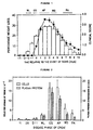
- mice were injected with 1 mg of spinal cord homogenate (SCH) emulsified with Freund's incomplete adjuvant supplemented with 60 ⁇ g mycobacteria ( Mycobacteria tuberculosis H37Ra and M. butyricum in an 8 to 1 ratio) on days 0 and 7 as described previously (Baker,D., et al. , J. Neuroimmunol. 28:261 (1990)). From day 11 (D11) post-inoculation (p.i.) onwards the mice were weighed and checked for clinical signs (Figure 1).
- SCH spinal cord homogenate
- FIG. 1 illustrates the kinetics of weight changes and clinical signs during acute phase chronic relapsing experimental allergic encephalomyelitis (CREAE) induced in Biozzi AB/H mice.
- CREAE chronic relapsing experimental allergic encephalomyelitis
- mice were injected intravenously with 2.5 x 10 7 51 Cr-labelled lymph node cells and 5 ⁇ Ci 125 I-albumin as described previously (Butter, C., et al. , J. Neurol. Sci. 104:9 (1991)). Eighteen hours later anesthetized animals were perfused with RPMI-1640 medium, via the left ventricle following the removal of a 20 ⁇ l blood sample. Brains and spinal cords of 4 - 14 animals per group were collected and estimations of the radioisotope concentrations were performed with a ⁇ -spectrometer.
- the results are expressed as the number of donor cells per gramme of target tissue and extravascular blood equivalents (EVBE), where 100 EVBE are equivalent to the 125 I-albumin plasma protein concentration in blood at the time of sampling, as described previously (Butter, C., et al. , J. Neurol. Sci. 104:9 (1991)).
- Figure 2 illustrates the blood-brain barrier permeability during acute phase chronic relapsing EAE.
- Open bars represent the permeability of the spinal cord to cells and hatched bars represent the permeability of the spinal cord to plasma protein.
- the results represent the mean ⁇ SEM of between 4-14 animals per group.
- BBB breakdown was first detectable in the brain (data not shown), which is relatively uninvolved during EAE in AB/H mice (Baker, D., et al. , J. Neuroimmunol. 28:261 (1990); (Butter, C., et al. , J. Neurol. Sci. 104:9 (1991)), and the spinal cord (Figure 2) when animals experienced weight loss. Blood-brain barrier permeability dramatically increased as clinical signs (OS and AP) developed and weight loss (a total of 25-35% of the body weight compared with day 12) progressed. Once weight gain became apparent in paralysed animals, BBB permeability markedly declined.
- CSF samples were prepared following exsanguination into the thoracic cavity, of terminally anesthetized animals during various phases of EAE. Cerebrospinal fluid (CSF) samples (1-3 ⁇ l/animal) were withdrawn from foramen magnum into a haematocrit tube. Following centrifugation to remove cells, these samples were stored at -20°C prior to assay.
- CSF Cerebrospinal fluid
- Tumour necrosis activity was assessed using either the TNF-sensitive mouse fibroblast cell line L929, as described previously (Beutler B., Science 229:869 (1985)), or 1:2 dilutions of serum, and 1:50 dilutions of CSF were assayed using the Factor-Test mouse TNF ⁇ ELISA kit (Genzyme, UK), according to the manufacturer's instructions.
- the ELISA assay could detect 50 pg/ml - 3.2 ng/ml of TNF ⁇ .
- Acetone-fixed cryostat sections of cervical spinal cord were stained within 1 week of preparation by an indirect immunoperoxidase technique essentially as described previously (Baker, D., et al. , J. Neuroimmunol. 28:261 (1990)). Briefly, endogenous peroxidase activity warn blocked. Sections were incubated with 5% normal mouse serum (NMS) for 30 minutes followed by a 1 hour incubation with primary monoclonal antibody reactive with mouse TNF ⁇ / ⁇ , for example with TN3.19.12 monoclonal antibody, a hamster immunoglobulin G1 (IgG1) monoclonal antibody which neutralizes mouse TNF ⁇ and TNF ⁇ (Sheehan, K.C.F., et al.
- IgG1 hamster immunoglobulin G1
- the primary monoclonal antibodies were detected by sequential 30 minute incubations with biotinylated goat anti-hamster immunoglobulin or rabbit anti-rat immunoglobulin, avidin:biotin peroxidase complex and peroxidase conjugated rabbit anti-goat immunoglobulin or swine anti-rabbit immunoglobulin, respectively.
- the reaction product was developed using the chromogen diaminobenzidine. Sections were counterstained with haematoxylin.
- the primary monoclonal antibody was diluted with excess recombinant mouse TNF ⁇ (200-500 ⁇ g/ml) prior to use for immunocytochemistry. This process failed to inhibit the staining of sections with a CD8-specific monoclonal antibody.
- Double immunofluorescence staining was then performed by incubating these sections with 1:50-1:100 dilutions (in 5% NMS) of either: FITC conjugated anti-Factor VIII, anti-GFAP, H-2A specific mouse monoclonal antibody, or phycoerythrin conjugated rat immunoglobulin monoclonal antibody specific for B cell restricted B220, CD4 or CD8 antigens for 30 minutes. Sections were observed by fluorescence microscopy.
- TNF-specific monoclonal antibodies MX6-XT22 [HB10649] and TN3.19.12
- MP6-XT3 (20 ⁇ l of 4-8 ⁇ g/ml) revealed staining ( Figure 3a) which was blocked by co-incubation of the monoclonal antibody with recombinant mouse TNF ⁇ ( Figure 3b).
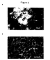
- Immunostaining demonstrated TNF ⁇ activity within lesions present in the cervical spinal cord of paralysed animals ( Figure 3a) and on some lesions of post-acute animals although the intensity of staining appeared reduced compared with that observed in paralysed (AP) animals.
- TNF ⁇ was present in mononuclear cells within perivascular lesions and was often concentrated at the parenchyma/lesion edge where positive cells appeared macrophage/glial-like. Although some positive cells also appeared to have the morphology of astrocytes, the resolution of the immunoperoxidase stained tissue precluded accurate identification of the cells expressing TNF.
- Figures 4a through 4h show the immunofluorescence detection of TNF ⁇ on CD4 + T lymphocytes, astrocytes and macrophages in spinal cord lesions during chronic relapsing EAE. This distribution is similar to that observed in multiple sclerosis lesions (Hoffman, F.M., et al. , J. Exp. Med. 170:607 (1989); Selmaj, K., et al. , J. Clin. Invest. 87:949 (1991)).
- Tumour necrosis factor (TNF ⁇ ) was detected by either FITC ( Figure 4a) or TRITC ( Figures 4c, 4e and 4g) conjugated antibody in the spinal cord lesions of paralysed EAE animals. Sections were incubated with either a phycoerythrin conjugated CD4-specific monoclonal antibody ( Figure 4b) or FITC conjugated Factor VIII related-antigen (Figure 4d), GFAP ( Figure 4f) or H-2A-specific antibodies ( Figure 4h).
- FIGS. 4e and 4f indicate astrocytes expressing TNF ⁇ and the small arrows indicate astrocytes expressing TNF ⁇ localized to part of the astrocytic processes surrounding lesions.
- This astrocytic staining profile of TNF ⁇ is also depicted in Figures 4a, 4c and 4g.
- the arrow in Figures 4g and 4h indicates expression during EAE of TNF ⁇ within the central nervous system lesions.
- TNF- or CD4-specific monoclonal antibodies diluted in PBS (500 ⁇ g TN3.19.12, a TNF-specific monoclonal antibody, or approximately 250 ⁇ g YTS 177.9, a rat IgG2a monoclonal antibody which is a non-depleting mouse CD4-specific antibody (Qin, S., et al. , Eur. J. Immunol. 20:2737 (1990)) produced in ascites fluid.
- Figure 7 shows the results of the effect of TNF immunotherapy on an in vivo induced T cell proliferative response from two individual experiments.
- the data depicted shows that in contrast to the immunosuppressive action of the CD4-specific antibody, the anti-TNF antibody did not inhibit T cell proliferative function under the conditions tested, indicating that these immunomodulatory compounds operate via different mechanisms.
- the results represent the mean ⁇ SD of a minimum of 5 replicate wells.
- TN3.19.12 monoclonal antibody supplied by Dr. R. Schreiber, Washington University Medical School, St. Louis, USA
- TN3.19.12 monoclonal antibody has a serum half-life of approximately 7 days (Sheehan K.C.F., et al. , J. Immunol. 142:3884 (1989)) and a single injection of 300 ⁇ g of TN3.19.12 monoclonal antibody has been reported to inhibit the development of relapsing EAE induced by cell-transfer (Ruddle N.H., et al. , J. Exp Med.
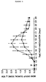
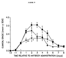
- Figure 5 illustrates the effect on EAE, specifically the effect on clinical signs, of a single injection of TNF-specific monoclonal antibody.
- the arrow indicates the day post-inoculation that the mice were injected intraperitoneally; the triangles represent the result from the mice that were injected intraperitoneally with 250 ⁇ g of TN3.19.12, a TNF-specific monoclonal antibody; and the inverse triangles represent the result from the mice that were injected intraperitoneally with 250 ⁇ g of L2 3D9, a control monoclonal antibody which is reactive with mouse interleukin-2.
- the results represent the mean clinical score of animals in each group ⁇ SEM, at various times post-inoculation.
- mice injected with TN3.19.12 monoclonal antibody compared with mice injected with L2 3D9 monoclonal antibody, appear to exhibit a delayed onset of weight loss (day 16.1 ⁇ 2.5 vs. 14.0 ⁇ 0.9) and clinical signs (day 17.0 ⁇ 2.0 vs. 15.4 ⁇ 1.2) and a lower severity of maximum clinical signs (2.1 ⁇ 1.1 vs. 3.1 ⁇ 0.9) and body weight loss (25.5 ⁇ 4.6% vs. 29.0 ⁇ 4.8%).
- TN3.19.12 monoclonal antibody administered just prior to the onset of clinical manifestations can partially inhibit the onset of EAE by 2-3 days. Therefore animals were given multiple (250 ⁇ g) antibody doses, intraperitoneally at three-daily intervals (days 14, 17 and 20), initiated prior to and during the anticipated development of clinical disease (e.g., weight loss) (Table 1). In comparison to PBS and L2 3D9-treated controls, multiple doses of TNF-specific monoclonal antibody significantly inhibited the development of EAE when assessed up to 3 days following the cessation of treatment. In the few instances where animals were injected when weight loss was first detected, TN3.19.12-treatment appeared to stabilize weight loss.
- mice were injected with TNF-specific monoclonal antibody when clinical signs were first manifested (day 0), that is when the animals were exhibiting a flaccid tail (grade 1).
- the mice were injected i.p. with 0.1 ml of either PBS, or 250 ⁇ g or 1 mg of TN3.19.12, a TNF-specific monoclonal antibody diluted in PBS, or 250 ⁇ g of L2 3D9, a hamster immunoglobulin monoclonal antibody diluted in PBS on days 0, 1 and 2 following the onset of clinical signs (Figure 6), or a single 250 ⁇ g injection i.p. of YTS 177.9, a CD4-specific monoclonal antibody diluted in PBS on day 0.
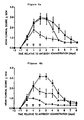
- Figure 6 shows the inhibition of the development of clinical disease following the injection of a TNF-specific monoclonal antibody.
- the arrows indicate days on which the mice were injected i.p.; the circles represent the results from the mice that were injected i.p. with 0.1 ml of PBS; the triangles represent the results from the mice that were injected i.p. with TNF-specific antibody; the inverse triangles represent results from the mice that were injected i.p. with 250 ⁇ g L2 3D9 monoclonal antibody; and the diamonds represent the results from the nice that were injected i.p. with 250 ⁇ g of YTS 177.9 monoclonal antibody on day 0.
- Figure 6a shows the results following injection i.p. of 250 ⁇ g of a TNF-specific antibody.
- Figure 6b shows the results following injection i.p. of 1 mg of a TNF-specific antibody.
- SCH-immunized EAE mice were injected intracerebrally (i.c.) in the cortex of the right frontal lobe as previously described (O'Neill, J.K., et al. , J. Neuroimmunol. 35:53 (1992)) with varying doses of TN3.19.12 monoclonal antibody (Figure 8): 150 ⁇ g, 15 ⁇ g, 1.5 ⁇ g, and 0 ⁇ g. Although 1.5 ⁇ g of TN3.19.12 monoclonal antibody failed to alter the clinical course of disease, 150 ⁇ g of monoclonal antibody stabilized clinical disease ( Figure 8).
- Figure 8 thus shows the dose-dependent inhibition of the progression of clinical EAE following injection of TNF-specific monoclonal antibody directly into the central nervous system.
- the arrow indicates the onset of clinical signs when the animals were exhibiting a flaccid tail (day 0); the circles represent the results from the mice that were injected intracerebrally with 30 ⁇ l of PBS; the inverse triangles represent results from the mice that were injected intracerebrally with 150 ⁇ g TN3.19.12 monoclonal antibody; the triangles represent results from the mice which were injected intracerebrally with 15 ⁇ g TN3.19.12 monoclonal antibody; and the diamonds represent results from the mice that were injected intracerebrally with 1.5 ⁇ g of TNF-specific TN3.19.12 monoclonal antibody.
- the results represent the mean group score ⁇ SEM of 5 animals per group.
- Figure 9 illustrates the inhibition of the development of more marked clinical disease following injection of TNF-specific monoclonal antibody directly into the central nervous system.
- the arrow indicates the treatment at onset of clinical signs when the animals were exhibiting a flaccid tail (day 0); the circles represent results from mice that were untreated; the triangles represent results from mice injected with 30 ⁇ l of PBS i.c. and 150 ⁇ g of TNF-specific monoclonal antibody i.p.; and the inverse triangles represent results from mice injected with 150 ⁇ g of TNF-specific monoclonal antibody i.c. and 30 ⁇ l of PBS i.p. ( Figure 9). The mean clinical group score of 5-7 animals per group following the onset of clinical signs is shown.
- SCH-immunized EAE mice were treated as follows: untreated; injected intraperitoneally with 250 ⁇ g of YTS.177.9, a CD4-specific monoclonal antibody; injected intraperitoneally with 150 ⁇ g TN3.19.12, a TNF-specific monoclonal antibody, and intracerebrally with 30 ⁇ l of PBS; or injected intraperitoneally with 30 ⁇ l of PBS and intracerebrally with 150 ⁇ g of TN3.19.12 monoclonal antibody.
- the results of this experiment indicate that intracerebral injection of TN3.19.12 monoclonal antibody significantly inhibited the progression of weight loss compared to both untreated animals and mice injected intraperitoneally with the TNF-specific antibody.
- the majority of anti-TNF i.c.-treated animals (5 of 6) subsequently relapsed on day 35.6 ⁇ 5.3 and the majority of the control animals (5 of 6) also relapsed on day 38.4 ⁇ 3.4, the results indicate that this treatment modulates the severity of clinical disease.
- CD4 + cells can be targeted in the peripheral circulation prior to extravasation into the central nervous system, TNF-pathogenesis/activity/secretion occurs mainly in the central nervous system, and that appropriately administered TNF-specific immunotherapy into the central nervous system can inhibit the progression of neuroimmunological disease.
- Direct Administration of Anti-TNF Antibody Into the CNS Inhibits Progressive Weight Loss During Clinical EAE Episodes Treatment i.p. none anti-CD4 anti-TNF PBS Treatment i.c.
- Example 8 Central Nervous System-directed Tumour Necrosis Factor Receptor Immunotherapy
- SCH-immunized EAE mice were injected i.c. with 15 ⁇ g of soluble human p55 SF2, a TNF receptor, or varying doses (0 ⁇ g, 15 ⁇ g, or 150 ⁇ g) of TN3.19.12, a TNF-specific monoclonal antibody (Figure 11).
- Figure 11 shows the inhibition of the development of clinical disease following the injection of a TNF-specific monoclonal antibody and following the injection of a soluble human p55 TNF receptor.
- the arrow indicates the treatment at onset of clinical signs when the animals were exhibiting a flaccid tail (day 0); the circles represent the results from the mice that were injected i.c. with 30 ⁇ l of PBS; the triangles represent the results from the mice that were injected i.c. with 15 ⁇ g of TNF-specific monoclonal antibody; the inverse triangles represent the results from the mice that were injected i.c. with 150 ⁇ g of TNF-specific monoclonal antibody; and the diamonds represent the results from the mice that were injected i.c.
- results represent the mean clinical score ⁇ SEM of 5-6 animals per group.
- the results observed when injecting i.c. with 15 ⁇ g of soluble human p55 SF2 TNF-R is similar to the results observed when injecting i.c. with 150 ⁇ g of anti-TNF monoclonal antibody, indicating, in this instance, the greater potency of the SF2 TNF-R.
- Figure 12 shows the dose-dependent inhibition of the progression of clinical EAE following the injection of soluble human p55 sTNF-R.
- the arrow indicates the treatment at onset of clinical signs when the animals were exhibiting a flaccid tail (day 0); the circles represent the results from the mice that were injected i.p. with 30 ⁇ l of PBS; the diamonds represent the results from the mice that were injected i.p. with 15 ⁇ g of SF2 TNF-R; and the triangles represent the results from the mice that were injected i.p. with 150 ⁇ g of SF2 TNF-R.
- the results represent the mean clinical score ⁇ SEM of 5-6 animals per group.
- the results show that the systemic effect of soluble human p55 TNF receptor is at 15 - 150 ⁇ g/injection, and thus is more effective than the systemic effect of the TNF monoclonal antibody TN3.19.12.
Claims (2)
- Utilisation d'un anticorps anti- facteur de nécrose tumorale pour la fabrication d'un médicament pour le traitement de la sclérose en plaques humaine, après survenue de manifestations cliniques, dans laquelle, pour ledit traitement, une quantité à efficacité thérapeutique de l'anticorps anti-facteur de nécrose tumorale est administrée directement dans le système nerveux central de l'être humain, par exemple par voie de cathéter.
- Utilisation selon la revendication 1 dans laquelle l'anticorps anti-facteur de nécrose tumorale est (a) un anticorps polyclonal ; ou (b) un anticorps monoclonal ; ou (c) un fragment d'anticorps.
Priority Applications (2)
| Application Number | Priority Date | Filing Date | Title |
|---|---|---|---|
| PT93917954T PT710121E (pt) | 1993-07-30 | 1993-07-30 | Metodo para o tratamento da esclerose multipla |
| EP99203261A EP1004312A1 (fr) | 1993-07-30 | 1993-07-30 | Procédé pour le traitement de sclérose en plaques |
Applications Claiming Priority (1)
| Application Number | Priority Date | Filing Date | Title |
|---|---|---|---|
| PCT/GB1993/001614 WO1995003827A1 (fr) | 1993-07-30 | 1993-07-30 | Procede de traitement de la sclerose en plaques |
Related Child Applications (1)
| Application Number | Title | Priority Date | Filing Date |
|---|---|---|---|
| EP99203261A Division EP1004312A1 (fr) | 1993-07-30 | 1993-07-30 | Procédé pour le traitement de sclérose en plaques |
Publications (2)
| Publication Number | Publication Date |
|---|---|
| EP0710121A1 EP0710121A1 (fr) | 1996-05-08 |
| EP0710121B1 true EP0710121B1 (fr) | 2000-10-11 |
Family
ID=10729416
Family Applications (2)
| Application Number | Title | Priority Date | Filing Date |
|---|---|---|---|
| EP99203261A Ceased EP1004312A1 (fr) | 1993-07-30 | 1993-07-30 | Procédé pour le traitement de sclérose en plaques |
| EP93917954A Expired - Lifetime EP0710121B1 (fr) | 1993-07-30 | 1993-07-30 | Procede de traitement de la sclerose en plaques |
Family Applications Before (1)
| Application Number | Title | Priority Date | Filing Date |
|---|---|---|---|
| EP99203261A Ceased EP1004312A1 (fr) | 1993-07-30 | 1993-07-30 | Procédé pour le traitement de sclérose en plaques |
Country Status (11)
| Country | Link |
|---|---|
| US (1) | US5958409A (fr) |
| EP (2) | EP1004312A1 (fr) |
| JP (1) | JPH09509646A (fr) |
| AT (1) | ATE196849T1 (fr) |
| AU (1) | AU4719093A (fr) |
| DE (1) | DE69329558T2 (fr) |
| DK (1) | DK0710121T3 (fr) |
| ES (1) | ES2153384T3 (fr) |
| GR (1) | GR3035196T3 (fr) |
| PT (1) | PT710121E (fr) |
| WO (1) | WO1995003827A1 (fr) |
Families Citing this family (26)
| Publication number | Priority date | Publication date | Assignee | Title |
|---|---|---|---|---|
| US6552170B1 (en) * | 1990-04-06 | 2003-04-22 | Amgen Inc. | PEGylation reagents and compounds formed therewith |
| US6270766B1 (en) | 1992-10-08 | 2001-08-07 | The Kennedy Institute Of Rheumatology | Anti-TNF antibodies and methotrexate in the treatment of arthritis and crohn's disease |
| US7070783B1 (en) * | 1995-05-09 | 2006-07-04 | The Mathilda And Terence Kennedy Institute Of Rheumatology | Small molecular weight TNF receptor multimeric molecule |
| US7012060B1 (en) | 1995-07-14 | 2006-03-14 | Applied Research Systems Ars Holding N.V. | TNF receptor and steroid hormone in a combined therapy |
| WO1997003686A1 (fr) * | 1995-07-14 | 1997-02-06 | Applied Research Systems | Recepteur de tnf et hormone steroïdienne en therapie combinee |
| US7608262B2 (en) * | 1996-02-16 | 2009-10-27 | The Kennedy Institute Of Rheumatology | Methods of preventing or treating thrombosis with tumor necrosis factor antagonists |
| TW555765B (en) | 1996-07-09 | 2003-10-01 | Amgen Inc | Low molecular weight soluble tumor necrosis factor type-I and type-II proteins |
| US6660843B1 (en) * | 1998-10-23 | 2003-12-09 | Amgen Inc. | Modified peptides as therapeutic agents |
| IL127851A0 (en) * | 1998-12-30 | 1999-10-28 | Applied Research Systems | Inhibition of TNF activity |
| US6982089B2 (en) * | 1999-02-24 | 2006-01-03 | Tact Ip, Llc | Cytokine antagonists for neurological and neuropsychiatric disorders |
| US8119127B2 (en) * | 1999-02-24 | 2012-02-21 | Tact Ip, Llc | Cytokine antagonists for neurological and neuropsychiatric disorders |
| USRE45976E1 (en) * | 1999-02-24 | 2016-04-19 | Tact Ip, Llc | Methods of inhibiting the action of TNF for neurological conditions by administering etanercept intrathecally |
| AU6929100A (en) * | 1999-08-23 | 2001-03-19 | Biocrystal Limited | Methods and compositions for immunotherapy of b cell involvement in promotion ofa disease condition comprising multiple sclerosis |
| CA2448956C (fr) * | 2001-05-30 | 2017-10-03 | Genentech, Inc. | Anticorps anti-ngf pour le traitement de divers troubles |
| US20050175585A1 (en) * | 2001-06-11 | 2005-08-11 | Transition Therapeutics Inc. | Combination therapies using vitamin B12 and interferon for treatment of viral proliferative and inflammatory disesases |
| US6908611B2 (en) * | 2001-06-11 | 2005-06-21 | Transition Therapeutics Inc. | Combination therapies using vitamin B12 and interferon for treatment of viral, proliferative and inflammatory diseases |
| CN1332711C (zh) * | 2002-02-20 | 2007-08-22 | 埃米球科技有限公司 | 施用glp-1分子的方法 |
| US9028822B2 (en) | 2002-06-28 | 2015-05-12 | Domantis Limited | Antagonists against TNFR1 and methods of use therefor |
| ES2347239T3 (es) | 2002-12-02 | 2010-10-27 | Amgen Fremont Inc. | Anticuerpos dirigidos al factor de necrosis tumoral y usos de los mismos. |
| US7101978B2 (en) | 2003-01-08 | 2006-09-05 | Applied Molecular Evolution | TNF-α binding molecules |
| US7435799B2 (en) | 2004-01-08 | 2008-10-14 | Applied Molecular Evolution | TNF-α binding molecules |
| US8236306B2 (en) * | 2004-12-18 | 2012-08-07 | Edward Lewis Tobinick | Methods to facilitate transmission of large molecules across the blood-brain, blood-eye, and blood-nerve barriers |
| JP2009531295A (ja) * | 2006-02-22 | 2009-09-03 | ユニバーシティ オブ チューリッヒ | 自己免疫疾患又は脱髄疾患を処置するための方法 |
| US8669281B1 (en) | 2013-03-14 | 2014-03-11 | Alkermes Pharma Ireland Limited | Prodrugs of fumarates and their use in treating various diseases |
| DK2970101T3 (en) | 2013-03-14 | 2018-08-20 | Alkermes Pharma Ireland Ltd | PRO-DRUGS OF FUMARATES AND THEIR USE IN TREATING DIFFERENT DISEASES |
| CA2940845C (fr) | 2014-02-24 | 2019-09-24 | Alkermes Pharma Ireland Limited | Sulfonamide et promedicaments de fumarates de sulfinamide et leur utilisation dans le traitement de diverses maladies |
Family Cites Families (5)
| Publication number | Priority date | Publication date | Assignee | Title |
|---|---|---|---|---|
| IL98078A0 (en) * | 1991-05-07 | 1992-06-21 | Yeda Res & Dev | Pharmaceutical compositions comprising an anticytokyne |
| DE10399036I1 (de) * | 1989-08-07 | 2004-04-01 | Peptide Technology Ltd | Bindeligande für Tumornekrosisfaktor. |
| DK0939121T4 (da) * | 1989-09-12 | 2008-02-04 | Ahp Mfg B V | TNF-bindende proteiner |
| US5656272A (en) * | 1991-03-18 | 1997-08-12 | New York University Medical Center | Methods of treating TNF-α-mediated Crohn's disease using chimeric anti-TNF antibodies |
| DE4202665A1 (de) * | 1992-01-28 | 1993-07-29 | Schering Ag | Prostacyclin- und carbacyclinderivate als mittel zur behandlung von multipler sklerose |
-
1993
- 1993-07-30 AT AT93917954T patent/ATE196849T1/de active
- 1993-07-30 DK DK93917954T patent/DK0710121T3/da active
- 1993-07-30 WO PCT/GB1993/001614 patent/WO1995003827A1/fr active IP Right Grant
- 1993-07-30 PT PT93917954T patent/PT710121E/pt unknown
- 1993-07-30 DE DE69329558T patent/DE69329558T2/de not_active Expired - Lifetime
- 1993-07-30 US US08/586,917 patent/US5958409A/en not_active Expired - Lifetime
- 1993-07-30 AU AU47190/93A patent/AU4719093A/en not_active Abandoned
- 1993-07-30 EP EP99203261A patent/EP1004312A1/fr not_active Ceased
- 1993-07-30 ES ES93917954T patent/ES2153384T3/es not_active Expired - Lifetime
- 1993-07-30 EP EP93917954A patent/EP0710121B1/fr not_active Expired - Lifetime
- 1993-07-30 JP JP7505635A patent/JPH09509646A/ja not_active Ceased
-
2001
- 2001-01-10 GR GR20010400014T patent/GR3035196T3/el unknown
Also Published As
| Publication number | Publication date |
|---|---|
| AU4719093A (en) | 1995-02-28 |
| PT710121E (pt) | 2001-04-30 |
| ES2153384T3 (es) | 2001-03-01 |
| DE69329558T2 (de) | 2001-05-31 |
| EP1004312A1 (fr) | 2000-05-31 |
| GR3035196T3 (en) | 2001-04-30 |
| EP0710121A1 (fr) | 1996-05-08 |
| JPH09509646A (ja) | 1997-09-30 |
| DK0710121T3 (da) | 2001-02-05 |
| DE69329558D1 (de) | 2000-11-16 |
| WO1995003827A1 (fr) | 1995-02-09 |
| US5958409A (en) | 1999-09-28 |
| ATE196849T1 (de) | 2000-10-15 |
Similar Documents
| Publication | Publication Date | Title |
|---|---|---|
| EP0710121B1 (fr) | Procede de traitement de la sclerose en plaques | |
| US5741488A (en) | Treatment of rheumatoid arthritis with anti-CD4 antibodies in conjunction with anti-TNF antibodies | |
| Baker et al. | Control of established experimental allergic encephalomyelitis by inhibition of tumor necrosis factor (TNF) activity within the central nervous system using monoclonal antibodies and TNF receptor‐immunoglobulin fusion proteins | |
| RU2263512C2 (ru) | Средства, подавляющие отторжение трансплантата | |
| US6210669B1 (en) | Methods and compositions for immunomodulation | |
| EP0449769B1 (fr) | Molécules liant à la CD25 | |
| US7332161B2 (en) | Treatment of disease with antibodies against high molecular weight kininogen domain 5 | |
| US7122374B1 (en) | Amyloid beta-protein 3(pE)-42 antibodies and uses thereof | |
| EP1018518A2 (fr) | Utilisation d'autoanticorps pour la thérapie et prophylaxie antitumorale | |
| US20030039651A1 (en) | Use of certain drugs for treating nerve root injury | |
| US20040197327A1 (en) | Treatment of T cell mediated autoimmune disorders | |
| JPH10503749A (ja) | IgEアンタゴニストを用いる寄生虫感染症の治療法 | |
| BRPI0317376B1 (pt) | proteína de fusão de anticorpo-il2 designada como hu14.18-il2, usos da mesma, vetor e composição farmacêutica | |
| WO1995009652A1 (fr) | Traitement des troubles auto-immuns et inflammatoires | |
| BG64436B1 (en) | Cd154 blockade therapy for the treatment of protein inhibition syndrome | |
| KR20000052800A (ko) | 면역 질환을 치료하기 위한 치료제로서의 용해성 림포톡신-베타수용체, 항림포톡신 수용체 항체 및 항림포톡신 리간드 항체 | |
| JP4339405B2 (ja) | 予防・治療剤 | |
| EP0667908B1 (fr) | Chaine alpha de receptor de lymphocytes t soluble, et derives utilises comme agents prophylactiques et therapeutiques de maladies auto-immunes | |
| US6770279B1 (en) | TNFα antagonists and cyclosporin in therapy of rheumatoid arthritis | |
| US20020068057A1 (en) | Treatment of autoimmune and inflammatory disorders | |
| US6759041B1 (en) | Preventives/remedies for autoimmune demyelinating diseases | |
| EP1227842A1 (fr) | Inhibition de l'angiogenese par des anticorps avec du kininogene de haut poids moleculaire du domaine 5 | |
| TW202136312A (zh) | 急性期之視神經脊髓炎之預防或治療劑 | |
| KR20230142834A (ko) | 항-cd38 항체 및 이의 용도 | |
| EP0765171A1 (fr) | Traitement des troubles auto-immuns et inflammatoires |
Legal Events
| Date | Code | Title | Description |
|---|---|---|---|
| PUAI | Public reference made under article 153(3) epc to a published international application that has entered the european phase |
Free format text: ORIGINAL CODE: 0009012 |
|
| 17P | Request for examination filed |
Effective date: 19960217 |
|
| AK | Designated contracting states |
Kind code of ref document: A1 Designated state(s): AT BE CH DE DK ES FR GB GR IE IT LI LU MC NL PT SE |
|
| 17Q | First examination report despatched |
Effective date: 19960905 |
|
| RAP3 | Party data changed (applicant data changed or rights of an application transferred) |
Owner name: KENNEDY INSTITUTE OF RHEUMATOLOGY |
|
| GRAG | Despatch of communication of intention to grant |
Free format text: ORIGINAL CODE: EPIDOS AGRA |
|
| GRAG | Despatch of communication of intention to grant |
Free format text: ORIGINAL CODE: EPIDOS AGRA |
|
| GRAG | Despatch of communication of intention to grant |
Free format text: ORIGINAL CODE: EPIDOS AGRA |
|
| GRAH | Despatch of communication of intention to grant a patent |
Free format text: ORIGINAL CODE: EPIDOS IGRA |
|
| GRAH | Despatch of communication of intention to grant a patent |
Free format text: ORIGINAL CODE: EPIDOS IGRA |
|
| GRAA | (expected) grant |
Free format text: ORIGINAL CODE: 0009210 |
|
| AK | Designated contracting states |
Kind code of ref document: B1 Designated state(s): AT BE CH DE DK ES FR GB GR IE IT LI LU MC NL PT SE |
|
| REF | Corresponds to: |
Ref document number: 196849 Country of ref document: AT Date of ref document: 20001015 Kind code of ref document: T |
|
| REG | Reference to a national code |
Ref country code: CH Ref legal event code: EP |
|
| REG | Reference to a national code |
Ref country code: IE Ref legal event code: FG4D |
|
| REF | Corresponds to: |
Ref document number: 69329558 Country of ref document: DE Date of ref document: 20001116 |
|
| ITF | It: translation for a ep patent filed |
Owner name: BARZANO' E ZANARDO MILANO S.P.A. |
|
| REG | Reference to a national code |
Ref country code: CH Ref legal event code: NV Representative=s name: PATENTANWAELTE SCHAAD, BALASS, MENZL & PARTNER AG |
|
| REG | Reference to a national code |
Ref country code: DK Ref legal event code: T3 |
|
| ET | Fr: translation filed | ||
| REG | Reference to a national code |
Ref country code: ES Ref legal event code: FG2A Ref document number: 2153384 Country of ref document: ES Kind code of ref document: T3 |
|
| REG | Reference to a national code |
Ref country code: PT Ref legal event code: SC4A Free format text: AVAILABILITY OF NATIONAL TRANSLATION Effective date: 20010111 |
|
| PLBE | No opposition filed within time limit |
Free format text: ORIGINAL CODE: 0009261 |
|
| STAA | Information on the status of an ep patent application or granted ep patent |
Free format text: STATUS: NO OPPOSITION FILED WITHIN TIME LIMIT |
|
| 26N | No opposition filed | ||
| REG | Reference to a national code |
Ref country code: GB Ref legal event code: IF02 |
|
| PGFP | Annual fee paid to national office [announced via postgrant information from national office to epo] |
Ref country code: LU Payment date: 20110725 Year of fee payment: 19 |
|
| PGFP | Annual fee paid to national office [announced via postgrant information from national office to epo] |
Ref country code: MC Payment date: 20110714 Year of fee payment: 19 Ref country code: FR Payment date: 20110729 Year of fee payment: 19 Ref country code: IE Payment date: 20110721 Year of fee payment: 19 Ref country code: CH Payment date: 20110725 Year of fee payment: 19 Ref country code: DK Payment date: 20110720 Year of fee payment: 19 |
|
| PGFP | Annual fee paid to national office [announced via postgrant information from national office to epo] |
Ref country code: DE Payment date: 20110722 Year of fee payment: 19 Ref country code: ES Payment date: 20110719 Year of fee payment: 19 Ref country code: SE Payment date: 20110722 Year of fee payment: 19 Ref country code: GR Payment date: 20110727 Year of fee payment: 19 Ref country code: PT Payment date: 20110725 Year of fee payment: 19 Ref country code: AT Payment date: 20110714 Year of fee payment: 19 |
|
| PGFP | Annual fee paid to national office [announced via postgrant information from national office to epo] |
Ref country code: NL Payment date: 20110726 Year of fee payment: 19 Ref country code: BE Payment date: 20110713 Year of fee payment: 19 Ref country code: IT Payment date: 20110729 Year of fee payment: 19 |
|
| PGFP | Annual fee paid to national office [announced via postgrant information from national office to epo] |
Ref country code: GB Payment date: 20120719 Year of fee payment: 20 |
|
| BERE | Be: lapsed |
Owner name: KENNEDY *INSTITUTE OF RHEUMATOLOGY Effective date: 20120731 |
|
| REG | Reference to a national code |
Ref country code: PT Ref legal event code: MM4A Free format text: LAPSE DUE TO NON-PAYMENT OF FEES Effective date: 20130130 |
|
| REG | Reference to a national code |
Ref country code: NL Ref legal event code: V1 Effective date: 20130201 |
|
| PG25 | Lapsed in a contracting state [announced via postgrant information from national office to epo] |
Ref country code: MC Free format text: LAPSE BECAUSE OF NON-PAYMENT OF DUE FEES Effective date: 20120731 |
|
| REG | Reference to a national code |
Ref country code: CH Ref legal event code: PL |
|
| REG | Reference to a national code |
Ref country code: GR Ref legal event code: ML Ref document number: 20010400014 Country of ref document: GR Effective date: 20130104 |
|
| REG | Reference to a national code |
Ref country code: SE Ref legal event code: EUG |
|
| REG | Reference to a national code |
Ref country code: AT Ref legal event code: MM01 Ref document number: 196849 Country of ref document: AT Kind code of ref document: T Effective date: 20120730 |
|
| REG | Reference to a national code |
Ref country code: DK Ref legal event code: EBP |
|
| REG | Reference to a national code |
Ref country code: FR Ref legal event code: ST Effective date: 20130329 |
|
| PG25 | Lapsed in a contracting state [announced via postgrant information from national office to epo] |
Ref country code: NL Free format text: LAPSE BECAUSE OF NON-PAYMENT OF DUE FEES Effective date: 20130201 Ref country code: LI Free format text: LAPSE BECAUSE OF NON-PAYMENT OF DUE FEES Effective date: 20120731 Ref country code: FR Free format text: LAPSE BECAUSE OF NON-PAYMENT OF DUE FEES Effective date: 20120731 Ref country code: DE Free format text: LAPSE BECAUSE OF NON-PAYMENT OF DUE FEES Effective date: 20130201 Ref country code: SE Free format text: LAPSE BECAUSE OF NON-PAYMENT OF DUE FEES Effective date: 20120731 Ref country code: CH Free format text: LAPSE BECAUSE OF NON-PAYMENT OF DUE FEES Effective date: 20120731 |
|
| REG | Reference to a national code |
Ref country code: IE Ref legal event code: MM4A |
|
| PG25 | Lapsed in a contracting state [announced via postgrant information from national office to epo] |
Ref country code: GR Free format text: LAPSE BECAUSE OF NON-PAYMENT OF DUE FEES Effective date: 20130204 Ref country code: PT Free format text: LAPSE BECAUSE OF NON-PAYMENT OF DUE FEES Effective date: 20130130 Ref country code: IT Free format text: LAPSE BECAUSE OF NON-PAYMENT OF DUE FEES Effective date: 20120730 Ref country code: BE Free format text: LAPSE BECAUSE OF NON-PAYMENT OF DUE FEES Effective date: 20120731 |
|
| PG25 | Lapsed in a contracting state [announced via postgrant information from national office to epo] |
Ref country code: AT Free format text: LAPSE BECAUSE OF NON-PAYMENT OF DUE FEES Effective date: 20120730 |
|
| PG25 | Lapsed in a contracting state [announced via postgrant information from national office to epo] |
Ref country code: DK Free format text: LAPSE BECAUSE OF NON-PAYMENT OF DUE FEES Effective date: 20120731 Ref country code: IE Free format text: LAPSE BECAUSE OF NON-PAYMENT OF DUE FEES Effective date: 20120730 |
|
| REG | Reference to a national code |
Ref country code: PT Ref legal event code: MM4A Free format text: MAXIMUM VALIDITY LIMIT REACHED Effective date: 20130730 |
|
| REG | Reference to a national code |
Ref country code: GB Ref legal event code: PE20 Expiry date: 20130729 |
|
| REG | Reference to a national code |
Ref country code: DE Ref legal event code: R119 Ref document number: 69329558 Country of ref document: DE Effective date: 20130201 |
|
| REG | Reference to a national code |
Ref country code: ES Ref legal event code: FD2A Effective date: 20131021 |
|
| PG25 | Lapsed in a contracting state [announced via postgrant information from national office to epo] |
Ref country code: PT Free format text: LAPSE BECAUSE OF EXPIRATION OF PROTECTION Effective date: 20130806 Ref country code: ES Free format text: LAPSE BECAUSE OF NON-PAYMENT OF DUE FEES Effective date: 20120731 |
|
| PG25 | Lapsed in a contracting state [announced via postgrant information from national office to epo] |
Ref country code: GB Free format text: LAPSE BECAUSE OF EXPIRATION OF PROTECTION Effective date: 20130729 |
|
| PG25 | Lapsed in a contracting state [announced via postgrant information from national office to epo] |
Ref country code: LU Free format text: LAPSE BECAUSE OF NON-PAYMENT OF DUE FEES Effective date: 20120730 |
|
| PG25 | Lapsed in a contracting state [announced via postgrant information from national office to epo] |
Ref country code: PT Free format text: LAPSE BECAUSE OF EXPIRATION OF PROTECTION Effective date: 20120730 |