EP0696437A2 - Method and apparatus for adjusting the length of a combined spinal-epidural needle - Google Patents
Method and apparatus for adjusting the length of a combined spinal-epidural needle Download PDFInfo
- Publication number
- EP0696437A2 EP0696437A2 EP95201635A EP95201635A EP0696437A2 EP 0696437 A2 EP0696437 A2 EP 0696437A2 EP 95201635 A EP95201635 A EP 95201635A EP 95201635 A EP95201635 A EP 95201635A EP 0696437 A2 EP0696437 A2 EP 0696437A2
- Authority
- EP
- European Patent Office
- Prior art keywords
- needle
- spinal
- epidural
- inner tube
- relative
- Prior art date
- Legal status (The legal status is an assumption and is not a legal conclusion. Google has not performed a legal analysis and makes no representation as to the accuracy of the status listed.)
- Granted
Links
Images


Classifications
-
- A—HUMAN NECESSITIES
- A61—MEDICAL OR VETERINARY SCIENCE; HYGIENE
- A61B—DIAGNOSIS; SURGERY; IDENTIFICATION
- A61B17/00—Surgical instruments, devices or methods, e.g. tourniquets
- A61B17/34—Trocars; Puncturing needles
- A61B17/3401—Puncturing needles for the peridural or subarachnoid space or the plexus, e.g. for anaesthesia
-
- A—HUMAN NECESSITIES
- A61—MEDICAL OR VETERINARY SCIENCE; HYGIENE
- A61M—DEVICES FOR INTRODUCING MEDIA INTO, OR ONTO, THE BODY; DEVICES FOR TRANSDUCING BODY MEDIA OR FOR TAKING MEDIA FROM THE BODY; DEVICES FOR PRODUCING OR ENDING SLEEP OR STUPOR
- A61M25/00—Catheters; Hollow probes
-
- A—HUMAN NECESSITIES
- A61—MEDICAL OR VETERINARY SCIENCE; HYGIENE
- A61B—DIAGNOSIS; SURGERY; IDENTIFICATION
- A61B17/00—Surgical instruments, devices or methods, e.g. tourniquets
- A61B17/34—Trocars; Puncturing needles
- A61B2017/347—Locking means, e.g. for locking instrument in cannula
-
- A—HUMAN NECESSITIES
- A61—MEDICAL OR VETERINARY SCIENCE; HYGIENE
- A61M—DEVICES FOR INTRODUCING MEDIA INTO, OR ONTO, THE BODY; DEVICES FOR TRANSDUCING BODY MEDIA OR FOR TAKING MEDIA FROM THE BODY; DEVICES FOR PRODUCING OR ENDING SLEEP OR STUPOR
- A61M19/00—Local anaesthesia; Hypothermia
-
- A—HUMAN NECESSITIES
- A61—MEDICAL OR VETERINARY SCIENCE; HYGIENE
- A61M—DEVICES FOR INTRODUCING MEDIA INTO, OR ONTO, THE BODY; DEVICES FOR TRANSDUCING BODY MEDIA OR FOR TAKING MEDIA FROM THE BODY; DEVICES FOR PRODUCING OR ENDING SLEEP OR STUPOR
- A61M5/00—Devices for bringing media into the body in a subcutaneous, intra-vascular or intramuscular way; Accessories therefor, e.g. filling or cleaning devices, arm-rests
- A61M5/46—Devices for bringing media into the body in a subcutaneous, intra-vascular or intramuscular way; Accessories therefor, e.g. filling or cleaning devices, arm-rests having means for controlling depth of insertion
Landscapes
- Health & Medical Sciences (AREA)
- Life Sciences & Earth Sciences (AREA)
- Surgery (AREA)
- Anesthesiology (AREA)
- Public Health (AREA)
- Engineering & Computer Science (AREA)
- Biomedical Technology (AREA)
- Heart & Thoracic Surgery (AREA)
- Veterinary Medicine (AREA)
- Animal Behavior & Ethology (AREA)
- General Health & Medical Sciences (AREA)
- Medical Informatics (AREA)
- Pathology (AREA)
- Nuclear Medicine, Radiotherapy & Molecular Imaging (AREA)
- Molecular Biology (AREA)
- Hematology (AREA)
- Biophysics (AREA)
- Pulmonology (AREA)
- Media Introduction/Drainage Providing Device (AREA)
- Infusion, Injection, And Reservoir Apparatuses (AREA)
- Feed For Specific Animals (AREA)
Abstract
Description
- This invention relates to a combined spinal-epidural needle for delivery of a medicament to the subarachnoid space, and more particularly, to a method and apparatus for adjusting the extension of a spinal needle relative to the epidural needle during a procedure for delivering medicament to the subarachnoid space.
- As is known in the art, there exist two basic techniques for introducing injectable medicament into the spinal area of a patient. Both of these techniques have their own unique advantages and disadvantages and both can be used to create spinal anesthesia or analgesia. In both of these procedures, of course, the medicaments can be any type of liquid therapeutic material including antibiotics, steroids or the like. In general, however, the medicaments are agents used for anesthesia and/or analgesia.
- The first procedure, known as the "epidural" technique, employs an epidural needle to deliver medicament to the epidural space of the patient. Certain epidural needles feature a curved distal end. Certain drawbacks exist with this technique. Because the medicament must percolate through semi-liquid fat to reach the nerve roots, the onset of the anesthetic block is oftentimes slow. Moreover, the potential exists for toxicity caused by the relatively large doses of medicament necessary to obtain an adequate block. After the initial dosage, a catheter is oftentimes inserted through the epidural needle into the epidural space to provide sustained or prolonged anesthesia/analgesia to the patient.
- The second procedure, known in the art as the "spinal" or "subarachnoid" technique, typically employs a relatively small gauge needle to deliver medicaments directly to the subarachnoid space of the spinal column. Because the anesthetic is delivered directly to the nerve roots, the onset of anesthetic effect is quite rapid, and the block achieved by the spinal technique is often deeper than that possible employing the epidural technique.
- The major disadvantage of the spinal technique relates to postoperative side effects. Unlike the epidural procedure, in the spinal technique, the dura mater must be punctured to reach the subarachnoid space. The resultant leakage of cerebrospinal fluid ("CSF") through the puncture oftentimes leads to severe postoperative headaches, known as "postdural puncture headache" ("PDPH"). In addition, while hypotension can result from either of the epidural or spinal techniques, it is believed that the rapid onset of the block in the spinal procedure causes a higher degree of hypotension than the epidural technique. Moreover, unlike the epidural procedure, which typically employs a catheter for continuous epidural blockage, a single shot spinal needle is often unable to extend the anesthetic block, once fixed.
- A survey of previous patent literature reports in this general area may be found, for instance, in U.S. Patent No. 5,085,631, which is directed to a method for placement of a subarachnoid catheter that utilizes a three component apparatus having an outer needle, an inner needle, and a catheter intermediate the two needles.
- In order to alleviate the disadvantages associated with both procedures while providing the advantages of each, a combined spinal-epidural technique, or "CSE", has been developed. In CSE, an epidural needle is inserted into the patient in the usual manner and advanced to the epidural space without puncturing the dura mater. Next, steadying his or her hand against the patient's back and using the fixed epidural needle as an introducer, a smaller gauge spinal needle is inserted through the lumen of the epidural needle and advanced so that the distal end of the spinal needle crosses the epidural space. The practitioner, relying on his sense of touch, continues to insert the spinal needle until the distal end is felt to puncture the dura mater and enter into the subarachnoid space. A "pop" sensation is often felt at the hub of the spinal needle by the practitioner when the dura mater has been punctured. As confirmation of proper placement in the subarachnoid space, the practitioner will normally look for the appearance of CSF at the proximal end of the spinal needle by removing the stylet of the spinal needle.
- Spinal anesthetic is administered in the usual manner, and the spinal needle is then withdrawn without displacing the epidural needle. Next, an epidural catheter is introduced through the epidural needle into the epidural space, and the epidural needle is thereafter removed from the back of the patient. Lastly, the epidural catheter is secured in place by taping same to the back of the patient.
- In general, the CSE technique provides the practitioner with the benefits associated with the individualized epidural or spinal techniques while offsetting the disadvantages experienced by each. The surgeon is able to gain the advantages of rapid onset of a deep block provided by the spinal procedure. The epidural catheter serves to provide sustained anesthetic effect and extend the block provided by the spinal anesthetic. The catheter also enhances the practitioner's options and choices in administering operative anesthetic or postoperative pain relief For example, the practitioner is able to administer a spinal anesthetic alone or in combination with epidural anesthetics and/or analgesics. Moreover, the practitioner can choose from a variety of medicaments or combinations thereof with various rates of delivery, not being limited by the single injection of the spinal technique alone.
- While providing the practitioner with a ready way to administer quality anesthetic relief to the patient, a number of drawbacks exist with current CSE practice. The CSE procedure is typically dependent on the individualized practitioner's experience with the method which, in turn, depends on the number and types of patients the doctor has had experience with. The exigencies of the operating environmental also greatly affect the procedure. As previously explained, CSE is performed by the relative insertion of two needles of differing gauges. Because the spinal needle is free to slide within the epidural needle, which itself is only retained by the dura mater once inserted, the danger exists that the spinal needle will be displaced during administration of the anesthetic. Thus, the doctor is required to utilize both hands, one to steady the spinal needle against the patient's body, the other hand to steady the syringe attached to the proximal end of the spinal needle. He must also utilize both hands when locking the spinal needle into place with the epidural needle. Because the doctor must steady his or her hand against the patient's back during insertion, smooth relative sliding is oftentimes difficult to achieve. Adequate tactile feedback, necessary to permit the practitioner to assess relative needle insertion, is also heavily dependent on the exigencies of the operating environment, which can vary at a moment's notice.
- In addition, it will be observed that human body structures differ. The relative dimensions of the body, and particularly those defining the epidural space, the thickness of the dura mater, and the distance to the subarachnoid space, will vary. The doctor's appreciation of these dimensions is critical to proper placement of the needles in the appropriate locations, and in particular, to avoid inadvertent puncture of the dura mater.
- Moreover, the practitioner must not only has to rely on his relative experience to make sure that the spinal needle is extended sufficiently through the dura mater, he must do so with two separate needles that may not often provide him with either sufficient tactile feedback or a discernible way to gauge relative insertion. A typical pencil-point spinal needle such as a Whitacre needle cannot always aspirate CSF, even when the dura mater is felt to "pop." In this situation, to be absolutely sure that the needles are properly placed, the practitioner must often withdraw both needles, repositioning them to reidentify the epidural space and, hence, the subarachnoid. This can cause unnecessary discomfort to both patient and practitioner.
- Furthermore, in some situations practitioners will not need the full degree of spinal needle extension provided when the hubs of the spinal and epidural needles engage. When this type of situation occurs, the practitioner is forced to overcome a potentially unsafe and unsecure condition caused by a portion of the spinal needle protruding unsupported from the hub of the epidural needle.
- The aforementioned difficulties can be amplified in that CSE is sometimes performed with individualized epidural and spinal needles sourced from different manufacturers. In these cases, owing to differing dimensions, tolerances, quality of finish or the like, precise sliding action between the needles may be compromised. Moreover, the hubs of differing spinal and epidural needles do not often fit, so that the practitioner cannot be sure of the relative extension achieved by the spinal needle. This can also affect the ability of the practitioners to rotate the spinal needle within the epidural needle in the locked state, useful if the practitioner suspects that the ports of the spinal needle are being blocked by the flap created in the dura mater during entry, or where the practitioner desires to better direct the extent of the anesthetic block provided by the spinal needle. The practitioner might wish to rotate the spinal needle so that the distal point is directed around the four quadrants of the subarachnoid space in an attempt to detect CSF. Faulty hub fit in the locked condition hampers the practitioner's ability to exploit the benefits of rotation.
- Some manufacturers have begun to market matched sets of spinal/epidural needles to provide good hub fit and establish a predetermined amount of extension between the spinal and epidural needles when both hubs engage. While to a certain extent alleviating some of the problems encountered with "mixing" needles, the practitioner is still constrained by a fixed extension when the hubs interlock. For some patients, the fixed extension may still be inadequate to reach the dura mater, while for others it may be more than necessary.
- Certain attempts in the art have sought to regulate the insertion or placement of a needle into the body. For instance, U.S. Patent No. 4,940,458 is directed to a placement system for an epidural needle. An internally threaded barrel is provided to guide the externally threaded epidural needle via a knurled wheel at the proximal end of the epidural needle. A pressure monitor serves to advise the practitioner when the epidural needle has entered the epidural space. U.S. Patent No. 5,312,375 is directed to a set for spinal anesthesia employing an introducer needle and a spinal needle. Either a screw or a toothed clamp arrangement may be provided to secure the spinal needle relative to the introducer needle once the spinal needle has been inserted through the dura mater. An analogous technique employing a metallic wing fixed to the epidural needle, with a relatively large L-shaped metallic bar engaged to the wing with two screws to fixedly adjust the position of the spinal needle relative to the epidural needle, has recently been proposed. See J. Simsa, "Use of 29 gauge spinal needles and a Fixation Device with Combined Spinal Epidural Technique", ACTA Anaesthesiologica Scandinavia, 1994 Vol. 38, pp. 439-441. The relative extension of the larger leg of the L-shaped bar past the wing is indicative of the spinal needle extension. Once the spinal needle has been extended to its desired position, a screw on the wing is tightened; two hands are required to operate this device. None of the aforementioned attempts sufficiently addresses the aforementioned problems of relative spinal needle insertion and inadequate (or non-existent) tactile feedback currently experienced with the CSE procedure.
- There exists a need, therefore, for a method and apparatus which will provide the practitioner with a ready way to support the spinal needle, gauge and insure precise insertion of the spinal needle through the epidural needle and into the subarachnoid space, maintain smooth sliding action and fit between the needles, and provide the practitioner with valuable tactile feedback during the procedure.
- The present invention alleviates in great part the drawbacks associated with present CSE practice and provides the practitioner with a ready way to precisely monitor the insertion or removal of a spinal needle during CSE, all the while preserving good tactile feedback and fit between the spinal needle and epidural needle.
- The invention is directed to a regulating device for extending and/or retracting the spinal needle relative to the epidural needle during CSE. The device, which may be provided as part of a CSE set, or with or attached to one of the spinal needle or epidural needles, or which can be provided as a separate unit for utilization with a separately sourced spinal needle or epidural needle or with a separately sourced CSE set, includes a pair of sliding members to which each of the epidural and spinal needles are separately fixed. The sliding members are disposed to permit relative sliding action between the spinal needle and the epidural needle. In one form, the sliding members may be configured as a pair of concentric tubes slidably disposed relative to one another. The interior surface of the outermost tube or, conversely, the exterior surface of the innermost tube, may be structured with a plurality of planar surface portions, with the opposing surface being relatively cylindrical. The mating of a planar surface portion with a rounded surface portion provides point contact between the inner and outer tubes, reducing the engagement surface area between the tubes, and, hence the frictional resistance between the tubes, providing for smoother sliding action and better tactile feedback to the practitioner.
- An actuating tab may be provided for regulating operation of the device between a free position and a locking position. In one version, the tab may be biasingly fixed at one end to the outermost tube. The tab, operable with a one-handed effort by the practitioner may include a mating portion for selectable engagement with the innermost tube responsive to the tab position selected by the practitioner. The mating portion may feature an engaging surface structured to permit secure locking action with a complimentary structured surface formed on or in the innermost tube. In one embodiment, the engaging surface may be formed as a male (or female) groove, with the tube surface formed with a series of female (or male) grooves located along the axis of the innermost tube. The series of grooves may be formed to correspond to a measured degree of extension of the innermost tube relative to the outermost tube which, in turn, relates to the degree of extension of the spinal needle relative to the epidural needle. Markings formed on the outside surface of the innermost tube provide the practitioner with visual indication of both the alignment of the distal tips of the spinal and epidural needles and with the relative extension length of the spinal needle relative to the epidural needle.
- The invention will now be described in greater detail by way of reference to the following drawings, wherein:
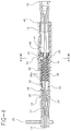
- Fig. 1 is a perspective view of the regulating device of the present invention as utilized in conjunction with a CSE set, showing the spinal needle in a retracted state;
- Fig. 2 shows a perspective view of the regulating device of the present invention, showing the spinal needle advanced through the lumen of the epidural needle;
- Fig. 3 depicts an exploded assembly view in perspective of the regulating device of the present invention;
- Fig. 4 illustrates a side view of the regulating device, as taken along line 4-4 of Fig. 1, showing the outermost tube in an extended state relative to the innermost tube;
- Fig. 5 is a side view of the regulating device, as taken along line 5-5 of Fig. 2, showing the outermost tube in a retracted state relative to the innermost tube;
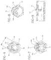
- Fig. 6 depicts a frontal view of the regulating device, as seen along line 6-6 of Fig. 3, showing the point contact relationship between the innermost and outermost tubes;
- Fig. 7 is a rear view of the regulating device, as seen along line 7-7 of Fig. 3, showing engagement of the actuating tab with the innermost tube;
- Fig. 8 is a cutaway view of the regulating device, as seen along line 8-8 of Fig. 4, with the innermost tube removed for clarity, showing the actuating tab and its mating portion as engaged with the innermost tube;
- Fig. 9 is a partial cutaway side view of the regulating device showing the actuating tab and mating portion;
- Fig. 10 is a side view illustrating placement of an epidural needle into the epidural space of a patient;
- Fig. 11 is a side view illustrating placement of a spinal needle within the lumen of the epidural needle and alignment of the distal tips of both needles prior to extension of the spinal needle; and
- Fig. 12 is a side view illustrating extension of the spinal needle through the dura mater of a patient into the subarachnoid space.
- Turning now to the drawings, wherein like numerals denote like components Figs. 1-9 depict one embodiment of a regulating
device 10 for adjusting the extension length of aspinal needle 12 relative to anepidural needle 14 during a CSE procedure. It will be understood that while directed in particular to regulating the extension of a spinal needle relative to an epidural needle during a combined CSE procedure, the device is readily applicable to any device and/or procedure employing a needle through needle technique and employing regulation of needle length extensions during that technique. - Referring to Figs. 1-5, the overall construction of the regulating
device 10 in conjunction with anepidural needle 14 and thespinal needle 12 is illustrated. Theepidural needle 14 will be well known to those skilled in the art and, in general, includes adistal end 14a and alumen 15 extending through the length of the needle. As illustrated, thedistal end 14a of the epidural needle may be curved for instance, to enhance a practitioner's efforts in directing placement of an epidural catheter (not shown) in the epidural space of a patient. Awing collar 20 may be provided to enable a practitioner to manipulate the needle and/or overall device during use. Theepidural needle 14 further features amale luer connector 22 permitting attachment of theepidural needle 14 to an appropriate fitting, a syringe, or the like. - The
spinal needle 12, equally well known to the skilled artisan, includes adistal end 12a together with ahub assembly 16. Thehub assembly 16 features astopper portion 18 configured to be placed within an appropriate fitting or the like. Thespinal needle 12 may also be provided with a stylet (not shown), as is known to those skilled in the art, both for blocking thelumen 13 of the spinal needle during insertion and for providing the practitioner with a way to check for CSF during the procedure. - In general, the
device 10 may be employed with any combination ofspinal needle 12 andepidural needle 14. It has been found, however, that to accommodate most patients, useful ranges of theepidural needle 14 include lengths between 8 centimeters ("cm") (3.1496") to about 8.890cm (3½"), while thespinal needle 12 can range from about 14.645cm (5 49/64") to about 15.558cm (6 1/8"). Thespinal needle 12 can be provided in various standardized diametral sizes ("gauges") depending on the particular anesthetic application desired by the practitioner, but in general it has been found thatspinal needles 12 between 22 gauge and 29 gauge will accommodate most applications. The following table provides diametral dimensions across the gauge range:Table of Hypodermic Tubing Nominal Sizes Gauge Outside Diameter (mm) Inside Diameter (mm) 30 0.30 0.18 29 0.33 0.20 28 0.36 0.20 27 0.40 0.25 26 0.46 0.30 25 0.51 0.30 24 0.56 0.36 23 0.64 0.38 22 0.71 0.46 21 0.82 0.56 20 0.90 0.65 19 1.08 0.80 18 1.27 0.96 17 1.50 1.17 16 1.65 1.32 - A general overall view of the regulating
device 10 in conjunction with thespinal needle 12 andepidural needle 14 is broadly depicted in Figs. 1-5. In the form depicted, the regulatingdevice 10 includes a first sliding member such as an outer cylinder ortube 51 disposed in sliding relation to a second sliding member such as an inner cylinder ortube 32, each of which are respectively fixed to one of thespinal needle 12 or theepidural needle 14. While other configurations may be envisioned, as here depicted, theepidural needle 14 is mounted to theinner tube 32 via a hub fitting 25 disposed at the distal end of theinner tube 32. The hub fitting 25 includes aproximal end 26 configured to mate with amale luer extension 30 disposed at thedistal end 28 of the inner tube, with the hub fitting 25 itself including a male luer fitting 24 at its distal end for snug insertion into thehub 22 of the epidural needle. It will be realized by those skilled in the art that the hub fitting 25 may be provided either as part of the regulatingdevice 10 or as part of theepidural needle 14. - As herein illustrated, the
spinal needle 12 may be secured to theouter tube 51 via its hub fitting 18 which may be configured for snug and secure engagement with theproximal end 46 of theouter tube 51. When assembled, thespinal needle 12 will project through thelumen 15 of theepidural needle 14, with thedistal end 12a of the spinal needle axially extendible relative to thedistal end 14a of the epidural needle by sliding action between theouter tube 51 andinner tube 32 of the regulating device. While various extension lengths "x" (see Fig. 2) of thespinal needle 12 relative to the epidural needle are possible depending on user need or desire, an extension length of approximately 1.501 cm (0.591") (inches) has been found to suffice for applications to most patients. However, one skilled in the art of catheters, needles and hypodermic delivery devices will recognize that for specialty applications such as neonates, pediatric patients, especially thin or obese individuals, and other specialty applications, it may be desirable to reduce or increase the sizes, gauges, component lengths, or extension lengths and/or other dimensions associated with the various components herein described for the specific application. - Turning our attention to construction of the regulating device, the
inner tube 32 may be formed as a hollow cylindrical tube extending between adistal end 28 and aproximal end 29. The tube 37 can be formed from any appropriate rigid material including a medical grade plastic such as polycarbonate, a metal, or the like, and, if desired, can be formed through an injection molding process. Thetube 32 features an axially extendingslot 36 providing access to a structuredinterior surface 38. As better seen in Figs. 4-7, the structuredinterior surface 38 may be formed as a plurality ofconcentric groove elements 38a formed along the axial length of theinner tube 32. Thegroove elements 38a may be formed in a variety of manners, such as male or female grooves; ramps or other similar projections; raised or recessed indentations; or various other configurations as may be envisioned by the skilled artisan. The spacing between thegroove elements 38a may be either equidistant or non-uniform as need or desire dictate. While the structuredsurface 38 can be formed along the entire axial length ofinner tube 32, it will be understood that the structuring may be effected along the axial length required to obtain the desired extension "x" of thespinal needle 12 relative to theepidural needle 14. While the overall length and diameter of the inside tube may be chosen as need or desire dictate, an outside diameter "a" (Fig. 1) of about 0.620 cm (0.244") and an overall length "c" (Fig. 3) of about 2.009cm (0.791") measured between thedistal end 28 andproximal end 29 should suffice for most applications. It will also be appreciated that when distal ends 14a, 12a of the epidural and spinal needles are aligned prior to use, a proximal length "d" (Fig. 3) should remain within theoutside tube 51 to provide stability. Here, a length "d" of about 0.508cm (0.200") may be provided for stability, with the remaining 1.501cm (0.591") of theinner tube 32 length representing the relative extension of thespinal needle 12 relative to theepidural needle 14 in use. - A plurality of
markings 34 may also be provided on the outside surface ofinner tube 32 to help the practitioner gauge the relative extension of theouter tube 51 respective to theinner tube 32. Themarkings 34 may be calibrated, as need or desire dictate, to any standard of measurement, such as millimeters, centimeters or the like. As will be discussed in greater detail below, themarkings 34 may be calibrated to the extension provided by the structuredsurface 38 of the inner tube and, in particular,individual markings 34a may be configured to correspond toindividualized grooves 38a in the structuredsurface 38 to assist the practitioner in gauging the relative extension length "x" of thespinal needle 12 relative to theepidural needle 14. - The
outer tube 51 includes aproximal end 46 and adistal end 44 and, as previously described, is disposed in sliding relation to theinner tube 32. Like theinner tube 32, the outer tube can be formed from a suitable material such as medical grade plastic, metal, or the like, and it can be injection molded. Theoutside surface 53 of the tube can be shaped in a variety of manners to enable secure gripping by the practitioner. Here, the outside surface is shaped as a hexagon, but other configurations are equally possible texturing. Moreover, theoutside surface 53 can be textured or roughened to enhance one's grip on the device. The outer diameter "b" and the length "l" (Fig. 2) of theoutside tube 51 can be constructed to any appropriate dimension both to provide easy one-handed manipulation by the practitioner and to accommodate the variously sizedepidural needles 14/spinal needles 12 utilized as previously described. In general, an outside diameter "b" of about 0.856cm (0.3371") and a length "l" of about 2.606cm (1.026") will suffice for most practitioners. - As illustrated in Figs. 1-5, the
outer tube 51 features an interior surface formed as a plurality ofplanar surfaces 58 circumferentially disposed around the central axis of theouter tube 51. While here illustrated as formed with a hexagonal configuration having sixplanar surfaces 58, it will be understood and appreciated by those skilled in the art that the invention is not so limited, and that the interior surface may be configured with any number of planar surfaces such as pentagonal, octagonal, etc. as need or desire dictate. - Referring to Figs. 6-8, it will also be seen that the
inner tube 32 is disposed within theouter tube 51 such that theoutside surface 33 of theinner tube 32 is in substantial sliding contact with theplanar surfaces 58 of theouter tube 51. A plurality of contact points 60 are established by the intersection of the relatively roundedoutside surface 33 of theinner tube 32 and each of the planar surfaces 58. It will be appreciated that by this arrangement, the outside circumferential surface area of theinner tube 32 is disposed in sliding contact with the interior of theouter tube 51. By reducing the contact area between the tubes and, in particular, by providing sliding point contact between theinner tube 32 andouter tube 51, frictional resistance between the tubes is substantially reduced, thereby enhancing smooth sliding action between the tubes, and resulting in better tactile feedback to the practitioner. - While it is desirable to maintain a relatively close diametral tolerance between the
inner tube 32 andouter tube 51 to promote stability and precise sliding action, the inside diameter "F" (Fig. 6) of theouter tube 51 should provide a slight clearance to prevent undue friction when sliding relative to theinner tube 32 Here, the diameter "F" may be configured to about 0.627cm (0.247") to prevent frictional resistance with theinner tube 32 having, for instance, an outside diameter "a" of 0.620 cm (0.244"). - It will be understood and appreciated that instead of providing the planar surfaces on the interior of the outer tube, with a rounded exterior surface on the inner tube, the plurality of planar surfaces may be structured on the exterior surface of the inner tube, with the interior of the outer tube rounded so as to provide point contact. As will be better evident from the discussion below, it will be further understood that the entire length of the
outer tube 52 need not be structured with the planar surfaces 58. Rather, only the axial portion of theouter tube 51 which will be subjected to relative sliding motion respective to theinner tube 32 need be structured so as to provide the benefits described above. Thus, for extension "x" of 1.501cm (0.591"), only an axial length of 1.501cm (0.591") measured from thedistal end 44 of theoutside tube 51 need be provided with the planar surfaces 58. - Turning to Figs. 1-9, and in particular to Figs. 4-9, an
actuating tab 50 is provided to enable the practitioner to regulate the axial position of theouter tube 51 relative to theinner tube 32 and, hence, to vary the extension of thespinal needle 12 relative to theepidural needle 14. For purposes of illustration, but not of limitation, thetab 50 is here illustrated configured as a cantilevered arm. However, it will be understood and realized by those skilled in the art that thetab 50 can be devised in numerous alternate manners. For instance, thetab 50 can be formed as a push-button configuration having, for instance, a spring or other biasing element for coordinating operation and use of the device, or thetab 50 can be formed as a sliding button or sliding tab configuration. Various other constructions as will be within the realm of the skilled artisan can be realized. - The
actuation tab 50, which may be molded or otherwise formed as a portion of theouter tube 51, may be resiliently fixed at oneend 52 to the outer tube. Afinger tab 54 is provided at the second end of thetab 50, permitting one-handed operation by a practitioner to bias the tab between a locked position, wherein theouter tube 51 is fixed in axial relation to theinner tube 32, and an unlocked position, wherein theouter tube 51 is axially slidable relative to theinner tube 32. - The
actuating tab 50 includes aneck portion 54a disposed both through aslot 56 formed in thesurface 53 of theouter tube 51 and through theaxial slot 36 formed in theinner tube 32. Amating portion 70 is provided at the end of theneck 54a in a manner so as to be located within the structuredinterior 38 of theouter tube 32. As here illustrated, themating portion 70 may be configured as a relatively flat, semi-circular tab, to accommodate the relatively circular interior surface of theinner tube 32. However, other shapes or configurations of themating portion 70 may be devised as need or desire dictate. - As shown,
mating portion 70 includes amating surface 72 for locking engagement with thegroove elements 38a forming thestructured surface 38. Here, themating surface 72 is illustrated as a female groove formed in opposed mating relationship to the structuredsurface 38 of theouter tube 32. In particular, thegroove 72 is dimensioned so as to lockingly mate with one of the individualconcentric grooves 38a which, taken together, form the structuredsurface 38. - While the structured
surface 38 is here illustrated as being formed along a major portion of theinner tube 32, as previously described it will be appreciated by those skilled in the art that the structuredsurface 38 can be formed in a variety of manners and/or configurations, as necessary or desired, so that the structuring need not encompass the entire inner circumferential area of theinner tube 32. For instance, the structuredsurface 38 may be provided on a portion of the surface area, such as on one quadrant of the circumferential surface area disposed within the interior of theinner tube 32. Thus, themating portion 70 may be appropriately designed in order to mate with the portion of the inner circumferential area of theinner tube 32 which has been structured as previously described. It will further be appreciated that themating surface 72 may be appropriately configured to the particular treatment and extent thereof chosen for the structuredsurface 38. For instance, if structuredsurface 38 were formed as a plurality offemale groove elements 38a, themating portion 72 may be configured as a male groove which is matingly disposed with the concentricfemale grooves 38a forming thestructured surface 38. As the structuredsurface 38 may be devised in varying manners, themating surface 72 may also be appropriately configured so as to matingly engage with the particular configuration chosen for the structuredsurface 38. Other variations and configurations may be envisioned by those skilled in the art. - Referring to Fig. 3, a
cap 40 may be provided at theproximal end 29 of theinner tube 32 to be securely mated to the proximal end via an appropriately sized malefitting portion 42. It will be appreciated that thecap 40 may be inserted into thedistal end 29 of theinner tube 32 during assembly, such that theinner tube 32 will be disposed within the interior of theouter tube 51, with thecap 40 positioned proximally of themating portion 70 of theactuating tab 50. In this manner, theinner tube 32 is prevented from inadvertent withdrawal from theouter tube 51 by a blocking action created between themating surface 70 and the cap 40 (see Fig. 4). As thespinal needle 12 is fitted to theproximal end 46 of theoutside tube 51, thespinal needle 12 is disposed through the center ofcap 40 via anopening 41. - Operation of the regulating
device 10 will now be explained with reference to Figs. 1-12. As previously explained, the regulatingdevice 10 can be provided either as part of the CSE set including theepidural needle 14 andspinal needle 12, or the device may be provided for use with an individual spinal needle or epidural needle separately sourced, or with a prematched CSE set separately sourced. For instance, thedevice 10 can be preattached or otherwise form an integral component of either a separately sourcedepidural needle 14 or separately sourcedspinal needle 12. For instance, thedevice 10 can form the hub portion of aspinal needle 12. - If for example, the device is provided with a separately sourced CSE set, the
epidural needle 14 is first affixed to theinner tube 32 via the hub fitting 25 as previously described, with theinner tube 32 thereafter slid through theouter tube 51. Theneck portion 54a andmating portion 70 of theactuating tab 50 will slide through theaxial opening 36 disposed in theinner tube 32, with thecap 40 thereafter fitted to thedistal end 29 of the inner tube to secure the inner tube against inadvertent withdrawal of theouter tube 51. Thespinal needle 12 may thereafter be fitted to theouter tube 51 and inserted through thehole 41 in thecap 40. The spinal needle will project through the interiors of both theouter tube 51 andinner tube 32, so that thespinal needle 12 is disposed through thelumen 15 of theepidural needle 14. It will be understood that if provided as part of a CSE set, the regulatingdevice 10 may be pre-assembled together with thespinal needle 12 andepidural needle 14. - In order to provide the practitioner with an effective way to gauge the axial extension of the
spinal needle 12 relative to theepidural needle 14, the dimensions of the various components such as theinner tube 32 andouter tube 51 may be chosen so that in a first locked position of theactuation tab 50, thedistal tip 12a of the spinal needle is aligned with thedistal tip 14a of the epidural needle, as illustrated in Fig. 11. As a practical matter, this may be accomplished by designating one of thegroove elements 38a which is engaged by themating portion 70 as corresponding to alignment between thedistal tips epidural needles distal end 44 of theouter tube 51 may be aligned with theindividual markings 34a to assist the practitioner with determining relative extension of thespinal needle 12. By correlating one of theindividual markings 34a on the inner tube to the designatedgroove element 38a to indicate when the distal points are aligned, the practitioner is provided with the ability to visually regulate the extension of thespinal needle 12. - In use, with the spinal and epidural needles aligned as previously described, the set is inserted into the
epidural space 100 of the patient until thedistal point 14a of the epidural needle is positioned by the practitioner in an appropriate location in the epidural space. Note that in this position,outer tube 51 is extended relative to theinner tube 32 so that the thespinal needle 12 is in a retracted state (Figs. 1 and 4), with thedistal tips mating portion 70 of the actuation tab locked with anindividual groove element 38a to maintain the position of the needles. - When the epidural needle has been properly positioned, the
finger tab 54 may be activated (depressed) by the practitioner, releasing themating portion 70 from engagement with the structuredsurface 38, thereby permitting theouter tube 51 to be axially slidable in the distal direction with respect to theinner tube 32, all with a one-handed effort by the practitioner. Theinner tube 32, itself fixed to theepidural needle 14, will remain fixed relative to the patient. As earlier described, a practitioner may additionally utilizewing collar 20 to provide additional support to theepidural needle 14, if need or desire dictate. - By continuing to slide the
tube 51 distally axially forward, thespinal needle 12 will be extended through the epidural needle 14 (Figs. 2 and 5) so as to puncture thedura mater 102 and come to rest in the subarachnoid space 104 (Fig. 12). Again, the practitioner may monitor the relative position of thedistal end 44 of theouter tube 51 relative to themarkings 34 as a means to assess relative insertion of the spinal needle. As earlier described, the dimensions of the various components may be chosen and selected as need or desire dictate so that thespinal needle 12 will have a relative extension "X" (see Fig. 2) relative to thespinal needle 14 when theouter tube 51 has been slid axially forward to a maximum position. Intermediate extension positions "Y" (see Fig. 12) may be selected by the practitioner based on the relative position of thedistal end 44 of theouter tube 51 to theinner tube 32. - Upon selecting the appropriate position, the practitioner will deactivate (release pressure against) the
finger tab 54, causing theactuating tab 50 to be biased upwards, forcing themating portion 70 to engage one of thevarious groove elements 38a to lock the position of theouter tube 51 relative to theinner tube 32. If a stylet has been provided, the same may be removed by the practitioner to detect for CSF. It will also be appreciated that by providing a rotating fit between the male luer fitting 24 andhub 22 of theepidural needle 14, and/or a rotating fit between themale luer extension 30 of the inner tube and the hub fitting 25, the practitioner will be able to rotate the spinal needle in all four quadrants of thesubarachnoid space 102 while maintaining the spinal needle in locked position relative to the epidural needle. Thus, a one-handed operation is easily achieved. - Thus, it will be seen that the regulating
device 10 provides the practitioner with a ready and sure way to practice a CSE procedure in a safe and sure manner. The device is easily operable with a one-handed effort and will guide the practitioner to accurate spinal needle extensions while providing him or her with smooth, steady sliding action and, hence, valuable tactile feedback. The spinal needle may be easily manipulated in the locked position improving safety and alleviating problems previously encountered in the procedure. - It will be appreciated and understood by those skilled in the art that additional and further forms of the invention may be devised without departing from the spirit and scope of the appended claims, the invention not being limited to the specific embodiments shown.
Claims (15)
- A device for regulating the extension of a spinal needle relative to an epidural needle, comprising:
a first member for securing said epidural needle;
a second member for securing said spinal needle and being slidably disposed relative to said first member; and
an actuating tab forming a selectably fixed connection between said first and second members, said actuating tab displaceable between a locked position wherein said first member is locked relative to said second member and an unlocked position wherein said second member is slidable relative to said first member to regulate the extension of the spinal needle relative to the epidural needle. - The device of Claim 1 wherein said second member defines an internal cavity having an axis, said first member slidingly disposed within said internal cavity.
- The device of Claim 2, wherein said internal cavity comprises a plurality of planar surfaces substantially circumferentially disposed about the axis of said second member, said first member comprising an exterior surface in point contact with said planar surfaces.
- A device for regulating the extension of a spinal needle relative to an epidural needle, comprising:
an inner tube having proximal and distal ends and defining an exterior surface and an interior surface, said epidural needle securable to said distal end;
an outer tube having proximal and distal ends and defining a cavity having an axis, the exterior surface of said inner tube substantially slidably disposed within the cavity and along the axis of said outer tube, the hub of said spinal needle securable to the proximal end of said outer tube; and
an actuating tab mounted on said outer tube and forming a selectably fixed connection between said inner tube and said outer tube, said actuating tab movable between a locked position wherein said inner tube is axially fixed relative to said outer tube and an unlocked position wherein said outer tube is axially slidable relative to said inner tube to vary the axial extension of said spinal needle relative to said epidural needle. - The device of Claim 4, wherein the interior surface of the inner tube comprises a plurality of locking formations disposed along the axis of the inner tube.
- The device of Claim 5, wherein said plurality of locking formations comprise grooves formed on at least a portion of the interior surface of the inner tube.
- The device of Claim 4, wherein said actuating tab comprises:
an arm member having a first end resiliently mounted to said outer tube and an inner tube engaging portion mounted to a second end of said arm member, said inner tube engaging portion disposed through the exterior surface of said inner tube in selective engagement with the interior portion of said inner tube. - The device of Claim 7, wherein said inner tube engaging portion comprises a mating surface disposed for user-selectable contact with the interior surface of said inner tube.
- The device of Claim 8, wherein said mating surface comprises a groove dimensioned to mesh with the locking formations formed along the axis of the inner tube.
- The device of Claim 4, wherein said inner tube includes a plurality of markings for gauging the axial position of said outer tube relative to said inner tube.
- The device of Claim 4, wherein the cavity of said outer tube comprises a plurality of planar surfaces substantially circumferentially disposed around the axis of the outer tube, wherein the exterior surface of said inner tube is configured for point contact with each of said plurality of planar surfaces.
- A method for regulating the extension of a spinal needle relative to an epidural needle, comprising the steps of:
forming a user-regulatable combined spinal epidural needle set by separately affixing each of said spinal needle and said epidural needle to a pair of substantially concentrically disposed sliding members, wherein said spinal needle is slidingly disposed in the lumen of said epidural needle;
deflecting an actuating tab forming a selectably fixed connection between said pair of substantially concentrically disposed sliding members; and
moving the sliding members relative to one another to regulate the extension of said spinal needle relative to said epidural needle. - The method of Claim 12, wherein the step of separately affixing each of said spinal needle and said epidural needle to said pair of concentrically disposed sliding members includes the steps of affixing said epidural needle to an innermost sliding member and affixing said spinal needle to an outermost sliding member.
- The method of Claim 13, wherein said step of regulating the extension of said spinal needle relative to said epidural needle further comprises the step of moving said outermost sliding member relative to a set of markings formed on said innermost sliding member, said markings calibrated to the degree of extension of said spinal needle relative to said epidural needle.
- The method of Claim 12, further including the step of forming sliding surfaces on each of said sliding members, the sliding surfaces disposed in point contact with one another.
Priority Applications (1)
| Application Number | Priority Date | Filing Date | Title |
|---|---|---|---|
| DE29522168U DE29522168U1 (en) | 1994-08-09 | 1995-06-17 | Device for adjusting the length of a combined spinal and epidural needle |
Applications Claiming Priority (2)
| Application Number | Priority Date | Filing Date | Title |
|---|---|---|---|
| US287995 | 1994-08-09 | ||
| US08/287,995 US5480389A (en) | 1994-08-09 | 1994-08-09 | Method and apparatus for adjusting the length of a combined spinal-epidural needle |
Publications (3)
| Publication Number | Publication Date |
|---|---|
| EP0696437A2 true EP0696437A2 (en) | 1996-02-14 |
| EP0696437A3 EP0696437A3 (en) | 1996-03-27 |
| EP0696437B1 EP0696437B1 (en) | 1999-09-08 |
Family
ID=23105298
Family Applications (1)
| Application Number | Title | Priority Date | Filing Date |
|---|---|---|---|
| EP95201635A Expired - Lifetime EP0696437B1 (en) | 1994-08-09 | 1995-06-17 | Apparatus for adjusting the length of a combined spinal-epidural needle |
Country Status (16)
| Country | Link |
|---|---|
| US (1) | US5480389A (en) |
| EP (1) | EP0696437B1 (en) |
| JP (1) | JP2787012B2 (en) |
| KR (1) | KR0161328B1 (en) |
| CN (1) | CN1116552A (en) |
| AT (1) | ATE184174T1 (en) |
| AU (1) | AU694518B2 (en) |
| BR (1) | BR9503558A (en) |
| CA (1) | CA2151006C (en) |
| CO (1) | CO4440487A1 (en) |
| DE (2) | DE29522168U1 (en) |
| DK (1) | DK0696437T3 (en) |
| ES (1) | ES2136240T3 (en) |
| FI (1) | FI953765A (en) |
| SG (1) | SG32447A1 (en) |
| TW (1) | TW308531B (en) |
Cited By (8)
| Publication number | Priority date | Publication date | Assignee | Title |
|---|---|---|---|---|
| GB2316320A (en) * | 1996-08-16 | 1998-02-25 | Smiths Industries Plc | Spinal-epidural needle assembly |
| EP0824894A1 (en) | 1996-08-16 | 1998-02-25 | Smiths Industries Public Limited Company | Needle assemblies |
| EP0982006A1 (en) * | 1998-07-17 | 2000-03-01 | Becton, Dickinson and Company | Variable extension combined spinal/epidural needle set and method for its use |
| WO2016112383A1 (en) * | 2015-01-10 | 2016-07-14 | University Of Florida Research Foundation, Inc. | Simulation features combining mixed reality and modular tracking |
| CN105873526A (en) * | 2013-08-21 | 2016-08-17 | Crh医疗公司 | Elastic band ligation device with locking mechanism and method for treatment of hemorrhoids |
| EP3035864A4 (en) * | 2013-08-21 | 2017-03-08 | CRH Medical Corporation | Elastic band ligation device with integrated obturator and method for treatment of hemorrhoids |
| EP3035865A4 (en) * | 2013-08-21 | 2017-03-08 | CRH Medical Corporation | Elastic band ligation device with anti-pinch feature and method for treatment of hemorrhoids |
| US10902677B2 (en) | 2010-04-09 | 2021-01-26 | University Of Florida Research Foundation, Incorporated | Interactive mixed reality system and uses thereof |
Families Citing this family (108)
| Publication number | Priority date | Publication date | Assignee | Title |
|---|---|---|---|---|
| US5628734A (en) * | 1995-03-23 | 1997-05-13 | Hatfalvi; Bela I. | Spinal needle with curved distal end and method of using said needle in a spinal injection to prevent post dural puncture headache |
| US5836916A (en) * | 1995-10-05 | 1998-11-17 | Children's Medical Center Corporation | Combined spinal epidural device |
| GB9601147D0 (en) * | 1996-01-19 | 1996-03-20 | Smiths Industries Ltd | Spinal epidural needle assemblies |
| US5792110A (en) * | 1996-06-26 | 1998-08-11 | Cunningham; Miles G. | Systems and methods for delivering therapeutic agents to selected sites in a subject |
| US6520951B1 (en) * | 1996-09-13 | 2003-02-18 | Scimed Life Systems, Inc. | Rapid exchange catheter with detachable hood |
| US6582401B1 (en) | 1996-09-13 | 2003-06-24 | Scimed Life Sytems, Inc. | Multi-size convertible catheter |
| US5921971A (en) | 1996-09-13 | 1999-07-13 | Boston Scientific Corporation | Single operator exchange biliary catheter |
| US6096009A (en) | 1996-09-13 | 2000-08-01 | Boston Scientific Corporation | Guidewire and catheter locking device and method |
| US6346093B1 (en) | 1996-09-13 | 2002-02-12 | Scimed Life Systems, Inc. | Single operator exchange biliary catheter with common distal lumen |
| US6606515B1 (en) | 1996-09-13 | 2003-08-12 | Scimed Life Systems, Inc. | Guide wire insertion and re-insertion tools and methods of use |
| US5906594A (en) * | 1997-01-08 | 1999-05-25 | Symbiosis Corporation | Endoscopic infusion needle having dual distal stops |
| US5871470A (en) * | 1997-04-18 | 1999-02-16 | Becton Dickinson And Company | Combined spinal epidural needle set |
| US5846226A (en) * | 1997-05-12 | 1998-12-08 | Urmey; William F. | Spinal-epidural administration system |
| US6077251A (en) * | 1997-10-30 | 2000-06-20 | Ting; Windsor | Medicinal agent administration system |
| USD430290S (en) * | 1998-04-28 | 2000-08-29 | Becton, Dickinson And Company | Splittable catheter introducer |
| USD417733S (en) * | 1998-04-28 | 1999-12-14 | Becton, Dickinson And Company | Catheter introducer needle assembly |
| US20010021824A1 (en) * | 1998-07-17 | 2001-09-13 | Marsh Ronald W. | Variable extension combined spinal/epidural needle set and method for its use |
| CA2320097C (en) * | 1998-12-09 | 2009-04-14 | Cook Incorporated | Hollow, curved, superelastic medical needle |
| US7081122B1 (en) * | 1999-10-19 | 2006-07-25 | Kyphon Inc. | Hand-held instruments that access interior body regions |
| US6575919B1 (en) * | 1999-10-19 | 2003-06-10 | Kyphon Inc. | Hand-held instruments that access interior body regions |
| AU1912601A (en) * | 1999-11-15 | 2001-05-30 | Sami onder | A sterile disposable spinal syringe set without mandrin |
| US7811250B1 (en) | 2000-02-04 | 2010-10-12 | Boston Scientific Scimed, Inc. | Fluid injectable single operator exchange catheters and methods of use |
| WO2001091651A1 (en) * | 2000-05-31 | 2001-12-06 | Hussain Karim | Epidural apparatus |
| US6893421B1 (en) * | 2000-08-08 | 2005-05-17 | Scimed Life Systems, Inc. | Catheter shaft assembly |
| US6613017B1 (en) * | 2000-08-08 | 2003-09-02 | Scimed Life Systems, Inc. | Controlled depth injection device and method |
| US6595958B1 (en) * | 2000-08-08 | 2003-07-22 | Scimed Life Systems, Inc. | Tortuous path injection device and method |
| ES2190713B1 (en) * | 2000-11-29 | 2005-02-01 | Jesus Sahagun De La Lastra | NEEDLE ASSEMBLY WITH PERFECTED IMMOBILIZER FOR THE APPLICATION OF RACHINE SURVEYS. |
| US6837878B2 (en) * | 2001-01-09 | 2005-01-04 | Icu Medical, Inc. | Bluntable needle assembly with open-ended blunting probe |
| US6558353B2 (en) * | 2001-01-25 | 2003-05-06 | Walter A. Zohmann | Spinal needle |
| US6764484B2 (en) * | 2001-03-30 | 2004-07-20 | Scimed Life Systems, Inc. | C-channel to o-channel converter for a single operator exchange biliary catheter |
| US6827718B2 (en) | 2001-08-14 | 2004-12-07 | Scimed Life Systems, Inc. | Method of and apparatus for positioning and maintaining the position of endoscopic instruments |
| AU2003231045A1 (en) | 2002-04-24 | 2003-11-10 | Becton Dickinson And Company | Apparatus for shielding a needle |
| US6893393B2 (en) * | 2003-02-19 | 2005-05-17 | Boston Scientific Scimed., Inc. | Guidewire locking device and method |
| JPWO2004091702A1 (en) * | 2003-04-15 | 2006-07-06 | ドクタージャパン株式会社 | Medical anesthesia needle |
| US8333734B2 (en) * | 2003-07-03 | 2012-12-18 | Walter A. Zohmann | Fenestrated peripheral nerve block needle and method for using the same |
| US7097637B2 (en) | 2003-08-27 | 2006-08-29 | C. R. Bard, Inc. | Safety needle with positive flush |
| US8480629B2 (en) * | 2005-01-28 | 2013-07-09 | Boston Scientific Scimed, Inc. | Universal utility board for use with medical devices and methods of use |
| US7608081B2 (en) * | 2005-05-23 | 2009-10-27 | Custom Spine, Inc. | Rod reducer |
| ATE424879T1 (en) | 2005-07-06 | 2009-03-15 | Vascular Pathways Inc | INTRAVENOUS CATHETER INSERTION DEVICE AND METHOD OF USE |
| JP4767655B2 (en) * | 2005-10-28 | 2011-09-07 | テルモ株式会社 | Protector and needle set |
| US20080045964A1 (en) * | 2006-08-16 | 2008-02-21 | Allan Mishra | Device for cartilage repair |
| US8449503B2 (en) | 2010-11-30 | 2013-05-28 | Custom Medical Applications | Neural injection system and related methods |
| US9480800B2 (en) | 2006-09-11 | 2016-11-01 | Custom Medical Applications | Neural injection system and related methods |
| US20080065029A1 (en) * | 2006-09-11 | 2008-03-13 | Racz N S | Nerve block needle and related methods |
| US9888940B2 (en) | 2006-09-11 | 2018-02-13 | Custom Medical Applications | Neural injection system and related methods |
| US8377005B2 (en) | 2006-09-11 | 2013-02-19 | Custom Medical Applications | Neural injection system and related methods |
| US20080171983A1 (en) * | 2006-10-27 | 2008-07-17 | Knutson Eric J | Needle hub assembly |
| US8372000B2 (en) | 2007-01-03 | 2013-02-12 | Boston Scientific Scimed, Inc. | Method and apparatus for biliary access and stone retrieval |
| US20080167628A1 (en) * | 2007-01-05 | 2008-07-10 | Boston Scientific Scimed, Inc. | Stent delivery system |
| US20080183192A1 (en) * | 2007-01-26 | 2008-07-31 | Laurimed Llc | Contralateral insertion method to treat herniation with device using visualization components |
| US20080188826A1 (en) * | 2007-02-01 | 2008-08-07 | Laurimed, Llc | Methods and devices for treating tissue |
| US8480570B2 (en) | 2007-02-12 | 2013-07-09 | Boston Scientific Scimed, Inc. | Endoscope cap |
| US20090062769A1 (en) * | 2007-04-13 | 2009-03-05 | Boston Scientific Scimed, Inc. | Rapid exchange catheter converter |
| CN100448413C (en) * | 2007-04-24 | 2009-01-07 | 温一辉 | Orbit type lumbar combined puncturing needle |
| DE602008003791D1 (en) | 2007-05-07 | 2011-01-13 | Vascular Pathways Inc | INTRODUCTION OF INTRAVENOUS CATHETER AND BLOOD DETECTING DEVICE AND METHOD OF USE |
| US9131960B2 (en) * | 2007-06-13 | 2015-09-15 | Custom Medical Applications | Safety neural injection system and related methods |
| US8690832B2 (en) | 2007-06-13 | 2014-04-08 | Custom Medical Applications | Retrofitted neural injection system and related methods |
| US8388521B2 (en) * | 2008-05-19 | 2013-03-05 | Boston Scientific Scimed, Inc. | Integrated locking device with active sealing |
| US8343041B2 (en) | 2008-05-19 | 2013-01-01 | Boston Scientific Scimed, Inc. | Integrated locking device with passive sealing |
| US20090247901A1 (en) * | 2008-03-25 | 2009-10-01 | Brian Zimmer | Latching side removal spacer |
| US20090247900A1 (en) * | 2008-03-25 | 2009-10-01 | Brian Zimmer | Push button adjustable spacer |
| CA2720452A1 (en) * | 2008-04-02 | 2009-10-08 | Laurimed, Llc | Methods and devices for delivering injections |
| US8043316B2 (en) * | 2008-05-02 | 2011-10-25 | Suros Surgical Systems, Inc. | Adjustable spacer |
| US10278725B2 (en) * | 2008-09-15 | 2019-05-07 | Paul M. Zeltzer | Lumbar puncture detection device |
| US8968210B2 (en) | 2008-10-01 | 2015-03-03 | Covidien LLP | Device for needle biopsy with integrated needle protection |
| US11298113B2 (en) | 2008-10-01 | 2022-04-12 | Covidien Lp | Device for needle biopsy with integrated needle protection |
| US9186128B2 (en) * | 2008-10-01 | 2015-11-17 | Covidien Lp | Needle biopsy device |
| US9332973B2 (en) | 2008-10-01 | 2016-05-10 | Covidien Lp | Needle biopsy device with exchangeable needle and integrated needle protection |
| US20110190662A1 (en) * | 2008-10-01 | 2011-08-04 | Beacon Endoscopic Corporation | Rapid exchange fna biopsy device with diagnostic and therapeutic capabilities |
| US9782565B2 (en) | 2008-10-01 | 2017-10-10 | Covidien Lp | Endoscopic ultrasound-guided biliary access system |
| BRPI0917035A2 (en) | 2008-12-04 | 2019-09-24 | Pivot Medical Inc | "telescope access cannula, telescope shutter, system, method for providing an access corridor from a first off-site location to a second on-site location" |
| EP2384779B1 (en) * | 2009-03-18 | 2014-05-07 | Unisis Corp. | Anesthetic compound needle |
| US8932258B2 (en) | 2010-05-14 | 2015-01-13 | C. R. Bard, Inc. | Catheter placement device and method |
| US10384039B2 (en) | 2010-05-14 | 2019-08-20 | C. R. Bard, Inc. | Catheter insertion device including top-mounted advancement components |
| US9950139B2 (en) | 2010-05-14 | 2018-04-24 | C. R. Bard, Inc. | Catheter placement device including guidewire and catheter control elements |
| US9872971B2 (en) | 2010-05-14 | 2018-01-23 | C. R. Bard, Inc. | Guidewire extension system for a catheter placement device |
| US11925779B2 (en) | 2010-05-14 | 2024-03-12 | C. R. Bard, Inc. | Catheter insertion device including top-mounted advancement components |
| IN2013CN00091A (en) | 2010-06-30 | 2015-07-03 | Laurimed Llc | |
| US8685052B2 (en) | 2010-06-30 | 2014-04-01 | Laurimed, Llc | Devices and methods for cutting tissue |
| US8690833B2 (en) | 2011-01-31 | 2014-04-08 | Vascular Pathways, Inc. | Intravenous catheter and insertion device with reduced blood spatter |
| EP3563898B1 (en) * | 2011-02-25 | 2020-11-11 | C.R. Bard, Inc. | Medical component insertion device including a retractable needle |
| USD903101S1 (en) | 2011-05-13 | 2020-11-24 | C. R. Bard, Inc. | Catheter |
| WO2013003450A1 (en) | 2011-06-27 | 2013-01-03 | Boston Scientific Scimed, Inc. | Stent delivery systems and methods for making and using stent delivery systems |
| WO2013119336A1 (en) | 2012-02-10 | 2013-08-15 | Laurimed, Llc | Vacuum powered rotary devices and methods |
| EP2740422A1 (en) | 2012-12-05 | 2014-06-11 | Custom Medical Applications | Safety neural injection system and related methods |
| US9522254B2 (en) | 2013-01-30 | 2016-12-20 | Vascular Pathways, Inc. | Systems and methods for venipuncture and catheter placement |
| US9358036B2 (en) * | 2013-03-12 | 2016-06-07 | Gyrus Acmi, Inc. | Blade positioning device |
| CA2846742C (en) | 2013-03-15 | 2017-11-28 | Custom Medical Applications | Safety neural injection system and related methods |
| ES2614991T3 (en) * | 2013-06-07 | 2017-06-02 | Stryker European Holdings I, Llc | Sleeve flange |
| US8815099B1 (en) | 2014-01-21 | 2014-08-26 | Laurimed, Llc | Devices and methods for filtering and/or collecting tissue |
| US9968373B1 (en) | 2014-02-21 | 2018-05-15 | Surgentec, Llc | Handles for needle assemblies |
| WO2016037127A1 (en) | 2014-09-05 | 2016-03-10 | C.R. Bard, Inc. | Catheter insertion device including retractable needle |
| USD903100S1 (en) | 2015-05-01 | 2020-11-24 | C. R. Bard, Inc. | Catheter placement device |
| WO2016187037A1 (en) | 2015-05-15 | 2016-11-24 | C.R.Bard, Inc. | Catheter placement device including an extensible needle safety component |
| US9681889B1 (en) * | 2015-06-09 | 2017-06-20 | Surgentec, Llc | Depth controlled needle assembly |
| JP7051821B2 (en) | 2016-09-12 | 2022-04-11 | シー・アール・バード・インコーポレーテッド | Blood control for catheter insertion device |
| CN106618686A (en) * | 2016-10-24 | 2017-05-10 | 常熟市第人民医院 | Combined spinal anaesthesia and dura mater puncture needle with three-dimensional relatively-fixed rotatable wing |
| KR101876807B1 (en) * | 2016-11-17 | 2018-07-10 | 임성빈 | Photo dynamics apparatus for medical treatment or needle |
| JP6953541B2 (en) | 2017-03-01 | 2021-10-27 | シー・アール・バード・インコーポレーテッドC R Bard Incorporated | Catheter insertion device |
| ES2686840A1 (en) * | 2017-04-19 | 2018-10-22 | Fundación Para El Fomento De La Investigación Sanitaria Y Biomédica De La Comunitat Valenciana | DURAL SEALING SYSTEM (Machine-translation by Google Translate, not legally binding) |
| CN110996753B (en) | 2017-08-11 | 2022-11-22 | 波士顿科学国际有限公司 | Biopsy cap for endoscope |
| CN109674515B (en) * | 2017-10-18 | 2021-08-06 | 江苏风和医疗器材股份有限公司 | Accommodation space adjusting method |
| WO2019173641A1 (en) | 2018-03-07 | 2019-09-12 | Bard Access Systems, Inc. | Guidewire advancement and blood flashback systems for a medical device insertion system |
| USD921884S1 (en) | 2018-07-27 | 2021-06-08 | Bard Access Systems, Inc. | Catheter insertion device |
| EP4010057A4 (en) | 2019-08-19 | 2023-10-18 | Becton, Dickinson and Company | Midline catheter placement device |
| EP4159143A1 (en) * | 2019-08-24 | 2023-04-05 | Guidestar Medical Devices | Epidural device for detection of and needle placement in epidural space |
| CN111110329A (en) * | 2020-01-14 | 2020-05-08 | 山东省千佛山医院 | Disposable infant lumbar puncture needle |
| WO2021159087A1 (en) * | 2020-02-07 | 2021-08-12 | The Johns Hopkins University | Extendable needle |
Citations (3)
| Publication number | Priority date | Publication date | Assignee | Title |
|---|---|---|---|---|
| US4601708A (en) * | 1985-09-09 | 1986-07-22 | Pavel Jordan | Automatic injection for syringe needle, and assembly |
| US5312351A (en) * | 1993-01-29 | 1994-05-17 | Gerrone Carmen J | Combined pneumo-needle and trocar apparatus |
| US5312375A (en) * | 1993-06-28 | 1994-05-17 | Simon Gurmarnik | Set for spinal anesthesia |
Family Cites Families (22)
| Publication number | Priority date | Publication date | Assignee | Title |
|---|---|---|---|---|
| US2922420A (en) * | 1957-11-29 | 1960-01-26 | Sierra Eng Co | Epidural needle |
| US3356089A (en) * | 1965-03-01 | 1967-12-05 | Howard R Francis | Injection needle guide |
| US3406687A (en) * | 1966-06-23 | 1968-10-22 | Resiflex Lab | Guide and positioning means for a needle |
| US3727613A (en) * | 1970-10-09 | 1973-04-17 | Voys Inc Le | Safety catheter placement assembly |
| US3964480A (en) * | 1974-10-03 | 1976-06-22 | Froning Edward C | Apparatus for sterotaxic lateral extradural disc puncture |
| US4230123A (en) * | 1978-10-31 | 1980-10-28 | Hawkins Jr Irvin F | Needle sheath complex and process for decompression and biopsy |
| US4362156A (en) * | 1979-04-18 | 1982-12-07 | Riverain Corporation | Intravenous infusion assembly |
| CA1221596A (en) * | 1984-03-09 | 1987-05-12 | David Evans | Surgical needle |
| US4801293A (en) * | 1985-10-09 | 1989-01-31 | Anthony Jackson | Apparatus and method for detecting probe penetration of human epidural space and injecting a therapeutic substance thereinto |
| US4760847A (en) * | 1986-08-18 | 1988-08-02 | Vincent Vaillancourt | Depth measuring device |
| ES2007667A6 (en) * | 1987-07-28 | 1989-07-01 | Espejo Martinez Antonio | A device for locating the epidural space. |
| US5195526A (en) * | 1988-03-11 | 1993-03-23 | Michelson Gary K | Spinal marker needle |
| US5085631A (en) * | 1988-08-02 | 1992-02-04 | Thomas Jefferson University | Method and kit for administering spinal subarachnoid anesthesia |
| FR2638359A1 (en) * | 1988-11-03 | 1990-05-04 | Tino Dalto | SYRINGE GUIDE WITH ADJUSTMENT OF DEPTH DEPTH OF NEEDLE IN SKIN |
| US4940458A (en) * | 1989-02-02 | 1990-07-10 | Cohn Arnold K | Epidural needle placement system |
| DE3918431C1 (en) * | 1989-06-06 | 1990-07-26 | B. Braun Melsungen Ag, 3508 Melsungen, De | |
| DE3922406C1 (en) * | 1989-07-07 | 1990-10-11 | B. Braun Melsungen Ag, 3508 Melsungen, De | |
| AU651745B2 (en) * | 1991-12-13 | 1994-07-28 | Covidien Ag | Locking pneumoneedle |
| CA2088024C (en) * | 1992-02-03 | 2004-03-30 | Daniel E. Andrew | A cannula clamp |
| US5246425A (en) * | 1992-09-21 | 1993-09-21 | Daniel Hunsberger | Trocar and cannula assembly |
| US5257972A (en) * | 1992-12-04 | 1993-11-02 | Simon Gurmarnik | Device for and method of length determination of epidural anesthesia catheter |
| US5380292A (en) * | 1993-12-22 | 1995-01-10 | Wilson-Cook Medical, Inc. | Gastrointestinal needle mechanism |
-
1994
- 1994-08-09 US US08/287,995 patent/US5480389A/en not_active Expired - Lifetime
-
1995
- 1995-06-05 CA CA002151006A patent/CA2151006C/en not_active Expired - Fee Related
- 1995-06-17 EP EP95201635A patent/EP0696437B1/en not_active Expired - Lifetime
- 1995-06-17 DE DE29522168U patent/DE29522168U1/en not_active Expired - Lifetime
- 1995-06-17 DK DK95201635T patent/DK0696437T3/en active
- 1995-06-17 AT AT95201635T patent/ATE184174T1/en active
- 1995-06-17 ES ES95201635T patent/ES2136240T3/en not_active Expired - Lifetime
- 1995-06-17 DE DE69511967T patent/DE69511967T2/en not_active Expired - Fee Related
- 1995-06-23 TW TW084106455A patent/TW308531B/zh active
- 1995-07-11 AU AU24935/95A patent/AU694518B2/en not_active Ceased
- 1995-08-04 CO CO95035009A patent/CO4440487A1/en unknown
- 1995-08-07 CN CN95115210A patent/CN1116552A/en active Pending
- 1995-08-07 SG SG1995001079A patent/SG32447A1/en unknown
- 1995-08-07 BR BR9503558A patent/BR9503558A/en not_active Application Discontinuation
- 1995-08-08 FI FI953765A patent/FI953765A/en not_active Application Discontinuation
- 1995-08-08 KR KR1019950024365A patent/KR0161328B1/en not_active IP Right Cessation
- 1995-08-09 JP JP7203323A patent/JP2787012B2/en not_active Expired - Lifetime
Patent Citations (3)
| Publication number | Priority date | Publication date | Assignee | Title |
|---|---|---|---|---|
| US4601708A (en) * | 1985-09-09 | 1986-07-22 | Pavel Jordan | Automatic injection for syringe needle, and assembly |
| US5312351A (en) * | 1993-01-29 | 1994-05-17 | Gerrone Carmen J | Combined pneumo-needle and trocar apparatus |
| US5312375A (en) * | 1993-06-28 | 1994-05-17 | Simon Gurmarnik | Set for spinal anesthesia |
Cited By (14)
| Publication number | Priority date | Publication date | Assignee | Title |
|---|---|---|---|---|
| GB2316320A (en) * | 1996-08-16 | 1998-02-25 | Smiths Industries Plc | Spinal-epidural needle assembly |
| EP0824894A1 (en) | 1996-08-16 | 1998-02-25 | Smiths Industries Public Limited Company | Needle assemblies |
| US5941853A (en) * | 1996-08-16 | 1999-08-24 | Smiths Industries Public Limited Company | Needle assemblies |
| AU725119B2 (en) * | 1996-08-16 | 2000-10-05 | Smiths Group Plc | Needle assemblies |
| EP0982006A1 (en) * | 1998-07-17 | 2000-03-01 | Becton, Dickinson and Company | Variable extension combined spinal/epidural needle set and method for its use |
| US6245044B1 (en) | 1998-07-17 | 2001-06-12 | Becton, Dickinson And Company | Variable extension combined spinal/epidural needle set and method for its use |
| US11361516B2 (en) | 2010-04-09 | 2022-06-14 | University Of Florida Research Foundation, Incorporated | Interactive mixed reality system and uses thereof |
| US10902677B2 (en) | 2010-04-09 | 2021-01-26 | University Of Florida Research Foundation, Incorporated | Interactive mixed reality system and uses thereof |
| EP3035865A4 (en) * | 2013-08-21 | 2017-03-08 | CRH Medical Corporation | Elastic band ligation device with anti-pinch feature and method for treatment of hemorrhoids |
| EP3035864A4 (en) * | 2013-08-21 | 2017-03-08 | CRH Medical Corporation | Elastic band ligation device with integrated obturator and method for treatment of hemorrhoids |
| EP3035863A4 (en) * | 2013-08-21 | 2017-03-08 | CRH Medical Corporation | Elastic band ligation device with locking mechanism and method for treatment of hemorrhoids |
| CN105873526A (en) * | 2013-08-21 | 2016-08-17 | Crh医疗公司 | Elastic band ligation device with locking mechanism and method for treatment of hemorrhoids |
| US11094223B2 (en) | 2015-01-10 | 2021-08-17 | University Of Florida Research Foundation, Incorporated | Simulation features combining mixed reality and modular tracking |
| WO2016112383A1 (en) * | 2015-01-10 | 2016-07-14 | University Of Florida Research Foundation, Inc. | Simulation features combining mixed reality and modular tracking |
Also Published As
| Publication number | Publication date |
|---|---|
| DE29522168U1 (en) | 2000-02-24 |
| DE69511967T2 (en) | 2000-01-20 |
| CA2151006C (en) | 1999-08-03 |
| JP2787012B2 (en) | 1998-08-13 |
| EP0696437A3 (en) | 1996-03-27 |
| US5480389A (en) | 1996-01-02 |
| ES2136240T3 (en) | 1999-11-16 |
| AU2493595A (en) | 1996-02-22 |
| TW308531B (en) | 1997-06-21 |
| AU694518B2 (en) | 1998-07-23 |
| CA2151006A1 (en) | 1996-02-10 |
| JPH0857052A (en) | 1996-03-05 |
| DK0696437T3 (en) | 2000-03-27 |
| CN1116552A (en) | 1996-02-14 |
| KR960006950A (en) | 1996-03-22 |
| CO4440487A1 (en) | 1997-05-07 |
| KR0161328B1 (en) | 1998-11-16 |
| DE69511967D1 (en) | 1999-10-14 |
| BR9503558A (en) | 1996-04-16 |
| ATE184174T1 (en) | 1999-09-15 |
| EP0696437B1 (en) | 1999-09-08 |
| SG32447A1 (en) | 1996-08-13 |
| FI953765A (en) | 1996-02-10 |
| FI953765A0 (en) | 1995-08-08 |
Similar Documents
| Publication | Publication Date | Title |
|---|---|---|
| EP0696437B1 (en) | Apparatus for adjusting the length of a combined spinal-epidural needle | |
| US5836914A (en) | Method and apparatus for variably regulating the length of a combined spinal-epidural needle | |
| US5846226A (en) | Spinal-epidural administration system | |
| US5417662A (en) | Injection needle arrangement | |
| US5871470A (en) | Combined spinal epidural needle set | |
| EP0982006A1 (en) | Variable extension combined spinal/epidural needle set and method for its use | |
| US4940458A (en) | Epidural needle placement system | |
| US5098389A (en) | Hypodermic needle assembly | |
| US8267895B2 (en) | Needle guide system | |
| US9205229B2 (en) | Catheter advancement device | |
| EP1372498B1 (en) | Epidural needle and combined spinal/epidural needle set of variable extension | |
| EP1618909A1 (en) | Medical anesthetic needle | |
| US10328214B2 (en) | Syringe guide and method for its use | |
| CN210354856U (en) | Auxiliary needle inserting device for epidural anesthesia | |
| US5061244A (en) | Pudendal/paracervical block needle assembly | |
| US5542930A (en) | Catheter assembly | |
| US20220168510A1 (en) | Injection Device | |
| US20230098397A1 (en) | Syringe with a patient contact surface | |
| US20180264206A1 (en) | Attachment for or on a device for injecting a fluid into or under the skin | |
| JPH1080483A (en) | Needle assembly | |
| MXPA99006397A (en) | Spinal needle set / epidu |
Legal Events
| Date | Code | Title | Description |
|---|---|---|---|
| PUAI | Public reference made under article 153(3) epc to a published international application that has entered the european phase |
Free format text: ORIGINAL CODE: 0009012 |
|
| PUAL | Search report despatched |
Free format text: ORIGINAL CODE: 0009013 |
|
| AK | Designated contracting states |
Kind code of ref document: A2 Designated state(s): AT BE CH DE DK ES FR GB GR IT LI NL SE |
|
| AK | Designated contracting states |
Kind code of ref document: A3 Designated state(s): AT BE CH DE DK ES FR GB GR IT LI NL SE |
|
| 17P | Request for examination filed |
Effective date: 19960417 |
|
| 17Q | First examination report despatched |
Effective date: 19970826 |
|
| GRAG | Despatch of communication of intention to grant |
Free format text: ORIGINAL CODE: EPIDOS AGRA |
|
| GRAG | Despatch of communication of intention to grant |
Free format text: ORIGINAL CODE: EPIDOS AGRA |
|
| GRAH | Despatch of communication of intention to grant a patent |
Free format text: ORIGINAL CODE: EPIDOS IGRA |
|
| GRAH | Despatch of communication of intention to grant a patent |
Free format text: ORIGINAL CODE: EPIDOS IGRA |
|
| GRAA | (expected) grant |
Free format text: ORIGINAL CODE: 0009210 |
|
| AK | Designated contracting states |
Kind code of ref document: B1 Designated state(s): AT BE CH DE DK ES FR GB GR IT LI NL SE |
|
| PG25 | Lapsed in a contracting state [announced via postgrant information from national office to epo] |
Ref country code: GR Free format text: LAPSE BECAUSE OF NON-PAYMENT OF DUE FEES Effective date: 19990908 Ref country code: AT Free format text: LAPSE BECAUSE OF FAILURE TO SUBMIT A TRANSLATION OF THE DESCRIPTION OR TO PAY THE FEE WITHIN THE PRESCRIBED TIME-LIMIT Effective date: 19990908 |
|
| REF | Corresponds to: |
Ref document number: 184174 Country of ref document: AT Date of ref document: 19990915 Kind code of ref document: T |
|
| RIN1 | Information on inventor provided before grant (corrected) |
Inventor name: ANTOSHKIW, WILLIAM T. Inventor name: GREGG, JOSEPH J. Inventor name: TALBOYS, NIGEL Inventor name: MCWHA, KEITH |
|
| REG | Reference to a national code |
Ref country code: CH Ref legal event code: EP |
|
| REF | Corresponds to: |
Ref document number: 69511967 Country of ref document: DE Date of ref document: 19991014 |
|
| REG | Reference to a national code |
Ref country code: CH Ref legal event code: NV Representative=s name: JACOBACCI & PERANI S.A. |
|
| ET | Fr: translation filed | ||
| REG | Reference to a national code |
Ref country code: ES Ref legal event code: FG2A Ref document number: 2136240 Country of ref document: ES Kind code of ref document: T3 |
|
| ITF | It: translation for a ep patent filed |
Owner name: JACOBACCI & PERANI S.P.A. |
|
| REG | Reference to a national code |
Ref country code: DK Ref legal event code: T3 |
|
| PGFP | Annual fee paid to national office [announced via postgrant information from national office to epo] |
Ref country code: DK Payment date: 20000606 Year of fee payment: 6 |
|
| PGFP | Annual fee paid to national office [announced via postgrant information from national office to epo] |
Ref country code: NL Payment date: 20000615 Year of fee payment: 6 |
|
| PLBE | No opposition filed within time limit |
Free format text: ORIGINAL CODE: 0009261 |
|
| STAA | Information on the status of an ep patent application or granted ep patent |
Free format text: STATUS: NO OPPOSITION FILED WITHIN TIME LIMIT |
|
| 26N | No opposition filed | ||
| PG25 | Lapsed in a contracting state [announced via postgrant information from national office to epo] |
Ref country code: DK Free format text: LAPSE BECAUSE OF NON-PAYMENT OF DUE FEES Effective date: 20010617 |
|
| PG25 | Lapsed in a contracting state [announced via postgrant information from national office to epo] |
Ref country code: NL Free format text: LAPSE BECAUSE OF NON-PAYMENT OF DUE FEES Effective date: 20020101 |
|
| REG | Reference to a national code |
Ref country code: GB Ref legal event code: IF02 |
|
| REG | Reference to a national code |
Ref country code: DK Ref legal event code: EBP |
|
| NLV4 | Nl: lapsed or anulled due to non-payment of the annual fee |
Effective date: 20020101 |
|
| PGFP | Annual fee paid to national office [announced via postgrant information from national office to epo] |
Ref country code: SE Payment date: 20020531 Year of fee payment: 8 |
|
| PGFP | Annual fee paid to national office [announced via postgrant information from national office to epo] |
Ref country code: CH Payment date: 20020603 Year of fee payment: 8 |
|
| PGFP | Annual fee paid to national office [announced via postgrant information from national office to epo] |
Ref country code: BE Payment date: 20020709 Year of fee payment: 8 |
|
| PG25 | Lapsed in a contracting state [announced via postgrant information from national office to epo] |
Ref country code: SE Free format text: LAPSE BECAUSE OF NON-PAYMENT OF DUE FEES Effective date: 20030618 |
|
| PG25 | Lapsed in a contracting state [announced via postgrant information from national office to epo] |
Ref country code: LI Free format text: LAPSE BECAUSE OF NON-PAYMENT OF DUE FEES Effective date: 20030630 Ref country code: CH Free format text: LAPSE BECAUSE OF NON-PAYMENT OF DUE FEES Effective date: 20030630 Ref country code: BE Free format text: LAPSE BECAUSE OF NON-PAYMENT OF DUE FEES Effective date: 20030630 |
|
| BERE | Be: lapsed |
Owner name: *BECTON DICKINSON AND CY Effective date: 20030630 |
|
| EUG | Se: european patent has lapsed | ||
| REG | Reference to a national code |
Ref country code: CH Ref legal event code: PL |
|
| PGFP | Annual fee paid to national office [announced via postgrant information from national office to epo] |
Ref country code: DE Payment date: 20070731 Year of fee payment: 13 |
|
| PG25 | Lapsed in a contracting state [announced via postgrant information from national office to epo] |
Ref country code: DE Free format text: LAPSE BECAUSE OF NON-PAYMENT OF DUE FEES Effective date: 20090101 |
|
| PGFP | Annual fee paid to national office [announced via postgrant information from national office to epo] |
Ref country code: GB Payment date: 20140627 Year of fee payment: 20 |
|
| PGFP | Annual fee paid to national office [announced via postgrant information from national office to epo] |
Ref country code: IT Payment date: 20140625 Year of fee payment: 20 Ref country code: ES Payment date: 20140626 Year of fee payment: 20 |
|
| PGFP | Annual fee paid to national office [announced via postgrant information from national office to epo] |
Ref country code: FR Payment date: 20140617 Year of fee payment: 20 |
|
| REG | Reference to a national code |
Ref country code: GB Ref legal event code: PE20 Expiry date: 20150616 |
|
| PG25 | Lapsed in a contracting state [announced via postgrant information from national office to epo] |
Ref country code: GB Free format text: LAPSE BECAUSE OF EXPIRATION OF PROTECTION Effective date: 20150616 |
|
| REG | Reference to a national code |
Ref country code: ES Ref legal event code: FD2A Effective date: 20150925 |
|
| PG25 | Lapsed in a contracting state [announced via postgrant information from national office to epo] |
Ref country code: ES Free format text: LAPSE BECAUSE OF EXPIRATION OF PROTECTION Effective date: 20150618 |