EP0615425B1 - VERFAHREN UND GERäT ZUR ATMUNGSUMSCHULUNG - Google Patents
VERFAHREN UND GERäT ZUR ATMUNGSUMSCHULUNG Download PDFInfo
- Publication number
- EP0615425B1 EP0615425B1 EP93901767A EP93901767A EP0615425B1 EP 0615425 B1 EP0615425 B1 EP 0615425B1 EP 93901767 A EP93901767 A EP 93901767A EP 93901767 A EP93901767 A EP 93901767A EP 0615425 B1 EP0615425 B1 EP 0615425B1
- Authority
- EP
- European Patent Office
- Prior art keywords
- cycles
- cycle
- amplitude
- breathing
- screen
- Prior art date
- Legal status (The legal status is an assumption and is not a legal conclusion. Google has not performed a legal analysis and makes no representation as to the accuracy of the status listed.)
- Expired - Lifetime
Links
- 230000029058 respiratory gaseous exchange Effects 0.000 title claims abstract description 21
- 238000000034 method Methods 0.000 title claims abstract description 16
- 230000001939 inductive effect Effects 0.000 claims description 9
- 230000003187 abdominal effect Effects 0.000 claims description 8
- 230000002685 pulmonary effect Effects 0.000 claims description 8
- 210000000115 thoracic cavity Anatomy 0.000 claims description 8
- 238000006243 chemical reaction Methods 0.000 claims description 4
- 238000012545 processing Methods 0.000 claims description 4
- 238000009499 grossing Methods 0.000 claims description 2
- 238000004804 winding Methods 0.000 claims 2
- 230000000241 respiratory effect Effects 0.000 abstract description 27
- 238000009432 framing Methods 0.000 abstract description 4
- 230000001419 dependent effect Effects 0.000 abstract 2
- 230000003519 ventilatory effect Effects 0.000 description 10
- 238000009423 ventilation Methods 0.000 description 8
- 230000006870 function Effects 0.000 description 7
- 210000004072 lung Anatomy 0.000 description 5
- 230000014509 gene expression Effects 0.000 description 4
- 201000004193 respiratory failure Diseases 0.000 description 4
- 208000004756 Respiratory Insufficiency Diseases 0.000 description 3
- 238000012544 monitoring process Methods 0.000 description 3
- 238000000554 physical therapy Methods 0.000 description 3
- 210000001015 abdomen Anatomy 0.000 description 2
- 210000000038 chest Anatomy 0.000 description 2
- 239000004744 fabric Substances 0.000 description 2
- 238000005259 measurement Methods 0.000 description 2
- 230000036412 respiratory physiology Effects 0.000 description 2
- 208000031968 Cadaver Diseases 0.000 description 1
- 206010009126 Chronic respiratory failure Diseases 0.000 description 1
- 208000000059 Dyspnea Diseases 0.000 description 1
- 206010013975 Dyspnoeas Diseases 0.000 description 1
- 241000897276 Termes Species 0.000 description 1
- 238000002679 ablation Methods 0.000 description 1
- 230000002159 abnormal effect Effects 0.000 description 1
- 230000006978 adaptation Effects 0.000 description 1
- 210000003484 anatomy Anatomy 0.000 description 1
- 230000009286 beneficial effect Effects 0.000 description 1
- 230000001684 chronic effect Effects 0.000 description 1
- 238000012937 correction Methods 0.000 description 1
- 230000001186 cumulative effect Effects 0.000 description 1
- 230000007423 decrease Effects 0.000 description 1
- 238000013461 design Methods 0.000 description 1
- 238000001514 detection method Methods 0.000 description 1
- 238000006073 displacement reaction Methods 0.000 description 1
- 230000003203 everyday effect Effects 0.000 description 1
- 230000007774 longterm Effects 0.000 description 1
- 238000012986 modification Methods 0.000 description 1
- 230000004048 modification Effects 0.000 description 1
- 230000001575 pathological effect Effects 0.000 description 1
- 239000000700 radioactive tracer Substances 0.000 description 1
- 230000004202 respiratory function Effects 0.000 description 1
- 238000005070 sampling Methods 0.000 description 1
- 230000002269 spontaneous effect Effects 0.000 description 1
- 230000009747 swallowing Effects 0.000 description 1
- 230000003442 weekly effect Effects 0.000 description 1
Images

Classifications
-
- A—HUMAN NECESSITIES
- A61—MEDICAL OR VETERINARY SCIENCE; HYGIENE
- A61B—DIAGNOSIS; SURGERY; IDENTIFICATION
- A61B5/00—Measuring for diagnostic purposes; Identification of persons
- A61B5/103—Measuring devices for testing the shape, pattern, colour, size or movement of the body or parts thereof, for diagnostic purposes
- A61B5/11—Measuring movement of the entire body or parts thereof, e.g. head or hand tremor or mobility of a limb
- A61B5/113—Measuring movement of the entire body or parts thereof, e.g. head or hand tremor or mobility of a limb occurring during breathing
- A61B5/1135—Measuring movement of the entire body or parts thereof, e.g. head or hand tremor or mobility of a limb occurring during breathing by monitoring thoracic expansion
-
- A—HUMAN NECESSITIES
- A61—MEDICAL OR VETERINARY SCIENCE; HYGIENE
- A61B—DIAGNOSIS; SURGERY; IDENTIFICATION
- A61B5/00—Measuring for diagnostic purposes; Identification of persons
- A61B5/48—Other medical applications
- A61B5/486—Biofeedback
Definitions
- the present invention relates to the field of rehabilitation of ventilatory movements in patients for whom respiratory physiotherapy sessions have been prescribed.
- Respiratory physiotherapy sessions may be prescribed for people who present a general pathological condition which is attributed to respiratory failure. This respiratory failure results, for example, in rapid breathlessness at the least physical effort. These sessions can also be prescribed for people who have had procedures that affect their respiratory function, such as lung ablation, to get them used to breathing in their new state.
- Physiotherapists have a number of devices that allow better monitoring of the movements indicated, but which are not intended to measure ventilation. There are devices that could be used to measure ventilation during a rehabilitation session, but their handling is too impractical for everyday use in a physical therapy office.
- Figure 1 illustrates such an apparatus. It is a plethysmograph with inductive turns such as that described in English patent 1596298.
- the apparatus is composed of a band comprising a thoracic coil 10 and a band comprising an abdominal coil 11, disposed respectively around the chest and the belly of a patient.
- Each of the turns is of variable section and determines the frequency of an associated oscillator 13 secured to the strip.
- the turns are of variable section thanks, for example, as shown, to a sinusoidal arrangement in the width of the bands.
- the oscillators 13 are connected to a processing circuit 14 comprising a frequency-voltage converter per oscillator, respectively 15 with gain K1 and 16 with gain K2.
- the converter 15 supplies a voltage C representative of the section of the thorax and the converter 16 supplies a voltage A representative of the section of the abdomen.
- the voltages C, A and their sum S supplied by a summator 18 can be displayed by any voltage measurement device 20, such as a voltmeter, an oscilloscope, a graphic recorder, etc.
- the sum voltage S can be made representative of the patient's lung volume and therefore of the ventilation. It is especially this calibration phase, to be performed at each session, which makes the device tedious to use.
- An object of the present invention is to provide a respiratory rehabilitation which can be carried out in part at home.
- Another object of the present invention is to provide respiratory rehabilitation which can lead to a cure.
- Another object of the present invention is to provide a respiratory rehabilitation device.
- the present invention provides a respiratory rehabilitation method comprising the following steps: measuring the respiratory amplitude of a patient; acquire a reference cycle representing the amplitude versus time of a regular respiratory cycle; determining upper and lower cycles framing the reference cycle; permanently display a couple of upper and lower cycles on a screen; and display on the same screen a point moving in a direction as a function of the respiratory amplitude and in an orthogonal direction as a function of time.
- the method comprises the step of scanning the screen horizontally by said point and of resetting a scan when the start of an inspiration is detected, the scanning speed being such that the horizontal dimension of the screen corresponds to a duration slightly greater than the period of the reference cycle.
- the method comprises the step of determining the upper and lower cycles so that the minimum distance between them gradually increases in the direction of increasing times.
- the maximums of the upper, lower and reference cycles are chosen aligned in the direction of the amplitudes.
- the upper and lower cycles are determined according to the following steps: choose a cloud of reference points corresponding to amplitudes of the reference cycle for distinct instants distributed between a start of scanning and an end sweep ; determining upper and lower point clouds from the reference cloud, each amplitude being obtained by adding and subtracting respectively from the amplitude of a reference point a term formed by the sum of a constant and a factor proportional to the absolute value of the slope at the reference point; and smooth the upper and lower clouds.
- the apparatus comprises a plethysmograph with inductive turns thoracic and abdominal connected to a microcomputer via an analog / digital conversion card.
- the respiratory amplitudes correspond to pulmonary volumes.
- the inductive turns are integrated in a body swimsuit that is inextensible in the direction of the height and between the legs.
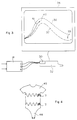
- FIG. 2 illustrates a typical evolution curve over time of the pulmonary volume V of a person at rest during a respiratory cycle (an inspiration followed by an expiration).
- This curve can be directly established from the sum signal S supplied by the plethysmograph of FIG. 1.
- the pulmonary volume gradually increases and reaches a maximum at an instant t 1 at the end of the inspiration phase I.
- time t 1 begins the expiration phase E.
- the lung volume gradually decreases, presents an inflection point and approximates the initial value at an instant t 0 + T where begins again a new phase of inspiration, and so on.
- the expiration phase is generally, as shown, longer than the inspiration phase.
- the curve in Figure 2 considered without amplitude and time scales, represents a form of respiratory cycle.
- a form of respiratory cycle is preserved when the curve undergoes deformations according to linear laws.
- FIG. 3 represents an embodiment of a respiratory rehabilitation device according to the invention.
- the processing circuit 14 of the plethysmograph of Figure 1 connected to the thoracic and abdominal turns (not shown).
- the signals of thoracic section C, of abdominal section A and their sum signal S are supplied to an analog / digital conversion card 30 connected to a microcomputer 32.
- a suitable program uses the signals C, S and A converted by the card 30.
- these three digitized signals can be stored over a given time interval and be displayed on a screen 34 using the microcomputer as an oscilloscope.
- the method according to the invention uses only a signal representative of the pulmonary volume of a patient, this signal possibly being the sum signal S provided by the plethysmograph or calculated by the operating program from the thoracic signals C and abdominal A.
- the program will be able to record information representative of the results of the sessions on a magnetic medium such as a floppy disk, a magnetic card or any other information medium.
- This information includes, for example, the parameters of successive sessions, such as dates, duration of sessions, indications on the success rate for monitoring the template.
- the patient will breathe in a manner which is adapted to him since he will breathe in a form which has been produced by him.
- he will gradually become aware of how to control his ventilatory movements.
- the patient has completed a certain number of sessions, he will be able to control his ventilatory movements in an easy manner and may on his own, or even unconsciously, correct them and tend towards more effective movements.
- This rehabilitation requires a certain attendance of the patient, but this is facilitated by the fact that the patient can perform a large part of this rehabilitation at home.
- the delimitation cycles 36, 37 are determined so that the minimum distance separating them is zero for the start of the reference cycle and gradually increases towards a maximum value at the end of the cycle reference.
- the minimum distance at a given instant is defined as the distance between the delimitation cycles measured according to the normal to the reference cycle at this instant.
- a template made up of the delimitation cycles thus determined is particularly convenient to respect because the patient will have everything instant enough room for maneuver.
- the patient will not be obliged, if necessary, to abruptly end his cycle before the end of the reference cycle (indicated by the arrow), which is beneficial if one seeks to extend the patient's respiratory cycles.
- the points of the delimitation cycles are not determined for each point of the reference cycle. We prefer to sample a few points of the reference cycle, calculate from these points the corresponding points of the delimitation cycles, and determine the smoothing curves of the calculated points to obtain each of the delimitation cycles. This avoids creating delimitation cycles which reproduce possible artifacts of the reference cycle, such as produced by swallowing.
- the coil plethysmograph as described in Figure 1 is not entirely suitable for home use. Indeed, the precision of the measurements can vary from one session to another depending on the positioning of the thoracic 10 and abdominal 11 turns relative to a previous session. Thus, the plethysmograph should be calibrated at each session (setting the gains K1 and K2 of the frequency-voltage converters 15 and 16) so that one can expect comparable results from one session to another.
- FIG. 4 illustrates a solution allowing good repetitivity of the positioning of the plethysmograph turns from one session to another so that the adjustments are made once and for all at the first session.
- the turns are integrated into the fabric of a undershirt 45.
- the fabric of this undershirt is inextensible in the direction of the height and a crotch 46 prevents any vertical displacement of the undershirt. With such an arrangement, it is certain that the turns find practically the same positions from one session to the next.
- Oscillators 13 will be provided with connectors so that they can be detached from the undershirt to wash the latter.
- the crotch 46 will be adjustable for a better adaptation to the patient's anatomy.
- the functions performed by the microcomputer 32 and the conversion card 30 can be performed by a specific microcontroller integrated in an apparatus of dimensions smaller than a microcomputer and that the 'we can connect to a television.
Landscapes
- Health & Medical Sciences (AREA)
- Life Sciences & Earth Sciences (AREA)
- Engineering & Computer Science (AREA)
- Medical Informatics (AREA)
- Physics & Mathematics (AREA)
- Dentistry (AREA)
- Biophysics (AREA)
- Pathology (AREA)
- Physiology (AREA)
- Biomedical Technology (AREA)
- Heart & Thoracic Surgery (AREA)
- Oral & Maxillofacial Surgery (AREA)
- Molecular Biology (AREA)
- Surgery (AREA)
- Animal Behavior & Ethology (AREA)
- General Health & Medical Sciences (AREA)
- Public Health (AREA)
- Veterinary Medicine (AREA)
- Measurement Of The Respiration, Hearing Ability, Form, And Blood Characteristics Of Living Organisms (AREA)
Claims (10)
- Verfahren zur Atmungsumschulung, beinhaltend den Schritt Messen der Atmungsamplitude eines Patienten;
dadurch gekennzeichnet, daß es die folgenden Schritte umfaßt:- Erfassen eines Referenzzyklus, der die Amplitude als Funktion der Zeit eines regulären Atmungszyklus darstellt;- Bestimmen eines oberen (36) und eines unteren (37) Zyklus, die den Referenzzyklus einschließen;- dauerhaftes Anzeigen eines Paares des oberen (36) und unteren (37) Zyklus auf einem Bildschirm (34); und- Anzeigen eines Punktes (40), der sich in einer Richtung als Funktion der Atmungsamplitude und in einer orthogonalen Richtung als Funktion der Zeit bewegt, auf demselben Bildschirm. - Verfahren nach Anspruchs 1, gekennzeichnet durch den Schritt horizontales Abtasten des Bildschirms (34) durch den Punkt (40) und Reinitialisieren einer Abtastung, wenn der Beginn einer Einatmung erfaßt wird, wobei die Geschwindigkeit der Abtastung so ist, daß die horizontale Länge des Bildschirms einer Zeitdauer entspricht, die geringfügig größer als eine Periode des Referenzzyklus ist.
- Verfahren nach Anspruch 1, gekennzeichnet durch den Schritt Bestimmen des oberen (36) und unteren (37) Zyklus in der Weise, daß ihr gegenseitiger Minimalabstand mit zunehmender Zeit fortschreitend zunimmt.
- Verfahren nach Anspruch 3, dadurch gekennzeichnet, daß die Maxima des oberen Zyklus (36), des unteren Zyklus (37) und des Referenzzyklus in Richtung der Amplituden ausgerichtet sind.
- Verfahren nach Anspruch 4, dadurch gekennzeichnet, daß der obere (36) und der untere (37) Zyklus gemäß den folgenden Schritten bestimmt werden:- Wählen einer Gruppe von Referenzpunkten, die Amplituden (yi) des Referenzzyklus für unterschiedliche Zeitpunkte (ti), welche zwischen einem Abtastungsbeginn und einem Abtastungsende verteilt sind, entsprechen;- Bestimmen von Gruppen von oberen und unteren Punkten in bezug auf die Referenzgruppe, wobei jede Amplitude (yMi,ymi) erhalten wird durch Addieren und Subtrahieren eines Gliedes, das aus der Summe einer Konstante und eines Faktors gebildet wird, welcher zu dem Absolutwert der Steigung in dem Referenzpunkt proportional ist, zu bzw. von der Amplitude eines Referenzpunktes; und- Glätten der oberen und der unteren Gruppe.
- Verfahren nach Anspruch 1, dadurch gekennzeichnet, daß die Atmungsamplituden pulmonalen Volumina entsprechen.
- Vorrichtung zur Atmungsumschulung, mit einer Einrichtung zum Messen (10, 11, 13, 14) der Atmungsamplitude eines Patienten;
dadurch gekennzeichnet, daß sie umfaßt:- eine Einrichtung zum Erfassen (30, 32) eines Referenzzyklus, der die Amplitude als Funktion der Zeit eines regulären Atmungszyklus darstellt;- eine Einrichtung (32) zum Berechnen und Speichern eines oberen (36) und unteren (37) Zyklus, die den Referenzzyklus einschließen;- einen Bildschirm (34), auf dem ein Paar des oberen und unteren Zyklus permanent angezeigt wird, und- eine Verarbeitungseinrichtung (32), die gestattet, einen Punkt (40), der sich in einer Richtung als Funktion der Atmungsamplitude und in einer orthogonalen Richtung als Funktion der Zeit bewegt, auf demselben Bildschirm anzuzeigen. - Vorrichtung nach Anspruch 7, dadurch gekennzeichnet, daß sie einen Plethysmograph mit thorakalen (10) und abdominalen (11) induktiven Windungen enthält, die mit einem Mikrocomputer (32) über eine Analog/Digital-Wandlerkarte (30) verbunden sind.
- Vorrichtung nach Anspruch 7, dadurch gekennzeichnet, daß die Atmungsamplituden pulmonalen Volumina entsprechen.
- Verfahren nach Anspruch 7, dadurch gekennzeichnet, daß die induktiven Windungen (10, 11) in ein in Höhenrichtung nicht dehnbares Leibtrikot (45) mit einer Schrittlasche (46) integriert sind.
Applications Claiming Priority (3)
| Application Number | Priority Date | Filing Date | Title |
|---|---|---|---|
| FR9115313 | 1991-11-28 | ||
| FR9115313A FR2684286B1 (fr) | 1991-11-28 | 1991-11-28 | Procede et dispositif de reeducation respiratoire. |
| PCT/FR1992/001092 WO1993010707A1 (fr) | 1991-11-28 | 1992-11-25 | Procede et dispositif de reeducation respiratoire |
Publications (2)
| Publication Number | Publication Date |
|---|---|
| EP0615425A1 EP0615425A1 (de) | 1994-09-21 |
| EP0615425B1 true EP0615425B1 (de) | 1996-08-14 |
Family
ID=9419880
Family Applications (1)
| Application Number | Title | Priority Date | Filing Date |
|---|---|---|---|
| EP93901767A Expired - Lifetime EP0615425B1 (de) | 1991-11-28 | 1992-11-25 | VERFAHREN UND GERäT ZUR ATMUNGSUMSCHULUNG |
Country Status (4)
| Country | Link |
|---|---|
| EP (1) | EP0615425B1 (de) |
| DE (1) | DE69212841T2 (de) |
| FR (1) | FR2684286B1 (de) |
| WO (1) | WO1993010707A1 (de) |
Families Citing this family (3)
| Publication number | Priority date | Publication date | Assignee | Title |
|---|---|---|---|---|
| DE19628356C2 (de) * | 1996-07-13 | 2003-04-17 | Detlef Tolksdorf | Verfahren zur Normalisierung des Atemmusters von mit Atmungsgeräten ausgestatteten Personen, insbesondere von Sport- und Berufstauchern, sowie Einrichtung zur Durchführung des Verfahrens |
| FR2787008B1 (fr) * | 1998-12-09 | 2001-04-13 | R B I | Dispositif de mesure de respiration |
| FR2940038A1 (fr) * | 2008-12-22 | 2010-06-25 | Rbi | Procede et systeme de surveillance de la deglutition |
Family Cites Families (3)
| Publication number | Priority date | Publication date | Assignee | Title |
|---|---|---|---|---|
| US4258718A (en) * | 1979-04-16 | 1981-03-31 | Goldman Michael D | Measuring respiratory air volume |
| US4373534A (en) * | 1981-04-14 | 1983-02-15 | Respitrace Corporation | Method and apparatus for calibrating respiration monitoring system |
| NL8500314A (nl) * | 1984-07-19 | 1986-02-17 | Drs Erna Peltzer | Inrichting voor het bepalen van het ademhalingsgedrag van een patient. |
-
1991
- 1991-11-28 FR FR9115313A patent/FR2684286B1/fr not_active Expired - Fee Related
-
1992
- 1992-11-25 DE DE69212841T patent/DE69212841T2/de not_active Expired - Fee Related
- 1992-11-25 EP EP93901767A patent/EP0615425B1/de not_active Expired - Lifetime
- 1992-11-25 WO PCT/FR1992/001092 patent/WO1993010707A1/fr not_active Ceased
Also Published As
| Publication number | Publication date |
|---|---|
| EP0615425A1 (de) | 1994-09-21 |
| DE69212841T2 (de) | 1997-01-30 |
| FR2684286A1 (fr) | 1993-06-04 |
| DE69212841D1 (de) | 1996-09-19 |
| WO1993010707A1 (fr) | 1993-06-10 |
| FR2684286B1 (fr) | 1994-03-04 |
Similar Documents
| Publication | Publication Date | Title |
|---|---|---|
| CN102973253B (zh) | 一种利用视觉信息监测人体生理指标的方法和系统 | |
| CN103445782B (zh) | 一种用于估算呼吸频率的视频处理技术 | |
| ES2250334T3 (es) | Valoracion no invasiva de la concentracion de lactato en el cuerpo en relacion con el ejercicio corporal. | |
| DE69417995T2 (de) | Oszillometrische Blutdruckmesseinrichtung | |
| US20150072326A1 (en) | System for the acquisition and analysis of muscle activity and operation method thereof | |
| EP0892618B1 (de) | Vorrichtung zur bestimmung der narkose-tiefe | |
| US20170360314A1 (en) | Method, apparatus and computer program for determining a blood pressure value | |
| CA2816608C (fr) | Simulateur destine a l'apprentissage de l'intubation tracheale | |
| US11406304B2 (en) | Systems and methods for physiological sign analysis | |
| JP6940067B2 (ja) | 協調運動評価装置及びプログラム | |
| CN105916436A (zh) | 用于测量动脉信号的设备和方法 | |
| JP7034524B2 (ja) | 血圧推定装置、血圧推定方法及び血圧推定プログラム | |
| WO2015121503A1 (fr) | Procédé et système de surveillance du système nerveux autonome d'un sujet | |
| FR2684286A1 (fr) | Procede et dispositif de reeducation respiratoire. | |
| EP1296594B1 (de) | Verfahren und vorrichtung zum bestimmen von physiologischen herz-atemparametern | |
| EP1804655B1 (de) | System und programm zur verarbeitung einer reihe von herzrhythmussignalen (rr) zur beurteilung von schmerz oder stress eines patienten | |
| CN106504235B (zh) | 基于图像处理的心率监测方法 | |
| FR2590151A1 (fr) | Appareil electronique ambulatoire d'enregistrement et/ou de reeducation de la locomotion | |
| FR2535964A1 (fr) | Appareillage de mesure, de traitement, de stockage et d'affichage de signaux biomedicaux | |
| EP3110322B1 (de) | Verfahren und vorrichtung zur automatischen überprüfung der qualität einer aus einem herzsignal ermittelten rr-intervallserie | |
| FR2948008A1 (fr) | Appareil d'evaluation de la capacite musculaire de la sangle abdominale d'un sujet | |
| JP4547804B2 (ja) | 皮下脂肪厚測定装置 | |
| EP2378962A1 (de) | Verfahren zur messung des lokalen steifheitsindexes der wand einer leitenden arterie und entsprechende ausrüstung | |
| CN106361350A (zh) | 基于图像处理的血氧饱和度监测方法 | |
| FR3024943A1 (fr) | Dispositif et procede de determination d'une frequence respiratoire d'un individu a partir de son rythme cardiaque |
Legal Events
| Date | Code | Title | Description |
|---|---|---|---|
| PUAI | Public reference made under article 153(3) epc to a published international application that has entered the european phase |
Free format text: ORIGINAL CODE: 0009012 |
|
| 17P | Request for examination filed |
Effective date: 19940525 |
|
| AK | Designated contracting states |
Kind code of ref document: A1 Designated state(s): BE DE FR GB IT NL |
|
| 17Q | First examination report despatched |
Effective date: 19950801 |
|
| GRAH | Despatch of communication of intention to grant a patent |
Free format text: ORIGINAL CODE: EPIDOS IGRA |
|
| GRAH | Despatch of communication of intention to grant a patent |
Free format text: ORIGINAL CODE: EPIDOS IGRA |
|
| GRAA | (expected) grant |
Free format text: ORIGINAL CODE: 0009210 |
|
| AK | Designated contracting states |
Kind code of ref document: B1 Designated state(s): BE DE FR GB IT NL |
|
| PG25 | Lapsed in a contracting state [announced via postgrant information from national office to epo] |
Ref country code: NL Free format text: LAPSE BECAUSE OF FAILURE TO SUBMIT A TRANSLATION OF THE DESCRIPTION OR TO PAY THE FEE WITHIN THE PRESCRIBED TIME-LIMIT Effective date: 19960814 |
|
| REF | Corresponds to: |
Ref document number: 69212841 Country of ref document: DE Date of ref document: 19960919 |
|
| GBT | Gb: translation of ep patent filed (gb section 77(6)(a)/1977) |
Effective date: 19960906 |
|
| ITF | It: translation for a ep patent filed | ||
| NLV1 | Nl: lapsed or annulled due to failure to fulfill the requirements of art. 29p and 29m of the patents act | ||
| PLBE | No opposition filed within time limit |
Free format text: ORIGINAL CODE: 0009261 |
|
| STAA | Information on the status of an ep patent application or granted ep patent |
Free format text: STATUS: NO OPPOSITION FILED WITHIN TIME LIMIT |
|
| 26N | No opposition filed | ||
| REG | Reference to a national code |
Ref country code: GB Ref legal event code: IF02 |
|
| PGFP | Annual fee paid to national office [announced via postgrant information from national office to epo] |
Ref country code: BE Payment date: 20021106 Year of fee payment: 11 |
|
| PGFP | Annual fee paid to national office [announced via postgrant information from national office to epo] |
Ref country code: DE Payment date: 20021113 Year of fee payment: 11 |
|
| PGFP | Annual fee paid to national office [announced via postgrant information from national office to epo] |
Ref country code: GB Payment date: 20021122 Year of fee payment: 11 |
|
| PG25 | Lapsed in a contracting state [announced via postgrant information from national office to epo] |
Ref country code: GB Free format text: LAPSE BECAUSE OF NON-PAYMENT OF DUE FEES Effective date: 20031125 |
|
| PG25 | Lapsed in a contracting state [announced via postgrant information from national office to epo] |
Ref country code: BE Free format text: LAPSE BECAUSE OF NON-PAYMENT OF DUE FEES Effective date: 20031130 |
|
| BERE | Be: lapsed |
Owner name: UNIVERSITE JOSEPH *FOURIER Effective date: 20031130 |
|
| PG25 | Lapsed in a contracting state [announced via postgrant information from national office to epo] |
Ref country code: DE Free format text: LAPSE BECAUSE OF NON-PAYMENT OF DUE FEES Effective date: 20040602 |
|
| GBPC | Gb: european patent ceased through non-payment of renewal fee |
Effective date: 20031125 |
|
| PG25 | Lapsed in a contracting state [announced via postgrant information from national office to epo] |
Ref country code: IT Free format text: LAPSE BECAUSE OF NON-PAYMENT OF DUE FEES;WARNING: LAPSES OF ITALIAN PATENTS WITH EFFECTIVE DATE BEFORE 2007 MAY HAVE OCCURRED AT ANY TIME BEFORE 2007. THE CORRECT EFFECTIVE DATE MAY BE DIFFERENT FROM THE ONE RECORDED. Effective date: 20051125 |
|
| PGFP | Annual fee paid to national office [announced via postgrant information from national office to epo] |
Ref country code: FR Payment date: 20111215 Year of fee payment: 20 |