EP0599456A1 - Procédé et appareil pour l'imagerie cinématographique RMN - Google Patents
Procédé et appareil pour l'imagerie cinématographique RMN Download PDFInfo
- Publication number
- EP0599456A1 EP0599456A1 EP93307437A EP93307437A EP0599456A1 EP 0599456 A1 EP0599456 A1 EP 0599456A1 EP 93307437 A EP93307437 A EP 93307437A EP 93307437 A EP93307437 A EP 93307437A EP 0599456 A1 EP0599456 A1 EP 0599456A1
- Authority
- EP
- European Patent Office
- Prior art keywords
- magnetic resonance
- frequency
- phase
- segment
- echoes
- Prior art date
- Legal status (The legal status is an assumption and is not a legal conclusion. Google has not performed a legal analysis and makes no representation as to the accuracy of the status listed.)
- Granted
Links
Images


Classifications
-
- A—HUMAN NECESSITIES
- A61—MEDICAL OR VETERINARY SCIENCE; HYGIENE
- A61B—DIAGNOSIS; SURGERY; IDENTIFICATION
- A61B5/00—Measuring for diagnostic purposes; Identification of persons
- A61B5/02—Detecting, measuring or recording pulse, heart rate, blood pressure or blood flow; Combined pulse/heart-rate/blood pressure determination; Evaluating a cardiovascular condition not otherwise provided for, e.g. using combinations of techniques provided for in this group with electrocardiography or electroauscultation; Heart catheters for measuring blood pressure
- A61B5/026—Measuring blood flow
- A61B5/0263—Measuring blood flow using NMR
-
- A—HUMAN NECESSITIES
- A61—MEDICAL OR VETERINARY SCIENCE; HYGIENE
- A61B—DIAGNOSIS; SURGERY; IDENTIFICATION
- A61B5/00—Measuring for diagnostic purposes; Identification of persons
- A61B5/05—Detecting, measuring or recording for diagnosis by means of electric currents or magnetic fields; Measuring using microwaves or radio waves
- A61B5/055—Detecting, measuring or recording for diagnosis by means of electric currents or magnetic fields; Measuring using microwaves or radio waves involving electronic [EMR] or nuclear [NMR] magnetic resonance, e.g. magnetic resonance imaging
-
- G—PHYSICS
- G01—MEASURING; TESTING
- G01R—MEASURING ELECTRIC VARIABLES; MEASURING MAGNETIC VARIABLES
- G01R33/00—Arrangements or instruments for measuring magnetic variables
- G01R33/20—Arrangements or instruments for measuring magnetic variables involving magnetic resonance
- G01R33/44—Arrangements or instruments for measuring magnetic variables involving magnetic resonance using nuclear magnetic resonance [NMR]
- G01R33/48—NMR imaging systems
- G01R33/54—Signal processing systems, e.g. using pulse sequences ; Generation or control of pulse sequences; Operator console
- G01R33/56—Image enhancement or correction, e.g. subtraction or averaging techniques, e.g. improvement of signal-to-noise ratio and resolution
- G01R33/561—Image enhancement or correction, e.g. subtraction or averaging techniques, e.g. improvement of signal-to-noise ratio and resolution by reduction of the scanning time, i.e. fast acquiring systems, e.g. using echo-planar pulse sequences
-
- G—PHYSICS
- G01—MEASURING; TESTING
- G01R—MEASURING ELECTRIC VARIABLES; MEASURING MAGNETIC VARIABLES
- G01R33/00—Arrangements or instruments for measuring magnetic variables
- G01R33/20—Arrangements or instruments for measuring magnetic variables involving magnetic resonance
- G01R33/44—Arrangements or instruments for measuring magnetic variables involving magnetic resonance using nuclear magnetic resonance [NMR]
- G01R33/48—NMR imaging systems
- G01R33/54—Signal processing systems, e.g. using pulse sequences ; Generation or control of pulse sequences; Operator console
- G01R33/56—Image enhancement or correction, e.g. subtraction or averaging techniques, e.g. improvement of signal-to-noise ratio and resolution
- G01R33/563—Image enhancement or correction, e.g. subtraction or averaging techniques, e.g. improvement of signal-to-noise ratio and resolution of moving material, e.g. flow contrast angiography
-
- G—PHYSICS
- G01—MEASURING; TESTING
- G01R—MEASURING ELECTRIC VARIABLES; MEASURING MAGNETIC VARIABLES
- G01R33/00—Arrangements or instruments for measuring magnetic variables
- G01R33/20—Arrangements or instruments for measuring magnetic variables involving magnetic resonance
- G01R33/44—Arrangements or instruments for measuring magnetic variables involving magnetic resonance using nuclear magnetic resonance [NMR]
- G01R33/48—NMR imaging systems
- G01R33/54—Signal processing systems, e.g. using pulse sequences ; Generation or control of pulse sequences; Operator console
- G01R33/56—Image enhancement or correction, e.g. subtraction or averaging techniques, e.g. improvement of signal-to-noise ratio and resolution
- G01R33/561—Image enhancement or correction, e.g. subtraction or averaging techniques, e.g. improvement of signal-to-noise ratio and resolution by reduction of the scanning time, i.e. fast acquiring systems, e.g. using echo-planar pulse sequences
- G01R33/5615—Echo train techniques involving acquiring plural, differently encoded, echo signals after one RF excitation, e.g. using gradient refocusing in echo planar imaging [EPI], RF refocusing in rapid acquisition with relaxation enhancement [RARE] or using both RF and gradient refocusing in gradient and spin echo imaging [GRASE]
- G01R33/5617—Echo train techniques involving acquiring plural, differently encoded, echo signals after one RF excitation, e.g. using gradient refocusing in echo planar imaging [EPI], RF refocusing in rapid acquisition with relaxation enhancement [RARE] or using both RF and gradient refocusing in gradient and spin echo imaging [GRASE] using RF refocusing, e.g. RARE
-
- G—PHYSICS
- G01—MEASURING; TESTING
- G01R—MEASURING ELECTRIC VARIABLES; MEASURING MAGNETIC VARIABLES
- G01R33/00—Arrangements or instruments for measuring magnetic variables
- G01R33/20—Arrangements or instruments for measuring magnetic variables involving magnetic resonance
- G01R33/44—Arrangements or instruments for measuring magnetic variables involving magnetic resonance using nuclear magnetic resonance [NMR]
- G01R33/48—NMR imaging systems
- G01R33/54—Signal processing systems, e.g. using pulse sequences ; Generation or control of pulse sequences; Operator console
- G01R33/56—Image enhancement or correction, e.g. subtraction or averaging techniques, e.g. improvement of signal-to-noise ratio and resolution
- G01R33/561—Image enhancement or correction, e.g. subtraction or averaging techniques, e.g. improvement of signal-to-noise ratio and resolution by reduction of the scanning time, i.e. fast acquiring systems, e.g. using echo-planar pulse sequences
- G01R33/5619—Image enhancement or correction, e.g. subtraction or averaging techniques, e.g. improvement of signal-to-noise ratio and resolution by reduction of the scanning time, i.e. fast acquiring systems, e.g. using echo-planar pulse sequences by temporal sharing of data, e.g. keyhole, block regional interpolation scheme for k-Space [BRISK]
-
- G—PHYSICS
- G01—MEASURING; TESTING
- G01R—MEASURING ELECTRIC VARIABLES; MEASURING MAGNETIC VARIABLES
- G01R33/00—Arrangements or instruments for measuring magnetic variables
- G01R33/20—Arrangements or instruments for measuring magnetic variables involving magnetic resonance
- G01R33/44—Arrangements or instruments for measuring magnetic variables involving magnetic resonance using nuclear magnetic resonance [NMR]
- G01R33/48—NMR imaging systems
- G01R33/54—Signal processing systems, e.g. using pulse sequences ; Generation or control of pulse sequences; Operator console
- G01R33/56—Image enhancement or correction, e.g. subtraction or averaging techniques, e.g. improvement of signal-to-noise ratio and resolution
- G01R33/563—Image enhancement or correction, e.g. subtraction or averaging techniques, e.g. improvement of signal-to-noise ratio and resolution of moving material, e.g. flow contrast angiography
- G01R33/56308—Characterization of motion or flow; Dynamic imaging
-
- G—PHYSICS
- G01—MEASURING; TESTING
- G01R—MEASURING ELECTRIC VARIABLES; MEASURING MAGNETIC VARIABLES
- G01R33/00—Arrangements or instruments for measuring magnetic variables
- G01R33/20—Arrangements or instruments for measuring magnetic variables involving magnetic resonance
- G01R33/44—Arrangements or instruments for measuring magnetic variables involving magnetic resonance using nuclear magnetic resonance [NMR]
- G01R33/48—NMR imaging systems
- G01R33/54—Signal processing systems, e.g. using pulse sequences ; Generation or control of pulse sequences; Operator console
- G01R33/56—Image enhancement or correction, e.g. subtraction or averaging techniques, e.g. improvement of signal-to-noise ratio and resolution
- G01R33/567—Image enhancement or correction, e.g. subtraction or averaging techniques, e.g. improvement of signal-to-noise ratio and resolution gated by physiological signals, i.e. synchronization of acquired MR data with periodical motion of an object of interest, e.g. monitoring or triggering system for cardiac or respiratory gating
- G01R33/5673—Gating or triggering based on a physiological signal other than an MR signal, e.g. ECG gating or motion monitoring using optical systems for monitoring the motion of a fiducial marker
Definitions
- This invention relates to a cine magnetic resonance imaging method and apparatus. It finds particular application in conjunction with cine imaging and will be described with particular reference thereto. It is to be appreciated, however, that the invention will also find application in conjunction with angiography, circulatory, cardiac quantitative flow, and other examinations in which flowing fluid or moving tissue is imaged.
- Cine images have commonly been acquired using field echoes.
- Field echoes permit a rapid, e.g. 5-10 msec, repetition rate.
- about 64 to 100 sequence repetitions can be made in a typical 600 to 1000 msec. cardiac cycle. If the view from each sequence repetition in a common cardiac cycle is allocated to a successive frame, then the cine sequence produces about 64 to 100 frames per cardiac cycle with a resolution of about 10 msec.
- Optimal cardiac gated cine images are achieved if all the views are collected within a single breath hold. Collecting all the views within a single breath hold eliminates the motion artifacts attributable to respiratory motion.
- a single breath hold is typically 16 - 20 heartbeats. Acquiring only one view per frame image in each cardiac cycle of a single breath hold would limit each frame image to 16-20 views. To increase the number of views per frame, data acquisition could be continued for several breath holds. However, the heart and surrounding tissue move with each respiratory cycle and typically do not return accurately to the same position in subsequent breath holds.
- the invention of the present application has recognized that this blurring is attributable to the acquisition of the central views, the views with the most signal power and image information, at different times in subsequent cardiac cycles.
- the central views in each group alternated between two different time displaced points in the cardiac cycle.
- a cine magnetic resonance imaging method in which k-space is segmented into at least (i) a central, lowest frequency phase encoding segment and (ii) a higher frequency phase encoding segment, the method comprising:
- a cine magnetic resonance imaging method in which k-space is segmented into n segments including a central segment having lowest frequency phase encoded views and a plurality of segments having progressively higher frequency phase encoded views, the method comprising:
- a cine magnetic resonance imaging apparatus comprising: a means for monitoring cardiac cycles of a subject in a magnetic resonance imaging region; a means for generating a series of magnetic resonance echoes in coordination with each monitored cardiac cycle, the magnetic resonance echoes being phase encoded cyclically with low frequency phase encodings and high frequency phase encodings; a means for receiving the magnetic resonance echoes and generating corresponding magnetic resonance signals, the magnetic resonance signals being phase encoded with the corresponding high and low frequency phase encodings; a plurality of data memory means each for storing a set of data that is reconstructible into a frame image representation, each frame image representation corresponding to a selected temporal point in the cardiac cycle; a means for sorting the magnetic resonance signals such that each of the low frequency phase encoded magnetic resonance signals is stored in a corresponding one of the data memory means and each of the high frequency phase encoded magnetic resonance signals is stored in two data memory means which correspond to the two most temporally contiguous frame image representations;
- a cine magnetic resonance imaging apparatus in which k-space is segmented into n segments including a central segment having lowest frequency phase encoded views and a plurality of segments having progressively higher frequency phase encoded views, the apparatus comprising: a means for monitoring cardiac cycles of a subject in a magnetic resonance imaging region; a means for generating a series of magnetic resonance echo sequences in coordination with each of a plurality of monitored cardiac cycles, each sequence generating a magnetic resonance echo with a phase encoding in one of a plurality of regions of k-space, the magnetic resonance echoes with a phase encoding in the lowest frequency region of k-space being generated at regular, spaced temporal intervals, the magnetic resonance echoes with phase encodings in higher frequency regions of k-space being generated between the echoes in the lowest frequency region of k-space; a receiving means for generating a magnetic resonance echo signal corresponding to each of the echoes; a means for reconstructing magnetic resonance signals corresponding
- One advantage of an apparatus and method embodying the present invention is that it enables more images to be collected per cardiac cycle.
- Another advantage of an apparatus and method embodying the present invention is that it reduces the phase encode gradient energy. Fewer of the higher frequency, higher energy phase encoding views are generated.
- Another advantage of an apparatus and method embodying the present invention is that it provides dynamic cine imaging in which a high frequency encoded echo is shared between two images.
- Another advantage of an apparatus and method embodying the present invention is that the resultant cine images have more views per frame with better temporal resolution.
- a main magnetic field control means 10 controls superconducting or resistive magnets 12 such that a substantially uniform main magnetic field is created longitudinally along a z-axis through an examination region 14.
- a magnetic resonance echo generating means applies sequences of RF and magnetic field pulses to cause magnetic resonance echoes, preferably field or gradient echoes, to be generated. More specifically, gradient pulse amplifiers 20 apply current pulses to gradient coils 22 to create gradient magnetic fields along orthogonal x, y and z-axes of the examination region.
- a radio frequency transmitter 24 transmits RF pulses to an RF coil 26 to transmit RF pulses into the examination to excite magnetic resonance and manipulate excited magnetic resonance.
- a radio frequency receiver 28 receives magnetic resonance signals emanating from the examination region that are picked up by the RF coil 26 or by surface coils (not shown).
- a sequence control means 30 controls the gradient pulse amplifiers 20 and the transmitter 24 to generate a series of field or gradient echo imaging sequences. More specifically to the preferred embodiment, a master sequence control 32 is gated by a cardiac monitor 34 to start a series of field echo sequences.
- the master sequence control means 32 implements one of a plurality of preselected sequences retrieved from a sequence memory 36 .
- the master control can retrieve and carry out motion sensitive and reference field echo imaging sequences in which the gradients along one of the axes are time or amplitude shifted relative to the other, such as shown for example, in U.S. Patent No. 4,689,560 of Nayler and Pattany.
- a polarity alternating means 38 causes 0°-180° cycling of the RF.
- the phase alternating means causes the phase of the radio frequency excitation pulse and the receiver 28 , preferably a digital receiver, to be reversed in alternate views.
- each magnetic resonance echo signal received by the receiver 28 corresponds to one view or line of data of one or more frames of the cine image sequence.
- Each image is reconstructed from 2n of views, e.g. 252 differently frequency encoded digital views.
- Each view is phase encoded with one of the 2n phase encodings.
- 2n phase encoded views of data lines are generated.
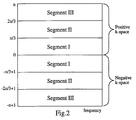
- a central view has zero phase encoding. Views to one side of the central view are phase encoded in progressive steps with more phase encoding in a positive sense. Views to the other side are phase encoded in progressive steps of the size but in a negative sense.
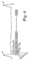
- the field echo resonance sequence excites resonance in a selected region, by application of an RF pulse 40 , e.g. with a 30° tip angle, and a slice select gradient 42 .
- a phase encoding gradient 44 is applied with a selected one of the 2n phase encoding steps concurrently with a read gradient 46 to phase encode the resultant resonance.
- the read gradient is reversed to an opposite polarity read gradient 48 .
- a field echo 50 is read during application of the read gradient 48 .
- An unwind phase encoding gradient 52 is applied to remove the phase encoding prior to application of the RF pulse of the next repetition. This sequence is repeated with a repeat time (TR) of about 10 milliseconds.
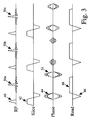
- an R-wave 54 of a patient's cardiac cycle is detected by the cardiac monitor 34 which causes the sequence control 32 to initiate the field echo sequence of FIGURE 3 which causes the series of echoes 50 during the following cardiac cycle at about 10 millisecond intervals. Typically, 61 repetitions of the gradient echo 50 are generated in the interval before a subsequent R-wave 56 .
- a sorting means 60 sorts the views into a series of data matrices or memories 62 .
- Each data memory 62a , 62b , etc. receives the data corresponding to one of a plurality of time displaced frame images.
- each magnetic resonance signal produces one view or horizontal data line which is encoded by frequency along its length.
- Each view is loaded into the data line of the temporally corresponding memory matrix 62a, 62b , etc. in the row corresponding to its phase encoding.
- a plurality of views are collected for each data memory matrix.
- a plurality M of adjacent views from temporally contiguous echoes are each encoded with different phase encodings for one of the frame images.
- M views for each image in each cardiac cycle only 1/Mth as many cardiac cycles are required to collect a full set of views.
- collecting M views of each image per cardiac cycle does not reduce the number of images generated per cardiac cycle to 1/Mth of the number of images that would be generated if only one view per image were collected in each cardiac cycle.
- the reconstruction means 64 reconstructs the data in each of data memories 62 into a frame image representation which is stored in a corresponding one of a plurality of image memories 66 .
- the reconstruction is carried out using an inverse two-dimensional Fourier transform.
- An image selecting means 70 is controlled by an operator control panel 72 or the like to select one or more frame images for display.
- a video formatting means 74 adjusts the format of each selected image into an appropriate format for display on a video monitor 76 .
- the operator can control the image selecting means 70 to select each frame image representation from image memories 66a , 66b , 66c , etc. in order at a selectable rate such that the video display illustrates movement of the heart during the cardiac cycle cinetographically and can freeze the image at any point in the cardiac cycle.
- FIGURES 1, 2, and 4 a specific example is set forth in which three views of each data set are collected in each cardiac cycle.
- the 2n views of k-space are divided into three segments, segment I which is the central most or lowest frequency views, segment II which is the intermediate frequency views, and segment III which is the highest frequency views, i.e. has the largest phase encode gradient applied.
- the sorting means 60 channels these three views to the appropriate data lines of the first memory data set 62a .
- the sorting means sends the view from a third repetition 50c to the second data set memory 62b as well.
- a fourth data line from a fourth repetition 50d is caused by the sequence control means 32 to have one of the phase encodings of the low frequency segment I and a fifth data line from a fifth repetition 50e has one of the phase encodings of k-space segment III . That is, the third excitation and the fifth excitation are phase encoded to produce one view with a phase encoding in group II and the other view with a phase encoding from group III .
- the views from the third, fourth, and fifth repetitions 50c , 50d and 50e are channeled to the second data memory 62b .
- the fifth view from the fifth repetition 50e is also sent by the sorting means 60 to a third data set memory 62c along with the two following views. That is, the sequence control means 32 causes the sixth view from a sixth repetition 50f to be phase encoded with one of the phase encode gradients of k-space segment I and causes a seventh view from a seventh repetition 50g of the field echo sequence to be phase encoded with one of the views from segment II . This process is repeated for all of the views collected during the cardiac cycle, i.e.
- a data set and corresponding frame image are generated at time intervals spaced about 20 milliseconds. More specifically, the central most or lowest frequency views which carry the most information are generated about every 20 milliseconds from every other repetition. The higher frequency views are generated between centrally phase encoded views and shared with the two most temporally adjacent data sets and images. It will be noted that segments II and III need not be maintained distinct. Rather, the views from segments II and III of k-space can be taken in any order or intermixed, provided that each of the data sets acquires a substantially full set of data. Because the first and last views are not shared, if there are 610 milliseconds between cardiac cycles, 61 views can be generated for reconstruction into 30 frame images. It might be noted that if each group of 3 were maintained distinct as in the parent application, only 20 frame images could be generated from the 60 views.
- k-space can be segmented in more than 3 segments, e.g., 4 segments, I , II , III , and IV .
- this data can be grouped such that only the central most data from segment I is unique to each data set and the remainder of the data is shared.
- the data from the central most segment I and from the next lowest frequency segment II can be unique to each data set and image and only the two highest frequency segments III and IV are shared.
- various groupings can be made such as: Analogous results can be obtained for yet larger segmentations of k-space.
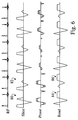
- the technique is used to encode flow. More specifically, with reference to FIGURE 6, the present technique can be used to encode flow in the slice select direction.
- a repetition 80 of the slice select gradient is 0th and 1st order moment zeroed.
- the second and third lobes of the slice select gradient are amplitude scaled to impart velocity encoding.
- the velocity compensated and velocity sensitized slice select gradients repeat alternately.
- the read gradient remains unchanged with each repetition.
- the phase encode gradient is changed as set forth in FIGURES 2-5 above. In this manner, the compensated and sensitized pairs are treated as a single unit in reference to the patterns in FIGURES 4 and 5 above.
- flow can be encoded in the read gradient direction by defining pairs of velocity compensated read gradient repetitions 84 and sensitized read gradient repetitions 86 .
- the slice select gradient remains unchanged with each repetition pair 84 , 86 .
- the phase encode gradient is changed as set forth in FIGURES 2-5 above. In this manner, the compensated and sensitized pairs are treated as a single unit in reference to the patterns in FIGURES 4 and 5 above.
- the motion compensated phase encode gradient repetitions can be velocity sensitized by amplitude scaling of alternate repetitions of the phase encode gradient.
- the motion sensitized and reference images can be collected in two separate sequences in two breath holds. This maximizes the number of frame images per cardiac cycle, but may create mis-registration between the motion sensitized and reference images.
- the motion sensitized and reference images may be collected in alternate cardiac cycles of the same breath hold. This is advantageous because the reference and motion sensitized scans occur at the same point in the cardiac cycle.
- the reference and motion sensitized views are alternated or interleaved within a single cardiac cycle as explained above and in greater detail in copending European patent application no. 93302041.4.
Applications Claiming Priority (2)
| Application Number | Priority Date | Filing Date | Title |
|---|---|---|---|
| US982569 | 1992-11-27 | ||
| US07/982,569 US5348011A (en) | 1991-11-14 | 1992-11-27 | Shared excitation phase encode grouping for improved throughput cardiac gated MRI cine imaging |
Publications (2)
| Publication Number | Publication Date |
|---|---|
| EP0599456A1 true EP0599456A1 (fr) | 1994-06-01 |
| EP0599456B1 EP0599456B1 (fr) | 1998-12-30 |
Family
ID=25529303
Family Applications (1)
| Application Number | Title | Priority Date | Filing Date |
|---|---|---|---|
| EP93307437A Expired - Lifetime EP0599456B1 (fr) | 1992-11-27 | 1993-09-20 | Procédé et appareil pour l'imagerie cinématographique RMN |
Country Status (4)
| Country | Link |
|---|---|
| US (1) | US5348011A (fr) |
| EP (1) | EP0599456B1 (fr) |
| JP (1) | JPH06217960A (fr) |
| DE (1) | DE69322841T2 (fr) |
Cited By (9)
| Publication number | Priority date | Publication date | Assignee | Title |
|---|---|---|---|---|
| EP0798566A1 (fr) * | 1996-03-26 | 1997-10-01 | Wisconsin Alumni Research Foundation | Angiographie tridimensionelle par sonstraction digitale an moyen de résonance magnétique |
| WO1998004928A1 (fr) * | 1996-07-26 | 1998-02-05 | Wisconsin Alumni Research Foundation | Angiographie numerique par resonance magnetique a soustraction, a suppression d'artefact d'image |
| WO1998032026A1 (fr) * | 1997-01-21 | 1998-07-23 | Wisconsin Alumni Research Foundation | Angiographie par resonance magnetique en 3d a contraste renforce et a resolution temporelle synchronisee |
| GB2325744A (en) * | 1997-04-25 | 1998-12-02 | Toshiba America Mri Inc | MRI with complex conjugation and shared echoes |
| US5873825A (en) * | 1997-04-11 | 1999-02-23 | Wisconsin Alumni Research Foundation | Three dimensional digital subtraction magnetic resonance angiography with limited k-space mask |
| DE4435106C2 (de) * | 1994-09-30 | 2000-11-02 | Siemens Ag | MR-Pulssequenz für die zeit- und ortsaufgelöste Darstellung funktioneller Gehirnaktivitäten |
| US6144201A (en) * | 1997-12-26 | 2000-11-07 | Kabushiki Kaisha Toshiba | MR imaging utilizing ECG gating technique |
| US6782286B2 (en) * | 1998-04-20 | 2004-08-24 | Kabushiki Kaisha Toshiba | MRI system and MR imaging method |
| WO2005024450A1 (fr) * | 2003-09-08 | 2005-03-17 | Koninklijke Philips Electronics, N.V. | Imagerie par resonance magnetique a declenchement retroactif d'un mouvement actif ou passif d'une articulation |
Families Citing this family (23)
| Publication number | Priority date | Publication date | Assignee | Title |
|---|---|---|---|---|
| US5435303A (en) * | 1993-08-04 | 1995-07-25 | General Electric Company | MRA image produced by temporal flow data sharing |
| DE4327325C1 (de) * | 1993-08-13 | 1995-01-12 | Siemens Ag | Verfahren zur zeitaufgelösten MR-Bildgebung |
| US5594336A (en) * | 1995-06-02 | 1997-01-14 | Picker International, Inc. | Three point technique using spin and gradient echoes for water/fat separation |
| US5602476A (en) * | 1995-08-17 | 1997-02-11 | Picker International, Inc. | Ultra-fast MR imaging data acquisition scheme using mixed bandwidth data |
| US5697370A (en) * | 1996-01-17 | 1997-12-16 | General Electric Company | Method for increasing temporal resolution of MR fluoroscopy |
| JPH10201736A (ja) * | 1997-01-22 | 1998-08-04 | Hitachi Ltd | 磁気共鳴を用いた検査装置 |
| US6144200A (en) * | 1998-02-20 | 2000-11-07 | General Electric Company | Acquisition of segmented MRI cardiac data using an EPI pulse sequence |
| US6201985B1 (en) * | 1998-08-14 | 2001-03-13 | General Electric Company | Segmented k-space method for three-dimensional MR imaging |
| JP3814157B2 (ja) * | 2001-04-17 | 2006-08-23 | ジーイー・メディカル・システムズ・グローバル・テクノロジー・カンパニー・エルエルシー | Mri装置 |
| US7450982B2 (en) * | 2003-03-14 | 2008-11-11 | Hitachi Medical Corporation | Magnetic resonance imaging system and method |
| US7398116B2 (en) | 2003-08-11 | 2008-07-08 | Veran Medical Technologies, Inc. | Methods, apparatuses, and systems useful in conducting image guided interventions |
| US8150495B2 (en) | 2003-08-11 | 2012-04-03 | Veran Medical Technologies, Inc. | Bodily sealants and methods and apparatus for image-guided delivery of same |
| US20070066881A1 (en) | 2005-09-13 | 2007-03-22 | Edwards Jerome R | Apparatus and method for image guided accuracy verification |
| WO2007033206A2 (fr) | 2005-09-13 | 2007-03-22 | Veran Medical Technologies, Inc. | Dispositif et procede de verification de precision utilisant un guidage par images |
| US7750632B2 (en) * | 2007-03-22 | 2010-07-06 | University Of Pittsburgh - Of The Commonwealth System Of Higher Education | Method for producing a magnetic resonance image of an object having a short T2 relaxation time |
| JP5575491B2 (ja) * | 2010-01-14 | 2014-08-20 | 株式会社東芝 | 医療画像診断装置 |
| CN103118596B (zh) | 2010-05-04 | 2015-11-25 | 开创治疗股份有限公司 | 用于使用伪特征进行腹部表面匹配的系统 |
| US10264947B2 (en) | 2010-08-20 | 2019-04-23 | Veran Medical Technologies, Inc. | Apparatus and method for airway registration and navigation |
| US8738113B2 (en) | 2010-09-30 | 2014-05-27 | University Of Utah Research Foundation | Retrospectively correlated turbo spin echo imaging |
| US9138165B2 (en) | 2012-02-22 | 2015-09-22 | Veran Medical Technologies, Inc. | Systems, methods and devices for forming respiratory-gated point cloud for four dimensional soft tissue navigation |
| US20150305612A1 (en) | 2014-04-23 | 2015-10-29 | Mark Hunter | Apparatuses and methods for registering a real-time image feed from an imaging device to a steerable catheter |
| US20150305650A1 (en) | 2014-04-23 | 2015-10-29 | Mark Hunter | Apparatuses and methods for endobronchial navigation to and confirmation of the location of a target tissue and percutaneous interception of the target tissue |
| JP6996930B2 (ja) * | 2017-10-13 | 2022-01-17 | キヤノンメディカルシステムズ株式会社 | 磁気共鳴イメージング装置 |
Citations (6)
| Publication number | Priority date | Publication date | Assignee | Title |
|---|---|---|---|---|
| US4710717A (en) * | 1986-12-29 | 1987-12-01 | General Electric Company | Method for fast scan cine NMR imaging |
| EP0269147A2 (fr) * | 1986-10-22 | 1988-06-01 | Philips Electronics North America Corporation | Encodeur de signaux cardio-respiratoires groupés dans le temps et méthode pour grouper des signaux cardio-respiratoires |
| DE3918625A1 (de) * | 1988-06-07 | 1989-12-14 | Hitachi Ltd | Verfahren und vorrichtung zur kinematographischen magnetresonanz-(mr)-abbildung |
| EP0471501A2 (fr) * | 1990-08-17 | 1992-02-19 | Wisconsin Alumni Research Foundation | Procédés et appareil de l'angiographie par RMN |
| EP0488496A2 (fr) * | 1990-11-26 | 1992-06-03 | Norbert J. Pelc | Système d'analyse non invasive de mouvement myocardiale utilisant des cartes de répartition de la vitesse myocardiale par imagerie à résonance magnétique en contrasté de phase |
| EP0512798A1 (fr) * | 1991-05-08 | 1992-11-11 | Kabushiki Kaisha Toshiba | Procédé et appareil pour la formation rapide d'image de résonance magnétique |
Family Cites Families (10)
| Publication number | Priority date | Publication date | Assignee | Title |
|---|---|---|---|---|
| US4724386A (en) * | 1985-09-30 | 1988-02-09 | Picker International, Inc. | Centrally ordered phase encoding |
| US4683431A (en) * | 1985-08-16 | 1987-07-28 | Picker International, Inc. | Magnetic resonance imaging of high velocity flows |
| US4689560A (en) * | 1985-08-16 | 1987-08-25 | Picker International, Inc. | Low R.F. dosage magnetic resonance imaging of high velocity flows |
| US4739766A (en) * | 1986-08-18 | 1988-04-26 | Duke University | NMR blood vessel imaging method and apparatus |
| US4767991A (en) * | 1986-12-03 | 1988-08-30 | Advanced Nmr Systems, Inc. | Method of high speed imaging with improved spatial resolution using partial k-space acquisitions |
| US5051903A (en) * | 1989-08-14 | 1991-09-24 | General Electric Company | Method and apparatus for predicting values of a varying periodic phenomenon |
| US4968935A (en) * | 1989-09-11 | 1990-11-06 | Mayo Foundation For Medical Education And Research | Selective rephasing of NMR signals to suppress motion artifacts |
| JPH04246327A (ja) * | 1991-01-31 | 1992-09-02 | Shimadzu Corp | ダイナミックmrイメージング法 |
| US5233302A (en) * | 1991-06-21 | 1993-08-03 | General Electric Company | Masking motion ghost artifacts in NMR images |
| US5273040A (en) * | 1991-11-14 | 1993-12-28 | Picker International, Inc. | Measurement of vetricle volumes with cardiac MRI |
-
1992
- 1992-11-27 US US07/982,569 patent/US5348011A/en not_active Expired - Fee Related
-
1993
- 1993-09-20 EP EP93307437A patent/EP0599456B1/fr not_active Expired - Lifetime
- 1993-09-20 DE DE69322841T patent/DE69322841T2/de not_active Expired - Fee Related
- 1993-10-22 JP JP5286200A patent/JPH06217960A/ja active Pending
Patent Citations (6)
| Publication number | Priority date | Publication date | Assignee | Title |
|---|---|---|---|---|
| EP0269147A2 (fr) * | 1986-10-22 | 1988-06-01 | Philips Electronics North America Corporation | Encodeur de signaux cardio-respiratoires groupés dans le temps et méthode pour grouper des signaux cardio-respiratoires |
| US4710717A (en) * | 1986-12-29 | 1987-12-01 | General Electric Company | Method for fast scan cine NMR imaging |
| DE3918625A1 (de) * | 1988-06-07 | 1989-12-14 | Hitachi Ltd | Verfahren und vorrichtung zur kinematographischen magnetresonanz-(mr)-abbildung |
| EP0471501A2 (fr) * | 1990-08-17 | 1992-02-19 | Wisconsin Alumni Research Foundation | Procédés et appareil de l'angiographie par RMN |
| EP0488496A2 (fr) * | 1990-11-26 | 1992-06-03 | Norbert J. Pelc | Système d'analyse non invasive de mouvement myocardiale utilisant des cartes de répartition de la vitesse myocardiale par imagerie à résonance magnétique en contrasté de phase |
| EP0512798A1 (fr) * | 1991-05-08 | 1992-11-11 | Kabushiki Kaisha Toshiba | Procédé et appareil pour la formation rapide d'image de résonance magnétique |
Non-Patent Citations (3)
| Title |
|---|
| D.J. ATKINSON ET AL.: "CINEANGIOGRAPHY OF THE HEART IN A SINGLE BREATH HOLD WITH A SEGMENTED TURBOFLASH SEQUENCE", RADIOLOGY, vol. 178, no. 2, 1 February 1991 (1991-02-01), NEW YORK, US, pages 357 - 360, XP009020253 * |
| S.J. WANG ET AL.: "FAST ANGIOGRAPHY USING SELECTIVE INVERSION RECOVERY", MAGNETIC RESONANCE IN MEDICINE., vol. 23, no. 1, 1 January 1992 (1992-01-01), DULUTH,MN US, pages 109 - 121 * |
| Z.H. CHO ET AL.: "MR FOURIER TRANSFORM ARTERIOGRAPHY USING SPECTRAL DECOMPOSITION", MAGNETIC RESONANCE IN MEDICINE., vol. 16, no. 2, 1 November 1990 (1990-11-01), DULUTH,MN US, pages 226 - 237, XP000175904 * |
Cited By (13)
| Publication number | Priority date | Publication date | Assignee | Title |
|---|---|---|---|---|
| DE4435106C2 (de) * | 1994-09-30 | 2000-11-02 | Siemens Ag | MR-Pulssequenz für die zeit- und ortsaufgelöste Darstellung funktioneller Gehirnaktivitäten |
| US5713358A (en) * | 1996-03-26 | 1998-02-03 | Wisconsin Alumni Research Foundation | Method for producing a time-resolved series of 3D magnetic resonance angiograms during the first passage of contrast agent |
| EP0798566A1 (fr) * | 1996-03-26 | 1997-10-01 | Wisconsin Alumni Research Foundation | Angiographie tridimensionelle par sonstraction digitale an moyen de résonance magnétique |
| US5881728A (en) * | 1996-07-26 | 1999-03-16 | Wisconsin Alumni Research Foundation | Digital subtraction magnetic resonance angiography with image artifact suppression |
| WO1998004928A1 (fr) * | 1996-07-26 | 1998-02-05 | Wisconsin Alumni Research Foundation | Angiographie numerique par resonance magnetique a soustraction, a suppression d'artefact d'image |
| WO1998032026A1 (fr) * | 1997-01-21 | 1998-07-23 | Wisconsin Alumni Research Foundation | Angiographie par resonance magnetique en 3d a contraste renforce et a resolution temporelle synchronisee |
| US5830143A (en) * | 1997-01-21 | 1998-11-03 | Wisconsin Alumnin Research Foundation | Gated time-resolved contrast-enhanced 3D MR angiography |
| US5873825A (en) * | 1997-04-11 | 1999-02-23 | Wisconsin Alumni Research Foundation | Three dimensional digital subtraction magnetic resonance angiography with limited k-space mask |
| GB2325744A (en) * | 1997-04-25 | 1998-12-02 | Toshiba America Mri Inc | MRI with complex conjugation and shared echoes |
| US6025714A (en) * | 1997-04-25 | 2000-02-15 | Toshiba America Mri, Inc. | Magnetic resonance imaging (MRI) using fast spin echo (FSE) imaging process |
| US6144201A (en) * | 1997-12-26 | 2000-11-07 | Kabushiki Kaisha Toshiba | MR imaging utilizing ECG gating technique |
| US6782286B2 (en) * | 1998-04-20 | 2004-08-24 | Kabushiki Kaisha Toshiba | MRI system and MR imaging method |
| WO2005024450A1 (fr) * | 2003-09-08 | 2005-03-17 | Koninklijke Philips Electronics, N.V. | Imagerie par resonance magnetique a declenchement retroactif d'un mouvement actif ou passif d'une articulation |
Also Published As
| Publication number | Publication date |
|---|---|
| US5348011A (en) | 1994-09-20 |
| JPH06217960A (ja) | 1994-08-09 |
| DE69322841T2 (de) | 1999-05-20 |
| DE69322841D1 (de) | 1999-02-11 |
| EP0599456B1 (fr) | 1998-12-30 |
Similar Documents
| Publication | Publication Date | Title |
|---|---|---|
| EP0599456B1 (fr) | Procédé et appareil pour l'imagerie cinématographique RMN | |
| EP0571071B1 (fr) | Imagerie cinématrographique RMN | |
| US5447155A (en) | High temporal resolution black blood cine imaging | |
| US6683454B2 (en) | Shifting of artifacts by reordering of k-space | |
| US5377680A (en) | MRI cardiac image produced by temporal data sharing | |
| US5423315A (en) | Magnetic resonance imaging system with thin cylindrical uniform field volume and moving subjects | |
| EP0117134B1 (fr) | Dispositif et procédé de projection d'image de vaisseau par résonance magnétique nucléaire | |
| EP0412695B1 (fr) | Imagerie par résonance magnétique | |
| DE19630758B4 (de) | Schnelle Herz-gesteuerte kernmagnetische Resonanz-Erfassung mit verbessertem T1-Kontrast | |
| US5435303A (en) | MRA image produced by temporal flow data sharing | |
| US8588889B2 (en) | Method and apparatus for breath-held MR data acquisition using interleaved acquisition | |
| US5912557A (en) | Centric phase encoding order for 3D NMR data acquisition | |
| JP4152630B2 (ja) | 可変サンプリングを用いた高速/息止め3dmrデータ獲得方法と装置 | |
| CN1575748A (zh) | 用于血管成像的快速多切片黑血双重反转恢复技术 | |
| EP0981058B1 (fr) | Procédé pour l'imagerie tridimensionelle par résonance magnétique à espace k segmenté | |
| CN1213694C (zh) | 用epi脉冲序列采集分段的mri心脏数据的系统和方法 | |
| US6515477B1 (en) | Magnetic resonance random trajectory collection method and apparatus | |
| US5528144A (en) | Interleaved slab inversion for enhanced throughput in fluid attenuated inversion recovery imaging | |
| JPH1033498A (ja) | 磁気共鳴映像法を使用するバッチ式の複数容積血管造像法 | |
| US6434413B1 (en) | Shifting sampling window and interleaves sparse K-space data acquisition for improved temporal resolution | |
| JPS63200745A (ja) | 磁気共鳴イメ−ジング装置 | |
| WO2002042787A2 (fr) | Technique fluoroscopique d'imagerie echo-planar a prises multiples | |
| DE19611007A1 (de) | Mangetresonanzabbildungs-Mittelpunkts-Artefakt-Beseitigung unter Verwendung einer Echtzeit-Empfänger-Phasensteuerung | |
| JPH05207989A (ja) | Mrイメージング法 | |
| JP2859264B2 (ja) | 磁気共鳴イメージング装置 |
Legal Events
| Date | Code | Title | Description |
|---|---|---|---|
| PUAI | Public reference made under article 153(3) epc to a published international application that has entered the european phase |
Free format text: ORIGINAL CODE: 0009012 |
|
| AK | Designated contracting states |
Kind code of ref document: A1 Designated state(s): DE FR NL |
|
| 17P | Request for examination filed |
Effective date: 19941122 |
|
| GRAG | Despatch of communication of intention to grant |
Free format text: ORIGINAL CODE: EPIDOS AGRA |
|
| 17Q | First examination report despatched |
Effective date: 19971105 |
|
| GRAG | Despatch of communication of intention to grant |
Free format text: ORIGINAL CODE: EPIDOS AGRA |
|
| GRAH | Despatch of communication of intention to grant a patent |
Free format text: ORIGINAL CODE: EPIDOS IGRA |
|
| GRAH | Despatch of communication of intention to grant a patent |
Free format text: ORIGINAL CODE: EPIDOS IGRA |
|
| GRAA | (expected) grant |
Free format text: ORIGINAL CODE: 0009210 |
|
| AK | Designated contracting states |
Kind code of ref document: B1 Designated state(s): DE FR NL |
|
| ET | Fr: translation filed | ||
| REF | Corresponds to: |
Ref document number: 69322841 Country of ref document: DE Date of ref document: 19990211 |
|
| PLBE | No opposition filed within time limit |
Free format text: ORIGINAL CODE: 0009261 |
|
| STAA | Information on the status of an ep patent application or granted ep patent |
Free format text: STATUS: NO OPPOSITION FILED WITHIN TIME LIMIT |
|
| 26N | No opposition filed | ||
| PGFP | Annual fee paid to national office [announced via postgrant information from national office to epo] |
Ref country code: NL Payment date: 20020618 Year of fee payment: 10 |
|
| PGFP | Annual fee paid to national office [announced via postgrant information from national office to epo] |
Ref country code: FR Payment date: 20030926 Year of fee payment: 11 |
|
| PGFP | Annual fee paid to national office [announced via postgrant information from national office to epo] |
Ref country code: DE Payment date: 20031126 Year of fee payment: 11 |
|
| REG | Reference to a national code |
Ref country code: FR Ref legal event code: CD |
|
| PG25 | Lapsed in a contracting state [announced via postgrant information from national office to epo] |
Ref country code: NL Free format text: LAPSE BECAUSE OF NON-PAYMENT OF DUE FEES Effective date: 20040401 |
|
| NLV4 | Nl: lapsed or anulled due to non-payment of the annual fee |
Effective date: 20040401 |
|
| REG | Reference to a national code |
Ref country code: FR Ref legal event code: TP |
|
| PG25 | Lapsed in a contracting state [announced via postgrant information from national office to epo] |
Ref country code: DE Free format text: LAPSE BECAUSE OF NON-PAYMENT OF DUE FEES Effective date: 20050401 |
|
| PG25 | Lapsed in a contracting state [announced via postgrant information from national office to epo] |
Ref country code: FR Free format text: LAPSE BECAUSE OF NON-PAYMENT OF DUE FEES Effective date: 20050531 |
|
| REG | Reference to a national code |
Ref country code: FR Ref legal event code: ST |