EP0535239A1 - Kollagenbestimmungsverfahren und testsatz - Google Patents
Kollagenbestimmungsverfahren und testsatz Download PDFInfo
- Publication number
- EP0535239A1 EP0535239A1 EP92906928A EP92906928A EP0535239A1 EP 0535239 A1 EP0535239 A1 EP 0535239A1 EP 92906928 A EP92906928 A EP 92906928A EP 92906928 A EP92906928 A EP 92906928A EP 0535239 A1 EP0535239 A1 EP 0535239A1
- Authority
- EP
- European Patent Office
- Prior art keywords
- collagen
- sample
- added
- assessed
- concentration
- Prior art date
- Legal status (The legal status is an assumption and is not a legal conclusion. Google has not performed a legal analysis and makes no representation as to the accuracy of the status listed.)
- Withdrawn
Links
Images






Classifications
-
- G—PHYSICS
- G01—MEASURING; TESTING
- G01N—INVESTIGATING OR ANALYSING MATERIALS BY DETERMINING THEIR CHEMICAL OR PHYSICAL PROPERTIES
- G01N33/00—Investigating or analysing materials by specific methods not covered by groups G01N1/00 - G01N31/00
- G01N33/48—Biological material, e.g. blood, urine; Haemocytometers
- G01N33/50—Chemical analysis of biological material, e.g. blood, urine; Testing involving biospecific ligand binding methods; Immunological testing
- G01N33/68—Chemical analysis of biological material, e.g. blood, urine; Testing involving biospecific ligand binding methods; Immunological testing involving proteins, peptides or amino acids
- G01N33/6881—Chemical analysis of biological material, e.g. blood, urine; Testing involving biospecific ligand binding methods; Immunological testing involving proteins, peptides or amino acids from skin
-
- G—PHYSICS
- G01—MEASURING; TESTING
- G01N—INVESTIGATING OR ANALYSING MATERIALS BY DETERMINING THEIR CHEMICAL OR PHYSICAL PROPERTIES
- G01N33/00—Investigating or analysing materials by specific methods not covered by groups G01N1/00 - G01N31/00
- G01N33/48—Biological material, e.g. blood, urine; Haemocytometers
- G01N33/50—Chemical analysis of biological material, e.g. blood, urine; Testing involving biospecific ligand binding methods; Immunological testing
- G01N33/68—Chemical analysis of biological material, e.g. blood, urine; Testing involving biospecific ligand binding methods; Immunological testing involving proteins, peptides or amino acids
- G01N33/6887—Chemical analysis of biological material, e.g. blood, urine; Testing involving biospecific ligand binding methods; Immunological testing involving proteins, peptides or amino acids from muscle, cartilage or connective tissue
Definitions
- the present invention relates to a method of measuring collagen and measuring tool kit and more particularly, to a method of measuring collagen making use of an immunoreaction.
- inventors of the present invention made strenuous efforts, found out that the reactivity between collagen included in a sample to be assessed and monoclonal antibodies becomes higher by adding chaotropic ion, and have completed the present invention.
- the method of measuring collagen according to claim 1 in this application is characterized in that a sample to be assessed is treated with chaotropic ions in an immunoassay using a monoclonal antibody against collagen.
- the method according to claim 2 is characterized in that chaotropic ions are added so that the concentration is 0.01 M or more.
- the method according to claim 3 is characterized in that a sample to be assessed is treated with heparins.
- the method according to claim 4 is characterized in that a sample to be assessed is treated with a chelating agent.
- the method according to claim 5 is characterized in that a sample to be assessed is treated with gelatins.
- the method according to claim 6 is characterized in that a sample to be assessed is treated with albumins.
- the method according to claim 7 is characterized in that chaotropic ions are added so that the concentration will be in a range from 0.1 to 1.6 M during an immunoreaction of the serum sample to be assessed.
- the method according to claim 8 is characterized in that chaotropic ions are added so that the concentration will be in a range from 0.2 to 0.6 M when a sample to be assessed is pretreated.
- the method according to claim 9 is characterized in that heparins are added so that the concentration will be in a range from 250 to 15000 U/ml and/or in a range from 5 to 60 mg/ml during an immunoreaction of a sample to be assessed.
- the method according to claim 10 is characterized in that heparins are added so that the concentration will be in a range from 250 to 5000 U/mil when a sample to be assessed is pretreated.
- the method according to claim 11 is characterized in that a sample to be assessed is treated with a chelating agent including bivalent cations at the concentration of 1 to 10 mM.
- the method according to claim 12 is characterized in that a sample to be assessed is treated with gelatins in an immunoassay using a monoclonal antibody against collagen.
- the method according to claim 13 is characterized in that a sample to be assessed is treated with heparins together with gelatins in the immunoassay as described in claim 12.
- the collagen measuring tool kit according to claim 14 is characterized in that said measuring tool kit includes; an antibody coated solid phase in which an antibody against collagen has been coated, a sample treating agent which includes an enzyme-labelled monoclonal antibody against collagen and chaotropic ions, and reacts to a sample to be assessed and to the aforesaid antibody coated solid phase, and an enzyme-substrate solution which includes a substrate of an enzyme in the aforesaid enzyme-labelled monoclonal antibody.
- the collagen measuring tool kit according to claim 15 is characterized in that said collagen measuring tool kit includes; a reaction phase wherein an enzyme-labelled monoclonal antibody against collagen, chaotropic ion and a carrier on which a monoclonal antibody against collagen has been coated coexists, and an enzyme-substrate solution including a substrate of an enzyme in the aforesaid enzyme-labelled monoclonal antibody.
- the chaotropic ion used in the assessing method according to the present invention is a univalent anion having a protein denaturing effect represented by such ions as thiocyanate ion, iodine ion, perchlorate ion, bromine ion, nitrate ion, chlorine ion, and salicylate ion; a group of substances having a large ionic radius.
- the treatment with chaotropic ions means to add a chaotropic salt such as sodium thiocyanate, sodium perchlorate, potassium iodide, or potassium bromide during an immunoreaction between a sample to be assessed and an antibody or when a sample to be assessed is pretreated, and this operation ensures an excellent result.
- a chaotropic salt such as sodium thiocyanate, sodium perchlorate, potassium iodide, or potassium bromide
- chaotropic ions should preferably be added so that the concentration is 0.01 M or more. If the concentration is less than 0.01 M, the effect to raise the sensitivity in assessment is low.
- the chaotropic ions should preferably be added so that the concentration is in a range from 0.1 to 1.6 M. If a sample to be assessed is serum, obstruction to the assessment by a coexisting substance is remarkable, but the sensitivity to collagen can largely be improved by raising the concentration of chaotropic ions to a relatively higher level.
- a serum sample to be assessed is pretreated with chaotropic ions
- concentration is less than 0.2 M or more than 0.6 M, the effect to raise the sensitivity in measurement is low.
- the heparins used according to the present invention is proteoglycan having a sulphonyl such as heparin sodium, chondroitin sulfate A or C sodium salt.
- a sulphonyl such as heparin sodium, chondroitin sulfate A or C sodium salt.
- heparins when heparins are added during an immunoreaction, sometimes an appropriate effect can not be obtained if the concentration of heparins in a sample to be assessed is less than 250 U/ml(or less than 5 mg/ml in case of chondroitin sulfate A or C sodium salt), and also that, if the concentration exceeds 15000 U/ml (or 60 mg/ml in case of chondroitin sulfurate A or C sodium salt), the effect of adding heparins can not be improved any more.
- heparins when heparins are added for pretreating a sample to be assessed, if the concentration of heparins in the sample to be assessed is less than 250 U/ml, sometimes an appropriate effect can not be obtained, and also that, if the concentration exceeds 5000 U/ml, often the effect of heparins can not be improved.
- the chelating agent including bivalent cations in an assessing method according to the present invention includes a group of substances having the effect to chelate bivalent cations represented by various types of ethylenediaminetetraacetic acid (such as EDTA) and ethylene glycol-0,0'-bis (2-aminoethyl)-N,N,N',N'-tetraacetate (EGTA).
- EDTA ethylenediaminetetraacetic acid
- EGTA ethylene glycol-0,0'-bis (2-aminoethyl)-N,N,N',N'-tetraacetate
- concentration of a chelating agent is less than 1 mM, sometimes an adequate effect may not be obtained, and also that, if the concentration exceeds 10 mM, improvement of the effect of adding the chelating agent can not be observed.
- gelatins used in the assessing method according to the present invention include not only high molecular gelatins, but also hydrolysates having a low molecular weight (e.g., PA10, 100, 200, and PE 20, 50: all available from Nippi).
- the albumins used according to the present invention are albumins from animals such as bovine serum albumin or human serum albumin.
- the albumins it is possible for the albumins to coexist with the aforesaid gelatins by adjusting the concentration, but it is more preferable to add albumins in place of gelatins, and a good result can be obtained by adding albumins so that the concentration is in a range from 0.2 to 15 % during an immunoreaction.
- IgG As a monoclonal antibody used in the assessing method according to the present invention, either IgG, F(ab')2 or Fab' may be used as it is.
- the collagen which is an object for measurement according to the present invention, should preferably be human type IV collagen.
- the human type IV collagen is human basement membrane-related collagen, and includes a collagen complex from endothelial basement membrane.
- an measuring method includes enzyme immunoassay (EIA), radio immunoassay (RIA) and fluorescence immunoassay (FIA), and the present invention is applicable to a method in which the immunoreaction is carried out in one step or two step.
- EIA enzyme immunoassay
- RIA radio immunoassay
- FIA fluorescence immunoassay
- the function provided by the present invention is not always clear, and although the sensitivity of purified collagen against various types of monoclonal antibodies is high, the present invention provides an extremely high effectiveness when the reactivity with a sample obtained from organism to be assessed is low.
- the cases where the reactivity between collagen and an antibody drops include (1) a case where a binding site of the collagen with the antibody is masked, and (2) a case where the reactivity of collagen with an antibody has been suppressed.
- reaction temperature during an immunoreaction or when a sample to be assessed is pretreated can be dropped to a room temperature.
- EIA. sandwich method was used as an immunoassay.
- the 3-step sandwich method is carried out as described below.
- an antibody 12 is combined to a well wall 10, and collagen 14 in a sample to be assessed is combined to this coated antibody 12.
- avidin-POD 18 is combined to the biotinylated antibody 16 (FIG. 1 (C)) to promote the chemical reaction with an enzyme active site of the avidin-POD 18, and a quantity of collagen is measured by checking the chemical reaction rate (FIG.1 (D)).
- preparation of pepsinized human type IV collagen was carried out as described below.
- the supernatant was collected by centrifugation (10,000 ⁇ g, for 1 hour), and then Tris and sodium hydroxide were added to adjust the pH to 8.5 to inactivate pepsin, and the solution was left for 2 days. After neutralization, sodium chloride was added so that the concentration would be 4 M, and furthermore glacial acetic acid was added to adjust the pH to 3, and the solution was left overnight. After centrifugation (10,000 ⁇ g, for 1 hour), the supernatant was collected, 0.5 M of acetic acid was added to the precipitate, and the solution was stirred for 5 hours. After centrifugation (10,000 ⁇ g, for 1 hour), the precipitate was collected, sodium chloride was added so that the concentration would be 1.2 M and the solution was left overnight.
- 0.1 ml of phosphate buffer solution containing 100 ⁇ g of human type IV collagen thus obtained was prepared, and the solution was injected into a BALB/C mouse by means of intraperitoneal injection.
- the second immunization was carried out according to the same procedure.
- 0.1 ml of BACTO-Adjuvant produced by DIFCO
- additional immunization was carried out by injecting the aforesaid human type IV collagen into the mouse.
- a spleen was taken out from this mouse under germfree conditions, cut into pieces by scissors, which were passed through a mesh, and thus a suspension of lymphocytes was obtained.
- cells were suspended in a buffer for hemolysis, and then centrifuged for 5 minutes at 1500 rpm, and washed by RPMI-1640 culture medium 2 times.
- mouse myeloma cells (SP 2/0) as confluent cells was cultivated in a RPMI-1640 culture medium containing 10% fetal bovine cell (FCS) for two days before confluence under the conditions of 10% CO2 and 37°C.
- a culture medium containing 100 ⁇ 106 of spleen cell and that containing 100 ⁇ 106 of myeloma cell were mixed, the mixture was centrifuged for 30 minutes at 1500 rpm, the supernatant was thrown away, 1 ml of 50% polyethylene glycol 4000 (produced by Merc Inc.) was dripped drop by drop to unbind the cells.
- 1 ml of the RPMI-1640 culture medium containing FCS was slowly dripped and mixed the cells, and the same operation was repeated for 1 minute.
- 7 ml of RPHI-1640 culture medium containing FCS was slowly added for 3 minutes, rotating the centrifugal tube.
- the culture medium was added so that 200 ⁇ 106 of cell would be contained in 50 ml of the culture medium, and this cell suspension was poured by 0.1 ml into each well on a 96 well micro plate respectively, and cultured therein under the conditions of 10% CO2 and 37°C (and 100% relative humidity).
- phagocyte from mouse had been put in each well on the micro plate at the rate of 104 well, also that HAT (hypoxanthin, aminobuthelin, thymidine) had been added to the RPMI-1640 culture medium containing bovine serum before the medium was poured into each well on the micro plate.
- FITC-labelled (fluorescent isothiocyanate)-labelled anti-mouse IgG ( F(ab)2 fragment) solution was added and left therein for 30 minutes, and then the secondary antibody solution was removed and the cells were fully washed.
- those emitting fluorescence were selected, 3 types of hybridoma corresponding to the supernatant were obtained, and these hybridomas were cloned according to the limiting dilution method.
- the samples were diluted with a culture medium so that the number of hybridomas in a well would be 0.3, 1.0 and 3 respectively.
- dilution was carried out very accurately by adding 1 million of thymic cell to each well respectively.
- the micro plate was kept under the conditions of 37 °C, 10% CO2, and 100 % relative humidity.
- the wells were checked with a magnifying glass on the 5th day, wells having 1 colony therein were checked, the cells were cultured for 2 weeks, and then the limiting dilution was repeated according to the previous procedure. This operation was repeated 3 times, and finally one type of colony was selected, and the colony was removed to ordinal culture dishes and each colony was cultured with 5 ml of a culture medium respectively until the number of cells increased up to 5 ⁇ 106/ml.
- the culture medium for the colony was removed to a centrifugal tube and centrifuged for 5 minutes at 1500 rpm to separate the sample to supernatant and cells, and 0.5 ml of RPMI-1640 culture solution was slowly added anew to the cells to suspend them. Then, RPMI-1640 culture medium containing 4.5 ml of FCS was added to the suspension of cells, and subculture was carried out. Namely, 1 ⁇ 106 of cells were suspended in an RPMI culture medium containing 5 ml of bovine serum, and the cells were cultured under the conditions of 5% CO2, 37°C and 100% relative humidity. The number of cells increased to 5 ⁇ 106 in 60 hours, and culture of one generation was over with this operation.
- the cells were separated from the culture medium by a centrifugal separator, and the cells were diluted by adding a new culture medium. Then, 4 ml of culture medium was added to 1 ml of the cell culture medium in which the cells had been diluted to the level of 1 ⁇ 106/ml. The subculture was carried out as described above. 50 ml of culture medium in which the cells had been cultured by 10 generations was centrifuged, and 5 ⁇ 107 of cells were obtained, and the cells were suspended in a culture medium 51.
- the suspension was divided and poured into 10 rotary culture bottles and cultured therein for 5 days under the conditions of 5 % CO2, 37°C, 100 % relative humidity and rotational speed of 1 rpm.
- the cells were removed from the culture media in which the number of cells had increased to a level of 5 ⁇ 106/ml, the culture media were sucked and filtered, ammonium sulfate was added to the filtrate so that the concentration would be 45 %, and precipitate was obtained.
- the precipitate was furthermore purified with protein A.
- 73 mg of monoclonal antibody against the human type IV collagen was obtained as powder from totally 151 culture media. It was found out by means of SDS- polyacrylamide gel electrophoresis that this monoclonal antibody against human type IV collagen made a single band, and the monoclonal antibody was named JK 199.
- JK 199 and JK 132 specifically recognized the type IV collagen which is a collagen fraction from a human basement membrane, but did not recognize other types of collagen fraction; namely human type I collagen fraction, human type III collagen fraction, bovine type IV collagen fraction, and also did not recognize human fibronectin, human laminin, nor mouse laminin.
- biotinylated monoclonal antibody was prepared as a labelled monoclonal antibody.
- the monoclonal antibody fraction obtained according to the monoclonal antibody preparing method (b) described above was added to 0.1 M sodium bicarbonate so that the concentration would be 1 mg/ml.
- a solution prepared by dissolving N-hydroxysucinimidobiotin in dimethylsuloxide so that the concentration would be 1 mg/ml was added by 0.06 ml, and the mixture was left, being stirred, for 4 hours under the room temperature so that the reaction would proceed. Then, the solution was fully dialyzed against phosphate buffered saline (pH: 7.4), and stored in cooled or frozen state.
- biotinylated monoclonal antibody was enzyme-labelled by the peroxidase.
- ABTS 2,2'-azino-di[3-ethyl-benzthiazoline] sulfonate
- the monoclonal antibody (JK 199) against human type IV collagen obtained according to the monoclonal antibody preparing method (b) described above was dissolved in a phosphate buffered saline (pH: 7.4), and the concentration was adjusted to 14.5 ⁇ g/ml.
- a sample diluted by a blocking buffer solution is added by 100 ⁇ l to each well on a microplate to which antibody has been coated respectively. After incubation (immunoreaction) for 2 hours at 37°C, the samples are washed with phosphate buffered sline (pH: 7.4, washing buffer solution) containing 0.05 % Tween 20 (Refer to FIG.1 (A)).
- washing buffer solution av-POD solution
- avidin-POD produced by Vector Laboratories
- a peroxidase substrate system (produced by Kirkegaard & Perry Laboratories) containing 2,2'-azino-di [ 3-ethyl-benzothiazoline] as the substrate is added by 100 ⁇ l to each well, and after incubation (coloring reaction) for 30 minutes at the room temperature, absorbance at 405 nm is observed by a microplate reader, and a difference between absorbance at the samples and that observed in blind test is checked (FIG.1 (D)).
- a quantity of type IV collagen corresponding to an absorbance of each sample was obtained from an calibration curve prepared by using a standard sample (See Table 2 for the measurement conditions).
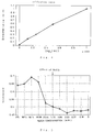
- FIG.2 An calibration curve for human type IV collagen is shown in FIG.2. As clearly shown in this figure, the quantitative sensitivity was 0.391 ng/0.1 ml, while a range of determination was from 0.391 to 125 ng/0.1 ml.
- Table 2 Samples Standard solution for calibration curve Heat-treated sample 100 ⁇ l 100 ⁇ l Antibody coated microplate 2 wells 2 wells Primary immunoreaction Statically left for 2 hours at 37°C after mixing Washing Washed with 350 ⁇ l of washing buffer solution 3 times Biotinylated antibody solution 100 ⁇ l 100 ⁇ l Secondary immunoreaction Statically left for 2 hours at 37°C after mixing Washing Washed with 350 ⁇ l of washing buffer solution 3 times av-POD solution 100 ⁇ l 100 ⁇ l Washing Washed with 350 ⁇ l of washing buffer solution 3 times Substrate solution 100 ⁇ l 100 ⁇ l Coloring reaction Statically left for 30 minutes at the room temperature Colorimetry Measurement of absorbance at 405 nm
- a monoclonal antibody (JK 132; 10 mg/2 ml) solution prepared according to the 3-step sandwich method was dialyzed against 0.1 M acetate buffer solution (pH:4.2), a pepsin solution (produced by Berlinger Manheim Corp.; 20 mg/ml, 0.1 M acetate buffer solution: 50 ⁇ l) was added to antibody solution above, and the mixture was left for 30 minutes under 37°C. Then the solution was centrifuged, the supernatant was applied to Sephacryl-S-200 column (Diameter 1.5 cm ⁇ 50 cm: eluate: 0.1 M phosphate buffer solution: pH 6.0), and F (ab')2 fraction was obtained.
- Dithiothreitol was added to this F (ab') fraction so that the concentration would be 20 mM, and the mixture was left for 1 hour at 37 °C for reaction. Then, the solution was centrifuged, the supernatant was applied to the Sephacryl-S-200 column (Diameter 1.5 cm ⁇ 50 cm, eluate : 0.1 M phosphate buffer (pH: 6.0), and F ab' fraction was obtained.
- MBB 3-(N-maleimidobuthylyl)-biocytin
- the monoclonal antibody (JK 199) obtained according to the method of preparing a monoclonal antibody (b) described above was diluted by 50 mM carbonate buffer solution (pH 9.6), and the concentration was adjusted to 15 ⁇ g/ml.
- This antibody solution (150 ⁇ l) was put in wells on a plate (Produced by Nunk Corp.; Immunoplate I, 96 holes), and was left therein for one night. After the wells were washed with saline several times, 1% BSA(bovine serum albumin) solution (0.1 M phosphate buffer solution, pH 7.0: 400 ⁇ l) was added, the mixture was left for 2 hours at the room temperature, and IgG immobilized solid phase was obtained.
- Human type IV collagen (produced by Sigma Corp.) was dissolved in human serum not containing type IV collagen, and samples with the collagen concentration of 0 to 1000 ng/ml were prepared.
- a substrate solution (1 mg/ml ABTS, 0.01% hydrogen peroxide, 0.1 M citric acid buffer solution, pH 4.2) was added by 150 ⁇ l, the mixture was heated for 10 minutes under 37°C, absorbance at 415 nm (main wavelength) and 650 nm (sub wavelength) was measured by microplate reader (FIG.3 (C)), and a calibration curve was prepared.
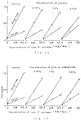
- the concentration should preferably be 0.01 M or more.
- the concentration should preferably be in a range from 0.1 to 1.6 M.
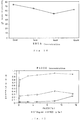
- Thiocyanic acid ions (sodium thiocyanate) were added to a sample to be assessed (serum) as chaotropic ions according to the 3-step sandwich method described above, and investigation was carried out using 5 types of serum, each having different collagen concentration.
- the concentration of the substance to be added should be in a range from 0.2 to 0.6 M, and preferably be in a range from 0.3 to 0.5 M.
- the concentration should preferably be in a range from 0.2 to 0.6 M.
- heparin sodium, chondroitin salfate A sodium salt, chondroitin sulfate C sodium salt, potassium dextran sulfate, and pentosan polysulfate sodium salt were used. The measurement was carried out according to the procedure like in the 1-step sandwich method described above, and the heparins were added during the immunoreaction.
- FIG.10 shows an analytical curve ( ⁇ - ⁇ ) of human type IV collagen in the absence of serum and also an analytical curve (+ ⁇ +) of human type IV collagen under the existence of serum stored in a frozen state.
- the concentration of gelatins should preferably be in a range from 1.5 to 4.5 %.
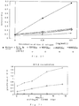
- FIG.19 shows a case where the concentration of gelatin hydrolysate was coated at 3 % and the concentration of heparin was raised from 250 to 500, 1000, 2500, and 5000 U/ml, and it can be understood that a degree of suppression by serum drops when the concentration of heparin is in a range from 250 to 5000 U/ml. For this reason, practically the concentration of heparin should preferably be in a range from 250 to 5000 U/ml.
- FIG.20 shows the effect of adding only heparin sodium
- the solid line ⁇ shows a result of the experiment when serum were not added
- the solid line + shows a result of the experiment when serum were added
- the dotted lines ⁇ , ⁇ , ⁇ , and ⁇ show results of the experiments when heparin sodium was added by 500, 1000, 2500, and 5000 U/ml respectively.
- heparins have both an effect to promote the immunological activity as described above and an effect to improve the effect provided by adding gelatins.
- BSA bovine serum albumin
- a quantity of type IV collagen in serum was checked in 20 cases of healthy persons, 24 cases of chronic hepatitis and 8 cases of liver cirrhosis, and distribution by disease were checked.
- the average was 24.5 ng/ml in cases of healthy persons, 55.7 ng/ml in case of chronic hepatitis, and 120.6 ng/ml in case of liver cirrhosis, and the value of type IV collagen raised as liver fibrosis proceeded.
- the values obtained through measurement according to the present invention on the concentration of human collagen in serum obtained from patients suffering from liver diseases are clearly higher than those on serum obtained from healthy people. For this reason, it is possible to detect liver diseases, especially liver fibrosis, liver cancer and digestive organ cancer metastasized to liver in their early stage without carrying out biopsy which is troublesome to the patients, by measuring the concentration of human type IV collagen in human blood according to the method by the present invention.
- this method is excellent as a measuring method.
- the method of coating antibodies is the same as that in the aforesaid 1-step sandwich method.
- the Fab'-B-A-POD is diluted by using a sample treating agent, and the product is used as an agent for treating samples.
- 50 mM-HEPES, 1.0M - KI, 2.5 mM-EDTA, 0.2 % PA-100, 4500 IU/ml-Heparin sodium, and 0.25 mg/ml-mouse IgG, all with pH of 8.0, are available as a solution for treating samples.
- the substrate 1 mg/ml-ABTS and 35 ⁇ l/dl-H2O2 are dissolved by a substrate dissolving solution, and the product is used as an enzyme substrate solution.
- the substrate dissolving solution is a mixture of 0.1 M citric acid and 1mM-EDTA and the pH is 4.2.
- the method for coating antibodies is the same as that in the aforesaid 1-step sandwich method.
- Fab'-B-A-POD solution was diluted by using a sample treating solution, and the product was used as an agent for treating samples.
- a sample treating solution D-PBS', 1.0 M-KI, 2.5 mM-EDTA, 0.2 % PA-100, 4500 IU/ml-heparin sodium, and 0.25 mg/ml-mouse IgG, all with pH of 7.0, are available.
- the substrate 1 mg/ml-ABTS and 35 ⁇ l/dl-H2O2 are dissolved by a substrate dissolving solution, and the product is used as an enzyme substrate solution.
- the substrate dissolving solution is a mixture of 0.1 M citric acid and 1mM-EDTA and the pH is 4.2.
- the solid phase was washed 6 times with saline, and 150 ⁇ l of the enzyme substrate agent was added and left therein for 10 minutes at 37°C.
- the method for coating antibodies is the same as that in the aforesaid 1-step sandwich method.
- Fab'-B-A-POD is diluted by a sample treating solution, and the product is used as an agent for treating samples.
- a sample treating solution 50 mM-HEPES, 0.1M sodium salicylate, 2.5 mM-EDTA, 0.2% PA-100, 4500 IU/ml-heparin sodium, and 0.25 mg/ml-mouse IgG, all with pH 8.0, are available.
- the substrate 1 mg/ml ABTS and 35 ⁇ l/dl-H2O2 are dissolved by a substrate dissolving solution, and the product is used as an enzyme substrate solution.
- the substrate dissolving solution is a mixture of 0.1 M citric acid and 1mM-EDTA and the pH is 4.2.
- the solid phase was washed 6 times with saline, and 150 ⁇ l of the enzyme substrate agent was added and left therein for 10 minutes at 37°C.
- the freeze-drying was carried out according to an ordinary method.
- Fab'-B-A-POD is diluted by using a sample treating solution.
- a sample treating solution 50 mM-HEPES, 1.0 M-KI, 2.5 mM-EDTA, 0.2 % PA-100, 4500 IU/ml-heparin sodium, and 0.25 mg/ml-mouse IgG, all with pH 8.0, are available.
- the substrate 1 mg/ml ABTS and 35 ⁇ l/dl-H2O2 are dissolved by a substrate dissolving solution, and the product is used as an enzyme substrate solution.
- the substrate dissolving solution is a mixture of 0.1 M citric acid and 1mM-EDTA and the pH is 4.2.
- the solid phase was washed 6 times with saline, and then 150 ⁇ l of the enzyme substrate solution was added and left therein for 10 minutes at 37°C.
- the reactant solution was measured by colorimetric determination at the wavelengths of 415 nm and 650 nm.
- liver fibrosis, liver cancer and digestive organ cancer metastasized to liver is difficult in measurement for ZTT (zinc sulfate turbid reaction), GOT (glutamine oxaloacetic transaminase), GPT (glutamine pyruvic acid transaminase), ALP (alkaline phosphatase), LDH (lactate dehydrogenase ), and ⁇ -GTP (glutamine transpeptitase), all of which are often used in determination of liver functions, and it can be expected that these types of disease can also be detected in their early stage by measuring human type IV collagen in blood according to the measuring method according the present invention.
- ZTT zinc sulfate turbid reaction
- GOT glutamine oxaloacetic transaminase
- GPT glutamine pyruvic acid transaminase
- ALP alkaline phosphatase
- LDH lactate dehydrogenase
- ⁇ -GTP glutamine transp
- the present invention has an extremely high utility.
Landscapes
- Life Sciences & Earth Sciences (AREA)
- Health & Medical Sciences (AREA)
- Engineering & Computer Science (AREA)
- Biomedical Technology (AREA)
- Molecular Biology (AREA)
- Chemical & Material Sciences (AREA)
- Urology & Nephrology (AREA)
- Hematology (AREA)
- Immunology (AREA)
- Biotechnology (AREA)
- Analytical Chemistry (AREA)
- Cell Biology (AREA)
- Proteomics, Peptides & Aminoacids (AREA)
- Food Science & Technology (AREA)
- Medicinal Chemistry (AREA)
- Physics & Mathematics (AREA)
- Microbiology (AREA)
- Biochemistry (AREA)
- General Health & Medical Sciences (AREA)
- General Physics & Mathematics (AREA)
- Pathology (AREA)
- Preparation Of Compounds By Using Micro-Organisms (AREA)
- Peptides Or Proteins (AREA)
Applications Claiming Priority (4)
| Application Number | Priority Date | Filing Date | Title |
|---|---|---|---|
| JP80892/91 | 1991-03-18 | ||
| JP8089291A JPH04289455A (ja) | 1991-03-18 | 1991-03-18 | コラーゲンの測定方法 |
| JP23713491 | 1991-08-24 | ||
| JP237134/91 | 1991-08-24 |
Publications (2)
| Publication Number | Publication Date |
|---|---|
| EP0535239A1 true EP0535239A1 (de) | 1993-04-07 |
| EP0535239A4 EP0535239A4 (en) | 1993-11-18 |
Family
ID=26421855
Family Applications (1)
| Application Number | Title | Priority Date | Filing Date |
|---|---|---|---|
| EP19920906928 Withdrawn EP0535239A4 (en) | 1991-03-18 | 1992-03-18 | Collagen assaying method and kit |
Country Status (2)
| Country | Link |
|---|---|
| EP (1) | EP0535239A4 (de) |
| WO (1) | WO1992016846A1 (de) |
Cited By (2)
| Publication number | Priority date | Publication date | Assignee | Title |
|---|---|---|---|---|
| WO2007111847A3 (en) * | 2006-03-24 | 2007-11-22 | Aokin Ag | Use of additives for the reduction of non-specific binding in assays |
| CN114137222A (zh) * | 2021-11-22 | 2022-03-04 | 江西乐成生物医疗有限公司 | 用于检测体液试样中组织金属白酶抑制剂-1的试剂盒 |
Family Cites Families (12)
| Publication number | Priority date | Publication date | Assignee | Title |
|---|---|---|---|---|
| FI792576A7 (fi) * | 1979-08-20 | 1981-01-01 | Orion Yhtymae Oy | Parannettu entsyymi-immunologinen määritysmenetelmä. |
| US4536478A (en) * | 1984-04-05 | 1985-08-20 | Seragan Diagnostics, Inc. | Method for reducing non-specific interferences in agglutination immunoassays |
| EP0165669A1 (de) * | 1984-05-04 | 1985-12-27 | Corning Glass Works | Methode zur Messung freien Ligands |
| JPS61241665A (ja) * | 1985-04-18 | 1986-10-27 | Toyobo Co Ltd | 安定化された固相試薬 |
| US4738932A (en) * | 1985-12-03 | 1988-04-19 | Advanced Polymer Systems, Inc. | Reaginic test for syphilis |
| CA1278259C (en) * | 1986-02-26 | 1990-12-27 | William C. Saxinger | Competitive elisa for the detection of antibodies |
| JPS6378067A (ja) * | 1986-09-20 | 1988-04-08 | Shiseido Co Ltd | 癌診断薬 |
| JPS63246396A (ja) * | 1987-03-31 | 1988-10-13 | Shiseido Co Ltd | モノクロ−ナル抗体 |
| DE68922846T2 (de) * | 1988-02-19 | 1995-11-16 | Fuji Yakuhin Kogyo Kk | Enzymimmuntest für menschliches typiv-collagen gemäss dem sandwichverfahren. |
| JPH0677017B2 (ja) * | 1988-02-19 | 1994-09-28 | 富士薬品工業株式会社 | ヒト▲iv▼型コラーゲンのサンドイツチ酵素免疫学的定量法 |
| GB8820375D0 (en) * | 1988-08-26 | 1988-09-28 | Boots Celltech Diagnostics | Assay |
| DE3901458A1 (de) * | 1989-01-19 | 1990-07-26 | Behringwerke Ag | Verwendung von zwei- oder dreiwertigen kationen in immunchemischen tests |
-
1992
- 1992-03-18 WO PCT/JP1992/000328 patent/WO1992016846A1/ja not_active Ceased
- 1992-03-18 EP EP19920906928 patent/EP0535239A4/en not_active Withdrawn
Cited By (3)
| Publication number | Priority date | Publication date | Assignee | Title |
|---|---|---|---|---|
| WO2007111847A3 (en) * | 2006-03-24 | 2007-11-22 | Aokin Ag | Use of additives for the reduction of non-specific binding in assays |
| US8124359B2 (en) | 2006-03-24 | 2012-02-28 | Aokin Ag | Use of additives for the reduction of non-specific binding in assays |
| CN114137222A (zh) * | 2021-11-22 | 2022-03-04 | 江西乐成生物医疗有限公司 | 用于检测体液试样中组织金属白酶抑制剂-1的试剂盒 |
Also Published As
| Publication number | Publication date |
|---|---|
| WO1992016846A1 (fr) | 1992-10-01 |
| EP0535239A4 (en) | 1993-11-18 |
Similar Documents
| Publication | Publication Date | Title |
|---|---|---|
| US11506674B2 (en) | Monoclonal antibody against D-dimer and diagnosis agent for detecting D-dimer, crosslinked fibrin and its derivatives containing D-dimer by using the antibody | |
| KR0148619B1 (ko) | Pivka-x의 측정방법 및 측정시약 | |
| EP1114064A1 (de) | Monoklonaler antikörper gegen oestrogen-stimulierte leucin-aminopeptidase | |
| EP0384130A2 (de) | Herzinfarkt-Immunoassay | |
| US20140227725A1 (en) | Competitive s100a9 immunoassays | |
| EP0741144A1 (de) | Monoklonaler antikörper gegen antiannexin-v, verfahren zu seiner herstellung und seiner verwendung | |
| JP3517754B2 (ja) | 抗ヒト可溶性フィブリン抗体,ハイブリドーマ及び免疫学的測定法 | |
| US5552292A (en) | Method of screening for colorectal cancer | |
| JP2003530089A (ja) | γ―カルボキシグルタミン酸を提示するエピトープを結合する抗体 | |
| EP1007570B1 (de) | Monospezifische antikörper gegen fibrinogen und fibrinopeptide b | |
| CN115856319B (zh) | 一种可溶性生长刺激表达基因2蛋白测定试剂盒及其制备方法和应用 | |
| EP1830189A1 (de) | Reagens und verfahren zum testen eines antigens | |
| CN105137071A (zh) | 心肌肌钙蛋白i均相酶免疫法测定试剂盒 | |
| EP1141726B1 (de) | Immunoassays zum nachweis von krankheiten oder krankheitsanfälligkeitsmerkmalen | |
| EP0298783A1 (de) | Methode, Vorrichtung und Testsatz zur Messung der Aktivität eines Gewebeplasminogenaktivators | |
| Katayama et al. | Increased fragmentation of urinary fibronectin in cancer patients detected by immunoenzymometric assay using domain-specific monoclonal antibodies | |
| KR960002740B1 (ko) | 안티-트롬빈-결합물질 모노클로날 항체, 이를 생산하는 하이브리도마 및 모노클로날 항체를 이용한 트롬빈-결합물질의 정제법 및 측정법 | |
| US7867717B2 (en) | Immunoassays to detect diseases or disease susceptibility traits | |
| EP0339302B1 (de) | Reagenzsystem zur Bestimmung des Komplexes des menschlichen Plasminogenaktivator-Inhibitors und des menschlichen Gewebeplasminogenaktivators und Testsatz dafür | |
| EP0324969A2 (de) | Verfahren zur Messung von Transferrin | |
| JPH05209883A (ja) | コラーゲンの測定方法及び測定キット | |
| US12429488B2 (en) | Monoclonal antibody against D-dimer and diagnosis agent for detecting D-dimer, crosslinked fibrin and its derivatives containing D-dimer by using the antibody | |
| CN111060689B (zh) | 一种子宫内膜癌检测试剂盒 | |
| Kropf et al. | Two sensitive time-resolved fluoroimmunoassays for cellular fibronectin | |
| JP2000028607A (ja) | 新規なモノクローナル抗体及びニックβ2グリコプロテインIの免疫学的分析方法 |
Legal Events
| Date | Code | Title | Description |
|---|---|---|---|
| PUAI | Public reference made under article 153(3) epc to a published international application that has entered the european phase |
Free format text: ORIGINAL CODE: 0009012 |
|
| AK | Designated contracting states |
Kind code of ref document: A1 Designated state(s): DE FR GB IT NL |
|
| 17P | Request for examination filed |
Effective date: 19930324 |
|
| A4 | Supplementary search report drawn up and despatched |
Effective date: 19930929 |
|
| AK | Designated contracting states |
Kind code of ref document: A4 Designated state(s): DE FR GB IT NL |
|
| 18D | Application deemed to be withdrawn |
Effective date: 19941001 |