EP0337410A2 - Nachweis von verändertem IgA1 in flüssigen Proben - Google Patents
Nachweis von verändertem IgA1 in flüssigen Proben Download PDFInfo
- Publication number
- EP0337410A2 EP0337410A2 EP89106443A EP89106443A EP0337410A2 EP 0337410 A2 EP0337410 A2 EP 0337410A2 EP 89106443 A EP89106443 A EP 89106443A EP 89106443 A EP89106443 A EP 89106443A EP 0337410 A2 EP0337410 A2 EP 0337410A2
- Authority
- EP
- European Patent Office
- Prior art keywords
- iga
- iga1
- complex
- lectin
- solid phase
- Prior art date
- Legal status (The legal status is an assumption and is not a legal conclusion. Google has not performed a legal analysis and makes no representation as to the accuracy of the status listed.)
- Withdrawn
Links
Images
Classifications
-
- G—PHYSICS
- G01—MEASURING; TESTING
- G01N—INVESTIGATING OR ANALYSING MATERIALS BY DETERMINING THEIR CHEMICAL OR PHYSICAL PROPERTIES
- G01N33/00—Investigating or analysing materials by specific methods not covered by groups G01N1/00 - G01N31/00
- G01N33/48—Biological material, e.g. blood, urine; Haemocytometers
- G01N33/50—Chemical analysis of biological material, e.g. blood, urine; Testing involving biospecific ligand binding methods; Immunological testing
- G01N33/53—Immunoassay; Biospecific binding assay; Materials therefor
- G01N33/543—Immunoassay; Biospecific binding assay; Materials therefor with an insoluble carrier for immobilising immunochemicals
- G01N33/54306—Solid-phase reaction mechanisms
-
- G—PHYSICS
- G01—MEASURING; TESTING
- G01N—INVESTIGATING OR ANALYSING MATERIALS BY DETERMINING THEIR CHEMICAL OR PHYSICAL PROPERTIES
- G01N33/00—Investigating or analysing materials by specific methods not covered by groups G01N1/00 - G01N31/00
- G01N33/48—Biological material, e.g. blood, urine; Haemocytometers
- G01N33/50—Chemical analysis of biological material, e.g. blood, urine; Testing involving biospecific ligand binding methods; Immunological testing
- G01N33/53—Immunoassay; Biospecific binding assay; Materials therefor
- G01N33/575—Immunoassay; Biospecific binding assay; Materials therefor for cancer
- G01N33/5756—Immunoassay; Biospecific binding assay; Materials therefor for cancer involving tumour-associated glycolinkage [TAG]
-
- G—PHYSICS
- G01—MEASURING; TESTING
- G01N—INVESTIGATING OR ANALYSING MATERIALS BY DETERMINING THEIR CHEMICAL OR PHYSICAL PROPERTIES
- G01N33/00—Investigating or analysing materials by specific methods not covered by groups G01N1/00 - G01N31/00
- G01N33/48—Biological material, e.g. blood, urine; Haemocytometers
- G01N33/50—Chemical analysis of biological material, e.g. blood, urine; Testing involving biospecific ligand binding methods; Immunological testing
- G01N33/68—Chemical analysis of biological material, e.g. blood, urine; Testing involving biospecific ligand binding methods; Immunological testing involving proteins, peptides or amino acids
- G01N33/6854—Immunoglobulins
-
- Y—GENERAL TAGGING OF NEW TECHNOLOGICAL DEVELOPMENTS; GENERAL TAGGING OF CROSS-SECTIONAL TECHNOLOGIES SPANNING OVER SEVERAL SECTIONS OF THE IPC; TECHNICAL SUBJECTS COVERED BY FORMER USPC CROSS-REFERENCE ART COLLECTIONS [XRACs] AND DIGESTS
- Y10—TECHNICAL SUBJECTS COVERED BY FORMER USPC
- Y10S—TECHNICAL SUBJECTS COVERED BY FORMER USPC CROSS-REFERENCE ART COLLECTIONS [XRACs] AND DIGESTS
- Y10S435/00—Chemistry: molecular biology and microbiology
- Y10S435/81—Packaged device or kit
-
- Y—GENERAL TAGGING OF NEW TECHNOLOGICAL DEVELOPMENTS; GENERAL TAGGING OF CROSS-SECTIONAL TECHNOLOGIES SPANNING OVER SEVERAL SECTIONS OF THE IPC; TECHNICAL SUBJECTS COVERED BY FORMER USPC CROSS-REFERENCE ART COLLECTIONS [XRACs] AND DIGESTS
- Y10—TECHNICAL SUBJECTS COVERED BY FORMER USPC
- Y10S—TECHNICAL SUBJECTS COVERED BY FORMER USPC CROSS-REFERENCE ART COLLECTIONS [XRACs] AND DIGESTS
- Y10S436/00—Chemistry: analytical and immunological testing
- Y10S436/811—Test for named disease, body condition or organ function
- Y10S436/813—Cancer
-
- Y—GENERAL TAGGING OF NEW TECHNOLOGICAL DEVELOPMENTS; GENERAL TAGGING OF CROSS-SECTIONAL TECHNOLOGIES SPANNING OVER SEVERAL SECTIONS OF THE IPC; TECHNICAL SUBJECTS COVERED BY FORMER USPC CROSS-REFERENCE ART COLLECTIONS [XRACs] AND DIGESTS
- Y10—TECHNICAL SUBJECTS COVERED BY FORMER USPC
- Y10S—TECHNICAL SUBJECTS COVERED BY FORMER USPC CROSS-REFERENCE ART COLLECTIONS [XRACs] AND DIGESTS
- Y10S436/00—Chemistry: analytical and immunological testing
- Y10S436/827—Lectins
Definitions
- This invention relates to a novel immunoassay method for the detection of immunoglobulin IgA1 with altered carbohydrate moieties in biological fluids.
- the invention is an immunoassay method for detecting IgA1 immunoglobulin by extracting total IgA from body fluids by reaction with an immobilized anti-IgA antibody and then detecting the IgA1 containing galactose Beta 1-3-N acetylgalactosamine protein using labeled lectin.
- Tumor markers are continually being sought to aid in both the monitoring of and the in-vitro diagnosis of cancer, as is well known in the art.
- commercial assays such as the carcinoembryonic antigen and alpha-fetoprotein immunoassays, are available to detect the presence of certain tumor markers in biological fluids.
- Serum secretory IgA(SIgA) has been reported in the literature as a tumor marker for various carcinomas. Some researchers report that assays for total SIgA are too non-specific for cancer and of no value for cancer detection, LoGerfo, P. et al., (1976) J. Surg. Res., 20:481. Other researchers showed that the measurement of total SIgA could be of value diagnostically in certain instances of carcinomas and chronic liver disease, Homburger, H.A., et al., (1984) A.J.C.P. 81:569; Kvale, D. et al., (1987) Cancer 59:203; Watanabe, T.
- Another problem associated with producing a specific and sensitive immunoassay is that repeated attempts to develop antibodies to a very specific antigen can be unsuccessful, thereby making an immunoassay to a specific antigen impossible. Also, the production of antibodies, when possible, is an elaborate and expensive process. The ability of a naturally occurring substance to replace antibodies in an assay would be beneficial.
- lectins In order to avoid using antibodies either partially or totally in an assay, and because they possess certain binding affinities, lectins have been employed in various assays. Lectins can recognize and bind to particular carbohydrate structures on the oligosaccharide moieties of glycoproteins.
- U.S. Patent Nos. 4,389,392 and 4,571,382 to Adachi describe methods of using lectins which bind to glycoproteins with a terminal galactose (Beta 1-3 or Beta 1-4) N-acetylglucosamine or terminal galactose (Beta 1-3 or Beta 1-4) -N-acetylgalactosamine in order to detect tumor associated glycolinkage containing substances.
- the methods described in these patents fail to use any immunological means to first specify which molecules are to be assayed, thereby losing a large measure of specificity.
- PCT Application No. WO 87/00289 discloses a test method in which lectins are used to form complexes with soluble desialylated glycoproteins which are then contacted with a detecting antibody.
- a variant of the test method is disclosed where the desialylated glycoprotein forms a complex with a specific antibody and the labeled lectin is then contacted with the complex.
- the antibody must be specific to the desialylated glycoprotein in order to selectively bind to the glycoprotein.
- the lectin is specific to a particular binding site on the oligosaccharide moiety of the desialylated glycoprotein, a substantial portion of the specificity of the test lies with the ability of the antibody to bind to the correct antigen.
- the oligosaccharide moieties of glycoproteins are attached to the protein moiety by O-glycosidic or N-glycosidic bonds.
- Carbohydrate structures of glycoprotein oligosaccharides synthesized by tumor cells are often different from the carbohydrate structures in the normal cell counterpart, thereby making such glycoproteins potential tumor markers. Many of these tumor markers have been identified as mucin glycoproteins.
- Mucins are high molecular weight glycoproteins characterized by unusually high carbohydrate content and carbohydrate side chains that are linked by O-glycosidic bonds to the protein chains. IgA1, but not IgA2, also contains carbohydrate side chains that are linked to the protein chain by O-glycosidic bonds.
- the O-linkage occurs between the hydroxyl groups of serine or threonine of the polypeptide and the reducing ends of N-acetylgalactosamine of the oligosaccharide.
- Certain lectins recognize and bind to the D galactose Beta (1-3)-N-acetyl-galactosamine (DGalBeta(1-3)DGalNAc) oligosaccharide structure O-linked to the polypeptide chains of IgA1 and mucin.
- Mucins and IgA1 are the major glycoproteins in serum and body fluids with this O-linkage; the vast majority of serum glycoproteins have N-glycosidic linkages. These N-linkages occur between the amide nitrogen of asparagine and the reducing end of N-acetylglucosamine.
- Oligosaccharide moieties of O-linked glycoproteins may be altered in cancer patients due to tumor cell necrosis and the release of glycosyltransferases and glycosidases in the vicinity of tumors.
- the glycoproteins IgA1 and mucin contain O-linked oligosaccharides and can serve as substrates for the released glycosyltransferases and glycosidases.
- glycosidases and glycosyltransferases are released from tumors, the O-linked carbohydrate structure of the IgA1 molecule may be altered.
- Unaltered IgA1 does contain DGalBeta(1-3)DGalNAc structures accessible to lectin binding, resulting in low levels or background normal levels of these O-linked carbohydrate side chains. However, when the IgA1 molecule is altered by glycosidases the number of these DGalBeta(1-3)DGalNAc structures accessible to lectin binding increases.
- the present invention is a novel immunoassay method for determining the presence or amount of altered IgA1 immunoglobulin in various body fluids, such as serum, plasma, sputum, urine and feces.
- altered IgA1 shall include all IgA1 with O-linked oligosaccharides and shall include both normal, non-altered IgA1 and IgA1 that has been affected by disease states, such as tumors.
- the method comprises contacting the sample with an anti-IgA antibody, forming an anti-IgA/IgA complex, then contacting this complex with a labeled lectin that can bind to DGalBeta(1-3)DGalNAc IgA1 but not the sialylated IgA1 or IgA2 which lacks the O-linked oligosaccharides, thereby forming an anti-IgA/IgA1/lectin complex.
- the presence or amount of the bound or unbound label measures the presence or amount of the altered IgA1 in the sample.
- the label may be any label capable of producing a detectable signal, such as, for example, an enzyme, a radioisotope or a fluorescent molecule.
- This invention also contemplates an in-vitro diagnostic kit to determine the presence or amount of altered IgA1 in a fluid sample which consists of polystyrene beads coated with anti-human IgA antibodies, a labeled lectin which has specificity for said altered IgA1 and not IgA2 specimen, dilution buffer and colostrum IgA standards.
- the present invention provides a method for determining the presence or amount of IgA1 with altered O-linked oligosaccharides in a fluid sample comprising:
- This anti-IgA immunoassay is directed by both the antibody and the lectin.
- the fluid samples assayed in this new immunoassay may be any fluid sample, but will normally be a body fluid, such as blood, serum, plasma, sputum, urine or feces, most preferably being blood, serum or plasma.
- the anti-IgA antibody may be a polyclonal or monoclonal antibody specific for IgA or IgA1 and is adsorbed or covalently bound to a solid phase, such as polystyrene beads, microparticles, wells, strips, plates or other suitable solid supports which are well known to those skilled in the art.
- a solid phase such as polystyrene beads, microparticles, wells, strips, plates or other suitable solid supports which are well known to those skilled in the art.
- Lectins useful in the practice of the invention include any lectins which recognize and bind to the DGalBeta(1-3)DGalNAc oligosacharide structure O-linked to the polypeptide chain of IgA1 and mucin.
- lectins for this purpose include peanut agglutinin (PNA) and Bauhinia purpurea alba agglutinin (BPA), although other lectins having the appropriate specificity may be employed.
- the label used may be an enzyme, a radioisotope, a fluorescent label or any other chemical or biological entity which will produce a detectable signal and is well known to those skilled in the art. If an enzyme is used as the label, the substrate forms a colored product in the presence of the enzyme. For example, if horseradish peroxidase is used, o-phenylenediamine is added as a substrate to form a colored product which can be measured spectrophotometrically.
- radioisotope is used as the label, no substrate is needed to activate the label, and after the appropriate amount of incubation of the radiolabeled lectin and the solid phase, unbound radiolabeled lectin is removed by washing, and bound radiolabeled lectin is determined by measuring radioisotope bound to the solid phase.
- the label on the lectin is an enzyme, such as alkaline phosphatase, B-galactosidase, and glucose oxidase.
- the label used with the lectin is horseradish peroxidase enzyme.
- the fluid sample is added to the anti-IgA antibody and incubated.
- Total IgA consisting of both the IgA1 and IgA2 fractions, are now bound to the anti IgA antibody.
- Any IgA in the sample that is not bound to the anti-IgA antibody and any residual sample components are then removed by washing.
- the enzyme labeled lectin which has specificity for the DGalBeta(1-3)DGalNAc receptors on the extracted IgA1, is added to the washed beads. The beads containing bound sample IgA and the labeled lectin are incubated.
- the beads are again washed and then transferred to reaction tubes, where a substrate is added.
- the reaction tubes are then incubated. The reaction is quenched and the label is detected in the solid phase as a measure of the lectin binding sites associated with the IgA1 in the sample.
- This example describes an assay to determine the presence or amount of IgA1 with altered carbohydrate moieties in serum, using an enzyme labeled lectin.
- the polyclonal antibody used to bind IgA was prepared from rabbit antisera to human IgA (alpha chain), obtained from ICN ImmunoBiologicals (catalogue #65-065). This antibody was prepared in the following manner: the IgG fraction was purified from the antisera by conventional protein A affinity chromatography. Antisera was applied to a protein A affinity column equilibrated in 20 mM Tris-HCl, 0.2 M NaCl, pH 8.3. The column is eluted with the same buffer while collecting fractions. Elution was continued until absorbance at 280 nm returned to baseline. The column was then eluted with 0.1 M glycine-HCl, pH 3.0, collecting fractions until the 280 nm absorbance returned to baseline.
- Fractions containing protein as determined by 280 nm absorbance were pooled from the pH 3.0 elution step and dialyzed against 10 mM sodium phosphate, pH 7.5.
- the dialyzed antibody was used to coat the polystyrene beads.
- the antibody is monospecific against human IgA (alpha chains) and not specific against heavy chains of human IgG and the heavy chains of Igm.
- the rabbit antihuman IgA antibody was used as the antibody for the anti-IgA-PNA enzyme immunoassay.
- the mixture was then centrifuged and the pellets dissolved in distilled water and dialyzed against water extensively, and then dialyzed against phosphate buffered saline (PBS).
- PBS phosphate buffered saline
- the dialyzed fraction was applied to a column of Sepharose 6B which had been acid treated and equilabrated in PBS.
- the column was eluted with PBS until 280 nm baseline absorbance was reached.
- the column was eluted with PBS containing 0.4 M D-galactose and the fractions demonstrating 280 nm absorbance were pooled and exhaustively dialyzed against (PBS).
- the dialyzed pool was purified PNA.
- HRPO Horseradish peroxidase
- HRPO sodium metaperiodate at a final concentration of 33 mM and the mixture was incubated 15 minutes at room temperature.
- the mixture was passed through a Sepharose G-25 column equilibrated in the acetate buffer and the brown band that was collected was activated HRPO.
- Activated HRPO at 1 mg/ml final concentration was added to PNA at 4 mg/ml (final concentration) in 50 mM bicarbonate pH 9.5 and incubated for 4 hours at room temperature.
- the reaction was quenched by adding sodium borohydride to a final concentration of 2.6 mM and allowing this to react 30 minutes at 4 degrees C.
- Acetone was added to a final concentration of 0.2% v/v to quench the sodium borohydride reaction.
- Polystyrene beads 0.25 inch in diameter, were coated by passive absorption with 10 micrograms/ml rabbit antihuman IgA at room temperature overnight. 25 microliters of serum specimen or colostrum IgA standard (Sigma Chemical Co., catalogue # I 0633), 200 microliters of assay diluent (0.1%BSA and 0.02% Tween 20 in 10 mM sodium phosphate, 150 mM NaCl, pH 7.2), and a coated bead were added to wells of a plastic reaction tray (Abbott Laboratories, list # 4046-16).
- Standards were prepared as 5, 20, 60 and 150 micrograms/ml of colostrum IgA diluted in a pool of normal human sera. This normal human sera pool was also tested as a reference control, and a panel of 845 serum specimens were the test samples.
- the tray was incubated at 37 degrees C for one hour.
- the beads were washed with distilled water and then incubated again at room temperature for two hours with 200 microliters of HRPO conjugated PNA diluted in assay diluent.
- the beads were washed with distilled water after this incubation and then transferred to reaction tubes.
- a 0.3 ml aliquot of o-phenylenediamine substrate (obtained from Abbott Laboratories OPD reagent kit, list # 6172-30) was added to each reaction tube, which was then incubated for thirty minutes at room temperature.
- To quench the peroxidase reaction 1.0 ml of 1 N sulfuric acid was added to each tube and the absorbance was measured at 492 nm with a spectrophotometer. The final result (R value) for each standard and specimen was reported as a ratio with respect to the reference control assay value.
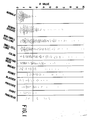
- Fig. 1 represents the results from the panel of 845 serum specimens.
- the mean R value of the normal specimens was 0.98 ⁇ 0.47 S.D. which was used to establish three cutoff R values, which are, 1.91 (mean plus two S.D ), 2.38 (mean plus three S.D.) and 3.0.
- R values of 1.91 and 2.38 5.1% and 1.2% respectively of normal specimens were greater than the R values; at the 3.0 value, 0% of normal specimens were elevated.
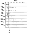
- the R values from all 845 serum specimens were analyzed with respect to these R values, as shown in Table I.
- the highest specificity was achieved with the 3.0 R value where all normal specimens were negative, 13% of the benign disease specimens were above the R value, and over 40% of the specimens for four different types of cancer were elevated.
- the cutoff R value was lowered, the sensitivity of the assay for detecting the cancer specimens increased, but there was poorer specificity due to an increased number of elevated benign disease specimens.
- Table II is a summary of the results of the benign diseases. At the 1.91 R value, all nine pulmonary specimens were elevated above the cutoff, whereas only two were so elevated above the 3.0 cutoff. Seven of eight of the bowel benign specimens were above the 1.91 cutoff, but only one was elevated above the 3.0 cutoff. The specimens from patients with diabetes had 30% of the specimens elevated above the 3.0 cutoff.
- CEA carcinoembryonic antigen
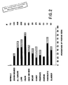
- Figure 3 illustrates the results of testing using the method of Example 1 wherein the specimen diluent was modified to a composition of 0.1% bovine serum albumin, 0.02% Tween 20, 5 mM EDTA, 10 mM sodium phosphate, 150 mM NaCl, at pH 7.2.
- the serum specimens tested by this assay were 29 normals, 36 colon cancer specimens with carcinoembryonic antigen assay (CEA) values of less than 5 ng/ml, 27 colon cancer specimens with CEA values greater than 5 ng/ml, 28 specimens from patients with colon polyps, 15 from patients with diverticulitis, 15 from patients with ulcerative colitis, 15 from patients with cirrhosis and 15 from patients with pancreatitis.
- CEA carcinoembryonic antigen assay
- a cutoff R value of the normal mean plus 2 standard deviations was used. 0% of normals, 36% of colon cancer specimens with CEA values of less than 5 ng/ml, 59% of the colon cancer specimens with CEA values greater than 5 ng/ml, 25% of the colon polyp specimens, 13% of the diverticulitis specimens, 73% of the ulcerative colitis specimens, 87% of the cirrhosis specimens, and 67% of the specimens with pancreatitis were elevated above the cutoff value.
- Fig. 3 demonstrate the use of this lectin assay as a supplemental assay to the widely used CEA assay for carcinomas. Values were elevated, to above the cutoff R value, for the premalignant conditions of colon polyps and ulcerative colitis and in 36% of the CEA negative colon cancer specimens (those with CEA levels of less than 5 ng/ml).
- CEA assay and the present assay can be used in an additive, or supplemental manner to jointly detect a greater number of premalignant and malignant conditions and for monitoring colon cancer patients for recurrence of disease.
- the benign conditions that were detected were cirrhosis and pancreatitis. These results also suggest that the present invention may be useful in the diagnosis of these types of diseases.
- This example describes an assay substantially the same as Example 1 the difference being the use of a radiolabeled lectin.
- Polystyrene beads 0.25 inch in diameter, are coated with rabbit antihuman IgA and are added to the wells of a reaction tray with 25 microliters of serum or standards, 200 microliters of assay diluent, as in Example 1.
- An assay blank is measured by substituting 25 microliters of assay diluent for the test sample.
- the trays are incubated for one hour at 37 degrees C and are washed with distilled water and then incubated at room temperature for two hours with PNA labeled with 125I.
- the beads are incubated with the labeled lectin, washed again with distilled water and then transferred to tubes for counting bound 125I.
- the anti-IgA-PNA enzyme immunoassay can be manufactured and distributed as an in-vitro diagnostic test kit.
- This kit containing 0.25 inch polystyrene beads that are coated with anti-human IgA antibodies, a labeled lectin that has specificity for O-linked oligosaccharide on IgA1, specimen dilution buffer and colostrum IgA standards.
Landscapes
- Health & Medical Sciences (AREA)
- Life Sciences & Earth Sciences (AREA)
- Immunology (AREA)
- Engineering & Computer Science (AREA)
- Chemical & Material Sciences (AREA)
- Molecular Biology (AREA)
- Biomedical Technology (AREA)
- Hematology (AREA)
- Urology & Nephrology (AREA)
- Food Science & Technology (AREA)
- Biochemistry (AREA)
- Cell Biology (AREA)
- Biotechnology (AREA)
- Medicinal Chemistry (AREA)
- Physics & Mathematics (AREA)
- Analytical Chemistry (AREA)
- Microbiology (AREA)
- General Health & Medical Sciences (AREA)
- General Physics & Mathematics (AREA)
- Pathology (AREA)
- Chemical Kinetics & Catalysis (AREA)
- Proteomics, Peptides & Aminoacids (AREA)
- Investigating Or Analysing Biological Materials (AREA)
- Measuring Or Testing Involving Enzymes Or Micro-Organisms (AREA)
Applications Claiming Priority (2)
| Application Number | Priority Date | Filing Date | Title |
|---|---|---|---|
| US181892 | 1988-04-15 | ||
| US07/181,892 US5051354A (en) | 1988-04-15 | 1988-04-15 | Detection of altered IGA1 in fluid samples |
Publications (2)
| Publication Number | Publication Date |
|---|---|
| EP0337410A2 true EP0337410A2 (de) | 1989-10-18 |
| EP0337410A3 EP0337410A3 (de) | 1990-10-17 |
Family
ID=22666251
Family Applications (1)
| Application Number | Title | Priority Date | Filing Date |
|---|---|---|---|
| EP19890106443 Withdrawn EP0337410A3 (de) | 1988-04-15 | 1989-04-11 | Nachweis von verändertem IgA1 in flüssigen Proben |
Country Status (5)
| Country | Link |
|---|---|
| US (1) | US5051354A (de) |
| EP (1) | EP0337410A3 (de) |
| JP (1) | JPH01305356A (de) |
| KR (1) | KR900016757A (de) |
| AU (1) | AU623640B2 (de) |
Cited By (5)
| Publication number | Priority date | Publication date | Assignee | Title |
|---|---|---|---|---|
| WO1991006865A1 (en) * | 1989-11-02 | 1991-05-16 | Biomira Inc. | Human tumor-associated thomsen-friedenreich antigen |
| US5242799A (en) * | 1989-11-02 | 1993-09-07 | Biomira, Inc. | Lectin-antibody immunoassays for TF epitope-bearing antigens |
| EP0829723A3 (de) * | 1996-09-13 | 1998-06-03 | Roche Diagnostics GmbH | Homogene Nachweisverfahren zur Bestimmung von Subpopulationen eines Analyten |
| EP1008852A1 (de) * | 1998-12-08 | 2000-06-14 | Aventis Behring Gesellschaft mit beschränkter Haftung | Verfahren zum spezifischen Nachweis von glykosilierten Proteinen |
| CN102043046A (zh) * | 2009-10-13 | 2011-05-04 | 上海慧普生物医药科技有限公司 | 一种检测糖链异常IgA肾病的蛋白芯片 |
Families Citing this family (1)
| Publication number | Priority date | Publication date | Assignee | Title |
|---|---|---|---|---|
| US9016493B2 (en) | 2011-11-16 | 2015-04-28 | Cooksmith, Inc. | Baking apparatus with multiple functions and sizes |
Family Cites Families (4)
| Publication number | Priority date | Publication date | Assignee | Title |
|---|---|---|---|---|
| GB2043890B (en) * | 1979-01-30 | 1983-09-07 | Otsuka Pharma Co Ltd | Determination of tumour associated glycolinkage and diagnosis of cancer |
| SE447028B (sv) * | 1980-06-30 | 1986-10-20 | Erik Audunn Cerven | Bestemning av endstaende sackaridgrupper i glykoprotein |
| JPS5729949A (en) * | 1980-07-30 | 1982-02-18 | Nippon Koutai Kenkyusho:Kk | Determination of sugar branch related to cancer and diagnostic technique of cancer |
| AU6126186A (en) * | 1985-07-01 | 1987-01-30 | Trustees Of Columbia University In The City Of New York, The | Lectin-antibody sandwich assay for desialylated glycoproteins |
-
1988
- 1988-04-15 US US07/181,892 patent/US5051354A/en not_active Expired - Fee Related
-
1989
- 1989-04-11 EP EP19890106443 patent/EP0337410A3/de not_active Withdrawn
- 1989-04-12 AU AU32709/89A patent/AU623640B2/en not_active Ceased
- 1989-04-13 KR KR1019890004882A patent/KR900016757A/ko not_active Abandoned
- 1989-04-13 JP JP1096200A patent/JPH01305356A/ja active Pending
Non-Patent Citations (3)
| Title |
|---|
| JOURNAL OF BIOLOGICAL CHEMISTRY, vol. 249, no. 22, 25th November 1974, pages 7270-7281, US; J. BAENZIGER et al.: "Structure of the carbohydrate units of IgA1 immunoglobulin. II. Structure of the O-glycosidically linked oligosaccharide untis" * |
| JOURNAL OF CLINICAL LABORATORY ANALYSIS, vol. 2, 1988, pages 225-234, Alan R. Liss, Inc., New York, US; J.G. HENSLEE et al.: "IgA with altered carbohydrate structure as a tumor-associated marker in serum of cancer patients" * |
| MOLECULAR IMMUNOLOGY, vol. 20, no. 9, 1983, pages 977-981, Pergamon Press Ltd, GB; M.E. CONLEY et al.: "Serum levels of IgA1 and IgA2 in children and in patients with IgA deficiency" * |
Cited By (7)
| Publication number | Priority date | Publication date | Assignee | Title |
|---|---|---|---|---|
| WO1991006865A1 (en) * | 1989-11-02 | 1991-05-16 | Biomira Inc. | Human tumor-associated thomsen-friedenreich antigen |
| EP0502078A1 (de) * | 1989-11-02 | 1992-09-09 | Biomira, Inc. | Menschliches tumor-assoziiertes thomsen-friedenreich-antigen |
| US5242799A (en) * | 1989-11-02 | 1993-09-07 | Biomira, Inc. | Lectin-antibody immunoassays for TF epitope-bearing antigens |
| EP0829723A3 (de) * | 1996-09-13 | 1998-06-03 | Roche Diagnostics GmbH | Homogene Nachweisverfahren zur Bestimmung von Subpopulationen eines Analyten |
| US6136545A (en) * | 1996-09-13 | 2000-10-24 | Roche Diagnostics Gmbh | Homogeneous detection methods for the determination of subpopulations of an analyte |
| EP1008852A1 (de) * | 1998-12-08 | 2000-06-14 | Aventis Behring Gesellschaft mit beschränkter Haftung | Verfahren zum spezifischen Nachweis von glykosilierten Proteinen |
| CN102043046A (zh) * | 2009-10-13 | 2011-05-04 | 上海慧普生物医药科技有限公司 | 一种检测糖链异常IgA肾病的蛋白芯片 |
Also Published As
| Publication number | Publication date |
|---|---|
| JPH01305356A (ja) | 1989-12-08 |
| EP0337410A3 (de) | 1990-10-17 |
| US5051354A (en) | 1991-09-24 |
| AU623640B2 (en) | 1992-05-21 |
| AU3270989A (en) | 1989-10-19 |
| KR900016757A (ko) | 1990-11-14 |
Similar Documents
| Publication | Publication Date | Title |
|---|---|---|
| US4659659A (en) | Diagnostic method for diseases having an arthritic component | |
| JP2579392B2 (ja) | 結合アッセイ法、該方法に用いる試薬およびキット | |
| US4289747A (en) | Immunological determination using lectin | |
| JP3486413B2 (ja) | 前立腺特異抗原の免疫検定法 | |
| US4447545A (en) | Bladder cancer detection | |
| AU654497B2 (en) | Analyte variant analysis | |
| EP0381450B1 (de) | Test für die alkalische Phosphatase aus Knochen | |
| US5051354A (en) | Detection of altered IGA1 in fluid samples | |
| EP0399464A2 (de) | Testverfahren für eine Substanz mit einer spezifischen Zuckerkette | |
| US4272504A (en) | Antibody adsorbed support method for carcinoembryonic antigen assay | |
| US6015680A (en) | Human lung adenocarcinoma-related monoclonal antibody and immunoassay method which uses the same | |
| EP0268296B1 (de) | Membran-Affinität-Konzentrationsimmuntest | |
| US20060073536A1 (en) | Immunoassays for determining vitamin B12, and reagents and kits therefor | |
| JP3667434B2 (ja) | 免疫測定に用いる非特異反応抑制剤、非特異反応抑制方法および測定キット | |
| US5380873A (en) | Homobifunctional agents for coupling enzymes and the like to antibodies and the like | |
| EP1837655B1 (de) | Blockierter enzymsondenkomplex | |
| JPS62168052A (ja) | Htlv−3対する抗体の免疫試験法 | |
| KR970001318B1 (ko) | 생물학적 유체내에서의 종양-관련된 표지물의 검출방법 | |
| AU601674B2 (en) | A method for determining a ligand | |
| JP3667435B2 (ja) | 非特異反応抑制剤、抑制方法及び測定キット | |
| JP7770666B2 (ja) | がん検出方法、がん検査方法、及びこれらに用いるキット | |
| Fujiwara et al. | Determination of urinary acetylpolyamines by a monoclonal antibody-based enzyme-linked immunosorbent assay (ELISA) | |
| JP2630436B2 (ja) | 肝癌診断薬 | |
| Loor et al. | Evaluation of a human pancreas-specific antigen by enzyme-linked immunosorbent assay | |
| Guesdon | Amplification systems for enzyme immunoassay |
Legal Events
| Date | Code | Title | Description |
|---|---|---|---|
| PUAI | Public reference made under article 153(3) epc to a published international application that has entered the european phase |
Free format text: ORIGINAL CODE: 0009012 |
|
| AK | Designated contracting states |
Kind code of ref document: A2 Designated state(s): AT BE CH DE ES FR GB IT LI NL |
|
| PUAL | Search report despatched |
Free format text: ORIGINAL CODE: 0009013 |
|
| AK | Designated contracting states |
Kind code of ref document: A3 Designated state(s): AT BE CH DE ES FR GB IT LI NL |
|
| 17P | Request for examination filed |
Effective date: 19910411 |
|
| 17Q | First examination report despatched |
Effective date: 19930224 |
|
| STAA | Information on the status of an ep patent application or granted ep patent |
Free format text: STATUS: THE APPLICATION IS DEEMED TO BE WITHDRAWN |
|
| 18D | Application deemed to be withdrawn |
Effective date: 19940830 |