EP0332677B1 - Virus d'herpes attenues, virus d'herpes comprenant de l'adn etranger de codage d'une sequence d'acides amines et vaccins les contenant - Google Patents
Virus d'herpes attenues, virus d'herpes comprenant de l'adn etranger de codage d'une sequence d'acides amines et vaccins les contenant Download PDFInfo
- Publication number
- EP0332677B1 EP0332677B1 EP88907889A EP88907889A EP0332677B1 EP 0332677 B1 EP0332677 B1 EP 0332677B1 EP 88907889 A EP88907889 A EP 88907889A EP 88907889 A EP88907889 A EP 88907889A EP 0332677 B1 EP0332677 B1 EP 0332677B1
- Authority
- EP
- European Patent Office
- Prior art keywords
- prv
- gene
- dna
- virus
- herpesvirus
- Prior art date
- Legal status (The legal status is an assumption and is not a legal conclusion. Google has not performed a legal analysis and makes no representation as to the accuracy of the status listed.)
- Expired - Lifetime
Links
- 241001529453 unidentified herpesvirus Species 0.000 title claims abstract description 245
- 229960005486 vaccine Drugs 0.000 title claims abstract description 69
- 230000002238 attenuated effect Effects 0.000 title claims description 11
- 125000003275 alpha amino acid group Chemical group 0.000 title abstract 2
- 108090000623 proteins and genes Proteins 0.000 claims abstract description 312
- 241000701093 Suid alphaherpesvirus 1 Species 0.000 claims abstract description 246
- 102000004169 proteins and genes Human genes 0.000 claims abstract description 60
- 230000000890 antigenic effect Effects 0.000 claims abstract description 30
- 108090000288 Glycoproteins Proteins 0.000 claims abstract description 23
- 102000003886 Glycoproteins Human genes 0.000 claims abstract description 21
- 102000007056 Recombinant Fusion Proteins Human genes 0.000 claims abstract description 15
- 108010008281 Recombinant Fusion Proteins Proteins 0.000 claims abstract description 15
- 108020004414 DNA Proteins 0.000 claims description 393
- 210000004027 cell Anatomy 0.000 claims description 115
- 241000125945 Protoparvovirus Species 0.000 claims description 98
- 241000282898 Sus scrofa Species 0.000 claims description 90
- 108020004440 Thymidine kinase Proteins 0.000 claims description 74
- 230000014509 gene expression Effects 0.000 claims description 67
- 102000006601 Thymidine Kinase Human genes 0.000 claims description 61
- 108090000765 processed proteins & peptides Proteins 0.000 claims description 50
- 241000700588 Human alphaherpesvirus 1 Species 0.000 claims description 45
- 229920001184 polypeptide Polymers 0.000 claims description 45
- 102000004196 processed proteins & peptides Human genes 0.000 claims description 45
- 108010076504 Protein Sorting Signals Proteins 0.000 claims description 40
- 241000588724 Escherichia coli Species 0.000 claims description 39
- 108091028043 Nucleic acid sequence Proteins 0.000 claims description 37
- 210000004899 c-terminal region Anatomy 0.000 claims description 15
- 201000004792 malaria Diseases 0.000 claims description 13
- 108090000565 Capsid Proteins Proteins 0.000 claims description 12
- 102100023321 Ceruloplasmin Human genes 0.000 claims description 11
- 230000003053 immunization Effects 0.000 claims description 4
- 102100031675 DnaJ homolog subfamily C member 5 Human genes 0.000 claims 6
- 241001465754 Metazoa Species 0.000 abstract description 75
- 108020001507 fusion proteins Proteins 0.000 abstract description 22
- 102000037865 fusion proteins Human genes 0.000 abstract description 22
- 239000013598 vector Substances 0.000 abstract description 22
- 241000700605 Viruses Species 0.000 description 316
- 238000000034 method Methods 0.000 description 251
- 239000012634 fragment Substances 0.000 description 208
- 108010005774 beta-Galactosidase Proteins 0.000 description 139
- 238000012217 deletion Methods 0.000 description 130
- 230000037430 deletion Effects 0.000 description 130
- 239000013612 plasmid Substances 0.000 description 112
- 108091007433 antigens Proteins 0.000 description 89
- 241000702626 Infectious bursal disease virus Species 0.000 description 82
- 239000000427 antigen Substances 0.000 description 81
- 102000036639 antigens Human genes 0.000 description 81
- 241001502481 Meleagrid alphaherpesvirus 1 Species 0.000 description 75
- 238000003780 insertion Methods 0.000 description 73
- 230000037431 insertion Effects 0.000 description 73
- LFQSCWFLJHTTHZ-UHFFFAOYSA-N Ethanol Chemical compound CCO LFQSCWFLJHTTHZ-UHFFFAOYSA-N 0.000 description 63
- 150000001413 amino acids Chemical class 0.000 description 63
- 108091008146 restriction endonucleases Proteins 0.000 description 61
- 238000010276 construction Methods 0.000 description 58
- 101150003725 TK gene Proteins 0.000 description 55
- 239000000523 sample Substances 0.000 description 55
- ISWSIDIOOBJBQZ-UHFFFAOYSA-N Phenol Chemical compound OC1=CC=CC=C1 ISWSIDIOOBJBQZ-UHFFFAOYSA-N 0.000 description 52
- 238000001890 transfection Methods 0.000 description 50
- 108091026890 Coding region Proteins 0.000 description 45
- 230000004927 fusion Effects 0.000 description 43
- 101150076489 B gene Proteins 0.000 description 42
- WQZGKKKJIJFFOK-FPRJBGLDSA-N beta-D-galactose Chemical compound OC[C@H]1O[C@@H](O)[C@H](O)[C@@H](O)[C@H]1O WQZGKKKJIJFFOK-FPRJBGLDSA-N 0.000 description 40
- 241000282887 Suidae Species 0.000 description 39
- 101710117490 Circumsporozoite protein Proteins 0.000 description 38
- 208000005562 infectious bovine rhinotracheitis Diseases 0.000 description 38
- 208000015181 infectious disease Diseases 0.000 description 38
- 102000005936 beta-Galactosidase Human genes 0.000 description 36
- 241000700584 Simplexvirus Species 0.000 description 35
- 238000006243 chemical reaction Methods 0.000 description 35
- 239000000243 solution Substances 0.000 description 35
- 239000002609 medium Substances 0.000 description 34
- 210000002966 serum Anatomy 0.000 description 34
- 239000000872 buffer Substances 0.000 description 33
- 238000010367 cloning Methods 0.000 description 33
- 238000002255 vaccination Methods 0.000 description 32
- 239000002299 complementary DNA Substances 0.000 description 30
- 108091032973 (ribonucleotides)n+m Proteins 0.000 description 29
- 101800000385 Transmembrane protein Proteins 0.000 description 29
- 239000008188 pellet Substances 0.000 description 29
- 238000007792 addition Methods 0.000 description 28
- 208000037265 diseases, disorders, signs and symptoms Diseases 0.000 description 28
- 230000006801 homologous recombination Effects 0.000 description 28
- 238000002744 homologous recombination Methods 0.000 description 28
- XLYOFNOQVPJJNP-UHFFFAOYSA-N water Chemical compound O XLYOFNOQVPJJNP-UHFFFAOYSA-N 0.000 description 28
- 238000004458 analytical method Methods 0.000 description 27
- KCXVZYZYPLLWCC-UHFFFAOYSA-N EDTA Chemical compound OC(=O)CN(CC(O)=O)CCN(CC(O)=O)CC(O)=O KCXVZYZYPLLWCC-UHFFFAOYSA-N 0.000 description 25
- FAPWRFPIFSIZLT-UHFFFAOYSA-M Sodium chloride Chemical compound [Na+].[Cl-] FAPWRFPIFSIZLT-UHFFFAOYSA-M 0.000 description 25
- 241000711484 Transmissible gastroenteritis virus Species 0.000 description 25
- 239000007983 Tris buffer Substances 0.000 description 25
- LENZDBCJOHFCAS-UHFFFAOYSA-N tris Chemical compound OCC(N)(CO)CO LENZDBCJOHFCAS-UHFFFAOYSA-N 0.000 description 25
- TWRXJAOTZQYOKJ-UHFFFAOYSA-L Magnesium chloride Chemical compound [Mg+2].[Cl-].[Cl-] TWRXJAOTZQYOKJ-UHFFFAOYSA-L 0.000 description 24
- 238000002105 Southern blotting Methods 0.000 description 24
- 208000009305 pseudorabies Diseases 0.000 description 24
- LINMATFDVHBYOS-MBJXGIAVSA-N (2s,3r,4s,5r,6r)-2-[(5-bromo-1h-indol-3-yl)oxy]-6-(hydroxymethyl)oxane-3,4,5-triol Chemical compound O[C@@H]1[C@@H](O)[C@@H](O)[C@@H](CO)O[C@H]1OC1=CNC2=CC=C(Br)C=C12 LINMATFDVHBYOS-MBJXGIAVSA-N 0.000 description 23
- 201000010099 disease Diseases 0.000 description 23
- 238000002360 preparation method Methods 0.000 description 23
- 241000271566 Aves Species 0.000 description 22
- HEDRZPFGACZZDS-UHFFFAOYSA-N Chloroform Chemical compound ClC(Cl)Cl HEDRZPFGACZZDS-UHFFFAOYSA-N 0.000 description 22
- 238000001262 western blot Methods 0.000 description 22
- 238000003556 assay Methods 0.000 description 21
- 238000002965 ELISA Methods 0.000 description 20
- 101150027427 ICP4 gene Proteins 0.000 description 20
- 239000000203 mixture Substances 0.000 description 20
- 241000701047 Gallid alphaherpesvirus 2 Species 0.000 description 19
- 241000702670 Rotavirus Species 0.000 description 19
- 229910001868 water Inorganic materials 0.000 description 19
- IAZDPXIOMUYVGZ-UHFFFAOYSA-N Dimethylsulphoxide Chemical compound CS(C)=O IAZDPXIOMUYVGZ-UHFFFAOYSA-N 0.000 description 18
- PEDCQBHIVMGVHV-UHFFFAOYSA-N Glycerine Chemical compound OCC(O)CO PEDCQBHIVMGVHV-UHFFFAOYSA-N 0.000 description 18
- 101710181600 Glycoprotein gp2 Proteins 0.000 description 18
- WCUXLLCKKVVCTQ-UHFFFAOYSA-M Potassium chloride Chemical compound [Cl-].[K+] WCUXLLCKKVVCTQ-UHFFFAOYSA-M 0.000 description 18
- 238000012360 testing method Methods 0.000 description 18
- 241000283690 Bos taurus Species 0.000 description 17
- 229930193140 Neomycin Natural products 0.000 description 17
- 230000000120 cytopathologic effect Effects 0.000 description 17
- 238000002474 experimental method Methods 0.000 description 17
- 229960004927 neomycin Drugs 0.000 description 17
- 230000028327 secretion Effects 0.000 description 17
- 239000013592 cell lysate Substances 0.000 description 16
- 239000003550 marker Substances 0.000 description 16
- 230000003472 neutralizing effect Effects 0.000 description 16
- 108020004705 Codon Proteins 0.000 description 15
- 241000287828 Gallus gallus Species 0.000 description 15
- 235000013330 chicken meat Nutrition 0.000 description 15
- 238000010586 diagram Methods 0.000 description 15
- 239000000499 gel Substances 0.000 description 15
- 238000012216 screening Methods 0.000 description 15
- 239000006228 supernatant Substances 0.000 description 15
- 210000003501 vero cell Anatomy 0.000 description 15
- 229920000936 Agarose Polymers 0.000 description 14
- 108091003079 Bovine Serum Albumin Proteins 0.000 description 14
- 108010067390 Viral Proteins Proteins 0.000 description 14
- 210000000234 capsid Anatomy 0.000 description 14
- 108700010070 Codon Usage Proteins 0.000 description 13
- 238000011160 research Methods 0.000 description 13
- 239000011780 sodium chloride Substances 0.000 description 13
- QKNYBSVHEMOAJP-UHFFFAOYSA-N 2-amino-2-(hydroxymethyl)propane-1,3-diol;hydron;chloride Chemical compound Cl.OCC(N)(CO)CO QKNYBSVHEMOAJP-UHFFFAOYSA-N 0.000 description 12
- 241000283973 Oryctolagus cuniculus Species 0.000 description 12
- 241000702619 Porcine parvovirus Species 0.000 description 12
- AIYUHDOJVYHVIT-UHFFFAOYSA-M caesium chloride Chemical compound [Cl-].[Cs+] AIYUHDOJVYHVIT-UHFFFAOYSA-M 0.000 description 12
- 238000000338 in vitro Methods 0.000 description 12
- 229910001629 magnesium chloride Inorganic materials 0.000 description 12
- 241000701087 Felid alphaherpesvirus 1 Species 0.000 description 11
- DBMJMQXJHONAFJ-UHFFFAOYSA-M Sodium laurylsulphate Chemical compound [Na+].CCCCCCCCCCCCOS([O-])(=O)=O DBMJMQXJHONAFJ-UHFFFAOYSA-M 0.000 description 11
- 238000013459 approach Methods 0.000 description 11
- 230000000694 effects Effects 0.000 description 11
- 238000000746 purification Methods 0.000 description 11
- 210000001519 tissue Anatomy 0.000 description 11
- 230000003612 virological effect Effects 0.000 description 11
- 241000680578 Canid alphaherpesvirus 1 Species 0.000 description 10
- 241000230501 Equine herpesvirus sp. Species 0.000 description 10
- 108020004511 Recombinant DNA Proteins 0.000 description 10
- 239000011543 agarose gel Substances 0.000 description 10
- 238000009396 hybridization Methods 0.000 description 10
- 239000002773 nucleotide Substances 0.000 description 10
- 125000003729 nucleotide group Chemical group 0.000 description 10
- 239000013600 plasmid vector Substances 0.000 description 10
- 241000702673 Bovine rotavirus Species 0.000 description 9
- 101150008820 HN gene Proteins 0.000 description 9
- 101710154606 Hemagglutinin Proteins 0.000 description 9
- 101710093908 Outer capsid protein VP4 Proteins 0.000 description 9
- 101710135467 Outer capsid protein sigma-1 Proteins 0.000 description 9
- 101710176177 Protein A56 Proteins 0.000 description 9
- VMHLLURERBWHNL-UHFFFAOYSA-M Sodium acetate Chemical compound [Na+].CC([O-])=O VMHLLURERBWHNL-UHFFFAOYSA-M 0.000 description 9
- 230000028993 immune response Effects 0.000 description 9
- 239000006166 lysate Substances 0.000 description 9
- 125000002924 primary amino group Chemical group [H]N([H])* 0.000 description 9
- 108010062697 pseudorabies virus glycoproteins Proteins 0.000 description 9
- 230000004044 response Effects 0.000 description 9
- DGVVWUTYPXICAM-UHFFFAOYSA-N β‐Mercaptoethanol Chemical compound OCCS DGVVWUTYPXICAM-UHFFFAOYSA-N 0.000 description 9
- 101710081079 Minor spike protein H Proteins 0.000 description 8
- 241001045988 Neogene Species 0.000 description 8
- 206010046865 Vaccinia virus infection Diseases 0.000 description 8
- 108020005202 Viral DNA Proteins 0.000 description 8
- 230000008901 benefit Effects 0.000 description 8
- 238000011534 incubation Methods 0.000 description 8
- 101150066555 lacZ gene Proteins 0.000 description 8
- 101150091879 neo gene Proteins 0.000 description 8
- 239000001103 potassium chloride Substances 0.000 description 8
- 235000011164 potassium chloride Nutrition 0.000 description 8
- 239000001632 sodium acetate Substances 0.000 description 8
- 235000017281 sodium acetate Nutrition 0.000 description 8
- 241000894007 species Species 0.000 description 8
- 229940031626 subunit vaccine Drugs 0.000 description 8
- 208000007089 vaccinia Diseases 0.000 description 8
- 210000002845 virion Anatomy 0.000 description 8
- 206010067484 Adverse reaction Diseases 0.000 description 7
- USFZMSVCRYTOJT-UHFFFAOYSA-N Ammonium acetate Chemical compound N.CC(O)=O USFZMSVCRYTOJT-UHFFFAOYSA-N 0.000 description 7
- 239000005695 Ammonium acetate Substances 0.000 description 7
- 108010054576 Deoxyribonuclease EcoRI Proteins 0.000 description 7
- 241000711450 Infectious bronchitis virus Species 0.000 description 7
- 108091036060 Linker DNA Proteins 0.000 description 7
- 239000000020 Nitrocellulose Substances 0.000 description 7
- 241000223960 Plasmodium falciparum Species 0.000 description 7
- 108091081024 Start codon Proteins 0.000 description 7
- 206010058874 Viraemia Diseases 0.000 description 7
- 230000006838 adverse reaction Effects 0.000 description 7
- 235000019257 ammonium acetate Nutrition 0.000 description 7
- 229940043376 ammonium acetate Drugs 0.000 description 7
- 230000000903 blocking effect Effects 0.000 description 7
- 230000001413 cellular effect Effects 0.000 description 7
- 230000029087 digestion Effects 0.000 description 7
- 239000012153 distilled water Substances 0.000 description 7
- 238000000605 extraction Methods 0.000 description 7
- 239000000185 hemagglutinin Substances 0.000 description 7
- 230000002458 infectious effect Effects 0.000 description 7
- 229920001220 nitrocellulos Polymers 0.000 description 7
- 239000013615 primer Substances 0.000 description 7
- 239000000758 substrate Substances 0.000 description 7
- WOVKYSAHUYNSMH-RRKCRQDMSA-N 5-bromodeoxyuridine Chemical compound C1[C@H](O)[C@@H](CO)O[C@H]1N1C(=O)NC(=O)C(Br)=C1 WOVKYSAHUYNSMH-RRKCRQDMSA-N 0.000 description 6
- QTBSBXVTEAMEQO-UHFFFAOYSA-N Acetic acid Chemical compound CC(O)=O QTBSBXVTEAMEQO-UHFFFAOYSA-N 0.000 description 6
- 241000711443 Bovine coronavirus Species 0.000 description 6
- 241001260012 Bursa Species 0.000 description 6
- 102100031780 Endonuclease Human genes 0.000 description 6
- 102000004190 Enzymes Human genes 0.000 description 6
- 108090000790 Enzymes Proteins 0.000 description 6
- 101150034814 F gene Proteins 0.000 description 6
- XQFRJNBWHJMXHO-RRKCRQDMSA-N IDUR Chemical compound C1[C@H](O)[C@@H](CO)O[C@H]1N1C(=O)NC(=O)C(I)=C1 XQFRJNBWHJMXHO-RRKCRQDMSA-N 0.000 description 6
- OKKJLVBELUTLKV-UHFFFAOYSA-N Methanol Chemical compound OC OKKJLVBELUTLKV-UHFFFAOYSA-N 0.000 description 6
- 206010033799 Paralysis Diseases 0.000 description 6
- 108010092799 RNA-directed DNA polymerase Proteins 0.000 description 6
- 108700026226 TATA Box Proteins 0.000 description 6
- 210000004102 animal cell Anatomy 0.000 description 6
- 230000015572 biosynthetic process Effects 0.000 description 6
- 229940098773 bovine serum albumin Drugs 0.000 description 6
- 238000004113 cell culture Methods 0.000 description 6
- 238000005119 centrifugation Methods 0.000 description 6
- 230000034994 death Effects 0.000 description 6
- 238000002405 diagnostic procedure Methods 0.000 description 6
- 238000010790 dilution Methods 0.000 description 6
- 239000012895 dilution Substances 0.000 description 6
- 239000012091 fetal bovine serum Substances 0.000 description 6
- 239000012530 fluid Substances 0.000 description 6
- 238000010353 genetic engineering Methods 0.000 description 6
- 238000002955 isolation Methods 0.000 description 6
- 238000013507 mapping Methods 0.000 description 6
- 230000004048 modification Effects 0.000 description 6
- 238000012986 modification Methods 0.000 description 6
- 239000002953 phosphate buffered saline Substances 0.000 description 6
- 239000000047 product Substances 0.000 description 6
- UCSJYZPVAKXKNQ-HZYVHMACSA-N streptomycin Chemical compound CN[C@H]1[C@H](O)[C@@H](O)[C@H](CO)O[C@H]1O[C@@H]1[C@](C=O)(O)[C@H](C)O[C@H]1O[C@@H]1[C@@H](NC(N)=N)[C@H](O)[C@@H](NC(N)=N)[C@H](O)[C@H]1O UCSJYZPVAKXKNQ-HZYVHMACSA-N 0.000 description 6
- 238000012546 transfer Methods 0.000 description 6
- 230000014616 translation Effects 0.000 description 6
- CURLTUGMZLYLDI-UHFFFAOYSA-N Carbon dioxide Chemical compound O=C=O CURLTUGMZLYLDI-UHFFFAOYSA-N 0.000 description 5
- 101710088235 Envelope glycoprotein C homolog Proteins 0.000 description 5
- 208000009889 Herpes Simplex Diseases 0.000 description 5
- 108700026244 Open Reading Frames Proteins 0.000 description 5
- 229920001213 Polysorbate 20 Polymers 0.000 description 5
- 206010037660 Pyrexia Diseases 0.000 description 5
- 108091036066 Three prime untranslated region Proteins 0.000 description 5
- 235000011089 carbon dioxide Nutrition 0.000 description 5
- 125000003178 carboxy group Chemical group [H]OC(*)=O 0.000 description 5
- RGWHQCVHVJXOKC-SHYZEUOFSA-J dCTP(4-) Chemical compound O=C1N=C(N)C=CN1[C@@H]1O[C@H](COP([O-])(=O)OP([O-])(=O)OP([O-])([O-])=O)[C@@H](O)C1 RGWHQCVHVJXOKC-SHYZEUOFSA-J 0.000 description 5
- ZMMJGEGLRURXTF-UHFFFAOYSA-N ethidium bromide Chemical compound [Br-].C12=CC(N)=CC=C2C2=CC=C(N)C=C2[N+](CC)=C1C1=CC=CC=C1 ZMMJGEGLRURXTF-UHFFFAOYSA-N 0.000 description 5
- 229960005542 ethidium bromide Drugs 0.000 description 5
- 210000002950 fibroblast Anatomy 0.000 description 5
- 230000012010 growth Effects 0.000 description 5
- 239000001963 growth medium Substances 0.000 description 5
- 210000003292 kidney cell Anatomy 0.000 description 5
- 230000001717 pathogenic effect Effects 0.000 description 5
- 230000008488 polyadenylation Effects 0.000 description 5
- 239000000256 polyoxyethylene sorbitan monolaurate Substances 0.000 description 5
- 235000010486 polyoxyethylene sorbitan monolaurate Nutrition 0.000 description 5
- 210000004927 skin cell Anatomy 0.000 description 5
- 238000001179 sorption measurement Methods 0.000 description 5
- 238000013518 transcription Methods 0.000 description 5
- 230000035897 transcription Effects 0.000 description 5
- 238000013519 translation Methods 0.000 description 5
- YBJHBAHKTGYVGT-ZKWXMUAHSA-N (+)-Biotin Chemical compound N1C(=O)N[C@@H]2[C@H](CCCCC(=O)O)SC[C@@H]21 YBJHBAHKTGYVGT-ZKWXMUAHSA-N 0.000 description 4
- JKMHFZQWWAIEOD-UHFFFAOYSA-N 2-[4-(2-hydroxyethyl)piperazin-1-yl]ethanesulfonic acid Chemical compound OCC[NH+]1CCN(CCS([O-])(=O)=O)CC1 JKMHFZQWWAIEOD-UHFFFAOYSA-N 0.000 description 4
- 206010003591 Ataxia Diseases 0.000 description 4
- 239000006144 Dulbecco’s modified Eagle's medium Substances 0.000 description 4
- 239000007995 HEPES buffer Substances 0.000 description 4
- 239000012981 Hank's balanced salt solution Substances 0.000 description 4
- 102000004160 Phosphoric Monoester Hydrolases Human genes 0.000 description 4
- 108090000608 Phosphoric Monoester Hydrolases Proteins 0.000 description 4
- 108010076039 Polyproteins Proteins 0.000 description 4
- 238000000246 agarose gel electrophoresis Methods 0.000 description 4
- 229960000723 ampicillin Drugs 0.000 description 4
- AVKUERGKIZMTKX-NJBDSQKTSA-N ampicillin Chemical compound C1([C@@H](N)C(=O)N[C@H]2[C@H]3SC([C@@H](N3C2=O)C(O)=O)(C)C)=CC=CC=C1 AVKUERGKIZMTKX-NJBDSQKTSA-N 0.000 description 4
- 210000003837 chick embryo Anatomy 0.000 description 4
- 239000003599 detergent Substances 0.000 description 4
- LOKCTEFSRHRXRJ-UHFFFAOYSA-I dipotassium trisodium dihydrogen phosphate hydrogen phosphate dichloride Chemical compound P(=O)(O)(O)[O-].[K+].P(=O)(O)([O-])[O-].[Na+].[Na+].[Cl-].[K+].[Cl-].[Na+] LOKCTEFSRHRXRJ-UHFFFAOYSA-I 0.000 description 4
- 238000001962 electrophoresis Methods 0.000 description 4
- 230000001900 immune effect Effects 0.000 description 4
- 238000001727 in vivo Methods 0.000 description 4
- 238000010348 incorporation Methods 0.000 description 4
- PHTQWCKDNZKARW-UHFFFAOYSA-N isoamylol Chemical compound CC(C)CCO PHTQWCKDNZKARW-UHFFFAOYSA-N 0.000 description 4
- 210000003734 kidney Anatomy 0.000 description 4
- 108020004999 messenger RNA Proteins 0.000 description 4
- 244000052769 pathogen Species 0.000 description 4
- VLTRZXGMWDSKGL-UHFFFAOYSA-N perchloric acid Chemical compound OCl(=O)(=O)=O VLTRZXGMWDSKGL-UHFFFAOYSA-N 0.000 description 4
- 238000001556 precipitation Methods 0.000 description 4
- 230000006798 recombination Effects 0.000 description 4
- 238000005215 recombination Methods 0.000 description 4
- 230000009467 reduction Effects 0.000 description 4
- 230000001105 regulatory effect Effects 0.000 description 4
- 230000010076 replication Effects 0.000 description 4
- 238000012163 sequencing technique Methods 0.000 description 4
- 239000002356 single layer Substances 0.000 description 4
- 239000001509 sodium citrate Substances 0.000 description 4
- NLJMYIDDQXHKNR-UHFFFAOYSA-K sodium citrate Chemical compound O.O.[Na+].[Na+].[Na+].[O-]C(=O)CC(O)(CC([O-])=O)C([O-])=O NLJMYIDDQXHKNR-UHFFFAOYSA-K 0.000 description 4
- 241000701161 unidentified adenovirus Species 0.000 description 4
- 229940125575 vaccine candidate Drugs 0.000 description 4
- 102000040650 (ribonucleotides)n+m Human genes 0.000 description 3
- CSCPPACGZOOCGX-UHFFFAOYSA-N Acetone Chemical compound CC(C)=O CSCPPACGZOOCGX-UHFFFAOYSA-N 0.000 description 3
- 101710154825 Aminoglycoside 3'-phosphotransferase Proteins 0.000 description 3
- 244000153158 Ammi visnaga Species 0.000 description 3
- 235000010585 Ammi visnaga Nutrition 0.000 description 3
- UXVMQQNJUSDDNG-UHFFFAOYSA-L Calcium chloride Chemical compound [Cl-].[Cl-].[Ca+2] UXVMQQNJUSDDNG-UHFFFAOYSA-L 0.000 description 3
- 241000282472 Canis lupus familiaris Species 0.000 description 3
- 241000283707 Capra Species 0.000 description 3
- 102000012410 DNA Ligases Human genes 0.000 description 3
- 108010061982 DNA Ligases Proteins 0.000 description 3
- 102000004594 DNA Polymerase I Human genes 0.000 description 3
- 108010017826 DNA Polymerase I Proteins 0.000 description 3
- 241001452028 Escherichia coli DH1 Species 0.000 description 3
- 241001131785 Escherichia coli HB101 Species 0.000 description 3
- 241000282324 Felis Species 0.000 description 3
- 241000282326 Felis catus Species 0.000 description 3
- ZRALSGWEFCBTJO-UHFFFAOYSA-N Guanidine Chemical compound NC(N)=N ZRALSGWEFCBTJO-UHFFFAOYSA-N 0.000 description 3
- 229920000209 Hexadimethrine bromide Polymers 0.000 description 3
- 108091026898 Leader sequence (mRNA) Proteins 0.000 description 3
- 241000282339 Mustela Species 0.000 description 3
- 208000010359 Newcastle Disease Diseases 0.000 description 3
- MUBZPKHOEPUJKR-UHFFFAOYSA-N Oxalic acid Chemical compound OC(=O)C(O)=O MUBZPKHOEPUJKR-UHFFFAOYSA-N 0.000 description 3
- 208000002606 Paramyxoviridae Infections Diseases 0.000 description 3
- 208000008071 Parvoviridae Infections Diseases 0.000 description 3
- 206010057343 Parvovirus infection Diseases 0.000 description 3
- 229930182555 Penicillin Natural products 0.000 description 3
- JGSARLDLIJGVTE-MBNYWOFBSA-N Penicillin G Chemical compound N([C@H]1[C@H]2SC([C@@H](N2C1=O)C(O)=O)(C)C)C(=O)CC1=CC=CC=C1 JGSARLDLIJGVTE-MBNYWOFBSA-N 0.000 description 3
- 241000288906 Primates Species 0.000 description 3
- 241000700159 Rattus Species 0.000 description 3
- 108091081062 Repeated sequence (DNA) Proteins 0.000 description 3
- 241000282695 Saimiri Species 0.000 description 3
- HEMHJVSKTPXQMS-UHFFFAOYSA-M Sodium hydroxide Chemical compound [OH-].[Na+] HEMHJVSKTPXQMS-UHFFFAOYSA-M 0.000 description 3
- 239000004098 Tetracycline Substances 0.000 description 3
- IQFYYKKMVGJFEH-XLPZGREQSA-N Thymidine Chemical compound O=C1NC(=O)C(C)=CN1[C@@H]1O[C@H](CO)[C@@H](O)C1 IQFYYKKMVGJFEH-XLPZGREQSA-N 0.000 description 3
- 241001314440 Triphora trianthophoros Species 0.000 description 3
- 206010051511 Viral diarrhoea Diseases 0.000 description 3
- ANAKGLHGKIVHLU-UHFFFAOYSA-N azanium;ethanol;acetate Chemical compound [NH4+].CCO.CC([O-])=O ANAKGLHGKIVHLU-UHFFFAOYSA-N 0.000 description 3
- 230000001580 bacterial effect Effects 0.000 description 3
- 239000001110 calcium chloride Substances 0.000 description 3
- 229910001628 calcium chloride Inorganic materials 0.000 description 3
- 235000011148 calcium chloride Nutrition 0.000 description 3
- 210000003169 central nervous system Anatomy 0.000 description 3
- 238000004590 computer program Methods 0.000 description 3
- 230000000875 corresponding effect Effects 0.000 description 3
- VHJLVAABSRFDPM-QWWZWVQMSA-N dithiothreitol Chemical compound SC[C@@H](O)[C@H](O)CS VHJLVAABSRFDPM-QWWZWVQMSA-N 0.000 description 3
- 229940079593 drug Drugs 0.000 description 3
- 239000003814 drug Substances 0.000 description 3
- 238000001976 enzyme digestion Methods 0.000 description 3
- ZDXPYRJPNDTMRX-UHFFFAOYSA-N glutamine Natural products OC(=O)C(N)CCC(N)=O ZDXPYRJPNDTMRX-UHFFFAOYSA-N 0.000 description 3
- 244000144980 herd Species 0.000 description 3
- RAXXELZNTBOGNW-UHFFFAOYSA-N imidazole Natural products C1=CNC=N1 RAXXELZNTBOGNW-UHFFFAOYSA-N 0.000 description 3
- 239000010410 layer Substances 0.000 description 3
- 239000000463 material Substances 0.000 description 3
- 230000007246 mechanism Effects 0.000 description 3
- 238000002156 mixing Methods 0.000 description 3
- 230000036961 partial effect Effects 0.000 description 3
- 239000002245 particle Substances 0.000 description 3
- 229940049954 penicillin Drugs 0.000 description 3
- 150000002989 phenols Chemical class 0.000 description 3
- 230000003362 replicative effect Effects 0.000 description 3
- 238000010839 reverse transcription Methods 0.000 description 3
- 230000000405 serological effect Effects 0.000 description 3
- 230000002269 spontaneous effect Effects 0.000 description 3
- 229960005322 streptomycin Drugs 0.000 description 3
- 238000003786 synthesis reaction Methods 0.000 description 3
- 229960002180 tetracycline Drugs 0.000 description 3
- 229930101283 tetracycline Natural products 0.000 description 3
- 235000019364 tetracycline Nutrition 0.000 description 3
- 150000003522 tetracyclines Chemical class 0.000 description 3
- 238000011144 upstream manufacturing Methods 0.000 description 3
- 230000009385 viral infection Effects 0.000 description 3
- 230000001018 virulence Effects 0.000 description 3
- BRZYSWJRSDMWLG-DJWUNRQOSA-N (2r,3r,4r,5r)-2-[(1s,2s,3r,4s,6r)-4,6-diamino-3-[(2s,3r,4r,5s,6r)-3-amino-4,5-dihydroxy-6-[(1r)-1-hydroxyethyl]oxan-2-yl]oxy-2-hydroxycyclohexyl]oxy-5-methyl-4-(methylamino)oxane-3,5-diol Chemical compound O1C[C@@](O)(C)[C@H](NC)[C@@H](O)[C@H]1O[C@@H]1[C@@H](O)[C@H](O[C@@H]2[C@@H]([C@@H](O)[C@H](O)[C@@H]([C@@H](C)O)O2)N)[C@@H](N)C[C@H]1N BRZYSWJRSDMWLG-DJWUNRQOSA-N 0.000 description 2
- IQFYYKKMVGJFEH-OYDXRQHMSA-N 1-[(2r,4s,5s)-4-hydroxy-5-(hydroxymethyl)oxolan-2-yl]-5-methylpyrimidine-2,4-dione Chemical compound O=C1NC(=O)C(C)=CN1[C@@H]1O[C@H]([14CH2]O)[C@@H](O)C1 IQFYYKKMVGJFEH-OYDXRQHMSA-N 0.000 description 2
- WOVKYSAHUYNSMH-UHFFFAOYSA-N BROMODEOXYURIDINE Natural products C1C(O)C(CO)OC1N1C(=O)NC(=O)C(Br)=C1 WOVKYSAHUYNSMH-UHFFFAOYSA-N 0.000 description 2
- 241000894006 Bacteria Species 0.000 description 2
- 241000282421 Canidae Species 0.000 description 2
- 206010010071 Coma Diseases 0.000 description 2
- 208000035473 Communicable disease Diseases 0.000 description 2
- 206010010904 Convulsion Diseases 0.000 description 2
- 206010010947 Coordination abnormal Diseases 0.000 description 2
- 239000003155 DNA primer Substances 0.000 description 2
- 108010014303 DNA-directed DNA polymerase Proteins 0.000 description 2
- 102000016928 DNA-directed DNA polymerase Human genes 0.000 description 2
- 108010067770 Endopeptidase K Proteins 0.000 description 2
- 241001524679 Escherichia virus M13 Species 0.000 description 2
- 208000034454 F12-related hereditary angioedema with normal C1Inh Diseases 0.000 description 2
- 229920001917 Ficoll Polymers 0.000 description 2
- ZHNUHDYFZUAESO-UHFFFAOYSA-N Formamide Chemical compound NC=O ZHNUHDYFZUAESO-UHFFFAOYSA-N 0.000 description 2
- 102000002464 Galactosidases Human genes 0.000 description 2
- 108010093031 Galactosidases Proteins 0.000 description 2
- DHMQDGOQFOQNFH-UHFFFAOYSA-N Glycine Chemical compound NCC(O)=O DHMQDGOQFOQNFH-UHFFFAOYSA-N 0.000 description 2
- 101800000342 Glycoprotein C Proteins 0.000 description 2
- 208000007514 Herpes zoster Diseases 0.000 description 2
- 241000282412 Homo Species 0.000 description 2
- 101000678879 Homo sapiens Atypical chemokine receptor 1 Proteins 0.000 description 2
- 101001002508 Homo sapiens Immunoglobulin-binding protein 1 Proteins 0.000 description 2
- 241000701074 Human alphaherpesvirus 2 Species 0.000 description 2
- MHAJPDPJQMAIIY-UHFFFAOYSA-N Hydrogen peroxide Chemical compound OO MHAJPDPJQMAIIY-UHFFFAOYSA-N 0.000 description 2
- 102100021042 Immunoglobulin-binding protein 1 Human genes 0.000 description 2
- 241000282560 Macaca mulatta Species 0.000 description 2
- 206010028851 Necrosis Diseases 0.000 description 2
- 241000772415 Neovison vison Species 0.000 description 2
- 108020002230 Pancreatic Ribonuclease Proteins 0.000 description 2
- 102000005891 Pancreatic ribonuclease Human genes 0.000 description 2
- 241001494479 Pecora Species 0.000 description 2
- 208000003251 Pruritus Diseases 0.000 description 2
- 238000002123 RNA extraction Methods 0.000 description 2
- 239000013616 RNA primer Substances 0.000 description 2
- 229920005654 Sephadex Polymers 0.000 description 2
- 239000012507 Sephadex™ Substances 0.000 description 2
- PXIPVTKHYLBLMZ-UHFFFAOYSA-N Sodium azide Chemical compound [Na+].[N-]=[N+]=[N-] PXIPVTKHYLBLMZ-UHFFFAOYSA-N 0.000 description 2
- 102000004357 Transferases Human genes 0.000 description 2
- 108090000992 Transferases Proteins 0.000 description 2
- 208000036142 Viral infection Diseases 0.000 description 2
- 206010000210 abortion Diseases 0.000 description 2
- 231100000176 abortion Toxicity 0.000 description 2
- 238000002835 absorbance Methods 0.000 description 2
- 230000001154 acute effect Effects 0.000 description 2
- 238000010171 animal model Methods 0.000 description 2
- 208000022531 anorexia Diseases 0.000 description 2
- 230000005875 antibody response Effects 0.000 description 2
- 239000008346 aqueous phase Substances 0.000 description 2
- 239000012148 binding buffer Substances 0.000 description 2
- 229960002685 biotin Drugs 0.000 description 2
- 235000020958 biotin Nutrition 0.000 description 2
- 239000011616 biotin Substances 0.000 description 2
- 210000004369 blood Anatomy 0.000 description 2
- 239000008280 blood Substances 0.000 description 2
- 230000036760 body temperature Effects 0.000 description 2
- 230000037396 body weight Effects 0.000 description 2
- 229950004398 broxuridine Drugs 0.000 description 2
- 239000007975 buffered saline Substances 0.000 description 2
- 244000309466 calf Species 0.000 description 2
- 239000006285 cell suspension Substances 0.000 description 2
- 239000001913 cellulose Substances 0.000 description 2
- 229920002678 cellulose Polymers 0.000 description 2
- 239000003153 chemical reaction reagent Substances 0.000 description 2
- 239000003795 chemical substances by application Substances 0.000 description 2
- 230000000052 comparative effect Effects 0.000 description 2
- 230000036461 convulsion Effects 0.000 description 2
- 210000000805 cytoplasm Anatomy 0.000 description 2
- 206010061428 decreased appetite Diseases 0.000 description 2
- 230000003247 decreasing effect Effects 0.000 description 2
- 238000001514 detection method Methods 0.000 description 2
- 239000012149 elution buffer Substances 0.000 description 2
- 238000005516 engineering process Methods 0.000 description 2
- 230000008029 eradication Effects 0.000 description 2
- 238000012869 ethanol precipitation Methods 0.000 description 2
- 239000000284 extract Substances 0.000 description 2
- 239000003889 eye drop Substances 0.000 description 2
- 230000002349 favourable effect Effects 0.000 description 2
- 239000012894 fetal calf serum Substances 0.000 description 2
- 230000001605 fetal effect Effects 0.000 description 2
- 238000004108 freeze drying Methods 0.000 description 2
- 230000006870 function Effects 0.000 description 2
- 101150030521 gI gene Proteins 0.000 description 2
- 230000002068 genetic effect Effects 0.000 description 2
- 229960004198 guanidine Drugs 0.000 description 2
- 238000003306 harvesting Methods 0.000 description 2
- 208000002672 hepatitis B Diseases 0.000 description 2
- 208000016861 hereditary angioedema type 3 Diseases 0.000 description 2
- 230000000521 hyperimmunizing effect Effects 0.000 description 2
- FDGQSTZJBFJUBT-UHFFFAOYSA-N hypoxanthine Chemical compound O=C1NC=NC2=C1NC=N2 FDGQSTZJBFJUBT-UHFFFAOYSA-N 0.000 description 2
- 210000000987 immune system Anatomy 0.000 description 2
- 229940031551 inactivated vaccine Drugs 0.000 description 2
- 208000016290 incoordination Diseases 0.000 description 2
- 239000002054 inoculum Substances 0.000 description 2
- 230000003834 intracellular effect Effects 0.000 description 2
- 238000005304 joining Methods 0.000 description 2
- 231100000518 lethal Toxicity 0.000 description 2
- 230000001665 lethal effect Effects 0.000 description 2
- KWGKDLIKAYFUFQ-UHFFFAOYSA-M lithium chloride Chemical compound [Li+].[Cl-] KWGKDLIKAYFUFQ-UHFFFAOYSA-M 0.000 description 2
- 210000004185 liver Anatomy 0.000 description 2
- 230000004807 localization Effects 0.000 description 2
- 210000004072 lung Anatomy 0.000 description 2
- 238000004519 manufacturing process Methods 0.000 description 2
- 239000012528 membrane Substances 0.000 description 2
- 244000005700 microbiome Species 0.000 description 2
- 235000019799 monosodium phosphate Nutrition 0.000 description 2
- 229940031348 multivalent vaccine Drugs 0.000 description 2
- 230000017074 necrotic cell death Effects 0.000 description 2
- 230000007935 neutral effect Effects 0.000 description 2
- 238000006386 neutralization reaction Methods 0.000 description 2
- 108020004707 nucleic acids Proteins 0.000 description 2
- 102000039446 nucleic acids Human genes 0.000 description 2
- 150000007523 nucleic acids Chemical class 0.000 description 2
- 230000007918 pathogenicity Effects 0.000 description 2
- 230000007170 pathology Effects 0.000 description 2
- 102000013415 peroxidase activity proteins Human genes 0.000 description 2
- 108040007629 peroxidase activity proteins Proteins 0.000 description 2
- 239000012071 phase Substances 0.000 description 2
- 229920003023 plastic Polymers 0.000 description 2
- 239000004033 plastic Substances 0.000 description 2
- 229920002401 polyacrylamide Polymers 0.000 description 2
- 239000001267 polyvinylpyrrolidone Substances 0.000 description 2
- 229920000036 polyvinylpyrrolidone Polymers 0.000 description 2
- 235000013855 polyvinylpyrrolidone Nutrition 0.000 description 2
- SCVFZCLFOSHCOH-UHFFFAOYSA-M potassium acetate Chemical compound [K+].CC([O-])=O SCVFZCLFOSHCOH-UHFFFAOYSA-M 0.000 description 2
- 230000008569 process Effects 0.000 description 2
- 230000000644 propagated effect Effects 0.000 description 2
- 230000002829 reductive effect Effects 0.000 description 2
- 230000000241 respiratory effect Effects 0.000 description 2
- 238000007790 scraping Methods 0.000 description 2
- 238000010187 selection method Methods 0.000 description 2
- 230000035945 sensitivity Effects 0.000 description 2
- 230000035939 shock Effects 0.000 description 2
- 235000020183 skimmed milk Nutrition 0.000 description 2
- AJPJDKMHJJGVTQ-UHFFFAOYSA-M sodium dihydrogen phosphate Chemical compound [Na+].OP(O)([O-])=O AJPJDKMHJJGVTQ-UHFFFAOYSA-M 0.000 description 2
- 229910000162 sodium phosphate Inorganic materials 0.000 description 2
- 239000012064 sodium phosphate buffer Substances 0.000 description 2
- 238000001228 spectrum Methods 0.000 description 2
- 210000000952 spleen Anatomy 0.000 description 2
- 210000004989 spleen cell Anatomy 0.000 description 2
- 230000000638 stimulation Effects 0.000 description 2
- 239000011550 stock solution Substances 0.000 description 2
- 239000000725 suspension Substances 0.000 description 2
- 210000001550 testis Anatomy 0.000 description 2
- 230000009466 transformation Effects 0.000 description 2
- PIEPQKCYPFFYMG-UHFFFAOYSA-N tris acetate Chemical compound CC(O)=O.OCC(N)(CO)CO PIEPQKCYPFFYMG-UHFFFAOYSA-N 0.000 description 2
- 230000003442 weekly effect Effects 0.000 description 2
- WQZGKKKJIJFFOK-SVZMEOIVSA-N (+)-Galactose Chemical compound OC[C@H]1OC(O)[C@H](O)[C@@H](O)[C@H]1O WQZGKKKJIJFFOK-SVZMEOIVSA-N 0.000 description 1
- VHJLVAABSRFDPM-UHFFFAOYSA-N 1,4-dithiothreitol Chemical compound SCC(O)C(O)CS VHJLVAABSRFDPM-UHFFFAOYSA-N 0.000 description 1
- MSWZFWKMSRAUBD-IVMDWMLBSA-N 2-amino-2-deoxy-D-glucopyranose Chemical compound N[C@H]1C(O)O[C@H](CO)[C@@H](O)[C@@H]1O MSWZFWKMSRAUBD-IVMDWMLBSA-N 0.000 description 1
- 102100027324 2-hydroxyacyl-CoA lyase 1 Human genes 0.000 description 1
- KUWPCJHYPSUOFW-YBXAARCKSA-N 2-nitrophenyl beta-D-galactoside Chemical compound O[C@@H]1[C@@H](O)[C@@H](O)[C@@H](CO)O[C@H]1OC1=CC=CC=C1[N+]([O-])=O KUWPCJHYPSUOFW-YBXAARCKSA-N 0.000 description 1
- HAEVLZUBSLBWIX-UHFFFAOYSA-N 2-octylphenol;oxirane Chemical compound C1CO1.CCCCCCCCC1=CC=CC=C1O HAEVLZUBSLBWIX-UHFFFAOYSA-N 0.000 description 1
- TVZGACDUOSZQKY-LBPRGKRZSA-N 4-aminofolic acid Chemical compound C1=NC2=NC(N)=NC(N)=C2N=C1CNC1=CC=C(C(=O)N[C@@H](CCC(O)=O)C(O)=O)C=C1 TVZGACDUOSZQKY-LBPRGKRZSA-N 0.000 description 1
- 208000004998 Abdominal Pain Diseases 0.000 description 1
- 208000030090 Acute Disease Diseases 0.000 description 1
- 229920001817 Agar Polymers 0.000 description 1
- QGZKDVFQNNGYKY-UHFFFAOYSA-O Ammonium Chemical compound [NH4+] QGZKDVFQNNGYKY-UHFFFAOYSA-O 0.000 description 1
- 206010003694 Atrophy Diseases 0.000 description 1
- 102100022717 Atypical chemokine receptor 1 Human genes 0.000 description 1
- 241000972773 Aulopiformes Species 0.000 description 1
- 101500001532 Avian infectious bursal disease virus Capsid protein VP2 Proteins 0.000 description 1
- 108090001008 Avidin Proteins 0.000 description 1
- DWRXFEITVBNRMK-UHFFFAOYSA-N Beta-D-1-Arabinofuranosylthymine Natural products O=C1NC(=O)C(C)=CN1C1C(O)C(O)C(CO)O1 DWRXFEITVBNRMK-UHFFFAOYSA-N 0.000 description 1
- 241000701083 Bovine alphaherpesvirus 1 Species 0.000 description 1
- 241000030939 Bubalus bubalis Species 0.000 description 1
- 101000583086 Bunodosoma granuliferum Delta-actitoxin-Bgr2b Proteins 0.000 description 1
- 241000282465 Canis Species 0.000 description 1
- 241000282693 Cercopithecidae Species 0.000 description 1
- 201000006082 Chickenpox Diseases 0.000 description 1
- 229910021580 Cobalt(II) chloride Inorganic materials 0.000 description 1
- 241000283716 Connochaetes Species 0.000 description 1
- 206010011469 Crying Diseases 0.000 description 1
- 241000701022 Cytomegalovirus Species 0.000 description 1
- 108020003215 DNA Probes Proteins 0.000 description 1
- 108010008286 DNA nucleotidylexotransferase Proteins 0.000 description 1
- 102100033215 DNA nucleotidylexotransferase Human genes 0.000 description 1
- 239000003298 DNA probe Substances 0.000 description 1
- 230000006820 DNA synthesis Effects 0.000 description 1
- 108010053770 Deoxyribonucleases Proteins 0.000 description 1
- 102000016911 Deoxyribonucleases Human genes 0.000 description 1
- 238000012286 ELISA Assay Methods 0.000 description 1
- 108010008655 Epstein-Barr Virus Nuclear Antigens Proteins 0.000 description 1
- 241000701081 Equid alphaherpesvirus 1 Species 0.000 description 1
- 241000283086 Equidae Species 0.000 description 1
- IAYPIBMASNFSPL-UHFFFAOYSA-N Ethylene oxide Chemical compound C1CO1 IAYPIBMASNFSPL-UHFFFAOYSA-N 0.000 description 1
- 208000010201 Exanthema Diseases 0.000 description 1
- 108060002716 Exonuclease Proteins 0.000 description 1
- 241000233866 Fungi Species 0.000 description 1
- 108010084884 GDP-mannose transporter Proteins 0.000 description 1
- 101000609762 Gallus gallus Ovalbumin Proteins 0.000 description 1
- 108700039691 Genetic Promoter Regions Proteins 0.000 description 1
- WQZGKKKJIJFFOK-GASJEMHNSA-N Glucose Natural products OC[C@H]1OC(O)[C@H](O)[C@@H](O)[C@@H]1O WQZGKKKJIJFFOK-GASJEMHNSA-N 0.000 description 1
- 239000004471 Glycine Substances 0.000 description 1
- 208000032843 Hemorrhage Diseases 0.000 description 1
- 208000001688 Herpes Genitalis Diseases 0.000 description 1
- 208000029433 Herpesviridae infectious disease Diseases 0.000 description 1
- 101001009252 Homo sapiens 2-hydroxyacyl-CoA lyase 1 Proteins 0.000 description 1
- 108010001336 Horseradish Peroxidase Proteins 0.000 description 1
- UGQMRVRMYYASKQ-UHFFFAOYSA-N Hypoxanthine nucleoside Natural products OC1C(O)C(CO)OC1N1C(NC=NC2=O)=C2N=C1 UGQMRVRMYYASKQ-UHFFFAOYSA-N 0.000 description 1
- 206010062016 Immunosuppression Diseases 0.000 description 1
- FFEARJCKVFRZRR-BYPYZUCNSA-N L-methionine Chemical compound CSCC[C@H](N)C(O)=O FFEARJCKVFRZRR-BYPYZUCNSA-N 0.000 description 1
- 208000032420 Latent Infection Diseases 0.000 description 1
- 208000030289 Lymphoproliferative disease Diseases 0.000 description 1
- 206010026749 Mania Diseases 0.000 description 1
- 208000006758 Marek Disease Diseases 0.000 description 1
- 241000711408 Murine respirovirus Species 0.000 description 1
- 241000699666 Mus <mouse, genus> Species 0.000 description 1
- 241000699670 Mus sp. Species 0.000 description 1
- BACYUWVYYTXETD-UHFFFAOYSA-N N-Lauroylsarcosine Chemical compound CCCCCCCCCCCC(=O)N(C)CC(O)=O BACYUWVYYTXETD-UHFFFAOYSA-N 0.000 description 1
- CHJJGSNFBQVOTG-UHFFFAOYSA-N N-methyl-guanidine Natural products CNC(N)=N CHJJGSNFBQVOTG-UHFFFAOYSA-N 0.000 description 1
- 238000011887 Necropsy Methods 0.000 description 1
- 208000012902 Nervous system disease Diseases 0.000 description 1
- 208000008457 Neurologic Manifestations Diseases 0.000 description 1
- 206010060860 Neurological symptom Diseases 0.000 description 1
- 101710163270 Nuclease Proteins 0.000 description 1
- XDMCWZFLLGVIID-SXPRBRBTSA-N O-(3-O-D-galactosyl-N-acetyl-beta-D-galactosaminyl)-L-serine Chemical compound CC(=O)N[C@H]1[C@H](OC[C@H]([NH3+])C([O-])=O)O[C@H](CO)[C@H](O)[C@@H]1OC1[C@H](O)[C@@H](O)[C@@H](O)[C@@H](CO)O1 XDMCWZFLLGVIID-SXPRBRBTSA-N 0.000 description 1
- 241000282936 Odocoileus hemionus Species 0.000 description 1
- 206010067152 Oral herpes Diseases 0.000 description 1
- 241000237502 Ostreidae Species 0.000 description 1
- 108091005804 Peptidases Proteins 0.000 description 1
- 241000577979 Peromyscus spicilegus Species 0.000 description 1
- 108010021757 Polynucleotide 5'-Hydroxyl-Kinase Proteins 0.000 description 1
- 102000008422 Polynucleotide 5'-hydroxyl-kinase Human genes 0.000 description 1
- 241000702665 Porcine rotavirus Species 0.000 description 1
- 239000004365 Protease Substances 0.000 description 1
- 244000088415 Raphanus sativus Species 0.000 description 1
- 235000006140 Raphanus sativus var sativus Nutrition 0.000 description 1
- 208000035415 Reinfection Diseases 0.000 description 1
- 102100037486 Reverse transcriptase/ribonuclease H Human genes 0.000 description 1
- 206010039101 Rhinorrhoea Diseases 0.000 description 1
- 206010051497 Rhinotracheitis Diseases 0.000 description 1
- 102100037968 Ribonuclease inhibitor Human genes 0.000 description 1
- 240000004808 Saccharomyces cerevisiae Species 0.000 description 1
- 241000701062 Saimiriine gammaherpesvirus 2 Species 0.000 description 1
- 238000012300 Sequence Analysis Methods 0.000 description 1
- 108010071390 Serum Albumin Proteins 0.000 description 1
- 102000007562 Serum Albumin Human genes 0.000 description 1
- PMZURENOXWZQFD-UHFFFAOYSA-L Sodium Sulfate Chemical compound [Na+].[Na+].[O-]S([O-])(=O)=O PMZURENOXWZQFD-UHFFFAOYSA-L 0.000 description 1
- 108010088160 Staphylococcal Protein A Proteins 0.000 description 1
- 206010042566 Superinfection Diseases 0.000 description 1
- 108020005038 Terminator Codon Proteins 0.000 description 1
- 108010046722 Thrombospondin 1 Proteins 0.000 description 1
- 108090000631 Trypsin Proteins 0.000 description 1
- 102000004142 Trypsin Human genes 0.000 description 1
- 206010064996 Ulcerative keratitis Diseases 0.000 description 1
- 206010046306 Upper respiratory tract infection Diseases 0.000 description 1
- 206010046862 Vaccination failure Diseases 0.000 description 1
- 206010046980 Varicella Diseases 0.000 description 1
- 241000700647 Variola virus Species 0.000 description 1
- 241000251539 Vertebrata <Metazoa> Species 0.000 description 1
- 206010047700 Vomiting Diseases 0.000 description 1
- 230000003187 abdominal effect Effects 0.000 description 1
- 239000008351 acetate buffer Substances 0.000 description 1
- 230000009471 action Effects 0.000 description 1
- 239000008272 agar Substances 0.000 description 1
- 238000007605 air drying Methods 0.000 description 1
- 229960003896 aminopterin Drugs 0.000 description 1
- 230000002788 anti-peptide Effects 0.000 description 1
- 239000002518 antifoaming agent Substances 0.000 description 1
- 230000037444 atrophy Effects 0.000 description 1
- MSWZFWKMSRAUBD-UHFFFAOYSA-N beta-D-galactosamine Natural products NC1C(O)OC(CO)C(O)C1O MSWZFWKMSRAUBD-UHFFFAOYSA-N 0.000 description 1
- IQFYYKKMVGJFEH-UHFFFAOYSA-N beta-L-thymidine Natural products O=C1NC(=O)C(C)=CN1C1OC(CO)C(O)C1 IQFYYKKMVGJFEH-UHFFFAOYSA-N 0.000 description 1
- 230000002146 bilateral effect Effects 0.000 description 1
- 239000001045 blue dye Substances 0.000 description 1
- 108010006025 bovine growth hormone Proteins 0.000 description 1
- 239000012888 bovine serum Substances 0.000 description 1
- UDSAIICHUKSCKT-UHFFFAOYSA-N bromophenol blue Chemical compound C1=C(Br)C(O)=C(Br)C=C1C1(C=2C=C(Br)C(O)=C(Br)C=2)C2=CC=CC=C2S(=O)(=O)O1 UDSAIICHUKSCKT-UHFFFAOYSA-N 0.000 description 1
- 239000007853 buffer solution Substances 0.000 description 1
- 238000010804 cDNA synthesis Methods 0.000 description 1
- 229910052792 caesium Inorganic materials 0.000 description 1
- TVFDJXOCXUVLDH-UHFFFAOYSA-N caesium atom Chemical compound [Cs] TVFDJXOCXUVLDH-UHFFFAOYSA-N 0.000 description 1
- 239000001506 calcium phosphate Substances 0.000 description 1
- 229910000389 calcium phosphate Inorganic materials 0.000 description 1
- 235000011010 calcium phosphates Nutrition 0.000 description 1
- 238000004364 calculation method Methods 0.000 description 1
- 239000000969 carrier Substances 0.000 description 1
- 230000010261 cell growth Effects 0.000 description 1
- 230000006037 cell lysis Effects 0.000 description 1
- 239000013553 cell monolayer Substances 0.000 description 1
- 230000008859 change Effects 0.000 description 1
- 239000011248 coating agent Substances 0.000 description 1
- 238000000576 coating method Methods 0.000 description 1
- 238000012790 confirmation Methods 0.000 description 1
- 230000002596 correlated effect Effects 0.000 description 1
- 238000005520 cutting process Methods 0.000 description 1
- SUYVUBYJARFZHO-RRKCRQDMSA-N dATP Chemical compound C1=NC=2C(N)=NC=NC=2N1[C@H]1C[C@H](O)[C@@H](COP(O)(=O)OP(O)(=O)OP(O)(O)=O)O1 SUYVUBYJARFZHO-RRKCRQDMSA-N 0.000 description 1
- SUYVUBYJARFZHO-UHFFFAOYSA-N dATP Natural products C1=NC=2C(N)=NC=NC=2N1C1CC(O)C(COP(O)(=O)OP(O)(=O)OP(O)(O)=O)O1 SUYVUBYJARFZHO-UHFFFAOYSA-N 0.000 description 1
- HAAZLUGHYHWQIW-KVQBGUIXSA-N dGTP Chemical compound C1=NC=2C(=O)NC(N)=NC=2N1[C@H]1C[C@H](O)[C@@H](COP(O)(=O)OP(O)(=O)OP(O)(O)=O)O1 HAAZLUGHYHWQIW-KVQBGUIXSA-N 0.000 description 1
- NHVNXKFIZYSCEB-XLPZGREQSA-N dTTP Chemical compound O=C1NC(=O)C(C)=CN1[C@@H]1O[C@H](COP(O)(=O)OP(O)(=O)OP(O)(O)=O)[C@@H](O)C1 NHVNXKFIZYSCEB-XLPZGREQSA-N 0.000 description 1
- 230000006378 damage Effects 0.000 description 1
- 230000007812 deficiency Effects 0.000 description 1
- 230000002950 deficient Effects 0.000 description 1
- 230000007850 degeneration Effects 0.000 description 1
- 230000001419 dependent effect Effects 0.000 description 1
- 238000013461 design Methods 0.000 description 1
- 238000011161 development Methods 0.000 description 1
- 239000008121 dextrose Substances 0.000 description 1
- 238000007865 diluting Methods 0.000 description 1
- SWSQBOPZIKWTGO-UHFFFAOYSA-N dimethylaminoamidine Natural products CN(C)C(N)=N SWSQBOPZIKWTGO-UHFFFAOYSA-N 0.000 description 1
- 239000006196 drop Substances 0.000 description 1
- 230000002500 effect on skin Effects 0.000 description 1
- 206010014599 encephalitis Diseases 0.000 description 1
- 201000002491 encephalomyelitis Diseases 0.000 description 1
- 238000009585 enzyme analysis Methods 0.000 description 1
- 239000003797 essential amino acid Substances 0.000 description 1
- 235000020776 essential amino acid Nutrition 0.000 description 1
- 238000011156 evaluation Methods 0.000 description 1
- 201000005884 exanthem Diseases 0.000 description 1
- 108010052305 exodeoxyribonuclease III Proteins 0.000 description 1
- 102000013165 exonuclease Human genes 0.000 description 1
- 239000012467 final product Substances 0.000 description 1
- 238000012224 gene deletion Methods 0.000 description 1
- 238000007429 general method Methods 0.000 description 1
- 201000004946 genital herpes Diseases 0.000 description 1
- 210000004392 genitalia Anatomy 0.000 description 1
- 239000011521 glass Substances 0.000 description 1
- 229960002442 glucosamine Drugs 0.000 description 1
- 210000002149 gonad Anatomy 0.000 description 1
- ZJYYHGLJYGJLLN-UHFFFAOYSA-N guanidinium thiocyanate Chemical compound SC#N.NC(N)=N ZJYYHGLJYGJLLN-UHFFFAOYSA-N 0.000 description 1
- 210000003128 head Anatomy 0.000 description 1
- 230000002008 hemorrhagic effect Effects 0.000 description 1
- 238000004128 high performance liquid chromatography Methods 0.000 description 1
- 230000005745 host immune response Effects 0.000 description 1
- 230000036039 immunity Effects 0.000 description 1
- 230000001506 immunosuppresive effect Effects 0.000 description 1
- 230000006872 improvement Effects 0.000 description 1
- 230000002779 inactivation Effects 0.000 description 1
- 206010022000 influenza Diseases 0.000 description 1
- 230000002401 inhibitory effect Effects 0.000 description 1
- 230000000968 intestinal effect Effects 0.000 description 1
- 230000003902 lesion Effects 0.000 description 1
- YFVGRULMIQXYNE-UHFFFAOYSA-M lithium;dodecyl sulfate Chemical compound [Li+].CCCCCCCCCCCCOS([O-])(=O)=O YFVGRULMIQXYNE-UHFFFAOYSA-M 0.000 description 1
- 239000012160 loading buffer Substances 0.000 description 1
- 210000004698 lymphocyte Anatomy 0.000 description 1
- 208000019420 lymphoid neoplasm Diseases 0.000 description 1
- 239000012139 lysis buffer Substances 0.000 description 1
- 210000001161 mammalian embryo Anatomy 0.000 description 1
- 238000005259 measurement Methods 0.000 description 1
- 238000002844 melting Methods 0.000 description 1
- 230000008018 melting Effects 0.000 description 1
- 229930182817 methionine Natural products 0.000 description 1
- KRZWEBVPFGCYMY-UHFFFAOYSA-M methylmercury(1+);hydroxide Chemical compound [OH-].[Hg+]C KRZWEBVPFGCYMY-UHFFFAOYSA-M 0.000 description 1
- 238000010369 molecular cloning Methods 0.000 description 1
- 231100000219 mutagenic Toxicity 0.000 description 1
- 230000003505 mutagenic effect Effects 0.000 description 1
- 208000010753 nasal discharge Diseases 0.000 description 1
- 210000004237 neck muscle Anatomy 0.000 description 1
- 231100000590 oncogenic Toxicity 0.000 description 1
- 230000002246 oncogenic effect Effects 0.000 description 1
- 230000008520 organization Effects 0.000 description 1
- 229960005030 other vaccine in atc Drugs 0.000 description 1
- 235000006408 oxalic acid Nutrition 0.000 description 1
- 235000020636 oyster Nutrition 0.000 description 1
- 210000002741 palatine tonsil Anatomy 0.000 description 1
- 208000003154 papilloma Diseases 0.000 description 1
- 235000020030 perry Nutrition 0.000 description 1
- 108010024226 placental ribonuclease inhibitor Proteins 0.000 description 1
- 238000002264 polyacrylamide gel electrophoresis Methods 0.000 description 1
- 229920000515 polycarbonate Polymers 0.000 description 1
- 239000004417 polycarbonate Substances 0.000 description 1
- 229920000136 polysorbate Polymers 0.000 description 1
- 239000013641 positive control Substances 0.000 description 1
- 235000011056 potassium acetate Nutrition 0.000 description 1
- HJRIWDYVYNNCFY-UHFFFAOYSA-M potassium;dimethylarsinate Chemical compound [K+].C[As](C)([O-])=O HJRIWDYVYNNCFY-UHFFFAOYSA-M 0.000 description 1
- 239000000843 powder Substances 0.000 description 1
- 239000002244 precipitate Substances 0.000 description 1
- 239000002243 precursor Substances 0.000 description 1
- 230000037452 priming Effects 0.000 description 1
- 238000012545 processing Methods 0.000 description 1
- 230000000750 progressive effect Effects 0.000 description 1
- 230000001681 protective effect Effects 0.000 description 1
- 238000001243 protein synthesis Methods 0.000 description 1
- 230000002285 radioactive effect Effects 0.000 description 1
- 206010037844 rash Diseases 0.000 description 1
- 239000011541 reaction mixture Substances 0.000 description 1
- 230000009257 reactivity Effects 0.000 description 1
- 238000011084 recovery Methods 0.000 description 1
- 208000012802 recumbency Diseases 0.000 description 1
- 230000001850 reproductive effect Effects 0.000 description 1
- 238000012827 research and development Methods 0.000 description 1
- 238000002271 resection Methods 0.000 description 1
- 230000000717 retained effect Effects 0.000 description 1
- 230000002441 reversible effect Effects 0.000 description 1
- 235000019515 salmon Nutrition 0.000 description 1
- 238000005070 sampling Methods 0.000 description 1
- 108700004121 sarkosyl Proteins 0.000 description 1
- 238000000926 separation method Methods 0.000 description 1
- 238000009589 serological test Methods 0.000 description 1
- 206010041232 sneezing Diseases 0.000 description 1
- 238000002415 sodium dodecyl sulfate polyacrylamide gel electrophoresis Methods 0.000 description 1
- 229910052938 sodium sulfate Inorganic materials 0.000 description 1
- 235000011152 sodium sulphate Nutrition 0.000 description 1
- 238000009987 spinning Methods 0.000 description 1
- 230000007480 spreading Effects 0.000 description 1
- 238000003892 spreading Methods 0.000 description 1
- 238000010561 standard procedure Methods 0.000 description 1
- 210000002784 stomach Anatomy 0.000 description 1
- 208000003265 stomatitis Diseases 0.000 description 1
- 239000000126 substance Substances 0.000 description 1
- 238000006467 substitution reaction Methods 0.000 description 1
- 239000013595 supernatant sample Substances 0.000 description 1
- 208000012153 swine disease Diseases 0.000 description 1
- 208000024891 symptom Diseases 0.000 description 1
- 238000010998 test method Methods 0.000 description 1
- 230000002992 thymic effect Effects 0.000 description 1
- 229940104230 thymidine Drugs 0.000 description 1
- 210000001541 thymus gland Anatomy 0.000 description 1
- 239000003104 tissue culture media Substances 0.000 description 1
- 231100000419 toxicity Toxicity 0.000 description 1
- 230000001988 toxicity Effects 0.000 description 1
- QORWJWZARLRLPR-UHFFFAOYSA-H tricalcium bis(phosphate) Chemical compound [Ca+2].[Ca+2].[Ca+2].[O-]P([O-])([O-])=O.[O-]P([O-])([O-])=O QORWJWZARLRLPR-UHFFFAOYSA-H 0.000 description 1
- 239000012588 trypsin Substances 0.000 description 1
- 210000001944 turbinate Anatomy 0.000 description 1
- 208000005925 vesicular stomatitis Diseases 0.000 description 1
- 230000008673 vomiting Effects 0.000 description 1
- 238000005406 washing Methods 0.000 description 1
- NWONKYPBYAMBJT-UHFFFAOYSA-L zinc sulfate Chemical compound [Zn+2].[O-]S([O-])(=O)=O NWONKYPBYAMBJT-UHFFFAOYSA-L 0.000 description 1
- 229910000368 zinc sulfate Inorganic materials 0.000 description 1
- 239000011686 zinc sulphate Substances 0.000 description 1
- 235000009529 zinc sulphate Nutrition 0.000 description 1
Images


Classifications
-
- C—CHEMISTRY; METALLURGY
- C12—BIOCHEMISTRY; BEER; SPIRITS; WINE; VINEGAR; MICROBIOLOGY; ENZYMOLOGY; MUTATION OR GENETIC ENGINEERING
- C12N—MICROORGANISMS OR ENZYMES; COMPOSITIONS THEREOF; PROPAGATING, PRESERVING, OR MAINTAINING MICROORGANISMS; MUTATION OR GENETIC ENGINEERING; CULTURE MEDIA
- C12N15/00—Mutation or genetic engineering; DNA or RNA concerning genetic engineering, vectors, e.g. plasmids, or their isolation, preparation or purification; Use of hosts therefor
- C12N15/09—Recombinant DNA-technology
- C12N15/63—Introduction of foreign genetic material using vectors; Vectors; Use of hosts therefor; Regulation of expression
- C12N15/79—Vectors or expression systems specially adapted for eukaryotic hosts
- C12N15/85—Vectors or expression systems specially adapted for eukaryotic hosts for animal cells
- C12N15/86—Viral vectors
-
- C—CHEMISTRY; METALLURGY
- C07—ORGANIC CHEMISTRY
- C07K—PEPTIDES
- C07K14/00—Peptides having more than 20 amino acids; Gastrins; Somatostatins; Melanotropins; Derivatives thereof
- C07K14/005—Peptides having more than 20 amino acids; Gastrins; Somatostatins; Melanotropins; Derivatives thereof from viruses
-
- C—CHEMISTRY; METALLURGY
- C07—ORGANIC CHEMISTRY
- C07K—PEPTIDES
- C07K14/00—Peptides having more than 20 amino acids; Gastrins; Somatostatins; Melanotropins; Derivatives thereof
- C07K14/435—Peptides having more than 20 amino acids; Gastrins; Somatostatins; Melanotropins; Derivatives thereof from animals; from humans
- C07K14/44—Peptides having more than 20 amino acids; Gastrins; Somatostatins; Melanotropins; Derivatives thereof from animals; from humans from protozoa
- C07K14/445—Plasmodium
-
- C—CHEMISTRY; METALLURGY
- C12—BIOCHEMISTRY; BEER; SPIRITS; WINE; VINEGAR; MICROBIOLOGY; ENZYMOLOGY; MUTATION OR GENETIC ENGINEERING
- C12N—MICROORGANISMS OR ENZYMES; COMPOSITIONS THEREOF; PROPAGATING, PRESERVING, OR MAINTAINING MICROORGANISMS; MUTATION OR GENETIC ENGINEERING; CULTURE MEDIA
- C12N15/00—Mutation or genetic engineering; DNA or RNA concerning genetic engineering, vectors, e.g. plasmids, or their isolation, preparation or purification; Use of hosts therefor
- C12N15/09—Recombinant DNA-technology
- C12N15/11—DNA or RNA fragments; Modified forms thereof; Non-coding nucleic acids having a biological activity
- C12N15/62—DNA sequences coding for fusion proteins
-
- A—HUMAN NECESSITIES
- A61—MEDICAL OR VETERINARY SCIENCE; HYGIENE
- A61K—PREPARATIONS FOR MEDICAL, DENTAL OR TOILETRY PURPOSES
- A61K39/00—Medicinal preparations containing antigens or antibodies
-
- C—CHEMISTRY; METALLURGY
- C07—ORGANIC CHEMISTRY
- C07K—PEPTIDES
- C07K2319/00—Fusion polypeptide
-
- C—CHEMISTRY; METALLURGY
- C07—ORGANIC CHEMISTRY
- C07K—PEPTIDES
- C07K2319/00—Fusion polypeptide
- C07K2319/01—Fusion polypeptide containing a localisation/targetting motif
- C07K2319/02—Fusion polypeptide containing a localisation/targetting motif containing a signal sequence
-
- C—CHEMISTRY; METALLURGY
- C07—ORGANIC CHEMISTRY
- C07K—PEPTIDES
- C07K2319/00—Fusion polypeptide
- C07K2319/40—Fusion polypeptide containing a tag for immunodetection, or an epitope for immunisation
-
- C—CHEMISTRY; METALLURGY
- C12—BIOCHEMISTRY; BEER; SPIRITS; WINE; VINEGAR; MICROBIOLOGY; ENZYMOLOGY; MUTATION OR GENETIC ENGINEERING
- C12N—MICROORGANISMS OR ENZYMES; COMPOSITIONS THEREOF; PROPAGATING, PRESERVING, OR MAINTAINING MICROORGANISMS; MUTATION OR GENETIC ENGINEERING; CULTURE MEDIA
- C12N2710/00—MICROORGANISMS OR ENZYMES; COMPOSITIONS THEREOF; PROPAGATING, PRESERVING, OR MAINTAINING MICROORGANISMS; MUTATION OR GENETIC ENGINEERING; CULTURE MEDIA dsDNA viruses
- C12N2710/00011—Details
- C12N2710/16011—Herpesviridae
- C12N2710/16311—Mardivirus, e.g. Gallid herpesvirus 2, Marek-like viruses, turkey HV
- C12N2710/16322—New viral proteins or individual genes, new structural or functional aspects of known viral proteins or genes
-
- C—CHEMISTRY; METALLURGY
- C12—BIOCHEMISTRY; BEER; SPIRITS; WINE; VINEGAR; MICROBIOLOGY; ENZYMOLOGY; MUTATION OR GENETIC ENGINEERING
- C12N—MICROORGANISMS OR ENZYMES; COMPOSITIONS THEREOF; PROPAGATING, PRESERVING, OR MAINTAINING MICROORGANISMS; MUTATION OR GENETIC ENGINEERING; CULTURE MEDIA
- C12N2710/00—MICROORGANISMS OR ENZYMES; COMPOSITIONS THEREOF; PROPAGATING, PRESERVING, OR MAINTAINING MICROORGANISMS; MUTATION OR GENETIC ENGINEERING; CULTURE MEDIA dsDNA viruses
- C12N2710/00011—Details
- C12N2710/16011—Herpesviridae
- C12N2710/16711—Varicellovirus, e.g. human herpesvirus 3, Varicella Zoster, pseudorabies
- C12N2710/16722—New viral proteins or individual genes, new structural or functional aspects of known viral proteins or genes
-
- C—CHEMISTRY; METALLURGY
- C12—BIOCHEMISTRY; BEER; SPIRITS; WINE; VINEGAR; MICROBIOLOGY; ENZYMOLOGY; MUTATION OR GENETIC ENGINEERING
- C12N—MICROORGANISMS OR ENZYMES; COMPOSITIONS THEREOF; PROPAGATING, PRESERVING, OR MAINTAINING MICROORGANISMS; MUTATION OR GENETIC ENGINEERING; CULTURE MEDIA
- C12N2710/00—MICROORGANISMS OR ENZYMES; COMPOSITIONS THEREOF; PROPAGATING, PRESERVING, OR MAINTAINING MICROORGANISMS; MUTATION OR GENETIC ENGINEERING; CULTURE MEDIA dsDNA viruses
- C12N2710/00011—Details
- C12N2710/16011—Herpesviridae
- C12N2710/16711—Varicellovirus, e.g. human herpesvirus 3, Varicella Zoster, pseudorabies
- C12N2710/16741—Use of virus, viral particle or viral elements as a vector
- C12N2710/16743—Use of virus, viral particle or viral elements as a vector viral genome or elements thereof as genetic vector
-
- C—CHEMISTRY; METALLURGY
- C12—BIOCHEMISTRY; BEER; SPIRITS; WINE; VINEGAR; MICROBIOLOGY; ENZYMOLOGY; MUTATION OR GENETIC ENGINEERING
- C12N—MICROORGANISMS OR ENZYMES; COMPOSITIONS THEREOF; PROPAGATING, PRESERVING, OR MAINTAINING MICROORGANISMS; MUTATION OR GENETIC ENGINEERING; CULTURE MEDIA
- C12N2720/00—MICROORGANISMS OR ENZYMES; COMPOSITIONS THEREOF; PROPAGATING, PRESERVING, OR MAINTAINING MICROORGANISMS; MUTATION OR GENETIC ENGINEERING; CULTURE MEDIA dsRNA viruses
- C12N2720/00011—Details
- C12N2720/12011—Reoviridae
- C12N2720/12311—Rotavirus, e.g. rotavirus A
- C12N2720/12322—New viral proteins or individual genes, new structural or functional aspects of known viral proteins or genes
-
- C—CHEMISTRY; METALLURGY
- C12—BIOCHEMISTRY; BEER; SPIRITS; WINE; VINEGAR; MICROBIOLOGY; ENZYMOLOGY; MUTATION OR GENETIC ENGINEERING
- C12N—MICROORGANISMS OR ENZYMES; COMPOSITIONS THEREOF; PROPAGATING, PRESERVING, OR MAINTAINING MICROORGANISMS; MUTATION OR GENETIC ENGINEERING; CULTURE MEDIA
- C12N2750/00—MICROORGANISMS OR ENZYMES; COMPOSITIONS THEREOF; PROPAGATING, PRESERVING, OR MAINTAINING MICROORGANISMS; MUTATION OR GENETIC ENGINEERING; CULTURE MEDIA ssDNA viruses
- C12N2750/00011—Details
- C12N2750/14011—Parvoviridae
- C12N2750/14311—Parvovirus, e.g. minute virus of mice
- C12N2750/14322—New viral proteins or individual genes, new structural or functional aspects of known viral proteins or genes
-
- C—CHEMISTRY; METALLURGY
- C12—BIOCHEMISTRY; BEER; SPIRITS; WINE; VINEGAR; MICROBIOLOGY; ENZYMOLOGY; MUTATION OR GENETIC ENGINEERING
- C12N—MICROORGANISMS OR ENZYMES; COMPOSITIONS THEREOF; PROPAGATING, PRESERVING, OR MAINTAINING MICROORGANISMS; MUTATION OR GENETIC ENGINEERING; CULTURE MEDIA
- C12N2760/00—MICROORGANISMS OR ENZYMES; COMPOSITIONS THEREOF; PROPAGATING, PRESERVING, OR MAINTAINING MICROORGANISMS; MUTATION OR GENETIC ENGINEERING; CULTURE MEDIA ssRNA viruses negative-sense
- C12N2760/00011—Details
- C12N2760/18011—Paramyxoviridae
- C12N2760/18111—Avulavirus, e.g. Newcastle disease virus
- C12N2760/18122—New viral proteins or individual genes, new structural or functional aspects of known viral proteins or genes
-
- C—CHEMISTRY; METALLURGY
- C12—BIOCHEMISTRY; BEER; SPIRITS; WINE; VINEGAR; MICROBIOLOGY; ENZYMOLOGY; MUTATION OR GENETIC ENGINEERING
- C12N—MICROORGANISMS OR ENZYMES; COMPOSITIONS THEREOF; PROPAGATING, PRESERVING, OR MAINTAINING MICROORGANISMS; MUTATION OR GENETIC ENGINEERING; CULTURE MEDIA
- C12N2760/00—MICROORGANISMS OR ENZYMES; COMPOSITIONS THEREOF; PROPAGATING, PRESERVING, OR MAINTAINING MICROORGANISMS; MUTATION OR GENETIC ENGINEERING; CULTURE MEDIA ssRNA viruses negative-sense
- C12N2760/00011—Details
- C12N2760/18011—Paramyxoviridae
- C12N2760/18611—Respirovirus, e.g. Bovine, human parainfluenza 1,3
- C12N2760/18622—New viral proteins or individual genes, new structural or functional aspects of known viral proteins or genes
-
- C—CHEMISTRY; METALLURGY
- C12—BIOCHEMISTRY; BEER; SPIRITS; WINE; VINEGAR; MICROBIOLOGY; ENZYMOLOGY; MUTATION OR GENETIC ENGINEERING
- C12N—MICROORGANISMS OR ENZYMES; COMPOSITIONS THEREOF; PROPAGATING, PRESERVING, OR MAINTAINING MICROORGANISMS; MUTATION OR GENETIC ENGINEERING; CULTURE MEDIA
- C12N2770/00—MICROORGANISMS OR ENZYMES; COMPOSITIONS THEREOF; PROPAGATING, PRESERVING, OR MAINTAINING MICROORGANISMS; MUTATION OR GENETIC ENGINEERING; CULTURE MEDIA ssRNA viruses positive-sense
- C12N2770/00011—Details
- C12N2770/20011—Coronaviridae
- C12N2770/20022—New viral proteins or individual genes, new structural or functional aspects of known viral proteins or genes
-
- C—CHEMISTRY; METALLURGY
- C12—BIOCHEMISTRY; BEER; SPIRITS; WINE; VINEGAR; MICROBIOLOGY; ENZYMOLOGY; MUTATION OR GENETIC ENGINEERING
- C12N—MICROORGANISMS OR ENZYMES; COMPOSITIONS THEREOF; PROPAGATING, PRESERVING, OR MAINTAINING MICROORGANISMS; MUTATION OR GENETIC ENGINEERING; CULTURE MEDIA
- C12N2770/00—MICROORGANISMS OR ENZYMES; COMPOSITIONS THEREOF; PROPAGATING, PRESERVING, OR MAINTAINING MICROORGANISMS; MUTATION OR GENETIC ENGINEERING; CULTURE MEDIA ssRNA viruses positive-sense
- C12N2770/00011—Details
- C12N2770/24011—Flaviviridae
- C12N2770/24311—Pestivirus, e.g. bovine viral diarrhea virus
- C12N2770/24322—New viral proteins or individual genes, new structural or functional aspects of known viral proteins or genes
Definitions
- the advent of recombinant DNA techniques has made it possible to manipulate the naturally occurring DNA sequences within an organism (the genome) in order to change in some manner the functions of the organism through genetic engineering.
- the present invention concerns organisms defined as viruses that infect animals and contain DNA as their genetic material; specifically viruses belonging to the herpesvirus group (herpesviruses) (23).
- This group of viruses comprise a number of pathogenic agents that infect and cause disease in a number of target species: swine, cattle, chickens, horses, dogs, cats, etc.
- Each herpesvirus is specific for its host species, but they are all related in the structure of their genomes, their mode of replication, and to some extent in the pathology they cause in the host animal and in the mechanism of the host immune response to the virus infection.
- the types of genetic engineering that have been performed on these herpesviruses consist of cloning parts of the virus DNA into plasmids in bacteria, reconstructing the virus DNA while in the cloned state so that the DNA contains deletions of certain sequences, and furthermore adding foreign DNA sequences either in place of the deletions or at sites removed from the deletions.
- the usual method is to make insertions of the foreign DNA into the viral sequences, although the foreign DNA could be attached to the end of the viral DNA as well.
- One utility of the addition of foreign sequences is achieved when the foreign sequence encodes a foreign protein that is expressed during viral infection of the animal.
- a virus with these characteristics is referred to as a vector, because it becomes a living vector which will carry and express the foreign protein in the animal. In effect it becomes an elaborate delivery system for the foreign protein.
- viruses to be engineered have been the smallest ones - the papovaviruses. These viruses contain 3000-4000 base pairs (bp) of DNA in their genome. Their small size makes analysis of their genomes relatively easy and in fact most of the ones studied (SV40, polyoma, bovine papilloma) have been entirely sequenced. Because these virus particles are small and cannot accommodate much extra DNA, and because their DNA is tightly packed with essential sequences (that is, sequences required for replication), it has not been possible to engineer these viruses as live vectors for foreign gene expression.
- the next largest DNA animal viruses are the adenoviruses. In these viruses there is a small amount of nonessential DNA that can be replaced by foreign sequences.
- the only foreign genes that seem to have been expressed in adenoviruses are the T-antigen genes from papovaviruses (2,3,4,5), and the herpes simplex virus thymidine kinase gene (28). It is possible, given this initial success, to envision the insertion of other small foreign genes into adenoviruses. However the techniques used in adenoviruses do not teach how to obtain the same result with herpesviruses.
- poxviruses Another group of animal viruses that have been engineered are the poxviruses.
- vaccinia One member of this group, has been the subject of much research on foreign gene expression.
- Poxviruses are large DNA-containing viruses that replicate in the cytoplasm of infected cells. They have a structure that is very unique among viruses - they do not contain any capsid that is based upon icosahedral symmetry or helical symmetry. In theorizing on the origin or viruses, the poxviruses are the most likely ones to have originated from bacterial-like microorganisms through the loss of function and degeneration. In part due to this uniqueness, the advances made in the genetic engineering of poxviruses cannot be directly extrapolated to other viral systems, including herpesviruses.
- Vaccinia recombinant virus constructs have been made in a number of laboratories that express the following inserted foreign genes: herpes simplex virus thymidine kinase gene (6,7), hepatitis B surface antigen (8,9,29), herpes simplex virus glycoprotein D gene (8,29), influenza hemagglutinin gene (10, 11), malaria antigen gene (12), and vesicular stomatitis glycoprotein G gene (13).
- the general overall features of the vaccinia recombinant DNA work are similar to the techniques used for all the viruses, especially as they relate to the techniques in reference (1). However in detail, the vaccinia techniques do not teach how to engineer herpesviruses.
- Vaccinia DNA is not infectious, so the incorporation of foreign DNA must involve an infection/transfection step that is not appropriate to other viruses, and vaccinia has unique stability characteristics that make screening easier.
- the signal sequence used by promoters in vaccinia are unique and will not work in other viruses.
- the utility of vaccinia as a vaccine vector is in question because of its close relationship to human smallpox and its known pathogenicity to humans. The use of host-specific herpesviruses promises to be a better solution to animal vaccination.
- Herpesviruses contain 100,000 to 150,000 base pairs of DNA as their genetic material, and several areas of the genome have been identified that are dispensible for the replication of virus in vitro in cell culture. Modifications of these regions of the DNA are known to lower the pathogenicity of the virus, i.e. to attenuate the virus, for an animal species. For example, inactivation of the thymidine kinase gene renders human herpes simplex virus non-pathogenic (45), and pseudorabies virus of swine non-pathogenic (46 and 47).
- the degree of attenuation of a virus is important in the utility of the virus as a vaccine. Deletions which cause too much attenuation of the virus will result in a vaccine that fails to elicit an adequate immune response.
- herpesviruses are known to cause a variety of latent and recurrent infections in human and other vertebrates and are even known to infect a fungus and an oyster.
- conditions associated with herpesvirus infections are fever blisters caused by herpes simplex type 1, genital herpes causes by herpes simplex type 2, and chickenpox in children and shingles in adults cause by herpes zoster infection.
- Pseudorabies virus (PRV) a Class D herpesvirus, induces Aujesky's disease, an acute and often fatal nervous condition, in domestic and wild animals.
- herpesviruses only herpes simplex of humans and, to a limited extent, herpes saimiri of monkeys have been engineered to contain foreign DNA sequences previous to this disclosure.
- the earliest work on the genetic manipulation of herpes simplex virus involved the rescue of temperature sensitive mutants of the virus using purified restriction fragments of DNA (14). This work did not involve cloning of the DNA fragments into the viral genome.
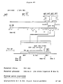
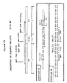
- the first use of recombinant DNA to manipulate herpes simplex virus involved cloning a piece of DNA from the L-S junction region into the unique long region of the DNA, specifically into the thymidine kinase gene (15).
- herpes simplex next involved the creation of deletions in the virus genome by a combination of recombinant DNA and thymidine kinase selection. The first step was to make a specific deletion of the thymidine kinase gene (16).
- the next step involved the insertion of the thymidine kinase gene into the genome at a specific site, and then the thymidine kinase gene and the flanking DNA at the new site were deleted by a selection against thymidine kinase (17). In this manner herpes simplex a pha-22 gene has been deleted (17). In the most recent refinement of this technique, a 15,000 bp sequence of DNA has been deleted from the internal repeat of herpes simplex virus (18).
- herpes simplex glycoprotein C back into a naturally occurring deletion mutant of this gene in herpes simplex virus (19); the insertion of glycoprotein D of herpes simplex type 2 into herpes simplex type 1 (20), again with no manipulation of promoters since the gene is not really 'foreign'; the insertion of hepatitis B surface antigen into herpes simplex virus under the control of the herpes simplex ICP4 promoter (21); and the insertion of bovine growth hormone into herpes saimiri virus with an SV40 promoter that in fact didn't work in that system (an endogenous upstream promoter served to transcribe the gene) (22).
- Two additional cases of foreign genes (chicken ovalbumin gene and Epstein-Barr virus nuclear antigen) have been inserted into herpes simplex virus (30), and glycoprotein X of pseudorabies virus has been inserted into herpes simplex virus (31).
- herpesvirus genomes demonstrate that it is possible to genetically engineer herpesvirus genomes by recombinant DNA techniques.
- the methods that have been used to insert genes involve homologous recombination between the viral DNA cloned on plasmids and purified viral DNA transfected into the same animal cell. In aggregate this is referred to as the homologous recombination technique.
- This technique with minor modifications has been adaptable to other herpesviruses that we have engineered.
- the extent to which one can generalize the location of the deletion and the sites for insertion of foreign genes is not obvious from these previous studies.
- non-primate herpesviruses are amenable to the same techniques as the primate herpesviruses, and that one could establish a targeted approach to the deletion, insertion, and expression of foreign genes.
- IBR virus Infectious bovine rhinotracheitis (IBR) virus, an alpha-herpesvirus with a class D genome, is an important pathogen of cattle. It has been associated with respiratory, ocular, reproductive, central nervous system, enteric, neonatal and dermal diseases (37). Cattle are the normal hosts of IBR virus, however it also infects goats, swine, water buffalo, wildebeest, mink and ferrets. Experimental infections have been established in muledeer, goats, swine, ferrets and rabbits (38).
- IBR has been analyzed at the molecular level as reviewed in (39). A restriction map of the genome is available in this reference, which will aid in the genetic engineering of IBR according to the methods provided by the present invention. No evidence has been presented that IBR has been engineered to contain a deletion or an insertion of foreign DNA.
- Marek's disease virus causes fowl paralysis, a common lymphoproliferative disease of chickens. The disease occurs most commonly in young chickens between 2 and 5 months of age.
- the prominant clinical signs are progressive paralysis of one or more of the extremeties, incoordination due to paralysis of legs, drooping of the limb due to wing involvement, and a lowered head position due to involvement of the neck muscles.
- severe depression may result.
- highly oncogenic strains there is characteristic bursal and thymic atrophy.
- lymphoid tumors affecting the gonads, lungs, liver, spleen, kidney and thymus (37).
- the multivalent vaccines engineered in this invention are a novel way to simultaneously vaccinate against a number of different pathogens.
- herpesviruses contemplated to be amenable to these procedures are feline herpesvirus (FHV), equine herpesvirus (EHV), and canine herpesvirus (CHV). These pathogens cause disease in each of their respective hosts.
- Feline herpesvirus causes feline rhinotracheitis, an acute upper respiratory tract infection characterized by fever, pronounced sneezing, nasal and lacrimal secretions, and depression. The virus may cause corneal ulceration and abortion. The nasal passages and turbinates show focal necrosis, and the tonsils are enlarged and hemorrhagic.
- Equine herpesvirus causes rhinopneumonitis, abortion, exanthema of the genitals and occasionally neurologic disease.
- the acute disease is characterized by fever, anorexia and a profuse, serous nasal discharge.
- the neurologic symptoms when present, consist of ataxia, weakness and paralysis.
- Canine herpesvirus causes severe illness in young puppies, where mortality may reach 80%.
- the disease is characterized by viremia, anorexia, respiratory illness, abdominal pain, vomiting and incessant crying. Generally, there is no fever.
- the principal lesions are disseminated necrosis and hemorrhages in the kidneys, liver and lungs.
- the molecular biology of the feline, equine and canine herpesviruses are in their initial phases. Partial restriction maps are available for equine herpesvirus, and in progress in at least one lab for the feline herpesvirus. Beyond this type of genome analysis, no evidence for the deletion or insertion of foreign genes into these viruses is available.
- the present invention involves the use of genetically engineered herpesvirus to protect animals against disease. It is not obvious which deletions in herpesviruses would serve to attenuate the virus to the proper degree. Even testing vaccine candidates in animal models, e.g. mice, does not serve as a valid predictor of the safety and efficacy of the vaccine in the target animal species, e.g. swine.
- Another subject of the present invention is a vaccine for pseudorabies virus (herpesvirus suis, suid herpesvirus 1, or Aujesky's disease virus) disease of swine.
- Swine are the natural host of pseudorabies virus in which infection in older animals is commonly inapparent but may be characterized by fever, convulsions, and death particularly in younger animals.
- Pseudorabies also infects cattle, sheep, dogs, cats, ferrets, foxes, and rats (37) where the infection usually results in death. Death is usually preceded by intense pruritus, mania, encephalitis, paralysis, and coma.
- Traditional live vaccines are available for use in swine, but they are lethal for the other animals.
- An improved vaccine for pseudorabies would induce a more reliable immune response in swine, would be specifically attenuated to be incapable of reversion to virulence, and would not cause disease in other hosts.
- Pseudorabies virus an alpha-herpesvirus of swine, has a genome of class D (23); that is it contains two copies of a single repeat region, one located between the unique long and unique short DNA region and one at the terminus of the unique short region (see Figure 1).
- Herpes simplex virus is an alpha-herpesvirus with a class E genome (23); that is it contains two copies of each of two repeats.
- Herpes saimiri is a gamma-herpesvirus with a class B genome; that is, it contains numerous reiterations of the same sequence at both termini (23).
- Pseudorabies virus has been studied using the tools of molecular biology including the use of recombinant DNA techniques. BamHI, KpnI, and BgIII restriction maps of the virus genome have been published (24, ,27). DNA transfection procedures have been utilized to rescue temperature sensitive and deletion mutants of the virus by the homologous recombination procedure (24). There are two examples of deletions that have been made in the pseudorabies virus genome - one is a thymidine kinase gene deletion (25), also disclosed in Patent No. 4,514,497 entitled "Modified Live Pseudorabies Viruses".
- This patent teaches thymidine kinase deletions only and does not suggest other attenuating deletions, nor does it suggest insertion of foreign DNA sequences.
- the other reference involves the deletion of a small DNA sequence around a HindIII restriction site in the repeat region (26) upon which European Patent Publication No. 0141458, published on May 15, 1985, corresponding to European Patent Application No. 84201474.8, filed on October 12, 1984 is based.
- This patent application does not teach or suggest attenuating deletions nor does it teach or suggest the insertion of DNA sequences into pseudorabies virus.
- the present invention concerns deletions which have been introduced into the pseudorabies virus genome at sites previously undisclosed. Foreign DNA sequences have also been introduced into the attenuated pseudorabies virus genome and expressed as proteins.
- One embodiment of the invention concerns a vaccine useful for preventing pseudorabies and other swine diseases with a single inoculum.
- pseudorabies literature concerns the presence of naturally-occurring deletions in the genome of two vaccine strains of pseudorabies viruses (27). These deletions are responsible, at least in part, for the attenuated nature of these vaccines however they do not occur in a repeat sequence and do not suggest the attenuation of pseudorabies virus by deleting a portion of a repeat sequence. Such naturally-occurring deletions do not teach methods for making these deletions starting with wild type pseudorabies virus DNA, nor do they suggest other locations at which to make attenuating deletions. There are no examples of naturally-occurring insertions of foreign DNA in herpesviruses.
- pseudorabies virus The natural host of pseudorabies virus is swine, in which infection is commonly inapparent but may be characterized by fever, convulsions and paralysis. Pseudorabies virus also infects cattle, sheep, dogs, cats, foxes and mink, where infection usually results in death of the host. The predominant visible feature of pseudorabies viral infection is intense pruritis generally resulting in host mutilation of the involved area. Violet excitement, fits and paralysis, all symptoms of encephalomyelitis, precede death which usually occurs within a few days following onset of clinical signs.
- Pseudorabies virus disease in swine is of serious concern to governmental bodies worldwide. In the United States, swine from infected herds cannot be sold except to slaughterhouses. Several individual states have separately enacted eradication control practices against pseudorabies. At the current time, any animal vaccinated for pseudorabies disease is treated as though it were infected with pseudorabies virus and is subject to the same regulatory constraints. This is due primarily to the lack of a diagnostic test to differentiate vaccinated from infected animals.
- Subunit vaccines also have limitations. They contain a limited number of viral antigens compared to those produced by live viruses. This paucity of antigens produces a weak immune response of short duration in the vaccinated animal at considerably greater cost than a live virus vaccination.
- the limited spectrum of antigens in the subunit vaccine allows the vaccinated swine to be distinguished from swine which have been infected with the wild-type virus.
- the ability to distinguish vaccinated from infected swine is a crucial property of a pseudorabies vaccine because none of the known vaccines prevent the vaccinated animals from being super-infected by the wild-type virus. While the vaccinated animals do not become sick upon super-infection, there is strong evidence that they may become carriers of the wild-type virus and pass the wild-type virus to other swine.
- a vaccine provided with characteristics which would allow vaccinated animals to be distinguished from animals infected with wild-type virus would be advantageous.
- the subunit vaccines have high cost and poor efficacy but an animal vaccinated with this type of vaccine will produce antibodies only to the limited spectrum of antigens present in the vaccine.
- By sampling the serum of the swine it is possible to show that the vaccinated animal has antibodies only to the antigens contained in the vaccine while an animal infected with the wild-type virus would have antibodies against a wider range of antigens.
- a subunit vaccine used in this way to differentiate vaccinated from pseudorabies infected animals has been disclosed in European Patent Application No.
- Van Oirschot, et al. have used a special monoclonal-based immunological detection system for gI of PRV and have shown that pigs inoculated with naturally-occuring vaccine strains which are missing at least a portion of the gI gene can be differentiated from pigs infected by wild-type PRV.
- this diagnostic test may be used for any of several vaccines against PRV that are already existing in both Europe and the U.S. without differentiating which vacccine was used. This limits the usefulness of this diagnostic, since the vaccines which are detectable have differing biological and virulence properties.
- the approach of deleting a gene to attenuate a virus coupled with a diagnostic for that gene provides a vaccine that can be differentiated from any of the currently used PRV vaccines and from wild-type PRV. It is important to be able to differentiate a new, safer vaccine from those currently used because pigs receiving the current vaccines are all regulated during eradication programs to the same extent as those infected with wild-type PRV.
- Antigens of choice for the purpose of a diagnostic marker would have the following characteristics: 1) the antigens and their genes would be non-essential for the production of infectious virus in tissue culture; and 2) the antigen would elicit a major serological response in the animal, but is preferably not an important neutralizing antigen.
- the present invention therefore involves the ability to attenuate pseudorabies virus of swine to create a live virus vaccine and the ability to distinguish whether an animal has been given the vaccination or whether the animal has been infected by wild-type pseudorabies virus.
- the present invention provides a recombinant nonprimate herpesvirus which comprises a foreign DNA sequence inserted into a non-essential region of the genomic DNA of a herpesvirus.
- the inserted foreign DNA sequence which is expressed in a host cell infected with the recombinant herpesvirus, encodes a recombinant fusion protein which comprises at least a portion of the pseudorabies virus glycoprotein X and a polypeptide which is fused to the carboxy terminus of the portion of the glycoprotein X.
- the present invention further provides a recombinant fusion protein which comprises at least a portion of pseudorabies virus glycoprotein X and a polypeptide which is fused to the carboxy terminus of the portion of the pseudorabies virus glycoprotein X.
- the present invention provides a recombinant herpesvirus which comprises a foreign DNA sequence inserted into a non-essential region of the genomic DNA of a herpesvirus.
- the inserted foreign DNA sequence which is expressed in a host cell infected with the recombinant herpesvirus, encodes a recombinant fusion protein which comprises the E. coli b -galactosidase fused at its carboxy terminus to a polypeptide.
- the present invention provides a vaccine which comprises an effective immunizing amount of the recombinant herpesvirus of the present invention and a suitable carrier.
- herpesviruses are described, at least in part, by reference to ATCC Accession Numbers. These herpesviruses have been deposited with the American Type Culture Collection (ATCC), 12301 Parklawn Drive, Rockville, Maryland 20852. These deposits were made pursuant to the Budapest Treaty on The International Recognition of the Deposit of Microorganisms for the Purposes of Patent Procedure.
- ATCC American Type Culture Collection
- the present invention provides a recombinant nonprimate herpesvirus which comprises a foreign DNA sequence inserted into a non-essential region of the genomic DNA of a herpesvirus.
- the inserted foreign DNA sequence encodes a recombinant fusion protein which comprises at least a portion of the pseudorabies virus glycoprotein X and a polypeptide which is fused to the carboxy terminus of the portion of the glycoprotein X.
- the inserted foreign DNA sequence is expressed in a host cell infected with the recombinant herpesvirus.
- the herpesvirus into which the foreign DNA sequence is inserted is a pseudorabies virus.
- the pseudorabies virus is attenuated.
- recombinant herpesvirus of the present invention examples are recombinant viruses designated S-PRV-040 (ATCC Accession No. VR 2214), S-PRV-098 (ATCC Accession No. VR 2219), S-PRV-088 (ATCC Accession No. VR 2217), and S-PRV-093 (ATCC Accession No. VR 2218).
- the expression of the inserted foreign DNA sequence is under control of a promoter located upstream of the foreign DNA sequence.
- promoter examples include herpes simplex virus type-1 thymidine kinase promoter and pseudorabies virus glycoprotein X promoter.
- the polypeptide fused to the carboxy terminus of the portion of the glycoprotein X is an antigenic polypeptide.
- the antigenic polypeptide may be a part of the swine parvovirus capsid protein, malaria CSP protein, or malaria CSP repeat region.
- the polypeptide is embedded in the pseudorabies virus glycoprotein X. In another embodiment, the polypeptide is fused to the amino terminal transmembrane signal sequence of the glycoprotein X.
- the present invention further provides a recombinant fusion protein which comprises at least a portion of pseudorabies virus glycoprotein X and a polypeptide which is fused to the carboxy terminus of the portion of the pseudorabies virus glycoprotein X.
- the polypeptide fused to the carboxy terminus of the portion of the glycoprotein X is an antigenic polypeptide.
- the antigenic polypeptide may be a part of the swine parvovirus capsid protein, malaria CSP protein, or malaria CSP repeat region.
- this invention provides a recombinant nonprimate herpesvirus which comprises a foreign DNA sequence inserted into a non-essential region of the genomic DNA of a herpesvirus.
- the inserted foreign DNA sequence encodes a recombinant fusion protein which comprises the E. coli b -galactosidase fused at its carboxy terminus to a polypeptide.
- the inserted foreign DNA sequence is expressed in a host cell infected with the recombinant herpesvirus.
- the herpesvirus into which the foreign DNA sequence is inserted is a pseudorabies virus.
- the pseudorabies virus is attenuated.
- recombinant herpesvirus of the present invention examples are recombinant viruses designated S-PRV-065 (ATCC Accession No. VR 2215) and S-PRV-086 (ATCC Accession No. VR 2216).
- the expression of the inserted foreign DNA sequence is under control of a promoter located upstream of the foreign DNA sequence.
- promoter examples include herpes simplex virus type-1 thymidine kinase promoter and pseudorabies virus glycoprotein X promoter.
- the polypeptide fused to the carboxy terminus of the E. coli b -galactosidase is an antigenic polypeptide.
- the antigenic polypeptide may be a part of the swine parvovirus capsid protein, malaria CSP protein, or malaria CSP repeat region.
- An effective immunizing amount of the recombinant herpesvirus of the present invention may be formulated into vaccines using methods well known to those skilled in the art.
- Vero cells for PRV; MDBK cells for IBR; CEF cells for HVT; Crandall feline kidney cells for FHV. Vero cells are suitable for EHV and MDCK cells are suitable for CHV.
- Herpesvirus stock samples were prepared by infecting tissue culture cells at a multiplicity of infection of 0.01 PFU/cell in Dulbecco's Modified Eagle Medium (DMEM) containing 2 mM glutamine, 100 units/ml penicillin, 100 units/ml streptomycin (these components were obtained from Irvine Scientific or equivalent supplier, and thereafter are referred to as complete DME medium) plus 1% fetal bovine serum. After cytopathic effect was complete, the medium and cells were harvested and the cells were pelleted at 3000 rpm for 5 minutes in a clinical centrifuge.
- DMEM Dulbecco's Modified Eagle Medium
- HVT herpesvirus
- the cells were resuspended in 1/10 the original volume of medium, and an equal volume of 2 times autoclaved skim milk (9% skim milk powder in H20 wgt/vol) was added.
- the virus sample was frozen and thawed 2 times, aliquoted, and stored frozen at -70 o C.
- the titer was usually about 108 plaque forming units per ml.
- infected cells were resuspended in complete medium containing 20% fetal bovine serum, 10% DMSO and stored frozen at - 70 o C.
- HERPESVIRUS DNA PREPARATION OF HERPESVIRUS DNA.
- a confluent monolayer of tissue culture cells in a 25 cm2 flask or a 60 mm petri dish was infected with 100 microliters of virus sample in 1 ml medium. Adsorption proceeded for 1-2 hours at 37 o C in a humidified incubator with 5% C02 in air. After adsorption, 4 mls of complete DME medium plus 1% fetal bovine serum were added. After overnight incubation, or when the cells were showing 100% cytopathic effect, the cells were scraped into the medium with a cell scraper (Costar brand). The cells and medium were centrifuged at 3000 rpm for 5 minutes in a clinical centrifuge.
- the medium was decanted, and the cell pellet was gently resuspended in 0.5 ml solution containing 0.01 M Tris pH 7.5, 1 mM EDTA, and 0.5% Nonidet P-40 (NP40, an ionic detergent comprising an octyl phenol ethylene oxide condensate containing an average of 9 moles ethylene oxide per molecule, purchased from Sigma Chemical Co., St. Louis, MO.).
- NP40 an ionic detergent comprising an octyl phenol ethylene oxide condensate containing an average of 9 moles ethylene oxide per molecule, purchased from Sigma Chemical Co., St. Louis, MO.
- the sample was centrifuged for 5 minutes at 3000 rpm in a clinical centrifuge to pellet nuclei.
- the DNA pellet was removed with a pasteur pipette or wooden stick and discarded.
- the supernatant fluid was decanted into a 1.5 ml Eppendorf tube containing 25 microliters of 20% sodium dodecyl sulfate (Sigma) and 25 microliters proteinase-K (10 mg/ml; Boehringer Mannheim supplier).
- the sample was mixed and incubated at 37 o C for 30-60 minutes. An equal volume of water-saturated phenol was added and the sample was mixed on a vortex mixer for 1 minute. The same was centrifuged in an Eppendorf minifuge for 5 minutes at full speed.
- the upper aqueous phase was removed to a new Eppendorf tube, and two volumes of -20 o C absolute ethanol were added and the tube put at -20 o C for 30 minutes to precipitate nucleic acid.
- the sample was centrifuged in an Eppendorf centrifuge at 4oC for 5 minutes. The supernatant was decanted, and the pellet was washed one time with cold 80% ethanol. The pellet was dried in a lyophilizer, and rehydrated in 17 microliters H20.
- the procedure was scaled up to start with a 850 cm2 roller bottle of Vero cells.
- the DNA was stored in H20 or in 0.01 M Tris pH 7.5, 1 mM EDTA at -20oC or +4oC.
- HVT virus DNA PREPARATION OF HERPES VIRUS OF TURKEY DNA. All manipulations requiring herpes virus of turkey (HVT) were made using strain FC-126 (ATCC #584-C).
- HVT virus DNA For the preparation of HVT virus DNA from the cytoplasm of infected cells, primary chicken embryo fibroblasts were infected at a MOI sufficient to cause extensive cytopathic effect before the cells overgrew. All incubations were carried out at 39°C in a humidified incubator with 5% CO2 in air. Best DNA yields were obtained by harvesting monolayers which were maximally infected, but showing incomplete cell lysis (typically 5-7 days). Infected cells were harvested by scraping the cells into the medium using a cell scraper (Costar brand).
- the cell suspension was centrifuged at 3000 rpm for 10 minutes at 5°C in a GS-3 rotor (Sorvall Instruments).
- the resultant pellet was resuspended in cold PBS (20ml/Roller Bottle) and subjected to another centrifugation for 10 minutes at 3000 rpm in the cold.
- the cellular pellet was resuspended in 4 ml/ roller bottle of RSB buffer (10 mM Tris pH 7.5, 1 mM EDTA, and 1.5 mM MgCl2).
- NP40 Nonidet P-40;Sigma
- the sample was centrifuged for 10 minutes at 3000 rpm in the cold to pellet the nuclei and remove cellular debris. The supernatant fluid was carefully transferred to a 15 ml Corex centifuge tube. Both EDTA (0.5M pH 8.0) and SDS (sodium dodecyl sulfate; stock 20%) were added to the sample to final concentrations of 5 mM and 1%, respectively.
- EDTA 0.5M pH 8.0
- SDS sodium dodecyl sulfate
- One hundred microliters of proteinase-K (10 mg/ml; Boehringer Mannheim) was added per 4 milliliters of sample, mixed, and incubated at 45°C for 1-2 hours. After this period, an equal volume of water-saturated phenol was added to the sample and gently mixed by hand.
- the sample was spun in a clinical centrifuge for 5 minutes at 3000 rpm to separate the phases. NaOAc was added to the upper aqueous phase to a final concentration of 0.3M (stock solution 3M pH 5.2), and the nucleic acid precipitated at -70°C for 30 minutes after the addition of 2.5 volumes of cold absolute ethanol. DNA is the sample was pelleted by spinning for 20 minutes at 8000 rpm in an HB-4 rotor at 5°C. The supernatant was carefully removed and the DNA pellet washed once with 25 ml of 80% ethanol.
- the DNA pellet was dried briefly by vacuum (2-3 minutes), and resuspended in 50 microliters/roller bottle of infected cells of T10E1 buffer (10 mM Tris pH 7.5, 1 mM EDTA). Typically, yields of viral DNA ranged between 5-10 micrograms/roller bottle of infected cells. All viral DNA was stored at approximately 10°C.
- HERPESVIRUS CELLYSATES PREPARATION HERPESVIRUS CELLYSATES.
- a conflunent monolayer of tissue culture cells in a 25 cm flask or a 60 mm petri dish was infected with 100 microliters of virus sample in 1 ml of medium. Adsorption proceeded for 1-2 hours at 37°C in a humidified incubator with 5% CO2 in air. After adsorption, 4 mls of medium were added. After overnight incubation, or when cells were showing 100% cytopathic effect, the cells were scraped into the medium with a cells scraper (Costar brand). The cells and medium were centrifuged at 3000 rpm for 5 minutes in a clinical centrifuge.
- the medium was decanted, and the cell pellet was suspended in 250 microliters of disruption buffer (2% sodium dodecyl sulfate, 2% beta-mercapto-ethanol). The sample was sonicated for 30 seconds on ice and stored at -20°C.
- disruption buffer 2% sodium dodecyl sulfate, 2% beta-mercapto-ethanol
- PHENOL EXTRACTION Phenol extraction was performed on any convenient volume of DNA sample, typically between 100 microliters to 1 ml.
- the DNA sample was diluted in 0.01M Tris pH 7.5, 1 mM EDTA and an equal volume of water saturated phenol was added.
- the sample was mixed briefly on a vortex mixer and placed on ice for 3 minutes. After centrifugation for 3 minutes in a microfuge, the aqueous layer was removed to a new tube and was precipitated by ethanol.
- AGAROSE GEL ELECTROPHORESIS OF DNA To visualize the restriction pattern of the DNA, 5 microliters of loading buffer (5X electrophoresis buffer, 0.01% bromphenol blue dye, 50 mM EDTA, and 50% glycerol) were added. The sample was loaded into a lane in a horizontal submarine electrophoresis unit containing a 0.6% agarose gel.
- the electrophoresis buffer was 40 mM Tris, 10 mM EDTA, adjusted to pH 7.8 with acetic acid, and with or without 0.5 micrograms/ml ethidium bromide.
- the gel was run at 40-50V for 18 hours, and the gel was removed and strained with 0.5 micrograms/ml ethidium bromide for 30 minutes.
- the DNA bands were visualized on a long wavelength UV transilluminator.
- PHOSPHATASE TREATMENT OF DNA Phosphatase treatment of DNA was performed by adding 1 microliter (25 units) of calf intestinal phosphatase (Boehringer Mannheim) directly to the restriction enzyme digestion reactions and continuing the incubation for 30 minutes at 37 o C. The phosphatase was inactivated for 60 minutes at 65 o C prior to phenol extraction.
- DNA was resuspended in 100 microliters of 60 mM Tris pH 8.0, 0.66 mM MgCl2, 1 mM beta-mercaptoethanol. The sample was warmed to 30 o C for 5 minutes, and 10 units of lambda exonuclease III (BRL) were added. At frequent time intervals (e.g. every 2.5 minutes), 10 microliter aliquots were diluted into 100 microliters of 30 mM sodium acetate pH 4.5, 250 mM NaCl, 1 mM ZnSO4, 4 micrograms/100 microliters yeast tRNA, 30 units/ 100 microliters S1 nuclease.
- BTL lambda exonuclease III
- stop buffer consisting of 625 mM Tris pH 9.0, 150 mM EDTA, 1% SDS were added. The samples were then phenol extracted and ethanol precipitated as above. The DNA digestion products were then analyzed and purified by agarose gel electrophoresis.
- T4 DNA ligase BBL
- Ligation reactions contained 10 fmoles DNA, 20 mM Tris pH 7.5, 10 mM MgCl2, 10 mM dithiothreitol (DTT), 200 micromolar ATP, and 20 units T4 DNA ligase in 10 microliters final reaction volume. The ligation was allowed to proceed for 3-16 hours at 15 o C. Typically DNA fragments to be ligated together were added to an equal molar ratio. Typically two different DNA fragments were joined during ligation, but joining of three or four different DNAs at once was also possible.
- Probe DNA was added that had been labelled by nick translation using a kit from Bethesda Research Laboratories (BRL) and one 32p-labeled nucleotide.
- the probe DNA was separated from the unincorporated nucleotides by NACS column (BRL) or on a Sephadex G50 column (Pharmacia). After overnight hybridization at 55 o C, the filter was washed once with 2X SSC at room temperature followed by two washes with 0.1X SSC, 0.1% sodium dodecyl sulfate (SDS) for 30 minutes at 55 o C. The filter was dried and autoradiographed.
- DNA TRANSFECTION FOR GENERATING RECOMBINANT VIRUS The method is based upon the calcium phosphate DNA precipitation procedure of Graham and Van der Eb (32) with the following modifications.
- 0.1-0.2 micrograms of plasmid DNA containing the foreign DNA flanked by appropriate herepesvirus cloned sequences (the homovector) were mixed with 0.3 micrograms of intact DNA. Both DNAs were stored either in H2O or 0.01 M Tris pH 7.5, 1 mM EDTA and the final volume should be less than 0.25 ml.
- the glycerol shock consisted of removing the medium and adding DME containing 20% glycerol for 3 minutes at room temperature, followed by a wash with 10% glycerol in DME, and a wash in 5% glycerol in DME, followed by the addition of fresh complete DME medium plus 10% fetal bovin serum.
- the cells were incubated at 37 o C as above for 3-4 days until cytopathic effect from the virus was 50-100%.
- Virus was harvested as described above for the preparation of virus stocks. This stock was referred to as a transfection stock and it was subsequently screened for recombinant virus either with or without a selection mechanism to enrich for recombinant plaques as described below.
- the cells were plated out the day before in CEF growth media (1X F10/199, 5% fetal calf serum, 2% glutamine, 1% non-essential amino acids, and 2% penicillin/streptomycin) containing 4 micrograms/milliliter polybrene (stock 4 mg/ml in 1X HBSS).
- CEF growth media (1X F10/199, 5% fetal calf serum, 2% glutamine, 1% non-essential amino acids, and 2% penicillin/streptomycin
- 5 micrograms of the plasmid homology vector was mixed with 5 micrograms of intact HVT DNA, and suspended in 1 ml of CEF media containing 30 micrograms/milliliter polybrene (stock 4 mg/ml in 1 X HBSS).
- the DNA-polybrene suspension (1ml) was then added to a 6 cm plate of CEF cells from which the media has been aspirated, and incubated at 39°C for 30 minutes. The plates were rocked periodically during this time to redistribute the inoculum. After this period, 4 ml of CEF growth media was added directly to each plate, and incubated an additional 2.5 hours at 39°C. At this time, the media was removed from each plate, and the cells shocked with 2 ml of 30% DMSO (Dimethyl Sulfoxide, J.T. Baker Chemical Co.) in 1X HBSS for 4 minutes at room temperature. The 30% DMSO was carefully removed and the monolayers washed once with 1X HBSS at room temperature.
- 30% DMSO Dimethyl Sulfoxide
- the cells were then incubated at 39°C after the addition of 5 mls of CEF growth media. The next day, the media was changed to remove any last traces of DMSO and to stimulate cell growth. Cytopathic effect from the virus becomes apparent within 6 days. Generation of a high titer stock (80%-90% CPE) can usually be made within 1 week from this date. HVT stock samples were prepared by resuspending the infected cells in CEF growth media containing 20% fetal calf serum, 10% DMSO and stored at -70°C.
- DIRECT LIGATION PROCEDURE FOR GENERATING RECOMBINANT HERPESVIRUSES Rather than using homovectors and relying upon homologous recombination to generate recombinant virus, the technique of direct ligation was developed to insert foreign genes into herpesviruses. In this instance, the cloned foreign gene did not require flanking herpesvirus DNA sequences but only required that it have restriction sites available to cut out the foreign gene fragment from the plasmid vector. A compatable restriction enzyme was used to cut the herpesvirus DNA. A requirement of the technique was that the restriction enzyme used to cut the herpesvirus DNA must cut at a limited number of sites, preferably less than 3 sites.
- PRV DNA For PRV DNA, we have used xbaI, which cut PRV DNA in two places, and contemplate the use of HindIII (2 cuts), EcoRV (2 or 3 cuts) or NdeI (3-5 cuts).
- the herpesvirus DNA was mixed with a 30-fold molar excess of plasmid DNA, and the mixture was cut with the appropriate restriction enzyme.
- the DNA mixture was phenol extracted and ethanol precipitated to remove restriction enzymes, and ligated together according to the ligation procedure detailed above.
- the ligated DNA mixture was then phenol extracted, ethanol precipitated, and resuspended in 298 microliters 0.01M Tris pH 7.5, 1 mM EDTA. Forty-two microliters of 2M CaCl2 were added, followed by an equal volume of 1X HEPES buffers saline (see above), and the sample was used to transfect animal cells as described above.
- the virus in the transfection stock was then screened for foreign DNA inserts as described below.
- the advantage of the direct ligation technique was that it required less construction of sub-clones in the plasmid state, and that the recombinant virus was present in the transfection stock at a much higher frequency than with homologous recombination.
- HAT SELECTION OF RECOMBINANT HERPESVIRUS EXPRESSING THYMIDINE KINASE Deletion mutants of herpesviruses which suffered deletions in the thymidine kinase (TK) gene were constructed. These PRV strains have been designated S-PRV-002 and S-PRV-003 and have been deposited with the ATCC under Accession No. VR 2107 and VR 2108 respectively. These TK minus (TK-) viruses have been used as recipients for the insertion of the foreign herpes simplex type 1 (HSV-1) TK gene.
- HSV-1 TK gene that we have used contains the HSV-1 ICP4 promoter and was from B. Roizman (16).
- the transfection stock was used to infect monolayers of 143 TK- cells in 60 mm culture dishes that had been preincubated in HAT medium for 16 hours at 37 o C (HAT medium: medium 199 containing 2 mM glutamine, 100 units/ml penicillin, 100 units/ml streptomycin, 10% fetal bovin serum, 5 x 10 ⁇ 5 M hypoxanthine, 10 ⁇ 5 M thymidine, 5 X 10 ⁇ 6 M aminopterin). Samples of the transfection stock virus were infected into the 143 TK- cells using 10 ⁇ 3 to 10 ⁇ 7 dilutions of virus.
- the dishes inoculated with the highest dilution of virus and still showing virus plaques were harvested for virus stocks, and the selection was repeated a second time.
- the virus stock harvested from the second HAT selection was used in a plaque assay and individual plaques were picked and tested for foreign DNA inserts as described below.
- BROMODEOXYURIDINE SELECTION OF RECOMBINANT HERPESVIRUS In order to insert a foreign gene in place of a TK gene already present in the herpesvirus genome, the foreign gene was cloned in plasmids so that it contained the same flanking homology regions as the TK genes. These flanking regions could be part of the TK gene itself, or parts of the herpesvirus that flank the TK gene. In either case, the plasmid DNA containing the foreign gene was transfected with intact herpesvirus genomic DNA containing the HSV-1 TK gene.
- the transfection stock of recombinant virus was grown for two selections in 143 TK- cells in the present of 40 micrograms/ml bromodeoxyuridine (BUDR, Sigma) in complete DME medium plus 10% fetal bovine serum.
- the drug BUDR is an analogue of thymidine that is recognized by the viral enzyme thymidine kinase (TK) and is ultimately incorporated into DNA.
- TK thymidine kinase
- BUDR mutagenic and lethal and thus selects against viruses that have an active TK gene.
- viruses that had exchanged their TK gene for a foreign gene by homologous recombination were enriched in the population. Screening for the recombinant viruses was then performed by one of the techniques detailed below.
- HYBRIDIZATION SCREEN FOR RECOMBINANT HERPESVIRUS One procedure used is described in (36). The technique involved doing a plaque assay on PRV under agarose, removing the agarose once plaques had formed, and lifting the cell monolayer from the dish onto a nitrocellulose membrane filter. The filter was then processed through the Southern procedure for DNA hybridization as detailed above. The DNA probe used in the procedure was made from the foreign gene that had been inserted into the virus. Thus plaques that contain the foreign gene were identified, and they were picked from the agarose overlay that had been saved.
- BLUOGAL SCREEN FOR RECOMBINANT HERPESVIRUS When the foreign gene encoded the enzyme beta-galactosidase, the plaques that contained the gene were visualized more easily.
- the chemical Bluogal ⁇ (BRL) was incorporated at the level of 200-300 micrograms/ml into the agarose overlay during the plaque assay, and the plaques that expressed active beta -galactosidase turned blue. The blue plaques were then picked and purified by further blue plaque isolations.
- Other foreign genes were inserted by homologous recombination such that they replaced the beta-galactosidase gene; in the instance non-blue plaques were picked for purification of the recombinant virus.
- a third method for screening the recombinant virus stock was to look directly for the expression of the foreign gene with antibodies.
- Herpesvirus plaques were spotted and picked by inserting a toothpick through the agarose above the plaque and scraping the plaque area on the dish. Viruses were then rinsed from the toothpick by inserting the toothpick ino a well of a 96-well microtiter dish (Falcon Plastics) containing a confluent monolayer of tissue culture cells that had been washed 3 times in DME medium without serum. It was important for the virus to grow without serum at this stage to allow the immunological procedure to work.
- the plates were put at -700C to freeze and lyse the cells.
- the medium was thawed, and the freeze/thaw procedure was repeated a second time.
- 50-100 microliters of medium were removed from each well and filtered under vacuum through a nitrocellulose membrane (S&S BA85) using a DotBlot ⁇ apparatus (BRL).
- the filter blots were soaked in a blocking solution of 0.01 M Tris pH 7.5, 0.1 M NaCl, 3% bovine serum albumin at room temperature for two hours with shaking.
- the filter blocks were then placed in a sealable bag (Sears Seal-A-Meal ⁇ or equivalent), and 10 mls of the blocking solution that contained 10 microliters of antibody specific for the foreign protein were added. After overnight incubation at room temperature with shaking, the blot was washed 3 times with 100 mls 0.01 M Tris, pH 7.5, 0.1 M NaCl, 0.05% Tween 20 detergent (Sigma). The blot was put in another sealable bag and 10 mls blocking solution containing 106 counts per minute of 125I-protein A (New England Nuclear) were added.
- the blot was washed as above, dried, and overlayed with an x-ray film and an intensifying screen (Dupont) and autoradiographed for 1-3 days at -70 o C.
- the film was developed by standard procedures. Virus from the positive wells which contained the recominant virus was further purified.
- a prewet piece of Whatman 3 mm paper was placed on top of the nitrocellulose filter to create a "sandwich", and the sandwich was placed into an electrophoretic transfer device (Biorad). The sandwich was completely submersed in transfer buffer. The electrophoretic transfer was carried out for 3 hours at 250 milliamps. After transfer, the nitrocellulose filter was removed from the assembly and placed in a dish containing 50 mls of blocking buffer (50 mg/ml bovin serum albumin, 10 mM magnesium chloride, 100 mM potassium chloride, 1 mM calcium chloride, 10 mM imidazole pH 7.0, 0.3% Tween-20, 0.02% sodium azide).
- blocking buffer 50 mg/ml bovin serum albumin, 10 mM magnesium chloride, 100 mM potassium chloride, 1 mM calcium chloride, 10 mM imidazole pH 7.0, 0.3% Tween-20, 0.02% sodium azide.
- the nitrocellulose blot was incubated for 1-2 hours in the blocking buffer at room temperature on a shaker. The blot was then placed in a sealable bag containing 15 mls of the blocking buffer plus the specific antiserum as a probe and incubated overnight at 37 o C on a shaker. The blot was then removed from the probe solution and rinsed with 5-6 changes of phosphate buffered saline over a period of 1 hour. The phosphate buffered saline was removed and 50 mls of blocking buffer containing 5 x 10⁵ cpm of 125I labeled protein A (Amersham) were added.
- the blot was incubated for 1 hour with a labeled protein A solution, the labeled protein A solution was removed and the blot was rinsed with 5-6 changes of phosphate buffered saline solution containing 0.3% Tween-20. The blot was air dried and autoradiographed overnight with an intensifying screen.
- the OSU strain of porcine rotavirus was propagated on MA-104 cells (Rhesus monkey kidney cells from MA Bioproducts). Confluent monolayers were infected at a multiplicity of infection of greater than 10 in DMEM containing 5 micrograms/ml trpysin. Cells were incubated with the virus for 48 hours or until a cytopathic effect was obtained. Media and cell debris were collected and centrifuged at 10,000 x g for 20 minutes at 4 o C.
- the supernatant containing the rotavirus was then centrifuged at 10,000 x g in a preparative Beckman Ti45 rotor at 4 o C.
- Virus pellets were resuspended in SM medium (50 mM Tris-HCl pH 7.5, 100 mM KCL, 10 mM MgCl2) and homogenized lightly in a Dounce-type homogenizer.
- the resuspended virus was centrifuged at 10,000 x g for 10 minutes then loaded onto 25-50% CsCl gradients in SM buffer. Gradients were centrifuged at 100,000 x g for 4 hours at 20 o C.
- the two blue-white bands representing intact virions and cores of rotavirus were collected, diluted, and the CsCl gradient procedure was repeated a second time.
- Virus obtained from the second gradient was dialyzed overnight against SM buffer at 4 o C.
- RNA ISOLATION VIRAL RNA ISOLATION. Dialyzed swine rotavirus was twice extracted with an equal volume of SDS/phenol then twice more with chloroform: isoamylalcohol (24:1). The double stranded RNA was precipitated with ethanol in the presence of 0.2 M sodium acetate, centrifuged and resuspended in water. The yield was typically 100 micrograms from 1,000 cm2 of infected cells.
- RNA-primer mixture was boiled for 3 minutes in a water bath then chilled on ice.
- the aqueous layer was removed and to it 250 microliters of 4 M ammonium acetate and 1.0 ml of 95% ethanol was added, the mixture was frozen in dry ice and centrifuged in the cold. The resulting pellet was resuspended in 100 microliters of 10 mM Tris-HC1 pH 7.5 and the ammonium acetate precipitation procedure was repeated. The pellet was resuspended in 100 microliters of 0.3 M KOH and incubated at room temperature overnight then at 37 o C for 2 hours. The solution was brought to neutral pH by addition of 10 microliters of 3.0 M HCl and 25 microliters of 1.0 M Tris-HCl pH 7.5.
- the resulting single-stranded cDNA was then precipitated two times by the above described ammonium acetate-ethanol procedure.
- the pellet obtained was resuspended in 50 microliters of 10 mM Tris-HCl pH 7.5, 100 mM NaCl, 1 mM EDTA, boiled in a water bath for 2 minutes then incubated at 59 o C for 16 hours.
- the solution was lyophilized to a volume of 15 microliters and the resulting double-stranded cDNA was run on a 1.0% agarose gel (Sigma agarose Type II).
- the ethidium bromide strained DNA migrating at 1,000-1,100 bas pair length was excised from the gel and electroeluted in a CBS electroeluter device.
- the solution was lyophilized, and the cDNA was resuspended in 25 microliters of water.
- the reaction was incubated at room temperature for 15 minutes, then chloroform/phenol extracted and ammonium acetate-ethanol precipitated as described above.
- the resulting cDNA was tailed with dCTP using terminal deoxynucleotide transferase (BRL buffer and enzyme used).
- the reaction was stopped with 2 microliters of 0.5 M EDTA, chloroform/phenol extracted and precipitated with sodium acetate in the presence of 10 micrograms of carrier tRNA.
- the resuspended cDNA was mixed with 200 ng of dGMP-tailed Pst I cut pBR322 (BRL catalog #5355SA) in 200 microliters of 10 mm Tris-HCl pH 7.5, 100 mM NaCl, 1 mM EDTA, heated to 65 o C for 5 minutes then 57 o C for 2 hours.
- the annealed cDNA-vector pBR322 was transformed onto E . coli DH-1 cells prepared for high efficiency transformation.
- Colonies that showed sensitivity to ampicillin and tetracycline resistance were grown and DNA was prepared and cut with Pst I to determine the size of the cDNA insert.
- Several clones having Pst I inserts of 1,050-1,100 base pairs were analyzed and found to have identical restrictrion enzyme digest patterns. The largest clone was designated pSY565 and have been deposited with the ATCC under accession number 53,340.
- the 1,100 base pair Pst I insert was subcloned into a M13 phage sequencing vector. The entire DNA sequence of this clone was determined and is shown in Fig. 10A and 10B. The location of the gp38 open reading frame was determined from the amino acid homology to human and bovine sequences already published (44).
- Virus pellets was resuspended in SM medium (50 mM Tris-HCl pH 7.5, 100 mM KCL, 10 mM MgCl2) and homogenized lightly in a Dounce-type homogenizer.
- the resuspended virus was centrifuged at 10,000 x g for 10 minutes then loaded onto 25-50% CsCl gradients in SM buffer. Gradients were centrifuged at 100,000 x g for 4 hours at 20 o C.
- the two blue-white bands representing intact virions and cores of rotavirus were collected, diluted, and the CsCl gradient procedure was repeated a second time. Virus obtained from the second gradient was dialyzed overnight against SM buffer at 4 o C.
- RNA ISOLATION VIRAL RNA ISOLATION. Dialyzed bovine rotavirus was twice extracted with an equal volume of SDS/phenol then twice more with chloroform: isoamylalcohol (24:1). The double stranded RNA was precipitated with ethanol in the presence of 0.2 M sodium acetate, centrifuged and resuspended in water. The yield was typically 100 micrograms from 1,000 cm2 of infected cells.
- RNA-primer mixture was boiled for 3 minutes in a water bath then chilled on ice.
- the aqueous layer was removed and to it 250 microliters of 4 M ammonium acetate and 1.0 ml of 95% ethanol was added, the mixture was frozen in dry ice and centrifuged in the cold. The resulting pellet was resuspended in 100 microliters of 10 mM Tris-HCl pH 7.5 and the ammonium acetate precipitation procedure was repeated. The pellet was resuspended in 100 microliters of 0.3 M KOH and incubated at room temperature overnight then at 37 o C for 2 hours. The solution was brought to neutral pH by addition of 10 microliters of 3.0 M HCl and 25 microliters of 1.0 M Tris-HCl pH 7.5.
- the resulting single-stranded cDNA was then precipitated two times by the above described ammonium acetate-ethanol procedure.
- the pellet obtained was resuspended in 50 microliters of 10 mM Tris-HCl pH 7.5, 100 mM NaCl, 1 mM EDTA, boiled in a water bath for 2 minutes then incubated at 59 o C for 16 hours.
- the solution was lyophilized to a volume of 15 microliters and the resulting double-stranded cDNA was run on a 1.0% agarose gel (Sigma agarose Type II).
- the ethidium bromide stained DNA migrating at 1,000-1,100 base pair length was excised from the gel and electroeluted in a CBS electroeluter device.
- the solution was lyophilized, and the cDNA was resuspended in 25 microliters of water.
- the reaction was incubated at room temperature for 15 minutes, then chloroform/phenol extracted and ammonium acteate-ethanol precipitated as described above.
- the resulting cDNA was tailed with dCTP using terminal deoxynucleotide transferase (BRL buffer and enzyme used).
- the reaction was stopped with 2 microliters of 0.5 M EDTA, chloroform/phenol extracted and precipitated with sodium acetate in the presence of 10 micrograms of carrier tRNA.
- the resuspended cDNA was mixed with 200 ng of dGMP-tailed Pst I cut pBR322 (BRL catalog #5355SA) in 200 microliters of 10 mM Tris-HCl pH 7.5, 100 mM NaCl, 1 mM EDTA, heated to 65 o C for 5 minutes then 57 o C for 2 hours.
- the annealed cDNA-vector pBR322 was transformed onto E . coli DH-1 cells prepared for high efficiency transformation.
- Colonies that showed sensitivity to ampicillin and tetracycline resistance were grown and DNA was prepared and cut with Pst I determine the size of the cDNA insert.
- Pst I inserts of 1,050-1,100 base pairs were analyzed and found to have identical restriction enzyme digest patterns.
- the 1,100 base pair Pst I insert was subcloned into a M13 phage sequencing vector. Part of the DNA sequence of this clone was determined and was found to be identical to the published sequence (24).
- the antibiotic G418 (GIBCO) has a wide range of inhibitory activity on protein synthesis.
- the recombinant virus expressed the aminoglycoside 3′-phosphotransferase, encoded by the NEO gene, upon acquiring the foreign gene and became resistant to G418.
- the transfection stocks of recombinant viruses were grown on MDBK (for IBR virus), Vero (for PRV) or QT35 (for HVT) cells in the presence of 500 micrograms/ml G418 in complete DME medium plus 1% fetal bovine serum.
- plaques from the dishes inoculated with the highest dilution of virus were picked for virus stocks.
- the selection was repeated a second or third time.
- the virus stocks generated from the G418 selection were tested for NEO gene insertion by the SOUTHERN BLOTTING OF DNA hybridization procedure described above.
- gpX was purified from the tissue culture medium of infected Vero cells grown in complete DME plus 1% bovine serum. Confluent Vero cells were infected at a multiplicaity of infection equal to 5, with wild-type, Iowa S-62 strain pseudorabies virus. The viral proteins were radiolabelled with 14C glucosamine and/or 35S methionine by adding the appropirate label to the flask eight hours after infection. The cells and media were harvested at twenty hours post infection, when the cells showed considerable cytopathic effect and the fluids were centrifuged.
- the supernatant fluid was concentrated 10X and dialyzed against 0.02M sodium sulfate/0.01M sodium phosphate buffer, pH 7.2 (16 hours, 0 o C), then against two changes of 0.01M sodium phosphate buffer, pH 7.2 (24 hours, 0 o C).
- the dialysate was treated for 30 minutes at 0 o C with 70% perchloric acid to a final concentration of 0.2M perchloric acid, then centrifuged at 10,000 rpm for 25 minutes.
- the supernatant fluid was then dialyzed against 0.02M Tris, pH 8.5.
- the acid-soluble proteins were separated on a Biogel TSK DXAE 5-PW column (75 x 75 mm) using a 60 minute linear gradient, flow rate 0.8 ml/minute.
- Starting buffer was 0.02M Tris, pH 8.5
- limit buffer was 0.02M Tris, pH 7.0 containing 0.75M NaCl.
- the gpX eluted as a major radioactive peak at 64% of the limit buffer.
- the recovered material represented 25% of the applied radioactivity.
- ELISA ASSAY A standard enzyme-linked immunosorbent assay (ELISA) protocol was used to determine the immune status of swine following vaccination and challenge.
- a purified gpX antigen solution (40 microliters) was allowed to a orb to the wells of polycarbonate microtiter dishes for 2 hours at room temperature.
- the antigen was in a (0.015M) carbonate-(0.04M) bicarbonate buffer, pH 9.6.
- the coated wells were rinsed 3 times with ELISA wash solution (0.05% Tween 20 non-ionic detergent in phosphate buffered saline, pH 7.5).
- the anti-serum was removed and the wells were washed 3 times with ELISA wash solution.
- a solution containing Staphylococcal protein A coupled to horseradsih peroxidase (Bio-Rad) (diluted 1:10,000 in the Tris/BSA/Tween buffer described above) was added (50 microliters) to visualize the wells containing antibody against the specific antigen.
- the solution was incubated 1 hour at 37 o C, then removed and the wells were washed 3 times with ELISA wash solution. 100 microliters of substrate solution (equal volumes of hydrogen peroxide and ATBS buffer (Bio-Rad) were added to each well and color as allowed to develop for 20 minutes.
- the reaction was terminated by addition of 50 microliters of 0.01M oxalic acid.
- the color was read at absorbance (A) 410nm on an automatic plate reader.
- VACCINATION STUDIES IN SWINE Weaned pigs (4-6 weeks old) and pregnant sows were obtained from swine herds known to be free of pseudorabies disease. Susceptibility of the test animals to pseudorabies was further verified by testing the pig serum for absence of neutralizing antibodies to pseudorabies virus (PRV).
- PRV pseudorabies virus
- Immunity was determined by measuring PRV serum antibody levels at weekly intervals and in some cases, by challenging the vaccinated pigs with virulent virus. In the latter case, the vaccinated animals and a group of non-vaccinated pigs were inoculated with virulent, Iowa S-62 strain PRV, using an amount of virus that caused PRV disease in at least 80% of the unvaccinated group of pigs. This was done about 28 days after vaccination. The challenged animals were observed daily for signs of disease and for increased body temperatures. A necropsy was conducted on animals that died and selected tissues were examined and cultured for PRV.
- cDNA CLONING refers to the methods used to convert RNA molecules into DNA molecules following state of the art procedures. Applicants' methods are described in (57). Bethesda Research Laboratories (Gaithersburg, MD) have designed a cDNA Cloning Kit that is very similar to the procedures used by applicants, and contains the best set of reagents and protocols to duplicate our results.
- RNA For cloning virus mRNA species, a host cell line sensitive to infection by the virus was infected at 5-10 plaque forming units per cell. When cytophatic effect was evident, but before total destruction, the medium was removed and the cells were lysed in 10 mls lysis buffer (4 M guanidine thiocyanate, 0.1% antifoam A, 25 mM sodium citrate pH 7.0, 0.5% N-lauroyl sarcosine, 0.1 M beta-mercaptoethanol). The cell lysate was poured into a sterilized Dounce homogenizer and homogenized on ice 8-10 times until the solution was homogenous.
- lysis buffer 4 M guanidine thiocyanate, 0.1% antifoam A, 25 mM sodium citrate pH 7.0, 0.5% N-lauroyl sarcosine, 0.1 M beta-mercaptoethanol.
- the cell lysate was poured into a sterilized Dounce homogenizer and homogenized on
- RNA purification 8 mls of cell lysate were gently layered over 3.5 mls of CsCl solution (5.7 M CsCl, 25 mM sodium citrate pH 7.0) in a Beckman SW41 centrifuge tube. The samples were centrifuged for 18 hrs at 20 o C at 36000 rpm in a Beckman SW41 rotor. The tubes were put on ice and the supernatants from the tubes were carefully removed by aspiration to leave the RNA pellet undisturbed.
- CsCl solution 5.7 M CsCl, 25 mM sodium citrate pH 7.0
- the pellet was resuspended in 400 microliters glass distilled water, and 2.6 mls of guanidine solution (7.5 M guanidine-HCl, 25 mM sodium citrate pH 7.0, 5 mM dithiothreitol) were added. Then 0.37 volumes of 1 M acetic acid were added, followed by 0.75 volumes of cold ethanol and the sample was put at -20 o C for 18 hrs to precipitate RNA. The precipitate was collected by centrifugation in a Sorvall centrifuge for 10 min at 4 o C at 10000 rpm in an SS34 rotor.
- guanidine solution 7.5 M guanidine-HCl, 25 mM sodium citrate pH 7.0, 5 mM dithiothreitol
- RNA containing polyadenylate tails was selected using oligo-dT cellulose (Pharmacia #27 5543-0). Three milligrams of total RNA was boiled and chilled and applied to a 100 mg oligo-dT cellulose column in binding buffer (0.1 M Tris pH 7.5, 0.5 M LiCl, 5 mM EDTA pH 8.0, 0.1% lithium dodecyl sulfate). The retained poly-A+ RNA was eluted from the column with elution buffer (5 mM Tris pH 7.5, 1 mM EDTA pH 8.0, 0.1% sodium dodecyl sulfate).
- RNA was reapplied to an oligo-dT column in binding buffer and eluting again in elution buffer.
- the sample was precipitated with 200 mM sodium acetate and 2 volumes cold ethanol at -20 o C for 18 hrs.
- the RNA was resuspended in 50 microliters distilled water.
- the reaction mixture for first strand cDNA synthesis in 0.25 ml contained 1 microgram oligo-dT primer (P-L Biochemicals) or 1 microgram synthetic primer, 28 units placental ribonuclease inhibitor (Bethesda Research Labs #5518SA), 100 mM Tris pH 8.3, 140 mM KCl, 10 mM MgCl2 , 0.8 mM dATP, dCTP, dGTP, and dTTP (Pharmacia), 100 microcuries 32P-labeled dCTP (New England Nuclear #NEG-013H), and 180 units AMV reverse transcriptase (Molecular Genetics Resources #MG 101).
- the reaction was incubated at 42 o C for 90 min, and then was terminated with 20 mM EDTA pH 8.0.
- the sample was extracted with an equal volume of phenol/chloroform (1:1) and precipitated with 2 M ammonium acetate and 2 volumes of cold ethanol -20 o C for 3 hrs. After precipitation and centrifugation, the pellet was dissolved in 100 microliters distilled water.
- the sample was loaded onto a 15 ml G-100 Sephadex column (Pharmacia) in buffer (100 mM Tris pH 7.5, 1 mM EDTA pH 8.0, 100 mM NaCl). The leading edge of the eluted DNA fractions were pooled, and DNA was concentrated by lyophilization until the volume was about 100 microliters, then the DNA was precipitated with ammonium acetate plus ethanol as above.
- the DNA was resuspended in 10 microliters distilled water, treated with 1 microgram RNase A for 10 min at 22 o C, and electrophoresed through a 1% agarose gel (Sigma Type II agarose) in 40 mM Tris-acetate buffer pH 6.85. The gel was stained with ethidium bromide, and DNA in the expected size range was excised from the gel and electroeluted in 8 mM Tris-acetate pH 6.85. Electroeluted DNA was lyophilized to about 100 microliters, and precipitated with ammonium acetate and ethanol as above. The DNA was resuspended in 20 microliters water.
- Oligo-dC tails were added to the DNA to facilitate cloning.
- the reaction contained the DNA, 100 mM potassium cacodylate pH 7.2, 0.2 mM dithiotreitol, 2 mM CoCl2, 80 micromoles dCTP, and 25 units terminal deoxynucleotidyl transferase (Molecular Genetic Resources #S1001) in 50 microliters. After 30 min at 37 o C, the reaction was terminated with 10 mM EDTA, and the sample was phenol/chloroform extracted and precipitated as above.
- the dC-tailed DNA sample was annealed to 200 ng plasmid vector pBR322 that contained oligo-dG tails (Bethesda Research Labs #5355 SA/SB) in 200 microliters of 0.01 M Tris pH 7.5, 0.1 M NaCl, 1 mM EDTA pH 8.0 at 65 o C for 2 min and then 57 o C for 2 hrs.
- Fresh competent E . coli DH-1 cells were prepared and transformed as described by Hanahan (58) using half the annealed cDNA sample in twenty 200 microliter aliquots of cells. Transformed cells were plated on L-broth agar plates plus 10 micrograms/ml tetracycline. Colonies were screened for the presence of inserts into the ampicillin gene using Ampscreen ⁇ (Bethesda Research Labs #5537 UA), and the positive colonies were picked for analysis.
- the first step in the analysis is to determine the overall G + C content of the gene in question.
- the G + C content of the foreign DNA must be equal to or higher than the G + C content of competing mRNAs. Said another way, the higher the G + C content of the foreign gene, the better will be the expression.
- the second step is to construct a "codon bias" table for known genes of the herpesvirus using a computer program such as the IBI DNA analysis system discussed in Example 24.
- the resulting table of "triplet frequencies" will form the final basis for comparison of the sequence.
- the IBI DNA analysis package provides a way to plot the similarity of any gene to a codon bias table. One can get from this plot a relative fit of the foreign gene to a herpesvirus gene (see Example 24).
- the analysis can then be used to make a prediction --will the gene be expressed well within the context of a herpesvirus genome?
- the deficiencies in the foreign gene become apparent. If the G + C content is low, then the G + C content must be raised -- this is one way to practice the invention. If raising the G + C content is to be practiced by DNA synthesis, then the method of choice in the practice of the invention is to consult the codon usage bias of the herpesvirus to utilize codons in the synthesis that best fit the herpesvirus codon usage.
- genes that are well expressed in pseudorabies virus include the E . coli beta-galactosidase gene, the neomycin resistance gene, and the HSV-1 thymidine kinase gene. These genes have a higher than average G + C content (55-60%) and by chance match the pseudorabies codon usage better than the average.
- a second method to practice the invention is to use one of these genes to drive the expression of the foreign gene in herpesvirus by linking the two genes together in a fusion.
- herpesviruses Most of the other genes that code for the antigenic proteins of animal viruses have a relatively low G + C content and do not match the herpesvirus codon usage, and their expression in herpesviruses can be improved by practicing this invention.
- Some examples of these viruses are swine parvovirus (37.8% G + C), swine and bovine rotavirus (34% G + C), swine transmissible gastroenteritis virus (37% G +C), parainfluenza type 3 (35% G + C), bovine viral diarrhea, Newcastles disease virus (46% G + C), infectious bronchitis virus (36% G + C), to a lesser extent, and infectious bursal disease virus (53% G + C).
- Pigs were given two doses of vaccine containing 106 PFU of virus, 21 to 28 days apart.
- One vaccine group was given a commercially available, inactivated parvovirus vaccine as the second dose.
- Serum samples were obtained weekly for evaluation of serum antibodies to pseudorabies virus and to porcine parvovirus.
- Twenty-one to 28 days after the second vaccination vaccinated pigs and two, non-vaccinated, age-matched pigs were challenged oronasally with virulent parvovirus. Parvovirus viremia was the criterion used for measuring vaccine efficacy.
- Parvovirus viremia was measured as follows: whole blood was collected and the cells were separated from plasma. Lymphocytes were separated by passage over a Ficoll gradient and their DNA was recovered following treatment of the cells with protease and phenol. The isolated DNA was reacted, in a slot blot assay (Schleicher & Schuell), with 12P-labelled parvovirus RNA. The slot blots were exposed to Kronex X-ray film (Kodak) and developed.
- Parvovirus serum neutralizing antibody was determined by infecting swine testis cell cultures with parvovirus that had been incubated with varying dilutions of vaccinated swine serum. Three days after infection, the cell culture monolayers were fixed in acetone/methanol and incubated with a polyclonal rabbit anti-PPV serum. The fluorescent tag was goat anti-rabbit IgG (Kirkegaard & Perry). The serum antibody titer was determined to be that dilution which resulted in a 50% or greater reduction of fluorescent foci.
- HSV-1 herpes simplex type 1
- TK thymidine kinase
- the homologous recombination procedure was used to create S-PRV-004 (see Fig. 2).
- the exact position of the junction region was determined by sequencing the DNA from SalI #1 fragment. It was found that the junction region was positioned between two StuI sites (Fig. 2A).
- Two fragments of DNA from the SalI clone were used to create the homology vector for recombination. One was a fragment from BamHI #8′ from StuI to BamHI and the other was from BamHI #8 from BamHI to StuI (see Figs. 1B and 2A). These fragments were cloned into the BamHI site of pSP64.
- This plasmid was cut with StuI, and a 3.8 kb PvuII fragment, obtained from B. Roizman (16), The University of Chicago, and containing the ICP4 promoter on the BamHI-N fragment and the HSV-1 TK gene on the BamHI-Q fragment, fused at the BamHI/BglII sites, was ligated into the StuI site.
- the net result from this series of clonings was a plasmid which had suffered a deletion of 3kb from between the StuI sites, and into which 3.8 kb of the foreign TK gene had been incorporated (see Fig. 2B).
- the TK gene was thus flanked by PRV DNA sequences to allow for insertion of the foreign gene into the PRV genome by homologous recombination.
- the plasmid DNA was transfected into rabbit skin cells along with the intact PRV DNA from S-PRV-003, which is a pseudorabies virus that has a deletion in the endogenous TK gene.
- the transfection stock of virus was selected in HAT medium and the virus was identified and selected by analysis of the restriction pattern of DNA isolated from the infected cells.
- S-PRV-004 contained the HSV-1 TK gene and was expressing this gene as demonstrated by the incorporation of 14C-thymidine in a plaque assay described in Tenser et al. (40) and by direct analysis of TK activity in infected cell extracts, following the procedure of Cheng et al. (41). The location of this gene in the genome of PRV is shown in Figure 2C.
- S-PRV-005 is a pseudorabies virus that has a deletion in the repeat region and in the endogenous PRV TK gene in the long unique region, and has an insertion of the HSV-1 TK gene under the control of the ICP4 promoter incorporated into both copies of the repeat region between the XbaI site and the HpaI site in the BamHI #5 fragment (See Fig. 3).
- the plasmid containing the BamHI #5 fragment was cut with XbaI and HpaI and the linearized plasmid was purified (see PHENOL EXTRACTON OF DNA FROM AGAROSE).
- the 3.8 kb PvuII fragment described in Example 1 and containing the TK gene and ICP4 promoter was likewise purified.
- the XbaI site was filled to yield a blunt end (see POLYMERASE FILL-IN REACTION), and the two DNAs were mixed and ligated together.
- the resulting plasmid that had incorporated the TK gene in the XbaI-HpaI deletion was selected and analyzed by restriction mapping (Fig. 3B).
- the plasmid containing the TK gene flanked by PRV Bam HI #5 sequences was used to transfect rabbit skin cells along with purified DNA from S-PRV-003, a pseudorabies virus that had a deletion in the endogenous TK gene.
- the resulting recombinant PRV that had incorporated the HSV-1 TK gene into the deletion in the repeats was screened and purified from the transfection stock by the HYBRIDIZATION SCREEN FOR RECOMBINANT HERPESVIRUS procedure without any prior selection.
- S-PRV-005 recombinant PRV was shown to express the HSV-1 TK gene by incorporation of 14C-thymidine in a plaque assay (40), by analysis of the TK activity in infected cell lysates (41), and by immunodetection of the HSV-1 TK protein according to the ANTIBODY SCREEN FOR RECOMBINANT HERPESVIRUS procedure outlined above. The location of this gene in the genome of PRV is shown in Fig. 3C.
- S-PRV-010 is a pseudorabies virus that has a deletion in the PRV TK gene in the long unique region, a deletion in the repeat region, and the insertion of the E . coli beta-galactosidase gene (lacZ gene) incorporated into both copies of the repeats at the XbaI site in BamHI #5 fragment (see Fig. 5A).
- the beta-galactosidase gene was constructed to be expressed using the HSV-1 TK gene promoter which we have shown in this construct to be active in PRV.
- the method used to insert the beta-galactosidase gene into S-PRV-010 was direct ligation (see DIRECT LIGATION PROCEDURE FOR GENERATING RECOMBINANT HERPESVIRUS).
- the beta-galactosidase gene was on plasmid pJF751, obtained from Jin Hoch, Scripps Clinic and Research Foundation. This gene is truncated at the 5′ end with a BamHI site that has removed the AGT initiation codon, and the AvaI site in pBR322 was used at the other end (see Fig. 4A).
- the HSV-1 TK promoter (Fig.
- Intact S-PRV-002 DNA was mixed with a 30-fold molar excess of plasmid DNA containing the beta-galactosidase gene under the control of the HSV-1 TK promoter, and this mixture was digested with XbaI restriction enzyme. The ligated DNA was used to transfect animal cells, and the transfection stock was analyzed for recombinant PRV. First, PRV DNA was prepared from cells infected with the transfection stock virus and this DNA was cut with restriction enzymes and analyzed on an agarose gel.
- the final result of the purification was the recombinant PRV designated S-PRV-010. It was shown to express the enzyme beta-galactosidase by the formation of blue plaques as noted above, and by the detection of the enzyme in infected cell extracts using the substrate O-nitrophenyl-beta-D-galactopyranoside (Sigma) following the procedure of Norton and Coffin (33). The location of this gene in the genome of PRV is shown in Fig. 5C.
- a group of weaned pigs and a litter of four-day-old piglets were vaccinated with S-PRV-010 and challenged three to four weeks later, according to VACCINATION STUDIES IN SWINE.
- the virus was inoculated into 4-day old piglets.
- the virus apparently was shed from vaccinates, since one (#67) two non-vaccinated, in-contact control piglets had developed PRV antibody by Day 24. After challenge, all vaccinated animals and the sero-positive in-contact control animal remained free of PRV disease. By comparison, the three non-vaccinated control pigs and the second in-contact control pig developed clinical signs of PRV and died.
- S-PRV-007 is a pseudorabies virus that has a deletion in the PRV TK gene in the unique long region, a deletion in the repeat region, and the swine rotavirus glycoprotein 38 gene under the control of the HSV-1 ICP4 promoter inserted into the repeat region.
- S-PRV-005 virus described in Example 2 above was further engineered to contain the rotavirus antigen (see Fig. 6) as follows.
- the swine rotavirus gp38 gene was cloned into plasmid pBR322 at the PstI site by procedures previously described herein.
- the resulting plasmid was called pSY565 (see Fig. 7).
- the 1090 bp PstI fragment containing the gp38 gene was cloned into vector pUC4K at the PstI site such that it became flanked by BamHI sites in a plasmid called pSY762.
- Plasmid pSY590 has had a complex origin as inferred from the flow chart. These clonings were routine in nature and are of historical interest but are not strictly required to practice the invention. Briefly this history is:
- Plasmid pSY590 was created from pSY583, pSY584, and pSY479 in a three fragment ligation of the following elements: the 3kb plasmid sequences from pSY479 (pSP66) cut with PstI, the 800 bp SmaI pA fragment cut from the polylinker in pSY583 with PstI and BamHI, and the 2200 bp BamHI N fragment cut from pSY583 with PstI and BamHI.
- Fig. 7 shows the final configuration of all of these DNA fragments in pSY590. There is a single BamHI site in the plasmid between the promoter in BamHI N and the TK pA signal that was used to insert the coding region of the gp38 gene.
- the plasmid pSY590 was opened with BamHI, and the 1090 bp gp38 gene was removed from pSY762 by cutting with BamHI, and these two fragments were ligated together to form pSY596.
- the correct orientation of the gp38 gene was confirmed by diagnostic restriction enzyme digestion utilizing sites with gp38 (see Fig. 10A and 10B).
- the gp38 gene resided between two flanking HSV-1 DNA fragments. These two regions were thus homologous to similar regions on the HSV-1 TK gene in S-PRV-005, and these regions were used for the homologous recombination to create S-PRV-007 (Fig. 6A).
- the plasmid and S-PRV-005 DNAs were mixed and used in the DNA TRANSFECTION PROCEDURE FOR GENERATING RECOMBINANT VIRUS. A virus that had incorporated the rotavirus antigen in place of the TK gene was selected with BUDR.
- Recombinants for the selected virus stock that had incorporated the rotavirus DNA were screened by the HYBRIDIZATION SCREEN FOR RECOMBINANT HERPESVIRUS and by analyzing restriction digests of DNA by the SOUTHERN BLOTTING OF DNA procedure using the rotavirus cloned gp38 gene as probe.
- S-PRV-007 which had the rotavirus gp38 gene incorporated into the repeat region between the XbaI and HpaI sites in PRV BamHI #5 fragment shown in Figure 6C.
- the presence in a host of gp38 expressed by S-PRV-007 has not yet been detected.
- S-PRV-012 is a pseudorabies virus that has a deletion in the PRV TK region in the unique long region, a deletion in the repeat region, and a deletion in the unique short region encoding the PRV glycoprotein X, called gpX and identified and mapped by Rea et al. (23).
- the HSV-1 TK gene under the control of the ICP4 promoter was inserted in place of the gpX gene.
- the following procedure was used to make the deletion of gpX and the simultaneous insertion of the HSV-1 TK gene.
- the flanking regions for homology to PRV were from cloned fragments of BamHI #10 fragment and BamHI #7 fragment extending from NdeI to BamHI (Fig. 8).
- the BamHI and NdeI sites were filled in according to the POLYMERASE FILL-IN REACTION, and the PvuII fragment of HSV-1 DNA was inserted by LIGATION.
- This plasmid was transfected with intact S-PRV-002 DNA according to the DNA TRANSFECTION FOR GENERATING RECOMBINANT VIRUS procedure.
- the recombinant virus was selected by HAT SELECTION OF RECOMBINANT HERPESVIRUS procedure, and screened by the ANTIBODY SCREEN FOR RECOMBINANT HERPESVIRUS procedure using antibodies specific for the HSV-1 protein.
- the recombinant virus selected by this procedure was designated S-PRV-012 and has been deposited with the ATCC under Accession No. VR-2119 and was shown by RESTRICTION MAPPING OF DNA and SOUTHERN BLOTTING OF DNA to contain the HSV-1 TK gene inserted in place of the gpX gene (Fig. 8B).
- the ANTIBODY SCREEN FOR RECOMBINANT HERPESVIRUS procedure showed that the virus was expressing the inserted HSV-1 TK gene.
- the structure of this virus is shown in Figure 8C.
- S-PRV-013 is a pseduorabies virus that has a deletion in the TK gene in the long unique region, a deletion in the repeat region and a deletion in the gpX coding region.
- the gene for E. coli beta-galactosidase (lacZ gene) was inserted in place of the gpX gene and is under the control of the endogenous gpX gene promoter.
- the flanking PRV homology regions were from the cloned BamHI #10 fragment which contained the gpX promoter, and from the cloned BamHI #7 fragment extending from the NdeI site to the BamHI site ( Figure 9A).
- the NdeI site was filled in according to the POLYMERASE FILL-IN REACTION, and the beta-galactosidase gene was inserted between the BamHI #10 and BamHI #7 fragments.
- This construct positioned the beta-galactosidase gene between the gpX promoter and the gpX poly A signal sequences with a deletion of almost all of the coding regions of gpX.
- the recombinant virus was screened and purified from the transfection stock by the BLUOGAL SCREEN FOR RECOMBINANT HERPESVIRUS procedure.
- the resulting virus from this screen was designated S-PRV-013 and has been deposited with the ATCC under Accession No. VR 2120. It contained the beta-galactosidase gene in place of the gpX coding regions ( Figures 9B and 9C) as determined by PREPARATION OF HERPESVIRUS DNA followed by SOUTHERN BLOTTING OF DNA. The expression of the beta-galactosidase gene was confirmed by the BLUOGAL SCREEN FOR RECOMBINANT HERPESVIRUS test, and by the o-nitrophenylgalactopyranoside substrate assay (33).
- DNA extracted from a stock of purified S-PRV-013 was digested with BamHI and the fragments were separated on agarose gel electrophoresis and analyzed by SOUTHERN BLOT HYBRIDIZATION.
- the hybridization probe was the BamHI-NDE fragment of pseudorabies BamHI #7 fragment from the unique short region. This probe fragment included 90% of the coding sequences of gpX. In this analysis, the gpX region was shown to be missing from S-PRV-013.
- S-PRV-013 may be used as a vaccine to protect swine against psuedorabies disease and that it produces an immune response which can readily be distinguished from wild-type infection.
- the serum samples collected from the vaccinated and challenged swine were assayed by the gpX ELISA assay. Because the gene for gpX was deleted from S-PRV-013, it is expected that swine vaccinated with S-PRV-013 would be sero-negative in the ELISA test for this antigen.
- the challenge virus carried the gpX gene.
- the vaccinated animals were protected by the vaccination from pseudorabies disease when challenged with the wild-type virus. However, vaccinated animals were asymptomatically super-infected by the challenge strain and would, therefore, be expected to produce antibodies to gpX upon challenge.
- S-PRV-013 is an effective vaccine strain which permits vaccinates to be distinguished from animals infected with wild-type virus by a sample serum diagnostic assay.
- S-PRV-014 is a pseudorabies virus that has a deletion in the gpX coding region.
- the gene for E. coli beta-galactosidase was inserted in place of the gpX gene and is under the control of the endogenous gpX promoter.
- the flanking PRV homology regions were from the cloned BamHI #10 fragment which contains the gpX promoter, and from the cloned BamHI #7 fragment extending from the NdeI site to the BamHI site ( Figure 9).
- the NdeI site was filled in according to the POLYMERASE FILL-IN REACTION, and the beta-galactosidase gene was inserted between the BamHI #10 and BamHI #7 fragments. This construct positioned the beta-galactosidase gene behind the gpX promoter and the gpX poly A signal sequence with a deletion of almost all of the coding region of gpX.
- the plasmid DNA and DNA from wild-type PRV were mixed and transfected according to the DNA TRANSFECTION FOR GENERATING RECOMBINANT VIRUS procedure.
- the recombinant virus was screened and purified from the transfection stock by the BLUOGAL SCREEN FOR RECOMBINANT HERPESVIRUS procedure.
- the resulting virus from this screen was designated S-PRV-014 and has been deposited with the ATCC under Accession No. VR 2135. It contains the beta-galactosidase gene in place of the gpX coding region as determined by PREPARATION OF HERPESVIRUS DNA followed by SOUTHERN BLOTTING DNA. The expression of the beta-galactosidase gene was confirmed by the BLUOGAL SCREEN FOR RECOMBINANT HERPESVIRUS test, and by the o-nitrophenylgalactopyranoside substrate assay (33). The structure of this virus is shown in Figure 9D.
- S-PRV-016 is a pseudorabies virus that has a deletion in both repeat sequences, and a deletion in the gpX coding region.
- the gene for E. coli beta-galactosidase was inserted in place of the gpX gene and is under the control of the endogenous gpX gene promoter.
- the following procedures were used to construct S-PRV-016 by homologous recombination.
- the flanking PRV homology regions were from the cloned BamHI #10 fragment which contains the gpX promoter, and from the cloned BamHI #7 fragment extending from the NdeI site to the BamHI site ( Figure 9).
- the NdeI site was filled in according to the POLYMERASE FILL-IN REACTION, and the beta-galactosidase gene was inserted between the BamHI #10 and BamHI #7 fragments.
- the construct positioned the beta-galactosidase gene behind the gpX promoter and the gpX poly A signal sequence with a deletion of almost all of the coding region of gpX.
- the plasmid DNA and DNA from S-PRV-001 were mixed and transfected according to the DNA TRANSFECTION FOR GENERATING RECOMBINANT VIRUS procedure.
- the recombinant virus was screened and purified for the transfection stock by the BLUOGAL SCREEN FOR RECOMBINANT HERPESVIRUS procedure.
- the resulting virus from this screen was designated S-PRV-016 and has been deposited with the ATCC Accession No. VR 2136. It contains the beta-galactosidase gene in place of the gpX coding region as determined by PREPARATION OF HERPESVIRUS DNA followed by SOUTHERN BLOTTING OF DNA. The expression of the beta-galactosidase gene was confirmed by the BLUOGAL SCREEN FOR RECOMBINANT HERPESVIRUS test, and by the o-nitrophenylgalactopyranoside substrate assay (33). The structure of this virus is shown in Figure 9E.
- S-PRV-020 is a pseudorabies virus that contains a deletion in the TK gene, a deletion in the repeat regions, and a deletion of the gpX gene, with an insertion of the swine parvovirus B capsid protein gene into the gpX region.
- the NADL-8 strain double-stranded replicative-form DNA was purified from swine parvovirus infected cells and was supplied by Dr. T. Molitor, University of Minnesota.
- the parvovirus NADL-8 DNA was cloned into the E. coli plasmid pSP64 by methods detailed in (15).
- the DNA was partially sequenced to allow the determination of the start and the end of the major caspid protein gene, the B gene. Identification was confirmed by comparison of related sequences in the rat HI parvovirus capsid gene (28,29).
- the sequence of the swine parvovirus B gene is shown in Fig. 11A and 11B.
- the PRV glycoprotein X (gpX) gene promoter was used to express the B gene and the gpX poly A signal sequence was used to terminate transcription.
- the parvovirus B gene from the AccI site at nucleotide #391 to the RsaI site at nucleotide #2051 was cloned between the BamHI and NdeI site of gpX (see Figure 7A and B).
- Plasmid pSY864 contained this fragment of the parvovirus B gene flanked by the gpX signal sequence as shown in Figure 13. It was used as the homologous DNA to promote homologous recombination between S-PRV-013 DNA and the plasmid DNA to facilitate the incorporation of the B gene into the PRV genome.
- the plasmid DNA and S-PRV-013 DNA were mixed and transfected together according to the DNA TRANSFECTION FOR GENERATING RECOMBINANT VIRUS procedure.
- the recombinant virus was screened and purified for the transfection stock by the BLUOGAL SCREEN FOR RECOMBINANT HERPESVIRUS procedure with the following modification.
- the parental S-PRV-013 contained the beta-galactosidase gene, it generated blue plaques in the screening procedure. Since the parvovirus B gene would replace the beta-galactosidase gene in the virus, the plaques with this insert would appear colorless. Therefore, colorless plaques were picked and analyzed during this screening.
- a virus that contained the B gene was isolated from this screening and was designated S-PRV-020. S-PRV-020 has been deposited with the ATCC under Accession No. VR 2137.
- S-PRV-020 DNA from S-PRV-020 was isolated by the PREPARATION OF HERPESVIRUS DNA procedure and used to confirm the insertion of the parvovirus B gene according to the SOUTHERN BLOTTING OF DNA procedure using the B gene as a probe. The test showed that the parvovirus B gene has been incorporated into the PRV genome as expected.
- the structure of S-PRV-020 is shown in Figure 13C.
- the DIRECT LIGATION PROCEDURE FOR GENERATING RECOMBINANT HERPESVIRUS was used to insert the parvovirus B gene into PRV.
- the plasmis pSY957 containing the B gene was mixed with S-PRV-002 DNA and they were cut with restriction enzyme XbaI.
- the DNA mixture was ligated as described in the method and the DNA was transfected into Vero cells.
- a virus that contained the B gene was isolated from the transfection stock of virus and was designated S-PRV-025.
- S-PRV-025 has been deposited with the ATCC under Accession No. VR 2138.
- S-PRV-025 DNA from S-PRV-025 was isolated by the PREPARATION OF HERPESVIRUS DNA procedure and used to confirm the insertion of the parvovirus B gene according to the SOUTHERN BLOTTING OF DNA procedure using the B gene a probe. The test showed that the parvovirus B gene has been incorporated into the PRV genome as expected.
- the structure of S-PRV-025 is shown in Figure 14.
- S-PRV-029 is a pseudorabies virus that has a deletion in the junction region between the unique long region and the internal repeat of PRV, and a deletion in the gpX gene in the unique short region.
- the E. coli beta-galactosidase gene under the control of the gpX promoter and polyadenylation signals has been inserted into both deletions in S-PRV-029.
- the SalI #1 fragment of PRV was first cloned.
- PRV DNA was prepared and then cut with SalI restriction enzyme.
- the cut DNA was electrophoresed on an agarose gel and the largest SalI band (15 kb) was purified from the gel (see PHENOL EXTRACTION OF DNA FROM AGAROSE).
- the purified DNA was ligated into the plasmid pSP64 (see LIGATION) and the DNA mixture was used to transform E. coli HB101 according to Maniatis et al. (1).
- the SalI #1 clone was mapped for restriction sites.
- the homologous recombination procedure was used to create S-PRV-029. The exact position of the junction region was determined by sequencing the DNA from the SalI #1 fragment. It was found that the junction region was positioned between two StuI sites (see Figure 15B). Two fragments of DNA from the SalI clone were used to create the homology vector for recombination. One was a fragment from BamHI #8′ from StuI to BamHI and the other was from BamHI to StuI ( Figure 15B).
- the E. coli beta-galactosidase gene was previously engineered to contain the gpX promoter and polyadenylation signals as described for S-PRV-013.
- a HindIII linker was first inserted into the StuI site between the BamHI #8 and BamHI #8′, and into this HindIII site was cloned a HindIII fragment containing the beta-galactosidase gene with the gpX signals.
- the resulting plasmid plus wild-type PRV DNA were transfected into Vero cells by the DNA TRANSFECTION FOR GENERATING RECOMBINANT VIRUS procedure.
- a virus was isolated from the transfection stock that contained the beta-galactosidase gene inserted into both the junction deletion (Fig. 15B) and the gpX deletion (Fig. 15A) due to the presence of homology to both of these regions in the plasmid.
- This virus was purified by the BLUOGAL SCREEN FOR RECOMBINANT HERPESVIRUS procedure and was designated S-PRV-029.
- S-PRV-029 has been deposited with the ATCC under Accession No. VR 2139.
- S-PVR-029 was shown to be expressing beta-galactosidase by the BLUOGAL SCREEN FOR RECOMBINANT HERPESVIRUS procedure and the o-nitrophenylgalactopyranoside assay (33). The structure of this virus is shown in Figure 15C.
- S-IBR-002 is an IBR virus that has a deletion of approximately 800 bp in the repeat region of the genome. This deletion removes the only two EcoRV restriction sites on the virus genome and an adjacent BglII site ( Figure 16).
- the DIRECT LIGATION PROCEDURE FOR GENERATING RECOMBINANT HERPESVIRUSES was performed.
- Purified IBR DNA (Cooper strain) digested with EcoRV restriction enzyme was mixed with DraI-restriction enzyme-digested plasmid DNA containing the beta-galactosidase gene under the control of the HSV-1 TK promoter. After ligation the mixture was used to transfect animal cells and the transfection stock was screened for recombinant IBR virus by the HYBRIDIZATION SCREEN FOR RECOMBINANT HERPESVIRUSES procedure. The final result of the purification was the recombinant IBR designated S-IBR-002.
- FIG. 16 shows the restriction map of the EcoRI B fragment which contains the EcoRV restriction sites and the map of S-IBR-002 which lacks the EcoRV sites. S-IBR-002 has been deposited with the ATCC under Accession No. VR 2140.
- S-IBR-004 is an IBR recombinant virus carrying an inserted foreign gene, Tn5 NEO (aminoglycoside 3′-phosphotransferase) gene, under the control of the pseudorabies virus (PRV) glycoprotein X promoter.
- Tn5 NEO aminoglycoside 3′-phosphotransferase
- the HindIII K DNA fragment from wild type IBR virus was cloned into the plasmid pSP64 at the HindIII site.
- This plasmid was designated pSY524.
- a map of the HindIII K fragment is shown in Figure 17.
- the DNA from the XhoI site to the HindIII site and containing the NdeI site from pSY524 was cloned into plasmid pSP65 and called pSY846.
- the NdeI to EcoRI fragment was removed from pSY846 by digestion with NdeI and EcoRI restriction enzymes, followed by POLYMERASE FILL-IN REACTION and LIGATION.
- the resulting plasmid was called pSY862.
- the plasmid pNEO (P.L. Biochemicals, Inc.) contains the aminoglycoside 3′-phosphotransferase (NEO) gene and confers resistance to ampicillin and neomycin on E. coli hosts.
- the coding region of this gene (BglII-BamHI fragment) was isolated and cloned between the PRV gpX promoter and the HSV-Tk poly A sequence in a plasmid called pSY845.
- the NEO gene construct in pSY845 was excised with HindIII, made blunt ended by the POLYMERASE FILL-IN REACTION, and cloned into the SacI site of plasmid pSY862. The final product was called pSY868.
- Wild type IBR DNA was mixed with pSY868 DNA and the mixture was transfected into rabbit skin cells to generate recombinant IBR.
- the recombinant IBR virus carrying a functional NEO gene was then isolated and purified according to the SELECTION OF G418 RESISTANT VIRUS method.
- S-IBR-004 recombinant IBR was shown to express the NEO gene by the fact that cells infected with this virus were resistant to the toxicity of G418.
- a detailed map of the plasmid construction is shown in Figure 17.
- the structure of S-IBR-004 is also shown in Figure 17.
- S-IBR-004 has been deposited with the ATCC under Accession No. VR 2134.
- S-IBR-008 is an IBR virus that has a deletion in the short unique region, and an insertion of the bovine rotavirus glycoprotein 38 (gp38) gene in the XbaI site in the long unique region.
- gp38 bovine rotavirus glycoprotein 38
- bovine rotavirus gp38 gene was engineered to contain herpesvirus regulatory signals as shown in Figure 18. This was accomplished by cloning the gp38 gene BamHI fragment contained in pSY1053 between the BamHI and BglII sites in pSY1052.
- the resulting plasmid, pSY1023, contained the PRV gpX promoter in front of the gp38 gene, and the HSV-1 TK polyadenylation signal behind the gp38 gene.
- the entire construct was flanked by XbaI sites to allow for the insertion of the XbaI fragment into IBR by direct ligation.
- S-IBR-004 was the starting virus for the generation of S-IBR-008.
- S-IBR-004 DNA and pSY1023 DNA were mixed together, cut with XbaI, and transfected into rabbit skin cells according to the DIRECT LIGATION FOR GENERATING RECOMBINANT HERPESVIRUS procedure.
- the transfection stock was screened for recombinant virus by the ANTIBODY SCREEN FOR RECOMBINANT HERPESVIRUS procedure using antibodies prepared against the rotavirus gp38 protein.
- S-IBR-008 contains the rotavirus gp38 gene plus the plasmid DNA inserted into the XbaI site in the long unique region of the virus genome, but no longer contains the NEO gene of parent S-IBR-004 in the unique short region. In fact, a small deletion was created in the unique short region at the location of the NEO gene, as evidenced by the absence of an XbaI site at this location in S-IBR-008.
- S-IBR-008 was shown to be expressing the rotavirus gp38 gene by analysis of RNA transcription in infected cells, and by the ANTIBODY SCREEN FOR RECOMBINANT HERPESVIRUS procedure using antibodies specific for the gp38 gene.
- S-IBR-008 has been deposited with the ATCC under Accession No. VR 2141, and its structure is shown in Figure 18.
- Herpes virus of turkeys is another herpesvirus that is similar in organization and structure to the other animal herpesvirus examples described above.
- the restriction enzyme map of HVT has been published (34). This information was used as a starting point to engineer the insertion of foreign genes into HVT.
- the Bam I restriction map of HVT is shown in Figure 19A. From this data, several different regions of HVT DNA into which insertions of foreign genes could be made were targeted.
- the foreign gene chosen for insertion was the E. coli beta-galactosidase gene (beta-gal), which we have used in PRV.
- the promoter was the PRV gpX promoter.
- the beta-gal gene was inserted into the unique long region of HVT, specifically into the XhoI site in the BamHI #16 (3300bp) fragment, and has been shown to be expressed in an HVT recombinant by the formation of blue plaques using the substrate Bluogal. Similarly, the beta-gal gene has been inserted into the SalI site in the repeat region contained within the BamHI #19 (900bp) fragment.
- Equine herpesvirus-1 EHV
- CHV canine herpesvirus-1
- FHV feline herpesvirus-1
- any animal herpesvirus whose genomic structure is related to these viruses are contemplated to be amenable to these methods. More specifically, the following procedures may be followed to construct such viruses.
- GROW ANIMAL HERPESVIRUS IN CELL CULTURE Established cell lines or primary cells may be used. The methodology for the growth of these viruses exists in the literature and does not require new art. EHV grows in Vero cells, CHV grows in Madin Darby canine kidney cells and FHV grows in Crandell feline kidney cells.
- PURIFY HERPESVIRUS DNA The procedure disclosed herein for purifying herpesvirus DNA was successful for all herpesviruses tested, including PRV, IBR, HVT and cytomegalovirus, and is a general method applicable to all herpesviruses.
- MAP RESTRICTION FRAGMENTS TO GENOME It is useful to have a restriction enzyme map of the virus genome to identify and select regions for deletion and insertion. Such maps are available for PRV and IBR, and partially for HVT. A map exists for EHV, but not for CHV or FHV. The creation of this map does not require any new technology and is detailed in Maniatis et al. (1).
- TRANSFECTION WITH MARKER GENE CLONE + HERPESVIRUS DNA The intent of this procedure is to put into the same cell the intact herpesvirus DNA and the repeat region clone with the deletion and containing the marker gene. Once these two DNAs are present in the same cell, normal mechanisms of homologous recombination ensure that a recombination will occur between the homologous regions in the clone and the same region in the herpesvirus DNA, thus substituting the marker gene for the deleted regions in the virus, with frequency of about 1%.
- the technique involves the TRANSFECTION PROCEDURE FOR GENERATING RECOMBINANT HERPESVIRUSES as detailed in the methods section.
- Herpesvirus DNA may be purified according to the methods described above.
- All the herpesviruses contemplated by this invention form plaques (foci of infection in cell culture) that enable their purification.
- a plaque results from infection by a single virus particle.
- picking a single plaque selects for the progeny of a single recombinational event. This technical feat requires a method to identify which plaque to pick.
- the methods used herein include SOUTHERN BLOTTING OF DNA to pick the plaque based upon the presence of the inserted gene, ANTIBODY SCREEN FOR RECOMBINANT HERPESVIRUS, to pick the plaque based upon the presence of protein made from the gene, BLUOGAL SCREEN FOR RECOMBINANT HERPESVIRUSES to pick a plaque that expresses the marker gene beta-galactosidase or G-418 SELECTION TO PURIFY RECOMBINAN HERPESVIRUSES to pick the plaque by its ability to form in the presence of the antibiotic G-418.
- the first two methods are applicable to any gene; the latter two are specific for the beta-galactosidase gene and neomycin resistance gene respectively.
- the biology of these screening and selection systems is such that they are applicable to any herpesvirus, including EHV, CHV, FHV, and any animal herpesvirus related to them.
- PURIFY RECOMBINANT VIRUS This procedure involves multiple plaque purifications in succession to completely purify the recombinant virus away from the parental virus. The screening is applied at each step to choose the plaque with which to continue. The procedures are known to those skilled in the art of virology.
- Multivalent vaccines for animals may be constructed by inserting a foreign antigen gene into a herpesvirus.
- the procedures and methodology are very analogous to those used for the initial insertion of the marker gene into the virus and may be performed as follows.
- the marker gene that is already present in the herpesvirus genome may be used to aid in the selection of the new recombinant. For example, it has proven useful to select white plaques instead of blue ones to test for the absence of beta-galactosidase in this step.
- white plaques instead of blue ones to test for the absence of beta-galactosidase in this step.
- One reason for the present of a white plaque is the replacement of the beta-galactosidase gene with the foreign antigen gene by homologous recombination (the desired outcome).
- SCREEN TRANSFECTION STOCK FOR FOREIGN GENE INSERTION Screening methods have been described above. They are a combination of indirect methods (screening for the absence of the marker) as well as direct methods (SOUTHERN BLOT for the antigen gene, and ANTIBODY SCREEN for the expressed protein). The methods may be applied sequentially during the purification of the virus.
- the recombinant virus containing the foreign antigen gene may be purified according to the procedures described above.
- the present invention involves the use of genetically engineered herpesviruses to protect animals against disease. It was not apparent at the outset of research which deletions in herpesviruses would serve to attenuate the viruses to the proper degree so as to render them useful as vaccines. Even testing vaccine candidates in animal models, e.g. mouse, does not serve as a valid indicator of the safety and efficacy of the vaccine in the target animal species, e.g. swine. To illustrate this point more clearly, Table VII shows summary data of the safety and efficacy of various pseudorabies viruses which were constructed and tested in swine according to the VACCINATION STUDIES IN SWINE procedure.
- S-PRV-001 has a deletion in both repeat regions
- S-PRV-002 has a deletion in both repeat regions and in the thymidine kinase gene
- S-PRV-003 has a deletion in the thymidine kinase gene
- S-PRV-004, S-PRV-010, S-PRV-013, S-PRV-014 and S-PRV-016 are described in Example #'s 1, 3, 6, 7 and 8 respectively.
- a superior vaccine product must not produce clinical signs in 3-4 day old piglets (the more sensitive age), and give 100% protection in pigs of all ages. From Table VII, it is apparent that each vaccine candidate provided some degree of attenuation and protection in swine, but each vaccine provided a unique response.
- the best vaccine candidate from this list to date is S-PRV-013, which contains three deletions; one in the repeat region; one in the TK gene, and one in the gpX gene. The utility of this combination of deletions was unexpected.
- S-PRV-055 is a pseudorabies virus that has a deletion in the TK gene, a deletion in the repeat region, and an insertion of the transmissible gastroenteritis virus (TGE) gp195 gene in the XbaI site in the repeat region.
- TGE transmissible gastroenteritis virus
- the Purdue strain of TGE was grown in swine testes cells in culture and RNA was extracted from infected cells. The RNA was used in a reverse transcription protocol as outlined in the cDNA CLONING procedure using poly-dT as primer for reverse transcriptase. From this procedure, a series of clones was obtained that comprised parts of the genome of the TGE virus. The location of the gene encoding the TGE gp195 gene has been published (59) and this information was used to locate the gene in our clones. The entire open reading frame of the gp195 gene was sequenced by applicants and is given in Figure 22.
- the PRV gpX promoter was used to express the TGE gp195 gene and the HSV TK poly-A signal was used to terminate transcription.
- the engineering of this construct was done as shown in Figure 23 A and B.
- the construct contained (5′ to 3′) the gX promoter, the gX TATA box, the gX cap site, the gX 5′ untranslated region, the gX start codon, 28 codons of the gX gene, an in-frame fusion to the TGE gp195 gene at the BstEII site, the TGE structural gene, the TGE stop codon, a fusion within the TGE 3′ untranslated region to the HSV TK 3′ untranslated region, and the HSV TK poly-A signal sequence.
- the TGE construction was used in the DIRECT LIGATION PROCEDURE FOR GENERATING RECOMBINANT HERPESVIRUSES. Restriction enzyme XbaI was used to cut the PRV DNA and the plasmid DNA prior to ligation. After the transfection step in the procedure, the resulting recombinant virus from the transfection stock was plaque purified by analysis of the DNA at each step. The virus that resulted from this screening was designated S-PRV-055.
- S-PRV-055 contained the TGE gp195 gene inserted at the XbaI site in the repeat region as shown by the SOUTHERN BLOTTING OF DNA procedure using the TGE coding region as probe.
- the expression of the gp195 protein was shown by the ANTIBODY SCREEN FOR RECOMBINANT HERPESVIRUS procedure.
- S-PRV-055 The structure of S-PRV-055 is shown in Figure 23 C.
- S-IBR-018 is an IBR virus that has three foreign genes inserted: the E . coli beta-galactosidase gene and the neomycin resistance gene in the XbaI site in the unique long region, and the parainfluenza-3 (PI-3) virus hemagglutinin gene (HN) in the HindIII site in the unique long region immediately adjacent to the XbaI site.
- PI-3 parainfluenza-3 virus hemagglutinin gene
- the location of the gene for the human PI-3 HN gene has been published (60,61) and this information was used to locate the gene in applicants' bovine PI-3 clones.
- the HSV ICP4 promoter was used to express the PI-3 HN gene and the HSV TK poly-A signal was used to terminate transcription.
- the engineering of this construct was done as shown in Figure 25 A and B.
- the construct contained (5′ to 3′) the HSV ICP4 promoter, the ICP4 TATA box, the ICP4 cap site, a fusion within the ICP4 5′ untranslated region to the PI-3 HN gene at the HhaI site, the HN gene start codon, the HN structural gene, the HN stop codon, a fusion within the HN 3′ untranslated region to the HSV TK untranslated 3′ region, and the HSV TK poly-A signal sequence.
- This plasmid also contained the beta-galactosidase gene under the control of the PRV gpX promoter with the gpX poly-A termination signal, as well as the neomycin resistance gene under the control of the gpX promoter with the TK poly-A termination signal.
- These latter two genes were cloned in tandem at the XbaI site in BamHI-C fragment ( Figure 25 A and B).
- This BamHI-C fragment contained the homology regions for use in the DNA TRANSFECTION FOR GENERATING RECOMBINANT VIRUS procedure.
- the resulting recombinant virus from the transfection stock was selected for by the SELECTION OF G418 RESISTANT HERPESVIRUS procedure, followed by the BLUOGAL SCREEN FOR RECOMBINANT HERPESVIRUS procedure, and subsequently analyzed for the insertion of the PI-3 HN gene by SOUTHERN BLOTTING OF DNA procedure.
- the virus that resulted from this screening was designated S-IBR-018.
- S-IBR-018 The structure of S-IBR-018 is shown in Figure 25 C.
- S-IBR-019 is an IBR virus that has three foreign genes inserted: the E . coli beta-galactosidase gene and the neomycin resistance gene in the XbaI site in the unique long region, and the parainfluenza-3 (PI-3) virus fusion gene (F) in the HindIII site in the long unique region adjacent to the XbaI site.
- PI-3 parainfluenza-3
- the location of the gene for the Sendai virus F gene has been published (62) and this comparative sequence information was used to locate the homologous gene in applicants' bovine PI-3 clones.
- the HSV ICP4 promoter was used to express the PI-3 F gene and the HSV TK poly-A signal was used to terminate transcription.
- the construct contained (5′ to 3′) the HSV ICP4 promoter, the ICP4 TATA box, the ICP4 cap site, a fusion in the ICP4 5′ untranslated region to the PI-3 F gene, the F start codon, the F structural gene, the F stop codon, a fusion in the F 3′ untranslated region to the HSV TK 3′ untranslated region, and the TK poly-A signal sequence.
- This plasmid also contained the beta-galactosidase gene under the control of the PRV gpX promoter with the gpX poly-A termination signal, as well as the neomycin resistance gene under the control of the gpX promoter with the TK poly-A termination signal.
- These latter two genes were cloned in tandem at the XbaI site in BamHI-C fragment ( Figure 26 A and B).
- This BamHI-C fragment contained the homology regions for use in the DNA TRANSFECTION FOR GENERATING RECOMBINANT VIRUS procedure.
- the resulting recombinant virus from the transfection stock was selected for by the SELECTION OF G418 RESISTANT HERPESVIRUS procedure, followed by the BLUOGAL SCREEN FOR RECOMBINANT HERPESVIRUS procedure, and subsequently analyzed for the insertion of the PI-3 F gene by SOUTHERN BLOTTING OF DNA procedure.
- the virus that resulted from this screening was designated S-IBR-019.
- S-IBR-019 The structure of S-IBR-019 is shown in Figure 26 C.
- S-HVT-003 is a herpesvirus of turkeys (HVT) that contains the E.coli beta-galactosidase gene plus the infectious bursal disease virus (IBDV) strain S40747 large segment of RNA (as a cDNA copy) inserted into the unique long region of the HVT genome.
- This IBDV DNA contains one open reading frame that encodes three proteins (5′VP2-VP4-VP3 3′), two of which are antigens to provide protection against IBDV infections of chickens.
- Expression of the genes for both beta-galactosidase and the IBDV polyprotein are under the control of the pseudorabies virus (PRV) gpX gene promoter.
- PRV pseudorabies virus
- the IBDV genes were cloned by the cDNA CLONING PROCEDURE. Clones representing the genome of IBDV were screened by SOUTHERN BLOTTING OF DNA procedure against blots containing authentic IBDV RNA. Positive clones were than characterized by restriction mapping to identify groups of clones. Two such clones were identified, that together were found to represent the entire coding region of the IBDV large segment of RNA (3.3 kb dsRNA). One cDNA clone (2-84) contained an approximately 2500 base pair fragment representing the first half of the IBDV gene. The second clone (2-40) contained an approximately 2000 base pair fragment representing the distal half of the IBDV gene.
- Plasmid 2-84/2-40 representing the entire IBDV gene, was constructed by joining clone 2-84 and 2-40 at a unique Pvu II site present in the overlapping sequences.
- the IBDV genome can be obtained from plasmid 2-84/2-40 as an approximately 3400 base pair Sma I to Hpa I fragment. Confirmation of the nature of the proteins encoded by the IBDV gene was obtained by expressing the clone (2-84/2-40) in E.coli and detecting VP3 antigen using antiserum made against purified IBDV capsid proteins on Western blots. Applicants' sequence of the large DNA segment that encodes the IBDV antigens is given in Figure 27.
- This sequence shows one open reading frame that will henceforth be referred to as the IBDV gene.
- the sequence of an Australian IBDV strain has been published which bears close homology to applicants' sequence (63). Comparison of the amino acid differences between the two viruses revealed 29 amino acid changes within the 1012 amino acid coding region. There were only 3 amino acid differences deduced for VP4 and only 8 in VP3. In contrast, VP2 contained 18 amino acid changes, 14 of which were clustered between amino acids 139 to 332.
- the coding region for the IBDV gene was cloned between the PRV gpX promoter and the HSV TK poly-A signal sequence, creating plasmid 191-23.
- the gpX promoted IBDV fragment from plasmid 191-23 was inserted behind (in tandem to) a beta-galactosidase gene controlled by a gpX promoter.
- the resultant plasmid, 191-47 contains the E.coli beta-galactosidase gene and the IBDV gene under the control of individual PRV gpX promoters.
- the first segment of DNA (segment 1, figure 28) contains the gpX promoter region including the residues encoding the first seven amino acids of the gpX gene, and was derived from a subclone of the PRV Bam HI number 10 fragment as an approximately 800 base pair Sal I to Bam HI fragment.
- the second segment of DNA contains the E.coli beta-galactosidase coding region from amino acid 10 to amino acid 1024 and was derived from the plasmid pJF751 (obtained from Jim Hoch, Scripps Clinic and Research Foundation) as an approximately 3300 base pair Bam HI to Bal I fragment followed by an approximately 40 base pair Ava I to Sma I fragment.
- the third segment of DNA contains the gpX poly A signal sequence and was derived from a subclone of the PRV Bam HI number 7 fragment as an approximately 700 base pair Nde I to Stu I fragment.
- Segment three was joined to segment two by ligating the Nde I end which had been filled in according to the POLYMERASE FILL-IN REACTION, to the Sma I site.
- the fourth segment of DNA (segment 4, figure 28) contains the gpX promoter (TATA box and cap site) and was derived from a subclone of the PRV Bam HI number 10 fragment as an approximately 330 base pair Nae I to Alu I fragment. Additionally, segment four contains approximately 36 base pairs of HSV TK 5′untranslated leader sequence as a Pst I to Bgl II fragment in which the Pst I site has been joined to the Alu I site through the use of a synthetic DNA linker (64).
- DNA segments four through six were inserted as a unit into the unique Kpn I site of segment three which is located 3′ of the gpX poly A signal sequence.
- the fifth segment of DNA (segment 5, figure 28) contains the entire coding region of the IBDV large segment of RNA (cDNA clone) as an approximately 3400 base pair Sma I to Hpa I fragment.
- the Sma I site of segment five was fused to the Bgl II site of segment four which had been filled in according to the POLYMERASE FILL IN REACTION.
- Expression of the IBDV gene (5′VP2-VP4-VP3 3′) is under the control of the gpX promoter (segment 4), but utilizes its own natural start and stop codons.
- the sixth segment of DNA contains the HSV TK poly-A signal sequence as an approximately 800 base pair Sma I fragment (obtained from Bernard Roizman, Univ. of Chicago).
- the Hpa I site of segment five was fused to the Sma I site of segment six through the use of a synthetic DNA liner.
- the construct used to create S-HVT-003 contains (5′ to 3′) the PRV promoter, the gpX TATA box, the gpX cap site, the first seven amino acids of gpx, the E.coli beta-galactosidase gene, the PRV poly-A signal sequence, the PRV gpX promoter, the gpX TATA box, the gpX cap site, a fusion within the gpX untranslated 5′ leader to the IBDV gene, IBDV start codon, a fusion within the IBDV untranslated 3′ end to HSV TK untranslated 3′ end, and the TK poly-A signal sequence.
- the cassette containing these genes was engineered such that it was flanked by two Eco RI restriction endonuclease sites.
- an approximately 9100 base pair fragment containing both beta-galactosidase and the IBDV gene can be obtained by digestion with Eco RI.
- the 9161 base pair Eco RI fragment will be referred to as the IBDV/b-gal cassette.
- the following procedures were used to construct S-HVT-003 by homologous recombination.
- the IBDV/b-gal cassette was inserted into the unique Xho I site present within a subclone of the HVT Bam HI number 16 fragment.
- the Xho I site was first changed to an Eco RI site through the use of an Eco RI linker. This site had previously been shown to be nonessential in HVT by the insertion of beta-galactosidase (S-HVT-001). It was also shown that the flanking homology regions in Bam HI number 16 were efficient in homologous recombination. Shown in Figure 29, the genomic location of the Bam HI number 16 fragment maps within the unique long region of HVT.
- HVT DNA was prepared by the PREPARATION OF HERPES VIRUS OF TURKEY DNA procedure. Cotransfections of HVT DNA and plasmid DNA into primary chick embryo fibroblast (CEF) cells were done according to the DNA COTRANSFECTION FOR GENERATING RECOMBINANT HVT VIRUS procedure. The recombinant virus resulting from the cotransfection stock was purified by three successive rounds of plaque purification using the BLUOGAL SCREEN FOR RECOMBINANT HERPESVIRUS procedure.
- IBDV specific proteins from S-HVT-003 were assayed in vitro using the WESTERN BLOTTING PROCEDURE.
- Cellular lysates were prepared as described in PREPARATION OF HERPESVIRUS CELL LYSATES. Briefly, the proteins contained in the cellular lysates of S-HVT-003 were separated by polyacrylamide gel electrophoresis, transferred to nitrocellulose, and probed with either an antiserum made against denatured purified IBDV capsid proteins or antiserum made against a synthetic peptide corresponding to a predicted imuno dominant region of the IBDV 40 kd (VP2) capsid protein.
- VP2 predicted imuno dominant region of the IBDV 40 kd
- FIG 31 shows the results obtained using the antiserum made against denatured purified IBDV capsid proteins, which have been shown by the applicants to primarily react with VP3 (32 kd protein).
- S-HVT-003 produces a protein which is immunologically indistinguishable from the authentic VP3 protein from intact IBDV virions.
- the polyprotein appears to be processed correctly, producing a VP3 species that comigrates with the authentic VP3 protein.
- Recent evidence using an Australian IBDV strain indicates that VP4 is involved in the processing of the precursor polyprotein into mature VP2 and VP3 protein species (65).
- Figure 32 shows the results obtained using a rabbit antiserum raised against a synthetic peptide that is homologous to a 14 amino acid region of the IBDV VP2 (40 kd) capsid protein.
- S-HVT-003 produces a protein that is immunologically indistinguishable from the authentic viral VP2 protein.
- the VP2 protein produced from S-HVT-003 comigrates with the 40 kd species of VP2 isolated from intact IBDV virions. This species represents a major component of infectious (complete) viral particles.
- the first experiment was designed to show the seroconversion of chickens to IBDV upon being vaccinated with S-HVT-003. Eleven 11-week-old chickens, seronegative to HVT and IBDV were obtained from SPAFAS Inc. Six birds were vaccinated subcutaneously in the abdominal region with one-half milliliter of a cellular suspension of CEF cells containing S-HVT-003 (40,000 PFU per milliliter). Serum samples were obtained every seven days for eight weeks for all birds in this study. On day 28 (4th week), three of these birds received a boost of S-HVT-003, while the other three birds received one-half milliliter of an inactivated IBDV vaccine inoculated subcutaneously in the cervical region.
- IBDV ELISA's were negative up to day 21 postvaccination, and were only detectable after challenge on day 27. The ELISA levels seen on day 27 postvaccination indicate a primary response to IBDV.
- Table XI comparing the Bursa-to-Body weight ratios for challenged controls and vaccinated/challenged groups show no significant differences.
- MAB anti-IBDV monoclonal antibodies
- Bursal homogenates were prepared by mixing 1 ml of 0.5% NP40 with one bursa. Bursa were then ground and briefly sonicated. Supernatants from the homogenates were reacted with the R63 MAB which had been affixed to 96-well Elisa plates via a protein A linkage. After incubation, a biotin labeled preparation of the R63 MAB was added. After washing, an avidin-horse radish peroxidase conjugate was added and incubated. Tests were developed with Tris-malcate buffer (TMB) + H202 substrate. The test results are presented in table XII.
- TMB Tris-malcate buffer
- the data show the presence of high levels of IBDV antigen in all bursa in the vaccinated-challenged group and in the challenged group. No IBDV antigen was detected in the controls. IBDV specific antigen could be detected at dilutions of over 1/1000, and there does not appear to be differences between vaccinated and non-vaccinated challenged groups.
- HVT titers as determined by ELISA were first detectable at day 7 in four out of the six birds vaccinated. By day 14, six out of six vaccinated birds showed titers to HVT. All six birds continued to show HVT titers throughout the experiment. No IBDV SN titers were seen prior to the challenge.
- S-HVT-003 shows the merit of the vaccine approach the applicants have invention.
- HVT has been engineered to simultaneously express the foreign antigens (b-galactosidase and IBDV antigens) that are recognized in the host by an immune response directed to these proteins.
- Applicants' invention will enable progression towards a product based on this technology.
- TABLE XII Serology Herpes/IBDV ELISA titer Bird # Bleed Date 11/3 11/10 11/14 11/24 12/1 12/8 12/15 12/22 Vacc.
- S-HVT-004 is a recombinant herpesvirus of turkeys that contains the Marek's disease virus (MDV) glycoprotein A (gpA) gene inserted into the long unique region, and the beta-galactosidase gene also inserted in the long unique region.
- MDV Marek's disease virus
- gpA glycoprotein A
- beta-galactosidase gene also inserted in the long unique region.
- the MDV antigen is more likely to elicit the proper antigenic response than the HVT equivalent antigen.
- the MDV gpA gene was cloned by standard DNA cloning gpA procedures. An EcoRI restriction fragment had been reported to contain the MDV gpA gene (66) and this fragment was identified by size in the DNA clones. The region of the DNA reported to contain the gpA gene was sequenced by applicants and found to contain a glycoprotein gene as expected. The DNA from this gene was used to find the corresponding gene in HVT by the SOUTHERN BLOTTING OF DNA procedure, and a gene in HVT was identified that contained a very similar sequence. This gene is the same gene previously called gpA (66).
- HVT DNA and plasmid DNA were co-transfected according to the DNA TRANSFECTION FOR GENERATING RECOMBINANT VIRUS procedure into primary chick embryo fibroblast (CEF) cells.
- the virus from the transfection stock was purified by successive plaque purifications using the BLUOGAL SCREEN FOR RECOMBINANT HERPESVIRUS procedure. At the end of this procedure, when 100% of the plaques were blue, the DNA was analyzed for the presence of the MDV gpA gene.
- S-HVT-004 is a recombinant virus that contains both the beta-galactosidase gene and the MDV gpA gene incorporated into the genome.
- Figure 33 C shows the structure of S-HVT-004.
- Bovine coronavirus is closely related to TGE virus in overall structure. We have cloned the major neutralizing antigens from BCV for use in a herpesvirus delivery system (Infectious bovine rhinotracheitis virus, IBR).
- IBR Infectious bovine rhinotracheitis virus
- Newcastle's disease virus is closely related to PI-3 in overall structure.
- HN hemagglutinin
- F fusion
- IBV Infectious bronchitis virus
- HVT herpesvirus delivery system
- Bovine viral diarrhea is a virus of cattle.
- IBR herpesvirus delivery system
- Synthetic sequences of DNA may be used to provide the favored triplet frequencies for expression within the herpesvirus genome.
- the herpesvirus is pseudorabies virus of swine (PRV) and the foreign gene is a fragment of DNA from the swine parvovirus B gene.
- PRV pseudorabies virus of swine
- This fragment of parvovirus DNA is small, but it illustrates dramatically the effect even such a short unfavorable sequence has on expression, and it is small enough that a synthetic DNA sequence can be readily synthesized for it. Larger unfavorable sequences have an even more dramatic effect on expression, and they too can be made synthetically at some cost of materials and manpower.
- Figure 34 shows the sequence of the entire parvovirus B gene, with the sequence of the fragment (hereinafter called 039-fragment) overlined.
- Figure 35A shows the amino acids that are coded for by the 039-fragment in the parvovirus B protein.
- the design of the synthetic DNA fragment starts with the amino acid sequence of the authentic gene. This amino acid sequence is "reverse translated" back to DNA using a computer program.
- One such program is sold by International Biotechnologies, Inc., New Haven, Connecticut, and is called the IBI DNA/protein Sequence Analysis System.
- changes can be incorporated into the synthetic DNA at any point using alternative codons for any amino acid.
- the next level of analysis is to approximate a new synthetic DNA based upon G + C content.
- the 039-fragment has a G + C content of 34%, while the herpesvirus PRV has a G + C content of about 70%. Therefore codons that are richer in G + C need to be substituted wherever possible into the synthetic DNA.
- the next step is to compare potential synthetic DNA pieces for the 039-fragment with actual coding regions from PRV to assign the best new sequence. This is accomplished by another program in the same computer package, which first creates a "codon bias" table for known PRV genes. Using this table, the synthetic DNA which best fits the PRV codon usage can be determined. This is the synthetic DNA of choice since it "looks most like" a PRV gene.
- Figure 35B shows the synthetic DNA fragment (called 039-synthetic) that was made to match the 039-fragment in amino acid sequence. It is a requirement of the invention that the amino acids encoded by both the natural and the synthetic DNA remain substantially the same. In practice some amino acids may be changed in order to create convenient restriction sites for the subsequent use of the synthetic DNA in constructions. Usually these changes can be limited to the addition of extra amino acids at the ends of the sequence of interest. Other changes within the body of the synthetic DNA are contemplated as well and are included within the scope of this invention, but they are in the main unnecessary in the practice of this invention.
- Figures 36 and 37 illustrate the degree to which the natural 039-fragment and the synthesized 039-synthetic match the codon bias of a PRV gene. These figures dramatically show that the synthetic DNA has been optimized for PRV codon usage and G + C content.
- a fusion protein may be used to provide the foreign antigen with the necessary triplet nucleotide frequencies to get expression in the herpesvirus genome.
- the fusion protein was the E.coli beta-galactosidase (beta-gal) gene which is efficiently expressed in the pseudorabies virus genome and which has a high G + C content and a triplet nucleotide frequency that is sufficiently similar to a real herpesvirus gene.
- the WESTERN BLOTTING METHOD was used to measure the amount of beta-gal present in the infected lysate, which did not rely upon active beta-gal expression. In all cases the amount of beta-gal determined enzymatically and the amount determined immunologically were the same. The size of the beta-gal fusion protein on Western blots showed that the protein contained the parvovirus amino acid sequence attached to the beta-gal.
- Table XIII shows the results of analysis of the expression of beta-gal in representative examples of the fusions. The results are normalized to a control for beta-gal expression, S-PRV-043, which contains no parvovirus sequences. Clearly, putting the parvovirus B-gene sequences in front of beta-gal (at the amino terminus) drastically reduced expression of beta-gal. Conversely, putting the parvovirus sequence behind beta-gal (at the carboxy terminus) resulted in significantly better expression of both the beta-gal part of the fusion (Table XIII) and the parvovirus part of the fusion) as determined by the size and amount of the fusion protein on Western blots). The best direct comparison to see this effect is to compare S-PRV-039 (6% expression) with S-PRV-061 (72% expression), where the same 44 amino acids of parvovirus are involved (Table XIII).
- This example provides a demonstration of the second method of expressing a gene in herpesvirus.
- a fusion should be made by putting at the amino terminus a gene that is well expressed, and putting at the carboxy terminus a gene that is less well expressed. The order of these two genes must not be altered to benefit from the invention.
- S-PRV-065 is a pseudorabies virus that has a deletion in the TK gene in the long unique region, and a deletion in the repeat region.
- a gene coding for a fusion protein between E.coli beta-galactosidase (lacZ gene) and the swine parvovirus A capsid was inserted into the repeat regions.
- This virus is an example of a unique method for expressing foreign antigens from a herpesvirus vector.
- This method involves the construction of herpesvirus containing a gene which codes for the foreign antigen as a carboxyl-terminal fusion to E.coli beta-galactosidase.
- This method has several advantages over previously known approaches. First and foremost this method often results in a dramatic increase in the absolute amount of antigen produced from the recombinant virus. Second the method as performed here results in an enzymatically active fusion protein, which allows the recombinant virus to be purified by the BLUOGAL SCREEN FOR RECOMBINANT HERPESVIRUS procedure. Third the resulting fusion protein can easily be characterized by the WESTERN BLOTTING procedure using commercially available antibody to E.coli beta-galactosidase.
- the DIRECT LIGATION PROCEDURE FOR GENERATING RECOMBINANT HERPESVIRUS was used to construct PRV-065. This procedure requires a parental virus DNA, a parental plasmid DNA, and a specific restriction enzyme.
- the parental virus DNA was from PRV-002 deposited under ATCC Accession No. VR2107 and described in parent application U.S. Serial No. 773,403, filed September 6, 1985.
- the parental plasmid DNA was from 244-25.3D (see fig. 40) and the restriction enzyme used was Xba I.
- the plasmid 244-25.3D contains an E.
- coli beta-galactosidase-swine parvovirus fusion gene as an Xba I fragment in the plasmid vector pSP65.
- Several segments of DNA were linked together utilizing either naturally occurring restriction sites or synthetic linker DNA. The detailed structure of this gene is shown in fig. 40.
- the first segment of DNA (segment 1 in fig. 40) contains the gpX promoter including the first seven amino acids of the gpX coding region and was derived from a subclone of the PRV Bam HI number 10 fragment as an approximately 400 base pair Sal I to Bam HI fragment.
- the second segment of DNA (segment 2 in fig. 40) contains the E.
- coli beta-galactosidase coding region from amino acid 10 to amino acid 1024 and was derived from the plasmid pJF751 (obtained from Jim Hoch, Scripps Clinic and Research Foundation) as two fragments of DNA, an approximately 3000 base pair Bam HI to Nde I fragment followed by an approximately 55 base pair Nde I to Pvu II fragment.
- the third segment of DNA (segment 3 in fig.
- swine parvovirus capsid A coding region from amino acid 64 to amino acid 729 and can be derived from swine parvovirus replicative form DNA (68) as two fragments of DNA, an approximately 1600 base pair Pvu II to Nde I fragment followed by an approximately 450 base pair Nde I to Rsa I fragment. These segments were assembled as indicated in fig. 40.
- the recombinant virus resulting from this construction was purified by the BLUOGAL SCREEN FOR RECOMBINANT HERPESVIRUS and confirmed by restriction analysis of viral DNA prepared from the purified virus. This virus was designated S-PRV-065 and has been deposited with the ATCC under Accession No. VR 2216. The structure of PRV-065 is shown in fig. 41.
- parvovirus antigen was assayed in vitro utilizing the WESTERN BLOTTING PROCEDURE.
- Cell lysates were prepared as described in PREPARATION OF HERPESVIRUS CELL LYSATES.
- Fig. 42 shows that a band of the expected size (185 kilodaltons) for a beta-galactosidase-swine parvovirus capsid A fusion protein reacts specifically with an antibody directed against beta-galactosidase. Based on comparisons to known amounts of purified beta-galactosidase protein it is estimated that ⁇ 30 ng of parvovirus capsid A are produced from an infection of a 60 mm petri dish of vero cells with S-PRV-065.
- ELISA ASSAY FOR PARVOVIRUS ANTIGEN A sandwhich ELISA was conducted by coating a microwell plate with polyclonal rabbit antiserum made against cesium chloride-purified porcine parvovirus. Lysates of infected cells were added to the coated plates and reacted for 90 minutes. The wells were washed and reacted with a polyclonal mouse antiserum against porcine parvovirus. A biotin/avidin anti-mouse IgG, conjugated to horse radish peroxidase, was then added and the reaction was read at 405 nm.
- S-PRV-086 is a pseudorabies virus that has a deletion in the TK gene in the long unique region, a deletion in the repeat region and a deletion in the gpX coding region.
- a gene coding for a fusion protein between E.coli beta-galactosidase (lacZ gene) and the swine parvovirus A capsid was inserted into the deletion in the gpX coding region.
- This virus is an example of combining the unique method of expression of a foreign antigen described for PRV-065 (Example 26) with increased safety and negative sero-logical marker described for PRV-013 (Example 6).
- S-PRV-086 was constructed by homologous recombination using the DNA TRANSFECTION FOR GENERATING RECOMBINANT PRV. This procedure requires a parental virus DNA and a homologous recombination plasmid DNA.
- the parental virus DNA was from PRV-002 deposited under ATCC Accession No. VR2107 and described in U.S. Serial No. 773,403, filed September 6, 1985, and the homologous recombination plasmid was 244-49.2H (see fig. 43).
- the plasmid 244-49.2H contains an E.coli beta-galactosidase-swine parvovirus fusion gene flanked by PRV DNA homologous to the gpX region cloned in the plasmid vector pSP65.
- Several segments of DNA were linked together utilizing either naturally occurring restriction sites and/or synthetic linker DNA. The detailed structure of this gene is shown in fig. 43.
- the first segment of DNA (segment 1 in fig. 43) contains the gpX promoter including the first seven amino acids of the gpX coding region and was derived from a subclone of the PRV Bam HI number 10 fragment as an approximately 1400 base pair Eco RV to Bam HI fragment.
- the second segment of DNA contains the E.coli beta-galactosidase coding region from amino acid 10 to amino acid 1024 and was derived from the plasmid pJF751 (obtained from Jim Hoch, Scripps Clinic and Research Foundation) as two fragments of DNA, an approximately 3000 base pair Bam HI to Nde I fragment followed by an approximately 55 base pair Nde I to Pvu II fragment.
- the third segment of DNA (segment 3 in fig.
- the fourth segment contains the gpX poly A addition signal and was derived from a sub-clone of the PRV Bam HI fragment number 7 as an approximately 1800 base pair Nde I to Stu I fragment. These segments were assembled as indicated in fig. 43.
- the recombinant virus resulting from this construction was purified by the BLUOGAL SCREEN FOR RECOMBINANT HERPESVIRUS and confirmed by restriction analysis of viral DNA prepared from the purified virus.
- This virus was designated S-PRV-086 and has been deposited with the ATCC under Accession No. VR 2216. The structure of PRV-086 is shown in fig. 44.
- parvovirus antigen was assayed in vitro utilizing the WESTERN BLOTTING PROCEDURE.
- Cell lysates were prepared as described in PREPARATION OF HERPESVIRUS CELL LYSATES.
- Fig. 42 shows that a band of the expected size (180 kilodaltons) for a beta-galactosidase-swine parvovirus capsid A fusion protein reacts specifically with an antibody directed against beta-galactosidase. Based on comparisons to known amounts of purified beta-galactosidase protein it is estimated that ⁇ 15 ng of parvovirus capsid A are produced from an infection of a 60 mm petri dish of vero cells with S-PRV-086.
- S-PRV-040 is a pseudorabies virus that has a deletion in the TK gene in the long unique region and a deletion in the repeat region.
- a gene coding for a fusion protein between the pseudorabies gpX secretion signal sequence and the swine parvovirus A capsid was inserted into the repeat regions.
- This virus is an example of a unique method for expressing foreign antigens from a herpesvirus vector.
- This method involves the construction of herpesvirus containing a gene which codes for the foreign antigen as a carboxyl-terminal fusion to a herpesvirus secretion signal sequence.
- This method has several advantages over previously known approaches. First, this method often results in an increase in the absolute amount of antigen produced from the recombinant virus. Second, the method often results in the secretion of the foreign antigen from cells infected with the herpesvirus vector. Third, the secretion signal is often cleaved from the fusion protein resulting in a protein more closely resembling the unfused antigen.
- the DIRECT LIGATION PROCEDURE FOR GENERATING RECOMBINANT HERPESVIRUS was used to construct PRV-040. This procedure requires a parental virus DNA, a parental plasmid DNA, and a specific restriction enzyme.
- the parental virus DNA was from PRV-002 deposited under ATCC Accession No. VR2107 and described in U.S. Serial No. 773,403, filed September 6, 1985, the parental plasmid DNA was from 167-72.54G (see fig. 45) and the restriction enzyme used was Xba I.
- the plasmid 167-72.54G contains a PRV gpX secretion signal-swine parvovirus fusion gene as an Xba I fragment in the plasmid vector pSP65.
- Several segments of DNA were linked together utilizing either naturally occurring restriction sites and/or synthetic linker DNA. The detailed structure of this gene is shown in fig. 45.
- the first segment of DNA (segment 1 in fig. 45) contains the gpX promoter including the first twenty-seven amino acids of the gpX coding region and was derived from a subclone of the PRV Kpn J′ fragment as an approximately 500 base pair Sal I to Sac I fragment.
- the second segment of DNA (segment 2 in fig.
- swine parvovirus capsid contains the swine parvovirus capsid A coding region from amino acid 64 to amino acid 729 and can be derived from swine parvovirus replicative form DNA (68) as two fragments of DNA, an approximately 1600 base pair Pvu II to Nde I fragment followed by an approximately 450 base pair Nde I to Rsa I fragment. These segments were assembled as indicated in fig. 45.
- a recombinant virus transfection stock generated using the DIRECT LIGATION PROCEDURE FOR GENERATING RECOMBINANT HERPESVIRUS was plated out. Twelve plaques were picked and analyzed for the presence of the approximately 2600 base pair parvovirus DNA containing Xba I fragment. Two of the viruses contained the expected Xba I fragment. One of these viruses was designated S-PRV-040 and has been deposited with the ATCC under Accession No. VR2214. The structure of PRV-040 is shown in fig 46.
- results of an experiment in which weaned pigs were vaccinated with S-PRV-040 as shown in Table XVI indicate that S-PRV-040 may be used as a vaccine to protect swine against parvovirus infection. Following vaccination all animals were free of adverse reactions and two out of three pigs developed serum neutralizing antibodies to swine parvovirus. After challenge vaccinated animals exhibited a significant reduction in viremia relative to non vaccinated control animals.
- S-PRV-098 is a pseudorabies virus that has a deletion in the TK gene in the long unique region, a deletion in the repeat region and a deletion in the gpX coding region.
- a gene coding for a fusion protein between E.coli beta-galactosidase (lacZ gene) and the swine parvovirus A capsid was inserted into the deletion in the gpX coding region.
- a second gene coding for a fusion protein between the pseudorabies gpX secretion signal sequence and the swine parvovirus A capsid was inserted into the repeat regions.
- This virus is an example of combining the unique methods for expression of a foreign antigen described for PRV-065 (Example 26) and for PRV-040 (Example 28) with increased safety and negative sero-logical marker described for PRV-013 (Example 6).
- the DIRECT LIGATION PROCEDURE FOR GENERATING RECOMBINANT HERPESVIRUS was used to construct PRV-098. This procedure requires a parental virus DNA, a parental plasmid DNA, and a specific restriction enzyme.
- the parental virus DNA was from PRV-086 (Example 27)
- the parental plasmid DNA was from 167-72.54G (see fig. 45) and the restriction enzyme used was Xba I.
- the plasmid 167-72.54G contains a PRV gpX secretion signal-swine parvovirus fusion gene as an Xba I fragment in the plasmid vector pSP65.
- a virus containing this XbaI insert was purified as described for PRV-040 (Example 28). This virus was designated S-PRV-098 and has been deposited with the ATCC under Accession No. VR 2219. The structure of PRV-098 is shown in figure 47.
- parvovirus antigen was assayed in vitro utilizing the ELISA ASSAY FOR PARVOVIRUS ANTIGEN.
- Table XIV shows that S-PRV-098 expresses the parvovirus antigen which reacts specifically with antibody directed against authentic parvovirus protein.
- S-PRV-066 is a pseudorabies virus that has a deletion in the TK gene in the long unique region, and a deletion in the repeat region.
- a gene coding for a fusion protein between the pseudorabies gpX secretion signal sequence and the plasmodium falciparum (P.fal.) circumsporozoite protein (CSP) was inserted into the repeat regions.
- This virus is an example of a unique method for expressing foreign antigens from a herpesvirus vector.
- This method involves the construction of herpesvirus containing a gene which codes for the foreign antigen as a carboxyl-terminal fusion to a herpesvirus secretion signal sequence.
- This method has several advantages over previously known approaches. First, this method often results in an increase in the absolute amount of antigen produced from the recombinant virus. Second, the method often results in the secretion of the foreign antigen from cells infected with the herpesvirus vector. Third, the secretion signal is often cleaved from the fusion protein resulting in a protein more closely resembling the unfused antigen.
- the DIRECT LIGATION PROCEDURE FOR GENERATING RECOMBINANT HERPESVIRUS was used to construct PRV-066. This procedure requires a parental virus DNA, a parental plasmid DNA, and a specific restriction enzyme.
- the parental virus DNA was from PRV-002 deposited under ATCC Accession No. VR2107 and described in parent application U.S. Serial No. 773,403, filed September 6, 1985, the parental plasmid DNA was from 181-42.2 (see fig. 48) and the restriction enzyme used was Xba I.
- the plasmid 181-42.2 contains a PRV gpX secretion signal-P.fal.CSP fusion gene as an Xba I fragment in the plasmid vector pSP18.
- the first segment of DNA contains the gpX promoter including the first twenty-seven amino acids of the gpX coding region and was derived from a subclone of the PRV Kpn J′ fragment as an approximately 500 base pair Sal I to Sac I fragment.
- the second segment of DNA contains the P.fal. CSP coding region from amino acid 19 to amino acid 412 and was derived from plasmid pUC8/lambda mpf5 (obtained from Institute of Immunology, dept.
- the third segment of DNA (segment 3 in fig. 48) contains the herpes simplex virus (HSV) thymidine kinase (TK) poly A addition signal and was derived form plasmid D 5′-0.20 (68) as an approximately 600 base pair Sma I to Pvu II fragment. These segments were assembled as indicated in fig. 48 and used in the DIRECT LIGATION PROCEDURE FOR GENERATING RECOMBINANT HERPESVIRUS.
- a virus containing the expected Xba I insert was purified as described for PRV-040 (Example 28). This virus was designated S-PRV-066. The structure of PRV-066 shown in figure 49.
- CSP antigen was assayed in vitro utilizing the WESTERN BLOTTING PROCEDURE.
- Cell lysates were prepared as described in PREPARATION OF HERPESVIRUS CELL LYSATES.
- Fig. 50 shows that a band of the expected size ( ⁇ 46 kilodaltons) for a gpX secretion signal sequence-P.fal.
- CSP fusion protein reacts specifically with an antibody directed against the repeat portion of the CSP protein. Based on comparisons to known amounts of purified CSP protein it is estimated that ⁇ 2 ng of CSP are produced from an infection of a 60 mm petri dish of vero cells with S-PRV-066.
- PRV-066 The potential for PRV-066 to induce an immune response in vivo was assayed. Three weaned pigs were vaccinated, and on day 21 post infection one of the pigs demonstrated a clear sero-conversion to the CSP antigen based on ELISA titers (69). Ths experiment demonstrates the utility of this expression approach to induce an immune response to the CSP antigen in a host animal.
- S-PRV-088 is a pseudorabies virus that has a deletion in the TK gene in the long unique region, a deletion in the repeat region and a deletion in the gpX coding region.
- a gene for E.coli beta-galactosidase (lacZ gene) was inserted into the deletion in the gpX coding region.
- a second gene coding for an embedded fusion protein between the PRV gpX and the P.fal.CSP was inserted into the repeat regions.
- This virus is an example of a unique method for expression of the antigenic determinate of a foreign antigen.
- This method involves the construction of a herpesvirus containing a gene which codes for a herpesvirus protein in which an antigenic determinate of the herpesvirus protein has been replaced with an antigenic determinant from the foreign antigen.
- a gene contains the foreign determinate as an embedded fusion in the herpesvirus protein.
- This method has several advantages over previously known approaches. First, this method often results in a dramatic (as much as 5000 fold) increase in the absolute amount of antigenic determinant produced from the recombinant virus.
- the foreign antigenic determinate often acquires advantageous properties of the herpesvirus protein, such as localization (secreted, surface, or intracellular) and stimulation of the host immune system.
- the high level of expression of this construct in vitro makes it feasible to purify this antigen from tissue culture for use as a subunit vaccine.
- the DIRECT LIGATION PROCEDURE FOR GENERATING RECOMBINANT HERPESVIRUS was used to construct PRV-088.
- This procedure requires a parental virus DNA, a parental plasmid DNA, and a specific restriction enzyme.
- the parental virus DNA was from PRV-013 (example 2)
- the parental plasmid DNA was from PSY1373 (see fig. 51) and the restriction enzyme used was Hind III.
- the plasmid PSY1373 contains a PRV gpX-P.fal.CSP embedded fusion gene as a Hind III fragment in the plasmid vector pSP65.
- the antigenic determinate to be replaced in gpX as identified by computer analysis of the gpX amino acid sequence.
- the site was confirmed by raising rabbit anti-serum against a chemically-synthesized peptide (peptide 85.12 amino acids 299 to 316) and showed that anti-serum to react specifically with the naturally synthesized gpX protein (see example 2, fig 4).
- the antigenic determinant of the P.fal.CSP was determined by Dame, et al. (69) and is composed of tandem repeats of the tetrapeptide asn-ala-asn-pro.
- fig. 51 The detailed structure of this gene is shown in fig. 51.
- the first segment of DNA (segment 1 in fig.
- the second segment of DNA contains the P.fal. antigenic determinate as ⁇ 32 tandem repeats of the tetrapeptide and can be derived from plasmid pUC8/lambda mpf5 (obtained from Institute of Immunology, Dept.
- the third segment contains the C-terminal region of gpX (amino acids 356 to 498) including the poly A addition signal and can be derived from a subclone of the PRV BAM HI number 10 fragment as an approximately 1100 base pair BstE II to Sal I fragment of DNA.
- CSP antigen was assayed in vitro utilizing the WESTERN BLOTTING PROCEDURE.
- Cell lysates were prepared as described in PREPARATION OF HERPESVIRUS CELL LYSATES.
- Fig. 50 shows that a band of the expected size ( ⁇ 60 kilodaltons) for the gpX-P.fal.
- CSP embedded fusion protein reacts specifically with an antibody directed against the repeat portion of the CSP protein. Based on comparisons to known amounts of purified CS protein it is estimated that ⁇ 25 lg of CSP are produced from an infection of a 60 mm petri dish of vero cells with S-PRV-088.
- S-PRV-093 is a pseudorabies virus that has a deletion in the TK gene in the long unique region, a deletion in the repeat region and a deletion in the gpX coding region.
- a gene for E.coli beta-galactosidase (lacZ gene) was inserted into the deletion in the gpX coding region.
- a second gene coding for a hybrid protein between the PRV gpX and the P.fal CSP was inserted into the repeat regions.
- This virus is an example of a unique method for expression of a foreign antigen.
- This method involves the construction of a herpesvirus containing a gene which codes for a hybrid protein the amino terminal portion of which is a herpesvirus protein and the carboxyl-terminal portion of which is the foreign antigen.
- This method has several advantages over previously known approaches. First, this method often results in an increase in the absolute amount of antigen produced from the recombinant virus. Second, the foreign antigen often has advantageous properties of both the herpesvirus protein and the foreign antigen. These properties may include localization (secreted, surface, or intracellular) and stimulation of the host immune system.
- PRV-093 The DIRECT LIGATION PROCEDURE FOR GENERATING RECOMBINANT HERPESVIRUS was used to construct PRV-093.
- This procedure requires a parental virus DNA was from PRV-013 (Example 2), the parental plasmid DNA was from 181-66.26 (see fig. 53) and the restriction enzyme used was Hind III.
- the plasmid 181-66.26 contains a PRV gpX-P.fal.CSP hybrid gene as a HIND III fragment in the plasmid vector pSP65.
- the detailed structure of this gene is shown in fig. 53.
- the first segment of DNA (segment 1 in fig.
- the third segment (segment 3 in fig.) contains the gpX promoter and the first two hundred and sixty-nine amino acids of the gpX coding region and was derived from a subclone of the PRV Kpn J′ fragment as an approximately 12000 base pair Sal to BstE II fragment.
- the second segment of DNA contains the P.fal.CSP coding region from amino acid 19 to amino acid 412 and was derived from plasmid pUC8/lambda mpf5 (obtained from Institute of Immunology, Dept. of the Army, Walter Reed Army Institute of Research) as an approximately 1200 base pair Stu I to Rsa I fragment.
- CSP antigen was assayed in vitro utilizing the WESTERN BLOTTING PROCEDURE.
- Cell lysates were prepared as described in PREPARATION OF HERPESVIRUS CELL LYSATES.
- Fig. 50 shows that a band of the expected size ( ⁇ 72 kilodaltons) for the gpX-P.fal.
- CSP hybid protein reacts specifically with an antibody directed against the repeat portion of the CSP protein. Based on comparisons to known amounts of purified CS protein it is estimated that ⁇ 600 ng of CSP are produced from an infection of a 60 mm petri dish of vero cells with S-PRV-093.
Abstract
Claims (32)
- Herpèsvirus recombiné ne provenant pas de primates, lequel comprend une séquence d'ADN étranger insérée dans une région non essentielle de l'ADN génomique d'un herpèsvirus, dans lequel la séquence d'ADN étranger code une protéine de fusion recombinée comprenant au moins une partie de la glycoprotéine gpX du virus pseudorabique et un polypeptide qui est fusionné à l'extrémité carboxyle terminale de la partie de la glycoprotéine gpX et dans lequel la séquence d'ADN étranger est exprimée dans une cellule hôte infectée par l'herpèsvirus recombiné.
- Herpèsvirus recombiné selon la revendication 1, dans lequel le polypeptide est un polypeptide antigénique.
- Herpèsvirus recombiné selon la revendication 1 ou 2, dans lequel le polypeptide est inclus dans la glycoprotéine gpX du virus pseudorabique.
- Herpèsvirus recombiné selon la revendication 1 ou 2, dans lequel la partie de la glycoprotéine gpX comprend la séquence signal transmembranaire amino-terminale de la glycoprotéine gpX.
- Herpèsvirus recombiné selon la revendication 1 ou 2, dans lequel l'expression de l'ADN étranger est sous le contrôle du promoteur thymidine-kinase du virus de l'herpès simplex de type 1 ou du promoteur glycoprotéine gpX du virus pseudorabique.
- Herpèsvirus recombiné selon la revendication 1, 2, 3, 4 ou 5, dans lequel l'herpèsvirus est un virus pseudorabique.
- Herpèsvirus recombiné selon la revendication 6, dans lequel le virus pseudorabique est atténué.
- Herpèsvirus recombiné selon la revendication 6, dans lequel le polypeptide antigénique fait partie de la protéine de capside du parvovirus du porc.
- Herpèsvirus recombiné selon la revendication 6, dans lequel le polypeptide antigénique fait partie de la protéine CSP du paludisme.
- Herpèsvirus recombiné selon la revendication 6, dans lequel le polypeptide antigénique fait partie de la région de répétition de la CSP du paludisme.
- Herpèsvirus recombiné selon la revendication 6, désigné par S-PRV-040 et déposé sous le numéro ATCC VR 2214.
- Herpèsvirus recombiné selon la revendication 6, désigné par S-PRV-098 et déposé sous le numéro ATCC VR 2219.
- Herpèsvirus recombiné selon la revendication 6, désigné par S-PRV-088 et déposé sous le numéro ATCC VR 2217.
- Herpèsvirus recombiné selon la revendication 6, désigné par S-PRV-093 et déposé sous le numéro ATCC VR 2218.
- Vaccin comprenant une quantité immunisante efficace de l'herpèsvirus recombiné ne provenant pas de primates selon les revendications 2, 6 ou 7, et un véhicule approprié.
- Protéine de fusion recombinée comprenant au moins une partie de la glycoprotéine gpX du virus pseudorabique et un polypeptide qui est fusionné à l'extrémité carboxyle terminale de la partie de la glycoprotéine gpX.
- Protéine de fusion recombinée selon la revendication 16, dans laquelle le polypeptide antigénique est inclus dans la glycoprotéine gpX du virus pseudorabique.
- Protéine de fusion recombinée selon la revendication 16, dans laquelle la partie de la glycoprotéine gpX comprend la séquence signal transmembranaire amino-terminale de la glycoprotéine gpX.
- Protéine de fusion recombinée selon la revendication 16, dans laquelle le polypeptide antigénique fait partie de la protéine de capside du parvovirus du porc.
- Protéine de fusion recombinée selon la revendication 16, dans laquelle le polypeptide antigénique fait partie de la protéine CSP du paludisme.
- Protéine de fusion recombinée selon la revendication 16, dans laquelle le polypeptide antigénique fait partie de la région de répétition de la CSP du paludisme.
- Herpèsvirus recombiné ne provenant pas de primates, lequel comprend une séquence d'ADN étranger insérée dans une région non essentielle de l'ADN génomique d'un herpèsvirus, dans lequel la séquence d'ADN étranger code une protéine de fusion recombinée comprenant la protéine β-galactosidase de E. coli fusionnée à son extrémité carboxyle terminale à un polypeptide et dans lequel la séquence d'ADN étranger est exprimée dans une cellule hôte infectée par l'herpèsvirus recombiné.
- Herpèsvirus recombiné selon la revendication 22, dans lequel le polypeptide est un polypeptide antigénique.
- Herpèsvirus recombiné selon les revendications 22 ou 23, dans lequel l'expression de l'ADN étranger est sous le contrôle du promoteur thymidine-kinase du virus de l'herpès simplex de type 1 ou du promoteur glycoprotéine gpX du virus pseudorabique.
- Herpèsvirus recombiné selon la revendication 22, 23 ou 24, dans lequel l'herpèsvirus est un virus pseudorabique.
- Herpèsvirus recombiné selon la revendication 25, dans lequel le virus pseudorabique est atténué.
- Herpèsvirus recombiné selon la revendication 25, dans lequel le polypeptide antigénique fait partie de la protéine de capside du parvovirus du porc.
- Herpèsvirus recombiné selon la revendication 25, dans lequel le polypeptide antigénique fait partie de la protéine CSP du paludisme.
- Herpèsvirus recombiné selon la revendication 25, dans lequel le polypeptide antigénique fait partie de la région de répétition de la CSP du paludisme.
- Herpèsvirus recombiné selon la revendication 25, désigné par S-PRV-065 et déposé sous le numéro ATCC VR 2215.
- Herpèsvirus recombiné selon la revendication 25, désigné par S-PRV-086 et déposé sous le numéro ATCC VR 2216.
- Vaccin, lequel comprend une quantité immunisante efficace de l'herpèsvirus recombiné selon la revendication 23, 25 ou 26 et un véhicule approprié.
Priority Applications (1)
| Application Number | Priority Date | Filing Date | Title |
|---|---|---|---|
| EP95100565A EP0658623A3 (fr) | 1987-07-27 | 1988-07-27 | Virus d'Herpes attenués, virus d'herpes comprenant de l'ADN étranger codante pour une séquence d'acides aminés, et vaccins les contenant. |
Applications Claiming Priority (3)
| Application Number | Priority Date | Filing Date | Title |
|---|---|---|---|
| US7851987A | 1987-07-27 | 1987-07-27 | |
| US78519 | 1987-07-27 | ||
| PCT/US1988/002540 WO1989001040A1 (fr) | 1987-07-27 | 1988-07-27 | Virus d'herpes attenues, virus d'herpes comprenant de l'adn etranger de codage d'une sequence d'acides amines et vaccins les contenant |
Related Child Applications (2)
| Application Number | Title | Priority Date | Filing Date |
|---|---|---|---|
| EP95100565A Division EP0658623A3 (fr) | 1987-07-27 | 1988-07-27 | Virus d'Herpes attenués, virus d'herpes comprenant de l'ADN étranger codante pour une séquence d'acides aminés, et vaccins les contenant. |
| EP95100565.1 Division-Into | 1995-01-17 |
Publications (3)
| Publication Number | Publication Date |
|---|---|
| EP0332677A1 EP0332677A1 (fr) | 1989-09-20 |
| EP0332677A4 EP0332677A4 (en) | 1990-10-10 |
| EP0332677B1 true EP0332677B1 (fr) | 1995-07-19 |
Family
ID=22144537
Family Applications (2)
| Application Number | Title | Priority Date | Filing Date |
|---|---|---|---|
| EP88907889A Expired - Lifetime EP0332677B1 (fr) | 1987-07-27 | 1988-07-27 | Virus d'herpes attenues, virus d'herpes comprenant de l'adn etranger de codage d'une sequence d'acides amines et vaccins les contenant |
| EP95100565A Withdrawn EP0658623A3 (fr) | 1987-07-27 | 1988-07-27 | Virus d'Herpes attenués, virus d'herpes comprenant de l'ADN étranger codante pour une séquence d'acides aminés, et vaccins les contenant. |
Family Applications After (1)
| Application Number | Title | Priority Date | Filing Date |
|---|---|---|---|
| EP95100565A Withdrawn EP0658623A3 (fr) | 1987-07-27 | 1988-07-27 | Virus d'Herpes attenués, virus d'herpes comprenant de l'ADN étranger codante pour une séquence d'acides aminés, et vaccins les contenant. |
Country Status (6)
| Country | Link |
|---|---|
| EP (2) | EP0332677B1 (fr) |
| JP (1) | JPH02500409A (fr) |
| AU (1) | AU628294B2 (fr) |
| CA (1) | CA1337120C (fr) |
| DE (1) | DE3854190T2 (fr) |
| WO (1) | WO1989001040A1 (fr) |
Cited By (2)
| Publication number | Priority date | Publication date | Assignee | Title |
|---|---|---|---|---|
| US8932604B2 (en) | 2011-10-21 | 2015-01-13 | Intervet Inc. | Recombinant non-pathogenic marek's disease virus constructs encoding infectious laryngotracheitis virus and newcastle disease virus antigens |
| US9096869B2 (en) | 2011-10-21 | 2015-08-04 | Intervet, Inc. | Recombinant nonpathogenic MDV vector providing multivalent immunity |
Families Citing this family (28)
| Publication number | Priority date | Publication date | Assignee | Title |
|---|---|---|---|---|
| US5763269A (en) * | 1985-09-06 | 1998-06-09 | Syntro Corporation | Recombinant infectious bovine rhinotrocheitis virus |
| US5928648A (en) * | 1985-09-06 | 1999-07-27 | Syntro Corporation | Recombinant herpesvirus of turkeys and uses thereof |
| US5965138A (en) * | 1985-09-06 | 1999-10-12 | Syntro Corporation | Recombinant chimeric virus and uses thereof |
| WO1987004463A1 (fr) * | 1986-01-27 | 1987-07-30 | Syntro Corporation | Virus herpetiques attenues, virus herpetiques comprenant de l'adn etranger contenant une sequence d'acide amine et vaccin les contenant |
| WO1988007088A1 (fr) * | 1987-03-19 | 1988-09-22 | Synergen, Inc. | Systeme de vecteur viral pour l'introduction de produits exprimant un adn etranger chez les volailles gallinacees |
| JP2583115B2 (ja) * | 1988-09-10 | 1997-02-19 | 財団法人化学及血清療法研究所 | 組換えマレック病ウイルスおよびその製法 |
| GB8821441D0 (en) * | 1988-09-13 | 1988-10-12 | Animal Health Inst | Viral vectors |
| US5695765A (en) * | 1989-08-17 | 1997-12-09 | Stichting Voor De Technische Wetenschappen | Mutant pseudorabies virus, and vaccines containing the same |
| NL8902087A (nl) * | 1989-08-17 | 1991-03-18 | Stichting Tech Wetenschapp | Niet-natuurlijk voorkomend pseudorabiesvirus en vaccins die dit bevatten. |
| ES2070997T3 (es) * | 1989-12-04 | 1995-06-16 | Akzo Nobel Nv | Virus de herpes recombinante de pavos y vacunas vector vivas derivadas de los mismos. |
| US5086372A (en) * | 1990-06-29 | 1992-02-04 | Amp Incorporated | Card edge power distribution system |
| US5231023A (en) * | 1990-07-30 | 1993-07-27 | Akzo N.V. | Recombinant Marek's disease virus |
| US5240703A (en) * | 1991-03-01 | 1993-08-31 | Syntro Corporation | Attenuated, genetically-engineered pseudorabies virus s-prv-155 and uses thereof |
| DE4110962A1 (de) | 1991-04-05 | 1992-10-08 | Bayer Ag | Equine herpesviren (ehv), die fremd-dna enthalten, verfahren zu ihrer herstellung und ihre verwendung in impfstoffen |
| ZA923041B (en) * | 1991-05-14 | 1993-04-28 | Akzo Nv | Recombinant vaccine against marek's disease |
| US5690939A (en) * | 1991-05-14 | 1997-11-25 | Akzo Nobel N.V. | Recombinant vaccine against marek's disease |
| EP0598759A4 (fr) * | 1991-07-18 | 1998-06-03 | Syntro Corp | Virus recombinant de la rhino-tracheite infectieuse des bovins. |
| US5658572A (en) * | 1991-07-26 | 1997-08-19 | Virogenetics Corporation | Infectious bursal disease virus recombinant poxvirus vaccine |
| US5853733A (en) * | 1993-02-26 | 1998-12-29 | Syntro Corporation | Recombinant herpesvirus of turkeys and uses thereof |
| FR2697534A1 (fr) * | 1992-11-02 | 1994-05-06 | Rhone Merieux | Virus herpès de la dinde recombinant pour la réalisation de vaccin de la maladie de Gumboro, son procédé de préparation et vaccin obtenu. |
| US6875856B2 (en) | 1993-09-24 | 2005-04-05 | Syntro Corporation | Recombinant infectious laryngotracheitis virus and uses thereof |
| FR2728794B1 (fr) * | 1994-12-30 | 1997-03-21 | Rhone Merieux | Vaccin recombinant aviaire a base de virus herpes aviaire, notamment contre la maladie de gumboro |
| FR2728795B1 (fr) * | 1994-12-30 | 1997-03-21 | Rhone Merieux | Vaccin vivant recombinant aviaire, utilisant comme vecteur un virus herpes aviaire |
| AU8246298A (en) * | 1997-07-08 | 1999-02-08 | Stichting Instituut Voor Dierhouderij En Diergezondheid | Epitopes and active sites of paramyxoviridae proteins and uses thereof |
| FR2824327B1 (fr) * | 2001-05-02 | 2003-07-11 | Agronomique Inst Nat Rech | Particules pseudovirales de birnavirus |
| US6764684B2 (en) | 2001-09-28 | 2004-07-20 | Zeon Corporation | Avian herpesvirus-based recombinant infectious bursal disease vaccine |
| PL2419132T3 (pl) | 2009-04-15 | 2015-03-31 | Ceva Sante Animale | Wektory z rekombinowanego herpeswirusa ptaków i szczepionki do immunizacji wodnych gatunków ptactwa |
| CN109563490A (zh) * | 2016-06-09 | 2019-04-02 | 大学之母博洛尼亚大学 | 具有经修饰的糖蛋白b的疱疹病毒 |
Family Cites Families (9)
| Publication number | Priority date | Publication date | Assignee | Title |
|---|---|---|---|---|
| EP0074808A3 (fr) * | 1981-09-16 | 1984-07-04 | University Patents, Inc. | Méthode recombinante et matériaux |
| US4593002A (en) * | 1982-01-11 | 1986-06-03 | Salk Institute Biotechnology/Industrial Associates, Inc. | Viruses with recombinant surface proteins |
| NL8303501A (nl) * | 1983-10-12 | 1985-05-01 | Centraal Diergeneeskundig Inst | Deletiemutant van een herpesvirus en vaccin, dat dit virus bevat. |
| US4514497B1 (en) * | 1983-12-30 | 1998-02-24 | Novagene Inc | Modified live pseudorabies viruses |
| EP0162738A1 (fr) * | 1984-04-09 | 1985-11-27 | MOLECULAR GENETICS RESEARCH & DEVELOPMENT LIMITED PARTNERSHIP | Production d'unités secondaires de vaccins de virus pseudorabique |
| US4810634A (en) * | 1985-07-29 | 1989-03-07 | The Upjohn Company | Pseudorabies virus mutants incapable of producing glycoprotein X |
| US4877737A (en) * | 1985-09-06 | 1989-10-31 | Prutech Research And Development Partnership | Attenuated pseudorabies virus which has a deletion in at least a portion of a repeat sequence and vaccine containing same |
| WO1987004463A1 (fr) * | 1986-01-27 | 1987-07-30 | Syntro Corporation | Virus herpetiques attenues, virus herpetiques comprenant de l'adn etranger contenant une sequence d'acide amine et vaccin les contenant |
| US5037742A (en) * | 1986-07-18 | 1991-08-06 | E. I. Du Pont De Nemours And Company | Pseudorabies virus recombinants and their use in the production of proteins |
-
1988
- 1988-07-27 EP EP88907889A patent/EP0332677B1/fr not_active Expired - Lifetime
- 1988-07-27 DE DE3854190T patent/DE3854190T2/de not_active Expired - Lifetime
- 1988-07-27 WO PCT/US1988/002540 patent/WO1989001040A1/fr active IP Right Grant
- 1988-07-27 JP JP63507135A patent/JPH02500409A/ja active Pending
- 1988-07-27 AU AU23083/88A patent/AU628294B2/en not_active Expired
- 1988-07-27 CA CA000573152A patent/CA1337120C/fr not_active Expired - Lifetime
- 1988-07-27 EP EP95100565A patent/EP0658623A3/fr not_active Withdrawn
Non-Patent Citations (4)
| Title |
|---|
| ADVANCES IN EXPERIMENTAL MEDICINE AND BIOLOGY, vol. 185, no. 3, 1985, pp. 63-81; S. HU et al. * |
| GENE, vol. 50, 1986, pp. 215-224, Elsievier Science Publ.; C.L. KEELER JR. et al. * |
| GENE, vol. 57, nos. 2, 3, 1987, pp. 261-265, Elsevier Science Publ. Amsterdam, NL; D.R. THOMSEN et al. * |
| SCIENCE, vol. 224, April 28, 1984, pp. 397-399; G.L. SMITH et al. * |
Cited By (3)
| Publication number | Priority date | Publication date | Assignee | Title |
|---|---|---|---|---|
| US8932604B2 (en) | 2011-10-21 | 2015-01-13 | Intervet Inc. | Recombinant non-pathogenic marek's disease virus constructs encoding infectious laryngotracheitis virus and newcastle disease virus antigens |
| US9096869B2 (en) | 2011-10-21 | 2015-08-04 | Intervet, Inc. | Recombinant nonpathogenic MDV vector providing multivalent immunity |
| US9409954B2 (en) | 2011-10-21 | 2016-08-09 | Intervet Inc. | Recombinant non-pathogenic marek's disease virus constructs encoding infectious laryngotracheitis virus and newcastle disease virus antigens |
Also Published As
| Publication number | Publication date |
|---|---|
| WO1989001040A1 (fr) | 1989-02-09 |
| EP0658623A2 (fr) | 1995-06-21 |
| DE3854190T2 (de) | 1996-03-28 |
| DE3854190D1 (de) | 1995-08-24 |
| AU628294B2 (en) | 1992-09-17 |
| JPH02500409A (ja) | 1990-02-15 |
| AU2308388A (en) | 1989-03-01 |
| EP0332677A4 (en) | 1990-10-10 |
| EP0332677A1 (fr) | 1989-09-20 |
| EP0658623A3 (fr) | 1995-09-27 |
| CA1337120C (fr) | 1995-09-26 |
Similar Documents
| Publication | Publication Date | Title |
|---|---|---|
| US5506128A (en) | Recombinant infectious bovine rhinotracheitis virus | |
| EP0332677B1 (fr) | Virus d'herpes attenues, virus d'herpes comprenant de l'adn etranger de codage d'une sequence d'acides amines et vaccins les contenant | |
| EP0256092B1 (fr) | Virus herpetiques attenues, virus herpetiques comprenant de l'adn etranger contenant une sequence d'acide amine et vaccin les contenant | |
| JP4339835B2 (ja) | 組換え型シチメンチョウ・ヘルペスウイルス、及びその使用 | |
| US6210961B1 (en) | Recombinant infectious bovine rhinotracheitis virus comprising foreign DNA inserted into a non-essential region of a infectious bovine rhinotracheitis virus genome | |
| US5905040A (en) | Parvovirus empty capsids | |
| US5451499A (en) | Attenuated, genetically-engineered pseudorabies virus S-PRV-155 and uses thereof | |
| US5853733A (en) | Recombinant herpesvirus of turkeys and uses thereof | |
| JP4440347B2 (ja) | 組換えキメラウイルスとその使用 | |
| AU645333B2 (en) | Recombinant marek's disease virus | |
| JPH08500969A (ja) | 七面鳥の組換えヘルペスウイルス及びそれらの使用 | |
| US5047237A (en) | Attenuated pseudorabies virus having a deletion of at least a portion of a gene encoding an antigenic, nonessential protein, vaccine containing same and methods of identifying animals vaccinated with the vaccine | |
| US5874279A (en) | Recombinant infectious bovine rhinotracheitis virus | |
| US5834305A (en) | Attenuated herpesvirus, herpesvirus which include foreign DNA encoding an amino acid sequence and vaccines containing same | |
| EP0794257B1 (fr) | Virus d'herpes atténués, virus d'herpes comprenant de l'ADN étranger codante pour une séquence d'acides aminés, et vaccins les contenant | |
| EP0326127A2 (fr) | Mutants d'insertion du virus de la rhinotrachéite infectieuse bovine, vaccins qui les contiennent et procédé pour leur préparation et applications | |
| JP3964458B2 (ja) | 組換え型伝染性喉頭気管炎ウイルスおよびその使用 | |
| US5068192A (en) | Attenuated pseudorabies virus which includes foreign DNA encoding an amino acid sequence | |
| US5763269A (en) | Recombinant infectious bovine rhinotrocheitis virus | |
| AU2393292A (en) | Recombinant infectious bovine rhinotracheitis virus | |
| US5783195A (en) | Recombinant infectious bovine rhinotracheitis virus S-IBR-052 and uses thereof | |
| ES2360298T3 (es) | Herpesvirus recombinantes de pavos y sus usos. | |
| FR2601689A1 (fr) | Herpes virus attenues, herpes virus qui renferment de l'adn etranger codant pour une sequence d'amino-acides, et vaccins les contenant |
Legal Events
| Date | Code | Title | Description |
|---|---|---|---|
| PUAI | Public reference made under article 153(3) epc to a published international application that has entered the european phase |
Free format text: ORIGINAL CODE: 0009012 |
|
| AK | Designated contracting states |
Kind code of ref document: A1 Designated state(s): AT BE CH DE FR GB IT LI LU NL SE |
|
| 17P | Request for examination filed |
Effective date: 19890728 |
|
| A4 | Supplementary search report drawn up and despatched |
Effective date: 19900822 |
|
| AK | Designated contracting states |
Kind code of ref document: A4 Designated state(s): AT BE CH DE FR GB IT LI LU NL SE |
|
| RAP1 | Party data changed (applicant data changed or rights of an application transferred) |
Owner name: SYNTRO CORPORATION |
|
| 17Q | First examination report despatched |
Effective date: 19920804 |
|
| RBV | Designated contracting states (corrected) |
Designated state(s): BE DE FR GB IT NL |
|
| GRAA | (expected) grant |
Free format text: ORIGINAL CODE: 0009210 |
|
| AK | Designated contracting states |
Kind code of ref document: B1 Designated state(s): BE DE FR GB IT NL |
|
| XX | Miscellaneous (additional remarks) |
Free format text: TEILANMELDUNG 95100565.1 EINGEREICHT AM 27/07/88. |
|
| REF | Corresponds to: |
Ref document number: 3854190 Country of ref document: DE Date of ref document: 19950824 |
|
| ITF | It: translation for a ep patent filed |
Owner name: JACOBACCI & PERANI S.P.A. |
|
| ET | Fr: translation filed | ||
| PLBE | No opposition filed within time limit |
Free format text: ORIGINAL CODE: 0009261 |
|
| STAA | Information on the status of an ep patent application or granted ep patent |
Free format text: STATUS: NO OPPOSITION FILED WITHIN TIME LIMIT |
|
| 26N | No opposition filed | ||
| REG | Reference to a national code |
Ref country code: GB Ref legal event code: IF02 |
|
| REG | Reference to a national code |
Ref country code: GB Ref legal event code: 732E |
|
| PGFP | Annual fee paid to national office [announced via postgrant information from national office to epo] |
Ref country code: DE Payment date: 20070731 Year of fee payment: 20 |
|
| REG | Reference to a national code |
Ref country code: FR Ref legal event code: TP |
|
| PGFP | Annual fee paid to national office [announced via postgrant information from national office to epo] |
Ref country code: GB Payment date: 20070618 Year of fee payment: 20 |
|
| PGFP | Annual fee paid to national office [announced via postgrant information from national office to epo] |
Ref country code: NL Payment date: 20070619 Year of fee payment: 20 Ref country code: IT Payment date: 20070713 Year of fee payment: 20 Ref country code: BE Payment date: 20070727 Year of fee payment: 20 |
|
| PGFP | Annual fee paid to national office [announced via postgrant information from national office to epo] |
Ref country code: FR Payment date: 20070706 Year of fee payment: 20 |
|
| BE20 | Be: patent expired |
Owner name: SYNTRO CORP. Effective date: 20080727 |
|
| REG | Reference to a national code |
Ref country code: GB Ref legal event code: PE20 Expiry date: 20080726 |
|
| NLV7 | Nl: ceased due to reaching the maximum lifetime of a patent |
Effective date: 20080727 |
|
| PG25 | Lapsed in a contracting state [announced via postgrant information from national office to epo] |
Ref country code: NL Free format text: LAPSE BECAUSE OF EXPIRATION OF PROTECTION Effective date: 20080727 |
|
| PG25 | Lapsed in a contracting state [announced via postgrant information from national office to epo] |
Ref country code: GB Free format text: LAPSE BECAUSE OF EXPIRATION OF PROTECTION Effective date: 20080726 |