EP0326075B1 - Heparinbindende Gehirnmitogene - Google Patents
Heparinbindende Gehirnmitogene Download PDFInfo
- Publication number
- EP0326075B1 EP0326075B1 EP89101187A EP89101187A EP0326075B1 EP 0326075 B1 EP0326075 B1 EP 0326075B1 EP 89101187 A EP89101187 A EP 89101187A EP 89101187 A EP89101187 A EP 89101187A EP 0326075 B1 EP0326075 B1 EP 0326075B1
- Authority
- EP
- European Patent Office
- Prior art keywords
- hbbm
- lys
- heparin
- hbbms
- brain
- Prior art date
- Legal status (The legal status is an assumption and is not a legal conclusion. Google has not performed a legal analysis and makes no representation as to the accuracy of the status listed.)
- Expired - Lifetime
Links
Images


Classifications
-
- C—CHEMISTRY; METALLURGY
- C07—ORGANIC CHEMISTRY
- C07K—PEPTIDES
- C07K14/00—Peptides having more than 20 amino acids; Gastrins; Somatostatins; Melanotropins; Derivatives thereof
- C07K14/435—Peptides having more than 20 amino acids; Gastrins; Somatostatins; Melanotropins; Derivatives thereof from animals; from humans
- C07K14/475—Growth factors; Growth regulators
-
- A—HUMAN NECESSITIES
- A61—MEDICAL OR VETERINARY SCIENCE; HYGIENE
- A61P—SPECIFIC THERAPEUTIC ACTIVITY OF CHEMICAL COMPOUNDS OR MEDICINAL PREPARATIONS
- A61P17/00—Drugs for dermatological disorders
-
- A—HUMAN NECESSITIES
- A61—MEDICAL OR VETERINARY SCIENCE; HYGIENE
- A61P—SPECIFIC THERAPEUTIC ACTIVITY OF CHEMICAL COMPOUNDS OR MEDICINAL PREPARATIONS
- A61P43/00—Drugs for specific purposes, not provided for in groups A61P1/00-A61P41/00
-
- A—HUMAN NECESSITIES
- A61—MEDICAL OR VETERINARY SCIENCE; HYGIENE
- A61K—PREPARATIONS FOR MEDICAL, DENTAL OR TOILETRY PURPOSES
- A61K38/00—Medicinal preparations containing peptides
Definitions
- This invention relates to a group of novel protein growth factors which are believed to promote angiogenesis and, therefore, should be useful in wound healing, bone-healing and the treatment of burns.
- the proteins induce mitogenesis in endothelial cells and, as such, may be considered to be growth factors for those cells.
- the proteins are also believed to promote the formation, maintenance and repair of tissue, in particular, neural tissue.
- the proteins are isolated from brain cells and may each be termed a heparin-binding brain mitogen (HBBM).
- HBBM heparin-binding brain mitogen
- the proteins are single chain and highly basic. Three such proteins have been isolated from bovine brain, and have been designated HBBM-1, HBBM-2 and HBBM-3, with molecular weights of 18, 16 and 15kd, respectively.
- the proteins possess a common 19 amino acid N-terminal sequence which differs from that of other known proteins.
- HBBMs have also been isolated from human brain tissue and have the same N-terminal sequence and the same type of mitogenic activity as bovine HBBMs. Rat and chicken brains have also been found to contain HBBMs.
- the proteins are isolated and purified from brain tissue by a combination of steps, which include extraction from the tissue, heparin affinity chromatography, and cation-exchange chromatography. Hydrophobic interaction chromatography may be used as an auxiliary method of purification.
- FGF fibroblast growth factor
- FGF was later purified from bovine brain (2).
- FGF can be isolated in either an acidic ("aFGF") or basic (“bFGF”) form, depending on the isolation procedures used (3,4).
- aFGF acidic
- bFGF basic
- a complete amino acid sequence for bovine pituitary bFGF (5) and bovine and human brain aFGF (6,7) has been published, together with the N-terminal sequences for bovine and human brain bFGF (5).
- the N-terminal sequences for bovine pituitary and bovine brain bFGF are identical (5,8).
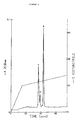
- Figure 1 depicts the fractionation of 400 ml of the 0.6 M NaCl eluate from cation exchange chromatography using heparin-Sepharose affinity chromatography.
- the horizontal bar A indicates the fractions containing the HBBMs which were thereafter rechromatographed for further purifaction.
- the horizontal bar B indicates the fractions containing aFGF, as determined by reverse-phase HPLC analysis.
- Figure 2 depicts the results of Mono-S cation exchange chromatography on 20 ml of the material eluted from the heparin-Sepharose affinity chromatography (as indicated by the horizontal bar A, Fig. 1). Arrows indicate the elution positions of aFGF and bFGF standards.
- the numbers in the chromatogram refer to HBBM-1, -2, and -3.
- the horizontal bar indicates the fractions containing the HBBMs which were used as a pool or individually for further purification/characterization.
- Figure 3 depicts the reverse-phase HPLC of HBBMs.
- An aliquot of the pool of Mono-S-purified HBBMs (peaks 1-3, as indicated by the horizontal bar in Fig. 2) was subjected to reverse-phase HPLC. Individual peaks were identified and assigned to specific Mono-S fractions by analyzing aliquots of Mono-S chromatography fractions (as indicated by sections of the horizontal bar in Fig. 2) in independent reverse-phase HPLC runs. Peaks 1-3 refer to HBBM-1, HBBM-2, and HBBM-3, respectively.
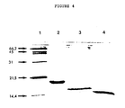
- Figure 4 depicts the analysis on SDS-PAGE of the HBBMs as follows: Lane 1: Protein standards (lysozyme, 14.4 kD; trypsin inhibitor, 21.5 kD; carbonic anhydrase, 31 kD; ovalbumin, 45 kD, serum albumin, 66.2 kD); Lane 2: HBBM-1; Lane 3: HBBM-2; Lane 4: HBBM-3.
- Protein standards lysozyme, 14.4 kD
- trypsin inhibitor 21.5 kD
- carbonic anhydrase 31 kD
- ovalbumin 45 kD
- serum albumin 66.2 kD
- Lane 2 HBBM-1
- Lane 3 HBBM-2
- Lane 4 HBBM-3.
- Figure 5 depicts the rechromatography of HBBM-3 on a Mono-S cation-exchange column.
- the fractions containing HBBM-3 from the chromatography depicted in Figure 2 were pooled, diluted three times with starting buffer, and the diluted samples chromatographed on a Mono-S column, under conditions identical to those shown in Figure 2.
- Aliquots of fractions were tested for their ability to stimulate the growth of bovine aortic arch endothelial cells (histogram). Retention times of aFGF and bFGF are shown with arrows.
- a horizontal bar marks the fractions which thereafter were used for concentrating HBBM-3.
- Figure 6 depicts the biological activity of HBBM-3 and HBBM-1 by means of dose response analysis and comparison with FGF activity.
- the ability of HBBM-3 and HBBM-1 to stimulate the growth of bovine aortic arch endothelial cells was tested.
- Figure 7 depicts the chromatography of HBBM-3 on an hydrophobic interaction column. An aliquot of Mono-S-concentrated HBBM-3 was subjected to HIC chromatography. Arrows indicate the retention times of aFGF and bFGF under identical chromatographic conditions. Aliquots of column fractions were tested for their ability to stimulate bovine aortic arch endothelial cell profileration. The results are indicated by the number of cells grown in each test well.
- novel growth factors of this invention are single chain, basic, heparin-binding brain mitogens.
- HBBMs have been identified in the brain tissues of all species tested, which include human, bovine, rat and chicken. Based on this distribution, it is expected that other species will also contain HBBMs.
- the N-terminal sequences of the HBBMs differ markedly from those published for bovine or human brain aFGF and bFGF (5,6,7).
- the N-terminal sequences of the first 19 amino acids have been found to be identical for human and bovine HBBMs.
- the N-terminal sequences are as follows: Gly-Lys-Lys-Glu-Lys-Pro-Glu-Lys-Lys-Val-Lys-Lys-Ser-Asp-Cys-Gly-Glu-Trp-Gln.
- the rat sequence is identical with the exception of the cysteine residue in position 15, because a determination of the presence or absence of cysteine was not conducted.
- HBBMs have been found in three forms in tissue from bovine brain.
- HBBM-1, HBBM-2 and HBBM-3 have molecular weights of 18, 16 and 15 kD, respectively.
- Their amino acid compositions are set forth in Table I below and also differ from the amino acid compositions of the bovine brain FGFs (5,6,7).
- HBBM-2 and HBBM-3 are C-terminally truncated forms of HBBM-1, lacking approximately 13 and 29 amino acids, respectively.
- the ratios of the three forms varied between different isolation batches.
- the overall isolation yield of HBBMs was estimated at approximately 30, 10 and 40 ⁇ g/kg brain tissue for HBBM-1, HBBM-2, and HBBM-3 respectively.
- the ratios may depend on variables during tissue storage and extraction, in particular proteolysis. Proteolysis during tissue extraction may cause carboxy-terminal truncation of HBBM-1, the largest of the HBBM forms, which would yield HBBM-2 and HBBM-3 in varying amounts.
- the process for isolating the novel HBBMs of this invention in substantially pure form from a source of brain tissue comprises the sequence of steps of:
- the extraction is accomplished by treating the tissue sequentially with 0.15 M ammonium sulfate, adjusting to pH 4.5 with hydrochloric acid, stirring and centrifugation, treating with ammonium sulfate after the pH is adjusted to 6-6.5 with sodium hydroxide, stirring and centrifugation, followed by dialysis and centrifugation.
- the first cation-exchange chromatography step comprises batch adsorption on a carboxymethyl-Sephadex column, followed by washing with 100 mM sodium phosphate buffer at pH 6 and elution with 100 mM sodium phosphate, pH 6.0/0.6 M NaCl.
- heparin affinity chromatography is performed on a heparin-Sepharose (Pharmacia) column by washing with a buffer containing 10 mM Tris-HCl, pH 7.0/0.6 M NaCl, followed by elution with a linear gradient of from 0.6 to 2.0 M NaCl in 10 mM Tris-HCl, pH 7.0.
- the second cation-exchange chromatography step is performed on a Mono-S (Pharmacia column equilibrated and, after loading of the sample, washed with 50 mM sodium phosphate, pH 6.8, followed by elution of HBBMs with a linear gradient of from 0 to 0.6 m NaCl in 50mM sodium phosphate, pH 6.8.
- the hydrophobic-interaction chromatography step comprises preequilibrating a HIC column (LKB Ultropac-TSK-Phenyl-5PW) with a buffer of 100 mM sodium phosphate, pH 7.0, and 1.5 M sodium sulfate, followed by elution with a linear gradient of from 1.5 to 0.6 M sodium sulfate.
- HIC column LLB Ultropac-TSK-Phenyl-5PW
- the HBBMs may be separated easily and quantitatively from the FGFs by reverse-phase HPLC, this procedure reduces the biological activity of the FGFs due to the conditions used in reverse-phase HPLC. Therefore, in order to separate and isolate the HBBMs and the FGFs in biologically active form for comparative testing, the process set forth above was developed.
- the use of reverse-phase HPLC was limited to the testing of small aliquots of fractions from heparin-Sepharose affinity chromatography and cation-exchange chromatography for the presence of HBBMs and FGFs.
- the reverse-phase HPLC steps were performed on a C4 column (The Separations Group) eluted with a shallow gradient of acetonitrile in 0.1% trifluoroacetic acid.
- HBBM-2 The biological activity of the HBBMs has been established by tests evidencing the induction of mitogenesis in endothelial cells. Although HBBM-2 was not tested, the activity demonstrated for HBBM-1 and HBBM-3 suggests that HBBM-2 will also possess this activity. The testing was performed on bovine aortic arch endothelial cells.
- HBBM-1 and HBBM-3 are each equipotent to aFGF.
- the ED50 are in the range of 50-150 ng/ml.
- HBBM-3 and the FGFs are clearly separated by highly resolutive Mono-S chromatography, as shown in Figure 5, and are also clearly separated by a second high resolution technique with different selectivities, hydrophobic interaction chromatography, as shown in Figure 7.
- the HBBMs did not contain measurable quantitites of aFGF as determined by quantitative reverse-phase HPLC.
- the HBBMs were not cross-reactive in an immuno-dot assay using polyclonal antibodies raised against synthetic peptides corresponding to the N-terminal 15 residues of aFGF (1-15) and bFGF (30-50), the latter yielding an antibody which cross-reacts with aFGF (antibodies provided by A. Baird, Salk Institute; growth factor nomenclature according to refs. 5,7).
- the concentration of HBBM and aFGF preparations were carefully determined by quantitative amino acid analysis as a basis for potency comparisons (Fig. 6).
- the HBBMs are further distinguished from the FGFs by a lack of amino acid sequence homology between the two groups of proteins.
- the presently available sequence information (approximately 75 of 130 residues of HBBM-3) shows no sequence homology to the published 146 residue sequence of the FGFs (5).
- the HBBMs were not present in bovine kidney tissue that was extracted using the procedure for brain tissue. Therefore, the HBBMs are novel proteins which may also play a role in the formation, maintenance and/or repair of tissue, in particular, neural tissue.
- HBBMs have the same biological and heparin-binding activities as aFGF; (2) the HBBMs are brain-specific; (3) higher amounts of the HBBMs than aFGF are found in the brain; and (4) aFGF and bFGF are known to have very prominent neural activities, such as neurotrophic (neuron survival) activity in vivo and in vitro , they are mitogenic for neuroblasts and glial cells, they promote neurite outgrowth and induce brain-specific protein synthesis.
- neurotrophic neurotrophic
- the protein structures of the HBBMs and the FGFs are dissimilar, their similarity in terms of heparin binding and biological activity suggests that both groups of mitogens act through a similar mechanism.
- the mitogens could bind to cell surface or extracellular, matrix-associated heparinlike structures.
- Therapeutic compositions in accordance with this invention include HBBMs, either singly or in mixtures, dispersed in a conventional pharmaceutically acceptable liquid or solid carrier.
- the therapeutic compositions may be administered topically in the form of creams, lotions and so forth, or orally in such forms as tablets, capsules, dispersible powders, granules or suspensions, or parenterally in the form of sterile injectable solutions or suspensions.
- These therapeutic compositions may be administered to human or veterinary patients to promote angiogenesis and the repair and maintenance of neural tissue.
- bovine HBBM isolation and characterization of bovine HBBM are set forth in detail. Results using human material were very similar and will not be described in detail unless differences from bovine material were found.
- the homogenate was then extracted by stirring of the suspension for 2 hours at 4°C followed by centrifugation at 4°C for 60 minutes at either 11,500 rpm (GSA rotor) or 9000 rpm (GS-3 rotor) to remove cells and debris.
- the supernatant was adjusted to pH 6-6.5 with NaOH, ammonium sulfate (230 g/l) was added slowly, and the resulting suspension was stirred for at least 30 minutes at 4°C and centrifuged again as described above. The pellet was discarded. More ammonium sulfate (300 g/l) was slowly added to the supernatant, the suspension was stirred for at least 30 minutes at 4°C and centrifuged as above.
- the resulting pellet was dissolved in cold water (100 ml per kg of starting material) and dialysed at 4°C for 20 hours against 20 l of water using a Spectrapor membrane (molecular weight cut-off 6-8 kD, diameter 31.8 mm). The dialysate was centrifuged again to remove precipitated material and the supernatant subjected to chromatographic purification (see below) after determining its conductivity.
- the tissue extract resulting from the above procedure was subjected to batch adsorption/elution by cation exchange chromatography as follows: The sample was diluted with water as required to bring the conductivity below that of a 0.1 M sodium phosphate buffer (pH 6)/0.15 M NaCl solution. The sample was then loaded onto a column of carboxymethyl-Sephadex (5.5 x 3 cm) which was preequilibrated with 100 mM sodium phosphate (pH 6)/0.15 M Cl. The column was washed with the equilibration buffer and a protein fraction was eluted with 100 mM sodium phosphate, pH 6.0/0.6 M NaCl and collected. All operations were carried out at room temperature and a flow rate of 500 ml/hour.
- the 0.6 M NaCl eluate from cation exchange chromatography was loaded on a heparin-Sepharose column (Pharmacia, 5 x 1.5 cm) at a flow rate of 125 ml/hour.
- the column was washed with 200 ml of a buffer containing 10 mM Tris-HCl, pH 7.0/0.6 M NaCl until the absorbance of the column eluate at 280 nm became negligible.
- Protein bound to the column was eluted with a 120-minute linear gradient from 0.6 M to 2 M NaCl in 10 mM Tris-HCl, pH 7.0 at a flow rate of 35 ml/h.
- Chromatography was performed at room temperature using a LKB peristaltic pump and a low pressure LKB programmable gradient former. Fractions of 1.4 ml were collected and aliquots subjected to bioassay.
- Fig. 1 The results of this Heparin-Sepharose chromatography are shown in Fig. 1.
- the reverse-phase HPLC analysis was conducted on a C4 column (25 x 0.46 cm, 5 um particle size, 300 angstroms pore size, The Separations Group, Hesperia, CA).
- the proteins were eluted in a shallow gradient of acetonitrile (10%/hour) in 0.1% trifluoroacetic acid at a flow rate of 0.7 ml/minute.
- the heparin-Sepharose chromatography was performed at room temperature.
- fractions eluting at 1-1.2M NaCl were pooled, diluted sufficiently with a buffer containing 50 mM sodium phosphate, pH 6.8 (to reduce their ionic strength to approximately that of the diluent) and subjected to cation-exchange chromatography on a Mono-S column (Pharmacia) equilibrated with 50 mM sodium phosphate, pH 6.8.
- This material was pumped onto a Mono-S column (Pharmacia)equilibrated with 50 mM sodium phosphate, pH 6.8. After washing the column with the same buffer until the absorbance at 210 nm reached a minimum value, protein was eluted with a gradient from 0 to 0.6 M NaCl in 50 mM sodium phosphate, pH 6.8.
- the first and quantitatively minor peak which eluted from the Mono-S column at 0.3 M NaCl corresponded to aFGF as evidenced by co-elution with a reference standard of authentic aFGF in the same system under identical conditions (see arrow in Fig. 2), as well as by co-elution in reverse-phase HPLC on a previously-described C4 column, eluted under highly resolutive, shallow gradient conditions (data not shown).
- the presence of aFGF in this Mono-S column fraction was expected, because the originating fraction from heparin-Sepharose chromatography is known to contain aFGF.
- HBBMs are clearly distinguishable by chromatographic retention not only from aFGF but also bFGF (Fig. 2).
- HBBMs when analyzed by reverse-phase HPLC (Fig. 3), the HBBMs were also found to differ clearly from the FGFs with respect to retention times (data not shown). Finally, reverse-phase HPLC separated all three HBBMs from each other (Fig. 3) and thus provided a means for their preparation in high purity (as needed for structural characterization) and for relatively unambiguous identification.
- Human brain also yielded three forms of HBBMs with reverse-phase HPLC elution patterns identical to those of their bovine counterparts.
- cysteine residues of the HPLC-purified proteins were treated according to the procedure of Gautschi-Sova et al. (6). Briefly, the cysteine residues were reduced with a five-fold molar excess of dithiothreitol and alkylated by carboxymethylation using a three-fold molar excess of iodo-[2-14C]-acetic acid.
- the N-terminal sequences of all three HBBMs were found to be identical to each other for the first 19 amino acids.
- the sequences were determined as Gly-Lys-Lys-Glu-Lys-Pro-Glu-Lys-Lys-Val-Lys-Lys-Ser-Asp-Cys-Gly-Glu-Trp-Gln.
- Human HBBMs (probably analyzed as a mixture) were found to possess the same N-terminal sequence as bovine HBBMs.
- Protein samples were analyzed using sodium dodecyl sulfate polyacrylamide gel electrophoresis ("SDS-PAGE") as described by Gospodarowicz et al. (9). Briefly, molecular weight determinations were performed by SDS-PAGE as follows: Aliquots containing 40-80 ng of protein were added to the sample buffer composed of 30% (v/v) glycerol, 0.2 M dithiothreitol, 4% (w/v) sodium dodecyl sulfate, 4 mM EDTA, and 75 mM Tris-HCl, pH 6.8. Samples were boiled for 3 minutes and then applied to a 20% polyacrylamide gel slab (1.5 mm) with a 3% stacking gel.
- SDS-PAGE sodium dodecyl sulfate polyacrylamide gel electrophoresis
- Electrophoresis under non-reducing conditions was performed in identical fashion except that dithiothreitol was omitted from the sample buffer.
- a protein standard mixture containing lysozyme (14.4 kD), trypsin inhibitor (21.5kD), carbonic anhydrase (31 kD), ovalbumin (45 kD) and serum albumin (66.2 kD) was also applied to each gel.
- SDS-PAGE revealed the molecular weights of HBBM 1, -2, and -3 to be 18, 16 and 15 kD, respectively (Fig. 4). The molecular weights did not differ significantly regardless of whether the samples were electrophoresed in the presence or absence of reducing agent, indicating that the proteins are single chain polymers.
- the molecular weights of the human HBBMs have not yet been determined by SDS-PAGE.
- a 10-20 pmol protein sample was hydrolyzed according to the high sensitivity methodology of Bohlen and Schroeder (11). Briefly, the protein sample was added to a hydrolysis tube and dried in vacuo . Fifty ⁇ l constant boiling HCl containing 2% (v/v) thioglycollic acid were added. The tube was then evacuated with high vacuum (less than 50mm Torr) after freezing the sample in liquid nitrogen. The sample was then allowed to melt while under vacuum and the tube was flame-sealed.
- the tube was hydrolyzed by heating at 110° for 20 hours. After hydrolysis, the tube was opened, dried in vacuo , and the residue dissolved in 130 ⁇ l citrate buffer (0.067 sodium citrate, pH 2.20) prior to loading on the cation exchange chromatography column.
- citrate buffer 0.067 sodium citrate, pH 2.20
- the protein hydrolysates were chromatographed on a Chromakon 500 amino acid analyzer (Kontron, Zurich, Switzerland) equipped with a polystyrene-based cation exchange column and an o-phthalaldehyde fluorescence detection system for high-sensitivity detection (11).
- HBBMs Quantitation of amino acids was by the external standard method using an amino acid standard mixture. Based on quantitative amino acid analysis, the overall isolation yield of HBBMs was estimated at approximately 30, 10 and 40 ⁇ g/kg brain tissue for HBBM-1, HBBM-2, and HBBM-3, respectively.
- Amino acid compositional data agree with the elution order of the three proteins on Mono-S: the least basic protein (HBBM-3) elutes first, while the most basic protein (HBBM-1) elutes last.
- Expected molecular weights calculated from amino acid analyses are somewhat lower than those determined by SDS-PAGE. The discrepancies may be accounted for by proline and cysteine residues which were not quantitated by amino acid analysis. However, based on the N-terminal sequence analysis, it is known that proline and cysteine are in fact present. Amino acid analyses of human HBBMs are not yet available.
- bovine endothelial cells were seeded with column fractions obtained as described below at low density (10,000-20,000 cells/35 mm dish) in Dulbecco's modified Eagle's medium containing 10% calf serum (Hyclone, Sterile Systems, Logan, UT). Cultures were grown for 5 days in the presence of various concentrations of column fraction aliquots (added on days 0 and 2) and then counted in a Coulter particle counter. The biological activities of the proteins tested are indicated in the number of vascular endothelial cells grown in each test well for each fraction in Figures 5-7.
- HBBM-3 The eluant fractions corresponding to the peak for HBBM-3 from Mono-S chromatography, as shown in Figure 2, were pooled, diluted three times with starting buffer and rechromatographed on the same system. Aliquots of these fractions were tested as described above for their ability to stimulate the growth of bovine vascular endothelial cells. Comparative tests were run with aFGF and bFGF. The results, as shown in Figure 5, indicated that HBBM-3 stimulated mitogenic activity for bovine endothelial cells. Furthermore, the HBBM-3 and its activity were separable from the FGFs.
- the upper panel of Figure 6 indicates that HBBM-3 stimulated bovine aortic endothelial cells in a dose-dependent manner.
- the ED50 was in the order of 20-50 ng/ml, with the minimally stimulating dose being approximately 3 ng/ml.
- the dose-response curves of HBBM-3 and aFGF were indistinguishable, both qualitatively and quantitatively, under the assay conditions used.
- the response of HBBM-3 appeared to be distinguishable from that of bFGF.
- bFGF possessed much higher potency and apparently higher intrinsic activity than HBBM-3.
- HBBM-1 was also biologically active in the same test system.
- Human HBBM comprising a mixture of HBBM-1, -2 and -3, was tested in the same manner and was similarly active (data not shown). However, results have not yet been obtained for individual fractions of the human HBBMs.
- bovine HBBMs were also tested on human umbilical cord endothelial cells and found to be active (data not shown).
Landscapes
- Health & Medical Sciences (AREA)
- Chemical & Material Sciences (AREA)
- Organic Chemistry (AREA)
- Life Sciences & Earth Sciences (AREA)
- Medicinal Chemistry (AREA)
- General Health & Medical Sciences (AREA)
- General Chemical & Material Sciences (AREA)
- Engineering & Computer Science (AREA)
- Nuclear Medicine, Radiotherapy & Molecular Imaging (AREA)
- Chemical Kinetics & Catalysis (AREA)
- Pharmacology & Pharmacy (AREA)
- Bioinformatics & Cheminformatics (AREA)
- Animal Behavior & Ethology (AREA)
- Public Health (AREA)
- Veterinary Medicine (AREA)
- Toxicology (AREA)
- Zoology (AREA)
- Gastroenterology & Hepatology (AREA)
- Biochemistry (AREA)
- Biophysics (AREA)
- Genetics & Genomics (AREA)
- Molecular Biology (AREA)
- Proteomics, Peptides & Aminoacids (AREA)
- Dermatology (AREA)
- Pharmaceuticals Containing Other Organic And Inorganic Compounds (AREA)
- Peptides Or Proteins (AREA)
- Polysaccharides And Polysaccharide Derivatives (AREA)
- Insulated Conductors (AREA)
- Medicines That Contain Protein Lipid Enzymes And Other Medicines (AREA)
- Organic Insulating Materials (AREA)
- Dental Preparations (AREA)
- Compositions Of Macromolecular Compounds (AREA)
- Medicines Containing Material From Animals Or Micro-Organisms (AREA)
- Materials For Medical Uses (AREA)
Claims (11)
- Heparin-bindendes Hirn-Mitogen in im wesentlichen reiner Form, das die N-terminale Aminosäuresequenz H-Gly-Lys-Lys-Glu-Lys-Pro-Glu-Lys-Lys-Val- aufweist.
- Heparin-bindendes Hirn-Mitogen nach Anspruch 1 in im wesentlichen reiner Form, das die N-terminale Aminosäuresequenz H-Gly-Lys-Lys-Glu-Lys-Pro-Glu-Lys-Lys-Val-Lys-Lys-Ser-Asp-Cys-Gly-Glu-Trp-Gln- aufweist.
- Heparin-bindendes Hirn-Mitogen nach Anspruch 2, das aus Rinderhirn isoliert ist.
- Heparin-bindendes Hirn-Mitogen nach Anspruch 3, das aus der aus HBBM-1, HBBM-2 und HBBM-3 bestehenden Gruppe ausgewählt ist, worin HBBM-1 ein Molekulargewicht von ungefähr 18000 Dalton aufweist, HBBM-2 ein Molekulargewicht von ungefähr 16000 Dalton aufweist und HBBM-3 ein Molekulargewicht von ungefähr 15000 Dalton aufweist, worin die Molekulargewichte durch SDS-PAGE unter entweder reduzierenden oder nicht-reduzierenden Bedingungen bestimmt sind, und eine Aminosäurezusammensetzung hat von:
Aminosäure (Anzahl der Reste) HBBM-1 HBBM-2 HBBM-3 Aparagin und Asparaginsäure 10 10 9 Threonin 14 14 13 Serin 9 9 6 Glutamin und Glutaminsäure 23 20 17 Glycin 16 15 13 Alanin 10 8 8 Valin 4 4 4 Methionin 1 1 1 Isoleucin 2 2 2 Leucin 8 8 8 Tyrosin 2 2 1 Phenylalanin 2 2 2 Histidin 1 1 1 Lysin 35 28 23 Tryptophan 3 3 3 Arginin 8 8 8 - Verfahren zur Isolierung von Heparin-bindendem Hirn-Mitogen nach Anspruch 1 in im wesentlichen reiner Form, das die folgende Stufensequenz umfaßt:(a) Extraktion aus der Gewebequelle;(b) eine erste Kationenaustausch-Chromatographie;(c) Heparin-Affinitätschromatographie; und(d) eine zweite Kationenaustausch-Chromatographie.
- Verfahren nach Anspruch 5, das weiter nach Stufe (d) die Stufe hydrophobe Wechselwirkungs-Chromatographie umfaßt.
- Verfahren nach Anspruch 5, worin die eluierten Fraktionen der Stufen (c) und (d) durch Umkehrphasen-Hochleistungsflüssigkeitschromatographie auf die Anwesenheit von Heparin-bindendem Hirn-Mitogen analysiert werden.
- Verfahren nach Anspruch 7, worin die Gewebequelle Rinderhirn ist.
- Therapeutische Zusammensetzung von Material, das für die Förderung von Angiogenese nützlich ist, umfassend einen pharmazeutischen Träger und eine wirksame Angiogenesefördernde Menge des Heparin-bindenden Hirn-Mitogens nach Anspruch 1.
- Therapeutische Zusammensetzung von Material, das für die Förderung der Bildung, Aufrechterhaltung und Wiederherstellung von Gewebe nützlich ist, umfassend einen pharmazeutischen Träger und eine wirksame Menge des Heparin-bindenden Hirn-Mitogens nach Anspruch 1.
- Therapeutische Zusammensetzung nach Anspruch 10, worin das Gewebe neurales Gewebe ist.
Priority Applications (1)
| Application Number | Priority Date | Filing Date | Title |
|---|---|---|---|
| AT89101187T ATE94882T1 (de) | 1988-01-25 | 1989-01-24 | Heparinbindende gehirnmitogene. |
Applications Claiming Priority (2)
| Application Number | Priority Date | Filing Date | Title |
|---|---|---|---|
| US14783588A | 1988-01-25 | 1988-01-25 | |
| US147835 | 1988-01-25 |
Publications (3)
| Publication Number | Publication Date |
|---|---|
| EP0326075A2 EP0326075A2 (de) | 1989-08-02 |
| EP0326075A3 EP0326075A3 (en) | 1989-10-18 |
| EP0326075B1 true EP0326075B1 (de) | 1993-09-22 |
Family
ID=22523098
Family Applications (1)
| Application Number | Title | Priority Date | Filing Date |
|---|---|---|---|
| EP89101187A Expired - Lifetime EP0326075B1 (de) | 1988-01-25 | 1989-01-24 | Heparinbindende Gehirnmitogene |
Country Status (9)
| Country | Link |
|---|---|
| EP (1) | EP0326075B1 (de) |
| JP (1) | JPH01308300A (de) |
| AT (1) | ATE94882T1 (de) |
| AU (1) | AU624803B2 (de) |
| DE (1) | DE68909254T2 (de) |
| DK (1) | DK29889A (de) |
| ES (1) | ES2058350T3 (de) |
| FI (1) | FI890342A (de) |
| NO (1) | NO175594C (de) |
Families Citing this family (12)
| Publication number | Priority date | Publication date | Assignee | Title |
|---|---|---|---|---|
| SE8801426D0 (sv) * | 1988-04-15 | 1988-04-15 | Ulf Rothman | Forfarande och medel for blodbehandling |
| US5126323A (en) * | 1989-11-16 | 1992-06-30 | Genetics Institute, Inc. | Homogeneous purified k-fgf and compositions containing the same |
| US6448381B1 (en) * | 1990-01-08 | 2002-09-10 | Barnes-Jewish Hospital | DNA encoding heparin-binding growth factor |
| ATE154608T1 (de) * | 1990-04-12 | 1997-07-15 | Ebewe Arzneimittel | Verwendung einer mischung von peptiden and aminosäuren für die prophylaxis oder behandlung von dementia |
| FR2665448A1 (fr) * | 1990-08-01 | 1992-02-07 | Paris Val De Marne Universite | Procede d'obtention d'un peptide ayant une activite facteur de croissance, produit obtenu et son application comme medicament. |
| US5210026A (en) * | 1990-08-20 | 1993-05-11 | American Cyanamid Company | Human mk gene and method of expression |
| US6511823B1 (en) * | 1990-08-20 | 2003-01-28 | American Cyanamid Corporation | Heparin binding neurotrophic factor gene sequence |
| EP0535337A3 (en) * | 1991-09-30 | 1993-05-19 | American Cyanamid Company | Cell growth inhibiting activities of heparin binding neurite-outgrowth promoting factor |
| EP0535336A3 (en) * | 1991-09-30 | 1993-05-19 | American Cyanamid Company | Mk protein as cell growth inhibitor |
| FI980032A0 (fi) * | 1998-01-09 | 1998-01-09 | Heikki Matti Eemeli Rauvala | Ny anvaendning av en heparin bindande, med tillvaext foerknippad molekyl |
| US6364912B1 (en) * | 1999-09-17 | 2002-04-02 | Depuy Orthopeaedics, Inc. | Pleiotrophin-based compositions for enhancing connective tissue repair |
| US7888485B2 (en) | 2003-03-26 | 2011-02-15 | Georgetown University | Anti-pleiotrophin antibodies and methods of use thereof |
Family Cites Families (3)
| Publication number | Priority date | Publication date | Assignee | Title |
|---|---|---|---|---|
| EP0248088A4 (de) * | 1985-11-22 | 1988-05-03 | Teijin Ltd | Vaskularisierungsfaktor und herstellung. |
| EP0241136A3 (de) * | 1986-03-07 | 1989-12-27 | The President And Fellows Of Harvard College | Humaner, Klasse 1 Heparin bindender, Wachstumsfaktor |
| CA2005600A1 (en) * | 1988-12-20 | 1990-06-20 | Cal-Henrik Heldin | Endothelial cell growth factor |
-
1989
- 1989-01-24 JP JP1013330A patent/JPH01308300A/ja active Pending
- 1989-01-24 ES ES89101187T patent/ES2058350T3/es not_active Expired - Lifetime
- 1989-01-24 FI FI890342A patent/FI890342A/fi not_active IP Right Cessation
- 1989-01-24 DK DK029889A patent/DK29889A/da not_active Application Discontinuation
- 1989-01-24 AT AT89101187T patent/ATE94882T1/de active
- 1989-01-24 DE DE89101187T patent/DE68909254T2/de not_active Expired - Lifetime
- 1989-01-24 EP EP89101187A patent/EP0326075B1/de not_active Expired - Lifetime
- 1989-01-24 NO NO890295A patent/NO175594C/no unknown
- 1989-01-24 AU AU28737/89A patent/AU624803B2/en not_active Expired
Non-Patent Citations (2)
| Title |
|---|
| ANALYTICAL BIOCHEMISTRY, vol. 154, 1986, Academic Press, Inc.; R.R.LOBB et al., pp. 1-14# * |
| JOURNAL OF BIOLOGICAL CHEMISTRY, vol. 261, no. 4, 05 February 1986, Am. Society of Biol. Chem., Inc. (US); R.LOBB et al., pp. 1924-1928# * |
Also Published As
| Publication number | Publication date |
|---|---|
| DE68909254D1 (de) | 1993-10-28 |
| NO175594C (no) | 1994-11-02 |
| DE68909254T2 (de) | 1994-04-28 |
| NO890295D0 (no) | 1989-01-24 |
| JPH01308300A (ja) | 1989-12-12 |
| DK29889A (da) | 1989-07-26 |
| ES2058350T3 (es) | 1994-11-01 |
| AU624803B2 (en) | 1992-06-25 |
| NO890295L (no) | 1989-07-26 |
| FI890342A0 (fi) | 1989-01-24 |
| ATE94882T1 (de) | 1993-10-15 |
| AU2873789A (en) | 1989-07-27 |
| EP0326075A3 (en) | 1989-10-18 |
| DK29889D0 (da) | 1989-01-24 |
| EP0326075A2 (de) | 1989-08-02 |
| NO175594B (no) | 1994-07-25 |
| FI890342A (fi) | 1989-07-26 |
Similar Documents
| Publication | Publication Date | Title |
|---|---|---|
| US4785079A (en) | Isolation of fibroblast growth factor | |
| EP0128849B1 (de) | Gereinigter transformierender Wachstum-beta-Faktor aus humanen Plaketten und Plazenten stammend | |
| Gautschi-Sova et al. | Acidic fibroblast growth factor is present in nonneural tissue: isolation and chemical characterization from bovine kidney | |
| Risau et al. | Endothelial cell growth factors in embryonic and adult chick brain are related to human acidic fibroblast growth factor. | |
| EP0326075B1 (de) | Heparinbindende Gehirnmitogene | |
| US5489670A (en) | Human uroguanylin | |
| EP0797590B1 (de) | Neurotrophe peptide des aktivitätsabhängigen neurotrophen faktors | |
| EP0100641B1 (de) | Menschliches Lymphotoxin und seine Derivate, Verfahren zu seiner Herstellung und dasselbe enthaltende pharmazeutische Zusammensetzungen | |
| US4740587A (en) | Inhibin and method of purifying same | |
| Edmundson et al. | Isolation and characterization of concanavalin A polypeptide chains | |
| US5136025A (en) | Method to purify basic fibroblast growth factor | |
| US5641743A (en) | Therapeutic compositions and methods for use of heparin-binding brain mitogens | |
| US4902782A (en) | Isolation of fibroblast growth factor | |
| US4920196A (en) | Human lymphotoxin | |
| JP2010031055A (ja) | Tfpiおよびtfpiアナログを精製するための改良された方法 | |
| Piot et al. | Isolation and characterization of a bradykinin-potentiating peptide from a bovine peptic hemoglobin hydrolysate | |
| US5171842A (en) | Heparin-binding brain mitogens | |
| EP0228357B1 (de) | Von der Prostata stammender Wachstumsfaktor | |
| US5270449A (en) | Methods for the isolation of heparin-binding brain mitogens | |
| Mount et al. | Purification and characterization of epidermal growth factor (β-urogastrone) and epidermal growth factor fragments from large volumes of human urine | |
| US5656458A (en) | Expression and processing of amino terminus acetylated FGF's in yeast | |
| EP0384731A2 (de) | Osteogenische Wachstumfaktoren, die aus sich regenerierendem Knochenmark identifiziert werden | |
| Mizon et al. | Human pre-α-inhibitor: isolation from a by-product of industrial scale plasma fractionation and structural analysis of its H3 heavy chain | |
| EP0655251A1 (de) | Pharmazeutische Zusammensetzung zum Vorbeugen oder Behandeln von Brüchen | |
| CZ272895A3 (en) | Method of converting hydrophobic derivative of growth hormone to the growth hormone natural form and detection method of said hydrophobic derivative |
Legal Events
| Date | Code | Title | Description |
|---|---|---|---|
| PUAI | Public reference made under article 153(3) epc to a published international application that has entered the european phase |
Free format text: ORIGINAL CODE: 0009012 |
|
| AK | Designated contracting states |
Kind code of ref document: A2 Designated state(s): AT BE CH DE ES FR GB GR IT LI NL SE |
|
| PUAL | Search report despatched |
Free format text: ORIGINAL CODE: 0009013 |
|
| AK | Designated contracting states |
Kind code of ref document: A3 Designated state(s): AT BE CH DE ES FR GB GR IT LI NL SE |
|
| 17P | Request for examination filed |
Effective date: 19900306 |
|
| 17Q | First examination report despatched |
Effective date: 19911213 |
|
| GRAA | (expected) grant |
Free format text: ORIGINAL CODE: 0009210 |
|
| AK | Designated contracting states |
Kind code of ref document: B1 Designated state(s): AT BE CH DE ES FR GB GR IT LI NL SE |
|
| REF | Corresponds to: |
Ref document number: 94882 Country of ref document: AT Date of ref document: 19931015 Kind code of ref document: T |
|
| REF | Corresponds to: |
Ref document number: 68909254 Country of ref document: DE Date of ref document: 19931028 |
|
| ET | Fr: translation filed | ||
| ITF | It: translation for a ep patent filed |
Owner name: MODIANO & ASSOCIATI S.R |
|
| RAP2 | Party data changed (patent owner data changed or rights of a patent transferred) |
Owner name: AMERICAN CYANAMID COMPANY |
|
| REG | Reference to a national code |
Ref country code: GR Ref legal event code: FG4A Free format text: 3009691 Ref country code: CH Ref legal event code: PFA Free format text: AMERICAN CYANAMID COMPANY |
|
| NLT2 | Nl: modifications (of names), taken from the european patent patent bulletin |
Owner name: AMERICAN CYANAMID COMPANY TE WAYNE, NEW JERSEY, VE |
|
| PLBE | No opposition filed within time limit |
Free format text: ORIGINAL CODE: 0009261 |
|
| STAA | Information on the status of an ep patent application or granted ep patent |
Free format text: STATUS: NO OPPOSITION FILED WITHIN TIME LIMIT |
|
| 26N | No opposition filed | ||
| REG | Reference to a national code |
Ref country code: ES Ref legal event code: FG2A Ref document number: 2058350 Country of ref document: ES Kind code of ref document: T3 |
|
| EAL | Se: european patent in force in sweden |
Ref document number: 89101187.6 |
|
| PGFP | Annual fee paid to national office [announced via postgrant information from national office to epo] |
Ref country code: SE Payment date: 19951213 Year of fee payment: 8 |
|
| PGFP | Annual fee paid to national office [announced via postgrant information from national office to epo] |
Ref country code: GR Payment date: 19951219 Year of fee payment: 8 |
|
| PGFP | Annual fee paid to national office [announced via postgrant information from national office to epo] |
Ref country code: AT Payment date: 19951221 Year of fee payment: 8 |
|
| PGFP | Annual fee paid to national office [announced via postgrant information from national office to epo] |
Ref country code: NL Payment date: 19951231 Year of fee payment: 8 |
|
| PGFP | Annual fee paid to national office [announced via postgrant information from national office to epo] |
Ref country code: ES Payment date: 19960123 Year of fee payment: 8 |
|
| PGFP | Annual fee paid to national office [announced via postgrant information from national office to epo] |
Ref country code: BE Payment date: 19960207 Year of fee payment: 8 |
|
| PGFP | Annual fee paid to national office [announced via postgrant information from national office to epo] |
Ref country code: CH Payment date: 19960423 Year of fee payment: 8 |
|
| PG25 | Lapsed in a contracting state [announced via postgrant information from national office to epo] |
Ref country code: AT Effective date: 19970124 |
|
| PG25 | Lapsed in a contracting state [announced via postgrant information from national office to epo] |
Ref country code: SE Effective date: 19970125 Ref country code: ES Free format text: LAPSE BECAUSE OF NON-PAYMENT OF DUE FEES Effective date: 19970125 |
|
| PG25 | Lapsed in a contracting state [announced via postgrant information from national office to epo] |
Ref country code: BE Effective date: 19970131 Ref country code: LI Effective date: 19970131 Ref country code: CH Effective date: 19970131 |
|
| BERE | Be: lapsed |
Owner name: AMERICAN CYANAMID CY Effective date: 19970131 |
|
| PG25 | Lapsed in a contracting state [announced via postgrant information from national office to epo] |
Ref country code: GR Free format text: THE PATENT HAS BEEN ANNULLED BY A DECISION OF A NATIONAL AUTHORITY Effective date: 19970731 |
|
| PG25 | Lapsed in a contracting state [announced via postgrant information from national office to epo] |
Ref country code: NL Effective date: 19970801 |
|
| REG | Reference to a national code |
Ref country code: GR Ref legal event code: MM2A Free format text: 3009691 |
|
| REG | Reference to a national code |
Ref country code: CH Ref legal event code: PL |
|
| NLV4 | Nl: lapsed or anulled due to non-payment of the annual fee |
Effective date: 19970801 |
|
| EUG | Se: european patent has lapsed |
Ref document number: 89101187.6 |
|
| REG | Reference to a national code |
Ref country code: ES Ref legal event code: FD2A |
|
| REG | Reference to a national code |
Ref country code: GB Ref legal event code: IF02 |
|
| PGFP | Annual fee paid to national office [announced via postgrant information from national office to epo] |
Ref country code: IT Payment date: 20080129 Year of fee payment: 20 Ref country code: GB Payment date: 20080129 Year of fee payment: 20 |
|
| PGFP | Annual fee paid to national office [announced via postgrant information from national office to epo] |
Ref country code: FR Payment date: 20080117 Year of fee payment: 20 Ref country code: DE Payment date: 20080229 Year of fee payment: 20 |
|
| REG | Reference to a national code |
Ref country code: GB Ref legal event code: PE20 Expiry date: 20090123 |
|
| PG25 | Lapsed in a contracting state [announced via postgrant information from national office to epo] |
Ref country code: GB Free format text: LAPSE BECAUSE OF EXPIRATION OF PROTECTION Effective date: 20090123 |