EP0302997A2 - Verfahren zum Nachweis von Antikörpern gegen das menschliche Immunomangelhaftigkeitsvirus - Google Patents
Verfahren zum Nachweis von Antikörpern gegen das menschliche Immunomangelhaftigkeitsvirus Download PDFInfo
- Publication number
- EP0302997A2 EP0302997A2 EP88106076A EP88106076A EP0302997A2 EP 0302997 A2 EP0302997 A2 EP 0302997A2 EP 88106076 A EP88106076 A EP 88106076A EP 88106076 A EP88106076 A EP 88106076A EP 0302997 A2 EP0302997 A2 EP 0302997A2
- Authority
- EP
- European Patent Office
- Prior art keywords
- antibodies
- urine
- hiv
- urine sample
- immunodeficiency virus
- Prior art date
- Legal status (The legal status is an assumption and is not a legal conclusion. Google has not performed a legal analysis and makes no representation as to the accuracy of the status listed.)
- Withdrawn
Links
Images

Classifications
-
- C—CHEMISTRY; METALLURGY
- C12—BIOCHEMISTRY; BEER; SPIRITS; WINE; VINEGAR; MICROBIOLOGY; ENZYMOLOGY; MUTATION OR GENETIC ENGINEERING
- C12Q—MEASURING OR TESTING PROCESSES INVOLVING ENZYMES, NUCLEIC ACIDS OR MICROORGANISMS; COMPOSITIONS OR TEST PAPERS THEREFOR; PROCESSES OF PREPARING SUCH COMPOSITIONS; CONDITION-RESPONSIVE CONTROL IN MICROBIOLOGICAL OR ENZYMOLOGICAL PROCESSES
- C12Q1/00—Measuring or testing processes involving enzymes, nucleic acids or microorganisms; Compositions therefor; Processes of preparing such compositions
- C12Q1/70—Measuring or testing processes involving enzymes, nucleic acids or microorganisms; Compositions therefor; Processes of preparing such compositions involving virus or bacteriophage
-
- G—PHYSICS
- G01—MEASURING; TESTING
- G01N—INVESTIGATING OR ANALYSING MATERIALS BY DETERMINING THEIR CHEMICAL OR PHYSICAL PROPERTIES
- G01N33/00—Investigating or analysing materials by specific methods not covered by groups G01N1/00 - G01N31/00
- G01N33/48—Biological material, e.g. blood, urine; Haemocytometers
- G01N33/50—Chemical analysis of biological material, e.g. blood, urine; Testing involving biospecific ligand binding methods; Immunological testing
- G01N33/53—Immunoassay; Biospecific binding assay; Materials therefor
- G01N33/569—Immunoassay; Biospecific binding assay; Materials therefor for microorganisms, e.g. protozoa, bacteria, viruses
- G01N33/56983—Viruses
- G01N33/56988—HIV or HTLV
-
- Y—GENERAL TAGGING OF NEW TECHNOLOGICAL DEVELOPMENTS; GENERAL TAGGING OF CROSS-SECTIONAL TECHNOLOGIES SPANNING OVER SEVERAL SECTIONS OF THE IPC; TECHNICAL SUBJECTS COVERED BY FORMER USPC CROSS-REFERENCE ART COLLECTIONS [XRACs] AND DIGESTS
- Y10—TECHNICAL SUBJECTS COVERED BY FORMER USPC
- Y10S—TECHNICAL SUBJECTS COVERED BY FORMER USPC CROSS-REFERENCE ART COLLECTIONS [XRACs] AND DIGESTS
- Y10S435/00—Chemistry: molecular biology and microbiology
- Y10S435/81—Packaged device or kit
-
- Y—GENERAL TAGGING OF NEW TECHNOLOGICAL DEVELOPMENTS; GENERAL TAGGING OF CROSS-SECTIONAL TECHNOLOGIES SPANNING OVER SEVERAL SECTIONS OF THE IPC; TECHNICAL SUBJECTS COVERED BY FORMER USPC CROSS-REFERENCE ART COLLECTIONS [XRACs] AND DIGESTS
- Y10—TECHNICAL SUBJECTS COVERED BY FORMER USPC
- Y10S—TECHNICAL SUBJECTS COVERED BY FORMER USPC CROSS-REFERENCE ART COLLECTIONS [XRACs] AND DIGESTS
- Y10S435/00—Chemistry: molecular biology and microbiology
- Y10S435/974—Aids related test
-
- Y—GENERAL TAGGING OF NEW TECHNOLOGICAL DEVELOPMENTS; GENERAL TAGGING OF CROSS-SECTIONAL TECHNOLOGIES SPANNING OVER SEVERAL SECTIONS OF THE IPC; TECHNICAL SUBJECTS COVERED BY FORMER USPC CROSS-REFERENCE ART COLLECTIONS [XRACs] AND DIGESTS
- Y10—TECHNICAL SUBJECTS COVERED BY FORMER USPC
- Y10S—TECHNICAL SUBJECTS COVERED BY FORMER USPC CROSS-REFERENCE ART COLLECTIONS [XRACs] AND DIGESTS
- Y10S436/00—Chemistry: analytical and immunological testing
- Y10S436/807—Apparatus included in process claim, e.g. physical support structures
- Y10S436/808—Automated or kit
-
- Y—GENERAL TAGGING OF NEW TECHNOLOGICAL DEVELOPMENTS; GENERAL TAGGING OF CROSS-SECTIONAL TECHNOLOGIES SPANNING OVER SEVERAL SECTIONS OF THE IPC; TECHNICAL SUBJECTS COVERED BY FORMER USPC CROSS-REFERENCE ART COLLECTIONS [XRACs] AND DIGESTS
- Y10—TECHNICAL SUBJECTS COVERED BY FORMER USPC
- Y10S—TECHNICAL SUBJECTS COVERED BY FORMER USPC CROSS-REFERENCE ART COLLECTIONS [XRACs] AND DIGESTS
- Y10S436/00—Chemistry: analytical and immunological testing
- Y10S436/811—Test for named disease, body condition or organ function
Definitions
- the present invention relates to a method for detecting antibodies directed against Human Immunodeficiency Virus which can be used for diagnosing AIDS and related diseases, and identifying latent, asymptomatic carriers of such infections.
- AIDS Acquired Immune Deficiency Syndrome
- LAV Lymphadenopathy Associated Virus
- ARV AIDS-associated retrovirus
- AIDS patients may suffer from a broad spectrum of opportunistic infections such as Pneumocystis carinii , Candida albicans , herpes simplex virus and cytomegalovirus, and are also frequently afflicted with certain tumors, especially Kaposi's Sarcoma. It has been estimated that the number of patients with AIDS in the United States continues to double approximately every twelve months.
- the putative AIDS virus, HIV has been isolated from peripheral blood mononuclear cells, cerebrospinal fluid, semen, neural tissue, saliva, tears and rarely, urine.
- mass screening of the population for the presence of antibodies-directed against the AIDS virus be undertaken.
- antibody substances are generally found only in human blood and serum, the proposed screening techniques involve obtaining a blood or serum sample from the patient who is to be screened.
- All of the current AIDS detection methods employ invasive procedures to obtain a blood or serum sample to be analyzed for the presence of antibodies to the HIV virus, i.e. the insertion of a hollow needle or other means for withdrawing a fluid sample from a vein, artery of subcutaneous space.
- These procedures involve some degree of risk to the health care personnel who are involved in collecting and analyzing these samples as Acquired Immune Deficiency Syndrome may possibly be contracted through inadvertent exposure to a syringe or needle that has been employed to obtain a blood or serum sample from a patient that is afflicted with the disease.
- the present invention discloses a non-invasive method for determining whether a patient is infected with HIV virus by detecting the presence of antibodies directed against HIV in the urine of a patient to be screened for HIV.
- the present invention provides a non-invasive method for determining whether an individual has been exposed to a specific viral agent.
- the method comprises detecting the presence of antibodies directed against the specific viral agent in the urine of a patient who has not been immunized against the specific viral agent.
- HIV AIDS virus
- the present inventors have directly identified these HIV antibodies as being members of the IgG and IgA classes of immunoglobulins by immunodiffusion techniques. This discovery is surprising because the prior art suggests that meaningful titers of antiviral antibodies are only present in the urine of patients afflicted with kidney disease, or those who have been immunized by vaccination with the poliovirus vaccine.
- Urinary antibodies (or fragments of antibodies) directed against HIV present in the urine of infected individuals have not heretofore been reported in the literature.
- Urinary antibodies are usually found only in non-immunized patients suffering from diseases of the kidney and/or liver such as nephrotic syndrome, glomerulonephritis, hepatorenal syndrome or from those afflicted with multiple myeloma, an immunoproliferative disorder.
- the amount of HIV antibodies present in the urine is approximately 20 fold lower than that found in serum, and is below the limits of detection of all currently available diagnostic techniques. Therefore, the urine must be concentrated, i.e. its volume must be reduced relative to its initial void volume, before assaying for such antibodies. As more sensitive methods of antibody detection become available, it is contemplated that the urine concentration step may be eliminated.
- the method of the present invention comprises obtaining a urine sample voided by a patient to be screened for exposure to HIV, concentrating the urine sample by reducing the volume of such sample at least about 20 fold in relation to its initial (void) volume, and assaying the concentrated sample for the presence of antibodies to HIV.
- the assay is conducted using techniques that are well-known for detecting the presence of such antibodies in serum. Although in theory as little as 1-5 ml of urine could be examined for the presence of the antibodies sought to be detected, using currently available methods for antibody detection, 40-100 mls of urine are desirably recovered from the patient to be tested and used in the assay procedure.
- the concentrated urine can be used immediately, stored for 24 hours or longer at 4°C before use and can be frozen (but deteriorates upon multiple freezing and thawing).
- any of the numerous methods that are well known to those skilled in the art can be used to concentrate (reduce the volume) of the urine sample to be analyzed.
- Examples of techniques which can be used in practicing the method of the present invention include, but are not limited to, air evaporation, membrane dialysis, rotary evaporation, and preferably using a Minicon B15 concentrator (Amicon, Danvers, MA) with a 60,000 dalton membrane filter as detailed in Example 1 below.
- Urine can also be concentrated by lyophilization, but this requires larger volumes (e.g. at least 200 ml).
- substantially concentrated means a volume reduction of at least 20 fold in relation to the initial (void) urine volume, and preferably between about 40 fold and 200 fold with respect to urine volume reduction, i.e. a reduction in volume from an initial (void) volume of 40 ml to a final volume of 2 ml is a 20 fold reduction.
- the samples can also be lyophilized and resuspended (in, for example isotonic saline) in any volume desired, but as mentioned above, this requires larger volumes.
- the sample thus obtained can be assayed for antibodies to HIV proteins using standard antibody detection techniques including by way of non-limiting example, ELISA, Western Blotting, radioimmunoassay, and immunodiffusion.
- the well-known Western Blot Analysis method is employed for anti-HIV antibody detection.
- This technique has been found to be the most reliable currently available method for detecting HIV antibodies in the urine of mammals.
- the technique generally comprises separating proteins (in this case, HIV proteins) by gel electrophoresis on the basis of molecular weight, transferring the separated proteins to a suitable solid support, (such as a nitrocellulose filter or alternatively, a nylon filter), and incubating the serum (or urine) of an HIV-infected individual with the separated proteins.
- a suitable solid support such as a nitrocellulose filter or alternatively, a nylon filter
- HIV antibodies are then detected using labeled anti-human HIV antibodies.
- This method of detecting antibodies to HIV is preferred due to its sensitivity and the fact that specific antibodies to viral proteins are examined. The incidence of false positive results which are inherent when employing ELISA assays is substantially reduced by using Western Blot Analysis.
- An alternative embodiment of the present invention utilizes on Enzyme Linked Immunosorbent Assay (ELISA) as a means for detecting antibodies specific for HIV.
- ELISA assays for the detection of antibodies to the HIV virus can either be competitive or non-competitive (as described by E. Engvall in Methods in Enzymology , 70 : 419439, incorporated herein by reference).
- ELISA's are immunoassays used (in this case) to quantitate the amount of antibodies present in a sample to be analyzed.
- the assays employ a chromagen ( a color producing substance) for detecting the antibody; antigen complex formed.
- Antibodies used in ELISA are covalently coupled to these chromagens, such as ortho-phenylenediamine or ortho- dianisidine, the former producing a tangerine-colored product in the presence of a peroxide (such as hydrogen peroxide) and the latter a yellow-orange colored product in the presence of a peroxide.
- These colors absorb light at specific wavelengths (ortho-phenylenediamine at 492nm and ortho-dianisidine at 400nm) and are detected and quantitated using a spectrophotometer.
- Non-competitive ELISA tests employ an immobilized antigen in order to capture any antibodies and an enzyme-labeled second antibody directed against the species in which the test antibody has been elicited. If for example, human antibodies are being measured, then goat or rabbit anti-human antibodies are the labeled, second antibody. The amount of antibody present is directly proportional to the amount of bound, labeled second antibody.
- Competitive ELISA's comprise a reaction in which unlabeled (the biological sample to be tested) and enzyme-labeled antibodies compete for a limited, known amount of antigen. In this case, the reaction is performed until equilibrium is reached, and the concentration of unknown antibody is inversely proportional to the amount of bound, enzyme-labeled antibodies. For example, if there are no antibodies in the unknown sample, all of the labeled antibodies will bind to the antigen, and a high value, indicated by an increased color, will be obtained when measuring the amount of enzyme label present.
- ELISA assay kits which can be used in practicing the alternative embodiment of the present invention are available from Abbott Labs (Chicago, IL) under Catalog Number 1037 and as ENVACOR (Human T Cell Lymphotropic Virus III, EIA, Catalogue No. 2791-22, Abbott International Diagnostics, Weisbaden, West Germany).
- the 1037 assay utilizes a non-competitive ELISA (as described in Example 3 below) whereas the ENVACOR test uses a competitive ELISA (as described in Example 2 below).
- These kits contain the components listed in Examples 2 and 3 below and are offered for use in assaying serum (not urine) for antibodies to HIV.
- One important advantage of the method of the present invention is that the biological sample to be analyzed (urine) can be easily obtained by non-invasive techniques, and therefore, the risk of transmitting an HIV infection to laboratory and health care personnel (e.g. through accidental puncture with a contaminated needle) is essentially eliminated.
- the method of the present invention can be carried out rapidly, limited only by the time it takes to concentrate the urine sample to be tested. In broad terms, this can be done in a period of time of less than 90 minutes, when the samples are to be concentrated 200 fold. Concentrating the urine 20-40 fold can be accomplished in less than 60 minutes. The sample can then be analyzed using the commercially-available HIV Serum testing kits.
- Serum samples and 60-100 ml of urine were collected (and numerically coded for patient confidentiality) from the following groups of patients: 28 AIDS-associated Kaposi's sarcoma (AIDS-KS) patients; 21 ARC patients; 48 asymptomatic HIV-infected high risk individuals including (37 homosexual men, 5 female intravenous drug users and 1 female transfusion recipient); and 16 patients suffering from AIDS-related opportunistic infections (AIDS-OI), such as Pneumocystis Carinii and cytomegalovirus (Table I contains specific patient identifcation and assay results)
- AIDS-OI AIDS-related opportunistic infections
- Table I contains specific patient identifcation and assay results
- 17 non-AIDS disease patients' urine and serum including patients diagnosed as having cirrhosis and hepatoma (1), hepatitis (3), Lupus (3), glomerulonephritis (3), nephrotic syndrome (1), Lupus-nephritis
- the collected urine specimens were centrifuged in conical tubes at 1500 RPM for 15 minutes at room temperature. The supernatant was decanted and saved and the pellet was discarded.
- the supernatant obtained above was concentrated between 20 and 200 fold in relation to the volume of initial urine sample collected from the subject to be tested using a Minicon B15 concentrator with a 60,000 dalton membrane (Amicon, Danvers, MA).
- the concentrator operates by retaining any substance greater than 60,000 daltons molecular weight on the membrane filter, while the solution is evaporated. This takes approximately one hour.
- the concentrated urine sample can be stored at 4°C for up to 30 days before use, or used immediately for assay as detailed in Examples 2-5 below.
- sample volumes are preferably further concentrated 200 fold i.e. to 1/200 of the starting volume. The amount each sample was concentrated is listed in Tables 1, 2 and 4 below.
- Example I The serum and concentrated urine samples collected in Example I were assayed for the presence of antibodies to ENV and CORE HIV antigens as described (Allain, J.P. et al, Lancet I: 1233-1236, 1986, incorporated by reference) using an Abbott Envacore assay kit. This is a competitive ELISA in which known amounts of HIV proteins and antibodies are added together with the sample to be tested. The presence of antibodies in the sample is indicated by a reduction in the binding of the control antisera with the control antibody. The manufacturer's instructions were followed exactly as provided. In addition, blood samples were collected and analyzed simultaneously.
- the kit contains a specimen diluent (containing bovine and goat sera and 0.1% sodium azide), enzyme-conjugated polyclonal antibody to HIV envelope and core proteins, beads coated with recombinant gp41 or gp24 HIV proteins, ortho-phenylenediamene (OPD) substrate, a positive and a negative HIV antibody control and instructions for use.
- a specimen diluent containing bovine and goat sera and 0.1% sodium azide
- enzyme-conjugated polyclonal antibody to HIV envelope and core proteins beads coated with recombinant gp41 or gp24 HIV proteins, ortho-phenylenediamene (OPD) substrate, a positive and a negative HIV antibody control and instructions for use.
- OPD ortho-phenylenediamene
- Absorbance values of the solution were measured at 492 nm using a spectrophotometer sold by Abbott Labs under the name Quantum II, number 3303-11.
- a positive result for the presence of urine or serum antibodies to either the HIV p24 or gp41 proteins was defined as any specimen with absorbance values equal to or less than 0.5 times the sum of the optical density (O.D.) of mean negative control (provided by the manufacturer) optical density (O.D.) plus the O.D. of the mean positive control (provided by the manufacturer) as measured in above.
- KIT 1 HTLV III EIA kit, Lot No. 1590HR00, Abbott Laboratories, Chicago, IL
- Kit II EIA Clinical Diagnostic Kit, Catalog No. 1037, Abbott Laboratories, Chicago, IL
- kits contain HIV antigen-coated beads, goat anti-human antibody conjugated to horseradish peroxidase, a positive control, a negative control, specimen diluent containing bovine and goat sera, OPD and an OPD diluent containing citrate-phosphate buffer and 0.02% hydrogen peroxide, reactron trays,, assay tubes and instructions for use.
- the assay for the presence in the urine of a patient to be tested of antibodies to HIV is performed as follows. 10 microliters of control or diluted specimen is dispensed into preselected wells of the reaction tray. Each well can hold up to 400 microliters of fluid. Two hundred microliters of specimen diluent and one bead are added per well. The reactions are incubated for about 1 hour at 40°C. Thereafter, the supernatant (i.e. liquid) is discarded and the bead washed three times with 4 to 6 ml of distilled or deionized water. Two hundred microliters of labeled goat-anti-human antibodies are then added and incubated at 40°C for about 2 hours.
- the supernatant is removed and the bead is washed as above.
- the bead is transferred to an assay tube, 300 microliters of OPD substrate solution is added, and the solution is incubated for about 30 minutes.
- 1 ml of 1N sulfuric acid is added and the absorbance of the solution determined at 492 nm in a standard Abbott Labs spectrophotometer (Model 3303-11 available from Abbott Labs). A positive value is indicated if a sample is within the range of 0.5 to 1.5 times the positive control mean.
- the kit contains precut nitrocellulose membrane strips with immobilized viral antigens that have been separated by sodium dodecyl sulfate/polyacrylamide gel electrophoresis (SDS-PAGE) and electroblotted onto the membrane; control sera, including a negative control, a weak positive control and a strong positive control; blotting buffer (Tris buffered saline with 5% nonfat dry milk and also containing heat inactivated normal goat serum); wash buffer (Tris buffered saline containing tween-20 detergent); biotinylated goat anti-human IgG; avidin-horse radish peroxidase; 4-chloro-1-naphthol in solution; hydrogen peroxide; and an incubation tray.
- the kit was used exactly according to the manufacturer's instructions as described below.
- the assay comprises soaking the strips in 2 mls of wash buffer in a well of the wash tray for 30 minutes at room temperature; the liquid is then drained off and discarded. The strips are then washed with 2 mls of blotting buffer for 5-10 minutes at room temperature. Twenty microliters of a 200 fold concentrated urine sample are added to the wells containing the strips and blotting buffer and the reactant incubated overnight at room temperature. Thereafter, the mixture in the wells is aspirated and discarded and the wells are washed once with 2 mls of wash buffer. Two additional 2 ml washes (with wash buffer) are performed at room temperature, allowing 5 minutes soaking between each wash and discarding the wash afterwards.
- the nitrocellulose strips are developed as follows. Two mls of biotinylated goat anti-human IgG are added to each well and allowed to incubate for 60 minutes at room temperature on a rocking or rotary apparatus. The strips are then washed with 2 mls of wash buffer per strip for 5 minutes; this step is then repeated 3 additional times at room temperature, discarding the wash after each use. Two mls per strip of avidin-horseradish peroxidase are added and incubated for 60 minutes at room temperature on a rocking or rotary apparatus. The strips are washed 3 times as above.
- the strips are scored for the presence of antibodies to HIV as negative (-) i.e. no antibodies to HIV detected, weakly positive ( ⁇ ) or strongly positive (+) using the controls supplied by the manufacturer as references.
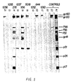
- the results are presented in Table 5 and Figure 1.
- p17 is a protein component produced upon cleavage of p55 to p24
- p24 is the viral core protein
- p31 is the viral endonuclease
- p41 is the mature envelope protein
- p51 and p66 are components of the viral reverse transcriptase
- p55 is a precursor to the viral core protein
- p160 is a precursor to the envelope protein
- p110/120 is a mixture of 2 proteins which co-migrate in this gel system: p120 is a protein component produced upon processing of the p160 protein to gp41; p110 is a protein component of the virus whose function is currently unknown.
- FIG. 1 An example of a typical a Western Blot analysis of the concentrated urine and serum samples obtained by practicing the method of the present invention is shown in Figure 1.
- lane 22 is the negative control
- lane 23 the weakly positive control
- lane 24 the strong positive control
- the numbers to the right represent the various HIV proteins discussed above.
- lines 12-16 and 18-21 are the corresponding patients as referred to in Table 5 above.
- urine obtained from patient No. 956 Figure 1, lane 16
- suffering from ARC contained antibodies directed against all of the HIV proteins except p24 (Core) and p17. The significance of this finding is presently unknown.
- Example 4 DOUBLE-DIFFUSION GRADIENT IMMUNOELECTROPHORETIC ANALYSIS OF HIV ANTI-BODY-CONTAINING URINE SAMPLES
- Concentrated urine sample No. 956 (Table I), which tested positive for HIV antigens using Western Blot analyses and antibodies directed against the envelope protein using ELISA, was further concentrated 200 fold and analyzed by double-diffusion gradient immuno-electrophoresis (DDG-IEP) as described by J.V. Chuba, in J.App. Biochem ., 1 : 37-50, 1979, (incorporated by reference). Serum samples from this patient were collected and analyzed in parallel to the patient urine samples as a positive control.
- DDG-IEP double-diffusion gradient immuno-electrophoresis
- the antisera troughs were completed by removing the corresponding segment of the gel between the precut slits, and conventional parallel-trough immunodiffusion was performed.
- 7.5 microliters of anti-human IgG, IgM and IgA (Behring Diagnostics, San Diego, CA) at a concentration of 5.5 micrograms per ml was added to the troughs and incubated for 20 minutes at room temperature. Thereafter, the gels were stained and examined for the development of percipitin lines.
- Anti-albumin antibodies were included in the serum samples as a positive control.
- IgG (11) and IgA (13) immunoglobulins were identified in the urine sample of patient No. 956.
- IgM although present in the serum did not appear in the urine (12). This is not surprising due to the large size of this immunoglobulin (approximately 900,000 daltons).
- Anti-albumin antibodies reacted with the serum samples, as expected (10), forming a sharp percipitin line of identity (lane 1).
Landscapes
- Health & Medical Sciences (AREA)
- Life Sciences & Earth Sciences (AREA)
- Chemical & Material Sciences (AREA)
- Immunology (AREA)
- Engineering & Computer Science (AREA)
- Virology (AREA)
- Molecular Biology (AREA)
- Hematology (AREA)
- Biomedical Technology (AREA)
- Urology & Nephrology (AREA)
- General Health & Medical Sciences (AREA)
- Microbiology (AREA)
- Biochemistry (AREA)
- Biotechnology (AREA)
- Physics & Mathematics (AREA)
- Analytical Chemistry (AREA)
- Organic Chemistry (AREA)
- Medicinal Chemistry (AREA)
- Proteomics, Peptides & Aminoacids (AREA)
- Cell Biology (AREA)
- Tropical Medicine & Parasitology (AREA)
- General Physics & Mathematics (AREA)
- Pathology (AREA)
- Zoology (AREA)
- Food Science & Technology (AREA)
- AIDS & HIV (AREA)
- Wood Science & Technology (AREA)
- Biophysics (AREA)
- Bioinformatics & Cheminformatics (AREA)
- General Engineering & Computer Science (AREA)
- Genetics & Genomics (AREA)
- Investigating Or Analysing Biological Materials (AREA)
- Measuring Or Testing Involving Enzymes Or Micro-Organisms (AREA)
Applications Claiming Priority (2)
| Application Number | Priority Date | Filing Date | Title |
|---|---|---|---|
| US40013 | 1987-04-17 | ||
| US07/040,013 US4865966A (en) | 1987-04-17 | 1987-04-17 | Method for detecting antibodies to human immunodeficiency virus |
Publications (2)
| Publication Number | Publication Date |
|---|---|
| EP0302997A2 true EP0302997A2 (de) | 1989-02-15 |
| EP0302997A3 EP0302997A3 (de) | 1990-11-22 |
Family
ID=21908598
Family Applications (1)
| Application Number | Title | Priority Date | Filing Date |
|---|---|---|---|
| EP19880106076 Withdrawn EP0302997A3 (de) | 1987-04-17 | 1988-04-15 | Verfahren zum Nachweis von Antikörpern gegen das menschliche Immunomangelhaftigkeitsvirus |
Country Status (10)
| Country | Link |
|---|---|
| US (1) | US4865966A (de) |
| EP (1) | EP0302997A3 (de) |
| JP (1) | JP2642721B2 (de) |
| KR (1) | KR960001503B1 (de) |
| AU (1) | AU617671B2 (de) |
| CA (1) | CA1340405C (de) |
| FI (1) | FI885868A7 (de) |
| OA (1) | OA09026A (de) |
| WO (1) | WO1988008039A1 (de) |
| ZA (1) | ZA882658B (de) |
Cited By (2)
| Publication number | Priority date | Publication date | Assignee | Title |
|---|---|---|---|---|
| EP0445650A3 (en) * | 1990-03-05 | 1992-02-12 | Abbott Laboratories | Detection of anti-hiv antibodies |
| KR101497726B1 (ko) * | 2007-04-27 | 2015-03-02 | 바셀 폴리올레핀 이탈리아 에스.알.엘 | 부텐-1 삼원중합체 및 이의 제조 방법 |
Families Citing this family (8)
| Publication number | Priority date | Publication date | Assignee | Title |
|---|---|---|---|---|
| US5122446A (en) * | 1987-04-17 | 1992-06-16 | New York University | Method for detecting antibodies to human immunodeficiency virus |
| EP0324834B1 (de) * | 1987-07-13 | 1995-11-08 | Verigen, Inc. | Verfahren zum schnellen und empfindlichen nachweis von hiv-1-antikörpern |
| US5447837A (en) * | 1987-08-05 | 1995-09-05 | Calypte, Inc. | Multi-immunoassay diagnostic system for antigens or antibodies or both |
| DE69028561T2 (de) * | 1989-03-09 | 1997-04-17 | Abbott Lab | Immunoassay zum Nachweis von Antikörpern gegen HIV |
| AU635008B2 (en) * | 1989-12-13 | 1993-03-11 | Genelabs Diagnostics Pte Ltd | Analytical apparatus and method for automated blot assay |
| AU9052491A (en) * | 1990-11-19 | 1992-06-11 | University Of Florida | Assay device and method for antibody and antigen detection |
| US6582962B1 (en) * | 1998-02-27 | 2003-06-24 | Ventana Medical Systems, Inc. | Automated molecular pathology apparatus having independent slide heaters |
| US20090181361A1 (en) * | 2008-01-14 | 2009-07-16 | Weidong Xu | Rapid test for detecting infection |
Family Cites Families (5)
| Publication number | Priority date | Publication date | Assignee | Title |
|---|---|---|---|---|
| US4464474A (en) * | 1980-07-09 | 1984-08-07 | Connaught Laboratories Limited | Non-A, non-B hepatitis assay and vaccine |
| JPS58182556A (ja) * | 1982-04-07 | 1983-10-25 | コンノ−ト.ラボラトリ−ズ.リミテツド | 非a非b型肝炎アツセイおよびワクチン |
| US4725669A (en) * | 1984-11-09 | 1988-02-16 | President And Fellows Of Harvard College | Assay for detecting infection by human T-cell lymphotropic virus-III |
| GB8707839D0 (en) * | 1987-04-02 | 1987-05-07 | Secr Social Service Brit | Immunoglobulin assay method |
| EP0317804B1 (de) * | 1987-11-24 | 1995-10-04 | Abbott Laboratories | HIV-Peptide und Methoden für den Nachweis von HIV |
-
1987
- 1987-04-17 US US07/040,013 patent/US4865966A/en not_active Expired - Lifetime
-
1988
- 1988-04-15 JP JP63504037A patent/JP2642721B2/ja not_active Expired - Lifetime
- 1988-04-15 FI FI885868A patent/FI885868A7/fi not_active IP Right Cessation
- 1988-04-15 CA CA000564232A patent/CA1340405C/en not_active Expired - Fee Related
- 1988-04-15 ZA ZA882658A patent/ZA882658B/xx unknown
- 1988-04-15 WO PCT/US1988/001304 patent/WO1988008039A1/en not_active Ceased
- 1988-04-15 EP EP19880106076 patent/EP0302997A3/de not_active Withdrawn
- 1988-04-15 AU AU17182/88A patent/AU617671B2/en not_active Ceased
- 1988-12-16 OA OA59493A patent/OA09026A/xx unknown
- 1988-12-16 KR KR88701685A patent/KR960001503B1/ko not_active Expired - Fee Related
Cited By (2)
| Publication number | Priority date | Publication date | Assignee | Title |
|---|---|---|---|---|
| EP0445650A3 (en) * | 1990-03-05 | 1992-02-12 | Abbott Laboratories | Detection of anti-hiv antibodies |
| KR101497726B1 (ko) * | 2007-04-27 | 2015-03-02 | 바셀 폴리올레핀 이탈리아 에스.알.엘 | 부텐-1 삼원중합체 및 이의 제조 방법 |
Also Published As
| Publication number | Publication date |
|---|---|
| JP2642721B2 (ja) | 1997-08-20 |
| KR890700683A (ko) | 1989-04-26 |
| EP0302997A3 (de) | 1990-11-22 |
| JPH01503487A (ja) | 1989-11-22 |
| KR960001503B1 (en) | 1996-01-31 |
| CA1340405C (en) | 1999-02-23 |
| US4865966A (en) | 1989-09-12 |
| AU617671B2 (en) | 1991-12-05 |
| FI885868L (fi) | 1988-12-19 |
| AU1718288A (en) | 1988-11-04 |
| OA09026A (en) | 1991-03-31 |
| ZA882658B (en) | 1989-03-29 |
| WO1988008039A1 (en) | 1988-10-20 |
| FI885868A0 (fi) | 1988-12-19 |
| FI885868A7 (fi) | 1988-12-19 |
Similar Documents
| Publication | Publication Date | Title |
|---|---|---|
| Grauballe et al. | Optimized enzyme‐linked immunosorbent assay for detection of human and bovine rotavirus in stools: Comparison with electron‐microscopy, immunoelectro‐osmophoresis, and fluorescent antibody techniques | |
| EP0216191B1 (de) | Immunologisches Testverfahren für HTLV-III-Antigene | |
| Araujo et al. | Antigenemia in recently acquired acute toxoplasmosis | |
| Naot et al. | IgM enzyme-linked immunosorbent assay test for the diagnosis of congenital Toxoplasma infection | |
| CA1303494C (en) | Simultaneous enzyme immunoassay for detecting antigen and/or antibody in humans | |
| US4294817A (en) | Method of fluoro immunoassay | |
| Bishai et al. | Enzyme-linked immunosorbent assay for detection of antibodies to influenza A and B and parainfluenza type 1 in sera of patients | |
| EP0202890A2 (de) | Konkurrierend-ELISA zum Nachweis von Antikörpern | |
| DK165958B (da) | Fremgangsmaade til isolering af htlv-iii proteiner, serologisk detektering af antistoffer til htlv-iii i serum fra aids- og prae-aids-patienter og detektering af htlv-iii infektioner ved immunoassays samt testsystem til brug ved fremgangsmaaden | |
| DK173687B1 (da) | Immunkemisk fremgangsmåde til bestemmelse af antistoffer, som er specifikke for et antigen, fra en af immunglobulin-klasserne A,M,D, eller E samt middel til udførelse af fremgangsmåden | |
| US5122446A (en) | Method for detecting antibodies to human immunodeficiency virus | |
| US5093230A (en) | Method for rapid and sensitive detection of IgM retroviral antibodies | |
| WO1992003579A1 (en) | An augmented western blot format and immunoassay for detection of viral antibodies | |
| US4865966A (en) | Method for detecting antibodies to human immunodeficiency virus | |
| Lin et al. | Standardized quantitative enzyme-linked immunoassay for antibodies to Toxoplasma gondii | |
| EP0265851B1 (de) | Verfahren zur Bestimmung von Anti-HIV, Mittel dazu und ihre Verwendung in diesem Verfahren | |
| Pyrhönen et al. | Immune reactions in epidermodysplasia verruciformis | |
| Vaheri et al. | Evaluation of solid‐phase enzyme‐lmmunoassay procedure in immunity surveys and diagnosis of rubella | |
| AU596404B2 (en) | Competitive elisa for the detection of antibodies | |
| US4816387A (en) | Method for detection of antibodies to HTLV-III and diagnostic test kit useful therewith | |
| EP0196873B1 (de) | Diagnose für Mikroorganismen | |
| CA2011783A1 (en) | Immunoassay for antibodies to hiv | |
| Matossian et al. | The serodiagnosis of human hydatid disease: 2. Additional studies on selected sera using indirect haemagglutination (IHA), enzyme linked immunosorbent assay (ELISA) and defined antigen substrate spheres (DASS) | |
| NO885617L (no) | Fremgangsmaate ved paavisning av menneskelig immunsviktvirus | |
| Gandhi et al. | A simple spot-test for circulating Entamoeba histolytica antigen-antibody complexes in patients with amoebic |
Legal Events
| Date | Code | Title | Description |
|---|---|---|---|
| PUAI | Public reference made under article 153(3) epc to a published international application that has entered the european phase |
Free format text: ORIGINAL CODE: 0009012 |
|
| AK | Designated contracting states |
Kind code of ref document: A2 Designated state(s): AT BE CH DE ES FR GB GR IT LI LU NL SE |
|
| RIN1 | Information on inventor provided before grant (corrected) |
Inventor name: CAO, YUNZHEN Inventor name: FRIEDMAN-KIEN, ALVIN E. Inventor name: BORKOWSKY, WILLIAM |
|
| PUAL | Search report despatched |
Free format text: ORIGINAL CODE: 0009013 |
|
| AK | Designated contracting states |
Kind code of ref document: A3 Designated state(s): AT BE CH DE ES FR GB GR IT LI LU NL SE |
|
| STAA | Information on the status of an ep patent application or granted ep patent |
Free format text: STATUS: THE APPLICATION IS DEEMED TO BE WITHDRAWN |
|
| 18D | Application deemed to be withdrawn |
Effective date: 19910523 |