EP0175617A2 - Conjugués d'anticorps et d'agent thérapeutique - Google Patents
Conjugués d'anticorps et d'agent thérapeutique Download PDFInfo
- Publication number
- EP0175617A2 EP0175617A2 EP85401776A EP85401776A EP0175617A2 EP 0175617 A2 EP0175617 A2 EP 0175617A2 EP 85401776 A EP85401776 A EP 85401776A EP 85401776 A EP85401776 A EP 85401776A EP 0175617 A2 EP0175617 A2 EP 0175617A2
- Authority
- EP
- European Patent Office
- Prior art keywords
- antibody
- therapeutic agent
- linker
- fragment
- fab
- Prior art date
- Legal status (The legal status is an assumption and is not a legal conclusion. Google has not performed a legal analysis and makes no representation as to the accuracy of the status listed.)
- Granted
Links
Images


Classifications
-
- A—HUMAN NECESSITIES
- A61—MEDICAL OR VETERINARY SCIENCE; HYGIENE
- A61K—PREPARATIONS FOR MEDICAL, DENTAL OR TOILETRY PURPOSES
- A61K51/00—Preparations containing radioactive substances for use in therapy or testing in vivo
- A61K51/02—Preparations containing radioactive substances for use in therapy or testing in vivo characterised by the carrier, i.e. characterised by the agent or material covalently linked or complexing the radioactive nucleus
- A61K51/04—Organic compounds
- A61K51/08—Peptides, e.g. proteins, carriers being peptides, polyamino acids, proteins
- A61K51/10—Antibodies or immunoglobulins; Fragments thereof, the carrier being an antibody, an immunoglobulin or a fragment thereof, e.g. a camelised human single domain antibody or the Fc fragment of an antibody
- A61K51/1093—Antibodies or immunoglobulins; Fragments thereof, the carrier being an antibody, an immunoglobulin or a fragment thereof, e.g. a camelised human single domain antibody or the Fc fragment of an antibody conjugates with carriers being antibodies
-
- A—HUMAN NECESSITIES
- A61—MEDICAL OR VETERINARY SCIENCE; HYGIENE
- A61K—PREPARATIONS FOR MEDICAL, DENTAL OR TOILETRY PURPOSES
- A61K41/00—Medicinal preparations obtained by treating materials with wave energy or particle radiation ; Therapies using these preparations
- A61K41/0042—Photocleavage of drugs in vivo, e.g. cleavage of photolabile linkers in vivo by UV radiation for releasing the pharmacologically-active agent from the administered agent; photothrombosis or photoocclusion
-
- A—HUMAN NECESSITIES
- A61—MEDICAL OR VETERINARY SCIENCE; HYGIENE
- A61K—PREPARATIONS FOR MEDICAL, DENTAL OR TOILETRY PURPOSES
- A61K41/00—Medicinal preparations obtained by treating materials with wave energy or particle radiation ; Therapies using these preparations
- A61K41/0057—Photodynamic therapy with a photosensitizer, i.e. agent able to produce reactive oxygen species upon exposure to light or radiation, e.g. UV or visible light; photocleavage of nucleic acids with an agent
- A61K41/0071—PDT with porphyrins having exactly 20 ring atoms, i.e. based on the non-expanded tetrapyrrolic ring system, e.g. bacteriochlorin, chlorin-e6, or phthalocyanines
-
- A—HUMAN NECESSITIES
- A61—MEDICAL OR VETERINARY SCIENCE; HYGIENE
- A61K—PREPARATIONS FOR MEDICAL, DENTAL OR TOILETRY PURPOSES
- A61K51/00—Preparations containing radioactive substances for use in therapy or testing in vivo
- A61K51/02—Preparations containing radioactive substances for use in therapy or testing in vivo characterised by the carrier, i.e. characterised by the agent or material covalently linked or complexing the radioactive nucleus
- A61K51/04—Organic compounds
- A61K51/08—Peptides, e.g. proteins, carriers being peptides, polyamino acids, proteins
- A61K51/10—Antibodies or immunoglobulins; Fragments thereof, the carrier being an antibody, an immunoglobulin or a fragment thereof, e.g. a camelised human single domain antibody or the Fc fragment of an antibody
-
- A—HUMAN NECESSITIES
- A61—MEDICAL OR VETERINARY SCIENCE; HYGIENE
- A61K—PREPARATIONS FOR MEDICAL, DENTAL OR TOILETRY PURPOSES
- A61K2121/00—Preparations for use in therapy
Definitions
- the present invention relates to the general area of antibody systems capable of delivering therapeutic agents to target sites in vivo.
- the therapeutic agents are covalently attached to antibodies or antibody fragments either through linkers or by direct attachment to form antibody conjugates.
- the antibody-therapeutic agent conjugates substantially retain the immunospecificity and immunoreactivity of the original antibody.
- the invention is directed to attachment of a therapeutic agent through a linker which may be either cleavable or non-cleavable.
- linker which may be either cleavable or non-cleavable.
- These antibody-therapeutic agent conjugates which comprise the therapeutic agent attached via the linker to the antibody molecule substantially retain the immunospecificity and immunoreactivity of the unconjugated antibody.
- Certain embodiments of the invention include attachment via linkers susceptible to cleavage by a proteolytic enzyme such that the resulting conjugate retains the ability to bind antigen and activate complement.
- the invention also includes attachment via linkers susceptible to cleavage by urokinase, plasmin, trypsin, a tissue plasminogen activator or other enzymes having proteolytic activity. In all of these embodiments cleavage of the linker promotes the release of the therapeutic agent in an active or activatable form at the target site.
- Another preferred embodiment of the invention relates to attachment of certain therapeutic agents to an antibody molecule such that the resulting conjugate is delivered to a specific target site and the therapeutic agent is not released.
- the therapeutic agent may be activated at the target site.
- Still another preferred embodiment relates to the attachment of an enzyme to an antibody molecule such that the resulting conjugate is delivered to a specific target site where the enzyme catalyzes reactions of therapeutic value.
- the invention also relates to several methods for preparing such antibody-therapeutic agent conjugates, intermediates which are useful in preparing the conjugates, novel therapeutic agents, and methods for using such therapeutic agents.
- carrier molecules have been utilized with limited success in the delivery of therapeutic agents to a site of action.
- the carrier should be non-toxic and target site specific.
- carrier molecules such as DNA, liposomes, proteins, steroid hormones and antibodies (whole antibody molecules or fragments) have been used in conjunction with a broad spectrum of pharmaceutical or cytotoxic agents such as: radioactive compounds (e.., 125 I, 131 I); agents which bind DNA, for instance, alkylating agents or various antibiotics; antimetabolites such as methotrexate; agents which act on cell surfaces (e.g., venom phospholipases and microbial toxins); and protein synthesis inhibitors (e.., diphtheria toxin and toxic plant proteins).
- radioactive compounds e.., 125 I, 131 I
- agents which bind DNA for instance, alkylating agents or various antibiotics
- antimetabolites such as methotrexate
- agents which act on cell surfaces e.g., venom phospholipases and microbial toxins
- protein synthesis inhibitors e.., diphtheria toxin and toxic plant proteins
- antibody carrier systems can be highly specific for the target site, a significant problem exists in that the therapeutic agent may not be released at that site. If release is necessary, the antibody-drug conjugates must be internalized by the tumor cell. There, release would occur through cleavage by lysosomal enzymes. Additionally, the non-site specific linkage of the therapeutic agent to (random) sites on the antibody molecule may interfere with antigen binding capacity, thus reducing the effectiveness of the system.
- the amino acid sequences of the light and heavy chains of immunoglobulins contain all of the amino acids relatively regularly and randomly dispersed throughout the molecule, including the antigen binding region. To the extent any chemical modification occurs in this antigen binding region, one has introduced a change in the recognition element of the antibody. Such changes would be expected to, and, in fact do, change the affinity and specificity of the antibody for antigen. In a population of different antibodies, such alteration in the antigen binding region results in complete inactivation of some antibodies and in lesser degrees of inactivation of others in relation to the proximity of the alterations to the antigen binding site.
- This inactivation may be due to a change within or very near the antigen binding site to alter the conformation of the binding site so as to make it unreactive, or may be due to a change in a region outside the antigen binding region so as to limit access of antigen to the antigen binding region.
- Methods involving amino acids which are relatively regularly and randomly dispersed throughout the antibody are referred to as non-site specific methods.
- Interchain disulfide bonds can also be used as sites of covalent attachment. However, even if one is successful in selectively reducing only the interchain disulfide bonds, several functional properties of the antibody may be adversely affected, such as functional affinity, agglutination ability and the ability to fix complement.
- Monoclonal antibodies produced by the hybridoma technique of Kohler and Milstein (1975, Nature 256:495-497; 1976, Eur. J. Immunol. 6:511-519) or related techniques provide distinct advantages for use as carriers for delivery of therapeutic agents to a site of action.
- monoclonal antibodies bind to only one molecular site (i.e., an epitope) with specific binding constants.
- Second, such antibodies are homogeneous and thus are purified with relative ease.
- monoclonal antibodies can be made in large quantities by particular hybridoma cell lines.
- tumor-produced or tumor-associated antigens have allowed the preparation of monoclonal antibodies which are immune-specific for solid tumors such as human colon, breast, hepatoma, melanoma and germ cell tumors (see reviews by Carrasquillo et al., 1984, Cancer Treatment Repts. 68:317-328; Kennel et al., 1984, Bio. Sci. 34:150-156).
- HpD hematoporphyrin derivative
- a cytotoxic mediator i.e., the singlet oxygen
- HpD seems to be preferentially taken up and retained by many tumors compared to adjacent tissues (see Dougherty, In Porphyrin Photosensitization, New York: Plenum Publishing Corp., 1983, pp. 3-13).
- the laser output beam may be connected to optical fibers of the appropriate size and applied either directly into the tumor or externally to the general location of the tumor.
- photochemicals e.g., hematoporphyrin and other photosensitizers
- hematoporphyrin and other photosensitizers have several disadvantages for clinical use.
- a second disadvantage is that patients receiving photoradiation therapy are generally extremely sensitive to sunlight and must avoid exposure to the sun, frequently for as long as four weeks.
- the dosage levels of photosensitizer required for therapy are very high and may have a negative effect on normal tissue.
- the ideal photosensitizer should be designed so that it has greater tumor specificity, requiring a lower therapeutic dose level, hence mitigating the deleterious effect of higher doses. Greater tumor specificity leads to more efficient localization to the site of action and less opportunity for dispersal throughout the body.
- a therapeutic agent is covalently attached to an antibody or antibody fragment.
- the covalent attachment of the therapeutic agent is accomplished so that the resulting antibody conjugate retains the ability to bind antigen.
- such methods include attachment to oxidized carbohydrate moieties of antibodies or antibody fragments, or to the sulfhydryl groups of reduced antibodies or reduced ( Fab ') 2 fragments.
- the invention concerns methods for preparing antibody-therapeutic agent conjugates, comprising:
- the first part would produce an antibody-linker intermediate which may be considered a step in the production of the final antibody-therapeutic agent conjugate.
- Such antibody-linker intermediates may be stored for later combination with the particular therapeutic agent.
- the first part of the two part method would involve steps (a) and (b) above to form the intermediate antibody-linker intermediates
- the second part possibly at a later point in time, would involve covalently attaching the linker portion of the antibody-linker intermediate to a therapeutic agent to produce the final antibody-therapeutic agent conjugate.
- Such antibody-therapeutic agent conjugates can also be made by alternate methods, as, for example, by first covalently attaching the linker to the therapeutic agent, and then reacting the antibody or antibody fragment with an amine group of the linker portion of the linker-therapeutic agent to form the antibody-therapeutic agent conjugate.
- the invention further includes a method for preparing an antibody-therapeutic agent conjugate, comprising:
- the linker may comprise a spacer element and a cleavable element.
- One function of the spacer element could be to position the cleavable element away from the core of the antibody molecule such that the cleavable element is more accessible to the enzyme responsible for cleavage.
- the above-described method for preparing antibody-therapeutic agent conjugates may be separated into two distinct parts.
- the first part (steps (a) and (b) above) would produce an antibody-linker intermediate which may be stored for later combination with a therapeutic agent (step (c) above).
- antibody-therapeutic agent conjugates in which the linker comprises a spacer element and a cleavable element may also be made by first covalently attaching the linker to the therapeutic agent, and then reacting the antibody or antibody fragment with the linker portion of the linker-therapeutic agent to form the antibody-therapeutic agent conjugate.
- this method for preparing an antibody-therapeutic agent conjugate (having a linker comprising a spacer element and a cleavable element) comprises:
- Such antibody-therapeutic agent conjugates in which the linker comprises a spacer element and a cleavable element may be made by still other methods, for instance by first attaching the antibody to the spacer element, and then attaching the spacer element of that intermediate to a cleavable element of another intermediate comprising a cleavable element covalently attached to a therapeutic agent.
- Such methods comprise:
- Still another method for preparing these antibody-therapeutic agent conjugates involves first preparing an antibody-spacer element intermediate, attaching to the spacer element of this intermediate a cleavable element to form an antibody-spacer element-cleavable element intermediate, and finally attaching to the cleavable element of this intermediate a therapeutic agent.
- This method comprises:
- another function of the spacer element could be to add multiple functional sites for subsequent attachment of cleavable elements or therapeutic agents, or cleavable element-therapeutic agent intermediates.
- sites may be aldehyde or sulfhydryl groups, or any chemical site to which a cleavable element, therapeutic agent or cleavable element-therapeutic agent intermediate may be attached.
- the linker could be a "branched linker" having multiple functional sites for attachment directly to a therapeutic agent.
- the functional sites may be aldehyde or sulfhydryl groups, or any chemical site to which a therapeutic agent may be attached.
- linkers including branched linkers, may be attached to the same antibody molecule to form conjugates having a large number of therapeutic agents per antibody molecule.
- the invention is also directed to intermediates and final products of the above-described methods.
- this invention encompasses antibody-linker intermediates, which comprise a linker attached via a covalent bond to a carbohydrate moiety of an oxidized antibody or antibody fragment, said antibody-linker intermediate having substantially the same immunoreactivity and immunospecificity of the original antibody or antibody fragment.
- the linkers may comprise a spacer element (attached to the carbohydrate moiety) which, in turn, is covalently attached to a cleavable element.
- the invention also relates to antibody-spacer element intermediates, cleavable element-therapeutic agent intermediates and linker-therapeutic agent intermediates described in the methods above. Intermediates in which the linker is not cleavable are also encompassed by the invention.
- the invention encompasses antibody-therapeutic agent conjugates which comprise a therapeutic agent covalently attached (directly or via a linker) to a carbohydrate moiety of an oxidized antibody or antibody fragment, said antibody-therapeutic agent conjugate having substantially the same immunoreactivity and immunospecificity as the unconjugated antibody or antibody fragment.
- an antibody-therapeutic agent conjugate in which the linker or therapeutic agent is attached to a sulfur atom of a reduced antibody or Fab' fragment.
- This embodiment of the invention involves a method for preparing an antibody-therapeutic agent conjugate, comprising:
- This method can be separated into two parts, the first part involving steps (a) and (b) above and the second step, separate in time, would involve producing the final conjugate as in step (c) above.
- the same antibody-therapeutic agent conjugates can be made by another method, comprising:
- the linker may comprise a spacer element covalently attached to a cleavable element.
- the spacer-element would enable positioning of the cleavable element away from the core of the antibody molecule so that the cleavable element is more accessible to the cleaving enzyme.
- the above-described method may be separated into two parts, the first involving steps (a) and (b), and the second involving step (c) above.
- These antibody-therapeutic agent conjugates having a linker comprising a spacer element and a cleavable element may be made by first covalently attaching the linker to the therapeutic agent, followed by reacting the reduced antibody or Fab' fragment with the linker portion of the linker-therapeutic agent to form the antibody-therapeutic agent conjugate.
- This method comprises:
- antibody-therapeutic agent conjugates having a spacer element and a cleavable element may be made by still other methods, such as by first attaching the antibody to the spacer elements, and then attaching the spacer element of that intermediate to a cleavable element of another intermediate comprising a cleavable element covalently attached to a therapeutic agent.
- These methods comprise :
- Still another method for preparing these antibody-therapeutic agent conjugates involves first preparing an antibody-spacer intermediate, followed by attaching to the spacer element of this intermediate a cleavable element to form an antibody-spacer element-cleevable element intermediate, and finally attaching to the cleavable element of this intermediate a therapeutic agent.
- This method comprises:
- spacer elements may have multiple functional sites for subsequent attachment of therapeutic agents, cleavable elements, or cleavable element-therapeutic agent intermediates. These functional sites may be aldehyde or sulfhydryl groups, or any chemical site to which the therapeutic agent, cleavable element or cleavable element-therapeutic agent may be attached.
- the invention includes the intermediates of these methods in which the attachment is to a sulfur atom of an antibody molecule, including antibody-linker intermediates, antibody-spacer element intermediates, antibody-spacer element-cleavable element intermediates, linker-therapeutic agent intermediates, cleavable element-therapeutic agent intermediates.
- the antibody-therapeutic agent conjugates comprise a therapeutic agent covalently attached (directly or through a linker) to a sulfur atom of a reduced antibody or Fab' fragment, said antibody-therapeutic agent conjugate having substantially the same immunoreactivity and immunospecificity as the unconjugated antibody or (Fab') 2 fragments.
- the antibody-therapeutic agent conjugates of the invention are ideally suited for in vivo therapy. Delivery of therapeutic agents to specific target sites involves administering to an animal or human an effective amount of an antibody-therapeutic agent conjugate, wherein said antibody-therapeutic agent conjugate is immunoreactive with and immunospecific for an antigenic determinant of said specific tissue and substantially non-immunoreactive with and non-immunospecific for non-specific tissue and said antigenic determinant is not found in substantial amount in non-specific tissue.
- This invention also encompasses the use of antibodies for delivery to specific cells, tissues, organs, or any other site in vivo, and the subsequent release or activation of the therapeutic agent at the target site.
- release of the compound may be mediated by activated complement, a plasminogen activator, plasmin, a urokinase, trypsin, or another enzyme having proteolytic activity.
- photosensitive chemicals or enzymes that catalyze substrate modification with the production of cytotoxic by-products are attached to the antibody molecule.
- the invention contemplates site selective attachment of therapeutic agents to those areas of antibodies or antibody fragments which are not a part of nor directly involved with the antigenic site of the molecule.
- the antibody conjugate formed after selective attachment to one of these sites (located outside the antigen binding region), the antibody conjugate formed has substantially the same immunoreactivity and immunospecificity as the unconjugated antibody or antibody fragment.
- Antibodies directed against any desired target may be used as carrier molecules.
- any desired target e.g., antigenic determinants of tumor cells, virus, fungi, bacteria or parasites
- monoclonal antibodies offer the advantages of increased specificity for antigen, improved efficiency of the delivery system and ease in production. ⁇
- a therapeutic agent is attached to an antibody carrier molecule of an immunoglobulin class that is capable of complement activation. This attachment is accomplished via linkers which are susceptible to cleavage by an enzyme as enumerated above.
- One or more different therapeutic agents may be attached to each antibody molecule.
- the resulting antibody-therapeutic agent conjugate is administered to an individual. Subsequent to the binding of the antibody-therapeutic agent conjugate to antigen in vivo, the individual's serum complement is activated and the compounds will be selectively cleaved and released at the target site.
- the same linker described supra may be attached to an antibody carrier molecule of a class that does not activate complement.
- a photosensitizer is attached to an antibody carrier molecule either by a non-cleavable linker or by direct attachment to the antibody molecule.
- the photosensitizer is activated by light of the appropriate wavelength and its cytolytic effects on nearby cells are mediated through the generation of singlet oxygen molecules and oxygen free radicals.
- cleavage of the linker at the target site may not be desirable.
- the linker utilized may be insensitive to serum proteins or the antibody molecule may be of a class or type that does not activate complement.
- a urokinase tissue plasminogen activator, plasmin, trypsin or a protease-sensitive linker attached to an antibody that does or does not fix complement.
- the therapeutic agent for the practice of this invention it is desirable to attach the therapeutic agent to the antibody molecule without interfering with either the antigen binding capacity of the antibody, with the ability to activate complement (also called complement fixation), or with enzyme cleavage or photoactivation of the therapeutic agent or with the process of conversion of enzyme substrates into cytotoxic by-products by the therapeutic agent.
- complement also called complement fixation
- the present invention describes the novel linkers and methods of attachment which may be used to attach therapeutic agents to any antibody capable of activating complement.
- the present invention concerns antibody-therapeutic agent conjugates prepared by attaching a therapeutic agent to an antibody or antibody fragment directed against a target antigen.
- the therapeutic agent is attached either directly or via a linker to the antibody or antibody fragment.
- Such therapeutic agents or linkers are selectively attached to those areas of antibodies or antibody fragments which are not a part of nor directly involved with the antigen binding site of the molecule.
- antibodies directed against any antigen or hapten may be used.
- monoclonal antibodies offer several advantages. Each monoclonal antibody is specific for one antigenic determinant. Additionally, large amounts of each monoclonal antibody can be produced.
- Antibodies used in the present invention may be directed against any determinant, e.g., tumor, bacterial, fungal, viral, parasitic, mycoplasmal, histocompatibility, differentiation and other cell membrane antigens, pathogen surface antigens, toxins, enzymes, allergens, drugs and any biologically active molecules.
- determinant e.g., tumor, bacterial, fungal, viral, parasitic, mycoplasmal, histocompatibility, differentiation and other cell membrane antigens, pathogen surface antigens, toxins, enzymes, allergens, drugs and any biologically active molecules.
- immunoglobulin classes that are known to activate complement are used.
- carrier immunoglobulins may be used which are not capable of complement activation.
- immunoglobulin carriers may include: certain classes of antibodies such as IgM, IgA, IgD, IgE; certain subclasses of IgG; or certain fragments of immunoglobulins, e.g., half antibody molecules (a single heavy: light chain pair), or Fab, Fab' or (Fab') 2 fragments.
- the Fab' fragments of IgG immunoglobulins are obtained by cleaving the antibody molecule with pepsin [resulting in a bivalent fragment, (Fab' ) 2 J or with papain [resulting in 2 univalent fragments, (2 Fab)].
- pepsin resulting in a bivalent fragment, (Fab' ) 2 J or with papain [resulting in 2 univalent fragments, (2 Fab)].
- the bivalent (Fab') 2 fragment can be split by mild reduction of one or a few disulfide bonds to yield univalent Fab' fragments.
- Fab and (Fab') 2 fragments are smaller than a whole antibody molecule and, therefore, permeate the target site or tissue more easily. This may offer an advantage for in vivo delivery since conjugates will more readily penetrate in vivo sites (e.g., tumor masses, infection sites, etc.). An additional advantage is obtained when using conjugates formed with antibody fragments because these fragments do not cross a placental barrier. As a result, using this embodiment of the present invention, a therapeutic agent may be delivered at an in vivo site (such as a tumor) to a pregnant female without exposing the fetus to the compound.
- an in vivo site such as a tumor
- the present invention utilizes several methods for attaching therapeutic agents to antibody molecules such as (a) attachment to the carbohydrate moieties of the antibody or antibody fragment, or (b) attachment to sulfhydryl groups of the antibody or antibody fragment. Whichever method is used, the attachment must not significantly change the essential characteristics of the antibody or antibody fragment, such as immunospecificity and immunoreactivity. Additional considerations include simplicity of reaction and stability of the antibody conjugate produced.
- Glycoproteins are biologically impcrtant macromolecules which share structural characteristics including carbohydrate residues covalently attached to a polypeptide backbone. Since antibodies are glycoproteins, compounds may be attached to the carbohydrate moiety of the molecule. Some of the carbohydrate moieties are located on the Fc region of the immunoglobulin and are required in order for Cl binding to occur. The carbohydrate moiety of the Fc region of an immunoglobulin may be utilized in the scheme described herein. Alternatively, the Fab or Fab' fragments of any immunoglobulins which contain carbohydrate moieties may be utilized in the reaction scheme described herein. An example of such an immunoglobulin is the human IgM sequenced by Putnam et al. (1973, Science 182: 287).
- the carbohydrate side chains of antibodies or Fab or Fab' fragments may be selectively oxidized to generate aldehydes.
- the resulting aldehydes may then be reacted with amine groups (e.g., ammonia derivatives such as primary amine, secondary amine, hydroxylamine, hydrazine, hydrazide, phenylhydrazine, semicarbazide or thiosemicarbazide) to form a Schiff base or reduced Schiff base (e . g., imine, enamine, oxime, hydrazone, phenylhydrazone, semicarbazone, thiosemicarbazone'or reduced forms thereof).
- amine groups e.g., ammonia derivatives such as primary amine, secondary amine, hydroxylamine, hydrazine, hydrazide, phenylhydrazine, semicarbazide or thiosemicarbazide
- the carbohydrate moiety of the antibody may be modified by enzymatic techniques so as to enable attachment to or reaction with other chemical groups.
- an enzyme is galactose oxidase which oxidized galactose in the presence of oxygen to form an aldehyde.
- Oxidation of the carbohydrate portion or moiety of antibody molecules leads to formation of aldehyde groups.
- a variety of oxidizing agents can be used, such as periodic acid, paraperiodic acid, sodium metaperiodate and potassium metaperiodate. Among these, oxygen acids and salts thereof are preferred since secondary or undesirable side reactions are less frequent.
- oxygen acids and salts thereof are preferred since secondary or undesirable side reactions are less frequent.
- Oxidation of antibodies with these oxidizing agents can be carried out by known methods.
- the antibody is used generally in the form of an aqueous solution, the concentration being generally less than 100 mg/ml, preferably 1 to 20 mg/ml.
- an oxygen acid or a salt thereof is used as the oxidizing agent, it is used generally in the form of an aqueous solution, and the concentration is generally 0.001 to 10 mM and preferably 1.0 to 10 mM.
- the amount of the oxygen acid or salt thereof depends on the kind of antibody, but generally it is used in excess, for example, twice to ten times as much as the amount of the oxidizable carbohydrate. The optimal amount, however, can be determined by routine experimentation.
- the optional ranges include a pH of from about 4 to 8, a temperature of from 0° to 37°C, and a reaction period of from about 15 minutes to 12 hours.
- Oxidation of the carbohydrate portion of antibody molecules may also be done with the enzyme, galactose oxidase (Cooper et al., 1959, J. Biol. Chem. 234: 445-448).
- the antibody is used in aqueous solution, the concentration being generally 0.5 to 20 mg/ml.
- the enzyme generally is used at about 5 to 100 units per ml of solution, at a pH ranging from about 5.5 to about 8.0. The influence of pH, substrate concentration, buffers and buffer concentrations of enzyme reaction are reported in Cooper et al., supra.
- linker or therapeutic agent having an available amine group selected from the group consisting of primary amine, secondary amine, hydrazine, hydrazide, hydroxylamine, phenylhydrazine, semicarbazide and thiosemicarbazide groups.
- the immediately resulting products contain a carbon-nitrogen double bond resulting from elimination of a molecule of water from the initial addition products:
- a solution of the oxidized antibody at a concentration of from about 0.5 to 20 mg/ml is mixed with the therapeutic agent or linker (molar ratios of reactive amine group ito antibody aldehyde ranging from about 1 to about 10,000) and the solution incubated for from about 1 to 18 hours. Suitable temperatures are from 0 to 37°C and pH may be from about 6 to 8.
- the antibody-therapeutic agent conjugates (or antibody-linker intermediates) have been formed between the antibody and therapeutic agent or linker as described in Section 5.2.1.3, they can optionally be stabilized with a suitable reducing agent, such as sodium cyanoborohydride or sodium borohydride: Reducing agent is generally added to a molar excess of from about 10 to 100 fold molar excess over available aldehyde groups.
- a suitable reducing agent such as sodium cyanoborohydride or sodium borohydride: Reducing agent is generally added to a molar excess of from about 10 to 100 fold molar excess over available aldehyde groups.
- Free sulfhydryl groups can be generated from the disulfide bonds of the immunoglobulin molecule. This is accomplished by mild reduction of the antibody molecule.
- the disulfide bonds of IgG which are generally most susceptible to reduction are those that link the two heavy chains.
- the disulfide bonds located near the antigen binding region of the antibody molecule remain relatively unaffected. Such reduction results in the loss of ability to fix complement but does not interfere with antibody-antigen binding ability (Karush et al., 1979, Biochem. 18:2226-2232).
- the free sulfhydryl groups generated in the intra-heavy chain region can then react with reactive groups of a linker or therapeutic agent to form a covalent bond which does not interfere with the antigen binding site of the immunoglobulin.
- Such reactive groups include, but are not limited to, reactive haloalkyl groups (including, for example, haloacetyl groups), p-mercuribenzoate groups and groups capable of Michael-type addition reactions (including, for example, maleimides and groups of the type described in Mitra and Lawton, 1979, J. Amer. Chem. Soc. 101: 3097-3110).
- haloalkyl is meant any alkyl group of one to three carbon atoms substituted with bromine, iodine or chlorine.
- Antibody-therapeutic agent conjugates which are produced by attachment to free sulfhydryl groups of reduced immunoglobulin or reduced antibody fragments do not activate complement.
- these conjugates may be used in in vivo systems where cleavage and release of the therapeutic agent is not desirable (e.g., where the therapeutic agent is a photosensitizer, or an enzyme that acts on a specific substrate).
- Such conjugates may also be used when non- complement mediated release is desired.
- the therapeutic agent may be linked to sulfhydryl groups on the reduced immunoglobulin or reduced antibody fragments via linkers which are susceptible to cleavage by enzymes having proteolytic activity, including but not limited to trypsin, urokinase, plasmin, tissue plasminogen activator and the like.
- a therapeutic agent joined to a complement-sensitive substrate linker can be attached to sulfhydryls of reduced IgG molecules or antibody fragments and delivered to the target in a mixture with intact antibody molecules that are capable of activating complement. The latter would activate complement which would cleave the therapeutic agent from the former.
- the use of antibody fragments as carrier molecules in the complement mediated release system would permit the treatment of pregnant females, and offers the advantage of more rapid penetration of the conjugate into target sites. ⁇
- the substrate linkers or the therapeutic agents are modified by attaching an iodoalkyl group to one end of the linker.
- the unmodified site on the linker may or may not be covalently attached to a therapeutic agent.
- the substrate linkers which are ester or amide linked to therapeutic agents as prepared in Section 5.3 are modified by the addition of an iodoalkyl group thus forming an iodoalkyl derivative as depicted below (N.B., the symbol * signifies an amide or ester bond):
- the linker may be one that is susceptible or resistant to cleavage by activated complement, trypsin, plasmin, tissue plasminogen activator, urokinase or another specific enzyme having proteolytic activity.
- Antibodies may be attached to any therapeutic agent which retains its essential properties after reaction with the antibody, and which enables the antibody to substantially retain immunospecificity and immunoreactivity.
- therapeutic agent includes chemical modifications and derivatives of therapeutic agents which substantially retain their biological activity. The major limiting factor is that any attachment reaction must be selective enough to limit competing, undesirable reactions and sufficiently mild so as not to severely interfere with antibody reactivity and selectivity.
- the therapeutic agent When it is desired to attach an aldehyde of the oxidized carbohydrate portion of an antibody or antibody fragment to a therapeutic agent, the therapeutic agent should contain an amine group selected from the group consisting of primary amine, secondary amine, hydrazine, hydrazide, hydroxylamine, phenylhydrazine, semicarbazide and thiosemicarbazide groups. If the therapeutic agent does not contain any such amino group, the agent can be modified to introduce a suitable amine group available for coupling.
- the therapeutic agent to be attached to an antibody for use in a delivery system is selected according to the purpose of the intended application (i.e, killing, prevention of cell proliferation, hormone therapy or gene therapy).
- Such therapeutic agents may include, for example, pharmaceutical agents, toxins, fragments of toxins, alkylating agents, enzymes, antibiotics, antimetabolites, antiproliferative agents, hormones, neurotransmitters, DNA, radioopaque dyes, radioactive isotopes, fluorogenic compounds, marker compounds, lectins, compounds which alter cell membrane permeability, and photochemical compounds.
- Table I lists some of the pharmaceutical agents that may be employed in the herein described invention and in no way is meant to be an exhaustive list. Finally, combinations of therapeutic agents may be used.
- photochemicals including photosensitizers and photothermolytic agents may be used as therapeutic agents.
- Efficient photosensitizers include, but are not Limited to porphyrins and modified porphyrins (e.g., hematoporphyrin, hematoporphyrin dihydrazide, deuteroporphyrin dihydrazide and protoporphyrin dihydrazide), rose bengal, acridines, thiazines, xanthenes, anthraquinones, azines, flavin and nonmetal-containing porphyrins, porphyrin-like compounds, methylene blue, eosin, erythrosin, psoralin and the like.
- photosensitizers include, but are not limited to tetracyclines (e.g., dimethylchlor tetracycline) sulfonamides (e.g., sulfanilamide), griseofulvin, phenothiazines, (e.., chlorpromazine), thiazides, sulfonylurea, and many others.
- Photochemicals may be designed or synthetically prepared to absorb light at specific wavelengths. Photothermolytic agents, such as Azure A, which are activated at the site of action by a light source (see Anderson and Parrish, 1983, Science 220: 524-527) may be utilized as therapeutic agents.
- enzymes that catalyze substrate modification with the production of cytotoxic by-products may be used as therapeutic agents.
- examples of such enzymes include but are not limited to glucose oxidase, galactose oxidase, xanthene oxidase and the like.
- antibodies may be covalently attached to a therapeutic agent through an intermediate linker having at least two reactive groups, one to react with antibody and one to react with the therapeutic agent.
- Suitable linkers for reaction with oxidized antibodies or oxidized antibody fragments include those containing an amine selected from the group consisting of primary amine, secondary amine, hydrazine, hydrazide, hydroxylamine, phenylhydrazine, semicarbazide and thiosemicarbazide groups. Such reactive functional groups may exist as part of the structure of the linker, or may be introduces by suitable chemical modification of linkers not containing such groups.
- suitable linkers for attachment to reduced antibodies or antibody fragments include those having certain reactive groups capable of reaction with a sulfhydryl group of a reduced antibody or Fab' fragment.
- reactive groups include, but are not limited to: reactive haloalkyl groups (including, for example, haloacetyl groups), p-mercuribenzoate groups and groups capable of Michael-type addition reactions (including, for example, maleimides and groups of the type described by Mitra and Lawton, 1979, J. Amer. Chem. Soc. 101: 3097-3110).
- haloalkyl is meant any alkyl group of one to three carbon atoms substituted with bromine, iodine or chlorine.
- the therapeutic agent may be attached to the linker before or after the linker is attached to the antibody molecule. In certain applications it may be desirable to first produce an antibody-linker intermediate in which the linker is free of an associated therapeutic agent. Depending upon the particular application, a specific therapeutic agent may then be covalently attached to the linker.
- branched linkers which have multiple sites for attachment of therapeutic'agents.
- a single covalent attachment to an antibody or antibody fragment would result in an antibody-linker intermediate capable of binding a therapeutic agent at a number of sites.
- the sites may be aldehyde or sulfhydryl groups or any chemical site to which therapeutic agents can be attached.
- higher specific activity can be achieved by attachment of a single site linker at a plurality of sites on the antibody or antibody fragment.
- This plurality of sites may be introduced into the antibody or antibody fragment by either of two methods either of two methods.
- the functional sites of the branched linker or multiple site linker may be aldehyde or sulfhydryl groups, or may be any chemical site to which linkers may be attached. Still higher specific activities may be obtained by combining these two approaches, that is, attaching multiple site linkers at several sites on the antibody or antibody fragment.
- Peptide linkers which are susceptible to cleavage by enzymes of the complement system, urokinase, tissue plasminogen activator, trypsin, plasmin, or another enzyme having proteolytic activity may be used in one embodiment of the present invention.
- a therapeutic agent is attached via a linker susceptible to cleavage by complement :
- the antibody is selected from a class which can activate complement.
- the antibody-therapeutic agent conjugate thus, activates the complement cascade and releases the therapeutic agent at the target site.
- attachment of a therapeutic agent to an antibody via sulfhydryl groups destroys the ability of the antibody therapeutic agent to activate complement.
- a therapeutic agent is attached via a linker susceptible to cleavage by enzymes having a proteolytic activity such as a urokinase, a tissue plasimogen activator, plasmin, or trypsin.
- therapeutic agents may be attached via disulfide bonds (for example, the disulfide bonds on a cystine molecule) to the antibody molecule. Since many tumors naturally release high levels of glutathione (a reducing agent) this can reduce the disulfide bonds with subsequent release of the therapeutic agent at the site of delivery.
- disulfide bonds for example, the disulfide bonds on a cystine molecule
- linker in such a way as to optimize the spacing between the therapeutic agent and the antibody. This may be accomplished by use of a linker of the general structure: wherein W is either -NH-CH 2 - or -CH 2 -; Q is an amino acid, peptide; and n is an integer from 0 to 20.
- the linker may comprise a spacer element and a cleavable element.
- the spacer element serves to position the cleavable element away from the core of the antibody molecule such that the cleavable element is more accessible to the enzyme responsible for cleavage.
- Certain of the "branched linkers" described above may serve as spacer elements.
- linker to therapeutic agent or of spacer element to cleavable element, or cleavable element to therapeutic agent
- attachment of linker to therapeutic agent need not be particular mode of attachment or reaction. Any reaction providing a product of suitable stability and biological compatibility is acceptable.
- an antibody of a class which can activate complement is used.
- the resulting retains the ability to bind antigen and activate the complement cascade.
- Complement is the collective name for a group of serum proteins which can be activated in one of two ways, the classical pathway and the properdin pathway (M U ller-Eberhard, Hospital Practice, August 1977:33-43).
- the classical pathway is initiated by the binding of antibodies of the IgM class or certain subclasses of IgG to its corresponding antigen whereas the properdin pathway is dependent upon the serum protein, properdin and other non-immunoglobulin serum factors (Reid and Porter, 1981, Ann. Rev. Biochem. 50:433-464).
- the classical pathway is a pathway of particular importance of the practice of the present invention.
- the classical pathway is characterized by the formation of certain antibody-antigen complexes (or immune complexes) which activate the proteolytic enzymes of the complement system (Borsos and Rapp, 1965, J. Immunol. 95:559-566; Cohen, 1968, J. Immunol. 100:407-413; Cohen and Becker, 1968, J. Immunol. 100:403-406; Ishizaka et al., 1968, J. Immunol. 100:1145-1153).
- These activated complement enzymes cleave and activate other components of the complement cascade.
- an "attack complex” or lytic complex
- the first component activated in the classical pathway is Cl which becomes a protease that acts on both C2 and C4.
- Activated Cl has a specific esterase activity.
- Activated C4,2 C 4,2
- C3 convertase is a complex which proteolytically cleaves C3, and together with activated C3 (C3b), cleaves C5.
- Cleavage of C3 is the first step in common between the classical and properdin pathways of complement activation.
- a therapeutic agent is joined to one end of the cleavable linker or cleavable element and the other end of the linker group is attached to a specific site on the antibody molecule.
- the therapeutic agent may be attached to the carboxy terminus of a peptide, amino acid or other suitably chosen linker via an ester or amide bond, respectively.
- such agents may be attached to the linker peptide via a carbodiimide reaction.
- the therapeutic agent contains functional groups that would interfere with attachment to the linker, these interfering functional groups can be blocked before attachment and deblocked once the product conjugate or intermediate is made. For example, FIG.
- Linkers may be of any desired length, one end of which can be covalently attached to specific sites on the antibody molecule. The other end of the linker or spacer element may be attached to an amino acid or peptide linker.
- Table III lists some cleavable elements that may be used as linker groups to prepare the antibody-therapeutic agent conjugates of the present invention. (In the table n may be an integer including zero.) These sequences were derived from those of the complement substrate sequences by substituting amino acids with similar acid-base properties. This list is not exhaustive.
- conjugates when these conjugates bind to antigen in the presence of complement the amide or ester bond which attaches the therapeutic agent to the linker will be cleaved, resulting in release of the agent in its active form.
- conjugates when administered to an individual, will accomplish delivery and release of the therapeutic agent at the target site, and are particularly effective for the in vivo delivery of pharmaceutical agents, antibiotics, antimetabolites, antiproliferative agents and the like.
- conjugates may be prepared by attaching the therapeutic agent to an antibody molecule or fragment that is not capable of activating complement via a linker that is mildly susceptible to cleavage by serum proteases. When this conjugate is administered to an individual, antigen-antibody complexes will form quickly whereas cleavage of the therapeutic agent will occur slowly, thus resulting in release of the compound at the target site.
- the substrate linkers are modified, for example, by attaching hydrazine or hydrazide derivative to one end of the linker.
- the unmodified sites on the linker may or may not be covalently attached to a therapeutic agent.
- the substrate linkers which are attached to a therapeutic agent via an ester or amide link, as described in Section 5.3 are modified by attaching a hydrazide (e.g., phenylhydrazine) to the opposite amino terminus of the peptide chain.
- a hydrazide e.g., phenylhydrazine
- hydrazine is in the para position
- compounds with the hydrazine moiety in the ortho or meta positions are then reacted with an oxidized immunoglobulin, or immunoglobulin fragment containing an oxidized carbohydrate.
- the linker utilized may be resistant to cleavage by either activated complement or serum proteases.
- linker is designed to be susceptible to cleavage by a serum protease
- one resulting structure is schematically represented below (N.B., the symbol * signifies an amide or ester bond):
- the conjugate may be designed so that the therapeutic agent is delivered to the target but not released. This may be accomplished by attaching a therapeutic agent to an antibody or antibody fragment either directly or via a non-cleavable linker.
- non-cleavable linkers may include-amino acids, peptides, D-amino acids or other organic compounds which may be modified to include functional groups that can subsequently be utilized in attachment to antibody molecules or antibody fragments by the methods described herein.
- a general formula for such an organic linker could be wherein W is either -NH-CH 2 - or -CH 2 -;
- a compound may be attached to antibody molecules or antibody fragments which do not activate complement.
- this attachment may be accomplished using linkers that are susceptible to cleavage by activated complement or using linkers that are not susceptible to cleavage by activated complement.
- the antibody-therapeutic agent conjugates of the invention are useful in a variety of therapeutic in vivo applications.
- cellular disorder is meant to include all neoplasms, including cancers, adenomas, and hyperplasias; certain immunological disorders, including autoimmune diseases, graft-versus-host diseases (e.g., after bone marrow transplantation), immune suppressive diseases, e.g., after kidney or bone marrow transplantation.
- Treatment of such cellular disorders involving, for example, bone marrow transplantation may include purging (by killing) undesired cells, e.g., malignant cells or mature T lymphocytes.
- Therapeutic applications center generally on treatment of various cellular disorders, including those broadly described above, by administering an effective amount of the antibody-therapeutic agent conjugates of the invention.
- the properties of the antibody are such that it is immunospecific for and immunoreactive with a particular antigen render it ideally suited for delivery of therapeutic agents to specific cells, tissues, organs or any other site having that particular antigen.
- the antibody or antibody fragment of the antibody therapeutic agent conjugate functions to deliver the conjugate to the target site.
- the choice of antibodies, linkers, and compounds used to make the conjugates depends upon the purpose of delivery.
- the delivery and release or activation of therapeutic agents at specific target sites may result in selective killing or inhibition of proliferation of tumor cells, cancer cells, fungi, bacteria, parasites, or virus.
- the targeted delivery of hormones, enzymes, or neurotransmitters to selected sites may also be accomplished.
- the method of the present invention may have an application in gene therapy programs wherein DNA or specific genes may be delivered in vivo or in vitro to target cells that are deficient in that particular gene.
- the conjugates may be used to "turn off" or prevent the activation of oncogenes, such as myc, ras and the like.
- In vivo administration may involve use of therapeutic agents of antibody therapeutic agent conjugates in any suitable adjuvant including serum or physiological saline, with or without another protein, such as human serum albumin. Dosage of the conjugates may readily be determined by one of ordinary skill, and may differ depending upon the nature of the cellular disorder and the therapeutic agent used. Route of administration may be parenteral, with intravenous administration generally preferred.
- photoradiation therapy also referred to in this context as photoimmunotherapy
- photoimmunotherapy which advantageously uses the antibody-therapeutic agent conjugates of this invention encompasses the treatment of disorders by combining the phototoxic effects of certain compounds and the site specific attachment of the antibody to a target site.
- the photosensitizer is activated by a light source and its cytotoxic effect is mediated through the production of singlet oxygen which results in toxicity to neighboring cells. This effect involves the participation of molecular oxygen.
- the specificity of the photochemical reaction can be maintained by selecting the proper wavelength and specific photosensitizer (or chromophore) to be used depending on the biologic effect desired. It may be possible to attach more than one photosensitizer for delivery to a target site. Depending upon the wavelength and effect desired therapeutic agents might be activated in a synergistic fashion.
- the photosensitizer may be activated at the target site with lasers or other light sources via optical fibers or any other appropriate method.
- substrate activation by the therapeutic agent may be used to mediate formation of singlet oxygen or peroxides and induce cell killing.
- the therapeutic agent is an enzyme.
- galactose oxidase will oxidize galactose and some galactose derivatives at the C 6 position.
- molecular oxygen is converted into hydrogen peroxide which is toxic to neighboring cells.
- glucose oxidase a flavoenzyme
- This enzyme is highly specific for S-D-glucose and can act as an antibiotic due to peroxide formation.
- the enzyme may be attached to an antibody molecule either directly or via a non-cleavable linker. An individual is given an effective dosage of this conjugate and is then perfused with substrate. Cell killing is mediated through the formation of peroxides by the methods described above.
- the toxic effect of peroxides may be amplified by administration of a second enzyme, preferably of human origin to convert the peroxide to a more toxic hypochlorous acid.
- suitable enzymes include but are not limited to myeloperoxidase, lactoperoxidase and chloroperoxidase.
- a therapeutic agent may be attached to an antibody directed against a target antigen.
- the chemical linking methods described herein allow the resulting antibody conjugate to retain the ability to bind antigen and to activate the complement cascade (when the unconjugated antibody or antibody fragment had such ability).
- the linker is designed to be susceptible to cleavage by complement, the compound will be cleaved at the target site by one or more of the enzymes of the complement cascade. Since release of the compound occurs after delivery to the target site the efficiency of the target delivery system is greatly improved.
- the method of the present invention offers another advantage over other targeting systems. For example, it is known that all cells of a tumor do not each possess the target antigenic determinant. Thus, delivery systems which require internalization into the target cell will effect successful delivery only to those tumor cells that possess the antigenic determinant and that are capable of internalizing the conjugate. Tumor cells that do possess the antigenic determinant or are incapable of this . internalization, will escape treatment.
- antibody carrier molecules deliver the therapeutic agent to the target cells. More importantly, however, once attached to the target cell, the method described in the present invention allows the release or activation of the active or activatable therapeutic compound. Release or activation may be mediated by the individual's activated complement enzymes, tissue plasminogen activator, urokinase, plasmin or another enzyme having proteolytic activity, or by activation of a photosensitizer or substrate modification. Once released, the therapeutic agent is then free to permeate the target sites, e.g., tumor mass. As a result, the therapeutic agent will act on tumor cells that do not possess the antigenic determinant. Additionally, the entire process is not dependent upon internalization of the conjugate.
- 1,6-diaminohexyl-EDDHA as well as unmodified EDDHA were attached to identical samples of IgM monoclonal antibody using the carbodiimide reaction. Under these conditions, the 1,6-diaminohexyl-EDDHA would couple to available aspartic and glutamic acid residues, while the unmodified EDDHA would couple to available lysines.
- a mouse monoclonal IgM antibody specific for the ligand, phosphorylcholine was oxidized at a concentration of 2 mg/ml in phosphate buffered saline (PBS, 0.01 M phosphate, 0.15 M sodium chloride), pH 6.0.
- PBS phosphate buffered saline
- This reaction mixture was incubated for one hour, after which 2 ⁇ l of ethylene glycol was added. This was incubated an additional thirty minutes.
- the sample was then passed through a Sephadex® G-25 column equilibrated with PBS and the protein fractions pooled.
- EDDHA (1.5 g, 4.2 mmole) and triethylamine (1.2 ml, 8.4 mmole) were mixed with 40 ml of water. This heterogeneous solution was heated to 60°C. and stirred vigorously for 0.5 hours. The solution was dried in vacuo and then was dissolved in 400 ml of dry N,N-dimethylformamide. The solution was then cooled in an ice bath and isobutylchloroformate (0.56 ml, 4.2 mmole) was added. The reaction mixture was stirred with cooling for 0.5 hours. The resulting triethylamine hydrochloride precipitate was removed by filtration and the filtrate containing the mixed carboxycarbonic anhydride of EDDHA was red in color.
- 1-amino-6-trifluoroacetamidohexane (0.8 g, 4.1 mmole) was added to the above carboxycarbonic anhydride of EDDHA. The homogeneous solution was stirred at 4 0 C for 0.5 hours, then was lyophilized to yield an oily product. The oil was washed with an actone/ether (4:1) mixture to yield a crude yellow product. The solid 1-amino-6-trifluoroacetamidohexyl-EDDHA was collected and hydrolyzed with 7% K 2 CO 3 and reprecipitated with HC1 at pH 4 to yield pure 1,6-diaminohexyl-EDDHA (1.4 g).
- the antibody oxidized by the method of Section 6.1, was incubated with an approximately 270-fold molar excess of 1,6-diaminohexyl-EDDHA, prepared by the method of Section 6.2, for one hour at roan temperature. This was followed by addition of solid sodium cyanoborohydride to a final concentration of 10 mM, and further incubation of 4 hours at room temperature. The mixture was then dialyzed at 4°C versus several changes of PBS, and concentrated by ultrafiltration.
- IgM antibody (1.9 mg/ml) was added 10 mg of 1-ethyl-3-(3-dimethylaminopropyl) carbodiimide (1 ml of 10 mg/ml solution, pH 5.0) and PBS (pH 5.0) to make up to 2.5 ml. The mixture was incubated two hours at room temperature. Then 275 ul of 0.1M 1,6-diaminohexyl-EDDHA in 2.5 ml of water (pH 5.5) was added, and the solution incubated for two hours at room temperature. Ten p I of 1M ethanolamine was then added and incubated for one hour at room temperature. This was then dialyzed overnight against PBS (pH 7.0).
- IgM antibody (1.9 mg/ml) was added 10 mg of 1-ethyl-3-(3-dimethylaminopropyl) carbodiimide (1 ml of 10 mg/ml solution, pH 5.0) and PBS (pH 5.0) to make up to 2.5 ml. The mixture was incubated for two hours at room temperature. To this was added 2.75 ml of 0.01M EDDHA (pH 5.5) and the solution was incubated for two hours at room temperature. Ten ul 1M of ethanolamine was added and the mixture incubated for one hour at room temperature.
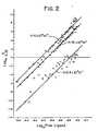
- the Sips plots presenting data for antibody carbodiimide conjugates and antibody conjugates of the invention are shown in FIG. 2.
- the binding measurements clearly demonstrate the retention of specificity, affinity, and homogeneity for the sample modified via the carbohydrate attachment methods of the invention, ( ⁇ ), when compared to
- 1-amino-6-trifluoroacetamidohexane (0.8 g, 4.1 mmole) was added to the above carboxycarbonic anhydride of EDDHA. The homogeneous solution was stirred at 4 °C for 0.5 hours, then was lyophilized to yield an oily product. The oil was washed with an actone/ether (4:1) mixture to yield a crude yellow product. The solid 1-amino-6-trifluoroacetamidohexyl-EDDHA was collected and hydrolyzed with 7% K 2 CO 3 and reprecipitated with HC1 at pH 4 to yield pure 1,6-diaminohexyl-EDDHA (1.4 g).
- the antibody oxidized by the method of Section 6.1, was incubated with an approximately 270-fold molar excess of 1,6-diaminohexyl-EDDHA, prepared by the method of Section 6.2, for one hour at room temperature. This was followed by addition of solid sodium cyanoborohydride to a final concentration of 10 mM, and further incubation of 4 hours at room temperature. The mixture was then dialyzed at 4°C versus several changes of PBS, and concentrated by ultrafiltration.
- IgM antibody (1.9 mg/ml) was added 10 mg of 1-ethyl-3-(3-dimethylaminopropyl) carbodiimide (1 ml of 10 mg/ml solution, pH 5.0) and PBS (pH 5.0) to make up to 2.5 ml. The mixture was incubated two hours at room temperature. Then 275 ⁇ l of 0.1M 1,6-diaminohexyl-EDDHA in 2.5 ml of water
- the antibody molecule used in this example was a monoclonal IgM (designated no. 171) specific for antigenic determinants on sheep red blood cells.
- IgM monoclonal IgM
- Lewis rats were immunized with a single injection of sheep red blood cells.
- spleen cells from the immunized rats were harvested and fused with the myeloma line SP2/O Ag14 according to the method of McKearn et al., 1979, Immunol. Rev. 47:91-115.
- Cloned cells were then grown and the resulting monoclonal antibody was purified as described by Kliman and McKearn, 1981, J. Immunol Meth. 42:1-9.
- Oxidation of the antibody carbohydrate moiety was accomplished by reacting the antibody with galactose oxidase by a modification of the method of Cooper et al., supra. To this end, 3.8 mg of no. 171 monoclonal antibody was added to 1 ml of buffer consisting of 0.135 M NaCl, 0.015 Tris-HCl (pH 7.0), 0.5 mM MgCl 2 , and 0.15 mM CaCl 2 . Subsequently, a 0.1 ml aliquot of a solution of galactose oxidase (Worthington Biochemical Co., Freehold, NJ) at a concentration of 52 units of enzyme/ml of the same buffer was added to the antibody solution.
- galactose oxidase Worthington Biochemical Co., Freehold, NJ
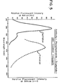
- FIG. 3 represents the excitation spectra for unoxidized (a) and oxidized (b) antibodies.
- a synthetic fluorogenic compound was utilized as the conjugate partner.
- the properties of this synthetic compound are such that the bound and free states of the fluorogenic compound are spectrofluorometrically distinguishable.
- the synthetic fluorogenic compound used was obtained from Serva Fine Biochemicals, Inc., Garden City Park, LI, NY (Catalog #51474).
- This compound consists of a tripeptide (Gly-Gly-Arg) attached via an amide linkage to the fluorescent compound 7-amino-4-methyl coumarin (AMC); the amino group of glycine is blocked by carbobenzoxy chloride (Cbz).
- Tripeptide-AMC or Gly-Gly7Arg-AMC is shown below:
- excitation and emission maxima of free AMC differ from those for AMC bound to the tripeptide (325 nm and 395 nm, respectively). This affords a means for distinguishing between the bound and free forms of the AMC molecule using a fluorometric assay. Excitation and emission wavelengths of 383 nm and 455 nm may be used for optimum differences for assay purposes; at these wavelengths, free AMC retains 20% of its maximal fluorescence but possesses a relative fluorescence 500-fold greater than an equimolar amount of bound AMC (Zimmerman et al., 1978, Proc. Natl. Acad. Sci., U.S.A. 75(2):750-753).
- a hydrazine derivative of the Tripeptide-AMC compound was prepared. Aldehyde groups of the oxidized carbohydrate side chain of the antibody molecule were then reacted with the hydrazine derivative to form a hydrazone.
- the Tripeptide-AMC was first deblocked at the glycine amino terminus by removal of the Cbz group. This was accomplished by dissolving the Tripeptide-AMC in trifluoroacetic acid (Sigma, St. Louis, MO), and bubbling HBr gas (Matheson, East Rutherford, NJ) through the solution for 45 minutes.
- the product, H 2 N-Gly-Gly-Arg-NH-AMC was precipitated by the addition of cold diethyl ether (Baker Chemical Co., Phillipsburgh, NJ), and dissolved in absolute ethanol (Publicker Industries Co., Linfield, PA).
- This compound was shown to be positive for fluorescence by exciting with ultraviolet light, and positive for the presence of a hydrazine group.
- the hydrazine linked to the tripeptide was detected by thin layer chromatography (TLC) using a spray of a 0.1% trinitrobenzene sulfonic acid aqueous solution for the colorimetric determination of a hydrazine (a pinkish or orange-brown color indicates the presence of hydrazine).
- TLC thin layer chromatography
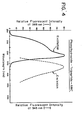
- the absorption and emission spectra for the Phenylhydrazine-Tripeptide-AMC compound as shown in FIG. 4 reveal a similarity to the Tripeptide-AMC spectra, but a shift in excitation and emission maxima consistent with the covalent modification of the Phenylhydrazine-Tripeptide-AMC.
- the maxima for excitation and emission of the Phenylhydrazine-Tripeptide-AMC compound are 345 nm and 385 nm, respectively.
- the product was precipitated from solution with cold diethyl ether, washed, and dissolved in dimethylsulfoxide (Baker Chemical Co., Phillipsburgh, NJ).
- Spectrofluorometric analysis of the protein fractions confirmed the presence of the Phenylhydrazine-Tripeptide-AMC covalently attached to the antibody (Antibody-Phenylhydrazine-Tripeptide-AMC).
- the excitation and emission maxima for the conjugate are 325 nm and 385 nm, respectively (FIG. 5).
- the large peak at 285 nm in the excitation spectrum of the conjugate may be explained by tryptophan absorption with residual fluorescence at 385 nm and may also be the result of resonance energy transfer from the amino acid tryptophan of the antibody molecule to AMC.
- the following examples illustrate specific release of the compound from the antibody conjugate prepared by the methods of Section 7. These antibody conjugates retain the ability to fix complement as revealed by a hemolytic complement fixation assay. Furthermore, the specific release of the compound from the antibody conjugate, at the antigenic cell surface, via enzymatic cleavage by the complement system is demonstrated by a non-hemolytic assay.
- the compound is fluorogenic.
- the complement mediated release of the fluorescent compound may be detected by an assay capable of differentiating between the bound and free forms of the fluorescent molecule.
- a therapeutic agent While the compound released in this example is not considered a therapeutic agent, its use illustrates enzymatic cleavage of a linker by the complement system or a serum enzyme having proteolytic activity.
- a therapeutic agent may be released from an antibody-therapeutic agent conjugate.
- the Tripeptide-AMC was prepared as described in Section 7.2.
- the properties of the fluorogenic compound (AMC) are such that the bound and free states of the fluorogenic compound are spectrofluorometrically distinguishable. This provides a definitive assay for measuring the complement fixation ability of the antibody conjugate. More importantly, it provides a means for quantitating the subsequent complement-mediated release of the compound.
- a 200 ul aliquot of a suspension of sheep red blood cells (Gibco Diagnostics, Madison, WI) at an approximate concentration of 2 x 10 8 cells/ml were mixed with 20 ul of the Antibody-Phenylhydrazine-Tripeptide-AMC conjugate mixture prepared in Section 7.3 (approximately 2 ⁇ g of protein). After 15 minutes of mixing and incubating at 37°C, 100 ul. of the human serum complement (prepared in Section 8.1.1.) was added to the mixture. After 30 minutes to 1 hour of incubation at 37°C, the mixture was centrifuged to pellet the cells. The extent of the complement-mediated cell lysis was determined by spectrophotometrically measuring hemoglobin released into the supernatant (412 nm).
- the results of this assay demonstrated complete hemolysis and essentially 100% binding of antibody to cell surface.
- addition of distilled water to a pellet formed be centrifuging 200 vl of the sheep red blood cell suspension completely lyses the cells, and releases hemoglobin.
- a 1:20 dilution of the supernatant of sheep red blood cells which were completely lysed in distilled water had an O.D. 412 of 0.646.
- An identical dilution of sheep red blood cells which were lysed by the addition of conjugate and complement had an O . D . 412 of 0.672.
- the conjugate retained the ability to bind antigen and to fix complement.
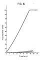
- the non-hemolytic assay was used to show the specific complement-mediated release of the AMC from the antibody conjugate. Similarly to the hemolytic assay, 200 ul of the glutaraldehyde-fixed sheep red blood cells, at an approximate concentration of 2 x 10 8 cells/ml, was incubated with the Antibody-Phenylhydrazide-Tripeptide-AMC conjugate at 37°C for 15 minutes.
- curve (a) which represents the conjugate incubated with glutaraldehyde-fixed sheep red blood cells and human complement
- curve (b) which represents the conjugate incubated with glutaraldehyde-fixed rat red blood cells and human complement
- curve (c) which represents the conjugate incubated with glutaraldehyde-fixed sheep red blood cells
- curve (d) which represents the conjugate alone demonstrate no release of AMC.
- a therapeutic agent may be attached to an antibody or antibody fragment via a linker that is susceptible to cleavage by complement or other proteolytic enzymes.
- Such antibody-conjugates are particularly useful for therapeutic applications where it is desired to release the therapeutic agent at the target site in the body.
- the antibody-therapeutic agent conjugates may be prepared either by attaching a therapeutic agent to an antibody-linker intermediate, or by attaching an antibody to a linker-therapeutic agent intermediate.
- the linkers e.g., Gly-Gly-Arg
- the linkers must be modified to contain a reactive group capable of reaction with the sulfhydryl group of a reduced antibody or Fab' fragment.
- reactive groups include, but are not limited to: reactive haloalkyl groups (including, for example, haloacetyl groups), p-mercuribenzoate groups and groups capable of Michael-type addition reactions (including, for example, maleimides and groups of the type described by Mitra and Lawton, 1979, J. Amer. Chem. Soc. 101: 3097-3110).
- haloalkyl is meant any alkyl group of one to three carbon atoms substitued with brornine, iodine or chlorine.
- the rates of cleavage for the peptide linker were measured by fluorescence quenching using a Perkin Elmer 650-10S fluorescence spectrometer (Perkin-Elmer Corporation, Norwalk, CT).
- the excitation and emission wavelengths were 380 nm and 460 nm, respectively and the temperature was maintained at 25°C with a Lauda k-2/R circulating water bath (Brinkmann Instruments, Westbury, NY).
- the initial concentrations of Gly-Gly-Arg-AMC were determined by optical density, IM 1325 - 16,000.
- the solutions were adjusted to 1 ⁇ M concentrations.
- One ml of the 1 ⁇ M solution in PBS, pH 7.4 was placed in a cuvette.
- the appropriate amounts of enzyme (1 ug/ml trypsin or 10 ⁇ g/ml urokinase) was then added at room temperature with stirring using a motor-driven syringe and the reaction kinetics were followed for several minutes.
- the fluorescence intensity of a known concentration of free AMC was established and used as a baseline to determine the change in units per nM/minute of free AMC in this fluorescence assay system. The results of the assay are described in Table IV.
- the Gly-Gly-Arg-AMC concentration was 100 ⁇ M and the trypsin used at 100 ⁇ g/ml.
- the results of this assay are also described in Table IV.
- the Gly-Gly-Arg-AMC was conjugated to oxidized dextran which can serve as a spacer as described in Section 5.4.3 (10,000 molecular weight) (Pharmacia Fine Chemicals, Piscataway, NJ) by the methods of the present invention using a molar ratio of Gly-Gly-Arg-AMC to dextran of 58:1. Dextran-Gly-Gly-Arg-AMC was separated with a 6.5 ml Sephadex® G-25 column (Pharmacia Fine Chemicals, Piscataway, NJ).
- Rates of enzymatic cleavage of the dextran-Gly-Gly-Arg-AMC, using the methods of this section, are also summarized in Table IV.
- Peptide sequences that are analogs of complement components were cleaved by trypsin (0.1 ⁇ g/ml), urokinase (0.1 ⁇ g/ml) , plasmin (1 ⁇ g/ml) and purified complement component(40 ⁇ g/ml).
- the data in Table V compare the cleavage rates (nM/minute) of these sequences.
- the fluorescence assay described in Section 8 for cleavage of Gly-Gly-Arg-AMC was used here.
- Complement components were purified and assayed by the method of Tack and Prahl (Biochem., 1976, 15:4513-4521). 9.3. CLEAVAGE OF GLY-GLY-ARG-TYR * BY
- the tetrapeptide Glycine-Glycine Arginine-Tyrosine was radiolabeled at the amino acid residue tyrosine with 125 Iodine ( 125 I) (Greenwood et al. 1963, Biochem. J. 88: 114-120).
- This radiolabeled compound is hereinafter referred to as Gly-Gly- A rg-Tyr * (wherein the * denotes 125 I ).
- this radiolabeled peptide is such that the bound and free states can be readily assayed for the release of the 125 labeled tyrosine residue after cleavage with the appropriate amount of enzyme (1 ⁇ g/ml trypsin or 10 ug/ml urokinase).
- the peptide Gly-Gly-Arg-Tyr was assayed for the release of the 1251 labeled tyrosine residue by agarose gel electrophoresis or thin-layer chromatography (TLC, Sono and Asakura, 1974, Biochem. 13:4386-4394). Briefly, appropriate amounts of enzyme were added to a reaction tube containing * 0.1 ⁇ M Gly-Gly-Arg-Tyr in PBS, pH 7.4. The mixture was incubated at 37°C for 30 minutes.
- Radiolabeled antibody-Gly-Gly-Arg-Tyr * conjugates were prepared according to one method of the present invention as described below.
- the carbohydrate moiety bf the antibody (LL 1151) was oxidized by reacting approximately 1 mg/ml of antibody in phosphate buffered saline (PBS) with 110 ul of 100 mM sodium metaperiodate (NaI0 4 ) at pH 6 (to give a final concentration of 10 mM (NaI0 4 ) for 1 hour on ice in the dark.
- PBS phosphate buffered saline
- Gly-Gly-Arg-Tyr * prepared by the method of Section 9.3, was coupled to normal human IgG or LL 1151 or NS 4.1 antibody oxidized by the method of Section 12.1 infra, by incubating the antibody at a 300-fold molar excess of Gly-Gly-Arg-Tyr * in the 0.1 M phosphate buffer, pH 6.0.
- sodium cyanoborohydride (NaCNBH 3 ) was added to a final concentration of 10 nM, and the reaction mixture was maintained at room temperature for 2 hours. Unreacted Gly-Gly-Arg-Tyr * was separated from the antibody conjugate by gel filtration.
- the sample was passed through a 5 ml Sephadex® G-50 column (Pharmacia Fine Chemicals, Piscataway, NJ), which had been pre-coated with 1 ml of 10% BSA and run in PBS, pH 7.4. The protein fraction were pooled.
- the antibody-Gly-Gly-Arg-Tyr * conjugates prepared by the method of Section 10.1 were assayed for the release of the 1251 tyrosine residue by gel filtration after cleavage of the linker.
- the conjugate was cleaved by the enzyme trysin (100 ug/ml or 10 ug/ml) or urokinase (100 ug/ml) as described in section 9.2, and the mixture was chromatographed on a Sephadex® G-50 column.. In this system, the cleaved Tyr * was retarded by the column. Therefore cleavage was determined by the loss of radioactivity in the void volume peak after gel filtration on the column.
- the antibody-peptide was cleaved as follows:
- This experiment illustrates cleavage of Tyr * from antibody-Gly-Gly-Arg-Tyr * conjugates, wherein the antibody conjugate was attached to an antigenic determinant of a cell.
- GRBC glutaraldehyde-fixed sheep red blood cells
- This experiment illustrates the cleavage of AMC from an antibody-Gly-Gly-Arg-AMC conjugate attached to a target cell.
- Gly-Gly-Arg-AMC conjugate prepared using the same methods described in Section 12.1 infra and incubated for 30 minutes at 37°C, and then for 30 minutes at 0°C. This mixture was washed with EDTA-VBS gel buffer and resuspended in 2.2 ml PBS, pH 7.4. The mixture was separated into two portions.
- porphyrin which can be used as photoactivatible cytolytic agents (or photosensitizers) for in vivo therapy when specifically attached to an antibody molecule.
- deuteroporphyrin dimethyl ester (98%, 0.05 g, 0.093 mmole), obtained from Aldrich Chemicals, Milwaukee, WI, was suspended in 10 ml dry methanol and 10 ul anhydrous hydrazine (9.0 mg, 0.28 mmole) was added. The solution was refluxed for approximately 4 hours and then evaporated to produce a deep red solid.
- protoporphyrin dimethyl ester (345 mg, 6.0 mmole) obtained from Aldrich Chemicals, Milwaukee, WI, was suspended in 30 ml dry methanol. Anhydrous hydrazine (48.0 mg, 1.5 mmole, 47.4 ul) was added and the solution refluxed for 24 hours. Evaporation gave a deep red solid which was only slightly soluble in many organic solvents.
- hematoporphyrin 3.0 g, 5 mmole
- hematoporphyrin 3.0 g, 5 mmole
- a solution of ethereal diazomethane (approximately 17 mmoles in about 125 ml ether) was added slowly over a period of 25 minutes with constant stirring. The mixture was stirred at 0°C for one hour and then at room temperature overnight in the dark.
- H ematoporphyrin free base (0.5 g, 8.35 x 10 mole, Sigma Chemical Co., St. Louis, MO) was dissolved in 200 ml of dry N,N-dimethylformamide and 0.24 ml of triethylamine was added and stirred under nitrogen gas for 20 minutes at room temperature. This homogeneous solution was cooled in an ice bath and 0.218 ml isobutylchloroformate (1.67 x 10 -3 mole) was added. After stirring for one-half hour, 0.132 ml (4.16 x 10 -3 mole) anhydrous hydrazine was added. The solution was stirred in the dark at 4°C for one hour and for one hour at room temperature.
- the solution was dried with a Rotary Evaporator (Rotavap, Buchi, Brinkmann Instruments, Westbury, NY) to give a deep red residue. Distilled water was added to the residue and the solution was adjusted to pH 12 with 1N NaOH to yield more of the precipitate. The solid product was removed by filtration, washed with distilled water and dried in a vacuum dryer to give a deep red crystals.
- Ficoll 70 was obtained from Pharmacia Fine Chemicals, Inc., (Piscataway, NJ).
- the carboxymethyl derivative of Ficoll 70 (CM-Ficoll) was prepared by a modification of the method of Inman (1975, J. Immunol. 114: 704-709).
- Two grams of CM-Ficoll were dissolved in 100 ml of water and 10 g of adipic dihydrazide were added slowly.
- the pH was adjusted to 4.7 by the dropwise addition of 1N HC1 then 1.25 g of l-ethyl-3-(3-dimethyl-aminopropyl) carbodiimide were added and the pH was readjusted to 4.7 with 1N HC1.
- the reaction mixture was then stirred for 20 hours at 23-25°C.
- the crude reaction product was purified by gel filtration chromatography on a 4.5 x 55 ml column of Sepahadex® G-25.
- the column was eluted with phosphate buffered saline (PBS, 0.01 M sodium phosphate, ⁇ .15 M sodium chloride, pH 7.4) and the void volume fractions were saved.
- PBS phosphate buffered saline
- the hydrazide-Ficoll was then dialyzed against water and lyophilized.
- the final product contained about 150 moles of hydrazide/mole of Ficoll.
- the hydrazide F icoll (0.25 g, 0.004 mmole Ficoll 0.23 meq. hydrazide) was dissolved in 10 ml water and adjusted to pH 5.0. Hematoporphyrin (0.069 g, 0.12 mmole) was added followed by 1-ethyl-3-(3-dimethyl-aminopropyl)(0.44 g, 2.3 mmole), and the reaction mixture was stirred overnight. The solution was then dialyzed against Na 2 C0 3 solution, pH 9.0 for approximately 5 days. The solution was then lyophilized and the brownish powder stored at 0°C.
- the specific mouse monoclonal antibody utilized in the following experiments was CYT-021.
- the antibody is a monoclonal IgG which is specific for a glycoprotein antigen on human cytotoxic/suppressor T-lymphocytes.
- the hematoporphyrin dihydrazide was attached to the CYT-021 antibody for use in photoradiation treatment of cellular disorders.
- Antibody-hematoporphyrin dihydrazide conjugates were prepared according to one method of the instant invention by first oxidizing the carbohydrate moiety of the antibody molecule.
- the carbohydrate moiety of CYT-021 antibody was oxidized by reacting approximately 1 mg/ml of antibody in phosphate buffered saline (PBS) with 110 ul of 100 mM sodium metaperiodate (NaI0 4 ) at pH 6 (to give a final concentration of 10 mM NaI0 4 ) for 1 hour on ice in the dark.
- PBS phosphate buffered saline
- Hematoporphyrin dihydrazide was coupled to oxidized CYT-021 antibody as follows.
- a solution of hematoporphyrin dihyrazide was prepared by dissolving 5 mg of hematoporphyrin dihydrazide in 50 ul of N,N-dimethylformamide (DMF).
- the concentration of hematoporphyrin dihydrazide was adjusted to 1 mg/ml by the addition of 5 ml of deionized water.
- the amount of hematoporphyrin dihydrazide bound to C YT -0 21 antibody was determined spectrophotometrically.-by using the extinction coefficients for protein at 280 nm and hematoporphyrin dihydrazide at 374 nm, and ranged from 4 to 8 moles/mole antibody.
- MOLT-4 In vitro cytotoxicity assays were carried out in 96 well U-bottom Costar tissue culture plates (Costar, Cambridge, MA).
- the target cells (MOLT-4, ATCC CRL 1582), were obtained from American Type Culture Collection, Rockville, MD as were control cells (Raji, ATCC CCL 86).
- the MOLT-4 cell line is a T-cell line derived from the human peripheral blood of a patient with acute lymphoblastic leukemia.
- the Raji cell line is a lymphoblastic-like cell line derived from a patient with Burkitt's lymphoma.
- the cells were washed 2 times with RPMI 1640 culture medium (10% Fetal Calf Serum, 2% Glutamine, 1% Penicillin-Streptomyocin) (M.A.
- the CYT-021 antibody- hematoporhyrin dihydrazide conjugate was filtered through a 0.45 micron syringe filter (Gelman, Ann Arbor, MI) and 1:10 dilutions of the conjugate were prepared.
- Viability of cells was then determined by measuring 3 H -thymidine (New England Nuclear, Boston, MA) uptake.
- the microtiter plates were centrifuged in a TJ-6 centrifuge (Beckman Instruments, Palo Alto, CA) for 5 minutes at 1500 rpm.
- the supernatants were removed and 100 ul of RPMI culture medium containing 1 uCi of 3 H-thymidine (New England Nuclear, Boston, MA) was added to each well.
- the cells were incubated for at least 4 hours in the C0 2 incubator before harvesting onto filter paper using a MASH (Skatron Inc., Sterling, VA).
- the filter paper was dissolved in 3 ml Aquasol-2 (New England Nuclear, Boston, MA) and counted on a LKB 1212 liquid scintillation counter.
- Ficoll 70 was obtained from Pharmacia Fine Chemicals, Inc. (Piscataway, NJ). The hydrazide-ficoll was prepared by the method described in Section 11.5.
- SIAB N-succinimidyl-(4-iodoacetyl)-aminobenzoate
- the reaction mixture was then stirred for 20 hours at 23-25°C.
- the organic solvents were removed by bubbling N 2 gas through the reaction mixture and the resultant cloudy solution was clarified by centrifugation. The clear supernatant was lyophilized.
- Excess SIAB was removed by extracting the dry powder with tetrahydrofuran and the excess solvent was removed by evaporation under reduced pressure. The dry powder was dissolved in 5 ml of water and dialyzed for 4 hours at 4°C.
- the (4-iodoacetyl)-aminobenzoichydrazide-Ficoll was stored frozen at -70°C and contained about 16 iodoacetyl groups per mole of hydrazide-Ficoll.
- mice anti-N. gonofrhoeae monoclonal IgG was diluted into 0.25 ml of PBS, pH 7.4. The IgG solution was then reduced with 5 mM dithiothreitol for 30 minutes at 23-25°C. The reduced antibody was then passed over 1x19 cm Sephadex * G-50 column and eluted with 0.1 M tris(hydroxymethyl)-aminomethane buffer, pH 8.0, containing 1mM ethylenediaminetetracetic acid (Tris/EDTA).
- Tris/EDTA tris(hydroxymethyl)-aminomethane buffer
- SMCC 4-(N-maleimidomethyl)-cyclohexane-1-carboxylic acid N-hydroxysuccinimide ester
- the organic solvents were then removed by bubbling N 2 through the reaction mixture and the resultant cloudy solution was clarified by centrifugation. The clear supernatant was lyophilized and the excess SMCC was extracted with tetrahydrofuran. The organic solvent was then removed by evaporation under reduced pressure. The final produce contained about 15 moles of maleimide/mole of Ficoll. This compound can be used in the same way as the (4-iodoacetyl)-aminobenzoichydrazide-Ficoll prepared by the method of Section 13.2.
- the following examples illustrate the novel derivatives of erythrosin which can be used as photoactivatible cytolytic agents (or photosensitizers) for in vivo therapy when specifically attached to an antibody molecule.
- Erythrosin 5-isothiocyanate (Aldrich Chemicals, Milwaukee, WI) was prepared according to the procedure of Moore and Garland (Biochemical Society Transactions, 1979, 7: 945-946) with certain modifications.
- the conversion of 5-amino erythrosin into the isothiocyanate was performed as follows: thiophosgene (0.34 g, 0.227 ml, 3.0 mmole) was added dropwise to a suspension of 5-amino erythrosin . (1.2 g, 1.4 mmole) in toluene (20 ml) at 0°C. After addition of the thiophosgene over 10 minutes, the solution was warmed to room temperature, then heated to reflux for 2 hours.
- Erythrosin 5-isothiocyanate (0.45 g, 0.5 mmole) was dissolved in 20 ml of tetrahydrofuran and cooled to 0°C.
- Ethylene diamine (0.3 g, 0.334 ml, 5.0 mmole) was added dropwise over ten minutes and the reaction stirred at room temperature overnight. Filtration of the solid red precipitate was followed by washing the tetrahydrofuran and drying at 80°C under vacuum. The red solid weighed 0.52 g and had no isothiocyanate peak in the infrared at approximately 2040 cm -1 . This product was suspended in 5% HC1 filtered and dried to give a red solid weighing 0.34 g.
- Solid sodium nitrite (0.01 g, 0.15 mmole) was added to a solution of erythrosin-5-amine (0.1 g, 0.11 mmole) (prepared by the method of Moore and Garland, 1979, Biochemical Society Transactions 7: 945-946) in 10 ml of 6N HC1 at 0°C.
- the diazotizing solution was stirred for 3 hours at 0°C and a solution of stannous chloride (0.1 g, 0.44 mole) in 2 ml of 3 M HC1 was added.
- the red solution was stirred at room temperature for 3 hours and then filtered.
- the deep red precipitate was washed with water, and dried.
- the red product showed binding to aldehydic oxidized Sepharose beads at pH 8.8 but no binding to non-oxidized non-aldehydic Sepharose, indicating the presence of a hydrazine group.
- Ficoll hydrazide (0.25 g 0.004 mmole, 0.23 meq. hydrazide) was dissolved in 40 ml of 50% ethanol: water. Erythrosin isothiocyanate (0.022 g, 0.025 mmole) was added and the solution stirred overnight. Dialysis against Na 2 Co 3 , pH 9.0 for 5 days, followed by lyophilization gave a pink powder which was water-soluble and displayed a maximum wavelength of about 524 nm in pH 9 buffer.
Priority Applications (1)
| Application Number | Priority Date | Filing Date | Title |
|---|---|---|---|
| AT85401776T ATE68974T1 (de) | 1984-09-13 | 1985-09-13 | Therapeutische antikoerpermittelkonjugate. |
Applications Claiming Priority (4)
| Application Number | Priority Date | Filing Date | Title |
|---|---|---|---|
| US65075484A | 1984-09-13 | 1984-09-13 | |
| US650754 | 1984-09-13 | ||
| US650375 | 1984-09-13 | ||
| US06/650,375 US4867973A (en) | 1984-08-31 | 1984-09-13 | Antibody-therapeutic agent conjugates |
Publications (3)
| Publication Number | Publication Date |
|---|---|
| EP0175617A2 true EP0175617A2 (fr) | 1986-03-26 |
| EP0175617A3 EP0175617A3 (en) | 1988-06-15 |
| EP0175617B1 EP0175617B1 (fr) | 1991-10-30 |
Family
ID=27095841
Family Applications (1)
| Application Number | Title | Priority Date | Filing Date |
|---|---|---|---|
| EP85401776A Expired - Lifetime EP0175617B1 (fr) | 1984-09-13 | 1985-09-13 | Conjugués d'anticorps et d'agent thérapeutique |
Country Status (7)
| Country | Link |
|---|---|
| EP (1) | EP0175617B1 (fr) |
| AU (2) | AU583854B2 (fr) |
| CA (1) | CA1326834C (fr) |
| DE (1) | DE3584559D1 (fr) |
| DK (1) | DK218386A (fr) |
| GR (1) | GR852217B (fr) |
| WO (1) | WO1986001720A1 (fr) |
Cited By (24)
| Publication number | Priority date | Publication date | Assignee | Title |
|---|---|---|---|---|
| EP0282057A2 (fr) * | 1987-03-11 | 1988-09-14 | The Board Of Regents Of The University Of Michigan | Produits associés chémo-radio-immunologiques |
| EP0336383A2 (fr) * | 1988-04-07 | 1989-10-11 | Takeda Chemical Industries, Ltd. | Lymphotoxine recombinante humaine |
| EP0525992A3 (fr) * | 1991-07-01 | 1994-03-16 | Lilly Co Eli | |
| EP0641796A1 (fr) * | 1988-07-19 | 1995-03-08 | The University Of British Columbia | Agents cytotoxiques de longueur d'onde spécifique |
| WO1997033620A3 (fr) * | 1996-03-15 | 1998-02-05 | Pulsion Verw Gmbh & Co Medical | Compose pour traiter des tumeurs |
| WO1998023286A2 (fr) * | 1996-11-26 | 1998-06-04 | Isprobi S.R.L. | Immunoconjugues comprenant des anticorps monoclonaux humains ou des fragments de ces anticorps, utilises a des fins diagnostiques et therapeutiques |
| WO1998047538A2 (fr) * | 1997-04-23 | 1998-10-29 | Institut für Diagnostikforschung GmbH an der Freien Universität Berlin | Colorants de synthese labiles aux acides et clivables par voie enzymatique pour le diagnostic avec de la lumiere dans le proche infrarouge et pour l'usage therapeutique |
| WO2000041727A1 (fr) * | 1999-01-15 | 2000-07-20 | Light Sciences Corporation | Traitement photodynamique transcutane de cellules cibles |
| WO2000041726A2 (fr) * | 1999-01-15 | 2000-07-20 | Light Sciences Corporation | Therapie vasculaire non invasive |
| EP1126782A1 (fr) * | 1998-11-02 | 2001-08-29 | Photogen, Inc. | Procede de visualisation et de therapie photodynamique ameliorees |
| EP1210078A1 (fr) * | 1999-08-13 | 2002-06-05 | Photogen, Inc. | Medicaments topiques ameliores et methodes permettant de traiter des maladies de maniere photodynamique |
| EP1284727A1 (fr) * | 2000-03-24 | 2003-02-26 | Photogen, Inc. | Medicaments intracorporels pour le traitement photodynamique de maladies |
| EP1311261A1 (fr) * | 2000-07-14 | 2003-05-21 | Photogen, Inc. | Medicaments pour le traitement chimiotherapeutique de maladies |
| USRE38994E1 (en) | 1988-07-20 | 2006-02-28 | Health Research, Inc. | Pyropheophorbides conjugates and their use in photodynamic therapy |
| USRE39094E1 (en) | 1988-07-20 | 2006-05-09 | Health Research, Inc. | Pyropheophorbides and their use in photodynamic therapy |
| EP1653996A2 (fr) * | 2003-08-08 | 2006-05-10 | Novo Nordisk Health Care AG | Utilisation de galactose oxydase pour la conjugaison chimique selective de molecules d'extraction a des proteines d'interet therapeutique |
| US9814780B2 (en) | 2010-08-10 | 2017-11-14 | Ecole Polytechnique Federale De Lausanne (Epfl) | Compositions for inducing antigen-specific tolerance |
| US9850296B2 (en) | 2010-08-10 | 2017-12-26 | Ecole Polytechnique Federale De Lausanne (Epfl) | Erythrocyte-binding therapeutics |
| US10392437B2 (en) | 2010-08-10 | 2019-08-27 | École Polytechnique Fédérale De Lausanne (Epfl) | Erythrocyte-binding therapeutics |
| US10821157B2 (en) | 2014-02-21 | 2020-11-03 | Anokion Sa | Glycotargeting therapeutics |
| US10940209B2 (en) | 2014-02-21 | 2021-03-09 | École Polytechnique Fédérale De Lausanne (Epfl) | Glycotargeting therapeutics |
| US10946079B2 (en) | 2014-02-21 | 2021-03-16 | Ecole Polytechnique Federale De Lausanne | Glycotargeting therapeutics |
| US10953101B2 (en) | 2014-02-21 | 2021-03-23 | École Polytechnique Fédérale De Lausanne (Epfl) | Glycotargeting therapeutics |
| US11253579B2 (en) | 2017-06-16 | 2022-02-22 | The University Of Chicago | Compositions and methods for inducing immune tolerance |
Families Citing this family (32)
| Publication number | Priority date | Publication date | Assignee | Title |
|---|---|---|---|---|
| US5140104A (en) * | 1982-03-09 | 1992-08-18 | Cytogen Corporation | Amine derivatives of folic acid analogs |
| US4831122A (en) * | 1986-01-09 | 1989-05-16 | Regents Of The University Of Minnesota | Radioimmunotoxins |
| CA1330378C (fr) * | 1986-05-08 | 1994-06-21 | Daniel J. Coughlin | Derives amine d'analogues de l'acide folique |
| US4801688A (en) * | 1986-05-27 | 1989-01-31 | Eli Lilly And Company | Hydrazone immunoglobulin conjugates |
| GB8628398D0 (en) * | 1986-11-27 | 1986-12-31 | Central Blood Lab Authority | Pharmaceutically-active conjugates |
| US4835258A (en) * | 1986-12-24 | 1989-05-30 | Northwestsern University | Conjugation of aromatic amines or nitro-containing compounds with proteins or polypeptides by photoirradiation of the azide derivatives with ultraviolet light in order to produce antibodies against the haptens |
| WO1988005433A1 (fr) * | 1987-01-15 | 1988-07-28 | Celltech Limited | Reactifs de reticulation presentant une reactivite aux thiols |
| US4883790A (en) * | 1987-01-20 | 1989-11-28 | University Of British Columbia | Wavelength-specific cytotoxic agents |
| US5095030A (en) * | 1987-01-20 | 1992-03-10 | University Of British Columbia | Wavelength-specific cytotoxic agents |
| US5171749A (en) * | 1987-01-20 | 1992-12-15 | University Of British Columbia | Wavelength-specific cytotoxic agents |
| US5283255A (en) * | 1987-01-20 | 1994-02-01 | The University Of British Columbia | Wavelength-specific cytotoxic agents |
| FR2610198B1 (fr) * | 1987-02-03 | 1990-06-15 | Ire Celltarg Sa | Conjugues d'hydrazide d'alcaloide de vinca lie a une immunoglobuline, procede de preparation et compositions pharmaceutiques en comprenant |
| FR2611141B1 (fr) * | 1987-02-20 | 1991-02-01 | Int Rech Cancer Centre | Ensemble ou kit pour utilisation comme agent pour la destruction selective de cellules transformees ou tumorales |
| US4937183A (en) * | 1988-02-03 | 1990-06-26 | Cytogen Corporation | Method for the preparation of antibody-fragment conjugates |
| US5047227A (en) * | 1988-02-08 | 1991-09-10 | Cytogen Corporation | Novel and improved antibodies for site specific attachment of compounds |
| DE3827940A1 (de) * | 1988-08-13 | 1990-03-01 | Schering Ag | 13,17-propionsaeure- und propionsaeurederivat- substituierte porphyrin-komplexverbindungen, verfahren zu ihrer herstellung und diese enthaltende pharmazeutische mittel |
| WO1990003401A1 (fr) * | 1988-09-30 | 1990-04-05 | Neorx Corporation | Conjugues de substance de ciblage et d'agent diagnostique/therapeutique comportant des liaisons de base de schiff |
| US5066789A (en) * | 1988-09-30 | 1991-11-19 | Neorx Corporation | Targeting substance-diagnostic/therapeutic agent conjugates having Schiff base linkages |
| GR890100827A (el) * | 1988-12-21 | 1991-03-15 | Abbott Lab | Μέ?οδος παραγωγής παραγώγων τετραπεπτιδίων ως ανταγωνιστών CCK. |
| US5270302A (en) * | 1988-12-21 | 1993-12-14 | Abbott Laboratories | Derivatives of tetrapeptides as CCK agonists |
| US5308608A (en) * | 1989-06-07 | 1994-05-03 | University Of British Columbia | Photosensitizing Diels-Alder porphyrin derivatives |
| AU641658B2 (en) * | 1989-06-07 | 1993-09-30 | University Of British Columbia, The | Photosensitizing diels-alder porphyrin derivatives |
| EP0449998A4 (en) * | 1989-06-30 | 1992-01-15 | Brunswick Corporation | Antibody-oxidase conjugates with non-systemic substrates |
| EP0505566A1 (fr) * | 1989-12-15 | 1992-09-30 | Takeda Chemical Industries, Ltd. | Anticorps biospecifique des cellules cancereuses et enzyme presentant des caracteristiques d'activation promedicamenteuse |
| CA2078118C (fr) * | 1990-04-25 | 1999-11-16 | Paul A. Aristoff | Analogues de cc-1065 |
| AU655942B2 (en) * | 1990-10-05 | 1995-01-19 | Queen's University At Kingston | Porphyrin derivatives |
| GR1001459B (el) * | 1992-10-08 | 1993-12-30 | Lilly Co Eli | Σύμπλεγμα συζυγών φαρμάκων με αντισώματα. |
| US8557298B2 (en) | 1998-08-06 | 2013-10-15 | Provectus Pharmatech, Inc. | Medicaments for chemotherapeutic treatment of disease |
| WO2005047507A1 (fr) * | 2003-11-12 | 2005-05-26 | The Austin Research Institute | Conjugue adn-excipient |
| US7541330B2 (en) | 2004-06-15 | 2009-06-02 | Kosan Biosciences Incorporated | Conjugates with reduced adverse systemic effects |
| EP2380909A1 (fr) | 2010-04-26 | 2011-10-26 | Max-Planck-Gesellschaft zur Förderung der Wissenschaften e.V. | Protéine PTK-7 impliquée dans le cancer du sein |
| US20200338128A1 (en) * | 2018-01-05 | 2020-10-29 | Gencyte Therapeutics, Inc. | Precision molecular adaptor system for car-t immunotherapy |
Citations (2)
| Publication number | Priority date | Publication date | Assignee | Title |
|---|---|---|---|---|
| JPS52141882A (en) * | 1976-05-20 | 1977-11-26 | Hidetoshi Tsuchida | Highhpolymeric complex of covalent type |
| EP0088695A2 (fr) * | 1982-03-09 | 1983-09-14 | Cytogen Corporation | Liaisons d'anticorps |
Family Cites Families (2)
| Publication number | Priority date | Publication date | Assignee | Title |
|---|---|---|---|---|
| US3970521A (en) * | 1974-08-07 | 1976-07-20 | Exxon Research And Engineering Company | Immobilized glycoenzymes |
| CA1083508A (fr) * | 1975-11-13 | 1980-08-12 | Jacques Grange | Supports porteurs de chaines laterales, procedes d'obtention de ces supports, procedes de fixation de composes organiques comportant un residu glucidique sur lesdits supports, produits et reactifs resultant de ladite fixation chimique |
-
1985
- 1985-09-10 WO PCT/US1985/001700 patent/WO1986001720A1/fr unknown
- 1985-09-10 AU AU48071/85A patent/AU583854B2/en not_active Ceased
- 1985-09-11 CA CA000490424A patent/CA1326834C/fr not_active Expired - Lifetime
- 1985-09-12 GR GR852217A patent/GR852217B/el unknown
- 1985-09-13 EP EP85401776A patent/EP0175617B1/fr not_active Expired - Lifetime
- 1985-09-13 DE DE8585401776T patent/DE3584559D1/de not_active Expired - Fee Related
-
1986
- 1986-05-12 DK DK218386A patent/DK218386A/da not_active Application Discontinuation
-
1989
- 1989-02-21 AU AU30161/89A patent/AU3016189A/en not_active Abandoned
Patent Citations (2)
| Publication number | Priority date | Publication date | Assignee | Title |
|---|---|---|---|---|
| JPS52141882A (en) * | 1976-05-20 | 1977-11-26 | Hidetoshi Tsuchida | Highhpolymeric complex of covalent type |
| EP0088695A2 (fr) * | 1982-03-09 | 1983-09-14 | Cytogen Corporation | Liaisons d'anticorps |
Non-Patent Citations (10)
| Title |
|---|
| CANCER RESEARCH, vol. 45, September 1985, pages 4380-4386, D. MEW et al.: "Ability of specific monoclonal antibodies and conventional antisera conjugated to hematoporphyrin to label and kill selected cell lines subsequent to light activation". * |
| CHEMICAL ABSTRACTS, vol. 102, no. 15, 15th April 1985, page 283, abstract no. 128005x, Columbus, Ohio, US; Ch. K. WAT et al.: "Photosensitizerprotein conjugates: potential use as photoimmunotherapeutic agents", & PROG. CLIN. BIOL. RES. 1984, 170 (Porphyrin localization treat tumors), 351-9 * |
| CHEMICAL ABSTRACTS, vol. 103, no. 15, 14th October 1985, page 558, abstract 121395m, Columbus, Ohio, US; D. Mew et al.: "Ability of specific monoclonal antibodies and conventional antisera conjugated to hematoporphyrin to label and kill selected cell lines subsequent to light activation"; & CANCER RES. 1985, 45 (9), 4380-6 * |
| CHEMICAL ABSTRACTS, vol. 103, no. 5, 5th August 1985, page 491, abstract no. 37259n, Columbus, Ohio, US; R. OCAMPO et al.: "Identification of polar porphyrins in oil shale"; & J. CHEM. SOC. CHEM. COMMUN. 1985,(4), 198-200 * |
| CHEMICAL ABSTRACTS, vol. 82, no. 3, 20th January 1975, page 361, abstract no. 15067u, Columbus, Ohio, US; W.T. SHEARER et al.: "Cytotoxicity with antibody-glucose oxidase conjugates specific for a human colonic cancer and carcioembryonic antigen"; & INT. J. CANCER 1974, 14(4), 539-47 * |
| CHEMICAL ABSTRACTS, vol. 89, no. 13, 25 September 1978, Columbus, Ohio, US; abstract no. 110773Z, page 29; & JP 52 141882 A * |
| CHEMICAL ABSTRACTS, vol. 89, no. 13, 25th September 1978, page 29, abstract no. 110773z, Columbus, Ohio, US; & JP-A-52 141 882 (E. HASEGAWA et al.) (26-11-1977) * |
| CHEMICAL ABSTRACTS, vol. 97, no. 7, 16th August 1982, page 224, abstract nr. 51193a, Columbus, Ohio, US; H. SAKURAI et al.: "Growth inhibition and photoxidative toxicity in the housefly. Musca domestica L. caused by xanthene dye in larval growth medium and after injection"; & ENVIRON. ENTOMOL. 1982, 11(2), 467-70 * |
| THE JOURNAL OF IMMUNOLOGY, vol. 125, no. 3, September 1980, page 1201, WILLIAMS & WILKINS Co., US; G.W. PHILPOTT et al.: "Selective binding and cytotoxicity of rat basophilic leukemia cells (RBL-1) with immunoglobulin E-biotin and avidin-glucose oxidase conjugates" * |
| THE JOURNAL OF IMMUNOLOGY, vol. 130, no. 3, March 1983, pages 1473-1477, American Association of Immunologists, US; D. MEW et al.: "Photoimmunotherapy: treatment of animal tumors with tumor-specific monoclonal antibody-hematoporphyrin conjugates" * |
Cited By (52)
| Publication number | Priority date | Publication date | Assignee | Title |
|---|---|---|---|---|
| EP0282057A2 (fr) * | 1987-03-11 | 1988-09-14 | The Board Of Regents Of The University Of Michigan | Produits associés chémo-radio-immunologiques |
| EP0282057A3 (fr) * | 1987-03-11 | 1990-03-07 | The Board Of Regents Of The University Of Michigan | Produits associés chémo-radio-immunologiques |
| EP0336383A2 (fr) * | 1988-04-07 | 1989-10-11 | Takeda Chemical Industries, Ltd. | Lymphotoxine recombinante humaine |
| EP0336383A3 (fr) * | 1988-04-07 | 1990-08-16 | Takeda Chemical Industries, Ltd. | Lymphotoxine recombinante humaine |
| EP0641796A1 (fr) * | 1988-07-19 | 1995-03-08 | The University Of British Columbia | Agents cytotoxiques de longueur d'onde spécifique |
| USRE38994E1 (en) | 1988-07-20 | 2006-02-28 | Health Research, Inc. | Pyropheophorbides conjugates and their use in photodynamic therapy |
| USRE39094E1 (en) | 1988-07-20 | 2006-05-09 | Health Research, Inc. | Pyropheophorbides and their use in photodynamic therapy |
| EP0525992A3 (fr) * | 1991-07-01 | 1994-03-16 | Lilly Co Eli | |
| WO1997033620A3 (fr) * | 1996-03-15 | 1998-02-05 | Pulsion Verw Gmbh & Co Medical | Compose pour traiter des tumeurs |
| US7863047B2 (en) | 1996-10-30 | 2011-01-04 | Provectus Pharmatech, Inc. | Intracorporeal medicaments for photodynamic treatment of disease |
| US7402299B2 (en) | 1996-10-30 | 2008-07-22 | Provectus Pharmatech, Inc. | Intracorporeal medicaments for photodynamic treatment of disease |
| US7390668B2 (en) | 1996-10-30 | 2008-06-24 | Provectus Pharmatech, Inc. | Intracorporeal medicaments for photodynamic treatment of disease |
| WO1998023286A3 (fr) * | 1996-11-26 | 1998-07-30 | Isprobi Srl | Immunoconjugues comprenant des anticorps monoclonaux humains ou des fragments de ces anticorps, utilises a des fins diagnostiques et therapeutiques |
| WO1998023286A2 (fr) * | 1996-11-26 | 1998-06-04 | Isprobi S.R.L. | Immunoconjugues comprenant des anticorps monoclonaux humains ou des fragments de ces anticorps, utilises a des fins diagnostiques et therapeutiques |
| WO1998047538A2 (fr) * | 1997-04-23 | 1998-10-29 | Institut für Diagnostikforschung GmbH an der Freien Universität Berlin | Colorants de synthese labiles aux acides et clivables par voie enzymatique pour le diagnostic avec de la lumiere dans le proche infrarouge et pour l'usage therapeutique |
| WO1998047538A3 (fr) * | 1997-04-23 | 1999-01-21 | Diagnostikforschung Inst | Colorants de synthese labiles aux acides et clivables par voie enzymatique pour le diagnostic avec de la lumiere dans le proche infrarouge et pour l'usage therapeutique |
| US7648695B2 (en) | 1998-08-06 | 2010-01-19 | Provectus Pharmatech, Inc. | Medicaments for chemotherapeutic treatment of disease |
| EP1126782A1 (fr) * | 1998-11-02 | 2001-08-29 | Photogen, Inc. | Procede de visualisation et de therapie photodynamique ameliorees |
| EP1126782A4 (fr) * | 1998-11-02 | 2003-06-04 | Photogen Inc | Procede de visualisation et de therapie photodynamique ameliorees |
| WO2000041726A2 (fr) * | 1999-01-15 | 2000-07-20 | Light Sciences Corporation | Therapie vasculaire non invasive |
| US6602274B1 (en) | 1999-01-15 | 2003-08-05 | Light Sciences Corporation | Targeted transcutaneous cancer therapy |
| WO2000041726A3 (fr) * | 1999-01-15 | 2000-11-02 | Light Sciences Ltd | Therapie vasculaire non invasive |
| WO2000041727A1 (fr) * | 1999-01-15 | 2000-07-20 | Light Sciences Corporation | Traitement photodynamique transcutane de cellules cibles |
| EP1210078A4 (fr) * | 1999-08-13 | 2004-06-09 | Photogen Inc | Medicaments topiques ameliores et methodes permettant de traiter des maladies de maniere photodynamique |
| EP1210078A1 (fr) * | 1999-08-13 | 2002-06-05 | Photogen, Inc. | Medicaments topiques ameliores et methodes permettant de traiter des maladies de maniere photodynamique |
| EP2020242A2 (fr) | 1999-08-13 | 2009-02-04 | Provectus Pharmatech, Inc. | Médicaments topiques améliorés et procédés de traitement photodynamique de maladies |
| EP2020242A3 (fr) * | 1999-08-13 | 2009-02-11 | Provectus Pharmatech, Inc. | Médicaments topiques améliorés et procédés de traitement photodynamique de maladies |
| EP1284727A4 (fr) * | 2000-03-24 | 2004-06-02 | Photogen Inc | Medicaments intracorporels pour le traitement photodynamique de maladies |
| EP1284727A1 (fr) * | 2000-03-24 | 2003-02-26 | Photogen, Inc. | Medicaments intracorporels pour le traitement photodynamique de maladies |
| EP1311261A4 (fr) * | 2000-07-14 | 2004-06-02 | Photogen Inc | Medicaments pour le traitement chimiotherapeutique de maladies |
| EP1311261A1 (fr) * | 2000-07-14 | 2003-05-21 | Photogen, Inc. | Medicaments pour le traitement chimiotherapeutique de maladies |
| EP1653996A2 (fr) * | 2003-08-08 | 2006-05-10 | Novo Nordisk Health Care AG | Utilisation de galactose oxydase pour la conjugaison chimique selective de molecules d'extraction a des proteines d'interet therapeutique |
| US9901645B2 (en) | 2010-08-10 | 2018-02-27 | Ecole Polytechnique Fedrale de Lausanne (EPFL) | Methods for reducing immune responses |
| US10919963B2 (en) | 2010-08-10 | 2021-02-16 | École Polytechnique Fédérale De Lausanne (Epfl) | Erythrocyte-binding therapeutics |
| US9878048B2 (en) | 2010-08-10 | 2018-01-30 | Ecole Polytechnique Federale De Lausanne (Epfl) | Compositions for generating immune tolerance by targeting erythrocytes |
| US9901646B2 (en) | 2010-08-10 | 2018-02-27 | Ecole Polytechnique Federale De Lausanne (Epfl) | Methods for induction of antigen-specific immune tolerance |
| US9814780B2 (en) | 2010-08-10 | 2017-11-14 | Ecole Polytechnique Federale De Lausanne (Epfl) | Compositions for inducing antigen-specific tolerance |
| US10392437B2 (en) | 2010-08-10 | 2019-08-27 | École Polytechnique Fédérale De Lausanne (Epfl) | Erythrocyte-binding therapeutics |
| US10471155B2 (en) | 2010-08-10 | 2019-11-12 | École Polytechnique Fédérale De Lausanne (Epfl) | Antigen-specific tolerance and compositions for induction of same |
| US10800838B2 (en) | 2010-08-10 | 2020-10-13 | École Polytechnique Fédérale De Lausanne (Epfl) | Erythrocyte-binding therapeutics |
| US11884721B2 (en) | 2010-08-10 | 2024-01-30 | École Polytechnique Fédérale De Lausanne (Epfl) | Erythrocyte-binding therapeutics |
| US9850296B2 (en) | 2010-08-10 | 2017-12-26 | Ecole Polytechnique Federale De Lausanne (Epfl) | Erythrocyte-binding therapeutics |
| US11246943B2 (en) | 2010-08-10 | 2022-02-15 | École Polytechnique Fédérale De Lausanne (Epfl) | Antigen-specific tolerance and compositions for induction of same |
| US10946079B2 (en) | 2014-02-21 | 2021-03-16 | Ecole Polytechnique Federale De Lausanne | Glycotargeting therapeutics |
| US10953101B2 (en) | 2014-02-21 | 2021-03-23 | École Polytechnique Fédérale De Lausanne (Epfl) | Glycotargeting therapeutics |
| US10940209B2 (en) | 2014-02-21 | 2021-03-09 | École Polytechnique Fédérale De Lausanne (Epfl) | Glycotargeting therapeutics |
| US11654188B2 (en) | 2014-02-21 | 2023-05-23 | Ecole Polytechnique Federale De Lausanne (Epfl) | Glycotargeting therapeutics |
| US11666638B2 (en) | 2014-02-21 | 2023-06-06 | Ecole Polytechnique Federale De Lausanne (Epfl) | Glycotargeting therapeutics |
| US11793882B2 (en) | 2014-02-21 | 2023-10-24 | École Polytechnique Fédérale De Lausanne (Epfl) | Glycotargeting therapeutics |
| US11801305B2 (en) | 2014-02-21 | 2023-10-31 | École Polytechnique Fédérale De Lausanne (Epfl) | Glycotargeting therapeutics |
| US10821157B2 (en) | 2014-02-21 | 2020-11-03 | Anokion Sa | Glycotargeting therapeutics |
| US11253579B2 (en) | 2017-06-16 | 2022-02-22 | The University Of Chicago | Compositions and methods for inducing immune tolerance |
Also Published As
| Publication number | Publication date |
|---|---|
| AU583854B2 (en) | 1989-05-11 |
| DK218386D0 (da) | 1986-05-12 |
| CA1326834C (fr) | 1994-02-08 |
| WO1986001720A1 (fr) | 1986-03-27 |
| AU3016189A (en) | 1989-07-13 |
| DK218386A (da) | 1986-07-11 |
| GR852217B (fr) | 1986-01-14 |
| DE3584559D1 (de) | 1991-12-05 |
| EP0175617B1 (fr) | 1991-10-30 |
| AU4807185A (en) | 1986-04-08 |
| EP0175617A3 (en) | 1988-06-15 |
Similar Documents
| Publication | Publication Date | Title |
|---|---|---|
| EP0175617B1 (fr) | Conjugués d'anticorps et d'agent thérapeutique | |
| US5156840A (en) | Amine-containing porphyrin derivatives | |
| US4867973A (en) | Antibody-therapeutic agent conjugates | |
| EP0088695B1 (fr) | Liaisons d'anticorps | |
| US4671958A (en) | Antibody conjugates for the delivery of compounds to target sites | |
| US4937183A (en) | Method for the preparation of antibody-fragment conjugates | |
| US4880935A (en) | Heterobifunctional linking agents derived from N-succinimido-dithio-alpha methyl-methylene-benzoates | |
| US5112954A (en) | Method of enhancing the effect of cytotoxic agents | |
| AU627785B2 (en) | Vasopermeability-enhancing conjugates | |
| EP0385601B1 (fr) | Anticorps réticulés et leurs procédés de préparation | |
| EP0173629A1 (fr) | Complexes d'ions métalliques conjugés avec des anticorps | |
| US4485093A (en) | Immunotoxin conjugate which comprises arsanilic acid, useful for treating malignant tumors, particularly pancreatic cancer | |
| US5354554A (en) | Crosslinked antibodies and processes for their preparation | |
| ZA200308093B (en) | Metal substituted non centrosimmetrical phthalocyanine analogues, their preparation and use in photodynamic therapy and in vivo diagnostic. | |
| JP3273608B2 (ja) | 治療薬の部位特異的インビボ活性化 | |
| JP2975676B2 (ja) | 架橋抗体とその製法 | |
| JPH10500410A (ja) | フリーラジカル薬物の製造用のセレンコンジュゲート | |
| US6007817A (en) | Vasopermeability enhancing immunoconjugates | |
| RU2106147C1 (ru) | Конъюгат производного 14-бромдаунорубицина с моноклональным антителом или его фрагментом, обладающий противоопухолевой активностью и имеющий специфичность к данной опухоли, и фармацевтическая композиция, обладающая противоопухолевой активностью | |
| US5151266A (en) | Use of anionic detergents with conjugates of monoclonal or polyclonal antibodies | |
| EP0485749A2 (fr) | Modification chimique d'anticorps pour la création d'immunoconjugués | |
| JPS62500175A (ja) | 抗体−治療剤接合体 | |
| Arnon | Antibodies and dextran as anti-tumour drug carriers | |
| CA2000479C (fr) | Conjuges accroissant la vasopermeabilite | |
| Whiteley | Drug Delivery with Protein and Peptide Carriers |
Legal Events
| Date | Code | Title | Description |
|---|---|---|---|
| PUAI | Public reference made under article 153(3) epc to a published international application that has entered the european phase |
Free format text: ORIGINAL CODE: 0009012 |
|
| AK | Designated contracting states |
Kind code of ref document: A2 Designated state(s): AT BE CH DE FR GB IT LI LU NL SE |
|
| PUAL | Search report despatched |
Free format text: ORIGINAL CODE: 0009013 |
|
| AK | Designated contracting states |
Kind code of ref document: A3 Designated state(s): AT BE CH DE FR GB IT LI LU NL SE |
|
| 17P | Request for examination filed |
Effective date: 19881107 |
|
| 17Q | First examination report despatched |
Effective date: 19900122 |
|
| GRAA | (expected) grant |
Free format text: ORIGINAL CODE: 0009210 |
|
| AK | Designated contracting states |
Kind code of ref document: B1 Designated state(s): AT BE CH DE FR GB IT LI LU NL SE |
|
| PG25 | Lapsed in a contracting state [announced via postgrant information from national office to epo] |
Ref country code: SE Effective date: 19911030 Ref country code: NL Effective date: 19911030 |
|
| REF | Corresponds to: |
Ref document number: 68974 Country of ref document: AT Date of ref document: 19911115 Kind code of ref document: T |
|
| ITF | It: translation for a ep patent filed |
Owner name: JACOBACCI & PERANI S.P.A. |
|
| REF | Corresponds to: |
Ref document number: 3584559 Country of ref document: DE Date of ref document: 19911205 |
|
| ET | Fr: translation filed | ||
| NLV1 | Nl: lapsed or annulled due to failure to fulfill the requirements of art. 29p and 29m of the patents act | ||
| PLBE | No opposition filed within time limit |
Free format text: ORIGINAL CODE: 0009261 |
|
| STAA | Information on the status of an ep patent application or granted ep patent |
Free format text: STATUS: NO OPPOSITION FILED WITHIN TIME LIMIT |
|
| PG25 | Lapsed in a contracting state [announced via postgrant information from national office to epo] |
Ref country code: AT Effective date: 19920913 |
|
| PG25 | Lapsed in a contracting state [announced via postgrant information from national office to epo] |
Ref country code: LU Free format text: LAPSE BECAUSE OF NON-PAYMENT OF DUE FEES Effective date: 19920930 Ref country code: LI Effective date: 19920930 Ref country code: CH Effective date: 19920930 Ref country code: BE Effective date: 19920930 |
|
| 26N | No opposition filed | ||
| BERE | Be: lapsed |
Owner name: CYTOGEN CORP. Effective date: 19920930 |
|
| REG | Reference to a national code |
Ref country code: CH Ref legal event code: PL |
|
| PGFP | Annual fee paid to national office [announced via postgrant information from national office to epo] |
Ref country code: GB Payment date: 19980907 Year of fee payment: 14 |
|
| PGFP | Annual fee paid to national office [announced via postgrant information from national office to epo] |
Ref country code: FR Payment date: 19980909 Year of fee payment: 14 |
|
| PGFP | Annual fee paid to national office [announced via postgrant information from national office to epo] |
Ref country code: DE Payment date: 19980921 Year of fee payment: 14 |
|
| PG25 | Lapsed in a contracting state [announced via postgrant information from national office to epo] |
Ref country code: GB Free format text: LAPSE BECAUSE OF NON-PAYMENT OF DUE FEES Effective date: 19990913 |
|
| GBPC | Gb: european patent ceased through non-payment of renewal fee |
Effective date: 19990913 |
|
| PG25 | Lapsed in a contracting state [announced via postgrant information from national office to epo] |
Ref country code: FR Free format text: LAPSE BECAUSE OF NON-PAYMENT OF DUE FEES Effective date: 20000531 |
|
| PG25 | Lapsed in a contracting state [announced via postgrant information from national office to epo] |
Ref country code: DE Free format text: LAPSE BECAUSE OF NON-PAYMENT OF DUE FEES Effective date: 20000701 |
|
| REG | Reference to a national code |
Ref country code: FR Ref legal event code: ST |