EP0067642A1 - Nachweis von bösartigen Tumorzellen - Google Patents
Nachweis von bösartigen Tumorzellen Download PDFInfo
- Publication number
- EP0067642A1 EP0067642A1 EP82302937A EP82302937A EP0067642A1 EP 0067642 A1 EP0067642 A1 EP 0067642A1 EP 82302937 A EP82302937 A EP 82302937A EP 82302937 A EP82302937 A EP 82302937A EP 0067642 A1 EP0067642 A1 EP 0067642A1
- Authority
- EP
- European Patent Office
- Prior art keywords
- malignin
- cells
- antibody
- cancer
- product
- Prior art date
- Legal status (The legal status is an assumption and is not a legal conclusion. Google has not performed a legal analysis and makes no representation as to the accuracy of the status listed.)
- Ceased
Links
Images
Classifications
-
- C—CHEMISTRY; METALLURGY
- C07—ORGANIC CHEMISTRY
- C07K—PEPTIDES
- C07K16/00—Immunoglobulins [IGs], e.g. monoclonal or polyclonal antibodies
- C07K16/18—Immunoglobulins [IGs], e.g. monoclonal or polyclonal antibodies against material from animals or humans
- C07K16/28—Immunoglobulins [IGs], e.g. monoclonal or polyclonal antibodies against material from animals or humans against receptors, cell surface antigens or cell surface determinants
- C07K16/30—Immunoglobulins [IGs], e.g. monoclonal or polyclonal antibodies against material from animals or humans against receptors, cell surface antigens or cell surface determinants from tumour cells
-
- C—CHEMISTRY; METALLURGY
- C07—ORGANIC CHEMISTRY
- C07K—PEPTIDES
- C07K14/00—Peptides having more than 20 amino acids; Gastrins; Somatostatins; Melanotropins; Derivatives thereof
- C07K14/435—Peptides having more than 20 amino acids; Gastrins; Somatostatins; Melanotropins; Derivatives thereof from animals; from humans
- C07K14/46—Peptides having more than 20 amino acids; Gastrins; Somatostatins; Melanotropins; Derivatives thereof from animals; from humans from vertebrates
- C07K14/47—Peptides having more than 20 amino acids; Gastrins; Somatostatins; Melanotropins; Derivatives thereof from animals; from humans from vertebrates from mammals
- C07K14/4701—Peptides having more than 20 amino acids; Gastrins; Somatostatins; Melanotropins; Derivatives thereof from animals; from humans from vertebrates from mammals not used
- C07K14/4731—Recognins, e.g. malignin
-
- C—CHEMISTRY; METALLURGY
- C07—ORGANIC CHEMISTRY
- C07K—PEPTIDES
- C07K16/00—Immunoglobulins [IGs], e.g. monoclonal or polyclonal antibodies
- C07K16/18—Immunoglobulins [IGs], e.g. monoclonal or polyclonal antibodies against material from animals or humans
-
- C—CHEMISTRY; METALLURGY
- C12—BIOCHEMISTRY; BEER; SPIRITS; WINE; VINEGAR; MICROBIOLOGY; ENZYMOLOGY; MUTATION OR GENETIC ENGINEERING
- C12N—MICROORGANISMS OR ENZYMES; COMPOSITIONS THEREOF; PROPAGATING, PRESERVING, OR MAINTAINING MICROORGANISMS; MUTATION OR GENETIC ENGINEERING; CULTURE MEDIA
- C12N1/00—Microorganisms, e.g. protozoa; Compositions thereof; Processes of propagating, maintaining or preserving microorganisms or compositions thereof; Processes of preparing or isolating a composition containing a microorganism; Culture media therefor
-
- C—CHEMISTRY; METALLURGY
- C12—BIOCHEMISTRY; BEER; SPIRITS; WINE; VINEGAR; MICROBIOLOGY; ENZYMOLOGY; MUTATION OR GENETIC ENGINEERING
- C12N—MICROORGANISMS OR ENZYMES; COMPOSITIONS THEREOF; PROPAGATING, PRESERVING, OR MAINTAINING MICROORGANISMS; MUTATION OR GENETIC ENGINEERING; CULTURE MEDIA
- C12N15/00—Mutation or genetic engineering; DNA or RNA concerning genetic engineering, vectors, e.g. plasmids, or their isolation, preparation or purification; Use of hosts therefor
-
- C—CHEMISTRY; METALLURGY
- C12—BIOCHEMISTRY; BEER; SPIRITS; WINE; VINEGAR; MICROBIOLOGY; ENZYMOLOGY; MUTATION OR GENETIC ENGINEERING
- C12P—FERMENTATION OR ENZYME-USING PROCESSES TO SYNTHESISE A DESIRED CHEMICAL COMPOUND OR COMPOSITION OR TO SEPARATE OPTICAL ISOMERS FROM A RACEMIC MIXTURE
- C12P1/00—Preparation of compounds or compositions, not provided for in groups C12P3/00 - C12P39/00, by using microorganisms or enzymes
-
- G—PHYSICS
- G01—MEASURING; TESTING
- G01N—INVESTIGATING OR ANALYSING MATERIALS BY DETERMINING THEIR CHEMICAL OR PHYSICAL PROPERTIES
- G01N33/00—Investigating or analysing materials by specific methods not covered by groups G01N1/00 - G01N31/00
- G01N33/48—Biological material, e.g. blood, urine; Haemocytometers
- G01N33/50—Chemical analysis of biological material, e.g. blood, urine; Testing involving biospecific ligand binding methods; Immunological testing
- G01N33/53—Immunoassay; Biospecific binding assay; Materials therefor
- G01N33/574—Immunoassay; Biospecific binding assay; Materials therefor for cancer
- G01N33/57484—Immunoassay; Biospecific binding assay; Materials therefor for cancer involving compounds serving as markers for tumor, cancer, neoplasia, e.g. cellular determinants, receptors, heat shock/stress proteins, A-protein, oligosaccharides, metabolites
- G01N33/57488—Immunoassay; Biospecific binding assay; Materials therefor for cancer involving compounds serving as markers for tumor, cancer, neoplasia, e.g. cellular determinants, receptors, heat shock/stress proteins, A-protein, oligosaccharides, metabolites involving compounds identifable in body fluids
-
- G—PHYSICS
- G01—MEASURING; TESTING
- G01N—INVESTIGATING OR ANALYSING MATERIALS BY DETERMINING THEIR CHEMICAL OR PHYSICAL PROPERTIES
- G01N33/00—Investigating or analysing materials by specific methods not covered by groups G01N1/00 - G01N31/00
- G01N33/48—Biological material, e.g. blood, urine; Haemocytometers
- G01N33/50—Chemical analysis of biological material, e.g. blood, urine; Testing involving biospecific ligand binding methods; Immunological testing
- G01N33/53—Immunoassay; Biospecific binding assay; Materials therefor
- G01N33/577—Immunoassay; Biospecific binding assay; Materials therefor involving monoclonal antibodies binding reaction mechanisms characterised by the use of monoclonal antibodies; monoclonal antibodies per se are classified with their corresponding antigens
-
- A—HUMAN NECESSITIES
- A61—MEDICAL OR VETERINARY SCIENCE; HYGIENE
- A61K—PREPARATIONS FOR MEDICAL, DENTAL OR TOILETRY PURPOSES
- A61K38/00—Medicinal preparations containing peptides
-
- C—CHEMISTRY; METALLURGY
- C12—BIOCHEMISTRY; BEER; SPIRITS; WINE; VINEGAR; MICROBIOLOGY; ENZYMOLOGY; MUTATION OR GENETIC ENGINEERING
- C12R—INDEXING SCHEME ASSOCIATED WITH SUBCLASSES C12C - C12Q, RELATING TO MICROORGANISMS
- C12R2001/00—Microorganisms ; Processes using microorganisms
- C12R2001/91—Cell lines ; Processes using cell lines
-
- G—PHYSICS
- G01—MEASURING; TESTING
- G01N—INVESTIGATING OR ANALYSING MATERIALS BY DETERMINING THEIR CHEMICAL OR PHYSICAL PROPERTIES
- G01N2333/00—Assays involving biological materials from specific organisms or of a specific nature
- G01N2333/435—Assays involving biological materials from specific organisms or of a specific nature from animals; from humans
- G01N2333/46—Assays involving biological materials from specific organisms or of a specific nature from animals; from humans from vertebrates
- G01N2333/47—Assays involving proteins of known structure or function as defined in the subgroups
- G01N2333/4701—Details
- G01N2333/473—Recognins, e.g. malignin
-
- Y—GENERAL TAGGING OF NEW TECHNOLOGICAL DEVELOPMENTS; GENERAL TAGGING OF CROSS-SECTIONAL TECHNOLOGIES SPANNING OVER SEVERAL SECTIONS OF THE IPC; TECHNICAL SUBJECTS COVERED BY FORMER USPC CROSS-REFERENCE ART COLLECTIONS [XRACs] AND DIGESTS
- Y10—TECHNICAL SUBJECTS COVERED BY FORMER USPC
- Y10S—TECHNICAL SUBJECTS COVERED BY FORMER USPC CROSS-REFERENCE ART COLLECTIONS [XRACs] AND DIGESTS
- Y10S436/00—Chemistry: analytical and immunological testing
- Y10S436/804—Radioisotope, e.g. radioimmunoassay
-
- Y—GENERAL TAGGING OF NEW TECHNOLOGICAL DEVELOPMENTS; GENERAL TAGGING OF CROSS-SECTIONAL TECHNOLOGIES SPANNING OVER SEVERAL SECTIONS OF THE IPC; TECHNICAL SUBJECTS COVERED BY FORMER USPC CROSS-REFERENCE ART COLLECTIONS [XRACs] AND DIGESTS
- Y10—TECHNICAL SUBJECTS COVERED BY FORMER USPC
- Y10S—TECHNICAL SUBJECTS COVERED BY FORMER USPC CROSS-REFERENCE ART COLLECTIONS [XRACs] AND DIGESTS
- Y10S436/00—Chemistry: analytical and immunological testing
- Y10S436/811—Test for named disease, body condition or organ function
- Y10S436/813—Cancer
-
- Y—GENERAL TAGGING OF NEW TECHNOLOGICAL DEVELOPMENTS; GENERAL TAGGING OF CROSS-SECTIONAL TECHNOLOGIES SPANNING OVER SEVERAL SECTIONS OF THE IPC; TECHNICAL SUBJECTS COVERED BY FORMER USPC CROSS-REFERENCE ART COLLECTIONS [XRACs] AND DIGESTS
- Y10—TECHNICAL SUBJECTS COVERED BY FORMER USPC
- Y10S—TECHNICAL SUBJECTS COVERED BY FORMER USPC CROSS-REFERENCE ART COLLECTIONS [XRACs] AND DIGESTS
- Y10S436/00—Chemistry: analytical and immunological testing
- Y10S436/815—Test for named compound or class of compounds
-
- Y—GENERAL TAGGING OF NEW TECHNOLOGICAL DEVELOPMENTS; GENERAL TAGGING OF CROSS-SECTIONAL TECHNOLOGIES SPANNING OVER SEVERAL SECTIONS OF THE IPC; TECHNICAL SUBJECTS COVERED BY FORMER USPC CROSS-REFERENCE ART COLLECTIONS [XRACs] AND DIGESTS
- Y10—TECHNICAL SUBJECTS COVERED BY FORMER USPC
- Y10S—TECHNICAL SUBJECTS COVERED BY FORMER USPC CROSS-REFERENCE ART COLLECTIONS [XRACs] AND DIGESTS
- Y10S530/00—Chemistry: natural resins or derivatives; peptides or proteins; lignins or reaction products thereof
- Y10S530/808—Materials and products related to genetic engineering or hybrid or fused cell technology, e.g. hybridoma, monoclonal products
- Y10S530/809—Fused cells, e.g. hybridoma
-
- Y—GENERAL TAGGING OF NEW TECHNOLOGICAL DEVELOPMENTS; GENERAL TAGGING OF CROSS-SECTIONAL TECHNOLOGIES SPANNING OVER SEVERAL SECTIONS OF THE IPC; TECHNICAL SUBJECTS COVERED BY FORMER USPC CROSS-REFERENCE ART COLLECTIONS [XRACs] AND DIGESTS
- Y10—TECHNICAL SUBJECTS COVERED BY FORMER USPC
- Y10S—TECHNICAL SUBJECTS COVERED BY FORMER USPC CROSS-REFERENCE ART COLLECTIONS [XRACs] AND DIGESTS
- Y10S530/00—Chemistry: natural resins or derivatives; peptides or proteins; lignins or reaction products thereof
- Y10S530/827—Proteins from mammals or birds
- Y10S530/828—Cancer
Definitions
- This invention relates to (1) the production of two products which are'distinct species of anti-malignin antibody; and (2) the production of three artificially-produced species of cell, each of which has the distinguishing characteristic of manufacturing either one or both species of anti-malignin antibody; whereby the above products, both the antibodies themselves and the cells which produce them, are useful for diagnostic, metabolic and therapeutic purposes.
- the anti-malignin antibodies of the present invention are monoclonal, and are the products of single artificially produced cells.
- the present monoclonal antibodies have unique properties, and should therefore be uniquely referred to in order to distinguish them from the anti-malignin antibody which is polyclonal, is produced in mammals, and has different properties.
- the artificially produced cell lines themselves which have been produced by the present invention, and have the ability to produce Monoclonal Anti-Malignin Antibodies, have themselves the characteristic novel feature that a single (monoclonal) line or type of cell has been produced artificially with the patented product Malignin, and perpetuated in vitro, with the ability to produce Monoclonal Anti-Malignin Antibody. Further, this novel cell line can produce Monoclonal Anti-Malignin Antibody in perpetuity and in any quantities desired. These new artificial cells are therefore herewith design- atedMonoclonal Anti-Malignin Antibody-Producing Cells. The new cells have immediate uses which are related to the uses of their product antibody, i.e. both diagnostic and therapeutic uses.
- malignant.or cancerous cells by the specific reaction of anti-malignin antibody with its specific antigen malignin, whether in solution or fixed in cells (see Examples 11, 11A, 12 for the use of the antibody to stain cancer cells specifically in immunofluoresence, and see Example 13 for the use of the antibody to identify or attach specifically to cancer cells carrying either a signal-emitter for identification and localization of the cancer cells in the body, or carrying an anti-cancer drug or chemical to be concentrated in the cancer cell for its destruction, as well as Examples 16, 17 where the antibody alone is used to treat (destroy) cancer cells).
- the method of production of each species however never yielded either one completely free of the other.
- the present invention is a marked improvement since it describes the production of an unique novel cell line which produces only the S-TAG species, one cell line which produces only the F-TAG species, and one cell line which produces both species.
- the species of combined antibody here produced for the first time by monoclonal producer cells designated MAMA-FS, as well as an artificial mixture of the two antibodies MAMA-S and MAMA-F, attach preferentially to cancer cells and destroy them.
- MAMA-FS monoclonal producer cells
- the separation of the attachment from the destruction functions of the species of the anti-malignin antibody described in this' invention has important applications in the separate uses of the antibody now possible for diagnosis on the one hand and for treatment (destruction of cancer cells) on the other.
- the respective novel producer cells are designated MAMA-S Producer, MAMA-F Producer, and MAMA-FS Producer, each being uniquely characterized by its specific antibody product.
- the nucleic acid of the producer cell which carries the specific information for manufacturing the particular monoclonal anti-malignin antibody produced by that particular cell can now be removed and isolated from the other cellular constituents and inserted into another, type of cell, for example, bacterial, which may, e.g., divide more quickly, be less susceptible to contamination during bulk manufacture, or be less costly to maintain in the laboratory continuously.
- bacterial which may, e.g., divide more quickly, be less susceptible to contamination during bulk manufacture, or be less costly to maintain in the laboratory continuously.
- Recognins are made by treating tumor cells or artificial cancer cells and separately the desired products.
- the Recognins may be used to prepare their Chemoreciprocals, i.e., by contacting the Recognins, or the Recognins on a support, with body fluids. These Chemoreciprocals are useful for diagnostic and therapeutic purposes, i.e., for diagnosing and treating cancers.
- Astrocyctin One of the Recognins of the present invention is Astrocyctin.
- Astrocytin has been produced from brain tumor tissue, preferably brain glioma tumor tissue.
- Protein fractions containing the Astrocytin precursor are first extracted from the tissue.
- a preferred method of accomplishing the extraction is to treat the tissue with a neutral buffer under conditions of homogenization, or other techniques, to disrupt the cells and tissues in order to solubilize protein fractions which contain the Astrocytin precursor.
- Astrocytin precursor is still bound to many large molecular weight substances, including proteins, glycoproteins, lipoproteins, nucleic acids and nucleo-proteins, for example.
- the solubilized proteins are then separated from the resulting tissue extract.
- the extract solution from the tissue is clarified to remove insoluble particles.
- the low molecular weight .contaminants are removed from the resulting solution, by a perevaporation concentration technique.
- the solution which is obtained is then treated to cleave Astrocytin precursor from other contaminants in order to obtain the protein fraction having a pK range between I and 4.
- the solution is placed on a chromatographic column and eluted with increasingly acidic solvents. All of the fractions which are. eluted in the neutral or acid range down to pK 4 are discarded and those fractions with pK range I - 4 are collected.
- the eluate is treated to yield a product having a molecular weight of 8,000.
- Astrocytin may be produced by extracting brain glioma tumor tissue with a neutral buffer, by repeated homogeni ' zation and by high speed centrifuging, separating from the resulting extract the fraction having a pK range of from 1 to 4, separating from said fraction the substances having a high molecular weight, i.e., up to 230,000, and isolating therefrom the. product Astrocytin having a molecular weight of 8,000.
- the product Astrocytin prepared in accordance with this process is characterized by forming a single line precipitate with its specific antibody in quantitative precipitin tests and Ouchterlony gel diffusion tests, being soluble in water and aqueous solutions having an acid or neutral pH, and insoluble at an alkaline pH, having a spectrophotometric absorption peak wave length of 280 mp and having a molecular weight of 8,000.
- Astrocytin is also characterized by having a very high percentage of residues of glutamic acid and aspartic acid and a very high ratio of these acids to histidine.
- Malignin In a manner similar to that described above, another Recognin, called Malignin, has been produced from artificial cancer cells, i.e., cancer cells grown in vitro. Malignin has a molecular weight of 10,000 and a similar but distinct aminoacid residue composition to Astrocytin, i.e., high amounts of glutaminic acid and aspartic acid and high ratios of these acids to histidine. A further analysis of Malignin is provided below.
- Malignin can be produced by extracting artificial cancer cells grown in culture with a neutral buffer by repeated homogenization and high speed centrifuging, separating from the resulting extract the fraction having a pK range ot I to 4, separating from said fraction the substances having a high molecular weight, i.e. up to 230,000 and isolating therefrom the product having a molecular weight of 10,000.
- Malignin prepared in accordance with this process is characterized by forming a single line precipitate with its speci-fic antibody in quantitative precipitin tests and Ouchterlony gel diffusion tests, being soluble in water and aqueous solutions having an acid or neutral pH, and insoluble at an alkaline pH, having a spectrophotometric absorption peak wave length of 280 m ⁇ and having a molecular weight of 10,000.
- Recognins are further characterized by being capable of complexing with bromoacetylcellulose to form bromoacetyl-cellulose-Recognin and producing specific Anti-Recognin antibodies upon injection into mammals, the Anti-Recognins being toxic to brain tumor cells in vitro and producing fluorescence of glioma cells when coupled with fluorescein, as described in further detail below.
- Recognins such as Astrocytin, Malignin and similar substances are useful as products which may be introduced into a biological system to reduce foreign reactions, such as by coating a material with a Recognin.
- a further example may be to introduce a Recognin inorder to produce the Chemoreciprocals in the biological system. They may also be used nutritionally to encourage the growth of a particular biological system of which they are a part.
- a further use for Recognins is the production of Target reagents which comprise the complexes of the Recognins with carriers to facilitate their applicability in biological systems.
- the complex conveys the physicochemical characteristics of the Recognin itself.
- the carrier should be selected from those which form a complex with the Recognin and which are substantially biologically inert.
- any substance known in the art which will form a stable complex with. polypeptides or proteins may oe useful for complexing with Recognins.
- An example is a cellulose-based material, such as bromoa.cetyl- cellulose.
- the carrier should not alter the specific physicochemical properties of the Recognin which are useful for the purposes set forth herein.
- the complex of the Recognin and its carrier is useful for producing, separating and identifying its chemoreciprocal in any biological system with which it is brought into contact.
- the Recognin-carrier complex is also useful for stimulating the production of its chemoreciprocal precursor in any biological system into which it is introduced.
- Chemoreciprocals are the anti-Recognins, e.g., anti-Astrocytin and anti-Malignin. These may be made by injecting the Recognin into a biological system. An immunologically effective dose of Recognin is brought into contact with bodily tissues or fluids in a manner which induces an antibody response in accordance with techniques known in the art for producing antibodies.
- the anti-Recognins may be used for the delivery of materials such as diagnostic, nutritional and therapeutic agents to specific cells or sites in a biological system which comprises introducing said agent in complexed form with the anti-Recognin into the biological system.
- the anti-Recognins are also useful for diagnosing the presence of tumor cells in an histology section, by applying the Anti-Recognin conjugated with a labelling substance such as a dye or radio-active substance, to said section, whereby staining or radio- active labelling occurs only with tumor cells.
- a labelling substance such as a dye or radio-active substance
- Yet another use for anti-Recognins is for increasing the yield of other useful Chemoreciprocal products (such as TAG, described below) from a mammal which comprises injecting an immunologically effective dose of the Recognin into the mammal, or other biological system.
- Target reagents complexed with their chemoreciprocals.
- the Target product of Astrocytin complexed with a carrier such as bromoacetylcellulose is brought into contact with anti-Astrocytin.
- This type of compound may be complexed with, and used for, the delivery of diagnostic, nutritional and therapeutic agents to specific cells or sites in a biological system. These compounds may also-be used for purification procedures.
- Anti-Astrocytin may be made by the de- complexing of Bromoacetylcellulose-Astrocytin-Anti-Astrocytin by hydrolytic treatment with an acid or proteinase enzyme.
- Target reagents are also useful for increasing the amount of TAG products (described below) in a biological system, such as by bringing an irrmunologically effective dose of Target into contact with bodily tissues or fluids.
- TAG reagents e.g., Target-Attaching Globulins
- the TAG products are produced by bringing Target reagents into contact with body fluids for varying periods of time to form a complex and cleaving TAG therefrom.
- Two useful embodiments are S-TAG and F-TAG.
- a process for producing S-TAG comprises reacting a blood serum or other body fluid with a Target product (e.g., Bromoacetyl- cellulose-Malignin) for approximately two hours or- more at a low temperature, e.g., 4°C, and cleaving S-TAG from the resulting material, e.g., with a dilute acid, for approximately two hours at a temperature of 37°C.
- a Target product e.g., Bromoacetyl- cellulose-Malignin
- the product S-TAG prepared in accordance with this process is characterized by being soluble in aqueous buffer solutions, forming a single line precipitate with its corresponding Recognin in Ouchterlony gel diffusion tests, being non-dialysable in cellophane membranes, being retained by millipore: filters which retain more- cules of over 10,000 molecular weight, having molecular weights in different states of aggregation as determined _ by thin layer gel chromatography of approximately 50,000 and multiples thereof into the macroglobulin range, and having a spectrophotometric absorption peak wave-length of 280 mp.
- a process for producing an F-TAG product comprises reacting a blood serum or other body fluid with a Target reagent (e.g., Bromoacetylcellulose-Malignin) for approximately 10 minutes at a low temperature, e.g., 4°C, and cleaving F-TAG from the resulting material, e.g., with a dilute acid, for approximately two hours at a temperature of 37°C.
- a Target reagent e.g., Bromoacetylcellulose-Malignin
- the product F-TAG prepared in accordance with this process is characterized by being soluble in aqueous buffered solutions, forming a single line precipitate with its corresponding Recognin in Ouchterlony gel diffusion tests, being non-dialysable in cellophane membranes, being retained by millipore filters which retain molecules over 25,000 molecular weight, having molecular weights in different states of aggregation as determined by thin layer gel chromatography of approximately 50,000 and multiples thereof into the macroglobulin range, and having a spectrophotometric absorption peak wave-length of 280 mp.
- TAG products are useful for detecting cancer tumors in living mammals by determining the concentrations of S-TAG and F-TAG produced by a known volume of the mmmal's blood serum or other body fluid and correlating this concentration with amounts determined to be indicative of cancer.
- TAG products are also useful for diagnosing the presence of tumor cells in an histology section, which comprises applying TAG conjugated with a labelling substance such as a dye or radioactive substance, to said section, whereby staining or radioactive labelling occurs only with tumor cells.
- TAG products have additionally been found to be cytotoxic to tumor cells.
- TAG products are also useful for directing the delivery of diagnostic, nutritional and therapeutic agents to specific cells or sites by introducing said agents in complexed form with the TAG product.
- Recognins are compounds with physicochemical characteristics which mimic those configurations characteristic of cancer cells in terms of their failure-to recognise and stop cell division. The use of Recognins goes beyond insight into the cancer mechanism, for immediate products and methods are thereby provided which are useful in the diagnosis and treatment of cancer, and for its prevention.
- Recognins ana the produers derived therefrom can be manufactured efficiently in virtually limitless quantities.
- Another aspect of this invention is the production of a valuable specific antibody-like product (Anti-Astrocytin) to a specific brain product (Astrocytin), permitting the use of this antibody-like product specifically to complex with and, as a specific delivery vehicle to, specific points in the nervous systems of various species.
- Anti-Astrocytin a valuable specific antibody-like product
- Astrocytin a specific brain product
- the TARGET-ATTACHING-GLOBULINS are so named because they are produced by two reactions, the first reacting biological fluids with a synthetic complex containing physicochemical configurations which mimic those of the MALIGNINS and called TARGET, the second, cleaving the specific TAG from the complex, and by the measure of the TAG so produced obtaining a quantitative indication from the biological fluids of living organisms as to whether there is present a tumor in that organism; and, hence, a diagnostic test for tumors.
- TAG products and ANTI-MALIGNIN are physico- chemi-cally complementary to MALIGNINS, they are termed CHEMORECIPROCALS.
- TAG products can be produced depending upon the time permitted for the reaction of serum with the specific TARGET reagent used, and depending upon the time permitted for the cleavage of the product which has been complexed.
- TAG and anti-RECOGNIN compounds attach to glial tumor cells preferentially in histological Sections of brain tumor and surrounding tissue removed at surgery of the brain tumor. This preferential labelling by TAG and Anti-RECOGNINS of tumor cells is demonstrated through standard immunofluorescent techniques.
- a method for determining through histological examination with a new degree of certainty whether tumor cells are present in the tissue removed, and whether these tumor cells have penetrated to the very edges of the tissue removed, indicating the likelihood that the tumor still remains in the brain or other organ, or that tumor cells are absent from the periphery of the tissue removed, indicating the possibility that all of the tumor has been removed from the brain or other organ.
- the TAG and Anti-RECOGNINS produced as described have ' been found to be cytotoxic to glioma brain tumor cells grown in tissue culture in vitro.
- TAG TARGET-ATTACHING-GLOBULINS
- TAG TARGET-ATTACHING-GLOBULINS
- the cytotoxicity of TAG and anti-RECOGNINS for tumor cells provides an additional new diagnostic test for the serum of patients who are suspected of suffering from a tumor.
- the serum or other body. fluid of these patients is reacted with a TARGET reagent to produce TAG and the product TAG is tested in tissue culture growths of tumor cells for cytotoxicity. Both the concentration of TAG and.
- the degree of cytotoxicity manifested by the TAG which can be produced from a given individual's serum may be not only diagnostic but also of value in tracing the course of the disorder preoperatively and postoperatively in a given patient.
- the coupling of radioactive and dye tracers to TAG provides new TAG products which are useful in vivo in the diagnosis of tumors and in their exact localization.
- the injection of suitably labelled TAG either intra- arterially or intravenously, into the cerebrospinal fluid, or directly into brain tissue or its cavities permits the demonstration by radioactive means, or by visualization of the coupled dye, of the presence of a brain tumor, for it is only to the tumor cells that the TAG specifically attaches. Further, this method permits the precise visualization of the location of the brain tumor.
- TAG reagents and anti-RECOGNINS have the chemical specificity which permits preferential attachment to ASTROCYTIN precursor containing tumor cells both in vitro and in vivo, these products may also be used therapeutically, as well as diagnostically, when coupled, e.g., with radioactive, proton capture agents, or other toxic physical or chemical agents, so that these toxic substances may be localized preferentially through these compounds' specificity of attachment in the tumor cells as compared with their neighbouring normal cells.
- TAG product has demonstrated efficacy in attaching preferentially to the tumor cells, and accordingly the properties of a therapeutic agent.
- S-TAG SLOW-TAG
- F-TAG FAST-TAG
- TARCET and TAG The possible relationship of the function of the actual synthetic producte TARCET and TAG to their precursors, and in turn to functions of postulated but not demonstrated cell "antigens” and circulating "antibodies” to them which may exist in vivo has yet to be elucidated.
- F-TAG and S-TAG produce single discrete lines of reaction with ASTROCYTIN in Ouchterlony gel diffusion, and the injection of TARGET into rabbits induces an increase in the yield of TAG products from rabbit serum after reacting with TARGET.
- TAG precursor P-TAG
- TARGET-like substances exist in vivo which function in the control of cell proliferation and cell death.
- the exposure of a cell constituent which normally is not directly exposed to serum proteins may occur during cell division.
- The. exposure of this cell constituent may result in that constituent becoming converted to a TARGET-like substance to which the attachment of a P-TAG-like molecule from serum may then occur, which would stimulate cell division or alternatively inhibit it.
- a non-dividing cell which is injured or malfunctioning may expose a TARGET-like substance to which the attachment of P-TAG-like molecules may be reparative.
- the attachment of P-TAG-like molecules may induce the destruction of the cell (e.g. ANTI-GLICMA-TAG synthetically produced as here described is markedly cytotoxic to- glioma tumor cells growing in tissue culture). This may thus represent a mirror of a normal mechanism for the control of cell division, and for either the repair or the removal of individual cells in the body throughout the life of the organisms.
- S-TAG SLOW-TAG
- Human brain glioma tumor tissue removed at surgery, is dissected to be as free as possible of surface blood vessels and normal .brain tissue. For a typical amount of dissected tumor tissue of 11 grams, the tissue is weighed into six 1.3 g. and two 1.0 g. aliquots., Each allquet is then treated as follows
- Each aliquot is homogenized in a neutral buffer solution by sonification or other mechanical means.
- each aliquot is homogenized in 100 cc per g. of tissue of 0.005 M phosphate buffer solution, pH 7, in a Waring blender. Homogenization should be done in the cold to prevent denaturing of the proteins present.
- the blender should be precooled in a cold room at 0 - 5°C and operated for only three minutes.
- the homogenate is then centrifuged for clarification, for example at 80,000 times gravity for 30 minutes in a refrigerated ultracentrifuge.
- the soluble supernatant is decanted and kept in the cold.
- the insoluble residue is rehomogenized with a further 100 cc of neutral buffer and centrifuged as before, and the second soluble extract combined with the first. Best yields are obtained when this procedure of homogenization and centrifuging is repeated until less than 50 micrograms of protein per ml. of solution are obtained in the supernate. With most tissues this is accomplished by the fifth extraction.
- the solutions thus obtained are combined and concentrated by perevaporation with subsequent dialysis, as by dialysis against 0.005 M phosphate buffer in the cold to produce a volume of 15 ml.
- the volume of this solution is noted, an aliquot is taken for total protein analysis, and the remainder is fractionated to yield the protein fraction having a pK range between 1 and 4.
- the preferred method of fractionation is chromatography as follows.
- solution (1) 4.04.g. NaH 2 PO 4 and 6.50 g. Na 2 HPO 4 are dissolve.d in 15 litres of distilled H 2 O (0.005 molar, pH 7);
- Each effluent tube is quantitatively analysed for protein.
- the eluates in the tube numbers 212 to 230 are combined, and contain the crude products from which ASTROCYTIN will be produced.
- fraction 10B While data have been published on this crude material, called fraction 10B in the past (Protein Meta- bolism of the Nervous System, pp. 555-569 (Plenum Press, 1970); Journal of Neurosurgery, Vol. 33, pp. 281 - 286. (Septemb.er, 1970)), the cleavage from fraction 10B can be prepared as a product in amounts between 0.1 and 10 mg. per gm. of original fresh nervous system tissue from which it was obtained.
- ASTROCYTIN-precursor contains varying amounts of covalently bound carbohydrate residues including a number of hexoses, namely glucose, galatose, mannose; hexosamines, in.cluding glucosamine, galatosamine and mannosamine; and occasionally other sugars, such as fucose, ribose and perhaps rhamnose. It also contains large molecular weight protein products and several lipids and nucleic acids.
- the ASTROCYTIN-Precursor-Containing fraction is further isolated from contaminants.
- the material from Example I is chromatograted on Sephadex G-50 resin with a typical column 40 cm.
- the first (flow-through) peak contains ASTROCYTIN-Precursor together with impurities, whereas subsequent peaks contain only impurities.
- the products in the above first flow-through peak are concentrated on Sephadex-G-15, and then passed onto a column of Cellex-D with the same solutions, (1) to (7), as in Example 1, and the same elution steps as are performed in Example 1.
- the product ASTROCYTIN is present as a sharp peak in the same tubes (numbers 212 - 230) as before, thus maintaining its behaviour on Cellex-D chromatography without the presence of a large number of contaminants.
- Low molecular weight contaminants may then be removed by techniques known to the art, such as millipore disc filtration.
- the product ASTROCYTIN is freed of salt and other small molecular weight contaminants by filtration through Millipor.e Pellicon Disc No. 1000, 13 mn., which retains substances. of molecular weight greater than 1000 and permits the passage of those of molecular weight below 1000.
- the product ASTRCCYTIN remains on the Pellicon Disc, and is recovered from it by washing with Solution (1) of Example 1.
- ASTRCCYTIN is then obtained by isolating the compound having a molecular weight of 8000 from the above solution.
- a preferred method uses thin layer gel (TLG) chromatography as follows:
- the plates are allowed to.dry at room temperature for 10 minutes and transferred to a moist chamber where they can be stored for two weeks, or they are used immediately after appropriate pre-equilibration (usually during the night for a minimum of 12 hours).
- the main function of equilibration is to normalize the ratio between the stationary and modile phase volumes.
- substances to be determined are applied with micropipettes as spots or as a streak at the start line. 10 ml. to 20 ml. of 0.2 - 2% protein solution are placed on the edge of a microscopic cover slide (18 x 18 mm.) and held against the gel surface.. In a few seconds, the solution will soak into the gel.
- the plates are replaced in the apparatus and the paper wick is pushed slightly downwards to ensure good contact with the gel layer.
- the paper wick must not drip. Excess moisture is wiped off.
- the liquid solvent in the reservoir is kept constant at 1 cm. from the upper end of the vessel.
- the runs are usually completed in 4 to 7 hours depending on the progress of separation. With coloured substances, separation follows directly.
- the separated spots of protein are easily made visible by transferring them to a paper sheet replica of TLG plate after the chromatographic separation has been completed, and by staining them on the prewashed methanol + H 2 O + acetic acid - 90:5:5, for 48 hours.
- the paper sheet is 3 mm, filter paper. A sheet of paper 20 x 18 cm.
- Sta.ining is performed by spraying the replica-paper with -0.03% diazotized sulfanilic acid in 10% Sodium Carbonate (Pauley's Reagent). Staining can also be accomplished with a saturated solution of Amido Black in Methanol-Acetic Acid (90:10v/v is used); the staining time is 5 - 10 minutes.
- the dry plates are allowed to swell for 10 minutes in a mixture of methanol + H 2 0 + acetic acid (75:20:5) and stained in a saturated Amido Black in the same solvent system for five hours and subsequently washed by bathing for two hours in the same solvent before they are dried.
- the distance from the starting line to the middle of each zone is measured with an accuracy of 0.05 mm. either directly on the print (replica) or on the densitogram. The.
- the product ASTROCYTIN is observed as a discrete spot at a distance of approximately 0.83 + 0.02 with reference to the standard Cytochrome C, yielding an approximate molecular weight of 8000 for ASTROCYTIN.
- ASTROCYTIN is separated in this procedurefrom ASTROCYTIN on the basis of slight differences in chemical structure and large differences in molecular weight.
- the product ASTROCYTIN is aspirated with the gel in which it is contained, in dry form, dissolved in Solution (1) and freed of resin by centrifuging or other similar means.
- the product ASTROCYTIN which has been produced at this stage is soluble in distilled water, soluble at neutral and acid pH, and insoluble at alkaline pH and has a spectrophotometric absorption peak wavelength of 280 mp. It is a polypeptide with molecular weight, as stated above, of.approximately 8000. Its covalently linked aminoacids are shown by hydrolysis with 6N HCI, then quantitative automatic determination to have the following average composition of aminoacids:
- Cysteic acid, hydroxyproline, norleucine, ammonia, isodesmosine, desmosine, hydroxylysine, lysinonorleucine and garrma-aminobutyric acid are all absent in detectable amounts, but a trace of glucosamine may be present.
- Example 2 From 11 grams of the starting brain tumor tissue in Example 1, approximately 3 mg. of purified ASTROCYTIN are produced by the above methods.
- a sterile technique is scrupulously maintained.
- All solutions e.g.-Hank's Balanced Salt (BSS), F-10 Nutrient medium, foetal calf serum, trypsin solution
- BSS Balanced Salt
- F-10 Nutrient medium e.g., foetal calf serum, trypsin solution
- Cells are removed from tumor tissue and grown in vitro for many generations using a suitable medium, such as that described below.
- a suitable medium such as that described below.
- Pre-rinse beakers to be used with a sterilizing solution, for example, 12-propanol plus Amphyl or creoline solution.
- the artificial cancer cells i.e., cells grown in vitro for many generations
- the liquid medium in which the cells are growing is discharged into the pre-rinsed beakers.
- the cells are then washed gently with 5 - 10 ml. of Hank's BSS or other similar solution for 30 seconds. Avoid agitation. All walls and surfaces are washed.
- the solution is clarified of cells by centrifuging.in the cold from 10 minutes at 3,000 rpm.
- the medium is poured. into a beaker as above. Add a small amount of buffered proteinase enzyme solution and rinse quickly to avoid digestion of the cells.
- 1 - 2 ml. of trypsin solution (EDTA) are added and rinsed for only 10 seconds. Pour off the trypsin solution.
- the cells may he extracted for the production of MALIGNIN after approximately seven days of growth.
- cells growing in each 250 ml. growth chamber as described above may be recovered as follows: The medium is transferred to a centrifuge tube and centrifuged at 3,000 rpm in the cold for 10 minutes. The medium is discarded. The cells remaining in the growth chamber are scraped from the chamber walls and washed into the centrifuge tubes with neutral buffer solution. The cells are washed twice with neutral buffer solution and centrifuged again at 3,000 rpm in the cold, and the medium is discarded. The washed cells are suspended in 10 ml. of neutral phosphate buffer until ready for extraction of the crude MALIGNIN-Precursor-Containing fraction.
- Washed cells suspended in neutral buffer from Example 3 are mechanically disrupted under conditions which avoid the denaturing of most proteins.
- the washed cells are treated in the cold with a sonifier for 20 seconds.
- the cell residues are centrifuged at 30,000 rpm for 30 minutes and the supernatant is decanted. Ten ml. aliquots of buffer solution are used to wash remaining cells from the chamber and these are added to the remaining cell residues. Sonify and centrifuge as above and combine the supernatants. Repeat the process once more.
- the combined supernatant is perevaporated to reduce the approximate 30 ml. volume to 6 - 7 ml.
- An aliquot is taken for total protein analysis and the remainder is fractionated according to the methods of Example 1 for ASTROCYTIN Precursor.
- the product MALIGNIN is further isolated from contaminants by the methods of Example 2 for ASTROCYTIN.
- the product MALIGNIN is observed at a discrete spot at a distance of approximately 0.91 + 0.02 with reference to the standard cytochrome C, yielding an approximate molecular weight of 10,000 for MALIGNIN.
- the product MALIGNIN which has been produced at this stage is soluble in distilled water, soluble at neutral or acid pH, and insoluble at alkaline pH and has a spectrophotometric absorption peak of 280 m ⁇ . It is a polypeptide with a molecular weight of approximately 10,000. Its covalently linked aminoacids are shown by hydrolysis with 6N HCl then quantitative determination to have the following average composition of aminoacids: the aminoacids cysteic acid, hydroxyproline, norleucine, ammonia, isodesmosine, desmosine, hydroxylysine, lysinonorleucine. and gamma-aminobutyric acid being absent in detectable amounts.
- a typical yield of pure MALIGNIN from twelve 250 ml. reaction chambers of Example 3 together is approximately 1 mg. of MALIGNIN.
- RECOGNIN in this case either Astrocytin or Malignin at pH between I and 2 is allowed to stand in the cold. After 7 to 14 days, TLG chromatography shows the product to have been reduced in molecular weight by approximately 200. When the solution is allowed to stand longer, further units of approximately 200 molecular weight are cleaved every 7 to 10 days. Thus, with Astrocytin the molecular weight is reduced from 8,000, and with MALIGNIN the molecular weight is reduced from 10,000 in each case by units of approximately 200 sequentially.
- the physicochemical specificities of ASTROCYTIN are retained by each product down to approximately 4,000 molecular weight.
- the physicochemical specificities of Malignin are retained by each product down to approximately 5,000 molecular weight. This is shown by Ouchterlony gel diffusion tests against Anti-Astrocytin and Anti-Malignin, respectively.
- This cleavage can also be accomplished en-zymat- ically, as with trypsin and other proteinases, with similar results.
- a rigid walled tube of plastic, metal, or other suitable rigid material is dipped in or impregnated with a highly concentrated (i.e., 10.mg/ml.) viscous solution of RECOGNIN, in this case either Astrocytin or Malignin, until all surfaces are fully coated with the RECOGNIN.
- RECOGNIN solution is passed through and around the tube under pressure until all surfaces are fully coated.
- the tube is then dried in air or in vacuo, or lyophilized. The process of coating is repeated several times in order to build up multiple molecular layers of RECOGNIN coating.
- the tube is now ready to be placed in a cavity or in a tissue which contains Astrocytin- or Malignin- like precursors in the neighbouring tissue or fluid of a living mammal.
- This artificial tissue or organ may be used to minimize or to eliminate reactions which foreign substances without the RECOGNIN coatin.g would incite.
- ASTROCYTIN prepared as in Example 2 above
- MALIGNIN prepared as in Example 5 above
- ASTROCYTIN or MALIGNIN is dissolved in 0.15 M. NaH 2 PO 4 - citrate buffer, pH 4.0.
- a bromoacetyl-resin for example bromoacetylcellulose (BAC) having 1.0 to 1.5 milli- equivalents Br per gram of cellulose, stored in the cold, is prepared in 0.15 M NaH 2 PO 4 buffer, pH 7.2. Convert the buffer to pH 4 by pouring off the pH 7.2 buffer solution and adding 0.15 M NaH 2 PO 4 - citrate buffer, pH 4.0.
- the ASTROCYTIN or MALIGNIN solution and the BAC solution are stirred together (10:1 BAC to RECOGNIN ratio) for 30 hours at room temperature, and then centrifuged.
- BAC-ASTROCYTIN or BAC-MALIGNIN
- the complexed BAC-ASTROCYTIN or BAC-MALIGNIN is resuspended in 0.1 M bicarbonate buffer pH 8.9 and stirred 24 hours at 4°C to permit the formation of chemical bonds between the BAC and the ASTRCCYTIN or MALIGNIN. After. the 24 hours, the suspension is centrifuged and the supernatant is analysed for protein.
- the complexed BAC-ASTROCYTIN or BAC-MALIGNIN is now resuspended in 0.05 M aminoethanol - 0.1 M bicarbonate buffer, pH 8.9 in order to block any unreacted bromine.
- the suspension is centrifuged, and the supernatant is kept but not analysed because of the presence of aminoethanol. Removal of all unbound ASTROCYTIN or MALIGNIN is then accomplished by centrifuging and resuspension for three washings in 0.15 M NaCl until no absorbence is measured on the spectrophotometer at 266 mp.
- the BAC-ASTROCYTIN or BAC-MALIGNIN complex is now stirred in 8 M urea for 2 hours at 38°C, centrifuged, then washed (three times usually suffices) with 8 M urea until no absorbence is shown in the washings at 266 m ⁇ .
- the complex is washed twice with 0.15 M NaCl to rid it of urea.
- the complex is then stirred at 37°C in 0.25 M acetic acid for 2 hours to demonstrate its stability. Centrifuge and read supernatant at 266 m ⁇ -- no absorbence should be present.
- BAC-ASTROCYTIN or BAC-MALIGNIN is therefore stable and can now be used as a reagent in the methods described below; in this stable reagent form, it is referred to as TARGET (TOPOGRAPHIC-ANTIGEN-LIKE-REAGENT-TEMPLATE) because it is a synthetically produced complex whose physical and chemical properties mimic the stable cell-bound precursor of ASTROCYTIN or MALIGNIN when it is in a potential reactive state with serum components.
- TARGET reagent is centrifuged and washed until neutralized with 0.15 M NaH 2 PO 4 buffer, pH 7.2.
- TARGET reagents may be prepared from bromoacetyl liganded carriers other than cellulose, such as bromoacetylated resins or even filter paper.
- Antisera to Astrocytin, Malignin or TARGET reagents may be produced by inducing an antibody response to them in a mammal. The following procedure has been found to be satisfactory.
- RECOGNIN Astrocytin or Malignin
- RECOGNIN Astrocytin or Malignin
- TARGET antigen injecting that amount of TARGET which contains I mg. of Astrocytin or Malignin as determined by a Folin-Lowry determination of protein.
- the specific antibody to Astrocytin is named Anti-Astrocytin.
- the specific antibody to Malignin is named Anti-Malignin.
- the specific antibody to TARGET reagent is named Anti-Target.
- the antiserum-containing specific Anti-Astrocytin can be reacted with BAC-Astrocytin.
- the specific antibodies to Astrocytin bind to their specific antigen Astrocytiri. Since Astrocytin is covalently bound to Bromoacetyl- cellulose, the specific antibody, Anti-Astrocytin, is now bound to BAC-Astrocytin to produce BAC-Astrocytin-Anti-Astrocytin (BACA-Anti-Astrocytin). This is proved by testing the remainder of the serum which is washed free from BAC-Astrocytin.
- Anti-Astrocytin antibodies previously shown to be present in the serum, have been absorbed to BAC-Astrocytin. Furthermore, when Anti-Astrocytin is releaed from its binding to BAC-Astrocytin, it is thereby isolated free of all contaminating antibodies. This release of Anti-Astrocytin may be accomplished by washing the BACA-Anti-Astrocytin complex with 0.25 M acetic acid (4°C., 2 hrs.) which has been shown above not to break the BAC-Astrocytin bond.
- Anti-Malignin when Anti-Malignin is released from its binding to BAC-Malignin, it is thereby isolated free of all contaminating antibodies. This release of Anti-Malignin may be accomplished by washing the BACM-Anti-Malignin complex with 0.25 M acetic acid (4°C., 2 hrs.), which has been shown above not to break the BAC-Malignin bond.
- the antibodies to TARGET show clearly on standard Ouchterlony gel diffusion tests for antigen-antibody reactions with specific single reaction lines produced with TARGET which show a line of identity with the line of reaction to ANTI-ASTROCYTIN or ANTI-MALIGNIN antisera (i.e., that produced to the injection of ASTROCYTIN or MALIGNIN themselves).
- Some rabbits it has bee.n noted, have levels of ANTI-TARGET in their blood prior to being injected with TARGET.
- These ANTI-TARGET substances when reacted specifically with TARGET reagent as described below in tests of human sera, lead to the production of approximately equivalent.amounts of the two types of TAG, S-TAG and F-TAG (see later Examples).
- TARGET reagent prepared in accordance with Example 8 is washed to remove any unbound RECOGNIN which may be present due to deterioration. The following procedure is satisfactory. TARGET reagent is stirred with acetic acid for two. hours at 37°C, centrifuged, the supernatant decanted, and the optical density of the supernatant read at 266 mp. If there is any absorbence, this wash is repeated until no further material is solubilized. The TARGET is then resuspended in phosphate buffered saline, pH 7.2.
- S-TAG Slow-Binding
- the clots are separated from the serum by centrifuging at 20,000 rpm at 4°C for 45 minutes.
- the serum is decanted into a centrifuge tube and centrifuged again at 2000 rpm at 4°C for 45 minutes.
- the serum is decanted and an 1% Solution of Methiolate (lg. in 95 ml. water and 5 ml. 0.2 M bicarbonate buffer pH 10) is added to the extent of 1% of the volume of serum.
- Serum samples prepared by the.above or other procedures, of 0.2 ml each are added to each of 0.2.0 ml. aliquots of TARGET suspension reagent containing 100-200 micrograms of RECOGNIN per 0.20 ml. of TARGET reagent, in duplicate determination.
- the suspension is mixed at 4°C in such a manner as to avoid pellet iorm- ation. For example, a small rubber cap rapid shaken may be used for 1 - 2 seconds and then, with the tubes slightly slanted, they may be shaken in a Thomas shaker for 2 hours or more.
- the TARGET reagent and protein bound to it are separated from the serum.
- One of the procedures which has been found to be satisfactory is the following.
- the tubes are centrifuged at 2000 rpm for 20 minutes at 4°C, the supernatant is decanted, the pellet which is formed by centrifuging is washed 3 times by remixing and shaking at room temperature with 0.2 - 0.3 ml. of 0.15 M Saline, and centrifuged and the supernatants are discarded.
- the protein which remains attached to the TARGET is cleaved therefrom and quantitatively determined. For example, 0.2 ml. of 0.25 M acetic acid is added, the suspension is shaken for I to 2 seconds with a rubber cap shaker, and then in a Thomas shaker for 2 hours in a 37°C incubator. The tubes are centrifuged at 2000 rpm at 4°C for 30 minutes. The supernatant is carefully decanted to avoid transferring particles and the optical density of the supernatant is read at 280 mp. The value of the optical density is divided by a factor of 1.46 for results in micrograms per ml. of serum protein (S-TAG). Duplicate determinations should not vary by more than 5%. Any other procedure effective for determining protein content may be used, such as Folin-Lowry determination, but standards must be specified to determine the range of control and tumor values of S-TAG and F-TAG concentration.
- S-TAG serum protein
- F-TAG Fast-Binding
- Serum samples prepared by the above or other procedures are allowed to stand at 4°C for 10 minutes less than the total time the S-TAG serum determinations were allowed to be in contact with TARGET reagent above (e.g., 1 hour 50 minutes if a "two hour" S-TAG determination was made). This procedure equilibrates the temperature histories of 5-TAG and F-TAG determinations.
- TARGET suspension reagent containing 100 - 200 micrograms of RECOGNIN per 0.20 ml. TARGET reagent, in duplicate determination.
- the suspension is then mixed at 4°C for approximately 10 minutes in such a manner as to avoid pellet formation.
- a small rubber cap rapid shaker may be used for 1 - 2 seconds and then, with the tubes slightly slanted, they may be shaken in a Thomas shaker for approximately 10 minutes.
- the TARGET reagent and protein bound to it are separated from the serum.
- One of the procedures which has been found to be satisfactory is the following.
- the tubes are centrifuged at 2000 rpm for 20 minutes at 4°C, the supernatant is decanted, the pellet which is formed by centrifuging is washed 3 times by remixing and shaking at room temperature with 0.2 - 0.3 ml. of 0.15 M Saline and centrifuged, and the supernatants are discarded.
- the protein which remains attached to the TARGET is cleaved therefrom and quantitatively determined.
- the procedure described above in this Example for determining S-TAG concentration is satisfactory. Any other procedure effective for determining protein content may be used, such as Folin-Lowry determination, but standards must be specified to determine the range of control and tumor values of S-TAG minus F-TAG concentration.
- TAG micrograms per ml. of Serum and equal S-TAG minus F-TAG.
- TAG values in non-brain-tumor patients and other controls currently range from zero (or a negative number) to 140 micrograms per ml of serum.
- TAG values in the first patients studied, brain tumor patients ranged from 141 to 500 micrograms per mi. of serum.
- TARGET reagent prepared from Astrocytin and brocnoacetylcellulose 11 out of 11 brain tumors and 28 out of 32 normals were correctly identified.
- One of the 4 supposed normals i.e., non-brain tumor controls
- the three remaining normals were individuals aged 60 - 70 who were in poor health, possibly having nondiagnosed cancer.
- three out of three cases of Hodgkin's Disease were correctly identified; one sample in the tumor range (141 - 500 ⁇ g. TAG/ml.) corresponded to patients having respectively, an intracranial mass diagnosis uncertain but non-tumor, and osteosarcoma (non-brain tumor) and a melanotic sarcoma (non-brain tumor).
- Example 10A Subsequent blind studies conducted according to the procedures of this example utilizing TARGET reagent prepared from MALIGNIN and bromoacetylcellulose correctly identified three out of three malignant brain tumors and all normals, then were continued as detailed in Example 10A which follows.
- the antibody to malignin a cancer cell 10,000 Dalton polypeptide of known composition, was quantitatively determined blind by specific immunoadsorption in 1,094 serum specimens from 1,026 cancer patients and controls.
- the concentration of anti-malignin antibody known to be cytotoxic to cancer cells in vitro, was elevated in 92.7% of sera from patients with clinically and pathologically active cancer (mean 2.73.7 + 156.5 micrograms/ml) compared with healthy normal subjects (mean 59.1 + 27.0 micrograms/ml) in a broad range of types of malignancy in support t of the hypothesis s that malignin is a general transformation antigen.
- the antibody was in the normal range (0 - 134 micrograms/ml) in 100% of sera of healthy normal subjects (first control group), in 94.6% of sera of out-patient hospital non-cancer controls (mean 64.3 ⁇ 46.3 micrograms/ml) (second control group) and in 91.2% of sera of in-patient medical-surgical disorder non-cancer patients (mean 81.2 ⁇ 67.3 micrograms/ml) (third control group).
- a general transformation antigen is one wh.ich cormmon to the process of malignant transformation rather than to the particular cell type involved.
- the general antigen therefore differs from cell-specific tumor markers which are related to the products of the particular type of cell transformed, as in the case of insulin or thyroid hormone excesses produced by pancreatic or thyroid neoplasms, respectively (7).
- Cancer patients were chosen by the clinical investigators at each of nine hospitals from various types of cancer in the approximate frequency of their rate of occurrence in their population or of the investigator's particular interest (see Table 1). Untreated as well as treated cases were accepted. Of the resulting 500 cancer sera studied, 247 (49,4%) were from patients who had clinically and pathologically defined and successfully treated cancer up to 15 years earlier and had no clinical or pathological evidence of disease at the time the antibody was determined (fourth control group, below). Of the active cancer group, 76 patients could be followed who were still alive beyond one year and up to 46 months.
- the medical-surgical diagnoses in the third control group included bacterial infections (26 sera), viral infections (28 sera), trauma (8 sera), cardiovascular disorders (30 sera), gastrointestinal and hematopoietic disorders (39 sera), thoracic disorders (6 sera), obstetrical and gynaecological disorders (7 metabolic and arthritic disorders (22 sera), neurologic disorders (62 sera), psychiatric disorders (6 sera), and skin disorders (16 sera).
- Serum anti-malignin antibody was quantitatively determined by an immunoabsorption method previously described in which the serum antibody is specifically adsorbed on to immobilized malignin ('Target' reagent) in a 2-hour (slow) and a 10-minute (fast) reaction, then released in soluble form and read by optical density at 280 millimicrons as micrograms of antibody protein (11).
- the values of anti-malignin antibody are expressed as net Target-attaching-globulins ('Net TAG') calculated: 2- hour immunoadsorption Slow (S) TAG less the 10-Minute immunoadsorption Fast (F) TAG. All values given repl es- ent Net TAG unless otherwise noted.
- the Net TAG does not appropriately reflect the antibody elevation when the F-TAG is markedly elevated to between 270 and 1100 micrograms/ml. In these instances, seen rarely in the four control groups (2 of 464 sera, 0.4%), but in 58 of 247 active cancer sera (23.5%) ; the S-TAG values are also elevated to above 400 and as much as 12,00 micrograms/ rnl. In the accompanying figures, to distinguish these cases of extraordinary increase in both forms of antibody, rather than adding the values for the two forms, only the S-TAG has been plotted as open circles. These cases have been examined statistically in two ways, separately, and as part of the clinically determined active cancer group. The antibody determinations were performed blind on coded specimens of sera by laboratory personnel who were in a different center from that in which the specimens were collected.
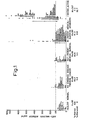
- Figure I of the accompanying drawings shows the concentration of anti-malignin antibody, in micrograms/ml serum in individual sera, in the four control groups and the active cancer group: that is (1) healthy normals, (2) cancer patients showing no evidence of disease after successful treatment, (3) out-patients (non-cancer) with medical-surgical symptoms but without defined disorders, (4) in-patients (non-cancer) with defined medical-surgical disorders, and,(5) patients with active cancer who lived one year or longer. While the four control groups did not differ from each .other at a statistically significant level, each differed from the active cancer group at the significant level of P ⁇ 0.000001.
- Figure 2A of the drawings shows the concentration of anti-malignin antibody in individual sera of patients with terminal cancer, that is, those who died within one year (mean 4.4 + 3.5 months).
- the concentration of antibody in this group differs statistically from the active cancer group at a level of P ⁇ 0.000001.
- Figure 2B of the drawings shows seven examples of the decrease before death observed in individual patients' serum anti-malignin antibody levels when determined serially.
- Table 1 shows the types of cancer patient studied, and the distribution of samples between active disease, terminal disease and no evidence of disease in each type of cancer.
- the distribution of type of cancer is fairly typical with the exception of an excess number of brain cancer cases which was the initial focus of interest of the study.
- the antibody level was elevated in the sera of 20.4% of patients with multiple sclerosis, 31.1% of patients with benign tumors, 30.2% of 'contacts' of active cancer patients, and 38.7% of blood relatives of active cancer patients.
- Anti-malignin antibody levels and the demonstration of malignin in cells may in the future help to clarify the definition in this group.
- the observation of a higher incidence of elevated anti-malignin antibody in contacts of active cancer patients is in agreement with several previously published studies on other tumor indexes demonstrating the same curious phenomenon (14 clinical studies and one laboratory study cited in reference 14). Whether this represents some form of irrmunization against a transmittable agent, either the malignin antigen itself or a substance which induces transformation and thus the appearance of the antigen, needs more work to clarify.
- the greatest incidence of antibody elevation in a 'non-cancer' group is observed in the blood relatives of active cancer patients.
- the anti-malignin antibody level may be related to survival in that the elevated values during active disease were associated with longer survival and low levels during active disease with early deaths After successful treatment, however, the presence of normal (low) antibody levels may be an aid in determining whether an active cancer state has been replaced by one in which there is 'no evidence of disease'.
- the laboratory value can have relevance only in relation to the clinical status, and it usually should not be difficult to separate the clinically nealthy from the cllnically terminal patient, both of whom have low levels of antibody, but for different reasons.
- anti-malignin antibody is specific for a cancer cell antigen, localized preferentially in malignant cells in vitro and in vivo, and ⁇ has been shown to be cytotoxic to malignant cells in vitro (7), the drop in antibody might be more central to the cancer process and be to the detriment of the patient.
- earlier data (6) showed antimali g nin antibody in human cancer sera to be largely 'disarmed', with its Fc portion cleaved from the Fab fragments, which would result in loss of cytotoxicity. This process might reflect one form of the cancer cell's defence against the antibody.
- the low levels of antibody observed here prior to death may be evidence of a second form of the cancer cell's defence,-the result of an increasing blockade of antibody production or release due to antigen excess as the tumor proliferates.
- malignin is not an 'onco-fetal' antigen is supported by the absence of malignin from fetal tissues.
- Malignin appears to be much older phylogenetically than those states conmonly thought of a: being recapitulated during fetal development; its only structural relatives, by computer search (16), are the ferredoxins of plants, lucaena glauca and alfalfa, the acyl carrier protein of E. coli, and cytochrome b5. These four share the property of being anaerobic enzymes, the ferredoxins being the most electro-negative oxidation-reduction enzymes in nature.
- Warburg observed the anaerobic advantage of malignant cells but was unable to account for this property in the activity of the then known anaerobic enzymes (17).
- malignin is a cleaved derivative of such an anaerobic enzyme system, that this system is common to all malignancies regardless of cell type, and that this system imparts an unique anaerobic advantage to cancer cells, would be consistent with the demonstrated increase in the yield of malignin with increasing malignancy of cell growth (1,2), the ubiquity of distribution of the. antigen, the cytotoxicity of the antibody and the antibody failure in the terminal state.
- purified human anti- malignin antibody is available (6,7), and monoclonal anti-malignin antibodies are available, the therapeutic uses of the antibody acting alone or as a carrier for anti-cancer drugs can be further systematically examined.
- the compounds Anti-Astrocytin, Anti-Malignin, and S-TAG have been shown to attach preferentially to - tumor cells. This specificity permits use of these compounds to diagnose tumor cells in histology sections by conjugating dyes or radioactive substances to Anti-Astrocytin, Anti-Malignin, or S-TAG. Standard labelling techniques may then beused.
- a procedure using S-TAG is as follows.
- the conjugate may be a standard anti-serum such as goat anti-rabbit conjugate.
- the conjugate is labelled by techniques known in the art with fluorescein or other labelling substance. Fluorescein-labelled goat anti-rabbit conjugate as commercially available may be used. The fluorescent technique used was a standard one in which a 1:200 to 1:400 solution of TAG is incubated for 30 minutes or more on the tumor section, followed by washes to remove unattached TAG.
- TAG products coupled with a signal emitter such as a dye or a radioactive label to detect cancer cells is described, for example, at pages 12-18 and Example 11 herein.
- a signal emitter such as a dye or a radioactive label
- Example 10 As described in Example 10 utilizing human serum in the determination of TAG, after the anti-malignin antibody was bound to the immobilized antigen and non- bound serumproteins were washed away, the antibody was cloven from the binding with 0.25 M acetic acid at 37°C for 2 hours and the TARGET reagent was separated out by centrifuging. The TAG antibody solution was quantitated by means of its absorption at 280 mp.
- TAG solutions were stored at -20°C, then thawed and combined, brought to pH 7 by titration with 6N NaOH, dialysed against phosphate buffered saline of pH 7, filtered and concentrated on Millipore Pellicon 1000 membranes, centrifuged to clear insoluble protein and the immune globulin complexes, concentrated and freed of immunologically non-active compounds by Cellex D and Blue Sepharose CI6B (Pharmacia) chromatography.
- This human anti-malignin antibody reacts with anti-human gamma globulin in Ouchterlony double diffusion.
- TAG When TAG is used with fluorscein conjugated to anti-human gamma globulin in standard double layer Coons irrmuno- fluoresence, it stains malignant glia, breast carcinoma, ovarian carcinoma, adenocarcinoma of the colon, and other types of cancer cells in postoperative and biopsy tissue sections, as well as inhuman sputum, bronchial washings, pleural effusion fluid, gastric aspirate and bladder urine.
- concentration of protein in TAG which yields clear fluorescence when controls are negative, is 1 to 10 ⁇ g per section.
- TAG The production of a "purified" TAG was undertaken by reacting the sera from patients with a variety of cancers.with bromoacetylcellulose - MALIGNIN by methods earlier described (Example 8).
- the antibody bound in this reaction was cleaved with 0.25 M acetic acid, quantified by measurement at O.D. 280 using a conversion factor of 1.46 for gamma globulin frozen and stored at -20°C.
- This antibody was found to contain immunoglobulin as determined by anti-hurnan gamma-globulin antiserum specific for gamma chains (BioRad Laboratories, Inc.) and with anti-FAB and anti-Fc .fragments (Miles Laboratories). It also reacts with rabbit anti-human albumin (BioRad Laboratories).
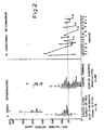
- Method_I - Fractionation of TAG chromatography with DEAE cellulose was first employed using step-wise elution with increasing ionic strength and decreasing pH, and the same sequence of eluants as that given in Example I for the production of Crude Astrocytin-Precursor-Containing Fraction. Good separation was obtained of the bulk of the protein into three fractions, Peak I obtained with Solution 1 (see Example 1) and Peak II obtained with Solution 1 (see Example 1) and Peak II obtained with Solution 6 and Solution 7. Ouchterlony double diffusion showed the TAG in Peak I still to contain appreciable protein with albumin mobility, and while Peak II contained most of the albumin, appreciable IgG could be detected.
- Fig. 1 shows the fractions obtained on chromatography of TAG utilizing Method III. After the first eluate of 200 mls., 50 ml. or smaller sub-fractions were collected. The protein content'of each eluate was determined by the optical density at 280 m ⁇ with an uniform factor of 1.46 based on gamma globulin used to convert to micrograms for calculating recoveries. The absolute amount of protein requires correction in those fractions in which there is appreciable albumin. The points at which the stepwise solvent changes were made are indicated by arrows. The subfractions are designated by Roman numerals I to VIII, inclusive.
- Photographs were prepared showing the line of reaction between anti-human gamma-globulin specific for gamma chains for each of Fractions I and VIII from above.
- TAG Fluorescence Activated Cell Sorting
- Non-cancer cells do not fluoresce.
- the TAG (1 to 10 ⁇ g in 0.1 ml phosphate buffered saline (PBS)) is applied to the surface of packed cells on a glass slide, incubated for 30 minutes, washed three times with PBS and then layered with fluorescein-conjugated anti-human IgG diluted until non-malignant control tissues give essentially no fluorescence.
- the cells are visualized with a Zeiss fluorescent microscope using a tungsten lamp and filters BG 23, BG 12 and 500.
- malignin was sought in cells collected from cancer patients and controls. Specimens were collected by thoracocentesis, paracentesis, bronchial or tracheal washings, sputum and pericardial effusion, from patients with lung, breast, prostatic, colon and und.ifferentiated cancers, as well as from non-cancer controls including patients with emphysema, heavy smoking and epilepsy; and sputum from a former cancer patient with no evidence of disease for two years following successful treatment. Cells were concentrated by centrifuging.
- the following Table shows correlation of the presence or absence of-malignin cells as determined blind by immunofluorescent staining with anti-malignin antibody (TAG), and the clinical-pathological diagnosis.
- TAG stain result was correct in 20/22 specimens (91%).
- Standard Papanicolaou stain examinations performed blind on duplicates of these specimens by other pathologists were correct in 17/22 specimens (77%).
- anti-malignin antibody was active at one nanogram antibody protein per cancer cell in producing the specific immunofluorescence seen and photographed in: A- bronchogenic carcinoma cells, from bronchial washings; B- lymphocytic leukaemia cell, from blood; C-ovarian carcinoma cells, at surgery; D- squamous cell carcinoma (2 cells), grown in tissue culture; E- astro- cytoma, anaplastic, at surgery.
- a portion of the labelled solution was also plated on an Ouchterlony gel plate to determine its ability to react with malignin in the antigen-antibody reaction. After a 3-hour period, the resulting sharp reactive lines were removed from the gel and their content of radio-activity was measured. An equal portion of the gel not involved in the reaction was also removed and its content of radioactivity was also measured as background.
- Wistar rats were injected intracerebrally with C6 glioma tumor cells which had had previous passages in rats and in tissue culture. The rats were observed for the first signs of growing tumor, such as weakness, tremor or unsteadiness. These symptoms first appear seven to ten days from injection, and with fast growing tumors result in death within three to four days in many animals, and one week in all. As soon as symptoms appeared, the animals were injected with labelled TAG intravenously in the tail vein, then the-animals were anaesthetized at varying times, the brain was removed, the tumor was dissected free of normal brain, and the radioactivities of the dissected specimens were compared with one another. Preliminar 99m Tc-TAG experiment
- Tumor and normal brain specimens were counted overnight in the gamna-well counter. All samples and standards were decay corrected for convenience to the mid- count of the first sample in the sequence.
- a myeloma cell line (P3.x63-Ag-8) was cultured in Dulbecco's minimum essential medium supplemented with 10%. foetal bovine serum (D 10 ), in a humidified incubator at 37°C and 5% CO 2 .
- mice Inbred female BALB/cJ mice (8 weeks of age) (Jackson Laboratory, Bar Harbor, Maine) were imrunized intraperitoneally, 4 times at weekly intervals with 1 mg. Malignin emulsified in complete Freund's adjuvant (Difco). Sera of the immunized mice were tested for the presence of anti-malignin antibody and antibody positive mice were further boosted 4 days prior to cell fusion.
- Immune spleen cells (10 8 ) were fused with the myeloma cell (10 ) using polyethylene glycol (PEG, 1000, J.T. Backer) as the fusion inducing agent as described by Galfre et al (Nature 266, 550-552, 1977).
- PEG polyethylene glycol
- the PEG treated cell mix was seeded into96 wells of microtiter plate (Costar 35 96) in D 10 supplemented with hypoxanthine, aminopterin, and thymidine (D10 HAT) (Littlefield, J.W., Science 145: 709 , 1964) . About one-half of D10 HAT was replaced twice weekly for two weeks.
- the spleen cells did not survive in vitro, while the infused myeloma cells were killed in D10 HAT. Only the hybrid cells remained actively growing after 10 days under the selective conditions. After two weeks in D10 HAT, the hybrid cells were fed for another week with medium which was the same as D 10 HAT except for the omission of aminopterin (D10 HT) then with D 10 . Whenever the wells were about 80% covered by hybrid cells, supernatants were aspirated for anti-malignin antibody assay..
- the above Table shows the quantities of monoclonal anti-malignin antibody produced by each antibody producing clone, in micrograms of protein per ml of extracellular fluid.
- the yields of antibody are seen to be good for the first four months of propagation of the clones, and to have increased by the fifth month of propagation.
- the cells continued to grow well through the eighth mopth and to successfully grow when transferred intraperitoneally to the mouse, where the yield of antibody again increased as expected to as much as 1 mg. of MAMA-S per ml of ascites fluid.
- the cells also grew successfully on soft agar and were frozen and stored in liquid nitrogen and grown again after thawing. Aliquots of each clone were frozen in liquid nitrogen for permanent storage and regrowth at later dates.
- the monoclonal antibody in each case was quantified as protein by optical density at 280 millimicrons, was non-dialysable, and migrated on SDS-polyacrylamide gel electrophoresis predominantly as gamma chain immunoglobulins.
- Second layer staining with fluorescent labels both fluorescein and rhodamine, at concentrations as low as 1:1,600 was observed and recorded. These very low concentrations of the second layer permitted dilution until background non-specific staining was eliminated, and, at those concentrations of second layer (FITC or rhodamine), highly specific staining was obtained with MAMA-F, MAMA-S and MAMA-FS.
- Standard tests for determining cytotoxicity may be used. Generally, the number of cells in a fixed counting chamber, usually arranged to contain about 100 live cells, is counted. These cells are then treated with the agent being tested and the number of cells which are still alive is counted.
- S-TAG produced death of all cells in the counting chamber.even when in high dilution of 1:00 and 1:1000, representing as little as 0.2 and 0.02 ⁇ g of S-TAG per ml. of solution. Similar results are obtained with high dilutions of Anti-Astrocytin and Anti-Malignin.
- Example 11A Both the specificity exhibited in Examples 11, 11A, 12, 13, 14 and 15 and the cytotoxicity demonstrated in this Example and Example 17 are highly relevant to the therapeutic possibilities of Anti-Astrocytin, Anti-Malignin and S-TAG for malignant tumors in man.
- the practical diagnostic potential of both of these phenomena for tumor tissue removed at operation but requiring diagnosis by histology is already demonstrated herein.
- MAMA-F or MAMA-S alone does not produce appreciable cytotoxicity with malignant cells
- the mixture is actively cytotoxic to malignant cells.
- the product MAMA-FS is cytotoxic. All three preparations are cytotoxic in approximately the same concentrations as previously observed for Anti-Astrocytin, Anti-Malignin and S-TAG products (Example 16). Approximately one nanogram of antibody per cell is cytotoxic, resulting in lysis of the cell.
- Cytotoxicity also was observed and recorded on both the Coulter cytofluorograph and on the Ortho cytofluorograph, each of which permits absolute counts of viable cells with time. Destruction of viable malignant cells (carcinoma of the pancreas, leukaemic and lymphoma) was observed over a period of 15 to 60 minutes. Those malignant cells which were identified by light scatter and specifically by fluorescence were destroyed by either the mixture of MAMA-F and MAMA-S or by MAMA-FS. The killing of cancer cells is, by definition, a therapeutic process, and the products which produce this killing, are by definition, therapeutic products.
- the various PRODUCER cells have had their desoxyribonucleic acid specifically programmed by MALIGNIN to produce the anti-MALIGNIN antibody.
- MALIGNIN monoclonal anti-Malignin antibody FAST producer cell, SLOW producer cell and FAST and SLOW producer cell. It will be appreciated that this invention covers both the desoxyribonucleic acids and the ribonucleic acids of all such PRODUCER cells.
Landscapes
- Health & Medical Sciences (AREA)
- Life Sciences & Earth Sciences (AREA)
- Chemical & Material Sciences (AREA)
- Engineering & Computer Science (AREA)
- Organic Chemistry (AREA)
- Immunology (AREA)
- Genetics & Genomics (AREA)
- Molecular Biology (AREA)
- General Health & Medical Sciences (AREA)
- Biochemistry (AREA)
- Zoology (AREA)
- Biotechnology (AREA)
- Biomedical Technology (AREA)
- Medicinal Chemistry (AREA)
- Wood Science & Technology (AREA)
- Microbiology (AREA)
- Bioinformatics & Cheminformatics (AREA)
- Hematology (AREA)
- General Engineering & Computer Science (AREA)
- Urology & Nephrology (AREA)
- Cell Biology (AREA)
- Biophysics (AREA)
- Proteomics, Peptides & Aminoacids (AREA)
- Physics & Mathematics (AREA)
- Oncology (AREA)
- Chemical Kinetics & Catalysis (AREA)
- Food Science & Technology (AREA)
- Pathology (AREA)
- Analytical Chemistry (AREA)
- General Physics & Mathematics (AREA)
- Hospice & Palliative Care (AREA)
- Mycology (AREA)
- Plant Pathology (AREA)
- General Chemical & Material Sciences (AREA)
- Toxicology (AREA)
- Gastroenterology & Hepatology (AREA)
- Tropical Medicine & Parasitology (AREA)
- Virology (AREA)
- Preparation Of Compounds By Using Micro-Organisms (AREA)
- Peptides Or Proteins (AREA)
Applications Claiming Priority (2)
| Application Number | Priority Date | Filing Date | Title |
|---|---|---|---|
| US271645 | 1981-06-08 | ||
| US06/271,645 US4486538A (en) | 1978-07-07 | 1981-06-08 | Detection of malignant tumor cells |
Publications (1)
| Publication Number | Publication Date |
|---|---|
| EP0067642A1 true EP0067642A1 (de) | 1982-12-22 |
Family
ID=23036465
Family Applications (2)
| Application Number | Title | Priority Date | Filing Date |
|---|---|---|---|
| EP82302937A Ceased EP0067642A1 (de) | 1981-06-08 | 1982-06-08 | Nachweis von bösartigen Tumorzellen |
| EP82901607A Withdrawn EP0080474A1 (de) | 1981-06-08 | 1982-06-08 | Nachweis von bösartigen tumorzellen |
Family Applications After (1)
| Application Number | Title | Priority Date | Filing Date |
|---|---|---|---|
| EP82901607A Withdrawn EP0080474A1 (de) | 1981-06-08 | 1982-06-08 | Nachweis von bösartigen tumorzellen |
Country Status (7)
| Country | Link |
|---|---|
| US (1) | US4486538A (de) |
| EP (2) | EP0067642A1 (de) |
| JP (2) | JPS58500837A (de) |
| CA (1) | CA1215919A (de) |
| DK (1) | DK51983A (de) |
| NO (1) | NO830404L (de) |
| WO (1) | WO1982004262A1 (de) |
Cited By (3)
| Publication number | Priority date | Publication date | Assignee | Title |
|---|---|---|---|---|
| GB2139645A (en) * | 1983-04-08 | 1984-11-14 | Kureha Chemical Ind Co Ltd | Antibody to human prostrate cancer |
| EP0221748A2 (de) * | 1985-11-01 | 1987-05-13 | Samuel Dr. Bogoch | Verfahren zur Herstellung von menschlichen Antikörpern |
| EP0287030A2 (de) * | 1987-04-14 | 1988-10-19 | Abbott Laboratories | Immunologische Methoden und Materialien zur Bestimmung von Tumor assoziierten Antigenen |
Families Citing this family (15)
| Publication number | Priority date | Publication date | Assignee | Title |
|---|---|---|---|---|
| US4624932A (en) * | 1978-09-13 | 1986-11-25 | Samuel Bogoch | Recognins and their chemoreciprocals |
| SE8401945L (sv) * | 1983-04-08 | 1984-11-13 | Kureha Chemical Ind Co Ltd | Antikropp mot cancer i urinblasan hos menniska |
| US4529693A (en) * | 1983-12-09 | 1985-07-16 | Children's Hospital Medical Center Of Northern California | Detecting neoplastic mammary epithelial cells by detecting thioesterase II marker |
| US4783330A (en) * | 1984-11-15 | 1988-11-08 | New England Medical Center Hospitals, Inc. | Monoclonal antibodies to activated platelets |
| WO1987004164A1 (en) * | 1986-01-06 | 1987-07-16 | The University Of Melbourne | Technetium-antibody conjugate |
| US6893625B1 (en) | 1986-10-27 | 2005-05-17 | Royalty Pharma Finance Trust | Chimeric antibody with specificity to human B cell surface antigen |
| IL84285A (en) * | 1986-10-27 | 1993-03-15 | Int Genetic Engineering | Chimeric antibody with specificity to human tumor antigen |
| IL85035A0 (en) * | 1987-01-08 | 1988-06-30 | Int Genetic Eng | Polynucleotide molecule,a chimeric antibody with specificity for human b cell surface antigen,a process for the preparation and methods utilizing the same |
| US5028594A (en) * | 1988-12-27 | 1991-07-02 | Naxcor | Use of photodynamic compositions for cytotoxic effects |
| US4983527A (en) * | 1989-05-26 | 1991-01-08 | Arizona Board Of Regents | Oocyte test for detection of tumor promoting compounds |
| AU6409894A (en) * | 1993-03-16 | 1994-10-11 | Samuel Bogoch | Recognin vaccines |
| US6242578B1 (en) * | 1994-02-17 | 2001-06-05 | Samuel Bogoch | Aglyco products and methods of use |
| US7420028B2 (en) * | 2001-03-27 | 2008-09-02 | Samuel Bogoch | Replikins and methods of identifying replikin-containing sequences |
| US7306919B1 (en) | 2001-10-31 | 2007-12-11 | Thornthwaite Jerry T | Antigen-antibody cancer recognition system |
| US9320784B2 (en) | 2009-08-07 | 2016-04-26 | Samuel Bogoch | Peptides shared among lethal cancers and therapeutic compositions comprising said peptides |
Citations (4)
| Publication number | Priority date | Publication date | Assignee | Title |
|---|---|---|---|---|
| US4172124A (en) * | 1978-04-28 | 1979-10-23 | The Wistar Institute | Method of producing tumor antibodies |
| EP0007214A1 (de) * | 1978-07-07 | 1980-01-23 | Samuel Dr. Bogoch | Mittel zum Nachweis der Anwesenheit krebsartiger oder bösartiger Tumorzellen |
| EP0015755A2 (de) * | 1979-03-09 | 1980-09-17 | Samuel Dr. Bogoch | Recognine, ihre Chemoreziprokale, am Target haftende Globuline und Verfahren zur Herstellung dieser Produkte; Verfahren zum Bestimmen von Krebstumoren |
| WO1981001469A1 (en) * | 1979-11-21 | 1981-05-28 | Wistar Inst | Monoclonal hybridoma antibody specific for high molecular weight carcinoembryonic antigen |
Family Cites Families (4)
| Publication number | Priority date | Publication date | Assignee | Title |
|---|---|---|---|---|
| US4196186A (en) * | 1975-10-09 | 1980-04-01 | Samuel Bogoch | Method for diagnosing malignant gliol brain tumors |
| US4298590A (en) * | 1975-02-25 | 1981-11-03 | Samuel Bogoch | Detection of malignant tumor cells |
| US4195017A (en) * | 1975-02-25 | 1980-03-25 | Samuel Bogoch | Malignin, derived from brain tumor cells, complexes and polypeptides thereof |
| US4196265A (en) * | 1977-06-15 | 1980-04-01 | The Wistar Institute | Method of producing antibodies |
-
1981
- 1981-06-08 US US06/271,645 patent/US4486538A/en not_active Expired - Lifetime
-
1982
- 1982-06-04 CA CA000404534A patent/CA1215919A/en not_active Expired
- 1982-06-08 EP EP82302937A patent/EP0067642A1/de not_active Ceased
- 1982-06-08 EP EP82901607A patent/EP0080474A1/de not_active Withdrawn
- 1982-06-08 WO PCT/GB1982/000170 patent/WO1982004262A1/en unknown
- 1982-06-08 JP JP57501690A patent/JPS58500837A/ja active Pending
-
1983
- 1983-02-08 DK DK51983A patent/DK51983A/da not_active Application Discontinuation
- 1983-02-08 NO NO830404A patent/NO830404L/no unknown
-
1989
- 1989-06-08 JP JP1146551A patent/JPH0372894A/ja active Pending
Patent Citations (4)
| Publication number | Priority date | Publication date | Assignee | Title |
|---|---|---|---|---|
| US4172124A (en) * | 1978-04-28 | 1979-10-23 | The Wistar Institute | Method of producing tumor antibodies |
| EP0007214A1 (de) * | 1978-07-07 | 1980-01-23 | Samuel Dr. Bogoch | Mittel zum Nachweis der Anwesenheit krebsartiger oder bösartiger Tumorzellen |
| EP0015755A2 (de) * | 1979-03-09 | 1980-09-17 | Samuel Dr. Bogoch | Recognine, ihre Chemoreziprokale, am Target haftende Globuline und Verfahren zur Herstellung dieser Produkte; Verfahren zum Bestimmen von Krebstumoren |
| WO1981001469A1 (en) * | 1979-11-21 | 1981-05-28 | Wistar Inst | Monoclonal hybridoma antibody specific for high molecular weight carcinoembryonic antigen |
Non-Patent Citations (3)
| Title |
|---|
| PROC. NATL. ACAD. SCI., vol.77, no.1, January 1980, (US) * |
| SCIENTIFIC AMERICAN, vol.243, no.4, October 1980, New York (US) * |
| THE LANCET, vol.2, no.8238, July 18, 1981, North American Edition * |
Cited By (5)
| Publication number | Priority date | Publication date | Assignee | Title |
|---|---|---|---|---|
| GB2139645A (en) * | 1983-04-08 | 1984-11-14 | Kureha Chemical Ind Co Ltd | Antibody to human prostrate cancer |
| EP0221748A2 (de) * | 1985-11-01 | 1987-05-13 | Samuel Dr. Bogoch | Verfahren zur Herstellung von menschlichen Antikörpern |
| EP0221748A3 (de) * | 1985-11-01 | 1988-10-12 | Samuel Dr. Bogoch | Verfahren zur Herstellung von menschlichen Antikörpern |
| EP0287030A2 (de) * | 1987-04-14 | 1988-10-19 | Abbott Laboratories | Immunologische Methoden und Materialien zur Bestimmung von Tumor assoziierten Antigenen |
| EP0287030A3 (en) * | 1987-04-14 | 1988-11-09 | Abbott Laboratories | Immunological methods and materials for detection of tumor associated antigens |
Also Published As
| Publication number | Publication date |
|---|---|
| US4486538A (en) | 1984-12-04 |
| JPS58500837A (ja) | 1983-05-26 |
| EP0080474A1 (de) | 1983-06-08 |
| WO1982004262A1 (en) | 1982-12-09 |
| DK51983D0 (da) | 1983-02-08 |
| JPH0372894A (ja) | 1991-03-28 |
| CA1215919A (en) | 1986-12-30 |
| NO830404L (no) | 1983-02-08 |
| DK51983A (da) | 1983-02-08 |
Similar Documents
| Publication | Publication Date | Title |
|---|---|---|
| US5866690A (en) | Detection of malignant tumor cells | |
| US4486538A (en) | Detection of malignant tumor cells | |
| US4298590A (en) | Detection of malignant tumor cells | |
| Inbar et al. | Crystallization with hapten of the Fab'fragment from a mouse IgA myeloma protein with antidinitrophenyl activity | |
| US4160019A (en) | Detection of cancer associated polypeptide antigen and preparation of antibodies thereto | |
| BE1000611A5 (fr) | Anticorps monoclonaux et antigene du carcinome pulmonaire humain a cellules non petites et de certains autres carcinomes humains. | |
| EP0782709A1 (de) | Nachweiss und behandlung krebsi | |
| Goldenberg et al. | Antigens associated with normal and malignant gastrointestinal tissues | |
| US4196186A (en) | Method for diagnosing malignant gliol brain tumors | |
| US5654408A (en) | Melanoma-associated antigens identified by monoclonal antibodies | |
| US4195017A (en) | Malignin, derived from brain tumor cells, complexes and polypeptides thereof | |
| US4624932A (en) | Recognins and their chemoreciprocals | |
| EP0244473A1 (de) | Verfahren zum nachweis von krebszellen | |
| EP0007214A1 (de) | Mittel zum Nachweis der Anwesenheit krebsartiger oder bösartiger Tumorzellen | |
| CA1286238C (en) | Anti-epiglycanin monoclonal antibodies | |
| EP0221561A2 (de) | Menschlichen Brustkrebs anzeigendes Antigen und darauf basierende Teste | |
| US4840915A (en) | Method for diagnosing malignant tumors | |
| US4976957A (en) | Process for the production of recognins and their chemoreciprocals | |
| EP0015755B1 (de) | Recognine, ihre Chemoreziprokale, am Target haftende Globuline und Verfahren zur Herstellung dieser Produkte; Verfahren zum Bestimmen von Krebstumoren | |
| JPS63500797A (ja) | 崩壊加速因子に対するモノクロ−ン抗体、その生成方法および使用 | |
| Hirai | Carcinofetal proteins | |
| CA1039650A (en) | Cancer associated polypeptide antigen, process for its preparation, process for preparing antibodies, process of cancer diagnosis and composition useful as an immunizing agent | |
| JPS6193A (ja) | 特異性がん関連抗原、フコシルシアロシルガングリオテトラオースおよびヒトの肺がんに関連する診断または治療処置におけるその使用 | |
| JPS62500304A (ja) | 膵臓がんに伴う胞状奇胎分化蛋白質、これらの蛋白質に対する抗血清およびモノクロ−ナル抗体、製造方法 | |
| US5087573A (en) | Monoclonal antibody against bone alkaline phosphatase |
Legal Events
| Date | Code | Title | Description |
|---|---|---|---|
| PUAI | Public reference made under article 153(3) epc to a published international application that has entered the european phase |
Free format text: ORIGINAL CODE: 0009012 |
|
| AK | Designated contracting states |
Designated state(s): AT BE CH DE FR GB IT LI LU NL SE |
|
| 17P | Request for examination filed |
Effective date: 19830609 |
|
| STAA | Information on the status of an ep patent application or granted ep patent |
Free format text: STATUS: THE APPLICATION HAS BEEN REFUSED |
|
| 18R | Application refused |
Effective date: 19890710 |
|
| APAF | Appeal reference modified |
Free format text: ORIGINAL CODE: EPIDOSCREFNE |