EP0057553B1 - Verfahren und Testpackung zur Feststellung beschädigter DNA durch Fluoreszenz - Google Patents
Verfahren und Testpackung zur Feststellung beschädigter DNA durch Fluoreszenz Download PDFInfo
- Publication number
- EP0057553B1 EP0057553B1 EP82300376A EP82300376A EP0057553B1 EP 0057553 B1 EP0057553 B1 EP 0057553B1 EP 82300376 A EP82300376 A EP 82300376A EP 82300376 A EP82300376 A EP 82300376A EP 0057553 B1 EP0057553 B1 EP 0057553B1
- Authority
- EP
- European Patent Office
- Prior art keywords
- dna
- cells
- solution
- alkaline
- fluorescent dye
- Prior art date
- Legal status (The legal status is an assumption and is not a legal conclusion. Google has not performed a legal analysis and makes no representation as to the accuracy of the status listed.)
- Expired
Links
- 238000000034 method Methods 0.000 title claims description 31
- 238000001514 detection method Methods 0.000 title claims description 5
- 210000004027 cell Anatomy 0.000 claims description 52
- 239000000243 solution Substances 0.000 claims description 41
- 238000004925 denaturation Methods 0.000 claims description 26
- 230000036425 denaturation Effects 0.000 claims description 26
- HEMHJVSKTPXQMS-UHFFFAOYSA-M Sodium hydroxide Chemical compound [OH-].[Na+] HEMHJVSKTPXQMS-UHFFFAOYSA-M 0.000 claims description 21
- 230000005855 radiation Effects 0.000 claims description 20
- 239000007850 fluorescent dye Substances 0.000 claims description 17
- 230000006378 damage Effects 0.000 claims description 16
- 239000002253 acid Substances 0.000 claims description 15
- 238000012360 testing method Methods 0.000 claims description 15
- 230000005778 DNA damage Effects 0.000 claims description 13
- 231100000277 DNA damage Toxicity 0.000 claims description 13
- 239000000126 substance Substances 0.000 claims description 13
- 239000003153 chemical reaction reagent Substances 0.000 claims description 11
- 238000001556 precipitation Methods 0.000 claims description 11
- NLXLAEXVIDQMFP-UHFFFAOYSA-N Ammonia chloride Chemical compound [NH4+].[Cl-] NLXLAEXVIDQMFP-UHFFFAOYSA-N 0.000 claims description 10
- 210000004369 blood Anatomy 0.000 claims description 10
- 239000008280 blood Substances 0.000 claims description 10
- 239000003381 stabilizer Substances 0.000 claims description 10
- 230000002934 lysing effect Effects 0.000 claims description 9
- XSQUKJJJFZCRTK-UHFFFAOYSA-N Urea Chemical compound NC(N)=O XSQUKJJJFZCRTK-UHFFFAOYSA-N 0.000 claims description 8
- 239000012670 alkaline solution Substances 0.000 claims description 7
- 210000003850 cellular structure Anatomy 0.000 claims description 7
- 239000003638 chemical reducing agent Substances 0.000 claims description 7
- 229960000367 inositol Drugs 0.000 claims description 7
- 238000000926 separation method Methods 0.000 claims description 7
- CDAISMWEOUEBRE-GPIVLXJGSA-N inositol Chemical compound O[C@H]1[C@H](O)[C@@H](O)[C@H](O)[C@H](O)[C@@H]1O CDAISMWEOUEBRE-GPIVLXJGSA-N 0.000 claims description 6
- 210000000265 leukocyte Anatomy 0.000 claims description 6
- 230000003204 osmotic effect Effects 0.000 claims description 6
- CDAISMWEOUEBRE-UHFFFAOYSA-N scyllo-inosotol Natural products OC1C(O)C(O)C(O)C(O)C1O CDAISMWEOUEBRE-UHFFFAOYSA-N 0.000 claims description 6
- 235000019270 ammonium chloride Nutrition 0.000 claims description 5
- 239000004202 carbamide Substances 0.000 claims description 5
- 230000007935 neutral effect Effects 0.000 claims description 5
- WQZGKKKJIJFFOK-GASJEMHNSA-N Glucose Natural products OC[C@H]1OC(O)[C@H](O)[C@@H](O)[C@@H]1O WQZGKKKJIJFFOK-GASJEMHNSA-N 0.000 claims description 4
- SQUHHTBVTRBESD-UHFFFAOYSA-N Hexa-Ac-myo-Inositol Natural products CC(=O)OC1C(OC(C)=O)C(OC(C)=O)C(OC(C)=O)C(OC(C)=O)C1OC(C)=O SQUHHTBVTRBESD-UHFFFAOYSA-N 0.000 claims description 4
- TWRXJAOTZQYOKJ-UHFFFAOYSA-L Magnesium chloride Chemical compound [Mg+2].[Cl-].[Cl-] TWRXJAOTZQYOKJ-UHFFFAOYSA-L 0.000 claims description 4
- 239000013592 cell lysate Substances 0.000 claims description 4
- 230000000254 damaging effect Effects 0.000 claims description 4
- ZMMJGEGLRURXTF-UHFFFAOYSA-N ethidium bromide Chemical compound [Br-].C12=CC(N)=CC=C2C2=CC=C(N)C=C2[N+](CC)=C1C1=CC=CC=C1 ZMMJGEGLRURXTF-UHFFFAOYSA-N 0.000 claims description 4
- 229960005542 ethidium bromide Drugs 0.000 claims description 4
- 239000008103 glucose Substances 0.000 claims description 4
- 239000000872 buffer Substances 0.000 claims description 3
- 230000006037 cell lysis Effects 0.000 claims description 3
- 239000001488 sodium phosphate Substances 0.000 claims description 3
- 229910000162 sodium phosphate Inorganic materials 0.000 claims description 3
- 235000000346 sugar Nutrition 0.000 claims description 3
- RYFMWSXOAZQYPI-UHFFFAOYSA-K trisodium phosphate Chemical compound [Na+].[Na+].[Na+].[O-]P([O-])([O-])=O RYFMWSXOAZQYPI-UHFFFAOYSA-K 0.000 claims description 3
- 239000007853 buffer solution Substances 0.000 claims description 2
- 229910001424 calcium ion Inorganic materials 0.000 claims description 2
- 229910001629 magnesium chloride Inorganic materials 0.000 claims description 2
- 229910001425 magnesium ion Inorganic materials 0.000 claims description 2
- 230000001681 protective effect Effects 0.000 claims 2
- 238000006243 chemical reaction Methods 0.000 claims 1
- MOTZDAYCYVMXPC-UHFFFAOYSA-N dodecyl hydrogen sulfate Chemical compound CCCCCCCCCCCCOS(O)(=O)=O MOTZDAYCYVMXPC-UHFFFAOYSA-N 0.000 claims 1
- 229940043264 dodecyl sulfate Drugs 0.000 claims 1
- 239000008363 phosphate buffer Substances 0.000 claims 1
- 239000011734 sodium Substances 0.000 claims 1
- 108020004414 DNA Proteins 0.000 description 64
- 238000003556 assay Methods 0.000 description 17
- 102000053602 DNA Human genes 0.000 description 15
- 238000011534 incubation Methods 0.000 description 10
- 239000003513 alkali Substances 0.000 description 8
- 239000003814 drug Substances 0.000 description 8
- 230000035945 sensitivity Effects 0.000 description 8
- 231100001074 DNA strand break Toxicity 0.000 description 7
- 230000003139 buffering effect Effects 0.000 description 6
- 229940079593 drug Drugs 0.000 description 6
- 150000007513 acids Chemical class 0.000 description 5
- 239000003795 chemical substances by application Substances 0.000 description 5
- 239000006166 lysate Substances 0.000 description 5
- 238000005259 measurement Methods 0.000 description 5
- 238000002156 mixing Methods 0.000 description 5
- 108020004682 Single-Stranded DNA Proteins 0.000 description 4
- 239000002585 base Substances 0.000 description 4
- 239000000975 dye Substances 0.000 description 4
- 230000000694 effects Effects 0.000 description 4
- 230000002093 peripheral effect Effects 0.000 description 4
- 230000008439 repair process Effects 0.000 description 4
- 239000000725 suspension Substances 0.000 description 4
- 206010028980 Neoplasm Diseases 0.000 description 3
- KWYUFKZDYYNOTN-UHFFFAOYSA-M Potassium hydroxide Chemical compound [OH-].[K+] KWYUFKZDYYNOTN-UHFFFAOYSA-M 0.000 description 3
- DBMJMQXJHONAFJ-UHFFFAOYSA-M Sodium laurylsulphate Chemical compound [Na+].CCCCCCCCCCCCOS([O-])(=O)=O DBMJMQXJHONAFJ-UHFFFAOYSA-M 0.000 description 3
- 230000001413 cellular effect Effects 0.000 description 3
- 210000004962 mammalian cell Anatomy 0.000 description 3
- 239000002609 medium Substances 0.000 description 3
- 210000000056 organ Anatomy 0.000 description 3
- 108090000623 proteins and genes Proteins 0.000 description 3
- 102000004169 proteins and genes Human genes 0.000 description 3
- 238000010008 shearing Methods 0.000 description 3
- 229940083575 sodium dodecyl sulfate Drugs 0.000 description 3
- 235000019333 sodium laurylsulphate Nutrition 0.000 description 3
- 230000033616 DNA repair Effects 0.000 description 2
- KCXVZYZYPLLWCC-UHFFFAOYSA-N EDTA Chemical compound OC(=O)CN(CC(O)=O)CCN(CC(O)=O)CC(O)=O KCXVZYZYPLLWCC-UHFFFAOYSA-N 0.000 description 2
- 101710163270 Nuclease Proteins 0.000 description 2
- 239000012979 RPMI medium Substances 0.000 description 2
- IQFYYKKMVGJFEH-XLPZGREQSA-N Thymidine Chemical compound O=C1NC(=O)C(C)=CN1[C@@H]1O[C@H](CO)[C@@H](O)C1 IQFYYKKMVGJFEH-XLPZGREQSA-N 0.000 description 2
- 210000000601 blood cell Anatomy 0.000 description 2
- 201000011510 cancer Diseases 0.000 description 2
- 230000000711 cancerogenic effect Effects 0.000 description 2
- 239000000356 contaminant Substances 0.000 description 2
- 238000009792 diffusion process Methods 0.000 description 2
- 239000006185 dispersion Substances 0.000 description 2
- 231100000673 dose–response relationship Toxicity 0.000 description 2
- 239000003256 environmental substance Substances 0.000 description 2
- 238000012921 fluorescence analysis Methods 0.000 description 2
- 238000000265 homogenisation Methods 0.000 description 2
- 238000000338 in vitro Methods 0.000 description 2
- 238000002347 injection Methods 0.000 description 2
- 239000007924 injection Substances 0.000 description 2
- 239000000463 material Substances 0.000 description 2
- 238000012544 monitoring process Methods 0.000 description 2
- 239000008188 pellet Substances 0.000 description 2
- 230000008569 process Effects 0.000 description 2
- DGVVWUTYPXICAM-UHFFFAOYSA-N β‐Mercaptoethanol Chemical compound OCCS DGVVWUTYPXICAM-UHFFFAOYSA-N 0.000 description 2
- FWBHETKCLVMNFS-UHFFFAOYSA-N 4',6-Diamino-2-phenylindol Chemical compound C1=CC(C(=N)N)=CC=C1C1=CC2=CC=C(C(N)=N)C=C2N1 FWBHETKCLVMNFS-UHFFFAOYSA-N 0.000 description 1
- 125000004203 4-hydroxyphenyl group Chemical group [H]OC1=C([H])C([H])=C(*)C([H])=C1[H] 0.000 description 1
- YHQDZJICGQWFHK-UHFFFAOYSA-N 4-nitroquinoline N-oxide Chemical compound C1=CC=C2C([N+](=O)[O-])=CC=[N+]([O-])C2=C1 YHQDZJICGQWFHK-UHFFFAOYSA-N 0.000 description 1
- 208000032484 Accidental exposure to product Diseases 0.000 description 1
- GUBGYTABKSRVRQ-XLOQQCSPSA-N Alpha-Lactose Chemical compound O[C@@H]1[C@@H](O)[C@@H](O)[C@@H](CO)O[C@H]1O[C@@H]1[C@@H](CO)O[C@H](O)[C@H](O)[C@H]1O GUBGYTABKSRVRQ-XLOQQCSPSA-N 0.000 description 1
- DWRXFEITVBNRMK-UHFFFAOYSA-N Beta-D-1-Arabinofuranosylthymine Natural products O=C1NC(=O)C(C)=CN1C1C(O)C(O)C(CO)O1 DWRXFEITVBNRMK-UHFFFAOYSA-N 0.000 description 1
- 108010077544 Chromatin Proteins 0.000 description 1
- 102000003844 DNA helicases Human genes 0.000 description 1
- 108090000133 DNA helicases Proteins 0.000 description 1
- QTANTQQOYSUMLC-UHFFFAOYSA-O Ethidium cation Chemical class C12=CC(N)=CC=C2C2=CC=C(N)C=C2[N+](CC)=C1C1=CC=CC=C1 QTANTQQOYSUMLC-UHFFFAOYSA-O 0.000 description 1
- 206010073306 Exposure to radiation Diseases 0.000 description 1
- 229930091371 Fructose Natural products 0.000 description 1
- 239000005715 Fructose Substances 0.000 description 1
- RFSUNEUAIZKAJO-ARQDHWQXSA-N Fructose Chemical compound OC[C@H]1O[C@](O)(CO)[C@@H](O)[C@@H]1O RFSUNEUAIZKAJO-ARQDHWQXSA-N 0.000 description 1
- GUBGYTABKSRVRQ-QKKXKWKRSA-N Lactose Natural products OC[C@H]1O[C@@H](O[C@H]2[C@H](O)[C@@H](O)C(O)O[C@@H]2CO)[C@H](O)[C@@H](O)[C@H]1O GUBGYTABKSRVRQ-QKKXKWKRSA-N 0.000 description 1
- 241000699670 Mus sp. Species 0.000 description 1
- FAPWRFPIFSIZLT-UHFFFAOYSA-M Sodium chloride Chemical compound [Na+].[Cl-] FAPWRFPIFSIZLT-UHFFFAOYSA-M 0.000 description 1
- CZMRCDWAGMRECN-UGDNZRGBSA-N Sucrose Chemical compound O[C@H]1[C@H](O)[C@@H](CO)O[C@@]1(CO)O[C@@H]1[C@H](O)[C@@H](O)[C@H](O)[C@@H](CO)O1 CZMRCDWAGMRECN-UGDNZRGBSA-N 0.000 description 1
- 229930006000 Sucrose Natural products 0.000 description 1
- 239000007983 Tris buffer Substances 0.000 description 1
- 231100000818 accidental exposure Toxicity 0.000 description 1
- 230000009471 action Effects 0.000 description 1
- 239000012491 analyte Substances 0.000 description 1
- 238000010171 animal model Methods 0.000 description 1
- 239000007864 aqueous solution Substances 0.000 description 1
- IQFYYKKMVGJFEH-UHFFFAOYSA-N beta-L-thymidine Natural products O=C1NC(=O)C(C)=CN1C1OC(CO)C(O)C1 IQFYYKKMVGJFEH-UHFFFAOYSA-N 0.000 description 1
- 210000001124 body fluid Anatomy 0.000 description 1
- 239000010839 body fluid Substances 0.000 description 1
- 210000001185 bone marrow Anatomy 0.000 description 1
- 231100000315 carcinogenic Toxicity 0.000 description 1
- 239000006285 cell suspension Substances 0.000 description 1
- 230000007248 cellular mechanism Effects 0.000 description 1
- 239000002738 chelating agent Substances 0.000 description 1
- 239000013043 chemical agent Substances 0.000 description 1
- 230000002925 chemical effect Effects 0.000 description 1
- 238000002512 chemotherapy Methods 0.000 description 1
- 210000003483 chromatin Anatomy 0.000 description 1
- 238000004587 chromatography analysis Methods 0.000 description 1
- 210000000349 chromosome Anatomy 0.000 description 1
- 238000012790 confirmation Methods 0.000 description 1
- 230000002596 correlated effect Effects 0.000 description 1
- 210000004748 cultured cell Anatomy 0.000 description 1
- 230000009089 cytolysis Effects 0.000 description 1
- CFCUWKMKBJTWLW-UHFFFAOYSA-N deoliosyl-3C-alpha-L-digitoxosyl-MTM Natural products CC=1C(O)=C2C(O)=C3C(=O)C(OC4OC(C)C(O)C(OC5OC(C)C(O)C(OC6OC(C)C(O)C(C)(O)C6)C5)C4)C(C(OC)C(=O)C(O)C(C)O)CC3=CC2=CC=1OC(OC(C)C1O)CC1OC1CC(O)C(O)C(C)O1 CFCUWKMKBJTWLW-UHFFFAOYSA-N 0.000 description 1
- 238000013461 design Methods 0.000 description 1
- 239000003599 detergent Substances 0.000 description 1
- 230000001627 detrimental effect Effects 0.000 description 1
- 230000029087 digestion Effects 0.000 description 1
- 230000005782 double-strand break Effects 0.000 description 1
- 230000000857 drug effect Effects 0.000 description 1
- 230000007613 environmental effect Effects 0.000 description 1
- 230000002255 enzymatic effect Effects 0.000 description 1
- 238000011067 equilibration Methods 0.000 description 1
- 238000002474 experimental method Methods 0.000 description 1
- 231100000446 genotoxin Toxicity 0.000 description 1
- 239000011521 glass Substances 0.000 description 1
- 229910052588 hydroxylapatite Inorganic materials 0.000 description 1
- 238000010348 incorporation Methods 0.000 description 1
- 239000003317 industrial substance Substances 0.000 description 1
- 230000002401 inhibitory effect Effects 0.000 description 1
- 230000000977 initiatory effect Effects 0.000 description 1
- 230000005865 ionizing radiation Effects 0.000 description 1
- 238000002955 isolation Methods 0.000 description 1
- 238000002372 labelling Methods 0.000 description 1
- 239000008101 lactose Substances 0.000 description 1
- 231100000518 lethal Toxicity 0.000 description 1
- 230000001665 lethal effect Effects 0.000 description 1
- 208000032839 leukemia Diseases 0.000 description 1
- 238000005374 membrane filtration Methods 0.000 description 1
- 230000002906 microbiologic effect Effects 0.000 description 1
- CFCUWKMKBJTWLW-BKHRDMLASA-N mithramycin Chemical compound O([C@@H]1C[C@@H](O[C@H](C)[C@H]1O)OC=1C=C2C=C3C[C@H]([C@@H](C(=O)C3=C(O)C2=C(O)C=1C)O[C@@H]1O[C@H](C)[C@@H](O)[C@H](O[C@@H]2O[C@H](C)[C@H](O)[C@H](O[C@@H]3O[C@H](C)[C@@H](O)[C@@](C)(O)C3)C2)C1)[C@H](OC)C(=O)[C@@H](O)[C@@H](C)O)[C@H]1C[C@@H](O)[C@H](O)[C@@H](C)O1 CFCUWKMKBJTWLW-BKHRDMLASA-N 0.000 description 1
- 231100000219 mutagenic Toxicity 0.000 description 1
- 230000003505 mutagenic effect Effects 0.000 description 1
- 238000006386 neutralization reaction Methods 0.000 description 1
- 102000039446 nucleic acids Human genes 0.000 description 1
- 108020004707 nucleic acids Proteins 0.000 description 1
- 150000007523 nucleic acids Chemical class 0.000 description 1
- XYJRXVWERLGGKC-UHFFFAOYSA-D pentacalcium;hydroxide;triphosphate Chemical compound [OH-].[Ca+2].[Ca+2].[Ca+2].[Ca+2].[Ca+2].[O-]P([O-])([O-])=O.[O-]P([O-])([O-])=O.[O-]P([O-])([O-])=O XYJRXVWERLGGKC-UHFFFAOYSA-D 0.000 description 1
- INAAIJLSXJJHOZ-UHFFFAOYSA-N pibenzimol Chemical compound C1CN(C)CCN1C1=CC=C(N=C(N2)C=3C=C4NC(=NC4=CC=3)C=3C=CC(O)=CC=3)C2=C1 INAAIJLSXJJHOZ-UHFFFAOYSA-N 0.000 description 1
- 229960003171 plicamycin Drugs 0.000 description 1
- 238000010791 quenching Methods 0.000 description 1
- 230000000171 quenching effect Effects 0.000 description 1
- 230000000191 radiation effect Effects 0.000 description 1
- 230000002285 radioactive effect Effects 0.000 description 1
- 238000001959 radiotherapy Methods 0.000 description 1
- 239000012266 salt solution Substances 0.000 description 1
- 150000003839 salts Chemical class 0.000 description 1
- 108700004121 sarkosyl Proteins 0.000 description 1
- 238000012216 screening Methods 0.000 description 1
- 238000004062 sedimentation Methods 0.000 description 1
- 230000005783 single-strand break Effects 0.000 description 1
- KSAVQLQVUXSOCR-UHFFFAOYSA-M sodium lauroyl sarcosinate Chemical compound [Na+].CCCCCCCCCCCC(=O)N(C)CC([O-])=O KSAVQLQVUXSOCR-UHFFFAOYSA-M 0.000 description 1
- 229940045885 sodium lauroyl sarcosinate Drugs 0.000 description 1
- 210000004989 spleen cell Anatomy 0.000 description 1
- 239000005720 sucrose Substances 0.000 description 1
- 150000008163 sugars Chemical class 0.000 description 1
- 230000004083 survival effect Effects 0.000 description 1
- 230000008961 swelling Effects 0.000 description 1
- LPSKDVINWQNWFE-UHFFFAOYSA-M tetrapropylazanium;hydroxide Chemical compound [OH-].CCC[N+](CCC)(CCC)CCC LPSKDVINWQNWFE-UHFFFAOYSA-M 0.000 description 1
- 229940124597 therapeutic agent Drugs 0.000 description 1
- 229940126585 therapeutic drug Drugs 0.000 description 1
- 229940104230 thymidine Drugs 0.000 description 1
- 231100000027 toxicology Toxicity 0.000 description 1
- 238000012546 transfer Methods 0.000 description 1
- 238000002604 ultrasonography Methods 0.000 description 1
Images

Classifications
-
- C—CHEMISTRY; METALLURGY
- C12—BIOCHEMISTRY; BEER; SPIRITS; WINE; VINEGAR; MICROBIOLOGY; ENZYMOLOGY; MUTATION OR GENETIC ENGINEERING
- C12Q—MEASURING OR TESTING PROCESSES INVOLVING ENZYMES, NUCLEIC ACIDS OR MICROORGANISMS; COMPOSITIONS OR TEST PAPERS THEREFOR; PROCESSES OF PREPARING SUCH COMPOSITIONS; CONDITION-RESPONSIVE CONTROL IN MICROBIOLOGICAL OR ENZYMOLOGICAL PROCESSES
- C12Q1/00—Measuring or testing processes involving enzymes, nucleic acids or microorganisms; Compositions therefor; Processes of preparing such compositions
- C12Q1/68—Measuring or testing processes involving enzymes, nucleic acids or microorganisms; Compositions therefor; Processes of preparing such compositions involving nucleic acids
- C12Q1/6813—Hybridisation assays
- C12Q1/6816—Hybridisation assays characterised by the detection means
-
- C—CHEMISTRY; METALLURGY
- C12—BIOCHEMISTRY; BEER; SPIRITS; WINE; VINEGAR; MICROBIOLOGY; ENZYMOLOGY; MUTATION OR GENETIC ENGINEERING
- C12Q—MEASURING OR TESTING PROCESSES INVOLVING ENZYMES, NUCLEIC ACIDS OR MICROORGANISMS; COMPOSITIONS OR TEST PAPERS THEREFOR; PROCESSES OF PREPARING SUCH COMPOSITIONS; CONDITION-RESPONSIVE CONTROL IN MICROBIOLOGICAL OR ENZYMOLOGICAL PROCESSES
- C12Q1/00—Measuring or testing processes involving enzymes, nucleic acids or microorganisms; Compositions therefor; Processes of preparing such compositions
- C12Q1/68—Measuring or testing processes involving enzymes, nucleic acids or microorganisms; Compositions therefor; Processes of preparing such compositions involving nucleic acids
-
- G—PHYSICS
- G01—MEASURING; TESTING
- G01N—INVESTIGATING OR ANALYSING MATERIALS BY DETERMINING THEIR CHEMICAL OR PHYSICAL PROPERTIES
- G01N33/00—Investigating or analysing materials by specific methods not covered by groups G01N1/00 - G01N31/00
- G01N33/48—Biological material, e.g. blood, urine; Haemocytometers
- G01N33/50—Chemical analysis of biological material, e.g. blood, urine; Testing involving biospecific ligand binding methods; Immunological testing
- G01N33/52—Use of compounds or compositions for colorimetric, spectrophotometric or fluorometric investigation, e.g. use of reagent paper and including single- and multilayer analytical elements
-
- G—PHYSICS
- G01—MEASURING; TESTING
- G01N—INVESTIGATING OR ANALYSING MATERIALS BY DETERMINING THEIR CHEMICAL OR PHYSICAL PROPERTIES
- G01N2800/00—Detection or diagnosis of diseases
- G01N2800/52—Predicting or monitoring the response to treatment, e.g. for selection of therapy based on assay results in personalised medicine; Prognosis
-
- Y—GENERAL TAGGING OF NEW TECHNOLOGICAL DEVELOPMENTS; GENERAL TAGGING OF CROSS-SECTIONAL TECHNOLOGIES SPANNING OVER SEVERAL SECTIONS OF THE IPC; TECHNICAL SUBJECTS COVERED BY FORMER USPC CROSS-REFERENCE ART COLLECTIONS [XRACs] AND DIGESTS
- Y10—TECHNICAL SUBJECTS COVERED BY FORMER USPC
- Y10T—TECHNICAL SUBJECTS COVERED BY FORMER US CLASSIFICATION
- Y10T436/00—Chemistry: analytical and immunological testing
- Y10T436/14—Heterocyclic carbon compound [i.e., O, S, N, Se, Te, as only ring hetero atom]
- Y10T436/142222—Hetero-O [e.g., ascorbic acid, etc.]
- Y10T436/143333—Saccharide [e.g., DNA, etc.]
Definitions
- This invention relates to the fluorescent detection of damage to DNA (deoxyribonucleic acid- the genetic material present in nearly all living cells) caused by low doses of radiation, chemicals, or other potentially damaging influences and a kit for use therein.
- DNA is generally considered to be the cellular target in living cells most sensitive to the lethal, mutagenic and carcinogenic effects of radiation, as well as numerous other environmental chemicals and drugs. These agents may damage DNA by altering or disrupting the base or the sugar-phosphate backbone. Although base damage is considered to have more serious consequences for a cell than disruption of the backbone (single-strand breaks), except in the case where both strands are disrupted at positions in close proximity (double-strand breaks), low levels of base damage are difficult to measure by physical or chemical means. In recent years, several methods have been devised for detecting the small number of DNA strand breaks produced by low doses of ionising radiation 2,58 10-2 C/kg or by exposure to certain chemicals. See: G. Ahnstr6m and K.
- Some fluorescent dyes have the potential for distinguishing between double-stranded and single-stranded DNA directly without the requirement for a physical or enzymatic step in separating the two. Morgan and Pulleyblank were the first to show that, for purified DNA, the fluorescent enhancement of single-stranded DNA is largely abolished at appropriate alkaine pH, with a lesser effect upon the fluorescent enhancement of double-stranded DNA. See A. R. Morgan et al, Nucleic Acids Res., Vol. 7, No. 3, 547-569, 1979.
- an assay method for detecting DNA strand breaks has been developed which is as sensitive as that of Rydberg mentioned above or substantially so, but which is simpler and more rapid to carry out and which does not require radioisotope labelling or physical and/or chemical means to separate the DNA before quantitation.
- the method of this invention for measuring the extent of DNA damage in cells which have been exposed to an actual or potential damaging influence is characterised in that after partially lysing the cells under test to render the DNA accessible and initiating the denaturing and separation of the DNA by treatment of the cell lysate with an alkaline reagent, the denaturation is interrupted at a chosen point of time during the denaturation period, by lowering the pH of each sample to a selected pH sufficiently low to stop the alkaline denaturation, but above that which causes any precipitation. Thereafter the sample is homogenised and treated with a fluorescent dye which interacts and fluoresces at the lowered pH with non-denatured double strand DNA but not with single strand DNA or other cell components.
- the resulting fluorescence is measured and compared with the fluorescence with that of known undamaged DNA subject to the same denaturation, the amount of any decrease in fluorescence being directly proportional to the extent of DNA damage.
- Such a method is simple, rapid (less than 3 hours), inexpensive and sensitive (radiation damage from 1,29 ⁇ 10 -3 ⁇ 2,58. 10- 3 C/kg can be detected).
- One preferred assay design is to utilize three sets of samples, one set subject to no denaturation, a second set to complete denaturation, and the third set to partial denaturation. From the fluorescence readings of the three sets, the % double-stranded DNA remaining after partial denaturation can be calculated. The % is compared for known undamaged and test (damaged) DNA, the amount of any decrease in the percentage being directly proportional to the extent of DNA damage.
- the method is partially adapted to screen drug, chemical or radiation effects on DNA and to monitor repair of DNA strand breaks, wherein selected DNA-containing cells are exposed to the drug, chemical, or radiation under test, a sample of the exposed cells lysed sufficiently to release DNA, and the DNA treated as above.
- the invention further includes a kit for quantitatively comparing the extent of damage in DNA molecules due to chemical or radiation influences, comprising in separately packaged units:
- the solution (ii) can be derived from (i) plus added alkali (see Examples).
- the kit includes:
- the method of the invention can be applied to any lysable cells containing DNA which is or is believed to be damaged by radiation, chemicals or other agencies, with mammalian cells usually being most suitable.
- Particularly meaningful assays can be obtained with cells from blood or other body fluids, bone marrow, various body organs or tumors.
- Intact cells are subjected to the influence believed to damage their DNA, and one or more samples of the exposed cells are separated and readied for test. Before lysing, it has been found desirable to suspend the test cells in a neutral buffer medium containing an osmotic stabilizer.
- a neutral buffer medium containing an osmotic stabilizer.
- One preferred buffer is sodium phosphate of pH 7-7.5.
- One stabilizer found to be preferred is inositol with MgCl 2 .
- a suitable concentration range for Mg ion is from about 1 to 3 mM, and for inositol is from about 0.2 M to about 0.3 M in the suspending medium (which has been found to minimize cell swelling).
- Ca ion may be used instead of Mg.
- Other operative stabilizers include isotonic saline.
- the cells are suspended in the buffer-stabilizer usually to a concentration of about 5x10 6 to about 10x10 6 cells/ml.
- This preferred buffer-stabilizer combination (phosphate-inositol) has been found to be most advantageous because (i) its contributes little buffering action in pH range 11-13; and (ii) its low salt content minimizes fluorescence quenching later in the method.
- test cells in suspension are lysed to at least partially release the DNA from protein and other cell components.
- a mild lysing agent such as urea and a chelator, such as cyclohexanediaminetetraacetate with or without an ionic detergent, particularly sodium dodecylsulfate or sodium lauroylsarcosinate.
- a suitable final concentration range for urea at cell lysis is from about 3 M to about 5 M in the cell suspension.
- the sodium dodecylsulfate or equivalent may be present in small amounts, e.g. from about 0.05 to about 0.2%. After sufficient lysis to release the DNA, it is not necessary to separate the cellular debris or otherwise purify the DNA.
- the alkaline denaturing reagent used to cause the unwinding or separation of the DNA strands is selected from strong bases of the type of sodium hydroxide and tetrapropylammonium hydroxide.
- Other operative alkaline reagents include potassium hydroxide, and NaOH+D 2 0.
- Sufficient alkaline denaturing reagent should be added to the DNA suspensions to give a pH of about 12 or above, preferably 12.2 to 12.9. Below pH 11.5 the naturation substantially ceases. While no significant effect of room light has been found to occur, incubations in the alkaline solutions preferably are carried out in the dark as a precaution for greatest sensitivity.
- samples are treated to lower the pH to a selected value where strand separation ceases but where any precipitation, particularly of DNA-protein complex, is avoided.
- the selection of pH to avoid any precipitation and to give the most fluorescence has been found important for high sensitivity. In most cases, this lowered pH will be about 11.
- acids such as acetic or 0.1 N HCI was found to lower the pH to about 10.5 or below and to lead to a precipitation which did not clear reliably on raising the pH to 11.0. Furthermore, these acids provided no buffering capacity at pH 11, which is desirable to maintain. Such precipitation, even in minor amounts, has been found to have a detrimental effect on reproducibility and accuracy of the assay.
- An important feature of the invention is the choice of weak acid used to stop the denaturation and avoid precipitation.
- a very weak acid with buffering capacity at about pH 11 preferably will be used. Glucose has been found most effective in lowering the pH sufficiently to stop the denaturation process (usually about 11), and provides very adequate buffering capacity at this pH.
- Other very weak acids, less effective than glucose but operative, are fructose, sucrose, lactose, and other soluble sugars.
- the samples are substantially homogeneous before proceeding to the fluorescence steps. In most cases, the samples are next subject to a shearing action sufficient to ensure homogenization.
- One way to carry out a suitable shearing is to pass the solution or suspension through a narrow tube, injection needle, fine tube syringe, capillary, etc. Vibration, e.g. as results from exposure to ultrasound, is another suitable alternative.
- a selected fluorescent dye is added to each sample and the fluorescence measured in a spectrofluorometer.
- the dye chosen should interact with double-strand DNA (at pH about 10.5-11.5) rather than single-strand, and show low base-pair specificity.
- ethidium bromide was found most reliable and is preferred.
- dyes such as ethidium dimer; 4',6 - diamidino - 2 - phenylindole 2HCI; mithramycin and Hoechst 33258 (2 - [2 - (4 - hydroxyphenyl) - 6 - benzimidazole] - 6 - (1 - methyl - 4 - piperazyl) - benzimidazole 3HCI), are expected to be operative at least with some systems.
- the kit may include means to shear the DNA solution such as a fine needle syringe or an injection needle; and thin-wall disposable glass sample tubes.
- a reducing agent to protect the fluorescent dye
- a reducing agent may be included alone, or with the weak acid or dye.
- appropriately packaged solutions comprising an osmotic stabilizer, preferably inositol. The packaged units may be appropriate for a single assay or for a series of assays.
- This example illustrates the measurement of the effect of gamma radiation on the kinetics of unwinding (or strand separation) of DNA from non-irradiated and from irradiated human white blood cells.
- Blood samples (3 ml) were collected in 5 ml tubes containing 3.6 mM EDTA (ethylenediaminetetraacetic acid) per tube. All tubes except controls were 60 C O gamma-irradiated to 2,58. 10-2 C/kg. The contents of each tube (3 ml) were mixed with 9 ml of solution I (described above). The 12 ml samples were held at 0°C for 20-30 min. until red cell lysis was complete. The lysate was centrifuged (0°C, 20 min., 400xg), the resulting pellet suspended in 3 ml of the solution I (above) and the cells again centrifuged for 10 min.
- EDTA ethylenediaminetetraacetic acid
- This second pellet was suspended in 2.7 ml of solution II to give a total white cell concentration of 5 ⁇ 10 ⁇ 10 6 cells per ml. Aliquots of this suspension (0.2 ml) were placed in tubes and to each tube was added 0.2 ml of solution III and incubated at 0°C for 10 min. After this incubation, the tubes were divided into three representatives for fluorescence measurement of (1) total fluorescence T (primarily double-strand DNA), (2) fully single-strand DNA plus other cell-derived material as background fluorescence B; and (3) partially unwound DNA after alkaline denaturation for a specified period as partial fluorescence P.
- T total fluorescence
- B fully single-strand DNA plus other cell-derived material
- P partially unwound DNA after alkaline denaturation for a specified period as partial fluorescence P.
- the percent of double-stranded DNA remaining after the period of alkaline denaturing (D) was calculated as follows for both irradiated and non-irradiated samples: Results were plotted (non-irradiated -0- and irradiated ⁇ ) to give Figure 1 from which it is evident that the radiation. caused the rate of alkaline denaturing or strand separation to increase, the amount of the increase being a good measure of the extent of DNA damage.
- the alkaline buffer system described allowed fluorescence measurements on the DNA complex to be made in crude lysates of cells.
- the assay procedure provided a relatively good signal to noise ratio, that is total fluorescence T was about 2.5 times the background fluorescence B.
- a reproducible dose-response curve for ionizing radiation has been found obtainable by plotting D (measured after 60 min. in alkali at 15°C) versus dose. Aliquots (3 ml total) of human whole blood were irradiated at 0°C at dose rates of 1,03 ⁇ 10 -3 ⁇ 4,12 ⁇ 10 -3 C/kg min using 60 Co gamma radiation. Percent residual double-stranded DNA (D) was determined as in Example 1 at 60 min. following transfer of samples to a 15°C bath. Quadruplicate determinations of D were carried out, and the difference (AD) between D values for non-irradiated and irradiated cells was calculated.
- D Percent residual double-stranded DNA
- This assay and resulting dose-response curve can be used as a biochemical dosimeter, i.e. one way of assessing the dose to an individual following a radiation accident or radiotherapy (provided a sample of blood was collected and held on ice soon after the exposure). The time between exposure to radiation or other influence and the assay would be important if strand breaks were repaired rapidly (as they are in cultured cells).
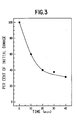
- the following example illustrates one assessment of the rate of DNA repair in blood cells in vitro.
- Samples of human blood (3 ml) were diluted 1:1 with RPMI Medium 1640 and irradiated (gamma) at 0°C to a dose of 2,58 ⁇ 10 -3 C/kg.
- the irradiated samples were analyzed as in Example 1 either immediately, or after a period of incubation at 37°C to allow repair of strand breaks in DNA to occur.
- Values for AD were determined as in Example 2, using non-irradiated blood treated in a similar manner.
- the initial AD value (immediately after irradiation) was 17.9%, i.e.
- RPMI Medium 1640 is described in Moore et al, J. Amer. Med. Assoc., Vol. 199, p. 519-524, 1967. It is available from Microbiological Associates and other suppliers of such media.
- This assay method has application in detecting DNA strand breaks induced in different cell types by other physical and chemical influences, as well as for monitoring subsequent repair. When spleen cells from irradiated mice were examined and assayed, DNA strand breaks were readily measured. Further experiments have shown that this assay readily detected strand breaks induced by chemical agents.
- AD is a measure of DNA damage, determined as in Examples 1 and 2.
Landscapes
- Life Sciences & Earth Sciences (AREA)
- Chemical & Material Sciences (AREA)
- Health & Medical Sciences (AREA)
- Engineering & Computer Science (AREA)
- Organic Chemistry (AREA)
- Immunology (AREA)
- Zoology (AREA)
- Proteomics, Peptides & Aminoacids (AREA)
- Molecular Biology (AREA)
- Wood Science & Technology (AREA)
- Biotechnology (AREA)
- Hematology (AREA)
- Microbiology (AREA)
- Analytical Chemistry (AREA)
- Physics & Mathematics (AREA)
- Biochemistry (AREA)
- General Health & Medical Sciences (AREA)
- General Engineering & Computer Science (AREA)
- Bioinformatics & Cheminformatics (AREA)
- Genetics & Genomics (AREA)
- Biophysics (AREA)
- Biomedical Technology (AREA)
- Urology & Nephrology (AREA)
- Cell Biology (AREA)
- Food Science & Technology (AREA)
- Medicinal Chemistry (AREA)
- General Physics & Mathematics (AREA)
- Pathology (AREA)
- Investigating Or Analysing Biological Materials (AREA)
- Measuring Or Testing Involving Enzymes Or Micro-Organisms (AREA)
Claims (19)
Applications Claiming Priority (2)
| Application Number | Priority Date | Filing Date | Title |
|---|---|---|---|
| US06/229,539 US4407942A (en) | 1981-01-29 | 1981-01-29 | Fluorescent detection of DNA damage |
| US229539 | 1981-01-29 |
Publications (2)
| Publication Number | Publication Date |
|---|---|
| EP0057553A1 EP0057553A1 (de) | 1982-08-11 |
| EP0057553B1 true EP0057553B1 (de) | 1985-05-08 |
Family
ID=22861670
Family Applications (1)
| Application Number | Title | Priority Date | Filing Date |
|---|---|---|---|
| EP82300376A Expired EP0057553B1 (de) | 1981-01-29 | 1982-01-26 | Verfahren und Testpackung zur Feststellung beschädigter DNA durch Fluoreszenz |
Country Status (6)
| Country | Link |
|---|---|
| US (1) | US4407942A (de) |
| EP (1) | EP0057553B1 (de) |
| JP (1) | JPS57144994A (de) |
| CA (1) | CA1161736A (de) |
| DE (1) | DE3263609D1 (de) |
| DK (1) | DK33982A (de) |
Families Citing this family (22)
| Publication number | Priority date | Publication date | Assignee | Title |
|---|---|---|---|---|
| US4906561A (en) * | 1981-09-14 | 1990-03-06 | Thornthwaite Jerry T | Nuclear isolation medium and procedure for separating cell nuclei |
| US4871661A (en) * | 1983-11-23 | 1989-10-03 | The Ohio State University Research Foundation | Process for testing the carcinogenicity of a material or the presence of cancer-inducing factors in an environment |
| US4704354A (en) * | 1984-02-15 | 1987-11-03 | University Of Cincinnati | Virion assay method for use in in vitro screening of teratogens and carcinogens |
| US4652517A (en) * | 1984-06-11 | 1987-03-24 | Diagnostic Research Limited Partnership | Methods for the in vitro detection and identification of unknown pathogens or genetic entities |
| EP0232967B1 (de) * | 1986-01-10 | 1993-04-28 | Amoco Corporation | Kompetitiver homogener Test |
| AU619170B2 (en) * | 1987-01-09 | 1992-01-23 | Abbott Laboratories | Diagnostic assays using nucleic acid probes |
| US4818685A (en) * | 1987-02-12 | 1989-04-04 | Temple University Of The Commonwealth Systems Of Higher Education | Method and kit for diagnosing bloom's syndrome |
| US5006460A (en) * | 1988-05-26 | 1991-04-09 | Pantox Corporation | Method for measuring DNA damage in single cells |
| US5359047A (en) * | 1988-09-22 | 1994-10-25 | Massachusetts Institute Of Technology | Nucleic acids encoding DNA structure-specific recognition protein and uses therefor |
| US5705334A (en) | 1988-09-22 | 1998-01-06 | Massachusetts Institute Of Technology | Uses for DNA structure-specific recognition protein |
| EP0435957A1 (de) * | 1988-09-22 | 1991-07-10 | Massachusetts Institute Of Technology | Dna-schadenbindender faktor und seine verwendung |
| FR2650839A1 (fr) * | 1989-08-08 | 1991-02-15 | Oris Ind Cie | Procede de detection en phase homogene liquide de sequences specifiques d'acides nucleiques et ses applications |
| US5369002A (en) * | 1992-04-01 | 1994-11-29 | Maruzen Petrochemical Co., Ltd. | Method of detecting injured nuclear DNA |
| EP0628817A1 (de) * | 1993-06-10 | 1994-12-14 | Nederlandse Organisatie voor toegepast-natuurwetenschappelijk onderzoek TNO | Verfahren zum Nachweis einzelsträngiger Brüche in DNS |
| US5723288A (en) * | 1994-05-06 | 1998-03-03 | The University Of North Carolina At Chapel Hill | Method of fluorescent detection of nucleic acids and cytoskeleton elements using bis-dicationic aryl furans, and kits useful therefor |
| WO1996024688A1 (en) * | 1995-02-07 | 1996-08-15 | Mosaic Technologies | Diagnostics based on dna duplex repair |
| US5702884A (en) * | 1996-03-12 | 1997-12-30 | Johnson & Johnson Clinical Diagnostics, Inc. | Whole blood sample preparation for polymerase chain reaction using ammonium chloride and a carboxylic acid or metal carboxylate for selective red blood cell lysis |
| DE19643721A1 (de) * | 1996-10-23 | 1998-05-07 | Deutsches Krebsforsch | Automatisierte Quantifizierung von DNA-Strangbrüchen in intakten Zellen |
| KR100785017B1 (ko) * | 2006-06-05 | 2007-12-12 | 삼성전자주식회사 | 혈액으로부터 핵산을 증폭하는 방법 |
| KR100790889B1 (ko) * | 2006-09-26 | 2008-01-02 | 삼성전자주식회사 | 전기 투석 장치 및 이를 이용한 전기 투석 방법 |
| US8911469B2 (en) * | 2010-03-25 | 2014-12-16 | Neocardium, Limited | Methods and apparatus for optimal remote ischemic preconditioning (ORIP) for preventing ischemia-reperfusion injuries to organs |
| SE539268C2 (sv) | 2012-04-16 | 2017-06-07 | Känsligt högeffektivt förfarande för DNA-skada |
Family Cites Families (5)
| Publication number | Priority date | Publication date | Assignee | Title |
|---|---|---|---|---|
| US3798131A (en) * | 1972-04-24 | 1974-03-19 | Pasadena Foundation For Medica | Assay for polymeric dna as a method for detecting malignancy |
| US3899297A (en) * | 1973-12-19 | 1975-08-12 | Block Engineering | Biological staining technique and mixture thereof |
| SU649751A1 (ru) * | 1977-06-14 | 1979-02-28 | Московский Ордена Ленина И Ордена Трудового Красного Знамени Государственный Университет Им.М.В.Ломоносова | Способ идентификации микроорганизмов |
| US4257774A (en) * | 1979-07-16 | 1981-03-24 | Meloy Laboratories, Inc. | Intercalation inhibition assay for compounds that interact with DNA or RNA |
| US4345027A (en) * | 1980-12-12 | 1982-08-17 | The United States Of America As Represented By The United States Department Of Energy | Fluorometric method of quantitative cell mutagenesis |
-
1981
- 1981-01-29 US US06/229,539 patent/US4407942A/en not_active Expired - Fee Related
- 1981-12-29 CA CA000393338A patent/CA1161736A/en not_active Expired
-
1982
- 1982-01-26 DE DE8282300376T patent/DE3263609D1/de not_active Expired
- 1982-01-26 EP EP82300376A patent/EP0057553B1/de not_active Expired
- 1982-01-26 DK DK33982A patent/DK33982A/da not_active Application Discontinuation
- 1982-01-28 JP JP57012500A patent/JPS57144994A/ja active Granted
Also Published As
| Publication number | Publication date |
|---|---|
| JPS57144994A (en) | 1982-09-07 |
| JPS6137920B2 (de) | 1986-08-26 |
| EP0057553A1 (de) | 1982-08-11 |
| DK33982A (da) | 1982-07-30 |
| US4407942A (en) | 1983-10-04 |
| DE3263609D1 (en) | 1985-06-13 |
| CA1161736A (en) | 1984-02-07 |
Similar Documents
| Publication | Publication Date | Title |
|---|---|---|
| EP0057553B1 (de) | Verfahren und Testpackung zur Feststellung beschädigter DNA durch Fluoreszenz | |
| Birnboim et al. | Fluorometric method for rapid detection of DNA strand breaks in human white blood cells produced by low doses of radiation | |
| Collins | The comet assay for DNA damage and repair: principles, applications, and limitations | |
| Olive et al. | Detection of etoposide resistance by measuring DNA damage in individual Chinese hamster cells | |
| Collins et al. | Direct enzymic detection of endogenous oxidative base damage in human lymphocyte DNA | |
| BR9712548A (pt) | Processo para identificar uma célula eritrócita ou eritroblástica fetal em uma amostra sanguìnea, kit para identificação de uma célula eritrócita ou eritroblástica nucleada fetal, e, kit para seleção ou isolamento de uma sequência de ácida nucléico dentro de uma célula eritrócita ou eritroblástica nucleada fetal. | |
| WO1989010566A1 (en) | Process for forming and using microdroplets | |
| EP1177315B1 (de) | Zell-assay, -verfahren und -reagenzien | |
| Briggs | An analysis of the inactivation of the frog sperm nucleus by toluidine blue | |
| FI77689B (fi) | Sensitiva tester baserade pao konstaterandet av dna foer paovisande av malignitet. | |
| EP1725873B1 (de) | Pharmakodynamische tests mit durchflusszytometrie | |
| CN111189761A (zh) | 基于质谱流式技术的细胞信号通路检测试剂盒及检测方法 | |
| US20040132037A1 (en) | Materials and methods for the induction of premature chromosone condensation | |
| Wada et al. | Detection of radiation-induced apoptosis using the comet assay | |
| Rudd et al. | Micronucleus assay in human fibroblasts: a measure of spontaneous chromosomal instability and mutagen hypersensitivity | |
| EP4112739A1 (de) | Verfahren zum nachweis von dna-ende(n) und verwendung davon | |
| KR101138343B1 (ko) | 알츠하이머 질병의 진단을 위한 신속한 시험법 | |
| Alves et al. | Application of the chemiluminescence systems to evaluate the role of Fcγ and complement receptors in stimulating the oxidative burst in neutrophils | |
| Reed et al. | Sample suitability for the detection of minor white cell populations (microchimerism) by polymerase chain reaction | |
| EP0711356A1 (de) | Nachweis von in dns eingebauten halogenierten vorläufern | |
| Sawazaki et al. | Deoxyribonuclease I (DNase I) typing from semen stains: low enzyme activity in vaginal fluids does not interfere with seminal DNase I typing from mixture stains | |
| US20230250465A1 (en) | Methods for detecting the presence of sepsis | |
| O'Melia et al. | Animalizing ability of evans blue in embryos of Arbacia punctulata: Effect on ribosomal RNA synthesis | |
| WO2025104680A1 (en) | Dna accessibility for the assessment of male fertility | |
| US20030017493A1 (en) | Methods for the detection and treatment of chronic immune diseases |
Legal Events
| Date | Code | Title | Description |
|---|---|---|---|
| PUAI | Public reference made under article 153(3) epc to a published international application that has entered the european phase |
Free format text: ORIGINAL CODE: 0009012 |
|
| AK | Designated contracting states |
Designated state(s): BE CH DE FR GB IT NL SE |
|
| 17P | Request for examination filed |
Effective date: 19830127 |
|
| ITF | It: translation for a ep patent filed | ||
| GRAA | (expected) grant |
Free format text: ORIGINAL CODE: 0009210 |
|
| AK | Designated contracting states |
Designated state(s): BE CH DE FR GB IT LI NL SE |
|
| REF | Corresponds to: |
Ref document number: 3263609 Country of ref document: DE Date of ref document: 19850613 |
|
| ET | Fr: translation filed | ||
| PGFP | Annual fee paid to national office [announced via postgrant information from national office to epo] |
Ref country code: NL Payment date: 19860131 Year of fee payment: 5 |
|
| PLBE | No opposition filed within time limit |
Free format text: ORIGINAL CODE: 0009261 |
|
| STAA | Information on the status of an ep patent application or granted ep patent |
Free format text: STATUS: NO OPPOSITION FILED WITHIN TIME LIMIT |
|
| 26N | No opposition filed | ||
| PG25 | Lapsed in a contracting state [announced via postgrant information from national office to epo] |
Ref country code: NL Effective date: 19870801 |
|
| NLV4 | Nl: lapsed or anulled due to non-payment of the annual fee | ||
| ITTA | It: last paid annual fee | ||
| PGFP | Annual fee paid to national office [announced via postgrant information from national office to epo] |
Ref country code: FR Payment date: 19920929 Year of fee payment: 12 |
|
| PGFP | Annual fee paid to national office [announced via postgrant information from national office to epo] |
Ref country code: BE Payment date: 19921113 Year of fee payment: 12 |
|
| PGFP | Annual fee paid to national office [announced via postgrant information from national office to epo] |
Ref country code: CH Payment date: 19921201 Year of fee payment: 12 |
|
| PGFP | Annual fee paid to national office [announced via postgrant information from national office to epo] |
Ref country code: SE Payment date: 19930115 Year of fee payment: 12 |
|
| PGFP | Annual fee paid to national office [announced via postgrant information from national office to epo] |
Ref country code: GB Payment date: 19930125 Year of fee payment: 12 |
|
| PGFP | Annual fee paid to national office [announced via postgrant information from national office to epo] |
Ref country code: DE Payment date: 19930223 Year of fee payment: 12 |
|
| PG25 | Lapsed in a contracting state [announced via postgrant information from national office to epo] |
Ref country code: GB Effective date: 19940126 |
|
| PG25 | Lapsed in a contracting state [announced via postgrant information from national office to epo] |
Ref country code: SE Effective date: 19940127 |
|
| PG25 | Lapsed in a contracting state [announced via postgrant information from national office to epo] |
Ref country code: CH Effective date: 19940131 Ref country code: LI Effective date: 19940131 Ref country code: BE Effective date: 19940131 |
|
| BERE | Be: lapsed |
Owner name: ATOMIC ENERGY OF CANADA LTD Effective date: 19940131 |
|
| GBPC | Gb: european patent ceased through non-payment of renewal fee |
Effective date: 19940126 |
|
| PG25 | Lapsed in a contracting state [announced via postgrant information from national office to epo] |
Ref country code: FR Effective date: 19940930 |
|
| REG | Reference to a national code |
Ref country code: CH Ref legal event code: PL |
|
| PG25 | Lapsed in a contracting state [announced via postgrant information from national office to epo] |
Ref country code: DE Effective date: 19941001 |
|
| REG | Reference to a national code |
Ref country code: FR Ref legal event code: ST |
|
| EUG | Se: european patent has lapsed |
Ref document number: 82300376.9 Effective date: 19940810 |