BR112013018117B1 - medical device - Google Patents
medical device Download PDFInfo
- Publication number
- BR112013018117B1 BR112013018117B1 BR112013018117-6A BR112013018117A BR112013018117B1 BR 112013018117 B1 BR112013018117 B1 BR 112013018117B1 BR 112013018117 A BR112013018117 A BR 112013018117A BR 112013018117 B1 BR112013018117 B1 BR 112013018117B1
- Authority
- BR
- Brazil
- Prior art keywords
- block stent
- catheter
- medical device
- stent
- hollow metal
- Prior art date
Links
Images


Classifications
-
- A—HUMAN NECESSITIES
- A61—MEDICAL OR VETERINARY SCIENCE; HYGIENE
- A61B—DIAGNOSIS; SURGERY; IDENTIFICATION
- A61B17/00—Surgical instruments, devices or methods, e.g. tourniquets
- A61B17/12—Surgical instruments, devices or methods, e.g. tourniquets for ligaturing or otherwise compressing tubular parts of the body, e.g. blood vessels, umbilical cord
- A61B17/12022—Occluding by internal devices, e.g. balloons or releasable wires
- A61B17/12027—Type of occlusion
- A61B17/12031—Type of occlusion complete occlusion
-
- A—HUMAN NECESSITIES
- A61—MEDICAL OR VETERINARY SCIENCE; HYGIENE
- A61B—DIAGNOSIS; SURGERY; IDENTIFICATION
- A61B17/00—Surgical instruments, devices or methods, e.g. tourniquets
- A61B17/12—Surgical instruments, devices or methods, e.g. tourniquets for ligaturing or otherwise compressing tubular parts of the body, e.g. blood vessels, umbilical cord
- A61B17/12022—Occluding by internal devices, e.g. balloons or releasable wires
- A61B17/12099—Occluding by internal devices, e.g. balloons or releasable wires characterised by the location of the occluder
- A61B17/12109—Occluding by internal devices, e.g. balloons or releasable wires characterised by the location of the occluder in a blood vessel
- A61B17/12113—Occluding by internal devices, e.g. balloons or releasable wires characterised by the location of the occluder in a blood vessel within an aneurysm
-
- A—HUMAN NECESSITIES
- A61—MEDICAL OR VETERINARY SCIENCE; HYGIENE
- A61B—DIAGNOSIS; SURGERY; IDENTIFICATION
- A61B17/00—Surgical instruments, devices or methods, e.g. tourniquets
- A61B17/12—Surgical instruments, devices or methods, e.g. tourniquets for ligaturing or otherwise compressing tubular parts of the body, e.g. blood vessels, umbilical cord
- A61B17/12022—Occluding by internal devices, e.g. balloons or releasable wires
- A61B17/12131—Occluding by internal devices, e.g. balloons or releasable wires characterised by the type of occluding device
- A61B17/12136—Balloons
-
- A—HUMAN NECESSITIES
- A61—MEDICAL OR VETERINARY SCIENCE; HYGIENE
- A61B—DIAGNOSIS; SURGERY; IDENTIFICATION
- A61B17/00—Surgical instruments, devices or methods, e.g. tourniquets
- A61B17/12—Surgical instruments, devices or methods, e.g. tourniquets for ligaturing or otherwise compressing tubular parts of the body, e.g. blood vessels, umbilical cord
- A61B17/12022—Occluding by internal devices, e.g. balloons or releasable wires
- A61B17/12131—Occluding by internal devices, e.g. balloons or releasable wires characterised by the type of occluding device
- A61B17/12168—Occluding by internal devices, e.g. balloons or releasable wires characterised by the type of occluding device having a mesh structure
- A61B17/12172—Occluding by internal devices, e.g. balloons or releasable wires characterised by the type of occluding device having a mesh structure having a pre-set deployed three-dimensional shape
-
- A—HUMAN NECESSITIES
- A61—MEDICAL OR VETERINARY SCIENCE; HYGIENE
- A61F—FILTERS IMPLANTABLE INTO BLOOD VESSELS; PROSTHESES; DEVICES PROVIDING PATENCY TO, OR PREVENTING COLLAPSING OF, TUBULAR STRUCTURES OF THE BODY, e.g. STENTS; ORTHOPAEDIC, NURSING OR CONTRACEPTIVE DEVICES; FOMENTATION; TREATMENT OR PROTECTION OF EYES OR EARS; BANDAGES, DRESSINGS OR ABSORBENT PADS; FIRST-AID KITS
- A61F2/00—Filters implantable into blood vessels; Prostheses, i.e. artificial substitutes or replacements for parts of the body; Appliances for connecting them with the body; Devices providing patency to, or preventing collapsing of, tubular structures of the body, e.g. stents
- A61F2/02—Prostheses implantable into the body
- A61F2/04—Hollow or tubular parts of organs, e.g. bladders, tracheae, bronchi or bile ducts
- A61F2/06—Blood vessels
-
- A—HUMAN NECESSITIES
- A61—MEDICAL OR VETERINARY SCIENCE; HYGIENE
- A61F—FILTERS IMPLANTABLE INTO BLOOD VESSELS; PROSTHESES; DEVICES PROVIDING PATENCY TO, OR PREVENTING COLLAPSING OF, TUBULAR STRUCTURES OF THE BODY, e.g. STENTS; ORTHOPAEDIC, NURSING OR CONTRACEPTIVE DEVICES; FOMENTATION; TREATMENT OR PROTECTION OF EYES OR EARS; BANDAGES, DRESSINGS OR ABSORBENT PADS; FIRST-AID KITS
- A61F2/00—Filters implantable into blood vessels; Prostheses, i.e. artificial substitutes or replacements for parts of the body; Appliances for connecting them with the body; Devices providing patency to, or preventing collapsing of, tubular structures of the body, e.g. stents
- A61F2/95—Instruments specially adapted for placement or removal of stents or stent-grafts
- A61F2/958—Inflatable balloons for placing stents or stent-grafts
-
- A—HUMAN NECESSITIES
- A61—MEDICAL OR VETERINARY SCIENCE; HYGIENE
- A61M—DEVICES FOR INTRODUCING MEDIA INTO, OR ONTO, THE BODY; DEVICES FOR TRANSDUCING BODY MEDIA OR FOR TAKING MEDIA FROM THE BODY; DEVICES FOR PRODUCING OR ENDING SLEEP OR STUPOR
- A61M25/00—Catheters; Hollow probes
- A61M25/10—Balloon catheters
- A61M25/1027—Making of balloon catheters
- A61M25/1029—Production methods of the balloon members, e.g. blow-moulding, extruding, deposition or by wrapping a plurality of layers of balloon material around a mandril
-
- A—HUMAN NECESSITIES
- A61—MEDICAL OR VETERINARY SCIENCE; HYGIENE
- A61M—DEVICES FOR INTRODUCING MEDIA INTO, OR ONTO, THE BODY; DEVICES FOR TRANSDUCING BODY MEDIA OR FOR TAKING MEDIA FROM THE BODY; DEVICES FOR PRODUCING OR ENDING SLEEP OR STUPOR
- A61M31/00—Devices for introducing or retaining media, e.g. remedies, in cavities of the body
-
- A—HUMAN NECESSITIES
- A61—MEDICAL OR VETERINARY SCIENCE; HYGIENE
- A61B—DIAGNOSIS; SURGERY; IDENTIFICATION
- A61B17/00—Surgical instruments, devices or methods, e.g. tourniquets
- A61B17/12—Surgical instruments, devices or methods, e.g. tourniquets for ligaturing or otherwise compressing tubular parts of the body, e.g. blood vessels, umbilical cord
- A61B17/12022—Occluding by internal devices, e.g. balloons or releasable wires
- A61B17/12131—Occluding by internal devices, e.g. balloons or releasable wires characterised by the type of occluding device
- A61B17/1214—Coils or wires
-
- A—HUMAN NECESSITIES
- A61—MEDICAL OR VETERINARY SCIENCE; HYGIENE
- A61B—DIAGNOSIS; SURGERY; IDENTIFICATION
- A61B17/00—Surgical instruments, devices or methods, e.g. tourniquets
- A61B17/12—Surgical instruments, devices or methods, e.g. tourniquets for ligaturing or otherwise compressing tubular parts of the body, e.g. blood vessels, umbilical cord
- A61B17/12022—Occluding by internal devices, e.g. balloons or releasable wires
- A61B17/12131—Occluding by internal devices, e.g. balloons or releasable wires characterised by the type of occluding device
- A61B17/12168—Occluding by internal devices, e.g. balloons or releasable wires characterised by the type of occluding device having a mesh structure
- A61B17/12177—Occluding by internal devices, e.g. balloons or releasable wires characterised by the type of occluding device having a mesh structure comprising additional materials, e.g. thrombogenic, having filaments, having fibers or being coated
-
- A—HUMAN NECESSITIES
- A61—MEDICAL OR VETERINARY SCIENCE; HYGIENE
- A61B—DIAGNOSIS; SURGERY; IDENTIFICATION
- A61B17/00—Surgical instruments, devices or methods, e.g. tourniquets
- A61B17/12—Surgical instruments, devices or methods, e.g. tourniquets for ligaturing or otherwise compressing tubular parts of the body, e.g. blood vessels, umbilical cord
- A61B17/12022—Occluding by internal devices, e.g. balloons or releasable wires
- A61B17/12131—Occluding by internal devices, e.g. balloons or releasable wires characterised by the type of occluding device
- A61B17/12181—Occluding by internal devices, e.g. balloons or releasable wires characterised by the type of occluding device formed by fluidized, gelatinous or cellular remodelable materials, e.g. embolic liquids, foams or extracellular matrices
-
- A—HUMAN NECESSITIES
- A61—MEDICAL OR VETERINARY SCIENCE; HYGIENE
- A61B—DIAGNOSIS; SURGERY; IDENTIFICATION
- A61B17/00—Surgical instruments, devices or methods, e.g. tourniquets
- A61B2017/00004—(bio)absorbable, (bio)resorbable, resorptive
-
- A—HUMAN NECESSITIES
- A61—MEDICAL OR VETERINARY SCIENCE; HYGIENE
- A61B—DIAGNOSIS; SURGERY; IDENTIFICATION
- A61B17/00—Surgical instruments, devices or methods, e.g. tourniquets
- A61B2017/00526—Methods of manufacturing
-
- A—HUMAN NECESSITIES
- A61—MEDICAL OR VETERINARY SCIENCE; HYGIENE
- A61B—DIAGNOSIS; SURGERY; IDENTIFICATION
- A61B17/00—Surgical instruments, devices or methods, e.g. tourniquets
- A61B2017/00831—Material properties
- A61B2017/0084—Material properties low friction
- A61B2017/00849—Material properties low friction with respect to tissue, e.g. hollow organs
-
- A—HUMAN NECESSITIES
- A61—MEDICAL OR VETERINARY SCIENCE; HYGIENE
- A61B—DIAGNOSIS; SURGERY; IDENTIFICATION
- A61B17/00—Surgical instruments, devices or methods, e.g. tourniquets
- A61B2017/00831—Material properties
- A61B2017/00867—Material properties shape memory effect
-
- A—HUMAN NECESSITIES
- A61—MEDICAL OR VETERINARY SCIENCE; HYGIENE
- A61B—DIAGNOSIS; SURGERY; IDENTIFICATION
- A61B17/00—Surgical instruments, devices or methods, e.g. tourniquets
- A61B2017/00831—Material properties
- A61B2017/00884—Material properties enhancing wound closure
-
- A—HUMAN NECESSITIES
- A61—MEDICAL OR VETERINARY SCIENCE; HYGIENE
- A61B—DIAGNOSIS; SURGERY; IDENTIFICATION
- A61B17/00—Surgical instruments, devices or methods, e.g. tourniquets
- A61B2017/00831—Material properties
- A61B2017/00893—Material properties pharmaceutically effective
-
- A—HUMAN NECESSITIES
- A61—MEDICAL OR VETERINARY SCIENCE; HYGIENE
- A61B—DIAGNOSIS; SURGERY; IDENTIFICATION
- A61B17/00—Surgical instruments, devices or methods, e.g. tourniquets
- A61B17/12—Surgical instruments, devices or methods, e.g. tourniquets for ligaturing or otherwise compressing tubular parts of the body, e.g. blood vessels, umbilical cord
- A61B17/12022—Occluding by internal devices, e.g. balloons or releasable wires
- A61B2017/1205—Introduction devices
- A61B2017/12054—Details concerning the detachment of the occluding device from the introduction device
- A61B2017/12059—Joint of soluble material
-
- A—HUMAN NECESSITIES
- A61—MEDICAL OR VETERINARY SCIENCE; HYGIENE
- A61B—DIAGNOSIS; SURGERY; IDENTIFICATION
- A61B17/00—Surgical instruments, devices or methods, e.g. tourniquets
- A61B17/12—Surgical instruments, devices or methods, e.g. tourniquets for ligaturing or otherwise compressing tubular parts of the body, e.g. blood vessels, umbilical cord
- A61B17/12022—Occluding by internal devices, e.g. balloons or releasable wires
- A61B2017/1205—Introduction devices
- A61B2017/12054—Details concerning the detachment of the occluding device from the introduction device
- A61B2017/12063—Details concerning the detachment of the occluding device from the introduction device electrolytically detachable
-
- A—HUMAN NECESSITIES
- A61—MEDICAL OR VETERINARY SCIENCE; HYGIENE
- A61B—DIAGNOSIS; SURGERY; IDENTIFICATION
- A61B17/00—Surgical instruments, devices or methods, e.g. tourniquets
- A61B17/12—Surgical instruments, devices or methods, e.g. tourniquets for ligaturing or otherwise compressing tubular parts of the body, e.g. blood vessels, umbilical cord
- A61B17/12022—Occluding by internal devices, e.g. balloons or releasable wires
- A61B2017/1205—Introduction devices
- A61B2017/12054—Details concerning the detachment of the occluding device from the introduction device
- A61B2017/12068—Details concerning the detachment of the occluding device from the introduction device detachable by heat
- A61B2017/12077—Joint changing shape upon application of heat, e.g. bi-metal or reversible thermal memory
-
- A—HUMAN NECESSITIES
- A61—MEDICAL OR VETERINARY SCIENCE; HYGIENE
- A61B—DIAGNOSIS; SURGERY; IDENTIFICATION
- A61B17/00—Surgical instruments, devices or methods, e.g. tourniquets
- A61B17/22—Implements for squeezing-off ulcers or the like on the inside of inner organs of the body; Implements for scraping-out cavities of body organs, e.g. bones; Calculus removers; Calculus smashing apparatus; Apparatus for removing obstructions in blood vessels, not otherwise provided for
- A61B2017/22038—Implements for squeezing-off ulcers or the like on the inside of inner organs of the body; Implements for scraping-out cavities of body organs, e.g. bones; Calculus removers; Calculus smashing apparatus; Apparatus for removing obstructions in blood vessels, not otherwise provided for with a guide wire
-
- A—HUMAN NECESSITIES
- A61—MEDICAL OR VETERINARY SCIENCE; HYGIENE
- A61B—DIAGNOSIS; SURGERY; IDENTIFICATION
- A61B90/00—Instruments, implements or accessories specially adapted for surgery or diagnosis and not covered by any of the groups A61B1/00 - A61B50/00, e.g. for luxation treatment or for protecting wound edges
- A61B90/03—Automatic limiting or abutting means, e.g. for safety
- A61B2090/037—Automatic limiting or abutting means, e.g. for safety with a frangible part, e.g. by reduced diameter
-
- A—HUMAN NECESSITIES
- A61—MEDICAL OR VETERINARY SCIENCE; HYGIENE
- A61B—DIAGNOSIS; SURGERY; IDENTIFICATION
- A61B90/00—Instruments, implements or accessories specially adapted for surgery or diagnosis and not covered by any of the groups A61B1/00 - A61B50/00, e.g. for luxation treatment or for protecting wound edges
- A61B90/39—Markers, e.g. radio-opaque or breast lesions markers
- A61B2090/3966—Radiopaque markers visible in an X-ray image
-
- A—HUMAN NECESSITIES
- A61—MEDICAL OR VETERINARY SCIENCE; HYGIENE
- A61F—FILTERS IMPLANTABLE INTO BLOOD VESSELS; PROSTHESES; DEVICES PROVIDING PATENCY TO, OR PREVENTING COLLAPSING OF, TUBULAR STRUCTURES OF THE BODY, e.g. STENTS; ORTHOPAEDIC, NURSING OR CONTRACEPTIVE DEVICES; FOMENTATION; TREATMENT OR PROTECTION OF EYES OR EARS; BANDAGES, DRESSINGS OR ABSORBENT PADS; FIRST-AID KITS
- A61F2250/00—Special features of prostheses classified in groups A61F2/00 - A61F2/26 or A61F2/82 or A61F9/00 or A61F11/00 or subgroups thereof
- A61F2250/0058—Additional features; Implant or prostheses properties not otherwise provided for
- A61F2250/0096—Markers and sensors for detecting a position or changes of a position of an implant, e.g. RF sensors, ultrasound markers
- A61F2250/0098—Markers and sensors for detecting a position or changes of a position of an implant, e.g. RF sensors, ultrasound markers radio-opaque, e.g. radio-opaque markers
-
- A—HUMAN NECESSITIES
- A61—MEDICAL OR VETERINARY SCIENCE; HYGIENE
- A61M—DEVICES FOR INTRODUCING MEDIA INTO, OR ONTO, THE BODY; DEVICES FOR TRANSDUCING BODY MEDIA OR FOR TAKING MEDIA FROM THE BODY; DEVICES FOR PRODUCING OR ENDING SLEEP OR STUPOR
- A61M2210/00—Anatomical parts of the body
- A61M2210/12—Blood circulatory system
-
- Y—GENERAL TAGGING OF NEW TECHNOLOGICAL DEVELOPMENTS; GENERAL TAGGING OF CROSS-SECTIONAL TECHNOLOGIES SPANNING OVER SEVERAL SECTIONS OF THE IPC; TECHNICAL SUBJECTS COVERED BY FORMER USPC CROSS-REFERENCE ART COLLECTIONS [XRACs] AND DIGESTS
- Y10—TECHNICAL SUBJECTS COVERED BY FORMER USPC
- Y10T—TECHNICAL SUBJECTS COVERED BY FORMER US CLASSIFICATION
- Y10T29/00—Metal working
- Y10T29/49—Method of mechanical manufacture
- Y10T29/49826—Assembling or joining
Abstract
DISPOSITIVO MÉDICO. A presente invenção refere-se a um dispositivo médico que compreende uma estrutura de metal expandida de paredes finas, oblonga ou cilíndrica, comprimida (um "stent de bloqueio") e um dispositivo alongado flexível (um "cateter de distribuição") para posicionar o stent de bloqueio comprimido no lúmen de um segmento de vaso sanguíneo a ser tratado e métodos de uso para oclusão de segmentos de vasos sanguíneos tratados. Um stent de bloqueio pode ser feito com metais dúcteis, tais como ouro, platina ou prata, de tal modo que o stent de bloqueio se conformará à forma do lúmen do segmento de vaso sanguíneo tratado durante a expansão e permitirá que a forma do stent de bloqueio seja permanentemente alterada pela aplicação de uma força externa. A superfície do stent de bloqueio pode ser configurada para promover trombo local na superfície externa do stent de bloqueio e para promover o crescimento do tecido na parede do stent de bloqueio a fim de ocluir o vaso sanguíneo tratado e fixar o stent de bloqueio no lugar. A parede do stent de bloqueio também pode ser configurada para liberar fármacos ou moléculas farmacologicamente ativas, tais como aquelas que promovem trombose, proliferação celular, deposição (...).MEDICAL DEVICE. The present invention relates to a medical device comprising a thin, expanded, oblong or cylindrical, compressed metal structure (a "locking stent") and a flexible elongated device (a "delivery catheter") for positioning the blocking stent compressed into the lumen of a blood vessel segment to be treated and methods of use for occlusion of treated blood vessel segments. A blocking stent can be made with ductile metals, such as gold, platinum or silver, such that the blocking stent will conform to the lumen shape of the treated blood vessel segment during expansion and will allow the shape of the blocking is permanently altered by the application of an external force. The surface of the blocking stent can be configured to promote local thrombus on the external surface of the blocking stent and to promote tissue growth on the wall of the blocking stent in order to occlude the treated blood vessel and fix the blocking stent in place. The blocking stent wall can also be configured to release drugs or pharmacologically active molecules, such as those that promote thrombosis, cell proliferation, deposition (...).
Description
[001] Este pedido reivindica prioridade do Pedido Provisório US 61/433.305 depositado em 17 de janeiro de 2011, que é incorporado por referência na sua totalidade.[001] This order claims priority for Provisional Order US 61 / 433,305 filed on January 17, 2011, which is incorporated by reference in its entirety.
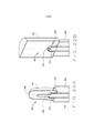
[002] A presente invenção refere-se a um dispositivo médico que compreende um stent em bloco (blockstent) e um cateter de distribuição para o tratamento de segmentos de vasos sanguíneos do sistema vascular. A presente invenção também se refere a várias formas de stents em bloco e cateteres de distribuição e métodos para a sua fabricação. A presente invenção também se refere a métodos de oclusão de segmentos de vasos sanguíneos usando vários dispositivos médicos, em que o stent em bloco permanece em último caso, no segmento do vaso sanguíneo. Stents em bloco são estruturas metálicas expansíveis, de paredes finas, cilíndricas constituídas por um dispositivo do tipo de stent e projetadas para encher o lúmen de um segmento de vaso sanguíneo. Os stents em bloco são configurados para: fixação aos cateteres de distribuição, compressão, avanço através do sistema vascular, expansão dentro do lúmen de segmentos de vasos sanguíneos, e então a separação a partir de cateteres de distribuição. Os cateteres de distribuição de vários tamanhos, formas, materiais e configurações podem ser usados para posicionar um stent em bloco comprimido em um segmento de vaso sanguíneo e expandir o stent em bloco no vaso sanguíneo pela passagem de fluidos ou sólidos através do cateter de distribuição e no interior do espaço ou espaço vazio central do stent em bloco. Além disso, a invenção refere-se aos componentes para, e métodos de, fixação do stent em bloco ao cateter de distribuição, assim como os componentes para, e métodos de, separação do stent em bloco expandido a partir do cateter de distribuição, tal que o stent em bloco permanece no lugar em um estado expandido dentro do vaso sanguíneo, enquanto o cateter de distribuição é removido do corpo.[002] The present invention relates to a medical device comprising a block stent and a delivery catheter for the treatment of blood vessel segments of the vascular system. The present invention also relates to various forms of block stents and delivery catheters and methods for their manufacture. The present invention also relates to methods of occlusion of segments of blood vessels using various medical devices, where the block stent remains ultimately in the segment of the blood vessel. Block stents are expandable, thin-walled, cylindrical metal structures made up of a stent-type device designed to fill the lumen of a blood vessel segment. Block stents are configured for: attachment to delivery catheters, compression, advancement through the vascular system, expansion within the lumen of blood vessel segments, and then separation from delivery catheters. Delivery catheters of various sizes, shapes, materials and configurations can be used to position a compressed block stent in a blood vessel segment and expand the block stent in the blood vessel by passing fluids or solids through the distribution catheter and inside the central empty space or space of the block stent. In addition, the invention relates to components for, and methods of, fixation of the block stent to the delivery catheter, as well as components to, and methods of, separation of the expanded block stent from the delivery catheter, such as that the block stent remains in place in an expanded state within the blood vessel, while the delivery catheter is removed from the body.
[003] Em certas situações clínicas, os pacientes podem se beneficiar da oclusão de certos segmentos de artérias ou veias através de meios endovasculares. Cenários clínicos onde a oclusão do vaso endovascular é benéfica incluem a redução de hemorragias a partir de um vaso danificado, redução do fluxo de sangue aos tumores, e re- encaminhamento da via de sangue no sistema vascular para outros fins.[003] In certain clinical situations, patients can benefit from the occlusion of certain segments of arteries or veins through endovascular means. Clinical scenarios where endovascular vessel occlusion is beneficial include reducing bleeding from a damaged vessel, reducing blood flow to tumors, and redirecting the blood pathway in the vascular system for other purposes.
[004] Alternativamente, os tratamentos endovasculares, baseados em cateter minimamente invasivos têm sido desenvolvidos para ocluir os segmentos dos vasos sanguíneos. Os dispositivos médicos endovasculares para a oclusão de vasos sanguíneos incluem cateteres de balão, em que o balão pode ser insuflado para encher o lúmen de um segmento de vaso sanguíneo e destacado do cateter. Existem duas grandes desvantagens para o uso de cateteres de balão destacáveis para a oclusão do vaso sanguíneo. Em primeiro lugar, os balões são feitos de polímeros que geralmente resistem à incorporação tecidual que limita a fixação dos dispositivos onde estão colocados. Em segundo lugar, os balões são configurados com paredes elásticas que são expandidos com a pressurização e as válvulas projetadas para manter a pressão após o desprendimento. Infelizmente, existe uma taxa substancial de falha da válvula e do balão, o que resulta no esvaziamento. Sem a incorporação tecidual, o esvaziamento do balão pode levar a migração do balão e a oclusão de segmentos de vasos não alvos. Os dispositivos médicos endovasculares para a oclusão de vasos sanguíneos incluem espirais de metal que são utilizadas para preencher uma porção do lúmen de um segmento de vaso sanguíneo para induzir a trombose e oclusão do segmento de vaso sanguíneo. Existem várias desvantagens principais para o uso de espirais de metal e estruturas de cesto para a oclusão do vaso sanguíneo. Em primeiro lugar, várias espirais são geralmente necessárias para ocluir o segmento de vaso sanguíneo, resultando em custos mais elevados e tempos de tratamento mais longos. Em segundo lugar, a colocação da bobina é difícil de controlar, muitas vezes resultando na colocação da bobina em segmentos de vasos não alvos. Em terceiro lugar, as espirais somente preenchem parcialmente o vaso sanguíneo. A acumulação de tecido cicatricial e trombo é necessária para a oclusão do vaso sanguíneo, um processo que leva semanas para ocorrer e é, por vezes, incompleto, muitas vezes resultando em oclusão ou incompleta ou recanalização e um tratamento falho. Mais recentemente, os dispositivos médicos endovasculares para a oclusão do vaso sanguíneo foram desenvolvidos os quais incluem estruturas de cesto que são usadas para preencher uma parte do lúmen de um segmento de vaso sanguíneo para induzir a trombose e oclusão do segmento de vaso sanguíneo. Apesar de apenas uma única estrutura de cesta ser geralmente necessária para ocluir um segmento de vaso sanguíneo, e os dispositivos serem geralmente mais fáceis de controlar, estes dispositivos só preenchem parcialmente o vaso sanguíneo e requerem a acumulação de tecido cicatricial e trombo para ocluir o vaso sanguíneo. Tal como acontece com as espirais, este processo leva semanas a ocorrer e é, por vezes, incompleto, muitas vezes resultando na oclusão ou recanalização incompleta e um tratamento falho.[004] Alternatively, endovascular treatments, based on minimally invasive catheters have been developed to occlude the segments of the blood vessels. Endovascular medical devices for blood vessel occlusion include balloon catheters, in which the balloon can be inflated to fill the lumen of a segment of the blood vessel and detached from the catheter. There are two major disadvantages to using detachable balloon catheters for blood vessel occlusion. First, the balloons are made of polymers that generally resist tissue incorporation that limits the fixation of the devices where they are placed. Second, the balloons are configured with elastic walls that are expanded with pressurization and valves designed to maintain pressure after detachment. Unfortunately, there is a substantial rate of valve and balloon failure, which results in deflation. Without tissue incorporation, balloon deflation can lead to balloon migration and occlusion of non-target vessel segments. Endovascular medical devices for blood vessel occlusion include metal spirals that are used to fill a portion of the lumen of a blood vessel segment to induce thrombosis and occlusion of the blood vessel segment. There are several major disadvantages to using metal spirals and basket structures for blood vessel occlusion. First, several coils are usually required to occlude the blood vessel segment, resulting in higher costs and longer treatment times. Second, the placement of the coil is difficult to control, often resulting in the placement of the coil in non-target vessel segments. Third, the spirals only partially fill the blood vessel. The accumulation of scar tissue and thrombus is necessary for blood vessel occlusion, a process that takes weeks to occur and is sometimes incomplete, often resulting in occlusion or incomplete or recanalization and a failed treatment. More recently, endovascular medical devices for blood vessel occlusion have been developed which include basket structures that are used to fill a portion of the lumen of a blood vessel segment to induce thrombosis and occlusion of the blood vessel segment. Although only a single basket structure is generally needed to occlude a segment of the blood vessel, and the devices are generally easier to control, these devices only partially fill the blood vessel and require the accumulation of scar tissue and thrombus to occlude the vessel blood. As with coils, this process takes weeks to occur and is sometimes incomplete, often resulting in incomplete occlusion or recanalization and failed treatment.
[005] Por conseguinte, continua a haver uma necessidade de dispositivos médicos baseados em cateter, sistemas, e métodos para a oclusão de segmentos de vasos sanguíneos, que são simples de executar, resultam em oclusão rápida, controlada e completa, tenham um baixo risco de recanalização, migração do dispositivo, ou outras complicações, e possam ser adquiridos a um custo razoável.[005] Therefore, there remains a need for catheter-based medical devices, systems, and methods for occlusion of blood vessel segments, which are simple to perform, result in rapid, controlled and complete occlusion, have a low risk recanalization, device migration, or other complications, and can be purchased at a reasonable cost.
[006] A presente invenção refere-se a um dispositivo médico para a oclusão, ou o bloqueio, de segmentos de vasos sanguíneos - incluindo artérias e veias, e outros condutos vasculares. Os dispositivos médicos compreendem um stent em bloco, um cateter de distribuição para distribuir e expandir o stent em bloco, e um componente para separar o stent em bloco expandido e o cateter de distribuição. A invenção diz ainda respeito a um stent em bloco expandido deixado no lúmen de um segmento de vaso sanguíneo. Além disso, a invenção inclui várias formas de stents em bloco, cateteres de distribuição, e componentes para a separação. Além disso, a invenção inclui sistemas e métodos relacionados com o uso dos dispositivos médicos, bem como kits que compreendem os dispositivos médicos e instruções de uso. A invenção também inclui métodos de fabricação de stents em bloco, cateteres de distribuição, e componentes para a separação.[006] The present invention relates to a medical device for the occlusion, or blockage, of segments of blood vessels - including arteries and veins, and other vascular conduits. Medical devices comprise a block stent, a delivery catheter for delivering and expanding the block stent, and a component for separating the expanded block stent and the delivery catheter. The invention further relates to an expanded block stent left in the lumen of a blood vessel segment. In addition, the invention includes various forms of block stents, delivery catheters, and components for separation. In addition, the invention includes systems and methods related to the use of medical devices, as well as kits comprising medical devices and instructions for use. The invention also includes methods of making block stents, delivery catheters, and components for separation.
[007] As paredes dos stents em bloco podem ser formadas a partir de uma variedade de materiais rígidos, expansíveis, preferencialmente, metais. O metal usado para fazer a parede de um stent em bloco pode ser selecionado a partir do grupo consistindo em ouro, platina, prata, titânio, vanádio, alumínio, níquel, tântalo, zircônio, cromo, prata, ouro, silício, magnésio, nióbio, escândio, platina, cobalto, paládio, manganês, molibdênio, ligas dos mesmos, e/ou suas combinações. Outros metais podem ser utilizados, desde que eles sejam seguros para uso como um dispositivo médico implantado, podem ser formados em paredes finas, e pode ser expandido a partir de um estado comprimido e expandido permanecem no corpo, mantendo a sua forma sob condições típicas. Preferencialmente, o stent em bloco é feita de um metal dúctil como o ouro, platina, prata, ligas dos mesmos, e/ou suas combinações. De uma forma totalmente expandida, o stent em bloco pode ser configurado em uma variedade de tamanhos e formas, dependendo da forma e tamanho do vaso sanguíneo a ser tratado, com formas preferíveis, incluindo um cilindro com extremidades arredondadas, hemisféricas, ou planas. As formas disponíveis incluem, mas não estão limitadas a, forma cilíndrica ou oblonga. Preferencialmente, o stent em bloco pode ter um diâmetro expandido variando de cerca de 2 mm a cerca de 30 mm. O stent em bloco oblongo pode ter um comprimento expandido de cerca de 5 mm a cerca de 60 mm. A parede do stent em bloco tem uma largura, ou espessura que varia entre cerca de 3 μm a cerca de 180 μm. Esta largura permite a compressão em um pequeno volume e facilita a passagem através dos vasos sanguíneos e cateteres. Por exemplo, os stents em bloco podem ser dobrados e comprimidos para um diâmetro suficientemente pequeno para passar através de cateteres 3Fr para 12Fr, de tal modo que os vasos sanguíneos de pequeno diâmetro, médio e grande podem ser tratados, ou controlados através de pequenos vasos, incluindo, mas não limitados às artérias cerebrais.[007] The walls of the block stents can be formed from a variety of rigid materials, expandable, preferably metals. The metal used to make the wall of a block stent can be selected from the group consisting of gold, platinum, silver, titanium, vanadium, aluminum, nickel, tantalum, zirconium, chrome, silver, gold, silicon, magnesium, niobium , scandium, platinum, cobalt, palladium, manganese, molybdenum, alloys thereof, and / or their combinations. Other metals can be used, as long as they are safe for use as an implanted medical device, can be formed on thin walls, and can be expanded from a compressed state and expanded to remain in the body, maintaining their shape under typical conditions. Preferably, the block stent is made of a ductile metal such as gold, platinum, silver, alloys thereof, and / or combinations thereof. In a fully expanded form, the block stent can be configured in a variety of sizes and shapes, depending on the shape and size of the blood vessel to be treated, with preferable shapes, including a cylinder with rounded, hemispherical, or flat ends. The shapes available include, but are not limited to, cylindrical or oblong shape. Preferably, the block stent may have an expanded diameter ranging from about 2 mm to about 30 mm. The oblong block stent can have an expanded length of about 5 mm to about 60 mm. The block stent wall has a width, or thickness, that varies from about 3 μm to about 180 μm. This width allows for compression in a small volume and facilitates passage through blood vessels and catheters. For example, block stents can be folded and compressed to a diameter small enough to pass through 3Fr to 12Fr catheters, such that small, medium and large diameter blood vessels can be treated, or controlled through small vessels , including, but not limited to, cerebral arteries.
[008] A parede do stent em bloco pode ser uniforme ou variável, com a espessura mudando em diferentes locais no stent em bloco. Em algumas modalidades do stent em bloco, a parede da região perto da fixação ao cateter de distribuição é mais espessa do que o corpo principal do stent em bloco, enquanto que em outras modalidades esta região é mais fina. Em outras modalidades, a parede do stent em bloco contém uma camada externa que é porosa. Esta porosidade pode ser geralmente uniformemente distribuída, ou pode ser aplicada apenas em certas regiões ou, um padrão sobre a superfície. Em certas modalidades, um stent em bloco pode ter uma pluralidade de poros que se estendem através de toda a parede.[008] The wall of the block stent can be uniform or variable, with the thickness changing in different places in the block stent. In some modalities of the block stent, the wall of the region close to the attachment to the delivery catheter is thicker than the main body of the block stent, while in other modalities this region is thinner. In other embodiments, the block stent wall contains an outer layer that is porous. This porosity can generally be uniformly distributed, or it can be applied only in certain regions or, a pattern on the surface. In certain embodiments, a block stent may have a plurality of pores that extend across the entire wall.
[009] Em outras modalidades, a superfície externa da parede do stent em bloco contém, que em certos casos, atuam para reduzir a migração do stent em bloco após a expansão. Estas projeções podem ser macroscópicas, tal como acontece com os ganchos ou farpas observadas em outros dispositivos médicos cardiovasculares implantados, tais como filtros de cava. Por exemplo, uma pluralidade de projeções, tais como farpas e ganchos, pode ser localizada sobre a camada externa para ancorar o stent em bloco ao tecido circundante. Em outra modalidade, estas projeções compreendem um metal expansível, tal como nitinol ou fibras. Para algumas modalidades, estas projeções são microscópicas, as quais variam em tamanho de 0,01 μm a cerca de 157 μm. Em outras modalidades, estas projeções são de ramificação.[009] In other embodiments, the external surface of the block stent wall contains, which in certain cases, act to reduce the migration of the block stent after expansion. These projections can be macroscopic, as with the hooks or splinters seen in other implanted cardiovascular medical devices, such as pit filters. For example, a plurality of projections, such as splinters and hooks, can be located on the outer layer to anchor the block stent to the surrounding tissue. In another embodiment, these projections comprise an expandable metal, such as nitinol or fibers. For some modalities, these projections are microscopic, which vary in size from 0.01 μm to about 157 μm. In other modalities, these projections have ramifications.
[0010] A superfície da parede do stent em bloco pode ser configurada para aumentar a formação de trombo local e o crescimento do tecido na parede do stent em bloco, a fim de prender o stent em bloco no lugar e reduzir o risco de migração do stent em bloco. A parede do stent em bloco pode ainda ser configurada para distribuir as soluções, que podem incluir fármacos, moléculas farmacologicamente ativas, ou composições farmacológicas, tais como aquelas que aumentam a formação de trombo local, estimulam a proliferação de células, ou a produção da matriz extracelular, ou aumentam as projeções da taxa ou grau de crescimento de tecido, tal como o crescimento de tecido nos poros, ou em torno, da parede do stent em bloco.[0010] The surface of the block stent wall can be configured to increase local thrombus formation and tissue growth on the block stent wall, in order to hold the block stent in place and reduce the risk of migration of the stent. block stent. The block stent wall can also be configured to deliver solutions, which may include drugs, pharmacologically active molecules, or pharmacological compositions, such as those that increase the formation of local thrombus, stimulate cell proliferation, or matrix production extracellular, or increase projections of the rate or degree of tissue growth, such as tissue growth in or around the block stent wall.
[0011] Em uma modalidade, o stent em bloco tem uma camada externa localizada na superfície externa da parede. A camada externa pode ser feita a partir dos mesmos materiais que a camada ou parede central, ou pode ser feita de materiais diferentes. A camada externa pode ser constituída de ouro, platina, prata, ligas dos mesmos, ou suas combinações. A camada externa também pode ser composta de um polímero, plástico, látex, borracha, um elastômero, material de fibras, e suas combinações. A camada externa pode ter uma espessura que varia entre cerca de 1 μm a cerca de 59 μm.[0011] In one embodiment, the block stent has an outer layer located on the outer surface of the wall. The outer layer can be made from the same materials as the central layer or wall, or it can be made of different materials. The outer layer may consist of gold, platinum, silver, alloys thereof, or combinations thereof. The outer layer can also be composed of a polymer, plastic, latex, rubber, an elastomer, fiber material, and combinations thereof. The outer layer can have a thickness ranging from about 1 μm to about 59 μm.
[0012] Em uma modalidade, a camada externa tem uma construção porosa. Para modalidades com uma camada externa porosa, a camada externa da parede do stent em bloco pode ter uma pluralidade de poros que variam em diâmetro de cerca de 0,01 μm a cerca de 100 μm. Os poros permitem que o tecido cresça na parede do stent em bloco. Os poros podem ser uniformemente distribuídos, ou podem ser aplicados apenas em algumas regiões, ou em um padrão sobre a superfície. Em uma outra modalidade da camada externa compreende uma pluralidade de projeções. Estas projeções podem variar em comprimento de cerca de 0,01 μm a cerca de 157 μm. Em outras modalidades, estas projeções são de ramificação. As projeções permitem que o tecido cresça em torno das porções da parede do stent em bloco. As projeções podem estar uniformemente distribuídas, ou podem ser aplicadas somente em certas regiões ou, um padrão sobre a superfície.[0012] In one embodiment, the outer layer has a porous construction. For modalities with a porous outer layer, the outer layer of the block stent wall may have a plurality of pores ranging in diameter from about 0.01 μm to about 100 μm. The pores allow the tissue to grow on the wall of the stent en bloc. The pores can be evenly distributed, or they can be applied only in some regions, or in a pattern on the surface. In another embodiment, the outer layer comprises a plurality of projections. These projections can vary in length from about 0.01 μm to about 157 μm. In other modalities, these projections have ramifications. The projections allow the tissue to grow around the portions of the block stent wall. The projections can be evenly distributed, or they can be applied only in certain regions or, a pattern on the surface.
[0013] Em uma modalidade, a camada externa porosa pode ser configurada para distribuir soluções, tais como fármacos, moléculas farmacologicamente ativas, composições farmacológicas, ou outras composições que aumentam a taxa de formação local de trombo, ou estimulam a proliferação celular, a formação da matriz extracelular, ou crescimento do tecido para dentro dos poros ou em torno das projeções de parede do stent em bloco. Exemplos de tais substâncias incluem, trombina, fator de crescimento derivado de plaquetas, Ethiodol®, Sotradecol®, e suas combinações, e podem incluir ambas as soluções e suspensões. A camada externa porosa pode ser constituída por qualquer material poroso, nomeadamente de metais, que podem conter material fluido ou sólido, incluindo os fármacos, moléculas farmacologicamente ativas, ou composições farmacológicas, ou qualquer material que promova a trombose, a proliferação celular, produções de matriz extracelular ou crescimento do tecido.[0013] In one embodiment, the porous outer layer can be configured to deliver solutions, such as drugs, pharmacologically active molecules, pharmacological compositions, or other compositions that increase the rate of local thrombus formation, or stimulate cell proliferation, formation extracellular matrix, or tissue growth into the pores or around the wall projections of the block stent. Examples of such substances include, thrombin, platelet-derived growth factor, Ethiodol®, Sotradecol®, and combinations thereof, and may include both solutions and suspensions. The porous outer layer may consist of any porous material, namely metals, which may contain fluid or solid material, including drugs, pharmacologically active molecules, or pharmacological compositions, or any material that promotes thrombosis, cell proliferation, productions of extracellular matrix or tissue growth.
[0014] Em alternativa, a camada externa pode ser mais macia, com porosidade ou projeções limitadas, tais como com uma superfície metálica polida. Em uma modalidade, as porções da camada externa podem ser lisas, enquanto que outras porções podem ser porosas ou conter projeções. Em uma modalidade, esta variação pode ter uma superfície padrão.[0014] Alternatively, the outer layer may be softer, with limited porosity or projections, such as with a polished metallic surface. In one embodiment, portions of the outer layer may be smooth, while other portions may be porous or contain projections. In one embodiment, this variation can have a standard surface.
[0015] Em uma modalidade, o stent em bloco tem uma camada interna localizada na superfície interna da camada central ou na parede. A camada interna pode ser feita a partir dos mesmos materiais que a camada central, ou pode ser feita de materiais diferentes. A camada interna pode ser composta de ouro, platina, prata, ligas dos mesmos, ou suas combinações. A camada interna pode também ser composta de um polímero, plástico, látex, borracha, um elastômero, material de fibras, e suas combinações. A camada interna pode ter uma espessura que varia entre cerca de 0,1 μm a cerca de 59 μm. Preferencialmente, a camada interna pode ser um revestimento elastomérico que fortalece a parede, reduz a fuga de fluido a partir do stent em bloco durante a expansão, ou facilita a dobragem, compressão ou expansão do stent em bloco.[0015] In one embodiment, the block stent has an inner layer located on the inner surface of the central layer or on the wall. The inner layer can be made from the same materials as the central layer, or it can be made of different materials. The inner layer can be composed of gold, platinum, silver, alloys thereof, or combinations thereof. The inner layer can also be composed of a polymer, plastic, latex, rubber, an elastomer, fiber material, and combinations thereof. The inner layer can have a thickness ranging from about 0.1 μm to about 59 μm. Preferably, the inner layer may be an elastomeric coating that strengthens the wall, reduces fluid leakage from the block stent during expansion, or facilitates the folding, compression or expansion of the block stent.
[0016] Em outra modalidade, o stent em bloco pode incluir duas ou mais regiões de metal ligadas por um polímero flexível e/ou articulação de elastômero. A articulação permite uma melhor maneabilidade e aumento da rastreabilidade na medida em que o stent em bloco é avançado para o local desejado. Em outras modalidades, o stent em bloco pode incluir três ou mais regiões metálicas que são unidas por meio de duas ou mais articulações flexíveis.[0016] In another embodiment, the block stent may include two or more regions of metal connected by a flexible polymer and / or elastomer joint. The articulation allows for better maneuverability and increased traceability as the block stent is advanced to the desired location. In other embodiments, the block stent may include three or more metal regions that are joined by means of two or more flexible joints.
[0017] A parede do stent em bloco define uma abertura que permite a passagem do fluido. Uma ligação entre o dispositivo de distribuição e o stent em bloco é formada através da qual o espaço vazio do stent em bloco definido pela superfície interna da parede pode ser unido em comunicação fluida com o lúmen de um membro cilíndrico oco do dispositivo de distribuição, que é configurado para permitir que a extremidade proximal do lúmen aceite uma fonte de fluido e que o fluido passe da fonte de fluido, através do lúmen do membro cilíndrico oco do dispositivo de distribuição, e para o espaço vazio do stent em bloco comprimido, resultando na expansão do stent em bloco.[0017] The block stent wall defines an opening that allows fluid to pass. A connection between the delivery device and the block stent is formed through which the empty space of the block stent defined by the internal surface of the wall can be joined in fluid communication with the lumen of a hollow cylindrical member of the delivery device, which is configured to allow the proximal end of the lumen to accept a fluid source and to allow fluid to pass from the fluid source, through the lumen of the hollow cylindrical member of the delivery device, and into the void space of the compressed block stent, resulting in expansion of the block stent.
[0018] Em uma modalidade, o fluido utilizado para expandir o stent em bloco é água ou uma solução salina. Em outra modalidade, o fluido é uma solução de material de contraste radiopaco. Em uma outra modalidade, os sólidos podem ser usados para expandir o stent em bloco, incluindo sólidos utilizados em combinação com os fluidos. Em uma modalidade, os sólidos usados para expandir o stent em bloco, ou para reduzir a compressão subsequente do stent em bloco expandido, são selecionados a partir do grupo de espirais ou arames metálicos ou poliméricos, estruturas expansivas metálicas ou poliméricas, grânulos, esferas, microesferas, materiais radialmente expansivos, estruturas de suporte, ou suas combinações. Em uma outra modalidade, o fluido que é usado para ampliar o stent em bloco pode conter fármacos ou moléculas farmacologicamente ativas, tais como aqueles que catalisam a formação de trombos, incluindo a trombina. P fluido, tal como definido, pode ser um gás, líquido, ou uma combinação dos mesmos.[0018] In one embodiment, the fluid used to expand the block stent is either water or a saline solution. In another embodiment, the fluid is a solution of radiopaque contrast material. In another embodiment, solids can be used to expand the stent en bloc, including solids used in combination with fluids. In one embodiment, the solids used to expand the block stent, or to reduce the subsequent compression of the expanded block stent, are selected from the group of metallic or polymeric wires or spirals, metallic or polymeric expansive structures, granules, spheres, microspheres, radially expanding materials, support structures, or combinations thereof. In another embodiment, the fluid that is used to enlarge the block stent may contain drugs or pharmacologically active molecules, such as those that catalyze the formation of thrombi, including thrombin. Fluid P, as defined, can be a gas, liquid, or a combination thereof.
[0019] A parede do stent em bloco define uma abertura que permite a passagem do fluido. Uma ligação entre o stent em bloco e o dispositivo de distribuição é formada por meio de que os dois dispositivos estão em comunicação de fluido. A abertura definida pela parede do stent em bloco pode ter um diâmetro que varia entre cerca de 0,25 mm e cerca de 5 mm. Opcionalmente, o stent em bloco tem um gargalo integral com a parede, pelo que o gargalo define uma abertura que pode se estender para fora do corpo principal do stent em bloco, tal como com um gargalo externo, ou pode se estender para o espaço vazio do stent em bloco, tal como com um gargalo interno. O gargalo do stent em bloco pode ser configurado para permanecer aberto no final do procedimento, ou pode ser configurado para ser vedado antes do final do procedimento.[0019] The block stent wall defines an opening that allows fluid to pass. A connection between the block stent and the delivery device is formed by means of which the two devices are in fluid communication. The opening defined by the block stent wall can have a diameter ranging from about 0.25 mm to about 5 mm. Optionally, the block stent has an integral neck with the wall, so the neck defines an opening that can extend outside the main body of the block stent, such as with an external neck, or can extend into the void block stent, such as with an internal neck. The neck of the block stent can be configured to remain open at the end of the procedure, or it can be configured to be sealed before the end of the procedure.
[0020] A presente invenção também inclui um dispositivo de distribuição para posicionar e expandir o stent em bloco. Várias configurações do dispositivo de distribuição podem ser utilizadas para avançar o stent em bloco para o local desejado e expandir o stent em bloco. Preferencialmente, o dispositivo de distribuição é um cateter de distribuição. O cateter de distribuição inclui um ou mais membros cilíndricos ocos que definem um ou mais lúmens. O cateter de distribuição pode ser construído como um cateter de lúmen único, em que o único membro cilíndrico é dimensionado de modo a distribuir o stent em bloco para um local desejado e distribuir o fluido de uma fonte de fluido na extremidade proximal para o espaço vazio central do stent em bloco na extremidade distal. Quando um membro cilíndrico único, com um único lúmen é usado, em geral, o dispositivo médico é avançado para a posição através do lúmen de um cateter-guia separado, que atua para guiar a porção de stent em bloco do dispositivo médico para o local desejado no lúmen do vaso sanguíneo. Uma vez no local desejado, o stent em bloco pode ser expandido e separado do cateter de distribuição de modo que possa permanecer no vaso sanguíneo, enquanto o cateter de distribuição é removido. Para esta modalidade de lúmen único, o cateter não inclui um membro cilíndrico que define um lúmen que é dimensionado para permitir a passagem de um membro de orientação, ou fio-guia. A parede do cateter de distribuição pode ser constituída por materiais de cateteres padrões, incluindo um material plástico ou polímero, tal como poliuretano. Além disso, a parede do cateter de distribuição pode ser adicionalmente composta de reforço metálico, tal como um reforço metálico que é enrolado em uma bobina ou entrançado, ou alguma combinação dos mesmos materiais, tal como descrito.[0020] The present invention also includes a delivery device for positioning and expanding the stent en bloc. Various configurations of the delivery device can be used to advance the block stent to the desired location and expand the block stent. Preferably, the delivery device is a delivery catheter. The delivery catheter includes one or more hollow cylindrical members that define one or more lumens. The delivery catheter can be constructed as a single lumen catheter, in which the single cylindrical member is sized to deliver the stent en bloc to a desired location and distribute the fluid from a fluid source at the proximal end to the void central block stent at the distal end. When a single cylindrical member with a single lumen is generally used, the medical device is advanced into position through the lumen of a separate guide catheter, which acts to guide the block stent portion of the medical device to the site. desired in the lumen of the blood vessel. Once in the desired location, the block stent can be expanded and separated from the delivery catheter so that it can remain in the blood vessel, while the delivery catheter is removed. For this single-lumen modality, the catheter does not include a cylindrical member that defines a lumen that is sized to allow the passage of a guiding member, or guidewire. The delivery catheter wall can be made up of standard catheter materials, including a plastic or polymer material, such as polyurethane. In addition, the delivery catheter wall may additionally be composed of metallic reinforcement, such as a metallic reinforcement that is wound on a coil or braided, or some combination of the same materials, as described.
[0021] Em uma modalidade, o dispositivo de distribuição compreende um único lúmen do cateter de distribuição em que a extremidade distal do cateter de distribuição é configurada para permitir uma conexão de fluido entre um lúmen do cateter de distribuição e o espaço vazio central do stent em bloco. Quando o stent em bloco é comprimido, este cateter de distribuição pode avançar o stent em bloco comprimido através de um cateter-guia e para dentro do lúmen do vaso sanguíneo. O cateter de distribuição também compreende, opcionalmente, um fio ou obturador de um tamanho que preenche pelo menos uma porção do lúmen do cateter. O fio ou obturador pode ainda compreender um cabo para auxiliar a remoção do fio ou obturador e permitir a passagem de fluido através do cateter e para dentro do espaço vazio central do stent em bloco para expandir o stent em bloco.[0021] In one embodiment, the delivery device comprises a single lumen of the delivery catheter in which the distal end of the delivery catheter is configured to allow a fluid connection between a lumen of the delivery catheter and the central empty space of the stent en bloc. When the block stent is compressed, this delivery catheter can advance the compressed block stent through a guide catheter and into the lumen of the blood vessel. The delivery catheter also optionally comprises a wire or plug of a size that fills at least a portion of the catheter lumen. The wire or plug may further comprise a cable to assist removal of the wire or plug and allow fluid to pass through the catheter and into the central empty space of the block stent to expand the block stent.
[0022] O cateter de distribuição também pode ser construído como um cateter de lúmen duplo, em que o primeiro membro cilíndrico é dimensionado para fornecer fluido a partir da fonte de fluido para dentro do espaço vazio central do stent em bloco e um segundo membro cilíndrico é dimensionado de modo a passar por cima do membro de orientação, que atua para guiar o dispositivo médico para o local desejado no lúmen do vaso sanguíneo. O membro de orientação é tipicamente um fio-guia flexível que pode ter uma ponta flexível macia e em uma configuração de ponta reta, em ângulo ou em forma de j.[0022] The delivery catheter can also be constructed as a double lumen catheter, in which the first cylindrical member is sized to deliver fluid from the fluid source into the central empty space of the block stent and a second cylindrical member it is sized to pass over the guiding member, which acts to guide the medical device to the desired location in the lumen of the blood vessel. The guide member is typically a flexible guidewire that can have a soft flexible tip and in a straight, angled or j-shaped configuration.
[0023] Em uma modalidade particular, o cateter de distribuição inclui um membro cilíndrico oco que define um lúmen. O membro cilíndrico tem uma extremidade proximal que está ligada ou pode ser ligada a uma fonte de fluido. O membro cilíndrico compreende poliuretano, com um reforço metálico sob a forma de uma bobina ou entrançado, e uma espessura de parede entre cerca de 0,05 mm e 0,25 mm. O lúmen definido tem um diâmetro entre cerca de 0,4 mm e 1,0 mm. Um fio composto de nitinol ou de fibras com um diâmetro entre cerca de 0,3 mm e 0,95 mm é colocado no lúmen. Um stent em bloco cilíndrico com uma parede e extremidades achatadas compostas de ouro com uma espessura de parede de 15 μm, um diâmetro expandido de 4 mm e um comprimento expandido de 6 mm, é ligado à extremidade distal do cateter de distribuição por atrito de uma maneira que permite a formação de uma ligação de fluido entre o lúmen do membro cilíndrico e o espaço vazio central do stent em bloco. Alternativamente pode, o stent em bloco pode ter extremidades arredondadas. O stent em bloco pode ser dobrado e comprimido em uma forma cilíndrica na ponta do cateter de distribuição.[0023] In a particular embodiment, the delivery catheter includes a hollow cylindrical member that defines a lumen. The cylindrical member has a proximal end that is connected or can be connected to a fluid source. The cylindrical member comprises polyurethane, with a metallic reinforcement in the form of a coil or braid, and a wall thickness between about 0.05 mm and 0.25 mm. The defined lumen has a diameter between about 0.4 mm and 1.0 mm. A wire composed of nitinol or fibers with a diameter between about 0.3 mm and 0.95 mm is placed in the lumen. A cylindrical block stent with a wall and flat ends composed of gold with a wall thickness of 15 μm, an expanded diameter of 4 mm and an expanded length of 6 mm, is attached to the distal end of the friction delivery catheter of a way that allows the formation of a fluid connection between the lumen of the cylindrical member and the central empty space of the block stent. Alternatively, the block stent may have rounded ends. The block stent can be folded and compressed into a cylindrical shape at the tip of the delivery catheter.
[0024] Vários métodos podem ser usados para comprimir o stent em bloco e permitir que ele se desloque através de um membro cilíndrico oco, ou lúmen, de um cateter-guia separado ou através de vasos sanguíneos de pequeno diâmetro. Em uma modalidade, o stent em bloco é dobrado para formar uma ou mais pregas antes ou depois de fixar o stent em bloco ao cateter de distribuição, e as pregas são enroladas e comprimidas, semelhante à dobragem de um balão de angioplastia não aderente. Em uma outra modalidade, o stent em bloco é achatado em uma forma plana, e enrolado em uma forma cilíndrica. Em uma outra modalidade, o stent em bloco é comprimido para uma forma esférica compacta. Em uma outra modalidade, o stent em bloco é dobrada e comprimido em uma forma similar à do origami. Em certas modalidades, o stent em bloco pode ser dobrado e enrolado em torno do eixo do cateter de distribuição.[0024] Various methods can be used to compress the stent en bloc and allow it to travel through a hollow cylindrical member, or lumen, through a separate guide catheter or through small diameter blood vessels. In one embodiment, the block stent is folded to form one or more pleats before or after attaching the block stent to the delivery catheter, and the pleats are rolled up and compressed, similar to the fold of a non-adherent angioplasty balloon. In another embodiment, the block stent is flattened into a flat shape and rolled into a cylindrical shape. In another embodiment, the block stent is compressed into a compact spherical shape. In another embodiment, the block stent is folded and compressed in a shape similar to that of origami. In certain embodiments, the block stent can be folded and wrapped around the axis of the delivery catheter.
[0025] O stent em bloco pode ser ligado ao cateter de distribuição usando uma variedade de materiais, componentes, sistemas e métodos. O stent em bloco pode ser ligado ao cateter de distribuição de um modo em que o tamanho e forma da extremidade distal do cateter de distribuição e o tamanho e forma da abertura na parede do stent em bloco são combinados de modo que um encaixe por atrito é formado entre o stent em bloco e o cateter de distribuição. Em uma modalidade de um encaixe por atrito, uma luva elástica ou envoltório pode ser colocada em torno do gargalo do stent em bloco e usada para manter ainda mais o stent em bloco e o cateter de distribuição juntos. Em uma outra modalidade de um encaixe por atrito, um espaço vazio pode ser formado no cateter para manter ainda mais o stent em bloco e o cateter de distribuição juntos. O stent em bloco pode ser ligado ao cateter de distribuição usando um adesivo ou cola. O stent em bloco pode ser ligado ao cateter de distribuição usando uma solda ou brazagem. O stent em bloco pode ser ligado ao cateter de distribuição por um encaixe de peças mecânicas no stent em bloco e do cateter de distribuição, tal como com um grampo que pode ser liberado com um fio, cadeia de polímero, filamento, linha ou fita, que pode ser desatada ou removida.[0025] The block stent can be attached to the delivery catheter using a variety of materials, components, systems and methods. The block stent can be connected to the delivery catheter in such a way that the size and shape of the distal end of the delivery catheter and the size and shape of the opening in the wall of the block stent are combined so that a friction fit is achieved. formed between the block stent and the delivery catheter. In a friction fit embodiment, an elastic sleeve or wrap can be placed around the neck of the block stent and used to further hold the block stent and delivery catheter together. In another embodiment of a friction fit, an empty space can be formed in the catheter to keep the block stent and the delivery catheter together. The block stent can be attached to the delivery catheter using an adhesive or glue. The block stent can be connected to the delivery catheter using a solder or brazing. The block stent can be connected to the delivery catheter by fitting mechanical parts on the block stent and the delivery catheter, such as with a clamp that can be released with a wire, polymer chain, filament, line or tape, which can be untied or removed.
[0026] Após a expansão do stent em bloco no lúmen de um segmento de vaso sanguíneo, o stent em bloco pode ser separado a partir do cateter de distribuição, usando uma variedade de materiais, componentes, dispositivos, sistemas, e métodos. Por exemplo, o stent em bloco expandido pode ser separado do cateter de distribuição usando componentes do dispositivo médico, usando um dispositivo médico separado e distinto, ou suas combinações. O stent em bloco pode ser separado a partir do cateter de distribuição, usando uma variedade de métodos, incluindo os métodos físicos, métodos mecânicos, métodos elétricos, métodos térmicos, métodos químicos, métodos hidráulicos, métodos sônicos, e suas combinações.[0026] After the expansion of the block stent in the lumen of a blood vessel segment, the block stent can be separated from the delivery catheter, using a variety of materials, components, devices, systems, and methods. For example, the expanded block stent can be separated from the delivery catheter using medical device components, using a separate and distinct medical device, or combinations thereof. The block stent can be separated from the delivery catheter, using a variety of methods, including physical methods, mechanical methods, electrical methods, thermal methods, chemical methods, hydraulic methods, sonic methods, and combinations thereof.
[0027] A título de exemplo e não como limitação, por métodos elétricos, o dispositivo médico pode ser configurado de tal modo que a eletrólise pode ser utilizada para dissolver um metal de solda ou de brazagem entre o stent em bloco e o cateter de distribuição, ou utilizada para dissolver uma porção do stent em bloco de metal em si. Em certas modalidades, um condutor isolado ou fio de eletrólise alongado pode transportar uma corrente elétrica a partir da extremidade proximal do cateter de distribuição para a extremidade distal do cateter de distribuição, onde ele pode ser acoplado eletricamente a solda ou brazagem, ou ao stent em bloco si. Uma porção da solda ou brazagem, ou uma porção do próprio stent em bloco pode carecer de isolamento de tal forma que a corrente elétrica que viaja através do fio de eletrólise isolado ou fio condutor isolado irá dissolver a porção da solda, brazagem, ou a porção do stent em bloco que carece de isolamento, resultando na separação do stent em bloco partir do cateter de distribuição. O stent em bloco pode ter um gargalo, por exemplo, que pode ser revestido na parede interna, parede externa, ou ambas, em que uma tira de material condutivo à esquerda é deixada exposta, não revestida ou não isolada e através da qual o fio se encontra em contato elétrico com o stent em bloco. Durante o processo de eletrólise pode-se separar uma porção do material de brazagem ou uma porção da parede do stent em bloco em íons carregados opostamente. A título de exemplo e não como limitação, para métodos mecânicos, o dispositivo médico pode ser configurado de tal modo que o cateter de distribuição é fisicamente separado do stent em bloco cortando ou rasgando uma porção do stent em bloco usando um loop de fio flexível, cadeia de polímero, filamento fita, linha, ou laço, ou usando uma ou mais lâminas. A separação mecânica também pode ocorrer quando o cateter de distribuição é fisicamente separado do stent em bloco por um desprendimento de peças acopladas mecanicamente, por exemplo, um grampo, ou através da remoção de um fio, cadeia de polímero, filamentos, fita, ou linha que mantém o stent em bloco e o cateter juntos. A título de exemplo e não como limitação, para métodos térmicos, o dispositivo médico pode ser configurado de tal modo que uma ligação adesiva é aquecida, fazendo com que o adesivo derreta e permitindo a separação do stent em bloco expandido e do cateter de distribuição, posteriormente, separando-os. A separação de um stent em bloco expandido e um cateter de distribuição pode também ocorrer pela aplicação de uma força hidráulica, através da dissolução de um meio de ligação, com um sal, um ácido ou base, ou uma substância química, ou através da aplicação de ondas sonoras, como ondas de ultrassom focadas ou pulsadas. Outro método envolve a perfuração do gargalo antes do uso, de modo que após a expansão o stent em bloco pode ser separado do cateter de distribuição por separação dos mesmos na linha da perfuração.[0027] As an example and not as a limitation, by electrical methods, the medical device can be configured in such a way that electrolysis can be used to dissolve a weld or brazing metal between the block stent and the delivery catheter , or used to dissolve a portion of the stent in the metal block itself. In certain embodiments, an isolated conductor or elongated electrolysis wire can carry an electrical current from the proximal end of the delivery catheter to the distal end of the delivery catheter, where it can be electrically coupled to the weld or brazing, or to the stent in block itself. A portion of the solder or brazing, or a portion of the block stent itself may lack insulation in such a way that the electrical current that travels through the insulated electrolysis wire or insulated conductor wire will dissolve the solder, brazing portion, or portion block stent that lacks isolation, resulting in the separation of the block stent from the delivery catheter. The block stent can have a neck, for example, which can be coated on the inner wall, outer wall, or both, in which a strip of conductive material on the left is left exposed, uncoated or uninsulated and through which the wire is in electrical contact with the block stent. During the electrolysis process, a portion of the brazing material or a portion of the block stent wall can be separated into oppositely charged ions. As an example and not as a limitation, for mechanical methods, the medical device can be configured in such a way that the delivery catheter is physically separated from the block stent by cutting or tearing a portion of the block stent using a flexible wire loop, polymer chain, filament tape, thread, or loop, or using one or more blades. Mechanical separation can also occur when the delivery catheter is physically separated from the block stent by loosening mechanically coupled parts, for example, a clamp, or by removing a wire, polymer chain, filaments, tape, or line that holds the block stent and the catheter together. As an example and not as a limitation, for thermal methods, the medical device can be configured in such a way that an adhesive bond is heated, causing the adhesive to melt and allowing the separation of the expanded block stent and the delivery catheter, later, separating them. The separation of an expanded block stent and a delivery catheter can also occur by applying a hydraulic force, by dissolving a bonding medium, with a salt, an acid or base, or a chemical, or by applying sound waves, such as focused or pulsed ultrasound waves. Another method involves perforating the neck before use, so that after expansion, the block stent can be separated from the delivery catheter by separating them from the perforation line.
[0028] A título de exemplo e não de limitação, para fixação por ligação de atrito, o stent em bloco expandido e o cateter de distribuição podem ser simplesmente puxados para longe. A título de exemplo e não de limitação, para fixação por um adesivo ou cola, o stent em bloco podem ser separado a partir do cateter de distribuição por um mecanismo mecânico como, cortando ou rasgando uma porção do stent em bloco ou porção distal do cateter, por eletrólise de uma solda, brazagem, ou uma porção do stent em bloco, ou por aquecimento da ligação adesiva, fazendo com que ele flua. A título de exemplo e não de limitação, para fixação por uma solda ou brazagem, o stent em bloco pode ser separado a partir do cateter de distribuição por eletrólise de uma solda, brazagem, ou uma porção do stent em bloco, ou por um mecanismo mecânico como corte ou rasgo de uma porção do stent em bloco ou porção distal do cateter.[0028] As an example and not as a limitation, for friction bond fixation, the expanded block stent and delivery catheter can simply be pulled away. As an example and not a limitation, for fixation by an adhesive or glue, the block stent can be separated from the delivery catheter by a mechanical mechanism such as, cutting or tearing a portion of the block stent or distal portion of the catheter , by electrolysis of a weld, brazing, or a portion of the stent en bloc, or by heating the adhesive bond, causing it to flow. As an example and not a limitation, for fixation by a weld or brazing, the block stent can be separated from the distribution catheter by electrolysis of a weld, brazing, or a portion of the block stent, or by a mechanism mechanical as cutting or tearing a portion of the stent en bloc or distal portion of the catheter.
[0029] A forma e o tamanho do stent em bloco podem ser modificados após a expansão. Por exemplo, antes da separação a partir do cateter de distribuição, a retirada do fluido a partir do espaço vazio do stent em bloco pode reduzir o tamanho do stent em bloco. Também antes da separação, a força pode ser aplicada ao stent em bloco através do cateter de distribuição com o avanço do cateter para a frente ou puxando para trás o cateter de distribuição, alterando assim a forma do stent em bloco. Após a separação, uma força externa pode ser aplicada ao stent em bloco inflando a porção do balão de um cateter-balão adjacente ao stent em bloco para modificar a forma do stent em bloco ou empurrar uma porção do stent em bloco para um vaso sanguíneo. Em certas modalidades, isto pode reduzir a quantidade de stent em bloco que se projeta a partir do vaso sanguíneo para dentro do lúmen do vaso de origem ou nativa. Além disso, a abertura do stent em bloco expandido pode ser vedada por meio de uma variedade de métodos, ou deixada aberta.[0029] The shape and size of the block stent can be modified after expansion. For example, prior to separation from the delivery catheter, withdrawal of fluid from the empty space of the block stent can reduce the size of the block stent. Also before separation, force can be applied to the block stent through the delivery catheter by advancing the catheter forward or by pulling the delivery catheter back, thus changing the shape of the block stent. After separation, an external force can be applied to the block stent by inflating the balloon portion of a balloon catheter adjacent to the block stent to modify the shape of the block stent or to push a portion of the block stent into a blood vessel. In certain embodiments, this can reduce the amount of block stent that protrudes from the blood vessel into the lumen of the original or native vessel. In addition, the opening of the expanded block stent can be sealed by a variety of methods, or left open.
[0030] A presente invenção refere-se também a um método para ocluir um segmento de vaso sanguíneo, com um dispositivo médico que compreende o stent em bloco e o cateter de distribuição. O método inclui as etapas de posicionamento do stent em bloco comprimido no lúmen do segmento de vaso sanguíneo a ser tratado através de um cateter de distribuição, a expansão do stent em bloco pela passagem de fluido através do cateter para o espaço vazio do stent em bloco, separação do cateter de distribuição do stent em bloco expandido e, a remoção do cateter de distribuição, enquanto deixando o stent em bloco no estado expandido no interior do segmento do vaso sanguíneo.[0030] The present invention also relates to a method for occluding a blood vessel segment, with a medical device comprising the block stent and the delivery catheter. The method includes the steps of positioning the compressed block stent in the lumen of the blood vessel segment to be treated through a delivery catheter, expanding the block stent by passing fluid through the catheter into the empty space of the block stent , separating the delivery catheter from the expanded block stent and, removing the delivery catheter, while leaving the block stent in the expanded state within the blood vessel segment.
[0031] Um método para a colocação de um stent em bloco expandido dentro de um segmento de vaso sanguíneo inclui as etapas de acesso à vasculatura com uma agulha, a inserção de um fio-guia através da agulha, remoção da agulha e, opcionalmente, inserção de uma bainha vascular no vaso sanguíneo. O método também inclui as etapas de avançar um cateter-guia sobre um fio-guia até que a ponta do cateter-guia esteja dentro ou perto do lúmen do vaso sanguíneo. O método também inclui a passagem do dispositivo médico que compreende um stent em bloco comprimido e o cateter de distribuição através do cateter-guia e o posicionamento no lúmen do vaso sanguíneo. Para este método, a porção do cateter de distribuição do dispositivo médico compreende, preferencialmente, um membro cilíndrico, com um único lúmen configurado para permitir que o fluido passe a partir da extremidade proximal do cateter de distribuição para a extremidade distal do cateter de distribuição e para o espaço vazio do stent em bloco, e não configurado para um membro de orientação ou fio-guia. Após o stent em bloco comprimido estar na posição, o stent em bloco é expandido pela passagem de fluido através do cateter de distribuição para o espaço vazio central do stent em bloco até o stent em bloco preencher pelo menos uma porção do vaso sanguíneo. O cateter de distribuição é separado do stent em bloco expandido e removido, enquanto o stent em bloco permanece no lugar em um estado expandido. O cateter-guia e a bainha são também removidos. Como resultado, o stent em bloco é expandido de modo que pelo menos 50%, pelo menos 90% e até 100% da superfície luminal do vaso sanguíneo é preenchida pelo stent em bloco expandido, ou, alternativamente, que pelo menos 50%, pelo menos 90% e até 100% da superfície luminal dos vasos sanguíneos está em contato com o stent em bloco expandido. O método pode ainda incluir as etapas de formação e/ou selagem do stent em bloco expandido. A superfície externa do stent em bloco compreende opcionalmente poros ou projeções. Os poros podem ter um diâmetro variando em diâmetro de cerca de 0,01 μm a cerca de 100 μm. As projeções podem ter um comprimento que varia entre cerca de 0,01 μm a cerca de 157 μm.[0031] A method for placing an expanded block stent within a blood vessel segment includes the steps of accessing the vasculature with a needle, inserting a guidewire through the needle, removing the needle and, optionally, insertion of a vascular sheath into the blood vessel. The method also includes the steps of advancing a guide catheter over a guide wire until the tip of the guide catheter is in or near the lumen of the blood vessel. The method also includes the passage of the medical device comprising a compressed block stent and the delivery catheter through the guide catheter and positioning in the lumen of the blood vessel. For this method, the delivery catheter portion of the medical device preferably comprises a cylindrical member, with a single lumen configured to allow fluid to pass from the proximal end of the delivery catheter to the distal end of the delivery catheter and into the empty space of the block stent, and not configured for a guiding member or guidewire. After the compressed block stent is in position, the block stent is expanded by passing fluid through the delivery catheter into the central empty space of the block stent until the block stent fills at least a portion of the blood vessel. The delivery catheter is separated from the expanded block stent and removed, while the block stent remains in place in an expanded state. The guide catheter and sheath are also removed. As a result, the block stent is expanded so that at least 50%, at least 90% and up to 100% of the luminal surface of the blood vessel is filled by the expanded block stent, or alternatively, that at least 50%, by minus 90% and up to 100% of the luminal surface of the blood vessels is in contact with the expanded block stent. The method may also include the steps of forming and / or sealing the expanded block stent. The external surface of the block stent optionally comprises pores or projections. The pores can have a diameter ranging in diameter from about 0.01 μm to about 100 μm. Projections can have a length ranging from about 0.01 μm to about 157 μm.
[0032] Um outro método para a colocação de um stent em bloco expandido dentro de um segmento de vaso sanguíneo inclui as etapas de acesso à vasculatura com uma agulha, a inserção de um fio-guia através da agulha, remoção da agulha e, opcionalmente, a inserção de uma bainha vascular no vaso sanguíneo. O método também inclui as etapas de avançar um cateter de diagnóstico sobre um fio-guia até que a ponta do fio-guia estar dentro ou perto do lúmen do vaso sanguíneo e a remoção do cateter de diagnóstico. O método inclui adicionalmente a passagem do dispositivo médico que compreende um stent em bloco comprimido e um cateter de distribuição ao longo do fio-guia, e posicionamento do stent em bloco comprimido no lúmen do vaso sanguíneo. Para este método, a porção do cateter de distribuição do dispositivo médico compreende, preferencialmente, pelo menos dois membros cilíndricos e dois lúmens, com um lúmen configurado para permitir que o fluido passe a partir da extremidade proximal do cateter de distribuição para a extremidade distal do cateter de distribuição e no espaço vazio do stent em bloco, e outro lúmen configurado para um membro de orientação ou fio-guia. Após o stent em bloco comprimido estar na posição, o stent em bloco é expandido pela passagem de fluido através de um dos membros cilíndricos do cateter de distribuição no stent em bloco até o stent em bloco ser expandido para preencher, pelo menos, uma porção do vaso sanguíneo. Em seguida, o cateter de distribuição é separado do stent em bloco expandido e removido, enquanto o stent em bloco permanece no lugar em um estado expandido. Em seguida, o fio-guia e a bainha são também removidos. Como resultado, o stent em bloco é expandido de modo que pelo menos 50% a pelo menos 90% e até 100% do vaso sanguíneo sejam preenchidos pelo stent em bloco expandido, ou, alternativamente, que pelo menos 50%, pelo menos 90% e até 100% da superfície luminal dos vasos sanguíneos esteja em contato com o stent em bloco expandido. O método pode ainda incluir as etapas de modelação e/ou selagem do stent em bloco expandido. A superfície externa do stent em bloco compreende opcionalmente poros ou projeções. Os poros podem ter um diâmetro variando em diâmetro de cerca de 0,01 μm a cerca de 100 μm. As projeções podem ter um comprimento que varia entre cerca de 0,01 μm a cerca de 157 μm.[0032] Another method for placing an expanded block stent within a blood vessel segment includes the steps of accessing the vasculature with a needle, inserting a guidewire through the needle, removing the needle and optionally , the insertion of a vascular sheath in the blood vessel. The method also includes the steps of advancing a diagnostic catheter over a guidewire until the tip of the guidewire is in or near the lumen of the blood vessel and removal of the diagnostic catheter. The method further includes the passage of the medical device comprising a compressed block stent and a delivery catheter along the guide wire, and positioning of the compressed block stent in the lumen of the blood vessel. For this method, the delivery catheter portion of the medical device preferably comprises at least two cylindrical members and two lumens, with a lumen configured to allow fluid to pass from the proximal end of the delivery catheter to the distal end of the delivery catheter and empty space of the block stent, and another lumen configured for a guiding member or guidewire. After the compressed block stent is in position, the block stent is expanded by passing fluid through one of the cylindrical members of the delivery catheter in the block stent until the block stent is expanded to fill at least a portion of the blood vessel. Then, the delivery catheter is separated from the expanded block stent and removed, while the block stent remains in place in an expanded state. Then, the guidewire and sheath are also removed. As a result, the block stent is expanded so that at least 50% to at least 90% and up to 100% of the blood vessel are filled by the expanded block stent, or alternatively, that at least 50%, at least 90% and up to 100% of the luminal surface of the blood vessels is in contact with the expanded block stent. The method can also include the modeling and / or sealing steps of the expanded block stent. The external surface of the block stent optionally comprises pores or projections. The pores can have a diameter ranging in diameter from about 0.01 μm to about 100 μm. Projections can have a length ranging from about 0.01 μm to about 157 μm.
[0033] A invenção inclui um kit com um dispositivo médico que compreende um stent em bloco e um cateter de distribuição, e instruções de uso. O dispositivo médico compreende ainda opcionalmente, componentes para a separação do stent em bloco expandido e do cateter de distribuição. Em uma modalidade, as instruções incluem as etapas de colocação de um cateter-guia próximo ou dentro do lúmen do vaso sanguíneo, passando o dispositivo médico através do cateter-guia, e posicionando o stent em bloco comprimido no lúmen do vaso sanguíneo. Após o stent em bloco comprimido estar na posição, as instruções de incluem ainda as etapas de expansão do stent em bloco até que preencha o vaso sanguíneo, seguido por separação do stent em bloco a partir do cateter de distribuição, e a remoção do cateter de distribuição, enquanto que o stent em bloco permanece no vaso sanguíneo em um estado expandido. As instruções podem incluir ainda as etapas de formação e/ou selagem do stent em bloco expandido. Em uma outra modalidade, as instruções incluem as etapas de colocação de um fio-guia próximo ou dentro do lúmen do vaso sanguíneo, passando o dispositivo médico sobre o fio- guia, posicionando o stent em bloco comprimido no lúmen do vaso sanguíneo, e removendo do fio-guia. Após o stent em bloco comprimido estar na posição, as instruções incluem ainda as etapas de expansão do stent em bloco até o preenchimento do vaso sanguíneo, seguido por separação do stent em bloco a partir do cateter de distribuição, e a remoção do cateter de distribuição, enquanto que o stent em bloco permanece no vaso sanguíneo um estado expandido. As instruções podem incluir ainda as etapas de formação e/ou selagem do stent em bloco.[0033] The invention includes a kit with a medical device comprising a block stent and a delivery catheter, and instructions for use. The medical device optionally further comprises components for the separation of the expanded block stent and the delivery catheter. In one embodiment, the instructions include the steps of placing a guide catheter close to or inside the lumen of the blood vessel, passing the medical device through the guide catheter, and placing the compressed block stent in the lumen of the blood vessel. After the compressed block stent is in position, the instructions further include the steps to expand the block stent until it fills the blood vessel, followed by separation of the block stent from the delivery catheter, and removal of the distribution, while the block stent remains in the blood vessel in an expanded state. The instructions may also include the steps of forming and / or sealing the expanded block stent. In another embodiment, the instructions include the steps of placing a guidewire close to or inside the lumen of the blood vessel, passing the medical device over the guidewire, placing the compressed block stent in the lumen of the blood vessel, and removing of the guidewire. After the compressed block stent is in position, the instructions also include the steps to expand the block stent until the blood vessel is filled, followed by separation of the block stent from the delivery catheter and removal of the delivery catheter , while the block stent remains in the blood vessel in an expanded state. The instructions may also include the steps of forming and / or sealing the block stent.
[0034] Em outras modalidades, a invenção inclui um método de fabricação do stent em bloco. O método pode incluir a formação da parede do stent em bloco através de eletrodeposição ou galvanização sobre um mandril cilíndrico, um mandril cônico, ou um molde. O método pode ainda incluir a formação de camadas internas ou externas através da eletrodeposição, galvanoplastia, pulverização catódica, deposição de vapor, ou uma combinação dos mesmas. O método para formar a camada externa pode incluir ainda métodos para formam poros ou projeções. O método inclui adicionalmente as etapas de colocar em contato o stent em bloco com uma solução ou suspensão de um produto farmacêutico, fármaco, ou moléculas farmacologicamente ativas de tal forma que o produto farmacêutico, fármacos, ou moléculas farmacologicamente ativas permanecem com o stent em bloco durante a colocação do stent em bloco em um vaso sanguíneo, assim distribuindo o produto farmacêutico, fármaco, ou moléculas farmacologicamente ativas para um segmento de vaso sanguíneo. Com este método, depois de posicionar o stent em bloco expandido no lúmen do vaso sanguíneo e deixa-lo no lugar, pelo menos algumas das moléculas deixam o stent em bloco e se difundem para dentro das células circundantes, espaços dos tecidos ou fluidos.[0034] In other embodiments, the invention includes a method of fabricating the block stent. The method may include forming the block stent wall through electroplating or galvanizing over a cylindrical mandrel, a conical mandrel, or a mold. The method may also include the formation of internal or external layers through electrodeposition, electroplating, sputtering, vapor deposition, or a combination thereof. The method for forming the outer layer can further include methods for forming pores or projections. The method additionally includes the steps of contacting the block stent with a solution or suspension of a pharmaceutical product, drug, or pharmacologically active molecules such that the pharmaceutical product, drugs, or pharmacologically active molecules remain with the block stent during the placement of the block stent in a blood vessel, thus delivering the pharmaceutical product, drug, or pharmacologically active molecules to a blood vessel segment. With this method, after placing the expanded block stent in the blood vessel lumen and leaving it in place, at least some of the molecules leave the block stent and diffuse into the surrounding cells, tissue spaces or fluids.
[0035] Como tal, um dispositivo médico que compreende um stent em bloco e um cateter de distribuição é fornecido o qual pode ser utilizado para ocluir um segmento de um vaso sanguíneo.[0035] As such, a medical device comprising a block stent and a delivery catheter is provided which can be used to occlude a segment of a blood vessel.
[0036] As FIGS. 1A-B são vistas em perspectiva das modalidades do stent em bloco do dispositivo médico.[0036] FIGS. 1A-B are seen in perspective of the modalities of the medical device block stent.
[0037] A FIG. 2 é uma vista plana de uma modalidade do cateter de distribuição do dispositivo médico.[0037] FIG. 2 is a plan view of an embodiment of the delivery catheter of the medical device.
[0038] As FIGS. 3A-B são vistas planas de uma modalidade do dispositivo médico.[0038] FIGS. 3A-B are plan views of an embodiment of the medical device.
[0039] As FIGS. 4A-E são vistas planas de uma modalidade do dispositivo médico em uma sequência de posicionamento, expansão do stent em bloco, seguido por separação do stent em bloco a partir do cateter de distribuição, em que o dispositivo médico não tem um membro cilíndrico com um lúmen configurado para um fio-guia.[0039] FIGS. 4A-E are plan views of a modality of the medical device in a positioning sequence, expansion of the block stent, followed by separation of the block stent from the delivery catheter, in which the medical device does not have a cylindrical member with a lumen configured for a guidewire.
[0040] As FIGS. 5A-B são vistas em perspectiva de modalidades do stent em bloco do dispositivo médico.[0040] FIGS. 5A-B are seen in perspective of modalities of the medical device block stent.
[0041] A FIG. 6 é uma vista plana de uma vista longitudinal de uma modalidade do cateter de distribuição do dispositivo médico.[0041] FIG. 6 is a plan view of a longitudinal view of an embodiment of the delivery catheter of the medical device.
[0042] As FIGS. 7A-B são vistas planas de uma modalidade do dispositivo médico.[0042] FIGS. 7A-B are plan views of an embodiment of the medical device.
[0043] As FIGS. 8A-E são vistas planas de uma modalidade do dispositivo médico em uma sequência de posicionamento, expansão do stent em bloco, seguido por separação do stent em bloco a partir do cateter de distribuição, em que o dispositivo médico é um membro cilíndrico, com um lúmen de fio-guia configurado.[0043] FIGS. 8A-E are plan views of a modality of the medical device in a positioning sequence, expansion of the block stent, followed by separation of the block stent from the delivery catheter, in which the medical device is a cylindrical member, with a configured guidewire lumen.
[0044] As FIGS. 9A-D são vistas em corte transversal hemisféricas tomadas ao longo de um diâmetro das modalidades do stent em bloco.[0044] FIGS. 9A-D are hemispherical cross-sectional views taken across a diameter of the modalities of the block stent.
[0045] A FIG. 10 é uma vista em perspectiva de uma modalidade do stent em bloco após a colocação de uma estrutura de suporte interna.[0045] FIG. 10 is a perspective view of a block stent modality after the placement of an internal support structure.
[0046] A FIG. 11 é uma vista em perspectiva de uma modalidade do stent em bloco em que a forma do stent em bloco está sendo alterada por aplicação de uma força externa, usando um balão.[0046] FIG. 11 is a perspective view of a block stent modality in which the shape of the block stent is being altered by applying an external force using a balloon.
[0047] As FIGS. 12A-B são vistas planas das modalidades do stent em bloco com projeções de superfície externa para ancoragem de meios do stent em bloco aos tecidos circundantes.[0047] FIGS. 12A-B are plan views of the modalities of the block stent with projections of external surface for anchoring the means of the block stent to the surrounding tissues.
[0048] A FIG. 13 é uma vista plana de uma modalidade do stent em bloco tendo uma junta de elastômero.[0048] FIG. 13 is a plan view of an embodiment of the block stent having an elastomeric joint.
[0049] A FIG. 14A é uma vista em perspectiva de uma modalidade de um stent em bloco comprimido contra um cateter de distribuição.[0049] FIG. 14A is a perspective view of an embodiment of a compressed block stent against a delivery catheter.
[0050] A FIG. 14B é uma vista em perspectiva de uma modalidade de um stent em bloco comprimido.[0050] FIG. 14B is a perspective view of an embodiment of a compressed block stent.
[0051] As FIGS. 15A-D são fotografias que representam uma forma exemplar de dobrar e comprimir um stent em bloco.[0051] FIGS. 15A-D are photographs that represent an exemplary way to fold and compress a stent en bloc.
[0052] As FIGS. 16A-B são vistas em corte transversal ao longo de um eixo longitudinal das modalidades do cateter de distribuição do dispositivo médico.[0052] FIGS. 16A-B are seen in cross-section along a longitudinal axis of the delivery device catheter modalities.
[0053] As FIGS. 17A-B são uma vista plana de uma modalidade do dispositivo médico com um lúmen configurado para aceitar um cateter-guia, em vez de um fio-guia.[0053] FIGS. 17A-B are a plan view of a medical device embodiment with a lumen configured to accept a guide catheter instead of a guide wire.
[0054] A FIG. 18 representa uma vista em corte transversal hemisférica tomada ao longo um diâmetro de uma modalidade do stent em bloco.[0054] FIG. 18 represents a hemispheric cross-sectional view taken along a diameter of a block stent modality.
[0055] A FIG. 19 é uma vista plana de um componente e um método para separar um stent em bloco a partir de um cateter de distribuição.[0055] FIG. 19 is a plan view of a component and a method for separating a block stent from a delivery catheter.
[0056] As FIGS. 20A-C são vistas planas de um componente e de um método para separar um stent em bloco a partir de um cateter de distribuição.[0056] FIGS. 20A-C are plan views of a component and a method for separating a block stent from a delivery catheter.
[0057] A FIG. 21 é uma vista plana de um componente e de um método para separar um stent em bloco a partir de um cateter de distribuição.[0057] FIG. 21 is a plan view of a component and a method for separating a block stent from a delivery catheter.
[0058] As FIGS. 22A-B são vistas em perspectiva de seções transversais parciais de uma modalidade do dispositivo médico, em que o stent em bloco tem um gargalo interno, invertido, que é ligado ao cateter de distribuição, em que 22A representa um stent em bloco comprimido e 22B descreve o stent em bloco gasto.[0058] FIGS. 22A-B are seen in perspective of partial cross-sections of a medical device modality, in which the block stent has an internal, inverted neck, which is connected to the delivery catheter, where 22A represents a compressed block stent and 22B describes the spent block stent.
[0059] As FIGS. 23A-B são uma vista em perspectiva e em corte transversal axial, respectivamente, das modalidades do cateter de distribuição do dispositivo médico, em que o cateter de distribuição foi avançado através do lúmen de um cateter-guia.[0059] FIGS. 23A-B are a perspective and axial cross-sectional view, respectively, of the delivery device catheter modalities, in which the delivery catheter has been advanced through the lumen of a guide catheter.
[0060] A FIG. 24 é uma vista em perspectiva de uma seção transversal parcial de uma modalidade do dispositivo médico, em que o gargalo do stent em bloco é fixo ao cateter de distribuição, com uma luva elastomérica que prende o gargalo do stent em bloco ao cateter de distribuição, e em que o stent em bloco é expandido.[0060] FIG. 24 is a perspective view of a partial cross-section of a medical device embodiment, in which the neck of the block stent is attached to the delivery catheter, with an elastomeric sleeve that secures the neck of the block stent to the delivery catheter, and in which the block stent is expanded.
[0061] As FIGS. 25A-B são uma vista em perspectiva e vista plana, respectivamente, de uma modalidade do dispositivo médico, em que o stent em bloco é ligado ao cateter de distribuição e com o adesivo que pode ser aquecido com um elemento de aquecimento resistivo.[0061] FIGS. 25A-B are a perspective view and a plan view, respectively, of an embodiment of the medical device, in which the block stent is connected to the delivery catheter and with the adhesive that can be heated with a resistive heating element.
[0062] A FIG. 26 representa um vaso sanguíneo preenchido por dois stents em bloco.[0062] FIG. 26 represents a blood vessel filled with two block stents.
[0063] A FIG. 27 é uma vista em perspectiva de um meio para preencher ou esvaziar um stent em bloco.[0063] FIG. 27 is a perspective view of a means to fill or empty a block stent.
[0064] A FIG. 28 é uma vista plana de uma modalidade do dispositivo médico, em que o stent em bloco é ligado ao cateter de distribuição com um adesivo e separado do cateter de distribuição por eletrólise.[0064] FIG. 28 is a plan view of an embodiment of the medical device, in which the block stent is attached to the delivery catheter with an adhesive and separated from the delivery catheter by electrolysis.
[0065] A presente invenção refere-se a um dispositivo médico que compreende uma estrutura metálica expansível conhecida como um "stent em bloco" e um cateter de distribuição. O stent em bloco é um dispositivo cilíndrico, tipo stent de parede fina, que pode ser expandido em uma forma semirrígido, que pode permanecer no corpo durante um período prolongado. Especificamente, o stent em bloco está configurado para uso na oclusão segmentos de artérias, veias e outros condutos biológicos. O cateter de distribuição está configurado para distribuir o stent em bloco a um vaso sanguíneo e para proporcionar uma via, através de um membro cilíndrico ou lúmen, para o fluido mover-se no espaço vazio central do stent em bloco, de modo a expandir e preencher pelo menos uma porção de do lúmen do vaso sanguíneo.[0065] The present invention relates to a medical device that comprises an expandable metallic structure known as a "block stent" and a delivery catheter. The block stent is a cylindrical, thin-walled stent type device, which can be expanded into a semi-rigid form, which can remain in the body for an extended period. Specifically, the block stent is configured for use in the occlusion of segments of arteries, veins and other biological conduits. The delivery catheter is configured to deliver the block stent to a blood vessel and to provide a route, through a cylindrical member or lumen, for the fluid to move in the central empty space of the block stent in order to expand and fill at least a portion of the lumen of the blood vessel.
[0066] Uma modalidade cilíndrica do stent em bloco 100 com extremidades achatadas é mostrada na FIG. 1A no estado expandido. Esta modalidade tem um gargalo proximal externo 116 que define uma abertura 112 para a passagem de fluidos, gases, líquidos ou sólidos, para o espaço vazio central do stent em bloco. Outra modalidade cilíndrica do stent em bloco 100 é mostrado na FIG. 1B em um estado expandido. Esta modalidade tem um gargalo interno 116, que define uma abertura 112, também para a passagem de fluidos, líquidos, gases, ou sólidos, para o espaço vazio central do stent em bloco. As modalidades do cateter de distribuição 400 são mostradas na FIG. 2 e nas FIGS. 3A-B.[0066] A cylindrical embodiment of the
[0067] Uma modalidade do dispositivo médico 500 é mostrada nas FIGS. 3A-B. Na FIG. 3A o stent em bloco 100 se encontra em um estado comprimido, que inclui opcionalmente pregas ou dobras. Na FIG. 3B o stent em bloco 100 está em um estado expandido. A expansão do stent em bloco 100, tal como é usado aqui, pode referir- se a expansão completa ou parcial do stent em bloco 100 usando um fluido, um líquido, um gás, um sólido, ou uma combinação dos mesmos. O cateter de distribuição 400 é usado para avançar o stent em bloco 100 para dentro do lúmen do vaso sanguíneo. O cateter de distribuição 400 é também utilizado para distribuir um fluido líquido, um gás, um sólido, ou uma combinação dos mesmos, para expandir o stent em bloco 100 no lúmen do vaso sanguíneo. Em uma modalidade, um fio de eletrólise 320 ou um fio condutor isolado é conectado a uma solda, ou brazagem de ligação do stent em bloco e o cateter de distribuição, ou para o stent em bloco em si.[0067] One embodiment of the
[0068] Tal como mostrado nas FIGS. 4A-E, em uma modalidade do dispositivo médico 500, o cateter de distribuição 400 avança o stent em bloco comprimido fixo 100 através do lúmen de um cateter-guia maior 800, para além da extremidade distal do cateter-guia, e para dentro do lúmen 701 do vaso sanguíneo 700. Uma vez que o stent em bloco comprimido 100 foi colocado no lúmen 701 do vaso sanguíneo 700, o fio ou obturador removível 404 é retirado do cateter de distribuição. O fio ou obturador removível 404 pode incluir um cabo 408 ou outro dispositivo para facilitar a inserção e remoção. Em seguida, uma fonte de fluido, tal como a seringa 314 pode ser conectada à porta de conexão 406 e o fluido pode ser transferido a partir da seringa 314 no espaço vazio ou espaço central 108 do stent em bloco 100, resultando na expansão do stent em bloco dentro do lúmen 701 do vaso sanguíneo 700 e o preenchimento do vaso sanguíneo. Como mostrado nas FIGS. 4D-E, após o stent em bloco 100 ser expandido, o cateter de distribuição 400 e o stent em bloco 100 são separados e o cateter de distribuição e o cateter-guia 800 são removidos, deixando o stent em bloco expandido no lúmen 701 do vaso sanguíneo 700. Uma variedade de métodos e dispositivos pode ser usada para separar o cateter do stent em bloco 100. Em uma modalidade, o cateter de distribuição 400 compreende um fio de eletrólise 320 ou um fio condutor isolado. Para esta modalidade, após o stent em bloco 100 ser expandido, uma corrente CC é aplicada ao fio de eletrólise 320 ou ao fio condutor isolado para dissolver uma porção da solda ou brazagem 316 entre o stent em bloco 100 e o cateter de distribuição 400 ou, alternativamente, para dissolver uma porção do stent em bloco 100. Uma vez que a solda ou brazagem 316 é dissolvida ou, alternativamente, uma porção do stent em bloco 100 está dissolvida, o cateter de distribuição 400 é separado do stent em bloco e o cateter de distribuição e o cateter-guia 800 são removidos.[0068] As shown in FIGS. 4A-E, in a
[0069] Uma outra modalidade cilíndrica do stent em bloco 100 é mostrada na FIG. 5A no estado expandido. Esta modalidade tem um gargalo proximal externo 116 que define uma abertura 112 para a passagem de fluidos, gases, líquidos ou sólidos, para o espaço vazio central do stent em bloco. Esta modalidade tem também um gargalo distal externo 118 que define uma abertura 114 para a passagem de um fio-guia 302. Outra modalidade do stent em bloco 100 é mostrada na FIG. 5B em um estado expandido. Esta modalidade tem um gargalo proximal interno 116, que define uma abertura 112, também para a passagem de fluidos, gases, líquidos ou sólidos, para o espaço vazio central do stent em bloco. Além disso, esta modalidade tem um gargalo distal interno 118 que define uma abertura 114 para a passagem de um fio-guia 302.[0069] Another cylindrical embodiment of the
[0070] Uma outra modalidade cilíndrica do dispositivo médico 500 é mostrada nas FIGS. 7A-B. Na FIG. 7A o stent em bloco 100 está no estado comprimido, que inclui opcionalmente pregas ou dobras. Na FIG. 7B o stent em bloco 100 está em um estado expandido. O cateter de distribuição 300 é utilizado para avançar o stent em bloco 100 sobre um fio-guia 302 e para dentro do lúmen do vaso sanguíneo. O cateter 300 também é utilizado para distribuir um fluido, líquido, gás, sólido, ou uma combinação dos mesmos, para expandir o stent em bloco 100 no lúmen 701 do vaso sanguíneo 700. Em uma modalidade, um fio condutor isolado ou um fio de eletrólise 320 é ligado a uma solda, brazagem que une o stent em bloco e o cateter de distribuição, ou para o próprio stent em bloco.[0070] Another cylindrical embodiment of the
[0071] Tal como mostrado nas FIGS. 8A-E, em uma modalidade do dispositivo médico 500, o cateter de distribuição 300 avança o stent em bloco comprimido fixo 100 ao longo de um fio-guia 302 e para dentro do lúmen 701 do vaso sanguíneo 700. Uma vez que o stent em bloco comprimido 100 foi colocado no lúmen 701 do vaso sanguíneo 700, o fio-guia 302 é removido. Em seguida, o fio ou obturador 404 é retirado do cateter de distribuição 300. O fio ou obturador 404 pode incluir um cabo 408 ou outro dispositivo para facilitar a inserção e remoção. Em seguida, uma fonte de fluido, tal como a seringa 314 é conectada à porta de conexão 308 e o fluido é movido a partir da seringa 314 para o espaço vazio ou espaço central 108 do stent em bloco 100 resultando na expansão do stent em bloco até preencher pelo menos uma porção do lúmen do vaso sanguíneo 701. Como mostrado na FIG. 8D-E, após o stent em bloco 100 ser expandido, o cateter de distribuição 300 e o stent em bloco 100 são separados e o cateter de distribuição é removido, deixando o stent em bloco expandido 100 no interior do lúmen 701 do vaso sanguíneo 700. Em uma modalidade, o cateter de distribuição compreende um fio de eletrólise ou um fio condutor isolado é conectado ou acoplado eletricamente a uma brazagem ou solda que une o stent em bloco e o cateter de distribuição, ou para o stent em bloco em si. Para esta modalidade, após o stent em bloco 100 ser expandido, uma corrente CC é aplicada ao fio de eletrólise 320 ou fio condutor isolado para dissolver uma porção da solda ou a brazagem 316 entre o stent em bloco 100 e o cateter de distribuição 300 ou, alternativamente, para dissolver uma porção do stent em bloco 100. Uma vez que a brazagem ou solda 316 é dissolvida ou, alternativamente, uma porção do stent em bloco 100 está dissolvida, o cateter de distribuição 300 é separado do stent em bloco 100 e o cateter de distribuição 100 e o cateter-guia 800 são removidos.[0071] As shown in FIGS. 8A-E, in a
[0072] O dispositivo médico 500 pode ser usado como parte de vários métodos e kits médicos para ocluir um vaso sanguíneo ou outro conduto biológico, tal como um ducto arterial, dos brônquios, ducto pancreático, ducto biliar, ureter, e trompa de Falópio. Alternativamente, estes sistemas, métodos e kits médicos podem ser usados para tratar uma variedade de condições médicas usando os sistemas, métodos e kits médicos que podem ser usados para ocluir condutos biológicos em pacientes com essa necessidade, os condutos biológicos incluindo artérias, veias estruturas vasculares, dutos, vias aéreas, dutos biliares, dutos pancreáticos, fístulas enterocutâneas, ureteres, trompas de falópio e uretras, entre outros. O kit médico inclui o dispositivo médico e instruções de uso. O kit médico também pode conter componentes adicionais para a realização de uma variedade de tratamentos usando o dispositivo médico 500.[0072] The
[0073] Um método típico para o uso do dispositivo médico 500 para ocluir um vaso sanguíneo inclui o acesso ao sistema vascular de um ser humano com uma agulha, passando por um membro de orientação, ou fio-guia, 302 dentro do vaso, opcionalmente, colocando uma bainha vascular, avançando o dispositivo médico que compreende um stent em bloco comprimido 100 e um cateter de distribuição 300 ou 400 e avançando até o stent em bloco comprimido permanecer no lúmen 701 de um vaso sanguíneo 700. Em seguida, o stent em bloco 100 é expandido por meio de um material fluido, líquido, gasoso, ou sólido, ou combinações dos mesmos, através do cateter e para dentro do espaço vazio ou espaço central ou interno 108 do stent em bloco. O cateter de distribuição e o stent em bloco expandido são então separados e a cateter de distribuição é removido do corpo, enquanto o stent em bloco expandido permanece no lugar no interior do lúmen 701 do vaso sanguíneo 700. A posição do stent em bloco 100 durante e após o processo pode ser monitorada por quaisquer métodos apropriados, incluindo fluoroscopia, tomografia computadorizada, MRI e ultrassom incluindo ultrassom intravascular.[0073] A typical method for using
[0074] O stent em bloco 100 pode ser composto por uma única camada ou parede contínua 102, como mostrado na FIG. 9A. A parede do stent em bloco 100 compreende um material, preferencialmente, um metal que é biocompatível e dúctil, que pode formar uma construção de parede fina, e pode assumir uma variedade de formas, após a expansão. A título de exemplo e não de limitação, o metal pode ser selecionado do grupo consistindo em ouro, platina, prata, níquel, titânio, vanádio, alumínio, tântalo, zircônio, cromo, prata, magnésio, nióbio, escândio, cobalto, paládio, manganês, molibdênio, ligas dos mesmos, e as suas combinações. Os metais preferidos incluem o ouro, platina e prata, ligas dos mesmos, e as suas combinações. Os materiais alternativos para o metal podem ser usados, tal como um polímero, plástico, látex, borracha, um elastômero, material de fibras, e suas combinações. Os stents em bloco podem ser feitos a partir de materiais alternativos que podem ser formados em estruturas de paredes finas que são suficientemente rígidas ou semirrígidas para tolerar a compressão e expansão, e podem manter um estado expandido in vivo. Outros materiais incluem polímeros ou plásticos, que são reforçados com espirais ou entrançados de metal, e outros materiais com propriedades semelhantes. Os materiais que constituem a parede do stent em bloco e a espessura da parede do stent em bloco são selecionados de tal modo que o stent em bloco 100 tenha rigidez suficiente para permanecer no estado expandido in vivo sob condições fisiológicas típicas após a expansão e a separação do cateter de distribuição, mesmo quando a pressão dentro e fora do espaço ou espaço vazio central 108 do stent em bloco é a mesmo ou similar. A camada central 122 da parede do stent em bloco 102 tem uma superfície interna 106 e superfície externa 124 que definem uma espessura de parede 120. Em particular, para as FIGS. 9A e 9B, a distância entre a superfície interna 106 e a superfície externa 124 é a espessura de parede total 120 da parede 102. Preferencialmente, a camada central 122 da parede do stent em bloco 102 tem uma espessura 120 de cerca de 3 μm a cerca de 180 μm. A espessura da parede 120 pode ser uniforme. Por exemplo, a parede do stent em bloco 102 pode ter uma espessura uniforme de 3 μm, 5 μm, 10 μm, 15 μm, 20 μm, 30 μm, 40 μm, 50 μm, 60 μm, 120 μm ou 180 μm. Alternativamente, a espessura da parede do stent em bloco em locais diferentes pode variar em espessura. Alternativamente, o stent em bloco 100 pode ser composto por uma única camada ou parede porosa 122, como mostrado na FIG. 9B, com poros 1300, em que pelo menos alguns dos poros se estendem a partir da superfície interna 106 para a superfície externa 124. Para esta modalidade, a parede 102 pode ser de uma espessura uniforme ou uma espessura variada.[0074] The
[0075] Alternativamente, o stent em bloco 100 pode ter um revestimento ou uma camada adicional sobre a superfície externa 124 da camada central 122, como mostrado na FIG. 9C. A parede do stent em bloco 102 e as camadas externas adicionais definem uma superfície externa 110 que, quando expandida, contata a parede interna do vaso sanguíneo. A camada externa 104 pode ser de uma espessura uniforme ou variada, preferencialmente, entre cerca de 1 μm e cerca de 59 μm. O revestimento ou camada externa 104 pode ser poroso e conter uma pluralidade de poros 200, como mostrado nas FIGS. 9C e 9D. Alternativamente, a camada exterior 104 pode ser lisa, com poucas projeções ou porosidade. Por exemplo, a camada externa 104 pode ser uma superfície metálica polida. Em uma modalidade, as porções da camada externa 104 podem ser lisas, enquanto outras porções podem ser porosas ou conter projeções. Em uma modalidade, as variações de superfície podem incluir um padrão. Em particular para as FIGS. 9C, a distância entre a superfície interna 106 e da superfície externa 110 é a espessura da parede total 120 da parede 102.Alternatively, the
[0076] A natureza porosa ou esponjosa da camada externa 104 pode conter (ou ser configurada para conter) soluções que incluem fármacos, moléculas farmacologicamente ativas, ou composições farmacêuticas dentro dos poros 200. Como tal, as soluções, tais como fármacos, moléculas farmacologicamente ativas, ou composições farmacêuticas podem ser distribuídas no local de tratamento. Os fármacos, moléculas farmacologicamente ativas, ou composições farmacêuticas que promovem a trombose, estimulam a proliferação celular ou a produção da matriz extracelular, ou o crescimento do tecido, são exemplos que podem ser colocados nos poros 200. Os fármacos, moléculas farmacologicamente ativas, ou composições farmacêuticas são incorporados nos poros da parede 200 ou camada externa 104 antes de posicionar o stent em bloco 100 no local desejado. As composições de fármaco podem ser distribuídas nos poros 200 por ação capilar ou de absorção. Os poros 200 variam entre cerca de 0,01 μm a cerca de 100 μm de diâmetro. Os diâmetros de poros para cada stent em bloco podem variar de acordo com os fármacos específicos, moléculas farmacologicamente ativas, ou composições farmacêuticas a serem incorporados e a taxa de distribuição desejada do stent em bloco in vivo. A título de exemplo e não de limitação, o stent em bloco 100 pode ter uma camada externa porosa 104, onde os diâmetros dos poros variam de cerca de 0,01 μm a cerca de 0,05 μm, cerca de 0,05 μm a cerca de 0,5 μm, 0,5 μm a cerca de 5 um, cerca de 5 um a cerca de 25 μm, cerca de 25 μm a cerca de 100 μm, cerca de 0,05 μm a cerca de 100 μm ou cerca de 0,01 μm a cerca de 100 μm para o stent em bloco.[0076] The porous or spongy nature of the
[0077] Os fármacos, moléculas farmacologicamente ativas, ou as composições farmacêuticas podem incluir trombina, fator de crescimento derivado de plaquetas, Ethiodol®, Sotradecol®, ou suas combinações. Também podem ser utilizados outros compostos e composições farmacêuticas que promovem a trombose e coagulação ou estimulam a proliferação celular, a síntese da matriz extracelular, ou o crescimento do tecido na parede externa porosa do stent em bloco 100. Tais fármacos ou moléculas farmacologicamente ativas ou composições farmacêuticas podem incluir moléculas para promover a proliferação celular, a produção da matriz extracelular, ou o crescimento do tecido, de tal modo que o stent em bloco expandido 100 se torne mais firmemente fixo ao tecido no local de tratamento. As dosagens e modos em que as moléculas farmacologicamente ativas, ou as composições farmacêuticas são incorporadas na parede do stent em bloco ou camada externa 104 são uma questão de escolha, dependendo do tratamento realizado. Outros compostos podem ser utilizados para promover a coagulação do sangue ou trombose em torno do stent em bloco. Para modalidades do stent em bloco 100 com uma camada porosa 104, ao longo do tempo, o stent em bloco 100 permanece expandido com o stent em bloco eventualmente tornando- se afixado ao tecido circundante. A superfície externa do stent em bola pode também compreender uma ou mais projeções, como descrito, que podem aumentar a resistência da fixação do stent em bloco expandido ao tecido adjacente, e assim reduzir o risco de movimento ou migração do stent em bloco. As projeções podem ter um comprimento que varia entre cerca de 0,01 μm a cerca de 157 μm. As projeções podem ser microscópicas e podem ter uma construção ramificada. Em algumas modalidades, as projeções são rígidas ou semirrígidas. Em outras palavras, e modalidades, as projeções são flexíveis e tipo cabelo, e podem ainda compreender extremidades globulares, semelhantes às projeções na superfície da pata da lagartixa.[0077] Drugs, pharmacologically active molecules, or pharmaceutical compositions may include thrombin, platelet-derived growth factor, Ethiodol®, Sotradecol®, or combinations thereof. Other compounds and pharmaceutical compositions that promote thrombosis and coagulation or stimulate cell proliferation, extracellular matrix synthesis, or tissue growth on the porous outer wall of the
[0078] Alternativamente, o stent em bloco 100 pode compreender uma camada ou revestimento de 1400 adicional sobre a superfície interna 106 da parede 102 ou camada central 122, como mostrado na FIG. 9D. A camada interna pode ser feita a partir dos mesmos materiais que a camada central, ou pode ser feita de materiais diferentes. A camada interna pode ser composta de ouro, platina, prata, ligas dos mesmos, ou as suas combinações. A camada adicional 1400 sobre a superfície interna da parede 106 da camada central 122 do stent em bloco 100 pode também ser constituída por um polímero, plástico, látex, borracha, tecido de malha ou material de fibras, metal ou outro material, ou suas combinações. Preferencialmente, a camada interna 1400 é um revestimento elastomérico que está ligado à superfície interna 106 da camada central 122. A camada interna 1400 pode ser de uma variedade de espessuras, variando preferencialmente entre cerca de 0,1 μm e cerca de 59 μm. A espessura total da parede 102, incluindo a camada central 122, a camada externa 104, e a camada interna 1400 é preferencialmente entre 2 μm e 60 μm, independentemente se a parede contém uma, duas, três ou mais camadas. A camada interna 1400 pode ser constituída por polímeros, látex, ou elastômeros. Em uma modalidade preferida, a camada interna 1400 é composta por Parylene™. A camada interna 1400 adiciona propriedades mecânicas (tais como a resistência), com a parede 102. Além disso, a camada interna 1400, opcionalmente, pode formar uma vedação que evita a fuga de fluidos a partir do stent em bloco 100, se a camada central 122 da parede 102 contém um defeito, tal como um defeito ou furo. A camada central 122 do stent em bloco e quaisquer camadas adicionais definem uma superfície interna 106 ou 1410, de tal modo que quando o stent em bloco é expandido, com um fluido, líquido, gás ou sólido, um espaço ou espaço vazio central 108 é definida. Em particular para a FIG. 9D, a distância entre a superfície interna 1410 e a superfície externa 110 é a espessura total da parede 120 da parede 102.Alternatively, the
[0079] Vantajosamente, o stent em bloco 100 pode ser fornecido no lúmen 701 de um segmento de vaso sanguíneo 700, expandido e depois separado do cateter de distribuição 300, tal que o cateter de distribuição pode ser removido enquanto o stent em bloco permanece no lugar preenchendo uma porção, substancialmente toda, ou toda, do lúmen do vaso sanguíneo em um estado expandido. O stent em bloco expandido 100 irá normalmente se conformar ao formato da cavidade do segmento do vaso sanguíneo na qual é colocado. O stent em bloco expandido 100 também pode ser moldado com força externa, como uma força física aplicada pela porção do balão inflado 1102 de um cateter-balão adjacente 1100, como mostrado na FIG. 11. Com colocação e moldagem precisas, o stent em bloco pode ser posicionado tal que o segmento do vaso sanguíneo tratado seja completamente ou substancialmente preenchido e obstruído sem qualquer porção do stent em bloco vedada, e ainda sem nenhum do stent em bloco, ou uma quantidade mínima do stent em bloco, se estendendo para dentro do lúmen de um segmento de vaso sanguíneo adjacente que não é destinado ao tratamento do vaso original 1202, a partir do qual o aneurisma foi formado.[0079] Advantageously,
[0080] Como ilustrado nas FIGS. 1A-B e FIGS. 3A-B, o stent em bloco 100 tem uma ou mais aberturas 112 e 114 definidas pela parede 102 ou por um ou mais gargalos 116 e 118. O fluido pode entrar na abertura 112 para se expandir e mover para dentro do espaço vazio ou espaço central 108 definido pela superfície interna 106 ou 1410, expandindo com isso o stent em bloco. Em várias modalidades, um ou ambos os gargalos 116 e 118 podem se projetar para fora a partir da parede 102 do stent em bloco 100 ou eles podem se projetar para dentro do espaço vazio ou espaço central 108 do stent em bloco 100. Os gargalos 116 e 118 podem ser usados para fixar o stent em bloco ao cateter de distribuição e podem funcionar na separação do stent em bloco 100 do cateter de distribuição. Adicionalmente, os gargalos 116 e 118 podem ser projetados e dimensionados tal que a abertura 112 possa ser fechada ou parcialmente fechada antes, durante ou depois da separação do stent em bloco expandido do cateter de distribuição. Uma ou mais aberturas 112 ou 114 podem permanecer abertas. Opcionalmente, antes, durante ou depois da separação, os gargalos 116 e 118 podem ser dobrados, apertados ou fechados para formar uma vedação. Os gargalos 116 e 118 tem um comprimento variando entre cerca de 0,5 mm e 60 mm, preferencialmente um comprimento entre cerca de 0,5 mm e cerca de 5 mm. Os gargalos 116 e 118 podem definir as aberturas 112 e 114, respectivamente, tendo diâmetros entre cerca de 0,25 mm e cerca de 2 mm. Os gargalos 116 e 118 podem se projetar para dentro do espaço vazio ou espaço central 108 para um comprimento variando entre cerca de 1 mm e 60 mm, e preferencialmente para um comprimento entre cerca de 0,5 mm e 5 mm, enquanto define as aberturas 112 e 114, respectivamente, tendo diâmetros entre cerca de 0,25 mm e cerca de 5 mm, e preferencialmente tendo diâmetros entre cerca de 0,25 mm e cerca de 5 mm. A espessura da parede de um ou outro ou de ambos os gargalos 116 e 118 pode ser a mesma que a do corpo principal do stent em bloco ou pode ser mais fina do que a parede do corpo principal do stent em bloco. Da mesma forma, a espessura da parede de um ou outro ou de ambos os gargalos 116 e 118 pode ser mais grossa do que a parede do corpo principal do stent em bloco.Preferencialmente, um ou outro ou ambos os gargalos 116 e 118 tem uma espessura de parede entre cerca de 3 μm e cerca de 60 μm. Com uma modalidade do stent em bloco em que o gargalo(s) se estende para dentro do espaço vazio ou espaço central 108 do stent em bloco 100 a superfície externa do stent em bloco conserva um contorno de superfície mais arredondado, e, por conseguinte, pode haver uma redução do risco de danos à parede do vaso sanguíneo ou ao tecido adjacente com a colocação do stent em bloco. Um ou ambos os gargalos 116 ou 118 pode ser revestido com isolamento na parede interna, parede externa, ou ambas, em que uma tira de material condutor, incluindo uma seção não revestida ou não isolada de uma solda ou brazagem, ou porção do próprio stent em bloco, é deixado exposto, não revestido, sem isolamento e pelo qual um fio condutor está em contato elétrico com o stent em bloco 100 porção não revestida ou não isolada da solda ou brazagem, ou stent em bloco 100.[0080] As illustrated in FIGS. 1A-B and FIGS. 3A-B,
[0081] Vários formatos do stent em bloco expandido são aceitáveis, conforme necessário para tratar o segmento de vaso sanguíneo de vários formatos, incluindo circular, retangular e irregular. Independente do formato formado, quando um stent em bloco é expandido dentro do lúmen ou cavidade 701 de um vaso sanguíneo 700, o stent em bloco é projetado para adequar-se, pelo menos parcialmente, ao formato da cavidade.[0081] Various formats of the expanded block stent are acceptable as needed to treat the blood vessel segment of various shapes, including circular, rectangular and irregular. Regardless of the shape formed, when a block stent is expanded into the lumen or
[0082] Em várias modalidades, as dimensões dos stents em bloco 100 são selecionadas com base no tamanho e formato do segmento do vaso sanguíneo sendo tratado. Formatos preferidos do stent em bloco 100 incluem cilíndrico, retangular e irregular. Por exemplo, o stent em bloco 100 pode ser um cilindro com extremidades arredondadas, hemisféricas ou planas. O diâmetro do stent em bloco expandido cilíndrico 100 varia de cerca de 2 mm a cerca de 30 mm, e preferencialmente tem um diâmetro expandido variando de cerca de 1 mm a cerca de 20 mm. O comprimento expandido dos stents em bloco retangulares preferencialmente varia entre cerca de 5 mm a cerca de 60 mm. O stent em bloco 100 pode ter um volume expandido que varia entre cerca de 0,005 cm3 a cerca de 65 cm3. Em modalidades preferidas, o diâmetro expandido do stent em bloco cilíndrico 100 varia de cerca de 2 mm a cerca de 10 mm, enquanto o volume expandido preferido varia de cerca de 0,004 cm3 a cerca de 40 cm3. Em modalidades preferidas, o comprimento expandido do stent em bloco retangular 100 varia entre cerca de 2 mm a cerca de 20 mm.[0082] In various modalities, the dimensions of
[0083] Em outras modalidades, uma ou mais porções da parede do stent em bloco 102 pode ser mais grossa do que as porções restantes da parede. Por meio de exemplo e não limitação, a parede no meio da porção central do corpo do stent em bloco pode ser mais grossa do que a parede nas porções proximais e distais do stent em bloco, ou no gargalo(s) a parede de um gargalo pode ser mais grossa ou mais fina do que o corpo principal do stent em bloco. Opcionalmente, toda a parede do stent em bloco pode ser porosa, como mostrado na FIG. 9B, com poros se estendendo a partir da superfície interna 106 para a superfície externa 124. Durante a expansão do stent em bloco desta modalidade, o fluido pode viajar sob pressão a partir do espaço vazio ou espaço central 108 do stent em bloco, através da parede 102 e deixa o stent em bloco na superfície externa 124. Preferencialmente, para esta modalidade, os poros variam de 10 μm - 1000 μm em diâmetro.[0083] In other embodiments, one or more portions of the
[0084] O stent em bloco compreende uma parede ou camada central 122, opcionalmente com uma parede ou camada externa 104, e também opcionalmente com uma parede ou camada interna 1400, como mostrado na FIG. 9C. Como mencionado, o constructo da parede ou camada central 122 e as camadas 104 e 1400 pode ser uniforme, poroso ou combinações dos mesmos.The block stent comprises a central wall or
[0085] Em uma construção, a parede ou camada central 122 do stent em bloco 100 é contínua e composta de ouro. Para esta construção preferida, uma camada externa 104 composta de ouro poroso pode ser adicionada. Adicionalmente, uma camada interna 1400 composta de Parylene™ pode estar presente. Em certas modalidades em que a eletrólise é usada para separar o stent em bloco expandido 100 do cateter de distribuição, determinadas porções do stent em bloco (como o gargalo ou corpo) são revestidas com um polímero isolante, como Parylene™ (incluindo a superfície externa, a superfície interna, ou ambas superfícies interna e externa) enquanto uma porção do gargalo ou corpo permanece não revestida ou não isolada. Neste exemplo, a porção não revestida ou não isolada é solubilizada pela passagem de uma corrente elétrica dentro da porção não revestida ou não isolada durante a eletrólise. Em certas modalidades, as porções não revestidas ou não isoladas são criadas pelo mascaramento durante o processo de revestimento. Em outras modalidades, o revestimento ou isolamento é removido das porções não revestidas, como através de gravação ou ablação, como com gravação a laser ou ablação a laser.[0085] In a construction, the central wall or
[0086] O espaço vazio ou espaço central 108 do stent em bloco 100 pode ser preenchido com fluidos, sólidos, ou combinações dos mesmos. Um fluido é uma substância que tem partículas que se movem facilmente e alteram a sua posição relativa sem a separação da massa. Os fluidos que podem ser usados para inflar ou expandir o stent em bloco 100 incluem líquidos, gases, e combinações dos mesmos. Por meio de exemplo e não limitação, o fluido pode ser água, uma solução salina, uma solução de contraste radiográfico, ou uma mistura dos mesmos. Em uma modalidade, o fluido pode ainda incluir uma solução ou suspensão de um fármaco ou moléculas farmacologicamente ativas ou uma preparação farmacêutica. Por meio de exemplo e não limitação, os fármacos, moléculas farmacologicamente ativas, ou preparação farmacêutica podem aumentar a trombose local, proliferação celular, produção da matriz extracelular, ou crescimento do tecido dentro ou em torno da parede 102 do stent em bloco expandido quando é posicionada no lúmen de um segmento de vaso sanguíneo.[0086] The empty space or
[0087] Em uma modalidade, o formato de um stent em bloco expandido é mantido pela colocação do material sólido ou estruturas de suporte dentro do espaço vazio ou espaço central 108 do stent em bloco expandido 100. Exemplos deste material sólido incluem metal ou espirais ou fios poliméricos, estruturas de suporte sólidas de metal ou poliméricas, materiais radialmente expansíveis, contas, partículas, esferas ou microesferas. Em certas modalidades, estes materiais sólidos também podem ser usados para ajudar a expandir o stent em bloco. Em outras modalidades, estes materiais sólidos são adicionados após a expansão do stent em bloco. Em uma modalidade, como mostrado na FIG. 10, o vaso sanguíneo 700 adjacente ao vaso sanguíneo 1202 é preenchido com um stent em bloco contendo pelo menos uma bobina ou fio expansível 1204. Em um aspecto, o stent em bloco 100 pode ser expandido pela bobina ou fio expansível 1204 somente, enquanto em outros aspectos, o stent em bloco 100 pode ser expandido por um fluido e os materiais sólidos podem ser adicionados para fornecer suporte para manter o formato expandido do stent em bloco. Outros materiais sólidos biocompatíveis adequados também podem ser usados. Os membros de preenchimento sólidos podem funcionar como uma treliça para garantir a integridade estrutural do stent em bloco 100. Por exemplo, a bobina 1204 pode promover a integridade estrutural do stent em bloco 100 e reduzir a compressão do stent em bloco. Em uma modalidade, material sólido pode ser projetado e fabricado para corresponder a um stent em esfera de um tamanho ou formato particular, e pode ser embalado como parte do dispositivo médico para uso com o stent em esfera embalado.[0087] In one embodiment, the shape of an expanded block stent is maintained by placing the solid material or support structures within the empty space or
[0088] Modalidades do stent em bloco podem incluir características projetadas para garantir a fixação do stent em bloco no lugar, uma vez que foi expandido no lúmen de um vaso sanguíneo. Estas características podem ser biológicas ou físicas, ou uma combinação destas. Em uma modalidade, a superfície externa 110 do stent em bloco 100 pode ser revestida com moléculas que podem se ligar ao trombo ou tecidos adjacentes. Estas moléculas podem ser afixadas ao stent em bloco através de uma variedade de métodos, incluindo ligações químicas, tais como com a ligação de hidrogênio ou ligações covalentes. Alternativamente, estas moléculas podem ser afixadas ao stent em bloco através da encapsulação de uma camada porosa ou encapsulação das projeções. Moléculas representativas que podem ser afixadas à parede dos stents em bloco incluem fibrina, e moléculas que podem se ligar à fibrina através de ligação covalente ou não-covalente. Com tal revestimento, o stent em bloco pode ser ancorado ao coágulo rico em fibrina que se forma entre o vaso sanguíneo e o stent em bloco. Em outra modalidade, o stent em bloco 100 pode compreender uma camada porosa ou uma parede externa 104, ou uma parede com projeções externas para promover a formação de trombos na superfície externa 110 ou nos poros 200 do stent em bloco e promover a proliferação celular, a produção da matriz extracelular, ou crescimento do tecido para dentro ou em torno da parede 102 do stent em esfera 100 da camada porosa, tal que o stent em bloco 100 se tornará, ao longo do tempo, mais fortemente ligado ao tecido na parede do vaso sanguíneo adjacente. Como mostrado em outra modalidade, a parede 102 ou superfície externa 124 ou 110 do stent em esfera 100 compreende ainda uma ou mais projeções da mesma, que podem ser usadas para ancorar o stent em bloco 100 às paredes circundantes do tecido especificamente do vaso sanguíneo e manter o stent em bloco na localização desejada. Em um formato macroscópico, as projeções podem ser compostas por nitinol ou fibras ou qualquer outro material biocompatível adequado. As projeções podem ser lineares, curvas, em forma de gancho, ou configuradas como ganchos tipo cauda de porco 1800 como mostrado na FIG. 12A. A FIG. 12B mostra um stent em bloco expandido 100 que está ancorado na parede 1802 de um vaso sanguíneo 1804. O tamanho e o formato das projeções podem ser selecionados com base na condição a ser tratada, e podem ser projetados e dimensionados de modo a fornecer o suporte de ancoragem suficiente sem causar danos excessivos à parede do vaso sanguíneo ou tecido circundante. Alternativamente, as projeções microscópicas ou filamentos podem ser utilizados para ancorar a stent em bloco. Para algumas modalidades, estas projeções microscópica variam em comprimento de 0,01 μm a cerca de 157 μm, e podem ser lineares ou ramificadas.[0088] Block stent modalities may include features designed to ensure the block stent is held in place once it has been expanded in the lumen of a blood vessel. These characteristics can be biological or physical, or a combination of these. In one embodiment, the
[0089] A fim de facilitar o avanço do stent em bloco através do sistema vascular, algumas modalidades do stent em bloco 100 compreendem duas ou mais porções metálicas 1900A-B, que são unidas por uma junta flexível 1902, como mostrado na FIG. 13. Em certas modalidades, esta junta flexível pode compreender uma variedade de materiais que são flexíveis e biocompatíveis, incluindo vários polímeros ou elastômeros. A junta 1902 permite uma melhor maneabilidade e rastreabilidade aumentada na medida em que o stent em bloco comprimido é avançado para o local desejado. Em outras modalidades, o stent em bloco 100 pode incluir três ou mais porções metálicas ou rígidas, que são unidas por meio de duas ou mais juntas flexíveis.[0089] In order to facilitate the advancement of the block stent through the vascular system, some embodiments of the
[0090] A fim de facilitar o avanço do stent em bloco através do sistema vascular, o stent em bloco 100 pode ser comprimido em vários formatos e dimensões. Opcionalmente, essa compressão pode incluir várias formas e padrões de dobras ou pregas. Por exemplo, uma ou mais pregas podem ser feitas no stent em bloco 100 e, em seguida, as pregas podem ser enroladas em um formato cilíndrico. Alternativamente, o stent em bloco 100 pode ser achatado em formato plano e em seguida enrolado em um formato cilíndrico. Alternativamente, o stent em bloco 100 pode ser comprimido em um formato esférico compacto. Além disso, as porções do stent em bloco 100 podem ser torcidas ou entrançadas, durante a compressão. Em certos casos, o stent em bloco pode ser comprimido em torno do cateter 300, como na FIG. 7A. Em outros casos, o stent em bloco pode ser comprimido em torno do obturador 404, tal como na FIG. 3A. Em outras modalidades, o stent em bloco 100 pode ser comprimido em si mesmo, sem um cateter ou obturador central.[0090] In order to facilitate the advancement of the block stent through the vascular system, the
[0091] Na Fig. 14A, o stent em bloco 100 foi pregueado, dobrado e dobrado em torno do membro cilíndrico oco do eixo 304 do cateter de distribuição 2900, como mostrado na FIG. 14A. Na FIG. 14B, o stent em bloco 100 foi similarmente pregueado e enrolado sem o cateter de distribuição. Em outra modalidade, o stent em bloco 100 é dobrado em pregas, em seguida, as pregas do stent em bloco dobrado são enroladas em torno do membro cilíndrico oco 304 do cateter de distribuição 2900, e o stent em bloco é comprimido contra o cateter de distribuição. Em outra modalidade, o stent em bloco 100 é dobrado em pregas, em seguida, as dobras pregueadas do stent em bloco dobrado são enroladas em torno do fio-guia removível 302 ou obturador 404, em seguida, o stent em bloco é comprimido contra o fio removível ou obturador 404. Em outra modalidade, o stent em bloco 100 é dobrado em pregas, e, em seguida, as dobras pregueadas são enroladas em um formato geralmente cilíndrico sem um fio removível, obturador, ou cateter atuando como ponto de fixação central.[0091] In Fig. 14A,
[0092] Em várias modalidades, o stent em bloco 100 é ligado ao cateter de distribuição 300, 400, em seguida, as pregas são formadas, e depois as dobras pregueadas são envoltas e comprimida sobre o cateter de distribuição 300 ou 2900, ou o obturador 404. Em outra modalidade, o stent em bloco 100 é dobrado em primeiro lugar para formar as pregas, em seguida, fixo ao cateter 300, 400, e, em seguida, as dobras pregueadas são enroladas e comprimidas sobre a superfície externa do cateter de distribuição 300, 2900, ou obturador 404. Em outra modalidade, o stent em bloco 100 pode ser dobrado e comprimido em uma variedade de formatos de um modo semelhante ao origami Japonês, como mostrado nas FIGS. 15A-D.[0092] In various embodiments, the
[0093] Em várias dessas modalidades, o stent em bloco 100 não precisa ser completamente expandido para obstruir um segmento de vaso sanguíneo. Por exemplo, o stent em bloco 100 pode ser parcialmente expandido, ou pode ser completamente expandido. Em todas as modalidades, o stent em bloco permanece em um estado expandido (parcialmente ou completamente) depois da separação a partir do cateter de distribuição. Um estado expandido refere-se a, pelo menos, a distensão parcial do stent em bloco 100, tal como pelo menos 10%, 20%, 50%, 75%, ou 90% e até 100% do volume do stent em bloco máximo.[0093] In several of these modalities,
[0094] A camada central 122 da parede do stent em bloco 102 e/ou as camadas internas e externas 1400 e 104, respectivamente, podem ser formadas por qualquer método adequado. Por exemplo, em uma modalidade preferida, a camada central 122 da parede 102 é formada por eletrodeposição ou galvanoplastia. Um mandril condutor é colocado em uma solução de íons metálicos, que revestem o mandril de modo a formar uma camada do stent em bloco 100. O formato do stent em bloco 100 pode ser modificado através da modificação do formato do mandril. A espessura da camada central 122 da parede 102 pode ser modificada pela variação do tempo de processo. Regiões de diferentes espessuras de parede e o padrão das diferenças da espessura podem ser produzidas através de mascaramento. Em outros métodos exemplares de formação do stent em bloco 100, a camada central 122 da parede 102 do stent em bloco 100 pode ser formada pela deposição de vapor, em que os vapores de um ou mais polímeros, metais puros ou ligas de metais são condensados em cima de um substrato ou molde (não representado). O molde pode ser removido para fornecer uma concha oca composta de metal puro ou liga metálica.[0094] The
[0095] Uma camada externa 104 pode ser formada no lado de fora da camada central 122 do stent em bloco 100 por galvanoplastia ou eletrodeposição adicional, por deposição de vapor ou por deposição por pulverização catódica, em que o material sofre erosão de um alvo (por exemplo, um metal ou liga metálica) e é, então, depositada sobre um substrato (por exemplo, um mandril ou molde) que forma uma camada fina sobre o substrato.[0095] An
[0096] Uma camada interna 1400 pode ser formada no interior da camada central 122 do stent em bloco 100 por galvanoplastia ou eletrodeposição adicional, ou por deposição de vapor ou por deposição por pulverização catódica.[0096] An
[0097] Uma camada externa 104 pode ser formada no lado de fora da camada central 122 do stent em bloco 100 por deposição de vapor adicional. Em alguns casos, a camada central 122 pode ser formada por eletrodeposição ou galvanoplastia e as camadas intermas e externas são formadas por deposição de vapor.[0097] An
[0098] Em alguns casos, pode ser desejável incorporar uma camada de elastômero no stent em bloco 100, quer como uma camada interna ou externa. Nestes casos, o elastômero pode ser adicionado por incorporação de um material pré-formado na orientação desejada, ou por deposição de vapor, ou por outros métodos.[0098] In some cases, it may be desirable to incorporate an elastomer layer in the
[0099] A parede 102 do corpo principal do stent em bloco 100 pode ser formada por métodos diferentes do que o gargalo 116. A camada central 122 do stent em bloco 100 pode ser formada por métodos diferentes do que a camada externa ou revestimento 104 ou a camada interna ou revestimento 1400.[0099] The
[00100] Folhas bidimensionais de metal podem ser manipuladas e fixadas na configuração desejada para formar a parede 102 e/ou a camada externa 104. Estas folhas bidimensionais podem ainda compreender borracha, plástico, polímero, materiais de fibras de tecido ou tricotados ou de outros materiais, ou suas combinações. A título de exemplo e não de limitação, uma ou mais folhas bidimensionais de um metal podem ser dobradas em um formato de stent em bloco e unidas, soldadas, coladas, ou ligadas em conjunto. Da mesma forma, as folhas bidimensionais do material podem ser manipuladas e fixadas para formar a camada externa 104 ou a camada interna 1400.[00100] Two-dimensional sheets of metal can be manipulated and fixed in the desired configuration to form the
[00101] Em várias modalidades, uma formação posterior em que a parede 102 do stent em bloco 100 compreende metal, um processo de recozimento é utilizado para melhorar a ductilidade e facilitar a dobragem, compressão, e/ou a expansão do stent em bloco 100. A título de exemplo e não como limitação, um processo típico de recozimento inclui o aquecimento do stent em bloco 100 a cerca de 300 °C durante um período de cerca de uma hora, seguido por um resfriamento imediato em água destilada à temperatura ambiente.[00101] In various embodiments, a later formation in which the
[00102] O stent em bloco 100 é avançado e posicionado no interior do corpo humano por uma porção alongada do dispositivo médico conhecido como o "dispositivo de cateter de distribuição". Tipicamente, um dispositivo de cateter de distribuição é um instrumento cirúrgico alongado que define, pelo menos, um lúmen, ou lúmen potencial, tendo uma extremidade proximal e uma extremidade distal, e que é dimensionado para fornecer fluido a partir de uma fonte de fluido na extremidade proximal para o vazio central ou espaço 108 do stent em bloco 100, que está ligado à extremidade distal. Além disso, qualquer dispositivo médico ou componente de um dispositivo médico que pode posicionar o stent em bloco 100 em um local desejado no sistema vascular, tal como o lúmen de um segmento de vaso sanguíneo, facilita a expansão do stent em bloco, e, em seguida, facilita a separação do stent em bloco a partir do dispositivo de distribuição é geralmente aceito como um dispositivo de distribuição. Tipicamente, o dispositivo de distribuição é um cateter (um "cateter de distribuição"). Preferencialmente, o cateter de distribuição pode ser qualquer cateter, fio oco, fio de núcleo removível, agulha, instrumento para drenagem, outro tipo de dispositivo, ou suas combinações, adequado para acessar os locais com o sistema vascular, incluindo cateteres de distribuição 300 e 400. O cateter de distribuição também pode ser qualquer outro tipo de cateter, o fio oco, ou o fio do núcleo removível, ou, alternativamente, uma agulha ou instrumento para drenagem, ou as suas combinações, adequado para acessar os locais com o sistema vascular.[00102] The
[00103] Um cateter é um dispositivo médico flexível, tubular, alongado configurado para inserção em compartimentos corporais, incluindo vasos sanguíneos, para permitir a injeção ou a retirada dos fluidos, entre outras funções. Os cateteres são frequentemente constituídos por polímeros ou plásticos e compreendem opcionalmente ainda metal, tal como em uma configuração de bobina ou de trança. Os cateteres podem ser configurados para permitir a fixação aos stents em bloco, para facilitar a entrega dos stents em bloco comprimidos ao lúmen de um vaso sanguíneo, para facilitar a expansão dos stents em bloco comprimidos, e separados da stents em bloco expandidos. O cateter de distribuição 300 ou 400 pode ser configurado para passar através do sistema vascular com o stent em bloco anexado 100 em uma forma comprimida, como mostrado nas FIGS. 3A e 7A. Após a expansão, o stent em bloco 100 é separado do cateter 300, permitindo assim que o stent em bloco expandido permaneça no lugar, enquanto o cateter de distribuição é removido do corpo. Desta forma, os cateteres de distribuição são semelhantes aos balões de angioplastia, os quais são configurados para permitir a fixação dos stents tubulares tradicionais, para facilitar a liberação dos stents tubulares tradicionais comprimidos anexos ao lúmen de um segmento específico de um vaso sanguíneo, permite a expansão dos stents tubulares tradicionais comprimidos e separados dos stents tubulares tradicionais expandidos.[00103] A catheter is a flexible, tubular, elongated medical device configured for insertion into body compartments, including blood vessels, to allow the injection or removal of fluids, among other functions. The catheters are often made of polymers or plastics and optionally also comprise metal, such as in a coil or braid configuration. The catheters can be configured to allow attachment to the block stents, to facilitate delivery of the compressed block stents to the lumen of a blood vessel, to facilitate the expansion of the compressed block stents, and to separate from the expanded block stents. The
[00104] Preferencialmente, o dispositivo de distribuição é um cateter 400, como mostrado na FIG. 2 e a FIG. 3A, o qual pode transportar um stent em bloco comprimido anexo 100 para o lúmen de um segmento de vaso sanguíneo. O cateter de distribuição 400 é composto por um material biocompatível. A título de exemplo e não de limitação, o cateter de distribuição 300 e 400 e os vários componentes do mesmo, podem ser compostos de borracha de silicone, borracha natural, cloretos de polivinila, poliuretano, polímeros de copoliéster, borrachas termoplásticas, copolímeros de silicone-policarbonato, copolímeros de polietileno de etil-vinil-acetato, fibras de poliéster de tecido, ou combinações dos mesmos. Em uma modalidade, a parede do membro cilíndrico oco, ou cateter de distribuição 300 e 400, podem ser reforçados com um metal, tal como em aço inoxidável espiral ou trançado, nitinol ou fibras, para melhorar o controle e reduzir a torção do cateter de distribuição 300 e 400 durante a utilização. Metais apropriados para reforço do cateter de distribuição incluem o aço inoxidável, nitinol ou fibras.[00104] Preferably, the delivery device is a
[00105] Como mostrado na FIG. 2, 3A-B, 6, 7A-B e 16A-B, o cateter de distribuição 300 e 400 terá um membro oco, ou potencialmente oco, cilíndrico, que define um lúmen para permitir a passagem do fluido a partir da extremidade proximal do cateter de distribuição para a extremidade distal do cateter de distribuição e para o vazio central 108 do stent em bloco. O cateter de distribuição 300 ou 400 é projetado e dimensionado de tal modo que ele pode ser inserido no corpo para libertar o stent em bloco comprimido 100 para uma localização desejada, para facilitar a expansão do stent em bloco, e facilitar a separação do stent em bloco expandido a partir do cateter de distribuição. Quando um cateter de distribuição de lúmen único 400 é usado, o stent em bloco comprimido pode ser posicionado no lúmen de um segmento de vaso sanguíneo depois de ser avançado através de um cateter-guia maior separado que está posicionado com a sua extremidade distal no interior ou nas proximidades do vaso sanguíneo. Uma vez dentro do lúmen do vaso sanguíneo e fora do cateter-guia, o stent em bloco comprimido 100 pode ser expandido, e depois o stent em bloco expandido e o cateter de distribuição podem ser separados, e o cateter de distribuição e cateter-guia podem ser removidos do corpo, enquanto o stent em bloco expandido continua no lugar. O membro oco, ou potencialmente oco, cilíndrico 306 do cateter de distribuição 400 tem uma espessura de parede que varia de cerca de 0,05 mm a cerca de 0,25 mm. Preferencialmente, a espessura da parede do membro cilíndrico oco 306 varia de cerca de 0,1 mm a cerca de 0,2 mm. O lúmen 312 definido pelo membro cilíndrico oco 306 com a finalidade de permitir a passagem de fluido para dentro do vazio central ou espaço do stent em bloco 108 tem um diâmetro que varia de cerca de 0,4 mm a cerca de 1,0 mm. A extremidade proximal do membro cilíndrico oco 306 inclui uma porta ou concentrador 308 ou 406 para se comunicar com uma fonte de fluido pressurizada, tal como uma seringa 314, ou uma bomba (não representada) que contém, por exemplo, água, solução salina ou uma solução de contraste radiográfico. Fluidos para expandir o stent em bloco são recebidos dentro do cateter de distribuição 300 ou 400 através do concentrador ou porta 308 ou 406.[00105] As shown in FIG. 2, 3A-B, 6, 7A-B and 16A-B, the
[00106] Para algumas modalidades, o dispositivo médico é avançado no corpo ao longo de um membro de orientação 302,conforme mostrado na FIG. 8B. Exemplos de um membro de orientação incluem um fio-guia flexível. O fio-guia 302 pode compreender metal sob a forma de um filamento flexível, bobina, ou haste delgada. Por exemplo, o fio-fio-guia de angiografia básico consiste em um núcleo de metal sólido fixo coberto por uma bobina de mola de metal. Em outras situações, um cateter de distribuição é avançado através de uma agulha ou instrumento para drenagem. O fio-guia 302 ocupa um lúmen no cateter de distribuição, com tal lúmen definido pela porção tubular do cateter de distribuição. Uma vez localizado no local, o fio-guia 302 ou instrumento para drenagem pode ser removido, a fim de permitir a injeção ou a retirada de fluidos.[00106] For some embodiments, the medical device is advanced in the body along an
[00107] Como mostrado na FIG. 6 e FIG. 16B, o cateter de distribuição 300 pode incluir um membro cilíndrico oco adicional que define um segundo lúmen 324 para receber um membro de orientação, como um fio-guia 302, para ajudar na orientação do stent em bloco 100 componente do dispositivo médico para o local desejado. Este segundo lúmen 324 é geralmente adjacente e paralelo ao primeiro lúmen 312. Como mostrado na FIG. 6 e FIG. 16B o cateter de distribuição pode ser um cateter de lúmen duplo, com um lúmen 312 configurado para possibilitar a passagem do fluido a partir de uma fonte de fluido na extremidade proximal do cateter de distribuição para o espaço vazio ou espaço central 108 do stent em bloco na extremidade distal do cateter de distribuição, e o outro lúmen 324 configurado para aceitar um membro de orientação, como um fio-guia 302, para facilitar o avanço e posicionamento do dispositivo médico no sistema vascular. Como mostrado na FIG. 16B, o cateter de distribuição 300 inclui dois membros cilíndricos ocos, cada um com um lúmen, em que os membros cilíndricos ocos 304 ou 306 tem uma espessura de parede variando de cerca de 0,05 mm a cerca de 0,25 mm. Preferencialmente, o membro cilíndrico oco 304 ou 306 espessura de parede varia de cerca de 0,1 mm a cerca de 0,2 mm. O lúmen definido pelo membro cilíndrico oco 304 para a aceitação de um fio-guia 302 tem um diâmetro variando de cerca de 0,25 mm a cerca de 0,5 mm. O diâmetro do lúmen para a passagem do fluido dentro do stent em bloco 312 e o diâmetro do lúmen para a aceitação de um membro de orientação 324 pode ser da mesma forma dimensionado. Alternativamente, o diâmetro do lúmen para a passagem do fluido dentro do stent em bloco pode ser maior ou menor do que o diâmetro do lúmen para a aceitação de um membro de orientação. Para um cateter de distribuição com dois lúmens, o primeiro e segundo membros cilíndricos ocos podem ser dimensionados da mesma forma. Alternativamente, o segundo membro cilíndrico oco pode ter um diâmetro maior para aceitar o membro de orientação, ou um diâmetro menor. A extremidade proximal do segundo membro cilíndrico oco 304 inclui uma porta do fio-guia 310. A porta do fio-guia 310 facilita a inserção do fio-guia 302 dentro do segundo membro cilíndrico oco 304. O fio-guia 302 é alimentado através do segundo membro cilíndrico oco 304 e estendido para fora da extremidade distal do cateter de distribuição 300. Nesta modalidade, o cateter de distribuição 300 é avançado sobre o fio-guia 302 até o stent em bloco comprimido 100 ser posicionado no lúmen de um segmento de vaso sanguíneo. Uma vez que o stent em bloco comprimido 100 está na posição desejada, o stent em bloco 100 é expandido pelo fluido fornecido para o primeiro membro cilíndrico oco 306 pela seringa 314 conectada à porta de expansão do stent em bloco 308 ou 406. Os fluidos como solução salina, soluções de agentes de contraste radiográficos, ou soluções de fármacos, como trombina, podem ser usados para expandir o stent em bloco comprimido. O fio-guia 302 é preferencialmente um fio angiográfico de comprimento suficiente para a ponta distal do fio-guia para alcançar o vaso sanguíneo, e uma extremidade proximal que se estende para fora e para longe do ponto de entrada dentro do sistema vascular. Em algumas modalidades, o fio-guia 302 tem uma ponta distal reta ou angulada, enquanto em outras modalidades, o fio-guia 302 tem uma ponta distal em forma de J curva, normalmente construída de uma liga de formato de memória ou um metal trançado que faz com que a ponta retorne para o formato de J após qualquer tensão aplicada ser removida. Os materiais e dimensões do fio-guia 302 podem ser selecionados com base no diâmetro, comprimento, e tortuosidade do vaso sanguíneo a ser atravessado. Normalmente, o fio-guia 302 pode ser composto de quaisquer materiais biocompatíveis adequados e ter um diâmetro externo que varia entre 0,3 mm a 0,95 mm.[00107] As shown in FIG. 6 and FIG. 16B,
[00108] As FIGS. 3A-B ilustram vistas longitudinais de uma modalidade de lúmen único da porção do cateter de distribuição do dispositivo médico 500. A FIG. 3A ilustra vistas longitudinais de uma modalidade de lúmen único do dispositivo médico 500 com o stent em bloco em uma forma comprimida. A FIG. 3B ilustra uma vista longitudinal de uma modalidade de lúmen única do dispositivo médico 500 com o stent em bloco em uma forma expandida. As FIGS. 7A-B ilustram vistas longitudinais de uma modalidade de lúmen duplo do cateter de distribuição porção 300 do dispositivo médico 500. A FIG. 7A ilustra uma vista longitudinal de uma modalidade de lúmen duplo do dispositivo médico 500 com o stent em bloco em uma forma comprimida. A FIG. 7B ilustra uma vista longitudinal de uma modalidade de lúmen duplo do dispositivo médico 500 com o stent em bloco em uma forma expandida. Como mostrado na FIGS. 8A-E, o cateter de distribuição 300 se move sobre o fio-guia 302 para distribuir o stent em bloco 100 para o lúmen de um segmento de vaso sanguíneo 701, para distribuir o fluido para expandir o stent em bloco no vaso sanguíneo, e depois separá-lo do mesmo. Em certas modalidades, um fio de infusão modificado tendo um núcleo removível pode ser usado como um cateter de distribuição de lúmen único. Um fio de infusão é um fio-guia modificado em que o núcleo de metal sólido pode ser removido para deixar um lúmen que pode ser usado para injetar fluidos. Um fio de infusão com um núcleo removível pode ser modificado tal que um stent em bloco pode ser fixado à extremidade distal e expandido através do lúmen do fio, após a remoção do fio núcleo.[00108] FIGS. 3A-B illustrate longitudinal views of a single-lumen embodiment of the dispensing catheter portion of the
[00109] A FIG. 2 ilustra uma vista longitudinal de uma modalidade de lúmen única do cateter de distribuição porção 400 do dispositivo médico 500. Como mostrado na FIGS. 4A-E, para a modalidade do lúmen único, o cateter de distribuição 300 se move através do lúmen de um cateter-guia 800 para distribuir o stent em bloco comprimido 100 para o lúmen 701 de um segmento de vaso sanguíneo 700. Para esta modalidade de lúmen único, o cateter de distribuição 400 não inclui um membro cilíndrico oco que define um lúmen que é dimensionado para permitir a passagem de um membro de orientação, ou fio-guia.[00109] FIG. 2 illustrates a longitudinal view of a single lumen embodiment of the
[00110] A FIG. 6 representa uma vista longitudinal de uma modalidade de lúmen duplo da porção do cateter de distribuição 300 do dispositivo médico 500. Como mostrado nas FIGS. 8A-E, para a modalidade de lúmen duplo, o cateter de distribuição 300 se move sobre um membro de orientação ou fio-guia 302 para distribuir o stent em bloco comprimido 100 para o lúmen 701 de um segmento de vaso sanguíneo 700.[00110] FIG. 6 represents a longitudinal view of a double lumen embodiment of the
[00111] Como mostrado nas FIGS. 17A-B, em outra modalidade, o cateter de distribuição do dispositivo médico pode ser configurado com um lúmen que pode aceitar um cateter-guia 800 como um membro de orientação. Com esta configuração, o dispositivo médico pode ser avançado em uma configuração tri-axial, com o dispositivo médico 500 avançado sobre um cateter-guia 800, que é avançado sobre um fio- guia. Em certas modalidades, o concentrador proximal no cateter-guia pode ser removido para permitir que o lúmen do membro cilíndrico oco 304 do cateter de distribuição 300 do dispositivo médico 500 aceita o cateter-guia 800. Em certos casos, esta modalidade do dispositivo médico pode resultar no melhor controle durante a liberação do stent em bloco comprimido para o vaso sanguíneo e melhor rastreabilidade do stent em bloco comprimido 100 na medida em que é avançado para o local desejado. Como mostrado, em um aspecto, o membro cilíndrico oco 304 do cateter de distribuição 300 pode ter o formato anular e cerca completamente o cateter-guia 800, enquanto em outros aspectos, o cateter de distribuição pode juntar 60%, 70%, 80%, 90% ou mais da circunferência do cateter-guia.[00111] As shown in FIGS. 17A-B, in another embodiment, the delivery catheter of the medical device can be configured with a lumen that can accept a
[00112] As dimensões do cateter de distribuição 300 ou 400 são uma questão de escolha de projeto que depende do tamanho do vaso sanguíneo a ser tratado e o local do vaso sanguíneo no sistema vascular. A distância entre o vaso sanguíneo a ser tratado e o local da inserção do dispositivo médico de distribuição dentro do sistema vascular, irá determinar, em parte, o comprimento do cateter de distribuição 300 ou 400. Os comprimentos do cateter de distribuição variam entre 5 cm e 300 cm, com as faixas preferíveis entre 75 cm e 225 cm. O segmento do vaso sanguíneo de menor diâmetro no caminho entre o local da inserção do dispositivo médico dentro do sistema vascular e o vaso sanguíneo a ser tratado, irá determinar, em parte, o diâmetro do cateter de distribuição. Os diâmetros do cateter de distribuição variam entre 2 Fr e 7 Fr, com as faixas preferíveis entre 3 Fr e 5 Fr.[00112] The dimensions of the
[00113] Em algumas modalidades, a extremidade proximal do cateter de distribuição 400 é configurada com um cubo ou cone de Luer 406 ou 308 que pode facilitar uma conexão do tipo Luer-LokTM ou Luer-SlipTM para conectar uma fonte de fluido, como uma seringa 314, ao lúmen 312 de um membro cilíndrico oco configurado para transmitir o fluido de uma extremidade proximal do cateter de distribuição para o espaço vazio ou espaço central do stent em bloco 100. Como mostrado, na FIG. 28, o lúmen 312 de um cateter de distribuição 400 é conectado a uma fonte de fluido, como a seringa 314, através de um encaixe de Luer fêmea 2802. Uma válvula reguladora 2804 pode ser posicionada entre a fonte de fluido e o cateter de distribuição 400 para permitir maior controle durante o movimento do fluido para dentro e para fora do cateter de distribuição.[00113] In some embodiments, the proximal end of the
[00114] O stent em bloco 100 pode ser ligado a, ou acoplado com o cateter de distribuição em uma variedade de maneiras. Por exemplo, o stent em bloco 100 pode ser afixado ao cateter de distribuição por um encaixe de fricção, utilizando um adesivo ou cola, por uma brazagem ou solda, por uma junção ou união de componentes, ou pela aplicação de uma força de compressão a partir de um grampo, anel, luva de elastômero ou envoltório, ou balão de compressão. Vários métodos e dispositivos podem ser usados para separar o stent em bloco expandido do cateter de distribuição. A título de exemplo e não de limitação, estes métodos e dispositivos podem ser genericamente classificados como físicos ou mecânicos, elétricos, térmicos, químicos, hidráulicos e sônicos.[00114]
[00115] Em uma modalidade, uma fixação física ou mecânica é feita entre um stent em bloco e um cateter de distribuição, em que as partes acopladas são configuradas para se encaixar firmemente e permanecem juntas por atrito. Após a expansão da stent em bloco, o médico desliza a extremidade distal do cateter para fora do gargalo do stent em bloco para efetuar a separação, um processo que pode ser facilitado pelo movimento de um cateter-guia 800 para frente para se encostar no stent em bloco expandido 100 antes de retirar o cateter de distribuição, como mostrado na FIG. 23B. Por exemplo, em uma modalidade mostrada na FIG. 18, o gargalo 1600 do stent em bloco 100 é invertido e localizado no interior do vazio central ou espaço 108 do stent em bloco. A superfície externa 1602 do gargalo 1600 engata a extremidade distal do membro cilíndrico oco 306 do cateter de distribuição 400 por atrito. Quando o stent em bloco 100 é comprimido, ele engata a extremidade distal 1706 do fio núcleo ou obturador 404 por atrito. Como mostrado nas FIGS. 18, 22A-B e 23A-B, a porção distal 1706 do fio núcleo ou do obturador 404 do cateter de distribuição 400 tem um diâmetro menor do que a porção mais proximal 1707. Em outras modalidades, a porção distal 1706 do fio núcleo ou do obturador 404 do cateter de distribuição 400 tem o mesmo diâmetro que a porção mais proximal 1707. Após o stent em bloco comprimido 100 ser posicionado no lúmen de um segmento de vaso sanguíneo, o fio núcleo ou obturador 404 é retirado. Isto cria um caminho de fluido 1710 através do cateter de distribuição 400 para o vazio central ou espaço 108 do stent em bloco 100. Uma vez que o obturador 404 é retirado, o stent em bloco 100 pode ser expandido. Após a stent em bloco 100 ser expandido, a extremidade distal do cateter-guia 800 é avançada para frente de encontro à parede do stent em bloco expandido 100 e a extremidade distal do cateter de distribuição 400 é retirada do gargalo do stent em bloco 1600 para separar o cateter de distribuição do stent em bloco expandido, permitindo que o cateter de distribuição seja removido, deixando o stent em bloco expandido no lúmen do segmento de vaso sanguíneo. Desta maneira, o cateter-guia 800 funciona como um suporte contra a superfície externa do stent em bloco 112, enquanto o stent em bloco expandido é separado do cateter de distribuição. Alternativamente, o stent em bloco e o cateter de distribuição podem ser separados por outros métodos físicos.[00115] In one embodiment, a physical or mechanical fixation is made between a block stent and a delivery catheter, in which the coupled parts are configured to fit tightly and remain together by friction. After the expansion of the block stent, the doctor slides the distal end of the catheter out of the neck of the block stent to effect the separation, a process that can be facilitated by the movement of a
[00116] Em outra modalidade, uma fixação mecânica é feita entre um stent em bloco e um cateter de distribuição em que um gargalo externo 1714 no stent em bloco 110 é configurado para se ajustar firmemente ao redor da extremidade distal do membro cilíndrico oco 306 do cateter de distribuição 400. Uma luva elástica ou invólucro 1724 é fixa à extremidade distal do membro cilíndrico oco 306 do cateter de distribuição 400 e estendida ao redor de pelo menos uma porção do gargalo externo do stent em bloco 1714 do stent em bloco 100 para prender o gargalo do stent em bloco contra a extremidade distal do membro cilíndrico oco 306 do cateter de distribuição 400, uma configuração mostrada na FIG. 24. Uma vez no lugar o stent em bloco é separado da extremidade distal do membro cilíndrico oco 306 do cateter de distribuição usando o cateter-guia, similar ao acima, para suportar o stent em bloco enquanto a extremidade distal do membro cilíndrico oco 306 do cateter de distribuição 400 é puxada para fora a partir do stent em bloco expandido.[00116] In another embodiment, a mechanical fixation is made between a block stent and a delivery catheter in which an
[00117] Em outra modalidade, o stent em bloco 100 é anexado à extremidade distal do membro cilíndrico oco 306 do cateter de distribuição 300 ou 400 com um adesivo, cola, solda ou brazagem. Nesta modalidade, o stent em bloco 100 é separado do cateter de distribuição 300 ou 400 por métodos mecânicos. O stent em bloco expandido 100 pode ser separado do dispositivo de distribuição por um número de métodos mecânicos que cortam, rasgam ou de outra maneira degradam fisicamente a porção do stent em bloco para separar o restante do stent em bloco do cateter de distribuição 300 ou 400.[00117] In another embodiment, the
[00118] Como mostrado na FIG. 19, em uma modalidade, um tubo flexível, fino do material 2200 pode ser posicionado para cercar o lado de fora do gargalo externo do stent em bloco 116 ou 2202. O tubo do material pode ser composto de vários materiais finos, fortes e flexíveis como um fio, filamento de polímero, corda, fio, linha ou laço. Após a expansão do stent em bloco, o tubo pode ser puxado em direção a uma extremidade proximal do cateter de distribuição 2204 para separar o gargalo 116 ou 2202 do stent em bloco 100, e separar o stent em bloco expandido do cateter de distribuição. Preferencialmente, o tubo é puxado através de um lúmen no cateter de distribuição dimensionado para aceitar o tubo na medida em que é puxado de volta. Em outra modalidade (não mostrada), um tubo de material fino flexível (em certas modalidades representando um laço do tubo ou laço do tubo modificado) pode ser avançado por um segundo cateter até o tubo ser colocado ao redor do lado de fora da porção proximal do gargalo externo de um stent em bloco expandido. O tubo pode então ser apertado contra o gargalo e retirado para dentro do segundo cateter a fim de separar o gargalo 116 do stent em bloco 100 e separar o stent em bloco do cateter de distribuição.[00118] As shown in FIG. 19, in one embodiment, a flexible, thin tube of material 2200 can be positioned to surround the outside of the outer neck of the 116 or 2202 block stent. The material tube can be composed of several thin, strong and flexible materials such as a yarn, polymer filament, rope, yarn, thread or loop. After the expansion of the block stent, the tube can be pulled towards a proximal end of the
[00119] Em outra modalidade, mostrada na FIG. 19, a extremidade distal 2500 de um tubo fino do material (como um fio, filamento de polímero, filamento, corda ou rosca) é afixado em um tubo ao gargalo do stent em bloco 2202, enquanto uma extremidade proximal 2506 do tubo material se estende para uma extremidade proximal do cateter de distribuição 2508. Após a expansão do stent em bloco 100, o tubo do material é puxado em direção à extremidade proximal do cateter de distribuição 2204, que rasga uma porção do gargalo 2202 para fora do stent em bloco expandido 100 para separar o stent em bloco do cateter de distribuição.[00119] In another embodiment, shown in FIG. 19, the distal end 2500 of a thin tube of the material (such as a wire, polymer filament, filament, rope or thread) is affixed to a tube at the neck of the
[00120] Em outra modalidade mostrada nas FIGS. 20A-C, o gargalo 2202 do stent em bloco 100 pode ser cortado por uma ou mais lâminas 2302A-D. Nesta modalidade, um dispositivo de corte 2304 é avançado sobre o cateter de distribuição 2204. O dispositivo de corte 2304 tem uma região de corte 2308 que inclui as lâminas 2302A-D. Quando o stent em bloco expandido 100 deve ser separado do cateter de distribuição, o dispositivo de corte 2304 é posicionado tal que o gargalo 2202 esteja dentro da região de corte 2308. As lâminas 2302A-D podem então ser acionadas para separar o gargalo 2202. Por meio de exemplo e não limitação, as lâminas 2302A-D podem ser acionadas pela rotação do dispositivo de corte, inserção de um fio, retração de um fio, ou outros métodos adequados. As FIGS. 20B-C são vistas transversais ao longo da linha B-B da região de corte antes (FIG. 20B) e durante o acionamento das lâminas (FIG. 20C).[00120] In another embodiment shown in FIGS. 20A-C, the
[00121] Em outra modalidade, mostrada na FIG. 21, o gargalo 2202 do stent em bloco 100 pode definir uma pluralidade de perfurações circunferenciais 2406 que podem ser resgadas para separar o stent em bloco do cateter de distribuição 2204.[00121] In another embodiment, shown in FIG. 21, the
[00122] Em outra modalidade, uma estrutura de anel é fixada à extremidade distal do cateter de distribuição, enquanto uma segunda estrutura de anel é fixada a uma extremidade proximal do stent em bloco, com uma correspondência dos dois anéis que fixam o stent em bloco ao cateter de distribuição. Após a expansão do stent em bloco, os anéis podem ser desengatados, resultando na separação do stent em bloco expandido 100 e o cateter de distribuição. O destravamento dos anéis poderia ser realizado pelo acionamento de um grampo carregado por mola ou outros métodos similares a fim de liberar o stent em bloco.[00122] In another embodiment, a ring structure is attached to the distal end of the delivery catheter, while a second ring structure is attached to a proximal end of the block stent, with a correspondence of the two rings that secure the block stent delivery catheter. After the expansion of the block stent, the rings can be disengaged, resulting in the separation of the expanded
[00123] Em outras modalidades, métodos hidráulicos podem ser usados para separar o stent em bloco expandido 100 do dispositivo do cateter de distribuição. Em uma modalidade, o stent em bloco expandido 100 se separa do cateter de distribuição após o fluido ser injetado através de um lúmen para acionar uma junta mecânica entre o stent em bloco 100 e o cateter de distribuição, resultando na separação do stent em bloco expandido 100 e o cateter de distribuição.[00123] In other embodiments, hydraulic methods can be used to separate the expanded
[00124] Em uma modalidade, uma ligação mecânica é feita entre um stent em bloco e um cateter de distribuição, em que uma porção do stent em bloco está fixa à porção distal do cateter de distribuição com um ou mais soldas ou soldaduras 316, que não são isoladas, e sensíveis a eletrólise. Para esta modalidade, um fio condutor isolado ou um fio de eletrólise 320 estende-se ao longo do comprimento do cateter de distribuição a partir da extremidade proximal do cateter de distribuição 300 ou 400. O fio de eletrólise 320 ou um fio condutor isolado pode acoplar eletricamente uma fonte de corrente elétrica no exterior do corpo do paciente, com a porção distal do cateter de distribuição, onde é acoplado à brazagem ou solda que une o stent em bloco para o cateter de distribuição. Desta maneira, o fio de eletrólise 320 ou o fio condutor isolado está em comunicação elétrica com a brazagem ou solda que une o stent em bloco ao cateter de distribuição. Em várias modalidades, o fio de eletrólise 320 ou o fio condutor isolado ou o fio de eletrólise 320 pode situar-se dentro da parede do cateter de distribuição 300 ou 400, ao longo da superfície externa do cateter de distribuição, ou dentro de um lúmen do cateter de distribuição. O fio de eletrólise 320 ou o fio condutor isolado está em comunicação elétrica com a brazagem ou solda entre o stent em bloco e o cateter de distribuição. Em algumas modalidades, o fio de eletrólise 320 é isolado, em que a brazagem ou solda não é isolada. Em outras modalidades, o fio de eletrólise 320 e a brazagem ou solda 316 não são isolados, mas uma porção do stent em bloco 100 não é isolada. Em algumas modalidades, o fio de eletrólise 320 e o stent em bloco 100 são isolados, enquanto que a brazagem ou solda 316 não é isolada. Uma corrente ou carga elétrica é aplicada ao fio de eletrólise 320 ou ao fio condutor isolado após o stent em bloco 100 ser expandido. A corrente é aplicada em uma quantidade e durante um tempo suficiente para dissolver pelo menos uma porção da solda ou brazagem e separar o cateter de distribuição do stent em bloco 100, deixando o stent em bloco expandido na posição desejada, enquanto o cateter é removido. Em uma modalidade, a corrente é aplicada em uma quantidade e durante um tempo suficiente para dissolver pelo menos uma porção do cateter de distribuição do stent em bloco e separada do stent em bloco 100, deixando o stent em bloco expandido na posição desejada, enquanto o cateter é removido. Em uma modalidade, a corrente é uma corrente contínua (CC), enquanto em outra modalidade, a corrente é uma corrente alternada (CA). O fio de eletrólise 320 ou o fio condutor isolado está em comunicação elétrica com a brazagem ou solda 316. Nesta modalidade, uma corrente contínua é aplicada ao fio de eletrólise 320 ou ao fio condutor isolado após o stent em bloco 100 ser expandido. A corrente contínua dissolve pelo menos uma porção da solda ou brazagem 316, resultando na separação do stent em bloco 100 e do cateter de distribuição, e deixando o stent em bloco 100 expandido na posição desejada, enquanto o cateter é removido.[00124] In one embodiment, a mechanical connection is made between a block stent and a delivery catheter, where a portion of the block stent is attached to the distal portion of the delivery catheter with one or more welds or
[00125] A FIG. 28 representa uma outra modalidade para a separação de um stent em bloco expandido e do cateter de distribuição por eletrólise. Para esta modalidade, uma porção do stent em bloco 100 está colocada no cateter de distribuição 400 por um adesivo 318. Um fio de eletrólise 320 ou um fio condutor isolado se estende ao longo do comprimento do cateter de distribuição a partir da extremidade proximal do cateter de distribuição 400, onde ele pode ser acoplado a uma fonte de energia ou fonte de corrente elétrica 3100 no exterior do corpo do paciente, para a porção distal do cateter de distribuição, onde é acoplado à porção proximal do stent em bloco 100. Desta maneira, o fio de eletrólise 320 ou fio condutor isolado está em comunicação elétrica com a porção 3102 do stent em bloco que não é isolada 3102 e que não está ligada ao cateter. Em várias modalidades, o fio de eletrólise 320 ou fio condutor isolado pode encontrar-se dentro da parede do cateter de distribuição 400, ao longo da superfície externa do cateter de distribuição, ou dentro de um lúmen do cateter de distribuição. Em outra emobidment, o fio condutor isolado ou o fio de eletrólise 320 está em comunicação elétrica com a porção proximal do stent em bloco 3102. Em algumas modalidades, o fio de eletrólise 320 é isolado, em que uma porção proximal 3102 do stent em bloco 100 não é isolada. Em algumas modalidades, o fio de eletrólise 320 e o restante do stent em bloco 100 e 116 são isolados, enquanto que uma porção proximal 3102 do stent em bloco 100 não é isolada. Uma corrente ou carga elétrica é aplicada ao fio de eletrólise 320 ou fio condutor isolado após o stent em bloco 100 ser expandido. A corrente é aplicada em uma quantidade e durante um tempo suficiente para dissolver pelo menos uma porção da porção não isolada do stent em bloco 3102, resultando na separação do cateter de distribuição do stent em bloco 100, deixando o stent em bloco expandido na posição desejada enquanto o cateter de distribuição é removido. Em uma modalidade, a corrente é uma corrente contínua (CC), enquanto em outra modalidade, a corrente é uma corrente alternada (CA). Nesta modalidade, uma corrente contínua é aplicada ao fio condutor isolado ou do fio de eletrólise 320 após o stent em bloco 100 ser expandido. Os stent em bloco 100 funciona como um cátodo, enquanto que uma almofada de terra 3106 funciona como um ânodo. A corrente contínua dissolve pelo menos uma porção da porção não isolada 3102 do stent em bloco 100, resultando na separação do stent em bloco 100 e do cateter de distribuição, e deixando o stent em bloco 100 expandido na posição desejada, enquanto o cateter é removido. Em uma modalidade, o exterior, o interior, ou ambos do gargalo do stent em bloco 116 pode ser revestido com uma substância de isolamento, tal como um polímero, incluindo mas não se limitando a ParyleneTM. Em outra modalidade, o exterior, o interior, ou ambos do gargalo 116 do stent em bloco e o stent em bloco (exceto para porção 3102) pode ser revestido com uma substância de isolamento, tal como um polímero, incluindo, mas não se limitando a Parylene™. O fio de eletrólise 320 ou o fio condutor isolado é então colocado em contato físico ou, de outra forma, eletricamente acoplado com uma porção 3102 do gargalo 116 que é não revestida e, de outro modo, não isolado. A porção 3102 não revestida do gargalo 116 pode ser deliberadamente deixada sem revestimento durante o processo de revestimento ou pode ser exposta após o revestimento por gravação ou ablação a laser, como com um laser, ou por outros processos adequados. O restante do stent em bloco pode ser revestido e isolado (a superfície interna, a superfície externa, ou ambas as superfícies) para reduzir o tempo necessário para dissolver a porção 3102 do stent em bloco que não é revestida ou isolada.[00125] FIG. 28 represents another modality for the separation of an expanded block stent and the delivery catheter by electrolysis. For this embodiment, a portion of the
[00126] Em outra modalidade, como mostrado nas FIGS. 25A-B, uma ligação mecânica é feita entre um stent em bloco e um cateter de distribuição, em que uma porção do stent em bloco está fixa à porção distal do cateter de distribuição usando uma ou mais ligações que são sensíveis a um agente adesivo ou de ligação 2700 que se derrete com o aquecimento, tal como agente de ligação com uma baixa temperatura de fusão aplicado entre o membro cilíndrico oco 306 do cateter de distribuição e o stent em bloco. Após a expansão do stent em bloco, uma corrente elétrica é passada através da ligação, gerando calor por meio de um elemento de aquecimento de resistência 2702 em comunicação elétrica com um fio condutor 2704, como mostrado, resultante do aquecimento do agente adesivo ou de ligação. Na medida em que o agente de ligação 2700 é fundido, o stent em bloco 100 é separado do cateter de distribuição 2706. O agente de ligação 2700 pode ser de metal (por exemplo, folha de ouro), ou um agente de ligação de polímero que é posicionado no gargalo do stent em bloco.[00126] In another embodiment, as shown in FIGS. 25A-B, a mechanical connection is made between a block stent and a delivery catheter, where a portion of the block stent is attached to the distal portion of the delivery catheter using one or more connections that are sensitive to an adhesive or
[00127] Em outra modalidade, uma ligação mecânica é feita entre um stent em bloco e um cateter de distribuição, em que uma porção do stent em bloco está fixa à porção distal do cateter de distribuição com uma ou mais ligações que são sensíveis à dissolução química. O meio de ligação pode ser composto de tal forma que o meio de ligação se dissolve quando em contato com uma solução com uma concentração elevada de sal, um ácido, uma base, ou um produto químico específico. A título de exemplo e não de limitação, uma tampa ou outro dispositivo de blindagem pode ser removida da região onde o stent em bloco 100 é unido ao cateter de distribuição para expor o meio de ligação. Também a título de exemplo e não como limitação, a injeção ou infusão de uma solução com uma concentração elevada de sal, um ácido, uma base, ou um produto químico específico para a região de ligação, após a expansão do stent em bloco no local desejo pode resultar na dissolução do meio de ligação e separação do stent em bloco expandido e do cateter de distribuição.[00127] In another embodiment, a mechanical connection is made between a block stent and a delivery catheter, in which a portion of the block stent is attached to the distal portion of the delivery catheter with one or more connections that are sensitive to dissolution chemistry. The bonding medium can be composed in such a way that the bonding medium dissolves when in contact with a solution with a high concentration of salt, an acid, a base, or a specific chemical. By way of example and not by way of limitation, a cap or other shielding device can be removed from the region where the
[00128] Em uma outra modalidade, uma ligação mecânica é feita entre um stent em bloco e um cateter de distribuição, em que uma porção do stent em bloco está fixa à porção distal do cateter de distribuição com um ou mais adesivos, colas, ligações, soldas ou soldaduras, que são sensíveis às ondas sonoras. Nesta modalidade, a ligação entre o stent em bloco 100 e o cateter de distribuição é quebrada através de ondas sonoras, tal como concentrando as ondas de ultrassom pulsadas, o que resulta na separação do cateter de distribuição e o stent em bloco expandido.[00128] In another embodiment, a mechanical connection is made between a block stent and a delivery catheter, in which a portion of the block stent is attached to the distal portion of the delivery catheter with one or more adhesives, glues, connections , welds or welds, which are sensitive to sound waves. In this embodiment, the connection between the
[00129] Em uma modalidade, a abertura da parede do stent em bloco expandido 100 é deixada aberta no final do procedimento. Em outras modalidades, a abertura da parede do stent em bloco expandido 100 é fechada antes do final do procedimento. A título de exemplo e não de limitação, uma abertura pode ser vedada pela aplicação de uma força externa, tal como com o enchimento da porção de balão do cateter-balão adjacente ao stent em bloco expandido. Em alternativa, uma abertura pode ser vedada apertando um loop de material flexível em torno da superfície externa do gargalo do stent em bloco 100 antes da separação do stent em bloco expandido e do cateter de distribuição. Neste método, o laço de material pode compreender um fio, cadeia de polímero, filamentos, fitas, linhas, ou laços.[00129] In one embodiment, the opening of the wall of the expanded
[00130] Em todas as modalidades, o stent em bloco 100 mantém a sua forma expandida, após separação e é resistente à compressão. O stent em bloco 100 permanece expandido mesmo que as pressões no interior e no exterior do stent em bloco expandido sejam iguais ou semelhantes, devido à rigidez da parede do stent em bloco. Em outro exemplo, a manutenção da expansão do stent em bloco é assistida pela colocação de materiais rígidos, semirrígidos, ou expansíveis no stent em bloco 100 conforme necessário. Exemplos destes materiais incluem espirais metálicas ou poliméricas, estruturas expansivas metálicas ou poliméricas, contas, bolas, esferas ou microesferas.[00130] In all modalities, the 100 block stent maintains its expanded shape after separation and is resistant to compression.
[00131] De acordo com qualquer um dos métodos em que o stent em bloco 100 é separado do cateter de distribuição, um ou mais marcadores radiopacos podem ser incorporados nas porções apropriadas do stent em bloco ou cateter de distribuição para auxiliar no posicionamento do stent em bloco, expansão do stent em bloco, separação do stent em bloco expandido a partir do cateter de distribuição, e a remoção do cateter de distribuição após a separação. Por exemplo, uma banda ou mancha de marcador radiopaco pode ser incorporada no dispositivo médico para identificar o local onde a separação é projetada e destinada a ocorrer. Além disso, o material radiopaco pode ser incorporado no stent em bloco. Além disso, uma mancha ou banda de marcador ou mancha radiopaco pode ser incorporada na extremidade distal do cateter de modo que a ponta do cateter pode ser vista sob fluoroscopia, ao puxa o cateter de distribuição para longe do stent em bloco expandido. Um marcador radiopaco pode também ser colocado sobre os componentes de descolamento, conforme seja preciso. A banda radiopaca ou mancha de marcação pode ser constituída por vários materiais radiodensos, incluindo, mas não limitados a, uma banda de metal, uma mancha ou linha de metal, ou uma linha de bário.[00131] According to any of the methods in which the
[00132] Os métodos da presente invenção geralmente incluem a colocação de um stent em bloco comprimido 100 para dentro do lúmen 701 do segmento 700 de um vaso sanguíneo usando um cateter de distribuição 300 ou 400 e expansão do mesmo para encher toda ou uma porção substancial do lúmen do vaso sanguíneo, assim, ocluindo- o. Como parte do método, o dispositivo de distribuição pode ser posicionado através de um cateter-guia 800 ou fio-guia 302, que foram colocados sobre, ou perto do vaso sanguíneo 700. Uma vez que o stent em bloco 100 é expandido, o cateter de distribuição 300 ou 400 é separado do stent em bloco, que permanece no lúmen 701 do vaso sanguíneo 700 em um estado expandido. A fixação do stent em bloco 100 para o cateter de distribuição 300 ou 400 e a separação do stent em bloco expandido e do cateter de distribuição pode ser realizada através de uma variedade de métodos, tal como é aqui divulgado.[00132] The methods of the present invention generally include placing a
[00133] O formato de um stent em bloco 100 que foi expandido no lúmen de um segmento de vaso sanguíneo é determinado, em parte, pelo formato do stent em bloco formado. Por exemplo, em algumas modalidades, o stent em bloco 100 é fabricado em uma orientação cilíndrica, oblonga, irregular ou não esférica para corresponder aos contornos da cavidade para um determinado segmento de vaso sanguíneo 700. O formato expandido é também determinado pelo tamanho e pelo formato do lúmen do segmento de vaso sanguíneo. O formato expandido pode também ser determinado pela aplicação de uma força externa, inflando a porção do balão de um cateter-balão adjacente ao stent em bloco expandido. Em certas modalidades dos métodos, a porção do balão 1102 de um cateter-balão 1100 é insuflada no lúmen do vaso sanguíneo de origem1202 adjacente ao stent em bloco expandido 100 no lúmen do vaso sanguíneo, empurrando, assim, a parede 1104 do stent em bloco 100 para o vaso sanguíneo. Em outras modalidades, o stent em bloco 100 é fabricado em uma orientação de não esférica para corresponder aos contornos da cavidade para um determinado segmento de vaso sanguíneo 700.[00133] The shape of a
[00134] Em todas as modalidades, o formato expandido do stent em bloco 100 é determinado por esses fatores: 1) o formato fabricado dos stent em bloco 100; 2) o grau de expansão do stent em bloco, 3) o tamanho e formato do vaso sanguíneo 700; e 4) o efeito de qualquer força externa aplicada sobre o stent em bloco após a expansão. A título de exemplo e não de limitação, o tamanho e formato do stent em bloco 100 fabricado pode ser determinado pela realização de medições do vaso sanguíneo 700. As medições podem ser feitas por meio de imagens médicas, incluindo reconstruções dimensionais e tridimensionais, e os marcadores de referência de distância padrões. Outros métodos de medição do vaso sanguíneo podem também ser utilizados.[00134] In all modalities, the expanded format of the
[00135] Em outra modalidade do stent em bloco 100 a posição, o tamanho e o formato do stent em bloco expandido podem ser manipulados e configurados in vivo ou in situ, enquanto ainda posicionados no interior do vaso sanguíneo 700. Nesta modalidade, não é necessário determinar os contornos exatos do vaso sanguíneo 700 antes da inserção do stent em bloco 100. O stent em bloco 100 é moldado pelo grau de expansão do stent em bloco e a aplicação de forças internas e/ou externas. Por exemplo, uma força externa pode ser aplicada pelo enchimento da porção de balão do cateter-balão adjacente ao stent em bloco expandido, ou por ferramentas inseridas através, ou em torno do, cateter de distribuição 400 ou guia de cateter 800. Em outras modalidades, o stent em bloco 100 pode ser moldado em uma etapa, antes ou após a etapa de separação do stent em bloco expandido a partir do cateter de distribuição 400.[00135] In another modality of the
[00136] Nas modalidades, o stent em bloco é projetado de modo que a superfície externa 110 do stent em bloco expandido 100 entra em contato com uma porção substancial da superfície interna 704 do vaso sanguíneo 700. Em algumas modalidade, a superfície externa 110 do stent em bloco 100 faz contato com pelo menos 50%, 75%, 90% ou mais da superfície interna 704 do vaso sanguíneo 700 incluindo até 100%. Em modalidades, o stent em bloco expandido é projetado para encher o lúmen do vaso sanguíneo 701. Em uma modalidade, o stent em bloco expandido 110 enche pelo menos 50%, 75%, 90% ou mais do volume do lúmen 701 do vaso sanguíneo, incluindo 700 até 100%.[00136] In the embodiments, the block stent is designed so that the
[00137] Em todas as modalidades, os stents em bloco são configurados para manter suas formas expandidas e os stents em bloco expandidos não são projetados para, ou destinados para serem, comprimidos ou achatados em estruturas tipo disco antes ou após a separação do cateter de distribuição.[00137] In all modalities, the block stents are configured to keep their shapes expanded and the expanded block stents are not designed to, or intended to be, compressed or flattened in disk-like structures before or after the catheter separation from distribution.
[00138] A título de exemplo e não como limitação, um método de uso do dispositivo 500 para o tratamento de um paciente pode incluir as etapas de examinar um paciente e recolher imagens médicas de diagnóstico para identificar um segmento de vaso sanguíneo. O sistema vascular pode ser acessado através de qualquer método adequado, incluindo o acesso de uma artéria ou veia através da técnica de Seldinger. Um fio-guia 302 é então introduzido no sistema vascular. Em seguida, um cateter-guia 800 é inserido no sistema vascular e avançado para dentro ou perto do lúmen do segmento de vaso sanguíneo. O vaso sanguíneo é visualizado pelo uso de um corante radiopaco injetado. O fio-guia 302 é removido e o dispositivo médico 500 é então inserido através do cateter-guia 800 até que o stent em bloco comprimido seja avançado para dentro do lúmen 701 do vaso sanguíneo 700. O stent em bloco 100 é, então, expandido no lúmen 701 do vaso sanguíneo 700. Uma solução de contraste radiográfico pode ser injetada no vaso adjacente 1202 perto do vaso sanguíneo 700 para confirmar que o tamanho do stent em bloco expandido 100 é apropriado e que ele está corretamente posicionado no vaso sanguíneo. Uma vez que a colocação e dimensionamento adequados do stent em bloco expandido 100 tenham sido confirmados, o stent em bloco expandido é separado do cateter de distribuição 300 ou 400 por qualquer um dos métodos aqui descritos, e o cateter é removido. O stent em bloco expandido 100 é deixado no paciente, quando o exame subsequente pode ser conduzido para determinar se é necessário o tratamento adicional. O stent em bloco expandido 100 deixado no paciente funciona para impedir o sangramento ou expansão dos vasos sanguíneos e alivia os futuros problemas médicos que o paciente pode experimentar caso tenha o vaso sanguíneo 700 não tratado.[00138] By way of example and not as a limitation, a method of using
[00139] A título de exemplo e não de limitação, um método de uso do dispositivo 500 para o tratamento de um paciente pode incluir as etapas de exame de um paciente e a recolha de imagens médicas de diagnóstico para identificar um segmento de vaso sanguíneo. O sistema vascular pode ser acessado através de qualquer método adequado, incluindo o acesso de uma artéria ou veia através da técnica de Seldinger. Um fio-guia 302 é então introduzido no sistema vascular. Em seguida, um cateter-guia 800 é inserido no sistema vascular e avançado com o fio-guia 302 até que o fio-guia 302 é posicionado sobre, ou próximo do lúmen do segmento de vaso sanguíneo. O vaso sanguíneo 700 é visualizado pelo uso de um corante radiopaco injetado. O cateter-guia 800 é removido e o dispositivo médico 500 é então inserido através do fio-guia até que o stent em bloco comprimido 100 é avançado para dentro do lúmen 701 do vaso sanguíneo 700. O fio-guia 302 é removido. O stent em bloco 100 é expandido no lúmen 701 do vaso sanguíneo 700. Uma solução de contraste pode ser injetada no vaso adjacente 1202 perto do vaso sanguíneo 700 para confirmar que o tamanho do stent em bloco 100 é adequado e que está adequadamente posicionado no vaso, e que o vaso tratado é ocluído. Uma vez que a colocação e dimensionamento adequados do stent em bloco expandido 100 tenham sido confirmados, o stent em bloco expandido é separado do cateter de distribuição 300 ou 400 por qualquer um dos métodos aqui descritos e o cateter é removido. O stent em bloco expandido 100 é deixado no paciente, quando o exame subsequente pode ser conduzido para determinar se é necessário tratamento adicional.[00139] By way of example and not by way of limitation, a method of using the
[00140] Em várias modalidades, um kit médico pode ser fornecido para o tratamento de um paciente com o dispositivo médico. O kit médico pode incluir o dispositivo médico 500, um fio-guia 302, um ou mais cateteres de guia 800, uma ou mais estruturas e métodos de suporte de stent em bloco para a separação do stent em bloco expandida 100 do cateter de distribuição 300 ou 400, incluindo os dispositivos médicos diferentes para a separação, componentes do dispositivo médico 500 para a separação, e métodos de uso. O kit médico pode ainda incluir instruções de uso.[00140] In several modalities, a medical kit can be provided for the treatment of a patient with the medical device. The medical kit may include the
[00141] Dois ou mais stents em bloco 100A-B podem ser utilizados em combinação para preencher o lúmen ou espaço vazio 701 do vaso sanguíneo 700, como ilustrado na FIG. 26. Além disso, um segundo, terceiro ou mais stents em bloco podem ser necessários para preencher a porção restante do vaso sanguíneo não preenchida pelo primeiro stent em bloco.[00141] Two or more 100A-B block stents can be used in combination to fill the lumen or void 701 of
[00142] Será apreciado que os dispositivos e os métodos da presente invenção são capazes de serem incorporados na forma de uma variedade de modalidades, apenas algumas das quais foram ilustradas e descritas acima. As divulgações aqui podem ser concretizadas em outras formas específicas sem se afastar de seu espírito ou de suas características essenciais. As modalidades descritas devem ser consideradas em todos os aspectos apenas como ilustrativas e não restritivas e o escopo da presente invenção é, portanto, indicado pelas reivindicações anexas em vez de pela descrição anterior. Todas as mudanças que fazem parte do significado e da faixa de equivalência das reivindicações devem ser abrangidas no seu escopo.[00142] It will be appreciated that the devices and methods of the present invention are capable of being incorporated in the form of a variety of modalities, only a few of which have been illustrated and described above. The disclosures here can be carried out in other specific ways without departing from their spirit or their essential characteristics. The described modalities are to be considered in all respects only as illustrative and not restrictive and the scope of the present invention is, therefore, indicated by the appended claims instead of the previous description. All changes that are part of the meaning and equivalence range of the claims must be covered in their scope.
Claims (26)
Applications Claiming Priority (3)
| Application Number | Priority Date | Filing Date | Title |
|---|---|---|---|
| US201161433305P | 2011-01-17 | 2011-01-17 | |
| US61/433,305 | 2011-01-17 | ||
| PCT/US2012/000030 WO2012099704A2 (en) | 2011-01-17 | 2012-01-17 | Blockstent device and methods of use |
Publications (2)
| Publication Number | Publication Date |
|---|---|
| BR112013018117A2 BR112013018117A2 (en) | 2016-11-08 |
| BR112013018117B1 true BR112013018117B1 (en) | 2021-03-02 |
Family
ID=46516296
Family Applications (3)
| Application Number | Title | Priority Date | Filing Date |
|---|---|---|---|
| BR112013018117-6A BR112013018117B1 (en) | 2011-01-17 | 2012-01-17 | medical device |
| BR112013017776-4A BR112013017776B1 (en) | 2011-01-17 | 2012-01-17 | medical device |
| BR112014017615-9A BR112014017615B1 (en) | 2011-01-17 | 2012-07-17 | SYSTEM FOR OCCLUSION OF THE BLOOD VESSEL SEGMENTS LUMEN |
Family Applications After (2)
| Application Number | Title | Priority Date | Filing Date |
|---|---|---|---|
| BR112013017776-4A BR112013017776B1 (en) | 2011-01-17 | 2012-01-17 | medical device |
| BR112014017615-9A BR112014017615B1 (en) | 2011-01-17 | 2012-07-17 | SYSTEM FOR OCCLUSION OF THE BLOOD VESSEL SEGMENTS LUMEN |
Country Status (13)
| Country | Link |
|---|---|
| US (9) | US9572697B2 (en) |
| EP (5) | EP2665508B1 (en) |
| JP (8) | JP6546718B2 (en) |
| KR (7) | KR102137221B1 (en) |
| CN (6) | CN107811668B (en) |
| AU (8) | AU2012207386A1 (en) |
| BR (3) | BR112013018117B1 (en) |
| CA (3) | CA2824284C (en) |
| ES (4) | ES2711325T3 (en) |
| HK (1) | HK1246131A1 (en) |
| IL (6) | IL227440B (en) |
| RU (6) | RU2666397C2 (en) |
| WO (3) | WO2012099910A2 (en) |
Families Citing this family (125)
| Publication number | Priority date | Publication date | Assignee | Title |
|---|---|---|---|---|
| US8425549B2 (en) | 2002-07-23 | 2013-04-23 | Reverse Medical Corporation | Systems and methods for removing obstructive matter from body lumens and treating vascular defects |
| EP1750619B1 (en) | 2004-05-25 | 2013-07-24 | Covidien LP | Flexible vascular occluding device |
| CA2758946C (en) | 2004-05-25 | 2014-10-21 | Tyco Healthcare Group Lp | Vascular stenting for aneurysms |
| US9675476B2 (en) | 2004-05-25 | 2017-06-13 | Covidien Lp | Vascular stenting for aneurysms |
| US8152833B2 (en) | 2006-02-22 | 2012-04-10 | Tyco Healthcare Group Lp | Embolic protection systems having radiopaque filter mesh |
| EP2460478B1 (en) | 2008-04-21 | 2021-09-08 | Covidien LP | Braid-ball embolic devices and delivery systems |
| US11464518B2 (en) | 2008-05-01 | 2022-10-11 | Aneuclose Llc | Proximal concave neck bridge with central lumen and distal net for occluding cerebral aneurysms |
| US11471164B2 (en) | 2008-05-01 | 2022-10-18 | Aneuclose Llc | Methods of occluding a cerebral aneurysm by inserting embolic members or material into an intrasacular implant |
| US11484322B2 (en) | 2018-01-03 | 2022-11-01 | Aneuclose Llc | Aneurysm neck bridge with a closeable opening or lumen through which embolic material is inserted into the aneurysm sac |
| US10716573B2 (en) | 2008-05-01 | 2020-07-21 | Aneuclose | Janjua aneurysm net with a resilient neck-bridging portion for occluding a cerebral aneurysm |
| US11583289B2 (en) | 2008-05-01 | 2023-02-21 | Aneuclose Llc | Aneurysm-occluding mesh ribbon with a series of loops or segments having distal-to-proximal variation in size, shape, and/or orientation |
| US11357511B2 (en) | 2008-05-01 | 2022-06-14 | Aneuclose Llc | Intrasacular aneurysm occlusion device with globular first configuration and bowl-shaped second configuration |
| US10028747B2 (en) | 2008-05-01 | 2018-07-24 | Aneuclose Llc | Coils with a series of proximally-and-distally-connected loops for occluding a cerebral aneurysm |
| US9675482B2 (en) | 2008-05-13 | 2017-06-13 | Covidien Lp | Braid implant delivery systems |
| US8409269B2 (en) | 2009-12-21 | 2013-04-02 | Covidien Lp | Procedures for vascular occlusion |
| US9358140B1 (en) | 2009-11-18 | 2016-06-07 | Aneuclose Llc | Stent with outer member to embolize an aneurysm |
| US8998947B2 (en) | 2010-09-10 | 2015-04-07 | Medina Medical, Inc. | Devices and methods for the treatment of vascular defects |
| US8974512B2 (en) | 2010-09-10 | 2015-03-10 | Medina Medical, Inc. | Devices and methods for the treatment of vascular defects |
| WO2012078678A1 (en) | 2010-12-06 | 2012-06-14 | Tyco Healthcare Group Lp | Vascular remodeling device |
| JP6546718B2 (en) | 2011-01-17 | 2019-07-17 | メタクティブ・メディカル・インコーポレイテッドMetactive Medical, Inc. | Block stent device and method of use |
| US11484318B2 (en) | 2011-01-17 | 2022-11-01 | Artio Medical, Inc. | Expandable body device and method of use |
| US9089332B2 (en) | 2011-03-25 | 2015-07-28 | Covidien Lp | Vascular remodeling device |
| AU2012253583B2 (en) | 2011-05-11 | 2014-09-25 | Covidien Lp | Vascular remodeling device |
| WO2013049448A1 (en) | 2011-09-29 | 2013-04-04 | Covidien Lp | Vascular remodeling device |
| US9072620B2 (en) | 2011-11-04 | 2015-07-07 | Covidien Lp | Protuberant aneurysm bridging device deployment method |
| JP6356612B2 (en) | 2012-01-17 | 2018-07-11 | メタクティブ・メディカル・インコーポレイテッドMetactive Medical, Inc. | Expandable device and usage |
| US9011480B2 (en) | 2012-01-20 | 2015-04-21 | Covidien Lp | Aneurysm treatment coils |
| US10182932B2 (en) | 2012-02-21 | 2019-01-22 | Allurion Technologies, Inc. | Methods and devices for deploying and releasing a temporary implant within the body |
| US9452070B2 (en) | 2012-10-31 | 2016-09-27 | Covidien Lp | Methods and systems for increasing a density of a region of a vascular device |
| CN108354645A (en) | 2012-11-13 | 2018-08-03 | 柯惠有限合伙公司 | plugging device |
| US20140142620A1 (en) * | 2012-11-19 | 2014-05-22 | Cook Medical Technologies Llc | Degradable balloon device and method for closure of openings in a tissue wall |
| TWI654962B (en) * | 2013-03-15 | 2019-04-01 | 美商麥塔克提夫醫療公司 | medical system |
| WO2014145012A2 (en) | 2013-03-15 | 2014-09-18 | Covidien Lp | Delivery and detachment mechanisms for vascular implants |
| EP2967571B1 (en) | 2013-03-15 | 2022-08-31 | Covidien LP | Occlusive device |
| WO2015073704A1 (en) | 2013-11-13 | 2015-05-21 | Covidien Lp | Galvanically assisted attachment of medical devices to thrombus |
| US9987014B2 (en) | 2014-02-06 | 2018-06-05 | Boston Scientific Scimed, Inc. | Occlusion device |
| US11076860B2 (en) | 2014-03-31 | 2021-08-03 | DePuy Synthes Products, Inc. | Aneurysm occlusion device |
| US11154302B2 (en) | 2014-03-31 | 2021-10-26 | DePuy Synthes Products, Inc. | Aneurysm occlusion device |
| US9713475B2 (en) | 2014-04-18 | 2017-07-25 | Covidien Lp | Embolic medical devices |
| EP3970635A1 (en) | 2014-04-30 | 2022-03-23 | Cerus Endovascular Limited | Occlusion device |
| US20150330158A1 (en) * | 2014-05-19 | 2015-11-19 | Crescent Point Energy Corp. | Apparatuses, systems, and methods for injecting fluids into a subterranean formation |
| CN105213076A (en) * | 2014-06-12 | 2016-01-06 | 微创心脉医疗科技(上海)有限公司 | A kind of artificial tumor neck and preparation method thereof |
| US8900304B1 (en) | 2014-06-17 | 2014-12-02 | Abdulrazzaq Alobaid | Kyphoplasty cement encapsulation balloon |
| US9579427B2 (en) | 2014-06-28 | 2017-02-28 | Cordis Corporation | Thin-film composite retrievable endovascular devices and method of use |
| US9918718B2 (en) * | 2014-08-08 | 2018-03-20 | DePuy Synthes Products, Inc. | Embolic coil delivery system with retractable mechanical release mechanism |
| US9814466B2 (en) | 2014-08-08 | 2017-11-14 | Covidien Lp | Electrolytic and mechanical detachment for implant delivery systems |
| RU2721288C2 (en) * | 2014-09-17 | 2020-05-18 | Метэктив Медикал, Инк. | Medical device for saccular aneurysm treatment |
| CN107249517B (en) * | 2014-12-31 | 2022-01-28 | 安德乐吉克斯有限责任公司 | Dual inflatable arterial prosthesis |
| US10478194B2 (en) | 2015-09-23 | 2019-11-19 | Covidien Lp | Occlusive devices |
| US10314593B2 (en) | 2015-09-23 | 2019-06-11 | Covidien Lp | Occlusive devices |
| CN106618660A (en) * | 2015-10-29 | 2017-05-10 | 西安东澳生物科技有限公司 | Self-crawling blood vessel blocking device |
| US10080875B2 (en) * | 2015-12-01 | 2018-09-25 | Medtronic Vascular, Inc. | Handle component for providing a pressurized material |
| EP3386402B1 (en) | 2015-12-07 | 2022-02-23 | Cerus Endovascular Limited | Occlusion device |
| CN108738305A (en) * | 2016-01-22 | 2018-11-02 | 韦恩堡金属研究产品公司 | Woven or woven tubular metal structure |
| US10729447B2 (en) | 2016-02-10 | 2020-08-04 | Microvention, Inc. | Devices for vascular occlusion |
| EP3426181B1 (en) | 2016-03-11 | 2020-10-21 | Cerus Endovascular Limited | Occlusion device |
| US10893869B2 (en) | 2016-03-24 | 2021-01-19 | Covidien Lp | Thin wall constructions for vascular flow diversion |
| CN105854161B (en) * | 2016-06-21 | 2022-06-24 | 中国福利会国际和平妇幼保健院 | Cervical anti-adhesion dilating rod and preparation method and application thereof |
| US10828039B2 (en) | 2016-06-27 | 2020-11-10 | Covidien Lp | Electrolytic detachment for implantable devices |
| US10828037B2 (en) | 2016-06-27 | 2020-11-10 | Covidien Lp | Electrolytic detachment with fluid electrical connection |
| US11051822B2 (en) | 2016-06-28 | 2021-07-06 | Covidien Lp | Implant detachment with thermal activation |
| US10478195B2 (en) | 2016-08-04 | 2019-11-19 | Covidien Lp | Devices, systems, and methods for the treatment of vascular defects |
| US10576099B2 (en) | 2016-10-21 | 2020-03-03 | Covidien Lp | Injectable scaffold for treatment of intracranial aneurysms and related technology |
| CA3054556A1 (en) | 2017-02-23 | 2018-08-30 | DePuy Synthes Products, Inc. | Aneurysm device and delivery system |
| US11812971B2 (en) | 2017-08-21 | 2023-11-14 | Cerus Endovascular Limited | Occlusion device |
| US10675036B2 (en) | 2017-08-22 | 2020-06-09 | Covidien Lp | Devices, systems, and methods for the treatment of vascular defects |
| KR20200038886A (en) * | 2017-09-01 | 2020-04-14 | 메타랙티브 메디컬, 인크. | Medical devices and methods of manufacture and use comprising removable balloons |
| KR102042450B1 (en) * | 2017-09-05 | 2019-11-08 | 한양대학교 산학협력단 | Catheter ballon coated with conductive material for multi-layered and the fabrication method thereof |
| WO2019055779A1 (en) * | 2017-09-14 | 2019-03-21 | Access Closure, Inc. | Arteriotomy positioning device and method of use therefor |
| AU2018342595B2 (en) * | 2017-09-29 | 2024-02-01 | Musc Foundation For Research Development | Expandable device |
| US10905430B2 (en) | 2018-01-24 | 2021-02-02 | DePuy Synthes Products, Inc. | Aneurysm device and delivery system |
| US11065136B2 (en) | 2018-02-08 | 2021-07-20 | Covidien Lp | Vascular expandable devices |
| US11065009B2 (en) | 2018-02-08 | 2021-07-20 | Covidien Lp | Vascular expandable devices |
| WO2019165449A1 (en) | 2018-02-26 | 2019-08-29 | Allurion Technologies, Inc. | Automatic-sealing balloon-filling catheter system |
| US10918390B2 (en) | 2018-03-30 | 2021-02-16 | DePuy Synthes Products, Inc. | Helical balloon assist device and method for using the same |
| US11596412B2 (en) | 2018-05-25 | 2023-03-07 | DePuy Synthes Products, Inc. | Aneurysm device and delivery system |
| US11058430B2 (en) | 2018-05-25 | 2021-07-13 | DePuy Synthes Products, Inc. | Aneurysm device and delivery system |
| US10939915B2 (en) | 2018-05-31 | 2021-03-09 | DePuy Synthes Products, Inc. | Aneurysm device and delivery system |
| CN108671363B (en) * | 2018-06-01 | 2021-05-04 | 湖南瀚德微创医疗科技有限公司 | Urethral dilator with induction device |
| CN114376777B (en) | 2018-07-06 | 2024-02-02 | 安瑞仁科技公司 | Binary fluid control valve system |
| WO2020027882A1 (en) | 2018-08-03 | 2020-02-06 | Nectero Medical, Inc. | Purified pentagalloyl glucose and devices for delivery |
| US11051825B2 (en) | 2018-08-08 | 2021-07-06 | DePuy Synthes Products, Inc. | Delivery system for embolic braid |
| US10912569B2 (en) | 2018-08-22 | 2021-02-09 | Covidien Lp | Aneurysm treatment coils and associated systems and methods of use |
| US10905432B2 (en) | 2018-08-22 | 2021-02-02 | Covidien Lp | Aneurysm treatment coils and associated systems and methods of use |
| KR102138632B1 (en) * | 2018-08-28 | 2020-07-28 | 인제대학교 산학협력단 | Stent structure and surgical instrument for stent including the structure |
| US11123077B2 (en) | 2018-09-25 | 2021-09-21 | DePuy Synthes Products, Inc. | Intrasaccular device positioning and deployment system |
| US11076861B2 (en) | 2018-10-12 | 2021-08-03 | DePuy Synthes Products, Inc. | Folded aneurysm treatment device and delivery method |
| CN111249601A (en) * | 2018-11-30 | 2020-06-09 | 上海微创医疗器械(集团)有限公司 | Porous balloon and preparation method thereof |
| US11406392B2 (en) | 2018-12-12 | 2022-08-09 | DePuy Synthes Products, Inc. | Aneurysm occluding device for use with coagulating agents |
| EP3886774A4 (en) * | 2018-12-13 | 2022-09-28 | Allurion Technologies, Inc. | Enhanced fluid delivery system |
| CN111388044A (en) | 2018-12-17 | 2020-07-10 | 柯惠有限合伙公司 | Occlusion device |
| US11272939B2 (en) | 2018-12-18 | 2022-03-15 | DePuy Synthes Products, Inc. | Intrasaccular flow diverter for treating cerebral aneurysms |
| WO2020146840A1 (en) * | 2019-01-10 | 2020-07-16 | Northeastern University | Titanium dioxide coatings for medical devices made by atomic layer deposition |
| KR102213798B1 (en) * | 2019-01-14 | 2021-02-09 | 계명대학교 산학협력단 | A catheter inserted into artery of the arm to prevent abdominal bleeding and use method thereof |
| US11134953B2 (en) | 2019-02-06 | 2021-10-05 | DePuy Synthes Products, Inc. | Adhesive cover occluding device for aneurysm treatment |
| CN109901146B (en) * | 2019-03-15 | 2023-03-14 | 西安电子科技大学 | Hemispherical array and differential angle decoupling method based on spiral distribution |
| US11337706B2 (en) | 2019-03-27 | 2022-05-24 | DePuy Synthes Products, Inc. | Aneurysm treatment device |
| US11607226B2 (en) | 2019-05-21 | 2023-03-21 | DePuy Synthes Products, Inc. | Layered braided aneurysm treatment device with corrugations |
| US11413046B2 (en) | 2019-05-21 | 2022-08-16 | DePuy Synthes Products, Inc. | Layered braided aneurysm treatment device |
| US11497504B2 (en) | 2019-05-21 | 2022-11-15 | DePuy Synthes Products, Inc. | Aneurysm treatment with pushable implanted braid |
| US11672542B2 (en) | 2019-05-21 | 2023-06-13 | DePuy Synthes Products, Inc. | Aneurysm treatment with pushable ball segment |
| US11602350B2 (en) | 2019-12-05 | 2023-03-14 | DePuy Synthes Products, Inc. | Intrasaccular inverting braid with highly flexible fill material |
| US10653425B1 (en) | 2019-05-21 | 2020-05-19 | DePuy Synthes Products, Inc. | Layered braided aneurysm treatment device |
| US11278292B2 (en) | 2019-05-21 | 2022-03-22 | DePuy Synthes Products, Inc. | Inverting braided aneurysm treatment system and method |
| CN110584770A (en) * | 2019-09-29 | 2019-12-20 | 上海凯利泰医疗科技股份有限公司 | High-pressure vertebral body expansion balloon and preparation method thereof |
| US11498165B2 (en) | 2019-11-04 | 2022-11-15 | Covidien Lp | Systems and methods for treating aneurysms |
| KR20220142431A (en) | 2019-12-18 | 2022-10-21 | 테루모 코퍼레이션 | Methods and devices for minimizing excess drug delivery |
| US11457926B2 (en) | 2019-12-18 | 2022-10-04 | DePuy Synthes Products, Inc. | Implant having an intrasaccular section and intravascular section |
| CN113081125B (en) * | 2019-12-23 | 2022-07-26 | 微创神通医疗科技(上海)有限公司 | Aneurysm plugging device |
| US11406404B2 (en) | 2020-02-20 | 2022-08-09 | Cerus Endovascular Limited | Clot removal distal protection methods |
| US11931041B2 (en) | 2020-05-12 | 2024-03-19 | Covidien Lp | Devices, systems, and methods for the treatment of vascular defects |
| RU199400U1 (en) * | 2020-05-16 | 2020-08-31 | Государственное бюджетное учреждение здравоохранения города Москвы «Научно-исследовательский институт скорой помощи им. Н.В. Склифосовского Департамента здравоохранения города Москвы» (ГБУЗ "НИИ СП ИМ. Н.В. СКЛИФОСОВСКОГО ДЗМ") | DEVICE FOR EXTERNAL INTESTINAL FISTORAL TREATMENT |
| US11224436B2 (en) * | 2020-05-21 | 2022-01-18 | Cesar Munoz | Hemorrhage control device |
| KR20210150657A (en) * | 2020-06-03 | 2021-12-13 | 주식회사 제노스 | Balloon catheter of drug-coated type |
| CN112206058B (en) * | 2020-10-21 | 2022-10-25 | 湖南雅美医疗美容医院有限公司 | Skin expansion device for burn plastic surgery |
| KR102531638B1 (en) * | 2020-12-15 | 2023-05-10 | 인제대학교 산학협력단 | Balloon device for space maintenance and drug injection in shoulder joint |
| WO2022170187A1 (en) * | 2021-02-05 | 2022-08-11 | Pyne Devaraj | Detachable balloon embolization device and methods |
| US20220280077A1 (en) * | 2021-03-08 | 2022-09-08 | Douglas Stockton | Glucose electrolysis method and apparatus |
| KR102337067B1 (en) | 2021-06-25 | 2021-12-07 | 전남대학교산학협력단 | Medical midline catheter |
| CN113559395B (en) * | 2021-07-27 | 2023-04-25 | 西藏自治区人民政府驻成都办事处医院 | Balloon catheter special for BRTO and application method |
| CN117835927A (en) * | 2021-08-11 | 2024-04-05 | 美敦力公司 | Apparatus and system for forming a chronically stable atrial shunt |
| WO2023114349A1 (en) * | 2021-12-17 | 2023-06-22 | The University Of Toledo | Bore embolization catheter |
| US20230263532A1 (en) | 2022-02-24 | 2023-08-24 | NV MEDTECH, Inc. | Intravascular flow diverter and related methods |
| CN114682471B (en) * | 2022-03-08 | 2022-11-29 | 吉林大学 | Ultrasonic control device and detection system |
| CN114948060B (en) * | 2022-07-27 | 2022-11-11 | 北京久事神康医疗科技有限公司 | Device for inhibiting expansion or rupture of aneurysm |
Family Cites Families (186)
| Publication number | Priority date | Publication date | Assignee | Title |
|---|---|---|---|---|
| US3459880A (en) * | 1967-09-19 | 1969-08-05 | Jack A Erdle | Flexible bus bar |
| US3794268A (en) | 1970-12-29 | 1974-02-26 | Tee Pak Inc | Tubular films or webs |
| US4471779A (en) * | 1976-08-25 | 1984-09-18 | Becton, Dickinson And Company | Miniature balloon catheter |
| US4341218A (en) | 1978-05-30 | 1982-07-27 | University Of California | Detachable balloon catheter |
| US4346712A (en) | 1979-04-06 | 1982-08-31 | Kuraray Company, Ltd. | Releasable balloon catheter |
| US4395806A (en) * | 1980-05-08 | 1983-08-02 | Sorenson Research Co., Inc. | Method of manufacturing a detachable balloon catheter assembly |
| US4311146A (en) | 1980-05-08 | 1982-01-19 | Sorenson Research Co., Inc. | Detachable balloon catheter apparatus and method |
| US4364392A (en) * | 1980-12-04 | 1982-12-21 | Wisconsin Alumni Research Foundation | Detachable balloon catheter |
| US4416028A (en) * | 1981-01-22 | 1983-11-22 | Ingvar Eriksson | Blood vessel prosthesis |
| US4441495A (en) | 1982-08-16 | 1984-04-10 | Becton, Dickinson And Company | Detachable balloon catheter device and method of use |
| US4638803A (en) * | 1982-09-30 | 1987-01-27 | Rand Robert W | Medical apparatus for inducing scar tissue formation in a body |
| US4517979A (en) * | 1983-07-14 | 1985-05-21 | Cordis Corporation | Detachable balloon catheter |
| US4770067A (en) * | 1987-06-08 | 1988-09-13 | Kollmorgen Corporation | Method of manufacturing surgical cutting tools for thermally aided surgery |
| US5527337A (en) * | 1987-06-25 | 1996-06-18 | Duke University | Bioabsorbable stent and method of making the same |
| US4819637A (en) * | 1987-09-01 | 1989-04-11 | Interventional Therapeutics Corporation | System for artificial vessel embolization and devices for use therewith |
| US4808773A (en) * | 1987-10-01 | 1989-02-28 | Digital Equipment Corporation | Low impedance cable |
| WO1989006551A1 (en) | 1988-01-12 | 1989-07-27 | Kievsky Nauchno-Issledovatelsky Institut Neirokhir | Occluding device |
| US6425893B1 (en) | 1990-03-13 | 2002-07-30 | The Regents Of The University Of California | Method and apparatus for fast electrolytic detachment of an implant |
| US5354295A (en) | 1990-03-13 | 1994-10-11 | Target Therapeutics, Inc. | In an endovascular electrolytically detachable wire and tip for the formation of thrombus in arteries, veins, aneurysms, vascular malformations and arteriovenous fistulas |
| JP2514087Y2 (en) * | 1990-05-25 | 1996-10-16 | 幸三 牧田 | Balloon with detachable double-sided check valve |
| US5167627A (en) * | 1990-09-13 | 1992-12-01 | Abbott Laboratories | Stoma creator gastrostomy device and method for placement of a feeding tube |
| US5282847A (en) | 1991-02-28 | 1994-02-01 | Medtronic, Inc. | Prosthetic vascular grafts with a pleated structure |
| US5222970A (en) | 1991-09-06 | 1993-06-29 | William A. Cook Australia Pty. Ltd. | Method of and system for mounting a vascular occlusion balloon on a delivery catheter |
| US5327885A (en) | 1991-10-08 | 1994-07-12 | Griffith James M | Combination catheter for invasive probe delivery and balloon dilation |
| EP0549100A1 (en) | 1991-12-20 | 1993-06-30 | Interventional Technologies Inc | Catheter balloon formed from a polymeric composite |
| US5250059A (en) | 1992-01-22 | 1993-10-05 | Devices For Vascular Intervention, Inc. | Atherectomy catheter having flexible nose cone |
| US5382261A (en) | 1992-09-01 | 1995-01-17 | Expandable Grafts Partnership | Method and apparatus for occluding vessels |
| US5370691A (en) | 1993-01-26 | 1994-12-06 | Target Therapeutics, Inc. | Intravascular inflatable stent |
| CA2114988A1 (en) * | 1993-02-05 | 1994-08-06 | Matthew O'boyle | Ultrasonic angioplasty balloon catheter |
| US5499995C1 (en) * | 1994-05-25 | 2002-03-12 | Paul S Teirstein | Body passageway closure apparatus and method of use |
| US5522836A (en) | 1994-06-27 | 1996-06-04 | Target Therapeutics, Inc. | Electrolytically severable coil assembly with movable detachment point |
| US5752522A (en) | 1995-05-04 | 1998-05-19 | Cardiovascular Concepts, Inc. | Lesion diameter measurement catheter and method |
| US6045557A (en) | 1995-11-10 | 2000-04-04 | Baxter International Inc. | Delivery catheter and method for positioning an intraluminal graft |
| US5843117A (en) | 1996-02-14 | 1998-12-01 | Inflow Dynamics Inc. | Implantable vascular and endoluminal stents and process of fabricating the same |
| US6270477B1 (en) | 1996-05-20 | 2001-08-07 | Percusurge, Inc. | Catheter for emboli containment |
| US20050245894A1 (en) | 1996-05-20 | 2005-11-03 | Medtronic Vascular, Inc. | Methods and apparatuses for drug delivery to an intravascular occlusion |
| US5833671A (en) | 1996-06-17 | 1998-11-10 | Cardeon Corporation | Triple lumen catheter with controllable antegrade and retrograde fluid flow |
| US5980514A (en) | 1996-07-26 | 1999-11-09 | Target Therapeutics, Inc. | Aneurysm closure device assembly |
| GB9616267D0 (en) | 1996-08-02 | 1996-09-11 | Ranier Ltd | Balloon catheter |
| US6186978B1 (en) * | 1996-08-07 | 2001-02-13 | Target Therapeutics, Inc. | Braid reinforced infusion catheter with inflatable membrane |
| US6395008B1 (en) | 1996-08-23 | 2002-05-28 | Scimed Life Systems, Inc. | Stent delivery device using stent cups and mounting collars |
| US5980530A (en) * | 1996-08-23 | 1999-11-09 | Scimed Life Systems Inc | Stent delivery system |
| WO1998034673A1 (en) | 1997-02-12 | 1998-08-13 | Prolifix Medical, Inc. | Apparatus for removal of material from stents |
| US5769817A (en) | 1997-02-28 | 1998-06-23 | Schneider (Usa) Inc. | Coextruded balloon and method of making same |
| GB9713624D0 (en) | 1997-06-28 | 1997-09-03 | Anson Medical Ltd | Expandable device |
| GB9715241D0 (en) | 1997-07-18 | 1997-09-24 | Jeffree Martin A | Device for treating aneurysms |
| ATE339919T1 (en) | 1997-08-04 | 2006-10-15 | Boston Scient Ltd | OCCLUSION SYSTEM FOR REPAIRING ANEURYSM |
| US6063070A (en) | 1997-08-05 | 2000-05-16 | Target Therapeutics, Inc. | Detachable aneurysm neck bridge (II) |
| DE69725324T2 (en) | 1997-10-23 | 2004-08-05 | Schneider (Europe) Gmbh | Seal for a catheter device with dilatation and occlusion balloon |
| WO1999049930A1 (en) | 1998-03-30 | 1999-10-07 | University Of Virginia Patent Foundation | Flow arrest, double balloon technique for occluding aneurysms or blood vessels |
| US7713297B2 (en) | 1998-04-11 | 2010-05-11 | Boston Scientific Scimed, Inc. | Drug-releasing stent with ceramic-containing layer |
| US6463317B1 (en) * | 1998-05-19 | 2002-10-08 | Regents Of The University Of Minnesota | Device and method for the endovascular treatment of aneurysms |
| US6293960B1 (en) | 1998-05-22 | 2001-09-25 | Micrus Corporation | Catheter with shape memory polymer distal tip for deployment of therapeutic devices |
| US6719773B1 (en) | 1998-06-01 | 2004-04-13 | Kyphon Inc. | Expandable structures for deployment in interior body regions |
| US7410482B2 (en) | 1998-09-04 | 2008-08-12 | Boston Scientific-Scimed, Inc. | Detachable aneurysm neck bridge |
| US6152144A (en) | 1998-11-06 | 2000-11-28 | Appriva Medical, Inc. | Method and device for left atrial appendage occlusion |
| US8016852B2 (en) | 1998-11-10 | 2011-09-13 | Stryker Corporation | Bioactive components for incorporation with vaso-occlusive members |
| US6723112B2 (en) | 1998-11-10 | 2004-04-20 | Scimed Life Systems, Inc. | Bioactive three loop coil |
| US6146372A (en) | 1998-12-24 | 2000-11-14 | Datascope Investment Corp | Apparatus and method for the percutaneous insertion of a pediatric intra-aortic balloon catheter |
| US7329236B2 (en) | 1999-01-11 | 2008-02-12 | Flowmedica, Inc. | Intra-aortic renal drug delivery catheter |
| US6022359A (en) | 1999-01-13 | 2000-02-08 | Frantzen; John J. | Stent delivery system featuring a flexible balloon |
| US6187034B1 (en) | 1999-01-13 | 2001-02-13 | John J. Frantzen | Segmented stent for flexible stent delivery system |
| JP2000223001A (en) * | 1999-01-27 | 2000-08-11 | Yazaki Corp | Power supply breaking device |
| US6312405B1 (en) | 1999-02-02 | 2001-11-06 | American Medical Systems, Inc. | Self-sealing detachable balloon |
| US6258099B1 (en) | 1999-03-31 | 2001-07-10 | Scimed Life Systems, Inc. | Stent security balloon/balloon catheter |
| US6270521B1 (en) | 1999-05-21 | 2001-08-07 | Cordis Corporation | Stent delivery catheter system for primary stenting |
| US20020169473A1 (en) | 1999-06-02 | 2002-11-14 | Concentric Medical, Inc. | Devices and methods for treating vascular malformations |
| US6375668B1 (en) * | 1999-06-02 | 2002-04-23 | Hanson S. Gifford | Devices and methods for treating vascular malformations |
| US20070299422A1 (en) | 1999-06-21 | 2007-12-27 | Olle Inganas | Surgical device, method for operation thereof and body-implantable device |
| EP1198271A4 (en) | 1999-06-25 | 2009-01-21 | Univ Emory | Devices and methods for vagus nerve stimulation |
| US6409754B1 (en) | 1999-07-02 | 2002-06-25 | Scimed Life Systems, Inc. | Flexible segmented stent |
| US7527622B2 (en) | 1999-08-23 | 2009-05-05 | Cryocath Technologies Inc. | Endovascular cryotreatment catheter |
| US6293968B1 (en) | 1999-09-02 | 2001-09-25 | Syde A. Taheri | Inflatable intraluminal vascular stent |
| US6733513B2 (en) | 1999-11-04 | 2004-05-11 | Advanced Bioprosthetic Surfaces, Ltd. | Balloon catheter having metal balloon and method of making same |
| US20070129746A1 (en) | 1999-12-09 | 2007-06-07 | Mische Hans A | Methods and devices for the treatment of neurological and physiological disorders |
| US6350270B1 (en) | 2000-01-24 | 2002-02-26 | Scimed Life Systems, Inc. | Aneurysm liner |
| US6346117B1 (en) * | 2000-03-02 | 2002-02-12 | Prodesco, Inc. | Bag for use in the intravascular treatment of saccular aneurysms |
| US6976950B2 (en) * | 2000-04-14 | 2005-12-20 | Solace Therapeutics, Inc. | Implantable valved pressure attenuation device |
| KR100387384B1 (en) | 2000-07-26 | 2003-06-12 | 규 호 이 | Embolic material detachment detection system and method and assembly for embolic treatments |
| US6855154B2 (en) | 2000-08-11 | 2005-02-15 | University Of Louisville Research Foundation, Inc. | Endovascular aneurysm treatment device and method |
| US6527761B1 (en) | 2000-10-27 | 2003-03-04 | Pulmonx, Inc. | Methods and devices for obstructing and aspirating lung tissue segments |
| US20060135947A1 (en) | 2000-10-27 | 2006-06-22 | Pulmonx | Occlusal stent and methods for its use |
| US6547804B2 (en) * | 2000-12-27 | 2003-04-15 | Scimed Life Systems, Inc. | Selectively permeable highly distensible occlusion balloon |
| ES2303851T3 (en) | 2001-01-19 | 2008-09-01 | Boston Scientific Limited | INTRODUCER FOR THE DEPLOYMENT OF RAMIFIED PROTESIS. |
| US6855153B2 (en) | 2001-05-01 | 2005-02-15 | Vahid Saadat | Embolic balloon |
| US6692510B2 (en) | 2001-06-14 | 2004-02-17 | Cordis Neurovascular, Inc. | Aneurysm embolization device and deployment system |
| US6454780B1 (en) | 2001-06-21 | 2002-09-24 | Scimed Life Systems, Inc. | Aneurysm neck obstruction device |
| US7572288B2 (en) * | 2001-07-20 | 2009-08-11 | Microvention, Inc. | Aneurysm treatment device and method of use |
| US6811560B2 (en) | 2001-09-20 | 2004-11-02 | Cordis Neurovascular, Inc. | Stent aneurysm embolization method and device |
| CA2462509A1 (en) | 2001-10-04 | 2003-04-10 | Neovasc Medical Ltd. | Flow reducing implant |
| US7572287B2 (en) | 2001-10-25 | 2009-08-11 | Boston Scientific Scimed, Inc. | Balloon expandable polymer stent with reduced elastic recoil |
| US20030135265A1 (en) | 2002-01-04 | 2003-07-17 | Stinson Jonathan S. | Prostheses implantable in enteral vessels |
| US7060089B2 (en) * | 2002-01-23 | 2006-06-13 | Boston Scientific Scimed, Inc. | Multi-layer stent |
| US7288111B1 (en) | 2002-03-26 | 2007-10-30 | Thoratec Corporation | Flexible stent and method of making the same |
| US20030187492A1 (en) | 2002-03-27 | 2003-10-02 | Mchale Tom | Curved wing balloon and manufacture thereof |
| US6793259B2 (en) * | 2002-03-29 | 2004-09-21 | Yazaki Corporation | Electric wire excess length absorbing device and sliding door-use power feeding apparatus using the same |
| US7338511B2 (en) * | 2002-05-24 | 2008-03-04 | Boston Scientific-Scimed, Inc. | Solid embolic material with variable expansion |
| AU2003245344A1 (en) | 2002-05-28 | 2003-12-12 | Endobionics, Inc. | Methods and apparatus for aspiration and priming of inflatable structures in catheters |
| US20040002752A1 (en) | 2002-06-26 | 2004-01-01 | Scimed Life Systems, Inc. | Sacrificial anode stent system |
| US7309334B2 (en) | 2002-07-23 | 2007-12-18 | Von Hoffmann Gerard | Intracranial aspiration catheter |
| US7037319B2 (en) | 2002-10-15 | 2006-05-02 | Scimed Life Systems, Inc. | Nanotube paper-based medical device |
| US8328710B2 (en) | 2002-11-06 | 2012-12-11 | Senorx, Inc. | Temporary catheter for biopsy site tissue fixation |
| JP4175117B2 (en) * | 2003-01-10 | 2008-11-05 | 株式会社カネカメディックス | Indwelling device for embolization |
| DE10302241A1 (en) | 2003-01-22 | 2004-08-05 | Czerwinski, Frank, Dr. | Balloon implant for the occlusion of aneurysms, e.g. in intracranial blood vessels, has a balloon of a porous fabric filled with a clotting fluid to reduce the risk of an aneurysm rupture |
| US7744583B2 (en) | 2003-02-03 | 2010-06-29 | Boston Scientific Scimed | Systems and methods of de-endothelialization |
| CA2523777C (en) * | 2003-03-14 | 2016-05-10 | Light Sciences Corporation | Light generating device to intravascular use |
| DE602004018282D1 (en) | 2003-03-17 | 2009-01-22 | Ev3 Endovascular Inc | STENT WITH LAMINATED THIN FILM LINKAGE |
| FR2852821B1 (en) * | 2003-03-31 | 2007-06-01 | Cie Euro Etude Rech Paroscopie | PARYLENE-COATED INTRA-GASTRIC BALLOON, PROCESS FOR PRODUCING SUCH BALLOON AND USE OF PARYLENE FOR COATING INTRA-GASTRIC BALLOON |
| US7527632B2 (en) * | 2003-03-31 | 2009-05-05 | Cordis Corporation | Modified delivery device for coated medical devices |
| US20040230290A1 (en) | 2003-05-15 | 2004-11-18 | Jan Weber | Medical devices and methods of making the same |
| US20040236278A1 (en) | 2003-05-22 | 2004-11-25 | Atrium Medical Corp. | Therapeutic agent delivery |
| US7632291B2 (en) * | 2003-06-13 | 2009-12-15 | Trivascular2, Inc. | Inflatable implant |
| US20050090888A1 (en) | 2003-10-28 | 2005-04-28 | Hines Richard A. | Pleated stent assembly |
| US8333798B2 (en) | 2003-11-07 | 2012-12-18 | Merlin Md Pte Ltd. | Implantable medical devices with enhanced visibility, mechanical properties and biocompatability |
| US8652502B2 (en) | 2003-12-19 | 2014-02-18 | Cordis Corporation | Local vascular delivery of trichostatin A alone or in combination with sirolimus to prevent restenosis following vascular injury |
| US20050137619A1 (en) | 2003-12-19 | 2005-06-23 | Scott Schewe | Molds and related methods and articles |
| US7316709B2 (en) * | 2004-01-13 | 2008-01-08 | Advanced Cardiovascular Systems, Inc. | Balloon catheter having a textured member for enhancing balloon or stent retention |
| US7803178B2 (en) * | 2004-01-30 | 2010-09-28 | Trivascular, Inc. | Inflatable porous implants and methods for drug delivery |
| US7744619B2 (en) * | 2004-02-24 | 2010-06-29 | Boston Scientific Scimed, Inc. | Rotatable catheter assembly |
| US9039724B2 (en) * | 2004-03-19 | 2015-05-26 | Aga Medical Corporation | Device for occluding vascular defects |
| US8777974B2 (en) * | 2004-03-19 | 2014-07-15 | Aga Medical Corporation | Multi-layer braided structures for occluding vascular defects |
| US7704545B2 (en) * | 2004-05-12 | 2010-04-27 | Medtronic Vascular, Inc. | Drug-polymer coated stent |
| US8388671B2 (en) * | 2004-07-15 | 2013-03-05 | Medtronic Vascular, Inc. | Methods for treatment of aneurysmal tissue |
| US20060079923A1 (en) * | 2004-08-09 | 2006-04-13 | Manik Chhabra | Aneurysm treatment using semi-compliant balloon |
| US8409282B2 (en) * | 2004-10-20 | 2013-04-02 | Vertiflex, Inc. | Systems and methods for posterior dynamic stabilization of the spine |
| US20090297582A1 (en) | 2004-11-26 | 2009-12-03 | Biomerix Corporation | Vascular occlusion devices and methods |
| US8292944B2 (en) | 2004-12-17 | 2012-10-23 | Reva Medical, Inc. | Slide-and-lock stent |
| AR055833A1 (en) * | 2005-01-07 | 2007-09-12 | Celonova Biosciences Inc | IMPLANTABLE THREE DIMENSIONAL BEAR SUPPORT |
| US20060155367A1 (en) * | 2005-01-07 | 2006-07-13 | Hines Richard A | Micro-pleated stent assembly |
| CN100589767C (en) | 2005-02-06 | 2010-02-17 | 桑戈S.A.S.迪卡塔尼里塔有限公司 | External brace for recovering venous valve function through tracting joint of venous valves |
| US20060206199A1 (en) | 2005-03-12 | 2006-09-14 | Churchwell Stacey D | Aneurysm treatment devices |
| US7879084B2 (en) * | 2005-03-30 | 2011-02-01 | Terumo Kabushiki Kaisha. | Stent and stent delivery device |
| DE102005032308A1 (en) | 2005-07-11 | 2007-01-18 | Campus Gmbh & Co. Kg | Endovascular implant for the occlusion of a blood vessel |
| WO2007006139A1 (en) * | 2005-07-12 | 2007-01-18 | Smart Biotech Inc. | Aneurysm occlusion device |
| CA2625638C (en) | 2005-11-08 | 2014-03-11 | Surmodics, Inc. | Ultra-thin photo-polymer coatings and uses thereof |
| US20070135826A1 (en) | 2005-12-01 | 2007-06-14 | Steve Zaver | Method and apparatus for delivering an implant without bias to a left atrial appendage |
| WO2007076308A2 (en) * | 2005-12-15 | 2007-07-05 | Stout Medical Group, L.P. | Expandable support device and method of use |
| US20070150041A1 (en) * | 2005-12-22 | 2007-06-28 | Nellix, Inc. | Methods and systems for aneurysm treatment using filling structures |
| US20070185231A1 (en) * | 2006-01-23 | 2007-08-09 | Liu Y K | Bone cement composite containing particles in a non-uniform spatial distribution and devices for implementation |
| US20080077164A1 (en) | 2006-02-24 | 2008-03-27 | National University Of Ireland, Galway | Minimally Invasive Intravascular Treatment Device |
| JP2007236472A (en) | 2006-03-06 | 2007-09-20 | Terumo Corp | Catheter |
| US7771451B2 (en) * | 2006-04-05 | 2010-08-10 | Boston Scientific Scimed, Inc. | Method and apparatus for the deployment of vaso-occlusive coils |
| US20070288083A1 (en) * | 2006-05-12 | 2007-12-13 | Hines Richard A | Exclusion Device and System For Delivery |
| US8111391B2 (en) | 2006-05-26 | 2012-02-07 | Mark Howard L | Optical cell |
| US8372114B2 (en) | 2006-11-13 | 2013-02-12 | Electroformed Stents, Inc. | Over-the-wire exclusion device and system for delivery |
| US20080281350A1 (en) * | 2006-12-13 | 2008-11-13 | Biomerix Corporation | Aneurysm Occlusion Devices |
| US20080188825A1 (en) * | 2007-02-01 | 2008-08-07 | Liliana Atanasoska | Catheters and medical balloons |
| US20080188923A1 (en) | 2007-02-01 | 2008-08-07 | Jack Fa-De Chu | Endovascular devices to protect aneurysmal wall |
| EP2460476B1 (en) | 2007-04-16 | 2020-11-25 | Occlutech Holding AG | Occluder for closing a cardiac auricle and manufacturing method therefor |
| WO2008144587A2 (en) | 2007-05-18 | 2008-11-27 | Boston Scientific Scimed, Inc. | Medical implant detachment systems |
| WO2009018289A2 (en) | 2007-07-30 | 2009-02-05 | Tini Alloy Company | Method and devices for preventing restenosis in cardiovascular stents |
| US8046897B2 (en) | 2007-09-28 | 2011-11-01 | Abbott Cardiovascular Systems Inc. | Method and apparatus for stent retention on a balloon catheter |
| US9414842B2 (en) * | 2007-10-12 | 2016-08-16 | St. Jude Medical, Cardiology Division, Inc. | Multi-component vascular device |
| US20090112158A1 (en) | 2007-10-29 | 2009-04-30 | Velasco Regina | Medical device including a thin metallic film component attached to a polymeric component and associated methods |
| US8728141B2 (en) | 2007-12-11 | 2014-05-20 | Cornell University | Method and apparatus for sealing an opening in the side wall of a body lumen, and/or for reinforcing a weakness in the side wall of a body lumen, while maintaining substantially normal flow through the body lumen |
| EP2460478B1 (en) * | 2008-04-21 | 2021-09-08 | Covidien LP | Braid-ball embolic devices and delivery systems |
| US8974487B2 (en) | 2008-05-01 | 2015-03-10 | Aneuclose Llc | Aneurysm occlusion device |
| US20160206321A1 (en) | 2008-05-01 | 2016-07-21 | Aneuclose Llc | Aneurysm Occlusion Device with Sequence of Shape-Changing Embolic Members |
| JP2011519632A (en) * | 2008-05-02 | 2011-07-14 | シークエント メディカル, インコーポレイテッド | Filament devices for the treatment of vascular disorders |
| US20090299327A1 (en) * | 2008-06-02 | 2009-12-03 | Lorna Vista Medical, Inc. | Inflatable medical devices |
| US20110213400A1 (en) * | 2008-09-05 | 2011-09-01 | Cardiopolymers, Inc. | Process for Generating Microwalled Encapsulation Balloons |
| WO2010068467A1 (en) | 2008-11-25 | 2010-06-17 | Attenuex Technologies, Inc. | Implant with high vapor pressure medium |
| US8308798B2 (en) | 2008-12-19 | 2012-11-13 | Edwards Lifesciences Corporation | Quick-connect prosthetic heart valve and methods |
| JP4312255B1 (en) | 2008-12-22 | 2009-08-12 | 規方 田熊 | Aneurysm embolizer and method of handling the same |
| JP5750051B2 (en) | 2009-01-22 | 2015-07-15 | コーネル ユニヴァーシティー | Method and apparatus for restricting flow through a lumen wall |
| EP2408402B1 (en) * | 2009-03-17 | 2020-05-06 | Stryker Corporation | Joint-spacing balloon catheter |
| CN101843949A (en) | 2009-03-26 | 2010-09-29 | 东莞市迪凯精密管材有限公司 | Antibacterial PTCA dilatation catheter |
| US8758423B2 (en) | 2009-06-18 | 2014-06-24 | Graftcraft I Goteborg Ab | Device and method for treating ruptured aneurysms |
| WO2011075745A2 (en) * | 2009-12-18 | 2011-06-23 | Palmaz Scientific, Inc. | Interosteal and intramedullary implants and method of implanting same |
| CN102770091B (en) | 2010-01-28 | 2015-07-08 | 泰科保健集团有限合伙公司 | Vascular remodeling device |
| US9211396B2 (en) | 2010-02-23 | 2015-12-15 | Covidien Lp | Devices and methods for vascular recanalization |
| US20110264185A1 (en) * | 2010-04-21 | 2011-10-27 | Thomas Haslinger | Balloon catheter and balloon mold to facilitate marker placement and method for using same |
| US8261423B2 (en) * | 2010-04-30 | 2012-09-11 | Abbott Cardiovascular Systems Inc. | Methods for crimping a polymeric stent onto a delivery balloon |
| US8268382B2 (en) | 2010-07-12 | 2012-09-18 | Medtronic Vascular, Inc. | Method of making a stent with hollow struts |
| JP6546718B2 (en) | 2011-01-17 | 2019-07-17 | メタクティブ・メディカル・インコーポレイテッドMetactive Medical, Inc. | Block stent device and method of use |
| US11484318B2 (en) | 2011-01-17 | 2022-11-01 | Artio Medical, Inc. | Expandable body device and method of use |
| US20120283768A1 (en) | 2011-05-05 | 2012-11-08 | Sequent Medical Inc. | Method and apparatus for the treatment of large and giant vascular defects |
| US9101507B2 (en) | 2011-05-18 | 2015-08-11 | Ralph F. Caselnova | Apparatus and method for proximal-to-distal endoluminal stent deployment |
| AU2012262331B2 (en) | 2011-06-03 | 2016-02-25 | Covidien Lp | Embolic implant and method of use |
| JP6356612B2 (en) | 2012-01-17 | 2018-07-11 | メタクティブ・メディカル・インコーポレイテッドMetactive Medical, Inc. | Expandable device and usage |
| US9084857B2 (en) | 2012-04-16 | 2015-07-21 | W. L. Gore & Associates, Inc. | Single access flow-reversal catheter devices and methods |
| US9283100B2 (en) | 2012-05-16 | 2016-03-15 | Abbott Cardiovascular Systems Inc. | Polymer scaffold with multi-pleated balloon |
| CN108354645A (en) | 2012-11-13 | 2018-08-03 | 柯惠有限合伙公司 | plugging device |
| US8992817B2 (en) | 2012-12-10 | 2015-03-31 | Abbott Cardiovascular Systems, Inc. | Process of making a medical balloon |
| TWI654962B (en) | 2013-03-15 | 2019-04-01 | 美商麥塔克提夫醫療公司 | medical system |
| RU2721288C2 (en) | 2014-09-17 | 2020-05-18 | Метэктив Медикал, Инк. | Medical device for saccular aneurysm treatment |
-
2012
- 2012-01-17 JP JP2013549436A patent/JP6546718B2/en active Active
- 2012-01-17 AU AU2012207386A patent/AU2012207386A1/en not_active Abandoned
- 2012-01-17 CN CN201710994867.7A patent/CN107811668B/en active Active
- 2012-01-17 US US13/980,274 patent/US9572697B2/en active Active
- 2012-01-17 EP EP12737004.7A patent/EP2665508B1/en active Active
- 2012-01-17 BR BR112013018117-6A patent/BR112013018117B1/en active IP Right Grant
- 2012-01-17 CN CN201710270208.9A patent/CN107320146B/en active Active
- 2012-01-17 AU AU2012207618A patent/AU2012207618B2/en active Active
- 2012-01-17 JP JP2013549618A patent/JP6522279B2/en active Active
- 2012-01-17 WO PCT/US2012/021621 patent/WO2012099910A2/en active Application Filing
- 2012-01-17 CA CA2824284A patent/CA2824284C/en active Active
- 2012-01-17 BR BR112013017776-4A patent/BR112013017776B1/en active IP Right Grant
- 2012-01-17 CN CN201910501355.1A patent/CN110464402A/en active Pending
- 2012-01-17 RU RU2013128987A patent/RU2666397C2/en active
- 2012-01-17 CA CA2822311A patent/CA2822311C/en active Active
- 2012-01-17 US US13/980,276 patent/US9572698B2/en active Active
- 2012-01-17 KR KR1020187034769A patent/KR102137221B1/en active IP Right Grant
- 2012-01-17 RU RU2018131107A patent/RU2018131107A/en unknown
- 2012-01-17 KR KR1020207012990A patent/KR102264552B1/en active IP Right Grant
- 2012-01-17 CN CN201280005586.XA patent/CN103476349B/en active Active
- 2012-01-17 AU AU2012207387A patent/AU2012207387B2/en active Active
- 2012-01-17 EP EP12736401.6A patent/EP2665510B1/en active Active
- 2012-01-17 ES ES12737004T patent/ES2711325T3/en active Active
- 2012-01-17 CN CN201280005574.7A patent/CN103391797B/en active Active
- 2012-01-17 EP EP18198855.1A patent/EP3501426B1/en active Active
- 2012-01-17 WO PCT/US2012/021620 patent/WO2012099909A2/en active Application Filing
- 2012-01-17 RU RU2013138347A patent/RU2695288C2/en active
- 2012-01-17 CN CN201280008971.XA patent/CN103747750B/en active Active
- 2012-01-17 RU RU2013138406A patent/RU2704539C2/en active
- 2012-01-17 KR KR1020137016009A patent/KR102139383B1/en active IP Right Grant
- 2012-01-17 EP EP19197615.8A patent/EP3679870A1/en active Pending
- 2012-01-17 KR KR1020137020337A patent/KR101965456B1/en active IP Right Grant
- 2012-01-17 KR KR1020207020958A patent/KR102311421B1/en active IP Right Grant
- 2012-01-17 RU RU2019120682A patent/RU2019120682A/en unknown
- 2012-01-17 WO PCT/US2012/000030 patent/WO2012099704A2/en active Application Filing
- 2012-01-17 JP JP2013549617A patent/JP6559396B2/en active Active
- 2012-01-17 KR KR1020197008898A patent/KR102109781B1/en active IP Right Grant
- 2012-01-17 ES ES12736401T patent/ES2754999T3/en active Active
- 2012-01-17 CA CA2823378A patent/CA2823378C/en active Active
- 2012-01-17 ES ES12736799.3T patent/ES2676661T3/en active Active
- 2012-01-17 US US13/980,278 patent/US11090176B2/en active Active
- 2012-01-17 EP EP12736799.3A patent/EP2665441B1/en active Active
- 2012-01-17 KR KR1020137020339A patent/KR101925815B1/en active IP Right Grant
- 2012-07-17 ES ES12865636.0T patent/ES2693550T3/en active Active
- 2012-07-17 BR BR112014017615-9A patent/BR112014017615B1/en active IP Right Grant
-
2013
- 2013-07-11 IL IL227440A patent/IL227440B/en active IP Right Grant
- 2013-07-11 IL IL227439A patent/IL227439B/en active IP Right Grant
- 2013-07-14 IL IL227465A patent/IL227465B/en active IP Right Grant
-
2016
- 2016-11-11 AU AU2016256789A patent/AU2016256789B2/en active Active
-
2017
- 2017-02-20 US US15/437,273 patent/US10537451B2/en active Active
- 2017-02-20 US US15/437,266 patent/US10543115B2/en active Active
- 2017-05-15 AU AU2017203233A patent/AU2017203233B2/en active Active
- 2017-05-15 AU AU2017203213A patent/AU2017203213B2/en active Active
- 2017-10-19 JP JP2017202670A patent/JP6463816B2/en active Active
- 2017-10-20 JP JP2017203411A patent/JP6463817B2/en active Active
- 2017-10-23 JP JP2017204554A patent/JP6463819B2/en active Active
-
2018
- 2018-05-02 HK HK18105632.3A patent/HK1246131A1/en unknown
- 2018-06-14 JP JP2018113623A patent/JP2018158136A/en active Pending
-
2019
- 2019-08-23 AU AU2019219865A patent/AU2019219865B2/en active Active
- 2019-10-15 RU RU2019132528A patent/RU2766742C2/en active
- 2019-11-11 IL IL270574A patent/IL270574B/en unknown
- 2019-11-28 IL IL271003A patent/IL271003B/en active IP Right Grant
-
2020
- 2020-01-21 US US16/748,693 patent/US11590011B2/en active Active
- 2020-01-23 AU AU2020200482A patent/AU2020200482B2/en active Active
- 2020-01-28 US US16/774,967 patent/US20200163784A1/en not_active Abandoned
- 2020-05-03 IL IL274404A patent/IL274404A/en unknown
- 2020-09-28 JP JP2020162388A patent/JP2020203163A/en active Pending
-
2021
- 2021-05-25 US US17/329,756 patent/US20210275187A1/en active Pending
- 2021-08-17 US US17/404,973 patent/US20220031486A1/en not_active Abandoned
Also Published As
Similar Documents
| Publication | Publication Date | Title |
|---|---|---|
| RU2766742C2 (en) | Medical device (options) |
Legal Events
| Date | Code | Title | Description |
|---|---|---|---|
| B25A | Requested transfer of rights approved |
Owner name: METACTIVE MEDICAL, INC. (US) |
|
| B06F | Objections, documents and/or translations needed after an examination request according [chapter 6.6 patent gazette] | ||
| B06A | Patent application procedure suspended [chapter 6.1 patent gazette] | ||
| B06A | Patent application procedure suspended [chapter 6.1 patent gazette] | ||
| B06I | Publication of requirement cancelled [chapter 6.9 patent gazette] |
Free format text: ANULADA A PUBLICACAO CODIGO 6.1 NA RPI NO 2589 DE 18/08/2020 POR TER SIDO INDEVIDA. |
|
| B09A | Decision: intention to grant [chapter 9.1 patent gazette] | ||
| B16A | Patent or certificate of addition of invention granted [chapter 16.1 patent gazette] |
Free format text: PRAZO DE VALIDADE: 20 (VINTE) ANOS CONTADOS A PARTIR DE 17/01/2012, OBSERVADAS AS CONDICOES LEGAIS. |
|
| B25D | Requested change of name of applicant approved |
Owner name: ARTIO MEDICAL, INC. (US) |
|
| B25G | Requested change of headquarter approved |
Owner name: ARTIO MEDICAL, INC. (US) |