WO2025009018A1 - キャピラリ電気泳動装置およびキャピラリ電気泳動法 - Google Patents
キャピラリ電気泳動装置およびキャピラリ電気泳動法 Download PDFInfo
- Publication number
- WO2025009018A1 WO2025009018A1 PCT/JP2023/024615 JP2023024615W WO2025009018A1 WO 2025009018 A1 WO2025009018 A1 WO 2025009018A1 JP 2023024615 W JP2023024615 W JP 2023024615W WO 2025009018 A1 WO2025009018 A1 WO 2025009018A1
- Authority
- WO
- WIPO (PCT)
- Prior art keywords
- component
- concentration
- sample
- signal intensity
- dna fragment
- Prior art date
- Legal status (The legal status is an assumption and is not a legal conclusion. Google has not performed a legal analysis and makes no representation as to the accuracy of the status listed.)
- Pending
Links
Images




Classifications
-
- G—PHYSICS
- G01—MEASURING; TESTING
- G01N—INVESTIGATING OR ANALYSING MATERIALS BY DETERMINING THEIR CHEMICAL OR PHYSICAL PROPERTIES
- G01N27/00—Investigating or analysing materials by the use of electric, electrochemical, or magnetic means
- G01N27/26—Investigating or analysing materials by the use of electric, electrochemical, or magnetic means by investigating electrochemical variables; by using electrolysis or electrophoresis
- G01N27/416—Systems
- G01N27/447—Systems using electrophoresis
- G01N27/44704—Details; Accessories
- G01N27/44717—Arrangements for investigating the separated zones, e.g. localising zones
- G01N27/44721—Arrangements for investigating the separated zones, e.g. localising zones by optical means
-
- G—PHYSICS
- G01—MEASURING; TESTING
- G01N—INVESTIGATING OR ANALYSING MATERIALS BY DETERMINING THEIR CHEMICAL OR PHYSICAL PROPERTIES
- G01N21/00—Investigating or analysing materials by the use of optical means, i.e. using sub-millimetre waves, infrared, visible or ultraviolet light
- G01N21/62—Systems in which the material investigated is excited whereby it emits light or causes a change in wavelength of the incident light
- G01N21/63—Systems in which the material investigated is excited whereby it emits light or causes a change in wavelength of the incident light optically excited
- G01N21/64—Fluorescence; Phosphorescence
-
- G—PHYSICS
- G01—MEASURING; TESTING
- G01N—INVESTIGATING OR ANALYSING MATERIALS BY DETERMINING THEIR CHEMICAL OR PHYSICAL PROPERTIES
- G01N27/00—Investigating or analysing materials by the use of electric, electrochemical, or magnetic means
- G01N27/26—Investigating or analysing materials by the use of electric, electrochemical, or magnetic means by investigating electrochemical variables; by using electrolysis or electrophoresis
- G01N27/416—Systems
- G01N27/447—Systems using electrophoresis
Definitions
- the present invention relates to a capillary electrophoresis device and a capillary electrophoresis method.
- Instrumental analysis in analytical chemistry is analysis using instruments that perform nuclear magnetic resonance spectroscopy, absorption spectroscopy, Raman spectroscopy, fluorescence spectroscopy, mass spectrometry, chromatography analysis, electrophoretic analysis, etc.
- the external standard method is a method for quantifying the unknown concentration of an analyte contained in a sample by first creating a calibration curve, which is the relationship between the concentration of the analyte and the signal intensity, using standard samples containing various known concentrations of the analyte.
- the signal intensity is proportional to the concentration of the analyte, meaning that the increase in signal intensity with respect to the concentration of the analyte is linear and that the rate of increase in signal intensity with respect to the concentration of the analyte is constant.
- the external standard method can still function when the increase in signal intensity relative to the analyte concentration is non-linear, for example when the rate of increase in signal intensity relative to the analyte concentration decreases with the analyte concentration.
- the external standard method has low quantitative accuracy due to the matrix effect of the sample and variability in the amount of sample injected into the analytical instrument.
- the internal standard method is a method for reducing these effects and improving quantitative accuracy. It uses standard samples containing various known concentrations of analytes and known concentrations of internal standards to create a calibration curve in advance, which is the relationship between the concentration ratio of the analyte to the internal standard and the signal intensity ratio of the analyte to the internal standard, and then quantifies the unknown concentration of the analyte contained in the sample.
- Non-Patent Documents 1 and 2 in the internal standard method, the signal intensities of the analyte and internal standard must be proportional to their respective concentrations, that is, the increase in each signal intensity relative to the concentration of the analyte and internal standard must be linear.
- the internal standard method does not work when the increase in signal intensity relative to the concentration of the analyte and the internal standard is non-linear. In other words, it is not possible to improve quantitative accuracy by reducing the influence of the matrix effect of the sample or the influence of variations in the amount of sample injected into the analytical instrument. On the contrary, as pointed out in Non-Patent Document 2, it is known that quantitative accuracy is actually reduced.
- the concentration C(tg) of the fluorescently labeled DNA fragment to be analyzed contained in a sample is quantified by capillary electrophoresis analysis using laser-induced fluorescence measurement.
- the subscripts used for coefficients or variables used in a formula may be shown in parentheses. For example, when the subscript tg is added to the variable C, it may be expressed as C(tg) or Ctg .
- FIG. 1 of Patent Document 1 a capillary electrophoresis device that processes four capillary electrophoresis analyses in parallel is used, and electrophoretic analysis is performed using one of these capillaries.
- the sample may contain fluorescently labeled DNA fragments other than the fluorescently labeled DNA fragments to be analyzed.
- salts (ions) other than the DNA fragments contained in the sample are removed in advance as much as possible using ethanol precipitation or column purification.
- a portion of the sample is injected from the sample injection end of the capillary by electric field injection.
- the amount of DNA fragments injected is proportional to the electric field strength E and time T of the electric field injection, as well as the concentration C(tg) of the DNA fragments in the sample.
- the injected DNA fragments are then separated by base length as they move by electrophoresis toward the sample elution end of the capillary.
- the DNA fragments that pass the measurement point on the capillary by electrophoresis are irradiated with a laser beam, causing the fluorophores labeled on the DNA fragments to emit fluorescence.
- the emitted fluorescence is measured sequentially by an image sensor, and the time series gives an electropherogram. On the electropherogram, peaks corresponding to the DNA fragments being analyzed are obtained.
- the signal intensity S(tg) of the DNA fragment to be analyzed is generally indicated by the area of the peak of the DNA fragment to be analyzed, but if the width of the peak can be considered constant, it is indicated by the height of the peak of the DNA fragment to be analyzed. Since the signal intensity S(tg) of the DNA fragment to be analyzed is proportional to the amount of the DNA fragment to be analyzed injected into the capillary, the proportionality coefficient is K(tg), and Here, E is the effective electric field strength in the sample near the sample injection end of the capillary during electric field injection, and T is the time of electric field injection.
- Equation 1 shows that the signal strength S(tg) of the DNA fragment to be analyzed is proportional to the concentration C(tg) of the DNA fragment to be analyzed in the sample, and is the calibration curve for the external standard method.
- the unknown concentration C(tg) of the analyte contained in the sample can be quantified from the measured signal strength S(tg) of the DNA fragment to be analyzed.
- a DNA fragment serving as an internal standard of known concentration is mixed with the sample and then subjected to capillary electrophoresis analysis.
- the DNA fragment serving as the internal standard is also fluorescently labeled.
- the peaks of the DNA fragment to be analyzed and the peaks of the DNA fragment serving as the internal standard are observed independently.
- the signal intensity S(st) of the DNA fragment serving as the internal standard is proportional to the amount of the DNA fragment serving as the internal standard injected into the capillary, so that the proportionality coefficient is K(st), and the following can be expressed:
- Equation 2 indicates that the signal strength S(st) of the internal standard DNA fragment is proportional to the concentration C(st) of the internal standard DNA fragment in the sample. Since the DNA fragment to be analyzed and the internal standard DNA fragment in the sample are electric-field-injected all at once, the electric-field strength E and time T of the electric-field injection in equations 1 and 2 are the same. Therefore, by taking the ratio of equations 1 and 2, Equation 3 indicates that the signal intensity ratio S(tg)/S(st) of the target DNA fragment to the internal standard is proportional to the concentration ratio C(tg)/C(st) of the target DNA fragment to the internal standard in the sample, and is the calibration curve for the internal standard method.
- the concentration ratio C(tg)/C(st) of the DNA fragment to be analyzed to the internal standard in the sample can be quantified from the signal intensity ratio S(tg)/S(st) of the DNA fragment to be analyzed to the internal standard measured. Since the concentration C(st) of the DNA fragment of the internal standard in the sample is known, this is equivalent to quantifying the unknown concentration Ctg of the DNA fragment to be analyzed in the sample.
- Equation 4 is the calibration curve. As in the case of (Equation 1), (Equation 4) can be used to quantify the unknown concentration C(tg) of the analyte contained in the sample from the measured signal intensity S(tg) of the DNA fragment to be analyzed.
- Patent Document 1 proposes improving the dynamic range of fluorescence measurement in a capillary electrophoresis device by optimizing the binning conditions according to the noise conditions of the image sensor. It is expected that according to Patent Document 1, it will be possible to quantify the concentration of DNA fragments in a sample over a wider concentration range than before.
- This phenomenon is a new issue that became apparent by using a capillary electrophoresis device with a wider dynamic range than conventional devices.
- concentration of the DNA fragments to be analyzed contained in the sample is low, a signal intensity proportional to the concentration of the DNA fragments to be analyzed is obtained, making it possible to improve the quantitative accuracy of the analyte using the internal standard method.
- concentration of the DNA fragments to be analyzed contained in the sample is high, a signal intensity that is not proportional to the concentration of the DNA fragments to be analyzed is obtained, making it impossible to improve the quantitative accuracy of the analyte using the internal standard method.
- the present invention has been made to solve these problems, and aims to provide a capillary electrophoresis device and a capillary electrophoresis method that can improve the quantitative accuracy of an analyte by using an internal standard method.
- An example of a capillary electrophoresis apparatus is A sample containing a first component and a second component is injected into the capillary; separating the injected first component and the injected second component by electrophoresis; irradiating light onto the capillary to induced luminescence from the first component and luminescence from the second component are measured by a detector, thereby obtaining a signal intensity of the first component and a signal intensity of the second component.
- the concentration range of the first component contained in the sample includes, in addition to a concentration range in which the signal intensity of the first component is proportional to the concentration of the first component, a concentration range in which the signal intensity of the first component is lower than the saturation signal intensity of the detector and deviates from the proportionality to the concentration of the first component to the saturation signal intensity;
- the capillary electrophoresis device is characterized in that it quantifies a ratio of a concentration of the first component to a concentration of the second component in the sample based on a ratio of a signal intensity of the first component to a signal intensity of the second component.
- An example of a capillary electrophoresis apparatus is A sample containing a first component and a second component is injected into the capillary; separating the injected first component and the injected second component by electrophoresis; irradiating light onto the capillary to induced luminescence from the first component and luminescence from the second component are measured by a detector, thereby obtaining a signal intensity of the first component and a signal intensity of the second component.
- the concentration range of the first component contained in the sample includes a concentration range in which the signal intensity of the second component is constant relative to the concentration of the first component, as well as a concentration range in which the signal intensity of the second component decreases relative to the concentration of the first component
- the capillary electrophoresis device is characterized in that it quantifies a ratio of a concentration of the first component to a concentration of the second component in the sample based on a ratio of a signal intensity of the first component to a signal intensity of the second component.
- An example of the capillary electrophoresis method according to the present invention is injecting a sample containing a first component and a second component into the capillary; separating the injected first and second components by electrophoresis; irradiating light onto the capillary to induced luminescence from the first component and luminescence from the second component are measured by a detector, thereby obtaining a signal intensity of the first component and a signal intensity of the second component;
- a capillary electrophoresis method comprising: the concentration range of the first component contained in the sample includes, in addition to a concentration range in which the signal intensity of the first component is proportional to the concentration of the first component, a concentration range in which the signal intensity of the first component is lower than the saturation signal intensity of the detector and deviates from the proportionality to the concentration of the first component to the saturation signal intensity;
- the capillary electrophoresis method is characterized in that it comprises quantifying a ratio of a concentration of the first component to a concentration of the second component
- An example of the capillary electrophoresis method according to the present invention is injecting a sample containing a first component and a second component into the capillary; separating the injected first and second components by electrophoresis; irradiating light onto the capillary to induced luminescence from the first component and luminescence from the second component are measured by a detector, thereby obtaining a signal intensity of the first component and a signal intensity of the second component;
- a capillary electrophoresis method comprising: the concentration range of the first component contained in the sample includes a concentration range in which the signal intensity of the second component is constant relative to the concentration of the first component, as well as a concentration range in which the signal intensity of the second component decreases relative to the concentration of the first component,
- the capillary electrophoresis method is characterized in that it comprises quantifying a ratio of a concentration of the first component to a concentration of the second component in the sample based on a ratio of a signal intensity of the first component to a
- Configuration of a capillary electrophoresis apparatus Configuration of a capillary electrophoresis method according to an embodiment of the present invention
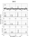
- Schematic diagram of electric field injection Electropherograms of samples with different concentrations Relationship between sample concentration and fluorescence intensity (part 1) Relationship between sample concentration and fluorescence intensity (part 2)
- Electropherogram of a sample containing two different size standards mixed at different ratios Part 1
- Electropherograms of samples containing two different size standards at different ratios Part 2 Relationship between sample concentration and fluorescence intensity and fluorescence intensity ratio of two types of DNA fragments
- Part 1 Relationship between sample concentration and fluorescence intensity of three types of DNA fragments Relationship between sample concentration and fluorescence intensity and fluorescence intensity ratio of two types of DNA fragments
- Part 2 Relationship between sample concentration and fluorescence intensity ratio of two types of DNA fragments
- saturation of the image sensor is not the cause, another possible cause is self-quenching of the fluorescent material at the measurement point. It is generally known that when the concentration of a fluorescent material becomes very high, self-quenching of the fluorescent material occurs, and the rate of increase in the fluorescent intensity decreases depending on the concentration of the fluorescent material, until the fluorescent intensity reaches saturation. It is also known that if the concentration of the fluorescent material increases further, the fluorescent intensity may begin to decrease.
- samples containing multiple types of DNA fragments with different concentrations were analyzed using the above-mentioned capillary electrophoresis device.
- the concentration of the DNA fragments in the sample was increased while maintaining the concentration ratio of the multiple types of DNA fragments in the sample.
- DNA fragments with high concentrations in the sample gave peaks of high intensity, and DNA fragments with low concentrations in the sample gave peaks of low intensity.
- a signal intensity proportional to the concentration of each DNA fragment was obtained.
- a signal intensity not proportional to the concentration of each DNA fragment was obtained.
- the degree of self-quenching of a fluorophore should depend on the concentration of the fluorophore at the measurement point on the capillary. However, the above change occurs independently of the concentration of the fluorophore at the measurement point on the capillary. From the above, it has been determined that the cause of this phenomenon is not self-quenching of the fluorophore. Therefore, in the above capillary electrophoresis analysis, the concentrations of the DNA fragments and the fluorophore labeled to the DNA fragments at the measurement point of the capillary do not reach a concentration that causes self-quenching. Of course, this phenomenon does not occur due to saturation of the image sensor, so the cause of this phenomenon is not due to saturation of the image sensor. Therefore, in the above capillary electrophoresis analysis, the concentrations of the DNA fragments and the fluorophore labeled to the DNA fragments at the measurement point of the capillary do not reach a concentration that causes saturation of the image sensor.
- FIG. 1A is a diagram of a capillary electrophoresis device.
- Capillary electrophoresis devices are widely used as analytical devices for DNA sequencing and DNA fragment analysis.
- Four capillaries 1 are used, and each capillary 1 can analyze a different sample.
- FIG. 1B shows the configuration of the capillary electrophoresis method.
- One capillary electrophoresis analysis is performed by the capillary electrophoresis method comprising steps (1) to (8) in Figure 1B.
- steps (1) to (8) By repeating steps (1) to (8), multiple capillary electrophoresis analyses can be performed.
- sample injection ends 2 of the four capillaries 1 are immersed in the cathode buffer solution 6, and the sample elution ends 3 are connected to the anode buffer solution 7 via the polymer solution 8 inside the pump block 10.
- valve 11 of the pump block 10 is closed, and the piston of the syringe 12 connected to the pump block 10 is pushed down to pressurize the polymer solution 8 inside, filling the polymer solution 8 inside each capillary 1 from the sample elution end 3 toward the sample injection end 2.
- each of the sample injection ends 2 of the four capillaries 1 is immersed in a different sample 9.
- a constant voltage is applied between the negative electrode 4 and the positive electrode 5 by the power source 13 for a fixed period of time, thereby electric-field-injecting a portion of the different sample 9 (containing at least the first component and the second component) from the sample injection end 2 into each capillary 1. This causes the sample 9 containing the first component and the second component to be injected into the capillary.
- the sample injection ends 2 of the four capillaries 1 are immersed in the negative electrode side buffer solution 6, and a high voltage is applied between the negative electrode 4 and the positive electrode 5 by the power supply 13 to start capillary electrophoresis.
- the DNA fragments labeled with a fluorescent substance are electrophoresed from the sample injection end 2 toward the sample elution end 3. This allows the injected first and second components to be separated by electrophoresis.
- each capillary 1 electrophoresed a certain distance from the sample injection end 2 is set as a measurement point 16, and a laser beam 14 emitted from a laser light source 15 is irradiated to each measurement point 16 at once.
- the coating of each capillary 1 near the measurement point 16 is removed in advance, each capillary 1 near the measurement point 16 is arranged on the same plane, the laser beam 14 is narrowed, and then it is introduced along the arrangement plane from the side of the arrangement plane.
- the fluorescence emitted from each measurement point 16 is measured by the detector 17. That is, the emitted fluorescence from the first component and the emitted fluorescence from the second component induced by irradiating the capillary 1 with a laser beam are measured by the detector 17, thereby obtaining the signal strength of the first component and the signal strength of the second component. An electropherogram, which is time series data of these signal strengths, is obtained and the sample 9 injected into each capillary 1 is analyzed.
- the detector 17 includes a spectrometer and an image sensor (not shown) and can simultaneously and independently perform spectroscopic measurement of the emitted fluorescence from the four measurement points 16. This makes it possible to distinguish between the emitted fluorescence of multiple types of fluorophores.
- the ratio of the concentration of the first component to the concentration of the second component in the sample is quantified based on the ratio of the signal intensity of the first component to the signal intensity of the second component. This quantification is possible based on the explanation of the [Principle] above and the method verified below.
- Figure 2 shows a schematic of the electric field injection of a sample 9 in capillary electrophoresis analysis.
- the sample injection end 2 of a capillary 1 filled with a polymer solution 8 is immersed in the liquid sample 9, with Figure 2(a) showing the state before electric field injection and Figure 2(b) showing the state after electric field injection. After Figure 2(b), the sample injection end 2 is immersed in the negative electrode side buffer solution and electrophoresis begins.
- Sample 9 contains a first DNA fragment 18 (first component), a second DNA fragment 19 (second component), which are negative ions, and negative ions 20 other than DNA fragments.
- the first DNA fragment 18 and the second DNA fragment 19 to be analyzed are both DNA fragments labeled with a fluorescent substance.
- the base length of the first DNA fragment 18 is different from the base length of the second DNA fragment 19.
- the first DNA fragment 18 and the second DNA fragment 19 have different concentrations in sample 9.
- the sample 9 may contain a size standard, and the second DNA fragment 19 may be a DNA fragment contained in the size standard.
- the sample 9 may contain a PCR product, and the first DNA fragment 18 may be a DNA fragment that is a PCR product or a DNA fragment derived from a PCR product.
- the sample 9 may contain a single-base extension product, and the first DNA fragment 18 may be a first DNA fragment that is a single-base extension product, and the second DNA fragment 19 may be a second DNA fragment that is a single-base extension product.
- sample 9 is pure water or formamide.
- Sample 9 is pre-prepared by ethanol precipitation or other methods to remove as many negative ions (salts) as possible other than DNA fragments, but it is not possible to reduce them to zero.
- the current I that flows when a constant voltage V is applied between the negative and positive electrodes is determined almost entirely by the electrical resistance R of the capillary 1 filled with the polymer solution 8, and is I ⁇ V/R.
- the electrical resistance R(i) between the negative electrode 4 (Fig. 1A) and the sample injection end 2 of the capillary 1 and the electrical resistance R(o) between the sample elution end 3 of the capillary (Fig. 1A) and the positive electrode 5 (Fig. 1A) are sufficiently small compared to R (R ⁇ R(i), R(o)).
- the combined resistance R(i) + R + R(o) between the negative electrode 4 and the positive electrode 5 is approximately equal to R (R(i) + R + R(o) ⁇ R).
- the current I flowing between the negative electrode and the sample injection end of the capillary, the current I flowing through the capillary, and the current I flowing between the sample elution end of the capillary and the positive electrode are all equal due to the continuity of the current.
- the current I that flows between the negative electrode and the sample injection end of the capillary during electric field injection i.e., the current I that flows through the sample
- the current I that flows through the sample is carried by the negative ions injected into the capillary, ignoring positive ions for simplicity. Therefore, the total amount of negative ions injected into the capillary by electric field injection caused by applying a constant voltage to both ends of the capillary for a certain period of time is constant regardless of the composition of the sample.
- the effective electric field strength in the sample near the sample injection end of the capillary during electric field injection is E
- the time of electric field injection is T
- the internal cross-sectional area of the capillary is A.
- the average of their mobilities and concentrations are ⁇ (0) and C(0). If the average charge per molecule of negative ions other than DNA fragments is q(0), the charge injection amount Q(0) of negative ions other than DNA fragments by electric field injection is It is.
- the mobility of the first DNA fragment and the second DNA fragment in the sample is ⁇
- the concentration of the first DNA fragment is C(1)
- the concentration of the second DNA fragment is C(2)
- the number of molecules of the first DNA fragment injected by electric field injection, J(1), and the number of molecules of the second DNA fragment injected, J(2) are given by: It is.
- the reason why the mobility of the first and second DNA fragments is made equal here is because the mobility of DNA fragments in a solution without the molecular sieve effect, i.e., in a sample, is constant regardless of base length.
- the charge amounts Q(1) and Q(2) injected into the first DNA fragment and the second DNA fragment by electric field injection are given by It is.
- E, T, and A are the same value. Since the total amount of injected charge of negative ions per unit time during electric field injection is equal to the current I, That is, It is.
- Equation 14 and (Equation 15) mean that the electric field injection is a competitive process between multiple types of negative ions, including DNA fragments contained in the sample, and the injected charge of each negative ion is distributed according to the product of the charge, mobility, and concentration of each negative ion.
- equation 14 the contribution of positive ions to the current, it is sufficient to multiply the right-hand side of (Equation 14) by the contribution rate of negative ions to the current.
- the signal intensities S(1) and S(2) of the peaks of the first and second DNA fragments on the electropherogram obtained by capillary electrophoresis are given by (Equation 8) and (Equation 9), respectively, when the sensitivity coefficients are m(1) and m(2).
- the sensitivity coefficient includes the average number of labeled fluorophores per molecule of a DNA fragment, the excitation efficiency of the fluorophores, the quantum yield of the fluorophores, the light collection efficiency of the emitted fluorescence, the sensitivity of the image sensor, and so on.
- Equation 12 the total signal intensity S of the peaks of the first DNA fragment and the second DNA fragment on the electropherogram obtained by capillary electrophoresis is given by (Equation 12) as follows:
- Equation 16 to Equation 21, Equation 24, Equation 25, and Equation 27 can be expressed as the following equations, where a and b are constants and y is a function of x.
- E is the effective electric field strength in the sample near the sample injection end of the capillary during electric field injection, and even if the voltage applied to both ends of the capillary is constant, i.e., the average electric field strength is constant, E can change depending on the composition of the sample.
- Equation 17 shows that when C(2) is small, J(2) is proportional to C(2), and when C(2) is large, J(2) saturates with respect to C(2) and approaches a constant value.
- Equation 18 shows that when C(1) is small, Q(1) is proportional to C(1), and when C(1) is large, Q(1) saturates with respect to C(1) and approaches a constant value.
- Equation 19 shows that when C(2) is small, Q(2) is proportional to C(2), and when C(2) is large, Q(2) saturates with respect to C(2) and approaches a constant value.
- Equation 20 shows that when C is small, J is proportional to C, and when C is large, J saturates with respect to C and approaches a constant value.
- Equation 21 shows that when C is small, Q is proportional to C, and when C is large, Q saturates with respect to C and approaches a constant value.
- Equation 24 shows that when C(1) is small, S(1) is proportional to C(1), and when C(1) is large, S(1) saturates with respect to C(1) and asymptotically approaches a constant value.
- Equation 25 shows that when C(2) is small, S(2) is proportional to C(2), and when C(2) is large, S(2) saturates with respect to C(2) and asymptotically approaches a constant value.
- Equation 27 shows that when C is small, S is proportional to C, and when C is large, S saturates with respect to C and asymptotically approaches a constant value.
- Equation 24 (Equation 24), (Equation 25), and (Equation 27) explain the phenomenon that in capillary electrophoresis analysis, when the concentration of the DNA fragment to be analyzed contained in the sample is low, a signal intensity proportional to the concentration is obtained, and when the concentration of the DNA fragment to be analyzed contained in the sample is high, the signal intensity saturates with respect to the concentration. In other words, the cause of the above phenomenon was identified. This knowledge was first obtained by introducing (Equation 14) and (Equation 15).
- the capillary electrophoresis apparatus of FIG. 1A can quantify the ratio of the concentration of the first DNA fragment to the concentration of the second DNA fragment in a sample based on the ratio of the signal intensity of the first DNA fragment to the signal intensity of the second DNA fragment.
- the concentration of the first DNA fragment in the sample can be quantified based on the ratio of the signal intensity of the first DNA fragment to the signal intensity of the second DNA fragment.
- (Equation 35) will have the same shape as (Equation 3), making it possible to avoid the effects of fluctuations in the electric field strength E and time T of the electric field injection, and to quantify the first DNA fragment, which is the subject of analysis, with high accuracy. Therefore, by using (Equation 35) as the calibration curve for the new internal standard method, similar to (Equation 3), which is the calibration curve for the conventional internal standard method, it becomes possible to quantify the DNA fragment to be analyzed with high accuracy.
- the conventional internal standard method was applicable only when the signal intensity was proportional to the concentration of the analyte.
- the new internal standard method as shown in (Equation 24), (Equation 25), and (Equation 27), is applicable not only when the signal intensity is proportional to the concentration of the analyte, but also when it is not proportional, approaches saturation, or reaches saturation.
- STR-PCR for DNA typing was performed using the human genome of a specific individual as a template, then desalted and the solvent was changed to formamide.
- Four types of samples were prepared in which the concentrations of the various types of DNA fragments that were the products of STR-PCR were varied over a four-digit concentration range: 1x, 0.05x, 0.002x, and 0.0001x the reference concentration.
- Each peak on the electropherogram indicates the signal of a DNA fragment that is the STR-PCR product of each locus.
- the single peak observed at around 4200 frames of electrophoresis time indicates the signal of a DNA fragment that is the STR-PCR product of locus D5S818.
- the fluorescence intensities measured when the same fluorescence intensity is generated at the measurement points of each of the four capillaries have been adjusted to be equal, so the differences in the measured fluorescence intensities faithfully reflect the differences in the fluorescence intensity emitted at the measurement points.
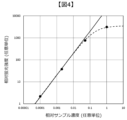
- Figure 4 is a double logarithmic graph plotting the peak fluorescence intensity of locus D5S818 against the concentration of DNA fragments contained in the sample, using the DNA fragments that are the STR-PCR products of locus D5S818 in Figure 3 as the analysis target.
- the peak width can be considered to be the same in Figures 3(a), (b), (c), and (d), so the peak height, not the peak area, is used as the fluorescence intensity.
- the DNA fragment concentration on the horizontal axis is the concentration of the DNA fragments that are the STR-PCR products of locus D5S818 contained in the sample, but it can also be considered as the total concentration of DNA fragments contained in the sample.
- the fluorescence intensity of the target DNA fragments is proportional to the concentration of the target DNA fragments in the sample.
- the fluorescence intensity of the target DNA fragments becomes less proportional to the concentration of the target DNA fragments in the sample and approaches saturation.
- Equation 28 the fluorescence intensity of the DNA fragment to be analyzed is S(1), and the concentration of the DNA fragment to be analyzed contained in the sample is C(1).
- S(1) is proportional to C(1), but as C(1) becomes high, S(1) deviates from the proportionality to C(1) and approaches saturation.
- Figure 5 is a log-log graph plotting the change in fluorescence intensity of the target DNA fragment against the change in concentration of the target DNA fragment contained in the sample for a sample different from those shown in Figures 3 and 4.
- the fluorescence intensity of the DNA fragment to be analyzed is S(1), and the concentration of the DNA fragment to be analyzed contained in the sample is C(1).
- S(1) is proportional to C(1), but as C(1) becomes high, it is confirmed that S(1) deviates from the proportionality to C(1) and approaches saturation.
- Example 1 The following measurements were performed using the capillary electrophoresis apparatus shown in Figure 1A.
- the " TM " symbol represents a trademark.
- Four types of samples were prepared by mixing two size standards, GeneScan TM 600 LIZ TM dye Size Standard (hereinafter, 600 LIZ) and GeneScan TM 500 ROX TM dye Size Standard (hereinafter, 500 ROX), in specific ratios using Hi-Di TM Formamide (hereinafter, formamide) from Thermo Fisher Scientific as a solvent, and analyzed using the capillary electrophoresis apparatus shown in Figure 1A.
- Each of the four capillaries had an inner diameter of 50 ⁇ m, a total length of 47 cm, and an effective length of 36 cm.
- Thermo Fisher Scientific's Applied Biosystems TM 310 and 31xx Running Buffer, 10X was used as the cathode buffer and anode buffer, diluted 10 times with pure water.
- POP-4 TM Polymer for 3500/SeqStudio TM Flex from Thermo Fisher Scientific was used.
- the concentrations of 600 LIZ contained in the four samples were changed to 1/2 (0.5), 1/20 (0.05), 1/200 (0.005), and 1/2000 (0.0005) of the reference concentration, respectively, while the concentrations of 500 ROX contained in the four samples were kept constant at 1/200 (0.005) of the reference concentration.
- the reference concentrations of 600 LIZ and 500 ROX are unrelated.
- 600 LIZ and 500 ROX each contain multiple types of DNA fragments, but the overall concentration was changed while maintaining the concentration ratio.
- Electric field injection of each sample was performed by applying a voltage of 1.2 kV to both ends of each capillary for only 9 seconds.
- Electrophoresis was performed by applying a voltage of 8.5 kV to both ends of each capillary.
- Figure 6(a) shows an electropherogram of a sample with 1/2000 times the concentration of 600 LIZ and 1/200 times the concentration of 500 ROX.
- Figure 6(b) shows an electropherogram of a sample with 1/200 times the concentration of 600 LIZ and 1/200 times the concentration of 500 ROX.
- Figure 7(a) shows an electropherogram of a sample with 1/20 times the concentration of 600 LIZ and 1/200 times the concentration of 500 ROX.
- Figure 7(b) shows an electropherogram of a sample with 1/2 times the concentration of 600 LIZ and 1/200 times the concentration of 500 ROX.
- the solid line indicates the fluorescence intensity of 600 LIZ and the dotted line indicates the fluorescence intensity of 500 ROX.
- the horizontal axis shows the electrophoresis time
- the left vertical axis shows the fluorescence intensity of 500 ROX
- the right vertical axis shows the fluorescence intensity of 600 LIZ.
- 600 LIZ contains 36 single-stranded DNA fragments with lengths of 20, 40, 60, 80, 100, 114, 120, 140, 160, 180, 200, 214, 220, 240, 250, 260, 280, 300, 314, 320, 340, 360, 380, 400, 414, 420, 440, 460, 480, 500, 514, 520, 540, 560, 580, and 600 bases, each of which is labeled with the fluorophore LIZ.
- the electropherograms in Figures 6 and 7 show peaks for 15 types of DNA fragments with lengths of 20, 40, 60, 80, 100, 114, 120, 140, 160, 180, 200, 214, 220, 240, and 250 bases. Furthermore, the peaks for DNA fragments with lengths of 40, 114, and 160 bases are indicated with arrows and labeled LIZ 40, LIZ 114, and LIZ 160.
- 500 ROX contains 16 single-stranded DNA fragments with lengths of 35, 50, 75, 100, 139, 150, 160, 200, 250, 300, 340, 350, 400, 450, 490, and 500 bases, each of which is labeled with the fluorescent ROX.
- the electropherograms in Figures 6 and 7 show peaks for nine of these DNA fragments, with lengths of 35, 50, 75, 100, 139, 150, 160, 200, and 250 bases. Furthermore, the peak for the 160-base-long DNA fragment is indicated by an arrow and labeled ROX 160.
- the peaks of DNA fragments of the same base length are observed at slightly different times due to the difference in mobility of the labeled fluorophores LIZ and ROX.
- Figure 8(a) is a log-log graph plotting the fluorescence intensity of the LIZ 160 peak and the ROX 160 peak obtained from the four electropherograms in Figures 6 and 7 against the concentration of 600 LIZ contained in the sample.
- the horizontal axis can be considered to be the concentration of LIZ 160 contained in the sample.
- the width of each peak can be considered to be approximately equal, so the height of each peak was taken as the fluorescence intensity.
- the black circles plot the fluorescence intensity of LIZ 160, and the white circles plot the fluorescence intensity of ROX 160.
- the fluorescence intensity of LIZ 160 is proportional to the concentration of 600 LIZ in the sample and can be approximated by a straight line with a slope of 1.
- the concentration of 600 LIZ in the sample is high, specifically in the range of 1/20 to 1/2 times the concentration of 600 LIZ, the fluorescence intensity of LIZ 160 deviates from proportionality to the concentration of 600 LIZ in the sample and approaches saturation. This phenomenon is the same as that observed in Figures 4 and 5.
- the concentration range of the first component contained in the sample includes not only the concentration range in which the signal strength of the first component is proportional to the concentration of the first component, but also the concentration range in which the signal strength of the first component is lower than the saturation signal strength of the detector and deviates from proportionality to the concentration of the first component, reaching the saturation signal strength.
- the capillary electrophoresis device of this embodiment is capable of measuring the concentration of such samples.
- the concentration range of the first component contained in the sample includes not only the concentration range in which the signal strength of the second component is constant relative to the concentration of the first component, but also the concentration range in which the signal strength of the second component deviates from constant and decreases relative to the concentration of the first component.
- the capillary electrophoresis device of this embodiment is capable of measuring the concentration of such samples.
- the first DNA fragment is considered to be all DNA fragments contained in 600 LIZ
- the second DNA fragment is considered to be all DNA fragments contained in 500 ROX.
- S(1) is the sum of the fluorescence intensities of all DNA fragments contained in 600 LIZ
- C(1) is the sum of the concentrations of all DNA fragments contained in 600 LIZ
- q(1) is the average charge of all DNA fragments contained in 600 LIZ
- S(2) is the sum of the fluorescence intensities of all DNA fragments contained in 500 ROX
- C(2) is the sum of the concentrations of all DNA fragments contained in 500 ROX
- q(2) is the average charge of all DNA fragments contained in 500 ROX.
- Figure 9 is a log-log graph plotting the peak fluorescence intensity of LIZ 160 (the same data as in Figure 8(a)), as well as, for example, the peak fluorescence intensity of LIZ 40 and LIZ 114, against the concentration of 600 LIZ in the sample.
- the horizontal axis can also be thought of as the concentration of LIZ 40, LIZ 114, or LIZ 160 in the sample.
- Figure 8(b) is a log-log graph in which the ratio of the peak fluorescence intensity of LIZ 160 to the peak fluorescence intensity of ROX 160 in Figure 8(a) is plotted against the concentration of 600 LIZ contained in the sample.
- the horizontal axis can be considered to be the concentration of LIZ 160 contained in the sample.
- equation (35) does not include variable parameters such as E, T, C(0), etc., it enables highly accurate quantification, similar to conventional internal quantification methods. Most importantly, equation (35) is compatible with equations (24) and (25). That is, as shown in Figure 8(a), whether the peak fluorescence intensity of LIZ 160 is proportional to the concentration of LIZ 160 in the sample or not, or whether the peak fluorescence intensity of ROX 160 is constant or decreasing, the ratio of the peak fluorescence intensity of LIZ 160 to the peak fluorescence intensity of ROX 160 is always proportional to the concentration of LIZ 160 in the sample.
- Figure 10 shows the experimental results corresponding to Figure 8, but in the same experiment as Figures 6 to 9, the electric field injection time was increased from 9 seconds to 18 seconds.
- Figure 10(a) shows the same tendency as Figure 8(a), even though it was obtained under different experimental conditions and by different electrophoretic analysis. However, the position of each plot varies slightly.
- Figure 10(b) shows almost the same result as Figure 8(b), even though it was obtained under different experimental conditions and by different electrophoretic analysis, and the variation in the position of each plot is also very small.
- the approximation line with a slope of 1 used in Figure 10(b) is the same as the approximation line with a slope of 1 used in Figure 8(a). The above results indicate that the new internal standard method has high quantitative accuracy.
- Figure 11 is a log-log graph showing the ratio of the LIZ 40 peak fluorescence intensity and the LIZ 114 peak fluorescence intensity to the LIZ 160 peak fluorescence intensity obtained in the four electropherograms of Figures 6 and 7 plotted against the concentration of 600 LIZ in the sample.
- the horizontal axis can be considered as the concentration of LIZ 40, LIZ 114, or LIZ 160 in the sample.
- This disclosure can be applied to any fragment analysis using capillary electrophoresis. Some specific examples are shown in the following examples.
- STR analysis reagent kits are on the market.
- Promega's PowerPlex (registered trademark) Fusion 6C System uses the human genome extracted from blood collected at the crime scene as a template to perform multiplex PCR of STRs at 27 loci on the human genome using five types of fluorophores.
- 500 WEN contains 21 types of single-stranded DNA fragments with base lengths of 60, 65, 80, 100, 120, 140, 160, 180, 200, 225, 250, 275, 300, 325, 350, 375, 400, 425, 450, 475, and 500, each of which is labeled with the fluorophore WEN.
- the sample is thermally denatured at 95°C, rapidly cooled on ice, and then subjected to capillary electrophoresis analysis to separate DNA fragments of various base lengths labeled with one of the six types of fluorophores contained in the sample, and the labeled fluorophores are detected while being identified.
- DNA typing is performed by analyzing the electropherograms obtained for each of the six types of fluorophores.
- the time at which the peaks of multiple types of DNA fragments belonging to the size standard are measured is utilized, but the fluorescence intensity of those peaks has not been utilized.
- the concentration of the size standard contained in the sample is known or fixed, it is possible to quantify the concentration of that DNA fragment contained in the sample from the fluorescence intensity of the peak of one of the multiple types of DNA fragments that are STR-PCR products contained in the sample by referring to the fluorescence intensity of that peak of the multiple types of DNA fragments that belong to the size standard.
- the human genome, which is the template be DNA type tested, but each of the multiple types of DNA fragments that are STR-PCR products contained in the sample can be quantified with high accuracy.
- the high accuracy of this quantification can be explained by taking the first DNA fragment as any DNA fragment contained in the STR-PCR product and the second DNA fragment as any DNA fragment contained in the size standard in (Equation 35).
- the concentration of the first DNA fragment in the sample relative to the concentration of the second DNA fragment in the sample can be calculated from the ratio of the peak fluorescence intensity of the first DNA fragment to the peak fluorescence intensity of the second DNA fragment.
- the concentration of the first DNA fragment in the sample can be determined from the peak fluorescence intensity of the first DNA fragment relative to the peak fluorescence intensity of the second DNA fragment. Since (Equation 35) does not include variable parameters such as E, T, C(0), etc., it enables highly accurate quantification, similar to conventional internal quantification methods. As shown in (Equation 24), this highly accurate quantification is valid both when the peak fluorescence intensity of the first DNA fragment is proportional to the concentration of the first DNA fragment contained in the sample, and when it is not proportional. In other words, the new internal standard method makes it possible to quantify the concentration of the first DNA fragment in a wider concentration range with high accuracy than before.
- the concentration of the template human genome contained in the solution immediately before STR-PCR is N
- the amplification efficiency of STR-PCR is E(f)
- the concentration of the human genome in the solution before STR-PCR reaction is C(f).
- N 29.
- E(f) 1, but the actual value can be determined in advance.
- an arbitrary STR-PCR product contained in the solution after STR-PCR reaction is the first DNA fragment, and its concentration is B(1).
- the concentration of the first DNA fragment in the sample used for electric field injection is C(1).
- Thermo Fisher Scientific's SNaPshot® Multiplex system is a kit that uses capillary electrophoresis to simultaneously type multiple SNPs (single nucleotide polymorphisms) on the human genome.
- a template DNA is prepared in advance by amplifying a region containing multiple SNPs on the human genome.
- Fluorescently unlabeled primers are hybridized to the template DNA at positions adjacent to each SNP, and a single-base extension reaction of each primer is performed using a fluorescently labeled terminator.
- the fluorescently labeled terminators are ddATP, ddCTP, ddGTP, and ddTTP, each labeled with four different fluorophores.
- each primer is changed according to the corresponding SNP.
- DNA fragments that are multiple types of single-base extension products are analyzed by capillary electrophoresis to obtain electropherograms for each of the four fluorophores.
- the location of the corresponding SNP is identified on the electropherogram from the electrophoresis time at which a peak is observed, i.e., the base length of the corresponding DNA fragment.
- the type of fluorescent substance in the same peak is used to identify whether the SNP is A, C, G, or T.
- the above SNP typing can be performed simultaneously for SNPs at multiple locations.
- each SNP may be a mixture of A, C, G, and T in any ratio.
- WT wild type of a given SNP
- MT mutant type
- M is an integer of 1 or more, and assume that the abundance ratio of A, C, G, and T in SNPs at M locations, and in particular the abundance ratio of MT base species relative to WT base species, is quantified. For each of the M SNPs, a maximum of four types of single-base extension products are obtained. The reaction solution thus containing a maximum total of 4 ⁇ M types of DNA fragments is desalted by ethanol precipitation, and then eluted in a desired amount of formamide to prepare the sample to be used for electric field injection.
- the fluorescence intensities of the peaks on the electropherogram of DNA fragments that are single-base extension products of base types A, C, G, and T of SNP number j are S(ja), S(jc), S(jg), and S(jt), their respective sensitivity coefficients are m(ja), m(jc), m(jg), and m(jt), their respective average charge amounts are q(ja), q(jc), q(jg), and q(jt), and their respective concentrations in the sample injected into the capillary are C(ja), C(jc), C(jg), and C(jt).
- the total injected charge amount Q of all DNA fragments injected into the capillary is given by the following equation (equation 13): On the other hand, as in (Number 15), From (39) and (40), as in (21), where q(0) ⁇ (0) C(0) represents the injected charge of negative ions other than the DNA fragments, and the ⁇ term represents the total injected charge of all DNA fragments.
- Equation 41 indicates that when Q is small compared to q(0) ⁇ (0) C(0), Q is proportional to the ⁇ term, but when the ⁇ term becomes large compared to q(0) ⁇ (0) C(0), Q deviates from proportionality to the ⁇ term and reaches saturation. Therefore, the total injection amount of all DNA fragments is proportional to the total concentration of all DNA fragments when the total concentration of all DNA fragments is low, but when the total concentration of all DNA fragments becomes high, Q deviates from proportionality and reaches saturation. Needless to say, an increase in the total concentration of all DNA fragments is brought about by an increase in the concentration of any DNA fragment. And when the total injection amount of all DNA fragments reaches saturation, the injection amount of any DNA fragment will either saturate or decrease.
- the concentration ratios C(jc)/C(ja), C(jg)/C(ja), C(jt)/C(ja) corresponding to the abundance ratio of the three types of MT relative to WT can be quantified with high accuracy using the peak fluorescence intensities S(jc)/S(ja), S(jg)/S(ja), and S(jt)/S(ja) on the electropherogram.
- m(ja), m(jc), m(jg), and m(jt) are calculated in advance.
Landscapes
- Health & Medical Sciences (AREA)
- Life Sciences & Earth Sciences (AREA)
- Chemical & Material Sciences (AREA)
- Molecular Biology (AREA)
- Physics & Mathematics (AREA)
- Analytical Chemistry (AREA)
- Biochemistry (AREA)
- General Health & Medical Sciences (AREA)
- General Physics & Mathematics (AREA)
- Immunology (AREA)
- Pathology (AREA)
- Electrochemistry (AREA)
- Chemical Kinetics & Catalysis (AREA)
- Nuclear Medicine, Radiotherapy & Molecular Imaging (AREA)
- Investigating, Analyzing Materials By Fluorescence Or Luminescence (AREA)
Priority Applications (5)
| Application Number | Priority Date | Filing Date | Title |
|---|---|---|---|
| PCT/JP2023/024615 WO2025009018A1 (ja) | 2023-07-03 | 2023-07-03 | キャピラリ電気泳動装置およびキャピラリ電気泳動法 |
| GB2514720.8A GB2643967A (en) | 2023-07-03 | 2023-07-03 | Capillary electrophoresis device and capillary electrophoresis method |
| JP2025530817A JPWO2025009018A1 (https=) | 2023-07-03 | 2023-07-03 | |
| DE112023005459.2T DE112023005459T5 (de) | 2023-07-03 | 2023-07-03 | Kapillarelektrophoresevorrichtung und kapillarelektrophoreseverfahren |
| CN202380094996.4A CN120813826A (zh) | 2023-07-03 | 2023-07-03 | 毛细管电泳装置以及毛细管电泳法 |
Applications Claiming Priority (1)
| Application Number | Priority Date | Filing Date | Title |
|---|---|---|---|
| PCT/JP2023/024615 WO2025009018A1 (ja) | 2023-07-03 | 2023-07-03 | キャピラリ電気泳動装置およびキャピラリ電気泳動法 |
Publications (1)
| Publication Number | Publication Date |
|---|---|
| WO2025009018A1 true WO2025009018A1 (ja) | 2025-01-09 |
Family
ID=94171597
Family Applications (1)
| Application Number | Title | Priority Date | Filing Date |
|---|---|---|---|
| PCT/JP2023/024615 Pending WO2025009018A1 (ja) | 2023-07-03 | 2023-07-03 | キャピラリ電気泳動装置およびキャピラリ電気泳動法 |
Country Status (5)
| Country | Link |
|---|---|
| JP (1) | JPWO2025009018A1 (https=) |
| CN (1) | CN120813826A (https=) |
| DE (1) | DE112023005459T5 (https=) |
| GB (1) | GB2643967A (https=) |
| WO (1) | WO2025009018A1 (https=) |
Citations (6)
| Publication number | Priority date | Publication date | Assignee | Title |
|---|---|---|---|---|
| JP2003310300A (ja) * | 2002-04-24 | 2003-11-05 | Hitachi Ltd | 遺伝子検査方法 |
| JP2016176764A (ja) * | 2015-03-19 | 2016-10-06 | 株式会社島津製作所 | キャピラリー電気泳動装置及びキャピラリー電気泳動による試料分析方法 |
| JP2019502367A (ja) * | 2015-11-03 | 2019-01-31 | アスラジェン, インコーポレイテッド | リピート配列の核酸サイズ検出のための方法 |
| JP2021072825A (ja) * | 2012-11-26 | 2021-05-13 | ザ・ユニバーシティ・オブ・トレド | 核酸の標準化された配列決定のための方法およびその使用 |
| JP2022526067A (ja) * | 2019-01-21 | 2022-05-23 | マックス-プランク-ゲゼルシャフト・ツア・フェルデルング・デア・ヴィッセンシャフテン・エー・ファオ | 炭水化物および炭水化物混合組成物パターンの自動化高性能同定のための先進的方法、ならびにそのためのシステム、ならびに新しい蛍光色素に基づく、そのための多波長蛍光検出システムの較正のための方法 |
| WO2023007567A1 (ja) * | 2021-07-27 | 2023-02-02 | 株式会社日立ハイテク | マルチキャピラリ電気泳動装置 |
-
2023
- 2023-07-03 GB GB2514720.8A patent/GB2643967A/en active Pending
- 2023-07-03 DE DE112023005459.2T patent/DE112023005459T5/de active Pending
- 2023-07-03 CN CN202380094996.4A patent/CN120813826A/zh active Pending
- 2023-07-03 JP JP2025530817A patent/JPWO2025009018A1/ja active Pending
- 2023-07-03 WO PCT/JP2023/024615 patent/WO2025009018A1/ja active Pending
Patent Citations (6)
| Publication number | Priority date | Publication date | Assignee | Title |
|---|---|---|---|---|
| JP2003310300A (ja) * | 2002-04-24 | 2003-11-05 | Hitachi Ltd | 遺伝子検査方法 |
| JP2021072825A (ja) * | 2012-11-26 | 2021-05-13 | ザ・ユニバーシティ・オブ・トレド | 核酸の標準化された配列決定のための方法およびその使用 |
| JP2016176764A (ja) * | 2015-03-19 | 2016-10-06 | 株式会社島津製作所 | キャピラリー電気泳動装置及びキャピラリー電気泳動による試料分析方法 |
| JP2019502367A (ja) * | 2015-11-03 | 2019-01-31 | アスラジェン, インコーポレイテッド | リピート配列の核酸サイズ検出のための方法 |
| JP2022526067A (ja) * | 2019-01-21 | 2022-05-23 | マックス-プランク-ゲゼルシャフト・ツア・フェルデルング・デア・ヴィッセンシャフテン・エー・ファオ | 炭水化物および炭水化物混合組成物パターンの自動化高性能同定のための先進的方法、ならびにそのためのシステム、ならびに新しい蛍光色素に基づく、そのための多波長蛍光検出システムの較正のための方法 |
| WO2023007567A1 (ja) * | 2021-07-27 | 2023-02-02 | 株式会社日立ハイテク | マルチキャピラリ電気泳動装置 |
Also Published As
| Publication number | Publication date |
|---|---|
| CN120813826A (zh) | 2025-10-17 |
| JPWO2025009018A1 (https=) | 2025-01-09 |
| GB2643967A (en) | 2026-03-11 |
| DE112023005459T5 (de) | 2025-11-20 |
Similar Documents
| Publication | Publication Date | Title |
|---|---|---|
| Butler et al. | Forensic DNA typing by capillary electrophoresis using the ABI Prism 310 and 3100 genetic analyzers for STR analysis | |
| Castro et al. | Single-molecule electrophoresis | |
| Karger et al. | Multiwavelength fluorescence detection for DNA sequencing using capillary electrophoresis | |
| Larkin et al. | Length-independent DNA packing into nanopore zero-mode waveguides for low-input DNA sequencing | |
| Lieberwirth et al. | Multiplex dye DNA sequencing in capillary gel electrophoresis by diode laser-based time-resolved fluorescence detection | |
| Butler et al. | Quantitation of polymerase chain reaction products by capillary electrophoresis using laser fluorescence | |
| JP3287361B2 (ja) | 統計的信頼区間の決定を含む多成分分析方法 | |
| Shewale et al. | Principles, practice, and evolution of capillary electrophoresis as a tool for forensic DNA analysis | |
| US20070158193A1 (en) | Analyte Identification in Transformed Electropherograms | |
| Gelfi et al. | Temperature-programmed capillary electrophoresis for detection of DNA point mutations | |
| US6942773B1 (en) | Particle sizer and DNA sequencer | |
| Datinská et al. | Preparative concentration of nucleic acids fragments by capillary isotachophoretic analyzer | |
| JP7016957B2 (ja) | 生体ポリマー分析方法、および生体ポリマー分析装置 | |
| IE912228A1 (en) | Neutral and positively charged dyes for electrophoresis sample loading solutions | |
| Li et al. | Capillary electrophoresis as a method to study DNA reassociation | |
| WO2025009018A1 (ja) | キャピラリ電気泳動装置およびキャピラリ電気泳動法 | |
| JPH05118991A (ja) | 塩基配列決定方法およびその装置 | |
| JP2011030502A (ja) | 塩基配列解析装置及びそのプログラム | |
| JP7318122B2 (ja) | 電気泳動装置及び分析方法 | |
| Mark et al. | Very fast capillary electrophoresis with electrochemical detection for high-throughput analysis using short, vertically aligned capillaries. | |
| Zhao et al. | Sensitive and selective label-free alkaline phosphatase detection based on DNA hairpin probe | |
| JP7253066B2 (ja) | 生体試料分析装置、生体試料分析方法 | |
| Zhang et al. | Recording the Reaction Process of Loop‐Mediated Isothermal Amplification (LAMP) by Monitoring the Voltammetric Response of 2′‐Deoxyguanosine 5′‐Triphosphate | |
| JP4021215B2 (ja) | 電気泳動方法 | |
| Zhao et al. | Quantification of the bisphosphonate alendronate using capillary electrophoresis mass spectrometry with dynamic pH barrage junction focusing |
Legal Events
| Date | Code | Title | Description |
|---|---|---|---|
| 121 | Ep: the epo has been informed by wipo that ep was designated in this application |
Ref document number: 23942637 Country of ref document: EP Kind code of ref document: A1 |
|
| WWE | Wipo information: entry into national phase |
Ref document number: 11202505731P Country of ref document: SG |
|
| WWE | Wipo information: entry into national phase |
Ref document number: 112023005459 Country of ref document: DE Ref document number: 202380094996.4 Country of ref document: CN |
|
| ENP | Entry into the national phase |
Ref document number: 2025530817 Country of ref document: JP Kind code of ref document: A |
|
| WWE | Wipo information: entry into national phase |
Ref document number: 2025530817 Country of ref document: JP |
|
| ENP | Entry into the national phase |
Ref document number: 202514720 Country of ref document: GB Kind code of ref document: A Free format text: PCT FILING DATE = 20230703 |
|
| WWE | Wipo information: entry into national phase |
Ref document number: 2514720.8 Country of ref document: GB |
|
| WWP | Wipo information: published in national office |
Ref document number: 202380094996.4 Country of ref document: CN |
|
| WWP | Wipo information: published in national office |
Ref document number: 112023005459 Country of ref document: DE |
|
| WWE | Wipo information: entry into national phase |
Ref document number: 11202505731P Country of ref document: SG |
|
| WWP | Wipo information: published in national office |
Ref document number: 11202505731P Country of ref document: SG |
|
| WWP | Wipo information: published in national office |
Ref document number: 2514720.8 Country of ref document: GB |