WO2025005232A1 - 軟組織修復用のインプラント医療機器及びその製造方法 - Google Patents
軟組織修復用のインプラント医療機器及びその製造方法 Download PDFInfo
- Publication number
- WO2025005232A1 WO2025005232A1 PCT/JP2024/023482 JP2024023482W WO2025005232A1 WO 2025005232 A1 WO2025005232 A1 WO 2025005232A1 JP 2024023482 W JP2024023482 W JP 2024023482W WO 2025005232 A1 WO2025005232 A1 WO 2025005232A1
- Authority
- WO
- WIPO (PCT)
- Prior art keywords
- medical device
- monomer
- layer
- implant medical
- less
- Prior art date
- Legal status (The legal status is an assumption and is not a legal conclusion. Google has not performed a legal analysis and makes no representation as to the accuracy of the status listed.)
- Ceased
Links
Images






Classifications
-
- A—HUMAN NECESSITIES
- A61—MEDICAL OR VETERINARY SCIENCE; HYGIENE
- A61L—METHODS OR APPARATUS FOR STERILISING MATERIALS OR OBJECTS IN GENERAL; DISINFECTION, STERILISATION OR DEODORISATION OF AIR; CHEMICAL ASPECTS OF BANDAGES, DRESSINGS, ABSORBENT PADS OR SURGICAL ARTICLES; MATERIALS FOR BANDAGES, DRESSINGS, ABSORBENT PADS OR SURGICAL ARTICLES
- A61L27/00—Materials for grafts or prostheses or for coating grafts or prostheses
- A61L27/14—Macromolecular materials
- A61L27/18—Macromolecular materials obtained otherwise than by reactions only involving carbon-to-carbon unsaturated bonds
Definitions
- the present invention relates to an implant medical device for soft tissue repair and a method for manufacturing the same.
- Tendons, the rotator cuff (a plate-like collection of tendons), and ligaments are tissues that connect muscles to bones or bones to bones, and play an important role in allowing the smooth movement of joints such as the shoulder, elbow, knee, wrist, and ankle.
- Tendons, rotator cuffs, and ligaments can become damaged or ruptured due to degeneration caused by aging, trauma, or overuse in sports, etc., interfering with daily movements and causing pain not only during exercise but also at rest and at night.
- meshes made of polylactic acid fibers with a controlled number of warp and weft threads and foams made of polylactic acid have been disclosed, which are reinforced with meshes that have improved resistance to dissolution in organic solvents by annealing and then rapidly cooling (Patent Documents 1 and 2).
- Patent Document 3 a biodegradable polyester made of lactic acid and caprolactone has been described as a synthetic polymer with flexibility
- Patent Document 4 a medical molded body using the biodegradable polyester described in Patent Document 3 has been disclosed that is intended to be placed in the body, and when there is no hard tissue around the placement site, the Young's modulus of the molded body is controlled to be low so that it can be treated in the same way as soft tissue
- the implant medical devices described in Patent Documents 1 and 2 use polylactic acid, a hard synthetic polymer, and so although it has excellent strength, it lacks flexibility and there are still issues with respect to conformity to soft tissue. Even when such a hard synthetic polymer is used, it may be possible to impart flexibility by making the thickness and density very small, but because the polymer is easily degraded, it will degrade and be absorbed before the soft tissue repair is complete.
- Patent Document 3 describes a biodegradable polyester made of lactic acid and caprolactone, but does not specifically describe its application to implant devices for soft tissue repair.
- Patent Document 4 describes a medical molded body, medical device, and nerve regeneration guide tube that use the biodegradable polyester described in Patent Document 3, but does not specifically describe its application to implant devices for soft tissue repair.
- the shape (particularly the thickness and bulk density) described in Patent Documents 1 and 2 is used, the strength will be insufficient. Therefore, even if the soft synthetic polymers described in Patent Documents 3 and 4 are used, the thickness and bulk density must be controlled within an appropriate range in order to apply them to implant devices for soft tissue repair.
- the present invention aims to provide an implant medical device for soft tissue repair that has excellent strength and flexibility by using a biodegradable polyester copolymer containing polyhydroxycarboxylic acid, a soft synthetic polymer whose main structural unit is two types of hydroxycarboxylic acid monomers, and by controlling the thickness and bulk density within an appropriate range.
- An implant medical device for soft tissue repair comprising a polyester copolymer containing polyhydroxycarboxylic acid having two types of hydroxycarboxylic acid monomers as main constituent units, and having a thickness of 1.0 mm or more and 20.0 mm or less and a bulk density of 0.40 g/ cm3 or more and 1.50 g/ cm3 or less.
- the implant medical device according to (1) which has a first layer and a second layer, and the bulk density of the first layer is 0.20 g/cm 3 or more and 0.95 g/cm 3 or less.
- An implant medical device for soft tissue repair comprising a first layer and a second layer, the first layer and the second layer having different bulk densities, a thickness of 1.0 mm or more and 20.0 mm or less, and an overall bulk density of 0.40 g/ cm3 or more and 1.50 g/cm3 or less.
- the implant medical device according to (3) which has a polyester copolymer containing a polyhydroxycarboxylic acid having two kinds of hydroxycarboxylic acid monomers as main structural units.
- An implant medical device for soft tissue repair comprising a polyester copolymer containing polyhydroxycarboxylic acid having two types of hydroxycarboxylic acid monomers as main constituent units, and having a thickness of 1.0 mm or more and 20.0 mm or less and a bulk density of 0.40 g/ cm3 or more and 1.50 g/ cm3 or less.
- the implant medical device according to (20) which has a first layer and a second layer, and the bulk density of the first layer is 0.20 g/cm3 or more and 0.95 g/ cm3 or less.
- Formula 1 [A] Molar fraction (%) of monomer A residues in polyhydroxycarboxylic acid
- B] Molar fraction (%) of monomer B residues in polyhydroxycarboxylic acid
- AB] mole fraction (%) of structures (AB and BA) in which monomer A residue and monomer B residue are adjacent to each other in polyhydroxycarboxylic acid
- the present invention can provide an implantable medical device suitable for soft tissue repair applications.
- FIG. 2 is a schematic diagram showing a rectangular parallelepiped having a curved attachment surface as viewed from the side.
- FIG. 2 is a schematic diagram showing a rectangular parallelepiped having projections and recesses on the attachment surface as viewed from the side.
- the implant medical device for soft tissue repair of the present invention is characterized in that it has a polyester copolymer containing polyhydroxycarboxylic acid whose main constituent units are two types of hydroxycarboxylic acid monomers, has a thickness of 1.0 mm or more and 20.0 mm or less, and has a bulk density of 0.40 g/cm3 or more and 1.50 g/cm3 or less.
- the implant medical device for soft tissue repair of the present invention is characterized in that it has a first layer and a second layer, the first layer and the second layer have different bulk densities, the thickness is 1.0 mm or more and 20.0 mm or less, and the overall bulk density is 0.40 g/cm3 or more and 1.50 g/cm3 or less.
- soft tissue refers to tissues of the human body excluding bones, including cartilage, tendons, rotator cuffs, and ligaments, and in particular tendons, rotator cuffs, and ligaments.
- the polyester copolymer refers to a polymer consisting of two or more types of monomers, among which the monomers are linked mainly by ester bonds.
- the term polyester also refers to a polymer in which some of the monomers are linked by bonds other than ester bonds, such as urethane bonds or amide bonds.
- the number of bonds other than ester bonds among the bonds used in monomer linkages is preferably 30% or less, more preferably 10% or less, even more preferably 5% or less, and most preferably 0%, i.e., no bonds other than ester bonds, when the number of bonds used in monomer linkages is taken as 100%.
- the polyester copolymer in the present invention contains polyhydroxycarboxylic acid.
- Polyhydroxycarboxylic acid refers to a polymer composed of hydroxycarboxylic acid.
- Hydroxycarboxylic acid refers to a compound that has both a hydroxy group and a carboxy group in the same molecule.
- polyester copolymers containing polyhydroxycarboxylic acids can be biodegradable.
- the implant medical device of the present invention containing a polyester copolymer is also biodegradable.
- the implant medical device of the present invention is biodegradable.
- the polyester copolymer of the present invention preferably contains 50% by mass or more of polyhydroxycarboxylic acid relative to 100% by mass of the polyester copolymer, more preferably 70% by mass or more, even more preferably 90% by mass or more, and most preferably contains 100% by mass, i.e., is composed only of polyhydroxycarboxylic acid.
- the implant medical device used in the present invention preferably contains 50% by mass or more of polyester copolymer relative to 100% by mass of the implant medical device, more preferably 70% by mass or more, even more preferably 90% by mass or more, and most preferably 100% by mass, i.e., the implant medical device is composed only of polyester copolymer.
- biodegradability refers to the property of being broken down in the body or in the environment. More specifically, it refers to a degree of biodegradation determined according to Measurement Example 1 described below of 60% or more.
- Terms that can be used interchangeably with biodegradability include bioabsorbability and biocompatibility, but these terms often refer primarily to the property of being broken down in the body.
- Polyhydroxycarboxylic acids can be obtained by polymerization of hydroxycarboxylic acids, polymerization of lactones, which are cyclic compounds formed by intramolecular dehydration condensation of the hydroxyl and carboxyl groups of hydroxycarboxylic acids, or polymerization of lactides, which are cyclic compounds formed by dehydration condensation of the hydroxyl and carboxyl groups of two molecules of hydroxycarboxylic acids.
- hydroxycarboxylic acids, lactones, or lactides can be used as hydroxycarboxylic acid monomers.
- Hydroxycarboxylic acids used in the hydroxycarboxylic acid monomer include lactic acid, glycolic acid, hydroxybutyric acid, hydroxyvaleric acid, hydroxycaproic acid, hydroxyheptanoic acid, hydroxyoctanoic acid, hydroxynonanoic acid, hydroxydecanoic acid, hydroxyundecanoic acid, hydroxydodecanoic acid, and (2-hydroxyethoxy)acetic acid.
- lactic acid, glycolic acid, hydroxyvaleric acid, and hydroxycaproic acid are preferred, and lactic acid and hydroxycaproic acid are particularly preferred.
- the lactone used in the hydroxycarboxylic acid monomer may be butyrolactone, valerolactone, caprolactone (hereinafter, CL), dioxepanone, ethylene oxalate, p-dioxanone, trimethylene carbonate, ⁇ -propiolactone, or pivalolactone, with butyrolactone, valerolactone, CL, p-dioxanone, or trimethylene carbonate being particularly preferred, and valerolactone or CL being even more preferred.
- CL caprolactone
- the lactide used in the hydroxycarboxylic acid monomer may be dilactide (hereinafter referred to as LA) which is formed by the dehydration condensation of two molecules of lactic acid, or glycolide or tetramethylglycolide which is formed by the dehydration condensation of two molecules of glycolic acid, with LA or glycolide being particularly preferred.
- LA dilactide
- the polyhydroxycarboxylic acid of the present invention has two types of hydroxycarboxylic acid monomers as its main constituent units. That a polyhydroxycarboxylic acid has two types of hydroxycarboxylic acid monomers as its main constituent units means that the sum of the number of the two types of monomer residues is 50 mol % or more when the sum of the number of all monomer residues contained in the entire polyhydroxycarboxylic acid, including other monomer residues, is taken as 100 mol %, and each of the two types of monomer residues is 10 mol % or more when the sum of the number of all monomer residues contained in the entire polyhydroxycarboxylic acid is taken as 100 mol %.
- the term "monomer residue” refers, in principle, to a repeating unit of a chemical structure derived from a monomer in the chemical structure of a polyhydroxycarboxylic acid obtained by polymerizing two or more monomers including the monomer.
- “having monomer A residues and monomer B residues as the main constituent units” means that the sum of the number of monomer A residues and monomer B residues is 50 mol % or more, and the monomer A residues are 10 mol % or more, and the monomer B residues are 10 mol % or more, when the sum of the number of all monomer residues contained in the entire polyhydroxycarboxylic acid is 100 mol %.
- the molar fractions (%) of monomer A residue, monomer B residue, and other residues can be determined from the area values of the signals derived from each residue by nuclear magnetic resonance (hereinafter referred to as "NMR") measurement.
- NMR nuclear magnetic resonance
- the monomer A residue is an LA residue
- the monomer B residue is a CL residue
- they can be determined by the measurement example 2 described below.
- the molecular weight of the repeating unit of the chemical structure derived from the monomer is used. For example, if the monomer A residue is an LA residue and the monomer B residue is a CL residue, the molecular weight of the repeating unit of monomer A residue is 144, and the molecular weight of the repeating unit of monomer B residue is 114.
- the molar fraction (%) of the sum of monomer A residues and monomer B residues is, based on the above definition, 50 mol % or more, preferably 75 mol % or more, and more preferably 90 mol % or more, when the sum of the number of all monomer residues contained in the entire polymer, including other monomer residues, is 100 mol %.
- the molar fractions (%) of monomer A residues and monomer B residues are each, similarly as defined above, 10 mol% or more, preferably 20 mol% or more, more preferably 30 mol% or more, even more preferably 40 mol% or more, and most preferably 50 mol% or more.
- a particularly preferred embodiment is one in which the sum of monomer A residues and monomer B residues is 100 mol% of the entire polymer, i.e., a polymer consisting only of monomer A and monomer B.
- a more preferred embodiment of the present invention is one in which the R value represented by the following formula 1 is 0.25 or more and 0.99 or less (hereinafter, a polyhydroxycarboxylic acid having an R value of 0.25 or more and 0.99 or less is referred to as a "random polymer").
- R value [AB] / (2 [A] [B]) x 100 ...
- Formula 1 [A] Molar fraction (%) of monomer A residues in polyhydroxycarboxylic acid
- B] Molar fraction (%) of monomer B residues in polyhydroxycarboxylic acid
- AB] mole fraction (%) of structures (AB and BA) in which monomer A residue and monomer B residue are adjacent to each other in polyhydroxycarboxylic acid
- the lower limit of the R value is preferably 0.25 or more, and more preferably 0.45 or more.
- the monomers A and B used in the present invention are preferably monomers selected from lactic acid, glycolic acid, hydroxybutyric acid, hydroxyvaleric acid, hydroxycaproic acid, hydroxyheptanoic acid, hydroxyoctanoic acid, hydroxynonanoic acid, hydroxydecanoic acid, hydroxyundecanoic acid, hydroxydodecanoic acid, (2-hydroxyethoxy)acetic acid, butyrolactone, valerolactone, CL, dioxepanone, ethylene oxalate, p-dioxanone, trimethylene carbonate, ⁇ -propiolactone, pivalolactone, LA, glycolide, and tetramethylglycolide.
- the monomer A is at least one monomer selected from the group consisting of lactic acid, glycolic acid, LA, and glycolide. It is also particularly preferred that the monomer B is at least one monomer selected from the group consisting of hydroxyvaleric acid, hydroxycaproic acid, valerolactone, and CL.
- the weight average molecular weight of the polyester copolymer in the present invention can be measured by the method described in Measurement Example 3. From the viewpoint of controlling the strength and biodegradability within a suitable range, the weight average molecular weight of the polyester copolymer is preferably 50,000 or more, more preferably 100,000 or more, even more preferably 150,000 or more, and most preferably 200,000 or more. As the weight average molecular weight increases, the strength increases and the number of days required for biodegradation increases. From the viewpoint of moldability, the weight average molecular weight of the polyester copolymer is preferably 1,000,000 or less, more preferably 800,000 or less, and even more preferably 500,000 or less.
- the polyester copolymer in the present invention may be a multi-polyester copolymer in which polyester copolymers are linked together.
- the polyester copolymer before linking is referred to as a "macromer" for convenience.
- the average number of macromer molecules constituting one molecule of the polyester copolymer is called the linkage number.
- the linkage number refers to the value obtained by dividing the weight average molecular weight of the polyester copolymer by the weight average molecular weight of the macromer.
- Multipolyester copolymers can be produced by linking polyester copolymers directly or by linking them using linker molecules. Whether the multipolyester copolymer has a linear or branched structure is determined by the structure of the polyester copolymer and the structure of the linker molecule.
- the multi-polyester copolymer will have a branched structure regardless of the method of linking. If the polyester copolymer has a linear structure, the multi-polyester copolymer will have a linear structure when linked as is or using a linker molecule with a linear structure, and the multi-polyester copolymer will have a branched structure when linked using a linker molecule with a branched structure.
- a linear linker molecule a compound having two functional groups, such as a carboxyl group, a hydroxyl group, or an amino group, can be used.
- examples of such compounds include oxalic acid, malonic acid, succinic acid, glutaric acid, adipic acid, pimelic acid, suberic acid, azelaic acid, sebacic acid, dodecanedioic acid, ethylene glycol, propanediol, butanediol, pentanediol, hexanediol, heptanediol, octanediol, polyethylene glycol, polypropylene glycol, polyethylene glycol-polypropylene glycol copolymer, ethylenediamine, propanediamine, butanediamine, diaminopentane, hexamethylenediamine, diaminoheptane, diaminooctane, lactic acid, glycolic acid,
- branched linker molecule a compound having two or more functional groups, such as a carboxyl group, a hydroxyl group, or an amino group, within the molecule can be used, and examples of such compounds include malic acid, tartaric acid, citric acid, tyrosine, serine, threonine, aspartic acid, glutamic acid, and lysine.
- the crystallization rate of at least one of the monomer A residue or monomer B residue is less than 14%. If the crystallization rate is less than 14%, the increase in the tensile modulus of the implant medical device is suppressed, and an implant medical device suitable for soft tissue repair applications can be obtained. It is more preferable that the crystallization rate of the monomer A residue or monomer B residue is 10% or less, and even more preferable that it is 5% or less. It is preferable that the crystallization rate of all monomer residues is low, but it is also acceptable for only the crystallization rate of some of the monomer residues to be low.
- the crystallization rate of a monomer residue is the ratio of the heat of fusion per unit weight of a monomer residue in the polyhydroxycarboxylic acid of the present invention to the product of the heat of fusion per unit weight of a homopolymer consisting of only a certain monomer residue and the weight fraction of that monomer residue in the polyhydroxycarboxylic acid of the present invention.
- the crystallization rate of a monomer A residue is the ratio of the heat of fusion per unit weight of a monomer A residue in the polyhydroxycarboxylic acid to the product of the heat of fusion per unit weight of a homopolymer consisting of only monomer A and the weight fraction of the monomer A residue in the polyhydroxycarboxylic acid of the present invention.
- the crystallization rates of monomer A residues and monomer B residues indicate the proportion of monomer A residues or monomer B residues in the polyhydroxycarboxylic acid of the present invention that form a crystalline structure.
- the crystallization rate of the lactic acid residue is preferably less than 14%, and more preferably 10% or less. Specifically, the crystallization rate is determined by the method described in Measurement Example 4 below.
- the implant medical device in Figure 1 has a rectangular parallelepiped structure formed by stacking a film-like layer (first layer 2) made of a polyester copolymer and a film-like layer (second layer 3) made of a polyester copolymer, each of which has a different thickness and bulk density.
- the implant medical device is a rectangular parallelepiped
- the surface with the largest area and the surface facing it that is included in the layer with the lower bulk density are referred to as the attachment surface for convenience.
- the implant medical device of the present invention is a cylinder
- the bottom surface of the layer with the lower bulk density of the two bottom surfaces is referred to as the attachment surface for convenience.
- the outer surface with the largest area of the first layer 2 is the attachment surface 1.
- a rectangular parallelepiped structure is shown as the external structure of the implant medical device of the present invention, but this is not limited to this.
- Specific examples of the external structure include a rectangular parallelepiped, pyramid, sphere, cylinder, and cone, but a rectangular parallelepiped or cylinder is preferable.
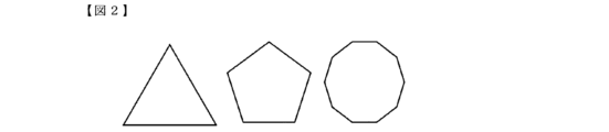
- the shape of the rectangular attachment surface is not limited to the quadrilateral shape of the implant device in FIG. 1, but also includes polygonal shapes such as a triangle, pentagon, or decagon as shown in FIG. 2, and a cross shape as shown in FIG. 3, etc.
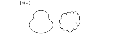
- the attachment surface of the cylinder is not limited to a circle, but may also be, for example, an ellipse, a gourd-shaped or cloud-shaped shape as shown in FIG. 4, a fan-shaped, half-moon-shaped or teardrop-shaped shape as shown in FIG. 5, a broach-shaped shape as shown in FIG. 6, a lens-shaped shape as shown in FIG. 7, etc.
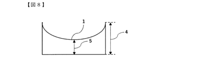
- the surfaces that make up the external structure may have one curved surface as shown in FIG. 8, or one concave or convex shape as shown in FIG. 9.
- the thickness of an implant medical device refers to the distance between the attachment surface and the surface facing it when the implant medical device is a rectangular parallelepiped or cylinder.
- the thickness is measured at multiple locations, including the maximum and minimum thicknesses, and is averaged to ensure that there is no deviation within the surface.
- the implant medical device is a pyramid
- the thickness is half the length of the short side of the base, when it is a sphere, the radius, and when it is a cone, the radius of the base.
- the distance between the apex of the curve in the curved surface and the other surface is the minimum thickness 4, and the distance between the end of the curved surface and the other surface is the maximum thickness 5.
- the distance between the apex of the concave surface and the other surface is the minimum thickness 4, and the distance between the surfaces without any unevenness is the maximum thickness 5.
- the thickness of these implant medical devices can be measured by the method described in Measurement Example 5.
- the thickness of the implant medical device of the present invention is preferably 1.0 to 20.0 mm, more preferably 1.2 to 10.0 mm, even more preferably 1.5 to 5.0 mm, and most preferably 2.0 to 3.0 mm.
- the direction perpendicular to the thickness direction is defined as the vertical direction
- the direction perpendicular to the thickness direction and the vertical direction is defined as the horizontal direction
- the vertical length of the implant medical device of the present invention defined as the vertical length and the horizontal length defined as the horizontal length.
- the vertical length and horizontal length are selected so that their sums are maximized.
- the range in which it can be placed in the affected area can be selected from ranges of 50 cm or less, 30 cm or less, 15 cm or less, or 5 cm or less.
- the lower limit is 0.1 cm or more.
- the thickness of the implant medical device is always smaller than the vertical and horizontal lengths of the implant medical device, and the horizontal length is the same as or smaller than the vertical length.
- the bulk density of the implant medical device refers to the value obtained by dividing the weight of the implant medical device by the volume including the pores and the internal voids, and can be measured by the method described in Measurement Example 6. From the viewpoint of controlling the strength and flexibility within a suitable range, the bulk density of the implant medical device is preferably 0.40 g/cm 3 or more and 1.50 g/cm 3 or less, more preferably 0.45 g/cm 3 or more and 1.40 g/cm 3 or less, even more preferably 0.60 g/cm 3 or more and 1.30 g/cm 3 or less, and most preferably 0.80 g/cm 3 or more and 1.20 g/cm 3 or less.
- PRP platelet-rich plasma
- the implant medical device of the present invention preferably includes at least two layers, a first layer and a second layer.
- a first layer with excellent PRP carrying ability and a second layer with high strength it is possible to exhibit functions suitable for soft tissue repair applications.
- layers other than the first and second layers may be included, but the thickness of these layers must be smaller than that of the first and second layers.
- the bulk density of the first layer and the bulk density of the second layer can be measured by the method described in Measurement Example 6.
- the bulk density of the first layer is preferably 0.20 g/cm3 or more and 0.95 g/cm3 or less , more preferably 0.25 g/cm3 or more and 0.85 g/cm3 or less , even more preferably 0.30 g/cm3 or more and 0.75 g/cm3 or less , and most preferably 0.35 g/cm3 or more and 0.60 g/cm3 or less .
- the bulk density of the second layer is preferably greater than 0.95 g/ cm3 and less than 2.00 g/cm3, more preferably greater than 1.00 g/ cm3 and less than 1.80 g/ cm3 , even more preferably greater than 1.05 g/cm3 and less than 1.65 g/ cm3 , and most preferably greater than 1.10 g/ cm3 and less than 1.50 g/cm3.
- the combination of bulk densities of the first and second layers may be any combination so long as the bulk density of each layer is within the above range and the bulk density of the implant medical device as a whole is within the above range; however, from the viewpoint of controlling the PRP loading capacity within a suitable range, the difference between the bulk density of the first layer and the bulk density of the second layer is preferably 0.10 g/cm3 or more and 0.90 g/cm3 or less, more preferably 0.15 g/cm3 or more and 0.80 g/cm3 or less , and even more preferably 0.20 g/cm3 or more and 0.75 g/ cm3 or less.
- the following method can be used as a pretreatment for measuring the bulk density of the first layer and the second layer.
- the cross section of the implant medical device is observed with an optical microscope, a scanning electron microscope, or the like to confirm the boundary surface between the first layer and the second layer, and the implant medical device is cut along the boundary surface at a plane sufficiently distant from the boundary surface to obtain all or part of the first layer and the second layer. All or part of the obtained first layer and second layer are measured using the method described in Measurement Example 6.
- the ratio of the thickness of the second layer to the overall thickness of the implant medical device can be measured by the method described in Measurement Example 7. From the viewpoint of controlling the bulk density of the implant medical device within a suitable range, the ratio of the thickness of the second layer to the thickness of the implant medical device is preferably 5% or more and 40% or less, more preferably 7% or more and 35% or less, and even more preferably 10% or more and 30% or less.
- the vertical and horizontal lengths of the first and second layers may be different, but it is preferable that the vertical and horizontal lengths of the first and second layers are the same.
- the first and second layers are preferably composed of the same components.
- the decomposition rates of the first and second layers are equal, and it is expected that the entire implant medical device will be decomposed and absorbed uniformly.
- the first and second layers it is possible to intentionally change the decomposition period and maximize the therapeutic effect on soft tissue.
- the present invention is used for soft tissue regeneration applications, it is preferable that the first layer is used as the application surface.
- the maximum tensile load of the implant medical device of the present invention can be measured by the method described in Measurement Example 8. Specifically, the maximum tensile load refers to the maximum load observed in a single measurement.
- the maximum tensile load of the implant medical device is preferably 20 N or more, more preferably 50 N or more, and even more preferably 70 N or more. There is no particular upper limit, but as a feasible range, 2,000 N or less, 1,000 N or less, or 500 N or less can be selected.
- the tensile modulus of the implant medical device of the present invention can be measured by the method described in Measurement Example 8.
- the tensile modulus of the implant medical device is preferably 100 MPa or less, more preferably 50 MPa or less, even more preferably 20 MPa or less, and most preferably 10 MPa or less. There is no particular lower limit, but the practicable range can be selected to be 0.01 MPa or more, 0.05 MPa or more, or 0.10 MPa or more.
- the storage length of the implant medical device of the present invention can be measured by the method described in Measurement Example 9.
- the storage length of the implant medical device of the present invention is preferably 50 mm or less, more preferably 30 mm or less, even more preferably 15 mm or less, and most preferably 10 mm or less.
- the smaller the storage length the smaller the tube that can be stored in, so by setting the storage length within the above range, it is possible to quickly deliver the device to the affected area during surgery using an endoscope.
- the implant medical device of the present invention After surgical treatment for soft tissue repair, platelet-rich plasma (PRP) may be injected into the affected area to promote tissue repair. For this reason, it is preferable that the implant medical device of the present invention has the ability to carry PRP.
- PRP platelet-rich plasma
- the PRP carrying capacity can be evaluated by the amount of PRP carried per unit weight of the implant medical device, and can be measured by the method described in Measurement Example 10.
- the amount of PRP injected in a typical treatment is 1 mL, and in order for the implant medical device to carry the entire amount of injected PRP, the PRP carrying capacity must be 0.20 g/g or more. Therefore, the PRP carrying capacity of the implant medical device of the present invention is preferably 0.20 g/g or more, more preferably 0.40 g/g or more, even more preferably 0.60 g/g or more, and most preferably 0.80 g/g or more.
- the implant medical device of the present invention can be produced by various methods such as injection molding, extrusion molding, freeze-drying, electrospinning, or 3D printing. From the viewpoint of precisely controlling the bulk density, however, it is preferable to produce it by freeze-drying, electrospinning, or 3D printing, and more preferably by 3D printing.
- 3D printers include, for example, fused deposition modeling (FDM), stereolithography (SLA or DLP), inkjet, powder sintering (SLS or SLM), and powder bonding.
- the implant medical device of the present invention is preferably produced by the fused deposition modeling (FDM), stereolithography (SLA or DLP) or powder sintering (SLS or SLM), and more preferably produced by the fused deposition modeling (FDM) or powder sintering (SLS or SLM).
- the thickness of each layer can be set.
- any layer pitch can be selected, for example, between 0.01 mm and 1 mm.
- Infill patterns include line, grid, triangle, trihexagon, cubic subdivision, octet, quarter cubic, zigzag, cross, cross 3D, star, cubic, honeycomb, 3D honeycomb, gyroid, Himbert curve, Archimedes code, and octagram spiral, but when producing the implant medical device of the present invention, line, grid, triangle, trihexagon, zigzag, star, cubic, concentric circle, honeycomb, 3D honeycomb, or gyroid are preferred.
- the implant medical device of the present invention is used to repair soft tissue, and is particularly suitable for repairing ruptured soft tissue. When used to repair ruptured soft tissue, it is preferably fixed to muscle and bone or bone and bone. In a method for repairing a ruptured tendon, rotator cuff, or ligament, it is preferable to have a fixing step of fixing the implant medical device of the present invention to muscle and bone or bone and bone.
- Biodegradability (%) (BOD-B)/TOD ⁇ 100...Formula 2
- BOD Oxygen consumption of polyester copolymer or implant medical device
- B Blank oxygen consumption
- TOD Theoretical oxygen consumption required when polyester copolymer or implant medical device is completely oxidized
- polyhydroxycarboxylic acid has two types of hydroxycarboxylic acid monomers as main constituent units
- the two types of hydroxycarboxylic acid monomers are designated as "monomer A” and “monomer B", respectively, and the molar fractions (%) of monomer A residues and monomer B residues were calculated.
- the peaks of monomer A and monomer B were separated by the signal derived from monomer A or monomer B of adjacent monomer residues by 1H homospin decoupling method, and the respective peak areas were quantified. [AB] was calculated from the respective peak area ratios, and the R value was calculated from the following formula 1.
- R value [AB] / (2 [A] [B]) x 100 ...
- Formula 1 [A]: Molar fraction (%) of monomer A residue in polyhydroxycarboxylic acid
- [AB] mole fraction (%) of structures in which monomer A residue and monomer B residue are adjacent to each other (AB and BA) in polyhydroxycarboxylic acid
- [AB] is the molar fraction (%) of structures (A-B and B-A) in which a monomer A residue and a monomer B residue are adjacent to each other in the polyhydroxycarboxylic acid, and specifically, is the ratio of the number of A-B and B-A to the total number of A-A, A-B, B-A, and B-B.
- 1H homospin decoupling method was performed under the following condition 1, and adjacent monomer residues were separated by signals derived from LA or CL for the methine group of LA (near 5.10 ppm), the ⁇ -methylene group of CL (near 2.35 ppm), and the ⁇ -methylene group (near 4.10 ppm), and the respective peak areas were quantified.
- Condition 1 Device name: JNM-ECZ400R (manufactured by JEOL Ltd.) 1 H homospin decoupling irradiation position: 1.66 ppm
- Solvent deuterated chloroform Measurement temperature: room temperature
- the obtained film (about 10 mg) was collected on an alumina PAN and subjected to DSC measurement under the following condition 3.
- the crystallization rate was calculated from the measurement results under temperature conditions (D) to (E) using the following formulas 3 and 4.
- the heat of fusion per unit mass of the homopolymer was determined as follows.
- the homopolymer was dissolved in chloroform to a concentration of 5% by mass, and the solution was transferred onto a PTFE petri dish and dried overnight at normal pressure and room temperature. This was dried under reduced pressure to obtain a film.
- the obtained film was collected on an alumina PAN and subjected to DSC measurement under the following condition 4, and the heat of fusion was read from the melting peak area of the graph obtained from the measurement results under temperature conditions (D) to (E).
- Condition 4 Device name: EXSTAR 6000 (Seiko Instruments Inc.) Temperature conditions: (A) 25°C ⁇ (B) 250°C (10°C/min) ⁇ (C) 250°C (5 minutes) ⁇ (D) -70°C (10°C/min) ⁇ (E) 250°C (10°C/min) ⁇ (F) 250°C (5 minutes) ⁇ (G) 25°C (100°C/min) Standard material: Alumina
- the thickness of the implant medical device was measured at 10 points, including the maximum and minimum thicknesses, without causing any in-plane bias, using a constant pressure thickness measuring device, and the average value was regarded as the thickness of the implant medical device.
- the bulk density of the implant medical device, first layer or second layer (hereafter referred to as the sample) was measured using the following procedure. To dry the sample, vacuum drying at room temperature may be performed, but no other pretreatment is required.
- Step 1 The mass of the sample in a dry state was measured using an electronic balance, and the displayed value was designated as W1.
- Step 2 Prepare a container filled with water on an electronic balance, and immerse the sample in the water so that the entire sample is immersed but does not touch the bottom. The value displayed at this time was W1-W2. The value of W2 was calculated from the value of W1 calculated in step 1 and the value of W1-W2.
- Step 3 The sample was taken out of the water, and the mass of the wet sample was measured using an electronic balance. The displayed value was designated as W3.
- PRP PRP Supporting Ability
- PRP was prepared as follows: 25 mL of bovine whole blood was placed in a 50 mL centrifuge tube, centrifuged under the following condition (1), and the supernatant was collected. The remaining liquid was then centrifuged under the following condition (2), and the supernatant was collected and mixed with the previous supernatant to prepare PRP.
- Condition (1) 140 ⁇ g, 10 min, 4° C. ⁇ 200 ⁇ g, 10 min, 4° C. ⁇ 300 ⁇ g, 10 min, 4° C.
- Conditions (2) 3000 rpm, 10 minutes, 4°C
- the weight of the implant medical device in a dry state was measured and designated as Wi.
- 15 mL of PRP was placed in a polystyrene container, and the implant medical device was immersed and left to stand for 5 minutes. The implant medical device was then removed and its weight was measured.
- the weight of PRP supported on the implant medical device was calculated from the weight difference of the implant medical device before and after immersion in PRP, and designated as Wp.
- the PRP support capacity was calculated from the following formula 7.
- PRP carrying capacity Wp/Wi...Formula 7
- the obtained crude copolymer was dissolved in 260 mL of chloroform and added dropwise to 3000 mL of hexane in a stirred state to obtain a precipitate.
- the precipitate was dried under reduced pressure at 50° C. to obtain macromer 1.
- the reaction mixture was diluted with 240 mL of chloroform, and 10.0 g of D,L-lactic acid (manufactured by Fujifilm Wako Pure Chemical Industries, Ltd.) was added and stirred for 30 minutes.
- 500 mL of 0.5 M hydrochloric acid was added, and the mixture was stirred for 20 minutes, and the aqueous layer was removed by decantation.
- 500 mL of ion-exchanged water was added, and the mixture was stirred for 10 minutes, and the process of removing the aqueous layer by decantation was repeated until the pH of the removed aqueous layer reached 7.
- the remaining organic layer was dropped into 3,000 mL of methanol while stirring to obtain a precipitate. This precipitate was dried under reduced pressure at 50°C to obtain polyester copolymer 1.
- the obtained crude copolymer was dissolved in 100 mL of chloroform and added dropwise to 1,400 mL of stirred methanol to obtain a precipitate. This procedure was repeated three times, and the precipitate was dried under reduced pressure at 70°C to obtain Macromer 2.
- dichloromethane dehydrated
- dicyclohexylcarbodiimide manufactured by Sigma-Aldrich Co., Ltd.
- the obtained crude copolymer was dissolved in 100 mL of chloroform and added dropwise to 1,400 mL of stirred methanol to obtain a precipitate. This operation was repeated three times, and the obtained precipitate was dried under reduced pressure at 50°C to obtain polyester copolymer 3.
- the obtained crude copolymer was dissolved in 100 mL of chloroform and added dropwise to 1,400 mL of stirred methanol to obtain a precipitate. This procedure was repeated three times, and the precipitate was dried under reduced pressure at 70°C to obtain Macromer 3.
- dichloromethane dehydrated
- dicyclohexylcarbodiimide manufactured by Sigma-Aldrich Co., Ltd.
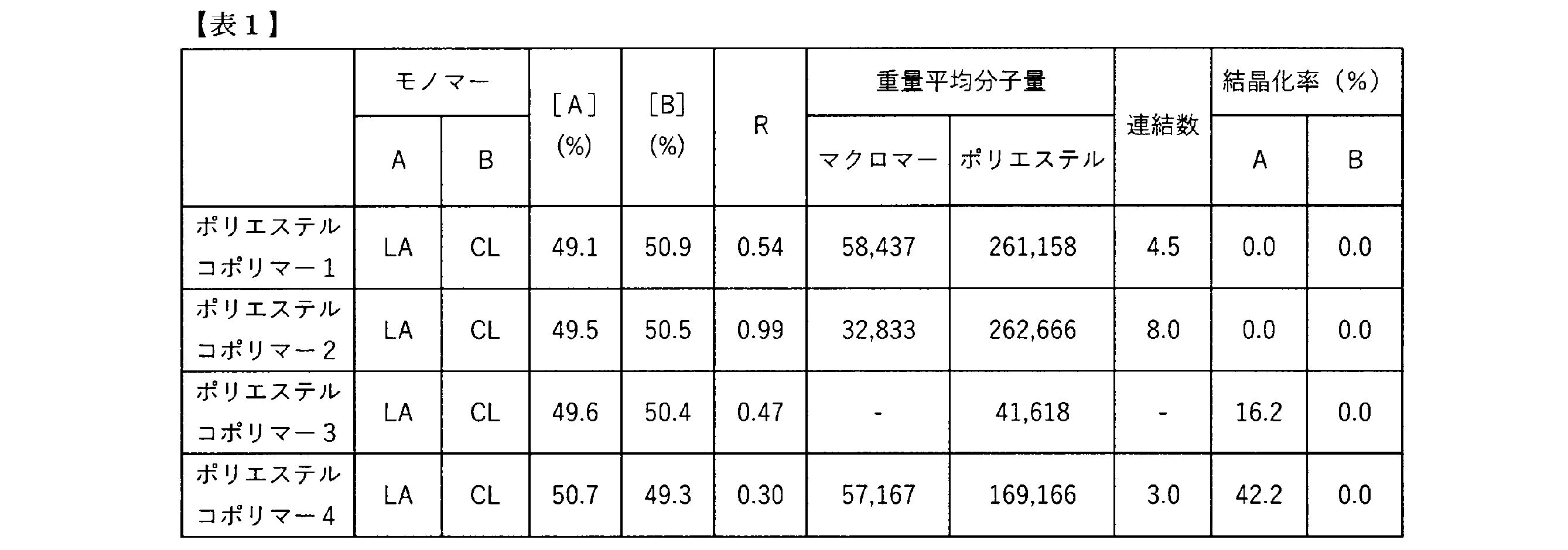
- polyester copolymers 1 to 4 were subjected to the measurements described in Measurement Examples 1 to 4. The results are shown in Table 1.
- Example 1 An implant medical device was produced by 3D printing using polyester copolymer 1 so that the length and width were 35 mm, the thickness was 2 mm, the bulk density of the first layer was 0.55 g/ cm3 , the bulk density of the second layer was 1.10 g/ cm3 , and the thickness ratio of the second layer was 30%.
- a fused deposition model (FDM) 3D printer was used to produce the device, with the layer pitch set to 0.2 mm and the infill pattern set to a grid.
- Example 2 An implant medical device was produced by 3D printing using polyester copolymer 1 so that the length and width were 35 mm, the thickness was 2 mm, the bulk density of the first layer was 0.35 g/ cm3 , the bulk density of the second layer was 1.10 g/ cm3 , and the thickness ratio of the second layer was 10%.
- a fused deposition model (FDM) 3D printer was used to produce the device, with the layer pitch set to 0.2 mm and the infill pattern set to a grid.
- Example 3 An implant medical device was produced by 3D printing using polyester copolymer 1 so that the length and width were 35 mm, the thickness was 2 mm, the bulk density of the first layer was 0.85 g/ cm3 , the bulk density of the second layer was 1.00 g/ cm3 , and the thickness ratio of the second layer was 10%.
- a fused deposition model (FDM) 3D printer was used to produce the device, with the layer pitch set to 0.2 mm and the infill pattern set to a grid.
- Example 4 An implant medical device was produced by 3D printing using polyester copolymer 1 so that the length and width were 35 mm, the thickness was 2 mm, and the bulk density was 1.10 g/ cm3 . Note that a fused deposition model (FDM) 3D printer was used, and the layer pitch was set to 0.2 mm and the infill pattern was set to a grid.
- FDM fused deposition model
- Example 5 Using polyester copolymer 2, an implant medical device was produced in the same manner as in Example 1.
- Example 6 Using polyester copolymer 3, an implant medical device was produced in the same manner as in Example 1.
- Example 7 Using polyester copolymer 4, an implant medical device was produced in the same manner as in Example 1.
- An implant medical device was produced by 3D printing using polyester copolymer 1 so that the length and width were 35 mm, the thickness was 0.4 mm, the first layer bulk density was 0.55 g/ cm3 , the second layer bulk density was 1.10 g/ cm3 , and the thickness ratio of the second layer was 50%.
- a fused deposition model (FDM) 3D printer was used to produce the device, with the layer pitch set to 0.2 mm and the infill pattern set to a grid.
- An implant medical device was produced by 3D printing using polyester copolymer 1 so that the length and width were 35 mm, the thickness was 2 mm, and the bulk density was 0.35 g/cm 3.
- a fused deposition model (FDM) 3D printer was used, and the layer pitch was set to 0.2 mm and the infill pattern was set to a grid.
- An implant medical device was produced by 3D printing using polyester copolymer 1 so that the length and width were 35 mm, the thickness was 20 mm, the bulk density of the first layer was 0.55 g/ cm3 , the bulk density of the second layer was 1.10 g/ cm3 , and the thickness ratio of the second layer was 30%.
- a fused deposition model (FDM) 3D printer was used to produce the device, with the layer pitch set to 0.2 mm and the infill pattern set to a grid.
- An implant medical device was produced by 3D printing using polyester copolymer 1 so that the length and width were 35 mm, the thickness was 2 mm, the bulk density of the first layer was 0.20 g/ cm3 , the bulk density of the second layer was 1.10 g/ cm3 , and the thickness ratio of the second layer was 30%.
- a fused deposition model (FDM) 3D printer was used to produce the device, with the layer pitch set to 0.2 mm and the infill pattern set to a grid.
- Example 10 An implant medical device was produced by 3D printing using polyester copolymer 1 so that the length and width were 35 mm, the thickness was 2 mm, the bulk density of the first layer was 0.15 g/ cm3 , the bulk density of the second layer was 1.10 g/ cm3 , and the thickness ratio of the second layer was 10%.
- a fused deposition model (FDM) 3D printer was used to produce the device, with the layer pitch set to 0.2 mm and the infill pattern set to a grid.
- Example 1 and Comparative Example 1 From the results of Example 1 and Comparative Example 1, it was revealed that a maximum tensile load suitable for soft tissue repair applications can be obtained by setting the thickness of the implant medical device to 1.0 mm or more . Also, from the results of Example 1 and Comparative Example 2, it was revealed that a maximum tensile load suitable for soft tissue repair applications can be obtained by setting the bulk density of the implant medical device to 0.40 g/cm3 or more.
- the implant medical device of the present invention can be used for repairing soft tissues, particularly as a repair material for cartilage, tendons, rotator cuffs, and ligaments.
Landscapes
- Health & Medical Sciences (AREA)
- Chemical & Material Sciences (AREA)
- Transplantation (AREA)
- Dermatology (AREA)
- Medicinal Chemistry (AREA)
- Oral & Maxillofacial Surgery (AREA)
- Chemical Kinetics & Catalysis (AREA)
- Epidemiology (AREA)
- Life Sciences & Earth Sciences (AREA)
- Animal Behavior & Ethology (AREA)
- General Health & Medical Sciences (AREA)
- Public Health (AREA)
- Veterinary Medicine (AREA)
- Materials For Medical Uses (AREA)
Abstract
本発明は、強度と柔軟性に優れた、インプラント医療機器を提供すること。本発明は、2種類のヒドロキシカルボン酸モノマーを主構成単位とするポリヒドロキシカルボン酸を含むポリエステルコポリマーを有し、厚みが1.0mm以上20.0mm以下、かつ、嵩密度が0.40g/cm3以上1.50g/cm3以下である、軟組織修復用のインプラント医療機器を提供する。
Description
本発明は、軟組織修復用のインプラント医療機器及びその製造方法に関する。
腱、腱が集合し板状となった腱板、及び靱帯は、筋肉と骨又は骨と骨を結合する組織であり、肩、肘、膝、手首、足首等の関節を円滑に可動するために重要な役割を果たしている。加齢による変性、外傷又はスポーツ等によるオーバーユースによって腱、腱板や靱帯が損傷、断裂することがあり、日常動作に支障を来し、運動時だけでなく安静時や夜間にも痛みを生じる。
腱、腱板や靱帯のような軟組織を損傷した場合の治療として、断裂した腱、腱板や靱帯と、骨とを物理的に結合する外科的手術が行われてきたが、例えば肩の回旋腱板修復手術の場合には40%以上の割合で再断裂が発生するとの報告があり、広範囲の断裂の再建の場合には90%以上で再断裂が発生するとの報告もある。
再断裂は、外科的に結合した腱、腱板や靱帯の組織が修復する過程において力学的強度が保持できていないことが原因で発生するとの考え方から、腱、腱板や靱帯と骨の結合部位にシート状の再建用材料で補強して力学的強度の保持や生物学的治癒促進を図る治療法が盛んに行われつつある。特に、修復した後に体内に残らないという点から生分解性の再建用材料が注目され、生物由来又は合成ポリマー由来の生分解性材料が本用途に使用されている。
合成ポリマーからなるインプラント医療機器として、ポリ乳酸の繊維からなり、縦糸と横糸の本数が制御されたメッシュやポリ乳酸からなるフォームであり、アニーリングしたあと急冷することで有機溶媒に対する耐溶解性を向上させたメッシュで補強されたフォームが開示されている(特許文献1及び2)。
また、柔軟性を有する合成ポリマーとして、乳酸とカプロラクトンからなる生分解性ポリエステルが記載されており(特許文献3)、特許文献3に記載の生分解性ポリエステルを用いた医療用成型体として、生体内に留置することを想定し、留置箇所周辺に硬組織がない場合にその成形体のヤング率を低く制御して軟組織と同様に扱うようにすることを可能とした医療用成型体が開示されている(特許文献4)。
しかしながら特許文献1及び2に記載のインプラント医療機器は、硬い合成ポリマーであるポリ乳酸を使用しているため、強度に優れるものの、柔軟性が不足しており、軟組織への追従性という観点で課題が残る。また、このような硬い合成ポリマーを使用した場合でも、厚みや密度を非常に小さくすることで柔軟性を付与できる可能性があるが、ポリマーが分解されやすいために軟組織の修復が完了する前に分解、吸収されてしまう。
一方、特許文献3には、乳酸とカプロラクトンからなる生分解性ポリエステルの記載はあるが、軟組織修復用のインプラント機器に適用するための具体的な記載はない。また、特許文献4には、特許文献3記載の生分解性ポリエステルを用いた医療用成形体、医療機器、神経再生誘導チューブが記載しているが、軟組織修復用のインプラント機器に適用するための具体的な記載はなく、例えば特許文献1及び2に記載の形状(特に厚みや嵩密度)とした場合、強度が不足してしまう。そのため、特許文献3及び4に記載されている柔らかい合成ポリマーを用いた場合であっても、軟組織修復用のインプラント機器に適用するためには厚みや嵩密度を適切な範囲に制御する必要がある。
そこで本発明は、柔らかい合成ポリマーである2種類のヒドロキシカルボン酸モノマーを主構成単位とするポリヒドロキシカルボン酸を含む生分解性のポリエステルコポリマーを用い、かつ、厚みと嵩密度を適切な範囲に制御することで、強度と柔軟性に優れた、軟組織修復用のインプラント医療機器を提供することを目的とする。
本発明者らは、上記課題を解決すべく鋭意研究を重ねた結果、以下の(1)~(19)の発明を見出した。
(1) 2種類のヒドロキシカルボン酸モノマーを主構成単位とするポリヒドロキシカルボン酸を含むポリエステルコポリマーを有し、厚みが1.0mm以上20.0mm以下、かつ、嵩密度が0.40g/cm3以上1.50g/cm3以下である、軟組織修復用のインプラント医療機器。
(2) 第一層及び第二層を有し、上記第一層の嵩密度が0.20g/cm3以上0.95g/cm3以下である、(1)記載のインプラント医療機器。
(3) 第一層及び第二層を有し、該第一層と該第二層の嵩密度は異なり、厚みが1.0mm以上20.0mm以下、かつ、全体の嵩密度が0.40g/cm3以上1.50g/cm3以下である、軟組織修復用のインプラント医療機器。
(4) 2種類のヒドロキシカルボン酸モノマーを主構成単位とするポリヒドロキシカルボン酸を含むポリエステルコポリマーを有する、(3)記載のインプラント医療機器。
(5) 上記第一層の嵩密度が0.20g/cm3以上0.95g/cm3以下である、(3)又は(4)記載のインプラント医療機器。
(6) 上記第一層の嵩密度と上記第二層の嵩密度の差は、0.10g/cm3以上0.90g/cm3以下である、(2)~(5)のいずれか記載のインプラント医療機器。
(7) 上記第二層の嵩密度が0.95g/cm3より大きく2.00g/cm3以下である、(2)~(6)のいずれか記載のインプラント医療機器。
(8) 上記インプラント医療機器の厚みに対する上記第二層の厚みの比率が5%以上40%以下である、(2)~(7)のいずれか記載のインプラント医療機器。
(9) 上記第一層と第二層が同じ成分で構成される、(2)~(8)のいずれか一項記載のインプラント医療機器。
(10) 引張最大負荷が20N以上2,000N以下である、(1)~(9)のいずれか記載のインプラント医療機器。
(11) 引張弾性率が0.01MPa以上100MPa以下である、(1)~(10)のいずれか記載のインプラント医療機器。
(12) 生分解性である、(1)~(11)のいずれか記載のインプラント医療機器。
(13) 上記2種類のヒドロキシカルボン酸モノマーをそれぞれ「モノマーA」、「モノマーB」とした場合に、上記ポリヒドロキシカルボン酸の下記の式1で表されるR値が0.25以上0.99以下である、(1)、(2)、(4)~(12)のいずれか記載のインプラント医療機器。
R値 = [AB]/(2[A][B])×100 ・・・式1
[A]:ポリヒドロキシカルボン酸中の、モノマーA残基のモル分率(%)
[B]:ポリヒドロキシカルボン酸中の、モノマーB残基のモル分率(%)
[AB]:ポリヒドロキシカルボン酸中の、モノマーA残基とモノマーB残基が隣り合った構造(A-B、及びB-A)のモル分率(%)
(14) 上記モノマーAは、乳酸、グリコール酸、ジラクチド及びグリコリドからなる群から選択されるモノマーであり、上記モノマーBは、ヒドロキシ吉草酸、ヒドロキシカプロン酸、バレロラクトン及びカプロラクトンからなる群から選択されるモノマーである、(13)記載のインプラント医療機器。
(15) ポリエステルコポリマーのみからなる、(1)~(14)のいずれか記載のインプラント医療機器。
(16) 上記軟組織は、腱、腱板又は靭帯である、(1)~(15)のいずれか記載のインプラント医療機器。
(17) 筋肉と骨、又は、骨と骨に固定化して使用される、(1)~(16)のいずれか記載の断裂した軟組織の修復用のインプラント医療機器。
(18) (1)~(17)のいずれか記載のインプラント医療機器を筋肉と骨又は骨と骨に固定化する固定工程を有する、断裂した腱、腱板又は靭帯を修復する方法。
(19) (1)~(17)のいずれか記載のインプラント医療機器の製造方法。
さらに、本発明者らは、上記課題を解決すべく鋭意研究を重ねた結果、以下の(20)~(23)の発明を見出した。
(20) 2種類のヒドロキシカルボン酸モノマーを主構成単位とするポリヒドロキシカルボン酸を含むポリエステルコポリマーを有し、厚みが1.0mm以上20.0mm以下、かつ、嵩密度が0.40g/cm3以上1.50g/cm3以下である、軟組織修復用のインプラント医療機器。
(21) 第一層及び第二層を有し、上記第一層の嵩密度が0.20g/cm3以上0.95g/cm3以下である、(20)記載のインプラント医療機器。
(22) 上記2種類のヒドロキシカルボン酸モノマーをそれぞれ「モノマーA」、「モノマーB」とした場合に、上記ポリヒドロキシカルボン酸の下記の式1で表されるR値が0.25以上0.99以下である、(20)又は(21)記載のインプラント医療機器。
R値 = [AB]/(2[A][B])×100 ・・・式1
[A]:ポリヒドロキシカルボン酸中の、モノマーA残基のモル分率(%)
[B]:ポリヒドロキシカルボン酸中の、モノマーB残基のモル分率(%)
[AB]:ポリヒドロキシカルボン酸中の、モノマーA残基とモノマーB残基が隣り合った構造(A-B、及びB-A)のモル分率(%)
(23) (20)記載のインプラント医療機器の製造方法。
本発明は、軟組織修復用途に適したインプラント医療機器を提供することができる。
本発明の軟組織修復用のインプラント医療機器は、2種類のヒドロキシカルボン酸モノマーを主構成単位とするポリヒドロキシカルボン酸を含むポリエステルコポリマーを有し、厚みが1.0mm以上20.0mm以下、かつ、嵩密度が0.40g/cm3以上1.50g/cm3以下であることを特徴とする。
また、本発明の軟組織修復用のインプラント医療機器は、第一層及び第二層を有し、該第一層と該第二層の嵩密度は異なり、厚みが1.0mm以上20.0mm以下、かつ、全体の嵩密度が0.40g/cm3以上1.50g/cm3以下であることを特徴とする。
本明細書における軟組織とは、人体の構成要素のうち、骨を除いた組織のことを指すが、その中でも軟骨、腱、腱板、靭帯のことを指し、特に腱、腱板及び靭帯のことを指す。
本発明におけるポリエステルコポリマーとは、主にエステル結合でモノマーが連結されたポリマーのうち、2種以上のモノマーからなるポリマーを指す。本明細書中においては、ウレタン結合やアミド結合等、エステル結合以外の結合でモノマーが一部連結されたポリマーもポリエステルという。
生分解性を好適な範囲に制御するためには、モノマー連結に用いられる結合のうち、エステル結合以外の結合の数は、モノマー連結に用いられる結合の数を100%とした場合のうち、30%以下が好ましく、10%以下がより好ましく、5%以下がさらに好ましく、0%、すなわちエステル結合以外を含まない場合が、もっとも好ましい。
本発明におけるポリエステルコポリマーは、ポリヒドロキシカルボン酸を含む。ポリヒドロキシカルボン酸とは、ヒドロキシカルボン酸から構成されるポリマーを指す。なお、ヒドロキシカルボン酸とは、同じ分子内にヒドロキシ基とカルボキシ基の両基をもつ化合物を指す。
ポリヒドロキシカルボン酸は生分解性を有するため、ポリヒドロキシカルボン酸を含むポリエステルコポリマーは、生分解性を備えうる。また、ポリエステルコポリマーが生分解性を有するため、ポリエステルコポリマーを含む本発明のインプラント医療機器も生分解性を備える。
軟組織修復用途に好適に用いるためには、本発明のインプラント医療機器が生分解性を有することが好ましい。好適な生分解性を持たせるため、本発明のポリエステルコポリマーは、ポリエステルコポリマー100質量%に対して、ポリヒドロキシカルボン酸を50質量%以上含むことが好ましく、70質量%以上含むことがより好ましく、90質量%以上含むことがさらに好ましく、100質量%含む、すなわちポリヒドロキシカルボン酸のみからなる場合がもっとも好ましい。
本発明のインプラント医療機器が生分解性を備えるという観点から、本発明に用いるインプラント医療機器は、インプラント医療機器100質量%に対して、ポリエステルコポリマーを50質量%以上含むことが好ましく、70質量%以上含むことがより好ましく、90質量%以上含むことがさらに好ましく、100質量%含むこと、すなわちインプラント医療機器がポリエステルコポリマーのみからなることがもっとも好ましい。
本明細書において生分解性とは、生体内や環境中で分解される性質のことを指す。より具体的には、後述の測定例1に従って求めた生分解度が60%以上であることを指す。生分解性と互換的に使用され得る用語として生体吸収性、生体適合性等が挙げられるが、これらの用語は主に生体内で分解される性質を指すことが多い。
ポリヒドロキシカルボン酸は、ヒドロキシカルボン酸の重合や、ヒドロキシカルボン酸のヒドロキシ基とカルボキシル基が分子内脱水縮合した環状化合物であるラクトンの重合、2分子のヒドロキシカルボン酸の互いのヒドロキシ基とカルボキシル基が脱水縮合した環状化合物であるラクチドの重合等で得ることができる。すなわち、ヒドロキシカルボン酸モノマーとして、ヒドロキシカルボン酸、ラクトン又はラクチドを用いることができる。
ヒドロキシカルボン酸モノマーに用いられるヒドロキシカルボン酸としては、乳酸、グリコール酸、ヒドロキシ酪酸、ヒドロキシ吉草酸、ヒドロキシカプロン酸、ヒドロキシヘプタン酸、ヒドロキシオクタン酸、ヒドロキシノナン酸、ヒドロキシデカン酸、ヒドロキシウンデカン酸、ヒドロキシドデカン酸及び(2-ヒドロキシエトキシ)酢酸等が挙げられる。なかでも、乳酸、グリコール酸、ヒドロキシ吉草酸又はヒドロキシカプロン酸が好ましく、特に乳酸又はヒドロキシカプロン酸が好ましい。
ヒドロキシカルボン酸モノマーに用いられるラクトンとしては、ブチロラクトン、バレロラクトン、カプロラクトン(以下、CL)、ジオキセパノン、エチレンオキサラート、p-ジオキサノン、トリメチレンカーボネート、β-プロピオラクトン又はピバロラクトン等を用いることができ、特にブチロラクトン、バレロラクトン、CL、p-ジオキサノン又はトリメチレンカーボネートが好ましく、バレロラクトン又はCLがさらに好ましい。
ヒドロキシカルボン酸モノマーに用いられるラクチドとしては、乳酸2分子が脱水縮合したジラクチド(以下、LA)や、グリコール酸2分子が脱水縮合したグリコリド又はテトラメチルグリコリドを用いることができ、特にLA又はグリコリドが好ましい。
本発明のポリヒドロキシカルボン酸は、2種類のヒドロキシカルボン酸モノマーを主構成単位とする。ポリヒドロキシカルボン酸が、2種類のヒドロキシカルボン酸モノマーを主構成単位とする、とは、当該2種類のモノマー残基数の和が、その他のモノマー残基を含めたポリヒドロキシカルボン酸全体に含まれる全てのモノマー残基数の和を100モル%とした場合に50モル%以上であり、かつ、それぞれの残基がポリヒドロキシカルボン酸全体に含まれる全てのモノマー残基数の和を100モル%とした場合に10モル%以上であることを意味する。
本明細書においてモノマー残基とは、原則として、当該モノマーを含む2種以上のモノマーを重合して得られたポリヒドロキシカルボン酸の化学構造中における、当該モノマーに由来する化学構造の反復単位をいう。
例えば、モノマーA残基とモノマーB残基とを主構成単位とする、とは、モノマーA残基とモノマーB残基の残基数の和が、ポリヒドロキシカルボン酸全体に含まれる全てのモノマー残基数の和を100モル%とした場合に50モル%以上であり、かつモノマーA残基が10モル%以上であり、かつモノマーB残基が10モル%以上であることを意味する。
ここで、モノマーA残基、モノマーB残基、その他の残基のモル分率(%)は、核磁気共鳴(以下「NMR」という)測定により、それぞれの残基に由来するシグナルの面積値より決定できる。例えば、モノマーA残基がLA残基、モノマーB残基がCL残基である場合には、後述する測定例2により求めることができる。
なお、モノマーA残基、モノマーB残基及びその他の残基のモル分率(%)を計算する際は、当該モノマーに由来する化学構造の反復単位の分子量を用いて算出する。例えば、モノマーA残基がLA残基、モノマーB残基がCL残基である場合には、モノマーA残基の反復単位の分子量は144、モノマーB残基の反復単位の分子量は114となる。
モノマーA残基とモノマーB残基の和のモル分率(%)は、上述の定義から、その他のモノマー残基を含めたポリマー全体に含まれる全てのモノマー残基数の和を100モル%とした場合に50モル%以上であり、75モル%以上であることが好ましく、90モル%以上であることがより好ましい。
また、モノマーA残基及びモノマーB残基のモル分率(%)は、同じく上述の定義からそれぞれ10モル%以上であり、20モル%以上であることが好ましく、30モル%以上であることがより好ましく、40モル%以上であることがさらに好ましく、50モル%以上であることがもっとも好ましい。モノマーA残基及びモノマーB残基の和がポリマー全体の100モル%である、すなわちモノマーA及びモノマーBのみからなるポリマーは、特に好ましい態様として挙げられる。
さらに、2種類のヒドロキシカルボン酸モノマーをそれぞれ「モノマーA」、「モノマーB」とした場合に、下記の式1で表されるR値が0.25以上0.99以下である(以下、R値が0.25以上0.99以下であるポリヒドロキシカルボン酸を「ランダムポリマー」という)場合、本発明のより好ましい態様である。
R値 = [AB]/(2[A][B])×100 ・・・式1
[A]:ポリヒドロキシカルボン酸中の、モノマーA残基のモル分率(%)
[B]:ポリヒドロキシカルボン酸中の、モノマーB残基のモル分率(%)
[AB]:ポリヒドロキシカルボン酸中の、モノマーA残基とモノマーB残基が隣り合った構造(A-B、及びB-A)のモル分率(%)
R値 = [AB]/(2[A][B])×100 ・・・式1
[A]:ポリヒドロキシカルボン酸中の、モノマーA残基のモル分率(%)
[B]:ポリヒドロキシカルボン酸中の、モノマーB残基のモル分率(%)
[AB]:ポリヒドロキシカルボン酸中の、モノマーA残基とモノマーB残基が隣り合った構造(A-B、及びB-A)のモル分率(%)
ポリヒドロキシカルボン酸としてランダムポリマーを用いる、つまりポリヒドロキシカルボン酸のR値を上記範囲とすることで、柔軟なインプラント医療機器が得られるため好ましい。柔軟性の観点から、R値の下限は0.25以上が好ましく、0.45以上がより好ましい。
本発明に用いられるモノマーA及びモノマーBは、乳酸、グリコール酸、ヒドロキシ酪酸、ヒドロキシ吉草酸、ヒドロキシカプロン酸、ヒドロキシヘプタン酸、ヒドロキシオクタン酸、ヒドロキシノナン酸、ヒドロキシデカン酸、ヒドロキシウンデカン酸、ヒドロキシドデカン酸、(2-ヒドロキシエトキシ)酢酸、ブチロラクトン、バレロラクトン、CL、ジオキセパノン、エチレンオキサラート、p-ジオキサノン、トリメチレンカーボネート、β-プロピオラクトン、ピバロラクトン、LA、グリコリド及びテトラメチルグリコリドから選択されるモノマーであることが好ましい。
モノマーA及びモノマーBにおいて、モノマーAは、乳酸、グリコール酸、LA及びグリコリドからなる群から選択される少なくとも1つのモノマーであることが特に好ましい。また、モノマーBは、ヒドロキシ吉草酸、ヒドロキシカプロン酸、バレロラクトン及びCLからなる群から選択される少なくとも1つのモノマーであることが特に好ましい。
本発明におけるポリエステルコポリマーの重量平均分子量は、測定例3に記載の方法で測定することができる。強度と生分解性を好適な範囲に制御する観点から、ポリエステルコポリマーの重量平均分子量は、50,000以上が好ましく、100,000以上がより好ましく、150,000以上がさらに好ましく、200,000以上がもっとも好ましい。重量平均分子量が大きくなるにつれて強度は高くなり、生分解に要する日数が多くなる。成形性の観点からは、ポリエステルコポリマーの重量平均分子量は、1,000,000以下が好ましく、800,000以下がより好ましく、500,000以下がさらに好ましい。
本発明におけるポリエステルコポリマーは、ポリエステルコポリマー同士が連結したマルチポリエステルコポリマーであってもよい。このとき連結前のポリエステルコポリマーを便宜上「マクロマー」と表現する。本明細書ではポリエステルコポリマー1分子を構成するマクロマーの平均分子数を連結数と呼ぶ。連結数はポリエステルコポリマーの重量平均分子量をマクロマーの重量平均分子量で除した値を指す。
マルチポリエステルコポリマーは、ポリエステルコポリマーをそのまま連結又はリンカー分子を用いて連結することで作製することができる。マルチポリエステルコポリマーが直鎖構造をとるか、分岐構造をとるかは、ポリエステルコポリマーの構造及びリンカー分子の構造によって決定される。
ポリエステルコポリマーが分岐構造の場合、連結の仕方によらずマルチポリエステルコポリマーは分岐構造となる。ポリエステルコポリマーが直鎖構造の場合、そのまま連結又は直鎖構造のリンカー分子を用いて連結すると、マルチポリエステルコポリマーは直鎖構造となり、分岐構造のリンカー分子を用いて連結すると、マルチポリエステルコポリマーは分岐構造となる。
直鎖構造のリンカー分子として、カルボキシル基、ヒドロキシ基及びアミノ基等の官能基を分子内に2つ有する化合物を用いることができ、例えばシュウ酸、マロン酸、コハク酸、グルタル酸、アジピン酸、ピメリン酸、スベリン酸、アゼライン酸、セバシン酸、ドデカン二酸、エチレングリコール、プロパンジオール、ブタンジオール、ペンタンジオール、ヘキサンジオール、ヘプタンジオール、オクタンジオール、ポリエチレングリコール、ポリプロピレングリコール、ポリエチレングリコール―ポリプロピレングリコール共重合体、エチレンジアミン、プロパンジアミン、ブタンジアミン、ジアミノペンタン、ヘキサメチレンジアミン、ジアミノヘプタン、ジアミノオクタン、乳酸、グリコール酸、カプロン酸、アラニン、イソロイシン、ロイシン、メチオニン、バリン、フェニルアラニン、トリプトファン、アスパラギン、システイン、グルタミン、アルギニン、ヒスチジン及びグリシン等が挙げられる。
また、分岐構造のリンカー分子として、カルボキシル基、ヒドロキシ基及びアミノ基等の官能基を分子内に2つ以上有する化合物を用いることができ、例えばリンゴ酸、酒石酸、クエン酸、チロシン、セリン、スレオニン、アスパラギン酸、グルタミン酸及びリジン等が挙げられる。
本発明のポリヒドロキシカルボン酸は、モノマーA残基又はモノマーB残基の少なくとも一方の結晶化率が14%未満であることが好ましい。当該結晶化率が14%未満であれば、インプラント医療機器の引張弾性率の上昇が抑えられ、軟組織修復用途に適したインプラント医療機器を得ることができる。モノマーA残基又はモノマーB残基の結晶化率は、10%以下であることがより好ましく、5%以下であることがさらに好ましい。なお、すべてのモノマー残基の結晶化率が低いことが好ましいが、一部のモノマー残基の結晶化率のみが低くてもよい。
本明細書においてモノマー残基の結晶化率とは、あるモノマー残基のみからなるホモポリマーの単位重量当たりの融解熱と本発明のポリヒドロキシカルボン酸中の当該モノマー残基の重量分率の積に対する、本発明のポリヒドロキシカルボン酸中の当該モノマー残基単位重量当たりの融解熱の割合である。すなわち、モノマーA残基の結晶化率とは、モノマーAのみからなるホモポリマーの単位重量あたりの融解熱と本発明のポリヒドロキシカルボン酸中のモノマーA残基の重量分率の積に対する、ポリヒドロキシカルボン酸中のモノマーA残基単位重量当たりの融解熱の割合である。
モノマーA残基及びモノマーB残基の結晶化率は、それぞれ本発明のポリヒドロキシカルボン酸のモノマーA残基又はモノマーB残基の中で結晶構造を形成している割合を示す。
特に、モノマーA残基がLAに由来する乳酸残基、モノマーB残基がCLに由来するカプロラクトン残基である場合には、乳酸残基の結晶化率は14%未満であることが好ましく、10%以下であることがより好ましい。結晶化率は、具体的には後述する測定例4に記載の方法で求めるものとする。
本発明のインプラント医療機器の構造の一例を図1に示す。図1のインプラント医療機器は、それぞれの厚みと嵩密度が異なる、ポリエステルコポリマーからなるフィルム状の層(第一層2)とポリエステルコポリマーからなるフィルム状の層(第二層3)が重ね合わせられて形成された直方体の構造を有する。
ここで、インプラント医療機器が直方体の場合は、もっとも面積の大きい面及びその向かい合う面のうち嵩密度の低い方の層が含む面を便宜上、貼付面と呼ぶ。また、本発明のインプラント医療機器が円柱の場合は、2つの底面のうち嵩密度の低い方の層が含む底面を便宜上、貼付面と呼ぶ。図1のインプラント用医療機器であれば、直方体の構造であるため、第一層2が有する面積の大きい外面が貼付面1となる。
本発明のインプラント医療機器の外形構造として、図1では直方体の構造を挙げたが、これに限定されない。外形構造の具体的な例として、直方体、角錐、球、円柱及び円錐等が挙げられるが、直方体又は円柱であることが好ましい。
なお、本発明において、直方体の貼付面の形状は、図1のインプラント機器が有する四角形だけでなく、例えば、図2に示すような三角形、五角形、十角形等の多角形や、図3に示す十字型の形状等も含む。
また、円柱の貼付面は、円だけでなく、例えば、楕円や、図4に示すひょうたん型及び雲型、図5に示す扇型、半月型及び涙滴型の形状、図6に示すブローチ型、図7に示すレンズ型等のような形状等も含む。また、外形構造を構成する面は図8のように一方の面が湾曲面であってもよく、図9に示すように一方の面が凹面や凸面を有する形状であってもよい。
本発明におけるインプラント医療機器の厚みとは、インプラント医療機器が直方体及び円柱の場合は、貼付面とその向かい合う面間の距離を指す。厚みが均一でない場合は、最大厚みと最小厚みを含み、かつ面内で偏りが出ないように複数箇所厚みを測定し、その平均値をインプラント医療機器の厚みとする。また、インプラント医療機器が角錐の場合は底面の短辺の長さの半分の値を、球の場合は半径を、円錐の場合は底面の半径をインプラント医療機器の厚みとする。具体的な例として、図8の湾曲面を持つ形状であれば、湾曲面内の湾曲の頂点ともう一方の面間の距離を最小厚み4とし、湾曲面の端ともう一方の面間の距離を最大厚み5とする。また、図9に示す凹面を有する形状の場合であれば、凹面の頂点ともう一方の面間の距離を最小厚み4とし、凹凸の無い面間の距離を最大厚み5とする。これらのインプラント医療機器の厚みは、測定例5に記載の方法で測定することができる。
本発明のインプラント医療機器の厚みは、強度と柔軟性を好適な範囲に制御する観点から1.0~20.0mmが好ましく、1.2~10.0mmがより好ましく、1.5~5.0mmがさらに好ましく、2.0~3.0mmがもっとも好ましい。
また、本明細書では厚み方向と垂直な方向を縦方向、厚み方向及び縦方向と垂直な方向を横方向とし、本発明のインプラント医療機器の縦方向の長さを縦長さ、横方向の長さを横長さとする。なお縦長さと横長さはそれぞれの和が最大になるように選択する。本発明のインプラント医療機器の縦長さ及び横長さの範囲は特に定めないが、患部に留置可能な範囲としては、50cm以下、30cm以下、15cm以下又は5cm以下の範囲を選択可能である。一方で作製可能という観点から下限は0.1cm以上である。なお上記インプラント医療機器の厚みはインプラント医療機器の縦長さ及び横長さよりも必ず小さく、横長さは縦長さと同じか縦長さよりも小さい。
上記インプラント医療機器の嵩密度は、インプラント医療機器の重量を、細孔も内部の空隙も含めた体積で除した値を指し、測定例6に記載の方法で測定することができる。上記強度と柔軟性を好適な範囲に制御する観点から、インプラント医療機器の嵩密度は、0.40g/cm3以上1.50g/cm3以下が好ましく、0.45g/cm3以上1.40g/cm3以下がより好ましく、0.60g/cm3以上1.30g/cm3がさらに好ましく、0.80g/cm3以上1.20g/cm3以下がもっとも好ましい。一方、後述する多血小板血漿(以下、PRP)の担持能を好適な範囲に制御する観点からは、1.10g/cm3以下が好ましく、1.00g/cm3以下がより好ましく、0.90g/cm3以下がさらに好ましく、0.80g/cm3以下がもっとも好ましい。
本発明のインプラント医療機器は、第一層及び第二層の少なくとも二つの層を含むことが好ましい。PRP担持能に優れた第一層と高い強度を有する第二層とを含む構造とすることで、軟組織修復用途に適した機能を発揮することができる。なお、第一層及び第二層以外の層を含んでもよいが、第一層及び第二層の厚みよりも小さくなければならない。
第一層の嵩密度及び第二層の嵩密度は、測定例6に記載の方法で測定することができる。PRPの担持能を好適な範囲に制御する観点から、第一層の嵩密度は、0.20g/cm3以上0.95g/cm3以下が好ましく、0.25g/cm3以上0.85g/cm3以下がより好ましく、0.30g/cm3以上0.75g/cm3以下がさらに好ましく、0.35g/cm3以上0.60g/cm3以下がもっとも好ましい。
強度及び柔軟性を好適な範囲に制御する観点から、上記第二層の嵩密度は、0.95g/cm3より大きく2.00g/cm3以下が好ましく、1.00g/cm3以上1.80g/cm3以下がより好ましく、1.05g/cm3以上1.65g/cm3以下がさらに好ましく、1.10g/cm3以上1.50g/cm3以下がもっとも好ましい。
第一層と第二層の嵩密度の組合せは、それぞれの層の嵩密度が上記の範囲であり、かつ、インプラント医療機器の全体としての嵩密度が上記の範囲であればどのような組み合わせであってもよいが、PRPの担持能を好適な範囲に制御する観点から、第一層の嵩密度と第二層の嵩密度の差が0.10g/cm3以上0.90g/cm3以下であることが好ましく、0.15g/cm3以上0.80g/cm3以下であることがより好ましく、0.20g/cm3以上0.75g/cm3以下であることがさらに好ましい。
第一層の嵩密度及び第二層の嵩密度を測定するための前処理としては次のような方法が挙げられる。インプラント医療機器の断面を光学顕微鏡や走査型電子顕微鏡等で観察することで、第一層と第二層の間に存在する境界面を確認し、境界面に沿って、境界面よりも十分に離れた面でインプラント医療機器を切断し、第一層及び第二層の全部又は一部を得る。得られた第一層及び第二層の全部又は一部を測定例6に記載の方法で測定を行う。
インプラント医療機器の全体の厚みに対する第二層の厚みの比率は、測定例7に記載の方法で測定できる。インプラント医療機器の嵩密度を好適な範囲に制御する観点から、インプラント医療機器の厚みに対する第二層の厚みの比率は、5%以上40%以下が好ましく、7%以上35%以下がより好ましく、10%以上30%以下がさらに好ましい。
第一層と第二層における縦長さ、横長さは、第一層と第二層でそれぞれの長さが異なっていてもよいが、第一層と第二層における縦長さ、横長さがともに同一の長さであることが好ましい。
本発明のインプラント医療機器は、第一層と第二層が同じ成分で構成されることが好ましい。同じ成分で構成されることにより、第一層と第二層の分解速度が等しくなり、インプラント医療機器全体が均一に分解、吸収されることが期待される。一方で、第一層と第二層に異なる成分を用いることで、意図的に分解期間を変化させ、軟組織の治療効果を最大化することも可能である。また、本発明を軟組織再生用途に使用する場合、第一層を貼付面とすることが好ましい。
本発明のインプラント医療機器の引張最大負荷は、測定例8に記載の方法で測定することができる。具体的には引張最大負荷はある一回の測定で観測される最大の負荷を指す。インプラント医療機器の引張最大負荷は20N以上が好ましく、50N以上がより好ましく、70N以上がさらに好ましい。上限は特に定めないが、実施可能な範囲としては、2,000N以下、1,000N以下又は500N以下を選択可能である。
本発明のインプラント医療機器の引張弾性率は、測定例8に記載の方法で測定することができる。インプラント医療機器の引張弾性率は、100MPa以下が好ましく、50MPa以下がより好ましく、20MPa以下がさらに好ましく、10MPa以下がもっとも好ましい。下限は特に定めないが、実施可能な範囲としては、0.01MPa以上、0.05MPa以上又は0.10MPa以上を選択可能である。
本発明のインプラント医療機器の収納長さは、測定例9に記載の方法で測定することができる。本発明のインプラント医療機器の収納長さは、50mm以下が好ましく、30mm以下がより好ましく、15mm以下がさらに好ましく、10mm以下がもっとも好ましい。下限は特に定めないが、実施可能な範囲としては、0.1mm以上、1mm以上又は3mm以上を選択可能である。収納長さが小さいほど内径の小さな筒に収納することが可能なため、収納長さを上記範囲とすることで、内視鏡を用いた手術の際に、患部に速やかに送達することが可能である。
軟組織修復のための外科的治療を行った後、組織の修復を促進させる目的で多血小板血漿(PRP)を患部に注入することがある。そのため本発明のインプラント医療機器はPRP担持能を有することが好ましい。
PRP担持能はインプラント医療機器の単位重量当たりのPRPの担持量で評価することができ、測定例10に記載の方法で測定することができる。一般的な治療におけるPRPの注入量は1mLであり、注入されたPRP全量をインプラント医療機器が担持するためには、PRP担持能は0.20g/g以上必要である。そのため、本発明のインプラント医療機器のPRP担持能は、0.20g/g以上が好ましく、0.40g/g以上がより好ましく、0.60g/g以上がさらに好ましく、0.80g/g以上がもっとも好ましい。
本発明のインプラント医療機器は、射出成型、押出成形、凍結乾燥、電界紡糸法又は3Dプリント等様々な方法で作製することが可能であるが、嵩密度を精密に制御する観点から、凍結乾燥、電界紡糸法又は3Dプリントで作製することが好ましく、3Dプリントで作製することがより好ましい。
3Dプリンターには、例えば熱溶解積層方式(FDM方式)、光造形方式(SLA方式又はDLP方式)、インクジェット方式、粉末焼結方式(SLS方式又はSLM方式)及び粉末固着方式等の方式が挙げられるが、本発明のインプラント医療機器は、熱溶解積層方式(FDM方式)、光造形方式(SLA方式又はDLP方式)又は粉末焼結方式(SLS方式又はSLM方式)で作製することが好ましく、熱溶解積層方式(FDM方式)又は粉末焼結方式(SLS方式又はSLM方式)で作製することがより好ましい。
3Dプリントで作製する場合、積層していく各層の厚み(積層ピッチ)を設定できる。本発明のインプラント医療機器を作製する際は、任意の積層ピッチを選択することができ、例えば0.01mm以上1mm以下である。
また、3Dプリントで作製する場合、造形物の内側の充填方法(インフィルパターン)を選択することができる。インフィルパターンとしては、ライン、グリッド、トライアングル、トライヘキサゴン、キュービックサブディビジョン、オクテット、クオーターキュービック、ジグザグ、クロス、クロス3D、スター、キュービック、ハニカム、3Dハニカム、ジャイロイド、ヒンベルト曲線、アルキメデスコード及びオクタグラムスパイラル等が挙げられるが、本発明のインプラント医療機器を作製する際には、ライン、グリッド、トライアングル、トライヘキサゴン、ジグザグ、スター、キュービック、同心円、ハニカム、3Dハニカム又はジャイロイドが好ましい。
本発明のインプラント医療機器は、軟組織を修復するために用いられるが、特に断裂した軟組織を修復するために好適に用いることができる。断裂した軟組織の修復用に用いるためには、筋肉と骨又は骨と骨に固定化して使用されることが好ましい。断裂した腱、腱板又は靭帯を修復する方法においては、本発明のインプラント医療機器を筋肉と骨又は骨と骨に固定化する固定工程を有することが好ましい。
以下、実施例及び比較例を挙げて本発明を詳細に説明するが、本発明はこれらに限定されるものではない。
(測定例1:生分解性評価)
JIS K6950(2000)に規定される方法に準じて測定を行った。具体的には、培養びんに無機塩溶液(300mL)、活性汚泥(9mg)、及びポリエステルコポリマー又はインプラント医療機器(30mg)を加え、25℃で90日間攪拌した。その際、溶存酸素計を用いて、撹拌前後の溶存酸素量を求め、その差を酸素消費量とし、各材料を加えた際の酸素消費量を基に下記の式2に従って生分解度を算出した。この際、生分解度が60%以上のものを、生分解性を有する、とした。
生分解度(%) = (BOD-B)/TOD×100 ・・・式2
BOD:ポリエステルコポリマー又はインプラント医療機器の酸素消費量
B :ブランクの酸素消費量
TOD:ポリエステルコポリマー又はインプラント医療機器が完全に酸化された場合に必要とされる理論的酸素消費量
(測定例2:各残基のモル分率及びR値)
精製したポリヒドロキシカルボン酸を重クロロホルムに溶解し、1H-NMRを測定してポリヒドロキシカルボン酸中のモノマー残基のモル分率(%)を算出した。
精製したポリヒドロキシカルボン酸を重クロロホルムに溶解し、1H-NMRを測定してポリヒドロキシカルボン酸中のモノマー残基のモル分率(%)を算出した。
なお、ポリヒドロキシカルボン酸が、2種類のヒドロキシカルボン酸モノマーを主構成単位とする場合、上記2種類のヒドロキシカルボン酸モノマーをそれぞれ「モノマーA」、「モノマーB」とし、モノマーA残基及びモノマーB残基のモル分率(%)をそれぞれ算出した。また、必要に応じて、1Hホモスピンデカップリング法により、モノマーA及びモノマーBのピークについて、隣り合うモノマー残基がモノマーA若しくはモノマーBに由来するシグナルで分離し、それぞれのピーク面積を定量した。それぞれのピーク面積比から、[AB]を計算し、下記式1よりR値を算出した。
R値 = [AB]/(2[A][B])×100 ・・・式1 [A]:ポリヒドロキシカルボン酸中の、モノマーA残基のモル分率(%)
[B]:ポリヒドロキシカルボン酸中の、モノマーB残基のモル分率(%)
[AB]:ポリヒドロキシカルボン酸中の、モノマーA残基とモノマーB残基が隣り合った構造(A-B及びB-A)のモル分率(%)
ここで、[AB]はポリヒドロキシカルボン酸中の、モノマーA残基とモノマーB残基が隣り合った構造(A-B、及びB-A)のモル分率(%)であり、具体的にはA-A、A-B、B-A、B-Bの総数に対するA-B、B-Aの数の割合である。
R値 = [AB]/(2[A][B])×100 ・・・式1 [A]:ポリヒドロキシカルボン酸中の、モノマーA残基のモル分率(%)
[B]:ポリヒドロキシカルボン酸中の、モノマーB残基のモル分率(%)
[AB]:ポリヒドロキシカルボン酸中の、モノマーA残基とモノマーB残基が隣り合った構造(A-B及びB-A)のモル分率(%)
ここで、[AB]はポリヒドロキシカルボン酸中の、モノマーA残基とモノマーB残基が隣り合った構造(A-B、及びB-A)のモル分率(%)であり、具体的にはA-A、A-B、B-A、B-Bの総数に対するA-B、B-Aの数の割合である。
例えば、モノマーAがLA、モノマーBがCLの場合、下記の条件1で1Hホモスピンデカップリング法を行い、LAのメチン基(5.10ppm付近)、CLのαメチレン基(2.35ppm付近)、εメチレン基(4.10ppm付近)について、隣り合うモノマー残基がLA又はCLに由来するシグナルで分離し、それぞれのピーク面積を定量した。
条件1:
機器名:JNM-ECZ400R(日本電子株式会社製)
1Hホモスピンデカップリング照射位置:1.66ppm
溶媒:重クロロホルム
測定温度:室温
条件1:
機器名:JNM-ECZ400R(日本電子株式会社製)
1Hホモスピンデカップリング照射位置:1.66ppm
溶媒:重クロロホルム
測定温度:室温
(測定例3:重量平均分子量)
ポリエステルコポリマーをクロロホルムに溶解し、0.45μmのシリンジフィルター(DISMIC-13HP;ADVANTEC社製)を通過させて不純物等を除去した後に下記の条件2にてGPC測定を行い、ポリエステルコポリマーの重量平均分子量を算出した。
条件2:
機器名:Prominence(登録商標;株式会社島津製作所製)
移動相:クロロホルム(HPLC用)(和光純薬工業株式会社製)
流速:1mL/分
カラム:TSKgel(登録商標) GMHHR-M(φ7.8mm×300mm;東ソー株式会社製)
検出器:UV(254nm)、RI
カラム、検出器温度:35℃
標準物質:ポリスチレン
ポリエステルコポリマーをクロロホルムに溶解し、0.45μmのシリンジフィルター(DISMIC-13HP;ADVANTEC社製)を通過させて不純物等を除去した後に下記の条件2にてGPC測定を行い、ポリエステルコポリマーの重量平均分子量を算出した。
条件2:
機器名:Prominence(登録商標;株式会社島津製作所製)
移動相:クロロホルム(HPLC用)(和光純薬工業株式会社製)
流速:1mL/分
カラム:TSKgel(登録商標) GMHHR-M(φ7.8mm×300mm;東ソー株式会社製)
検出器:UV(254nm)、RI
カラム、検出器温度:35℃
標準物質:ポリスチレン
(測定例4:DSC測定による結晶化率)
ポリヒドロキシカルボン酸を濃度が5質量%になるようにクロロホルムに溶解させ、その溶液をポリテトラフルオロエチレン(以下、PTFE)製シャーレ上に移して、常圧、室温下で1昼夜乾燥させた。これを50℃で一昼夜減圧乾燥させて、厚さ約100μmのフィルムを得た。
ポリヒドロキシカルボン酸を濃度が5質量%になるようにクロロホルムに溶解させ、その溶液をポリテトラフルオロエチレン(以下、PTFE)製シャーレ上に移して、常圧、室温下で1昼夜乾燥させた。これを50℃で一昼夜減圧乾燥させて、厚さ約100μmのフィルムを得た。
得られたフィルム(約10mg)をアルミナPANに採取し、下記の条件3でDSC測定を行い、温度条件(D)から(E)の測定結果から、下記の式3及び式4を用いて結晶化率を算出した。
条件3:
機器名:EXSTAR 6000(セイコーインスツル株式会社製)
温度条件:(A)25℃→(B)250℃(10℃/分)→(C)250℃(5分)→(D)-70℃(10℃/分)→(E)250℃(10℃/分)→(F)250℃(5分)→(G)25℃(100℃/分)
標準物質:アルミナ
モノマーAの結晶化率 = (ポリヒドロキシカルボン酸の単位質量当たりのモノマーA残基由来の融解熱)/{(モノマーA残基のみからなるホモポリマーの単位質量当たり融解熱)×(ポリヒドロキシカルボン酸中のモノマーA残基の質量分率)}×100 ・・・式3
モノマーBの結晶化率 = (ポリヒドロキシカルボン酸の単位質量当たりのモノマーB残基由来の融解熱)/{(モノマーB残基のみからなるホモポリマーの単位質量当たり融解熱)×(ポリヒドロキシカルボン酸中のモノマーB残基の質量分率)}×100 ・・・式4
ここで、ホモポリマーの単位質量当たり融解熱は、次のように求めた。ホモポリマーを濃度が5質量%になるようにクロロホルムに溶解させ、その溶液をPTFE製シャーレ上に移して、常圧、室温下で一昼夜乾燥させた。これを減圧乾燥させて、フィルムを得た。得られたフィルムをアルミナPANに採取し、下記の条件4でDSC測定を行い、温度条件(D)から(E)の測定結果から得られる、グラフの融解ピーク面積から融解熱を読み取った。
条件4:
機器名:EXSTAR 6000(セイコーインスツル株式会社製)
温度条件:(A)25℃→(B)250℃(10℃/分)→(C)250℃(5分)→(D)-70℃(10℃/分)→(E)250℃(10℃/分)→(F)250℃(5分)→(G)25℃(100℃/分)
標準物質:アルミナ
条件4:
機器名:EXSTAR 6000(セイコーインスツル株式会社製)
温度条件:(A)25℃→(B)250℃(10℃/分)→(C)250℃(5分)→(D)-70℃(10℃/分)→(E)250℃(10℃/分)→(F)250℃(5分)→(G)25℃(100℃/分)
標準物質:アルミナ
(測定例5:インプラント医療機器の厚み)
インプラント医療機器の最大厚みと最小厚みを含み、かつ、面内で偏りが出ないように10箇所の厚みを、定圧厚さ測定器を用いて測定し、その平均値をインプラント医療機器の厚みとした。
インプラント医療機器の最大厚みと最小厚みを含み、かつ、面内で偏りが出ないように10箇所の厚みを、定圧厚さ測定器を用いて測定し、その平均値をインプラント医療機器の厚みとした。
(測定例6:インプラント医療機器の嵩密度、第一層の嵩密度、第二層の嵩密度)
本明細書では以下のように単語を定義する。
開気孔 :物質表面に存在する、外気と接続された空隙。
閉気孔 :物質内部に孤立している、外気と接続されていない空隙。
乾燥質量:物質のみの質量。W1と表す。
液中質量:物質を液体に浸した際の質量。W2と表す。
湿潤質量:物質と開気孔に満たされた液体の質量の和。W3と表す。
本明細書では以下のように単語を定義する。
開気孔 :物質表面に存在する、外気と接続された空隙。
閉気孔 :物質内部に孤立している、外気と接続されていない空隙。
乾燥質量:物質のみの質量。W1と表す。
液中質量:物質を液体に浸した際の質量。W2と表す。
湿潤質量:物質と開気孔に満たされた液体の質量の和。W3と表す。
インプラント医療機器、第一層又は第二層(以下サンプルと呼ぶ)の嵩密度は次の手順で測定を行った。なおサンプルを乾燥させるために、常温で真空乾燥を行ってもよいが、その他の前処理は不要である。
手順1:乾燥状態のサンプルの質量を、電子天秤を用いて測定し、このとき表示された値をW1とした。
手順2:電子天秤の上に水の入った容器を準備し、サンプル全体が浸るようにかつサンプルが底に接触しないように水中に沈め、このとき表示された値をW1-W2とした。また、手順1で求めたW1の値とW1-W2の値からW2の値を求めた。
手順3:水中からサンプルを取り出し、湿潤状態のサンプルの質量を、電子天秤を用いて測定し、このとき表示された値をW3とした。
手順2:電子天秤の上に水の入った容器を準備し、サンプル全体が浸るようにかつサンプルが底に接触しないように水中に沈め、このとき表示された値をW1-W2とした。また、手順1で求めたW1の値とW1-W2の値からW2の値を求めた。
手順3:水中からサンプルを取り出し、湿潤状態のサンプルの質量を、電子天秤を用いて測定し、このとき表示された値をW3とした。
手順1~3で得られたW1、W2、W3の値と下記の式5を用いてサンプルの嵩密度を求めた。なお、サンプルが水に溶解してしまう場合は、水の代わりに密度が既知の液体を用いて上述の手順1~3を実施することで嵩密度を求めることができる。
嵩密度 = (W1×ρ)/(W3-W2) ・・・式5
ρ:手順1~3で用いた液体の密度。
ρの値について、上述の通り水を用いた場合はρ=1である。
嵩密度 = (W1×ρ)/(W3-W2) ・・・式5
ρ:手順1~3で用いた液体の密度。
ρの値について、上述の通り水を用いた場合はρ=1である。
(測定例7:全体の厚みに対する第二層の厚みの比率)
インプラント医療機器を厚み方向に切断した断面を電子顕微鏡にて観察し、得られた画像をもとに、インプラント医療機器の第二層の厚みを、視野を変えて3回測定した。さらに切断面を変えて前述の測定を3回行った。得られた9つの値の平均値をインプラント医療機器の第二層の厚みとし、下記の式6を用いて、全体の厚みに対する第二層の厚みの比率を求めた。
第二層の厚み比率(%) = 第二層の厚み/インプラント医療機器の厚み ・・・式6
インプラント医療機器を厚み方向に切断した断面を電子顕微鏡にて観察し、得られた画像をもとに、インプラント医療機器の第二層の厚みを、視野を変えて3回測定した。さらに切断面を変えて前述の測定を3回行った。得られた9つの値の平均値をインプラント医療機器の第二層の厚みとし、下記の式6を用いて、全体の厚みに対する第二層の厚みの比率を求めた。
第二層の厚み比率(%) = 第二層の厚み/インプラント医療機器の厚み ・・・式6
(測定例8:引張最大負荷、引張弾性率)
インプラント医療機器をJIS K7161(2014)に規定される方法に準じ、下記の条件5に基づく引張試験を行い、引張最大負荷及び引張弾性率を測定した。各3回測定し、その数平均値を算出することにより、インプラント医療機器の引張最大負荷、引張弾性率を求めた。なお、引張最大負荷及び引張弾性率は小数点第2位を四捨五入した値を用いた。
条件5:
機器名:EZ-LX(株式会社島津製作所製)
初期長:10mm
引張速度:25mm/分
ロードセル:1kN
インプラント医療機器をJIS K7161(2014)に規定される方法に準じ、下記の条件5に基づく引張試験を行い、引張最大負荷及び引張弾性率を測定した。各3回測定し、その数平均値を算出することにより、インプラント医療機器の引張最大負荷、引張弾性率を求めた。なお、引張最大負荷及び引張弾性率は小数点第2位を四捨五入した値を用いた。
条件5:
機器名:EZ-LX(株式会社島津製作所製)
初期長:10mm
引張速度:25mm/分
ロードセル:1kN
(測定例9:収納長さ)
インプラント医療機器を丸め、デジタルノギスを用いて直径を測定した。なお直径又は厚みは、長さ方向と垂直な方向で測定を行った。
インプラント医療機器を丸め、デジタルノギスを用いて直径を測定した。なお直径又は厚みは、長さ方向と垂直な方向で測定を行った。
(測定例10:PRP担持能)
以下の手順でPRPを調製した。牛全血25mLを50mL遠沈管に入れ、下記の条件(1)で遠心分離を行った後、上澄みを採取した。続いて残った液体について下記の条件(2)で遠心分離を行い、上澄みを採取し、先ほどの上澄みと混合し、PRPを調製した。
条件(1):140×g、10分、4℃→200×g、10分、4℃→300×g、10分、4℃
条件(2):3000rpm、10分、4℃
以下の手順でPRPを調製した。牛全血25mLを50mL遠沈管に入れ、下記の条件(1)で遠心分離を行った後、上澄みを採取した。続いて残った液体について下記の条件(2)で遠心分離を行い、上澄みを採取し、先ほどの上澄みと混合し、PRPを調製した。
条件(1):140×g、10分、4℃→200×g、10分、4℃→300×g、10分、4℃
条件(2):3000rpm、10分、4℃
乾燥状態のインプラント医療機器の重量を測定し、これをWiとした。続いてポリスチレン容器にPRP15mLを入れ、インプラント医療機器を浸し5分間静置した。その後インプラント医療機器を引き上げ、インプラント医療機器の重量を測定した。PRP浸漬前後のインプラント医療機器の重量差からインプラント医療機器に担持されているPRPの重量を算出し、これをWpとした。PRP担持能は下記の式7から求めた。
PRP担持能 = Wp/Wi・・・式7
PRP担持能 = Wp/Wi・・・式7
(合成例1)
50.0gのL,L-ジラクチド(PURASORB(登録商標) L;PURAC社製)と、39.6gのε-カプロラクトン(富士フイルム和光純薬工業株式会社製)とを、モノマーとして、0.45gのヒドロキシピバル酸を開始剤として、セパラブルフラスコに採取した。これらをアルゴン雰囲気下とし、37.4mLのキシレン(富士フイルム和光純薬工業株式会社製)を添加し、2.0mLのキシレン(富士フイルム和光純薬工業株式会社製)に溶解した触媒である0.28gのオクチル酸スズ(II)(富士フイルム和光純薬工業株式会社製)を加え、130℃で15時間反応させ、粗コポリマーを得た。得られた粗コポリマーを260mLのクロロホルムに溶解し、攪拌状態にある3000mLのヘキサンに滴下して、沈殿物を得た。沈殿物を50℃で減圧乾燥してマクロマー1を得た。
50.0gのL,L-ジラクチド(PURASORB(登録商標) L;PURAC社製)と、39.6gのε-カプロラクトン(富士フイルム和光純薬工業株式会社製)とを、モノマーとして、0.45gのヒドロキシピバル酸を開始剤として、セパラブルフラスコに採取した。これらをアルゴン雰囲気下とし、37.4mLのキシレン(富士フイルム和光純薬工業株式会社製)を添加し、2.0mLのキシレン(富士フイルム和光純薬工業株式会社製)に溶解した触媒である0.28gのオクチル酸スズ(II)(富士フイルム和光純薬工業株式会社製)を加え、130℃で15時間反応させ、粗コポリマーを得た。得られた粗コポリマーを260mLのクロロホルムに溶解し、攪拌状態にある3000mLのヘキサンに滴下して、沈殿物を得た。沈殿物を50℃で減圧乾燥してマクロマー1を得た。
マクロマー1を50gと、触媒である1.3gのp-トルエンスルホン酸4,4-ジメチルアミノピリジニウム(合成品)と、0.52gの4,4-ジメチルアミノピリジン(富士フイルム和光純薬工業株式会社製)を採取した。これらをアルゴン雰囲気下とし、184mLのジクロロメタン(脱水)(富士フイルム和光純薬工業株式会社製)に溶解し、縮合剤である0.56mLのジイソプロピルカルボジイミド(富士フイルム和光純薬工業株式会社製)を添加し、室温で2終夜縮合重合させた。
反応混合物を240mLのクロロホルムで希釈し、10.0gのD,L-乳酸(富士フイルム和光純薬工業株式会社製)を加え、30分間撹拌した。次に500mLの0.5M塩酸を添加した後20分間攪拌し、デカンテーションで水層を除去した。その後500mLのイオン交換水を加え、10分間攪拌し、デカンテーションで水層を除去する工程を除去した水層のpHが7になるまで繰り返した。残った有機層を、攪拌状態にある3000mLのメタノールに滴下して、沈殿物を得た。この沈殿物を50℃で減圧乾燥し、ポリエステルコポリマー1を得た。
(合成例2)
50.0gのL-ラクチド(PURASORB(登録商標) L;PURAC社製)と、38.5mLのεーカプロラクトン(富士フイルム和光純薬工業株式会社製)とを、モノマーとしてセパラブルフラスコに採取した。これらをアルゴン雰囲気下とし、14.5mLのトルエン(超脱水)(富士フイルム和光純薬工業株式会社製)に溶解した触媒である0.81gのオクチル酸スズ(II)(富士フイルム和光純薬工業株式会社製)、助開始剤としてイオン交換水を、モノマー/助開始剤比が285.7となるよう添加し、90℃で1時間助触媒反応を行ったあと、150℃で12時間共重合反応させて、粗コポリマーを得た。
50.0gのL-ラクチド(PURASORB(登録商標) L;PURAC社製)と、38.5mLのεーカプロラクトン(富士フイルム和光純薬工業株式会社製)とを、モノマーとしてセパラブルフラスコに採取した。これらをアルゴン雰囲気下とし、14.5mLのトルエン(超脱水)(富士フイルム和光純薬工業株式会社製)に溶解した触媒である0.81gのオクチル酸スズ(II)(富士フイルム和光純薬工業株式会社製)、助開始剤としてイオン交換水を、モノマー/助開始剤比が285.7となるよう添加し、90℃で1時間助触媒反応を行ったあと、150℃で12時間共重合反応させて、粗コポリマーを得た。
得られた粗コポリマーを100mLのクロロホルムに溶解し、攪拌状態にある1400mLのメタノールに滴下して、沈殿物を得た。この操作を3回繰り返し、沈殿物を70℃で減圧乾燥してマクロマー2を得た。
マクロマー2を30gと、触媒である1.12gのp-トルエンスルホン酸4,4-ジメチルアミノピリジニウム(合成品)と、0.40gの4,4-ジメチルアミノピリジン(富士フイルム和光純薬工業株式会社製)を採取した。これらをアルゴン雰囲気下とし、20%の濃度となるようジクロロメタン(脱水)(富士フイルム和光純薬工業株式会社製)に溶解し、20mLのジクロロメタンに溶解した縮合剤である5.52gのジシクロヘキシルカルボジイミド(シグマアルドリッチ社製)を添加し、室温で2日間縮合重合させた。
反応混合物に120mLのクロロホルムを添加し、攪拌状態にある2000mLのメタノールに滴下して、沈殿物を得た。この沈殿物を200mLのクロロホルムに溶解し、攪拌状態にある2000mLのメタノールに滴下して、沈殿物を得た。この操作を2回繰り返し、得た沈殿物を50℃で減圧乾燥し、ポリエステルコポリマー2を得た。
(合成例3)
50.0gのL-ラクチド(PURASORB(登録商標) L;PURAC社製)と、38.5mLのεーカプロラクトン(富士フイルム和光純薬工業株式会社製)とを、モノマーとしてセパラブルフラスコに採取した。これらをアルゴン雰囲気下とし、14.5mLのトルエン(超脱水)(富士フイルム和光純薬工業株式会社製)に溶解した触媒である0.81gのオクチル酸スズ(II)(富士フイルム和光純薬工業株式会社製)を添加し、150℃で6時間共重合反応させて、粗コポリマーを得た。
50.0gのL-ラクチド(PURASORB(登録商標) L;PURAC社製)と、38.5mLのεーカプロラクトン(富士フイルム和光純薬工業株式会社製)とを、モノマーとしてセパラブルフラスコに採取した。これらをアルゴン雰囲気下とし、14.5mLのトルエン(超脱水)(富士フイルム和光純薬工業株式会社製)に溶解した触媒である0.81gのオクチル酸スズ(II)(富士フイルム和光純薬工業株式会社製)を添加し、150℃で6時間共重合反応させて、粗コポリマーを得た。
得られた粗コポリマーを100mLのクロロホルムに溶解し、攪拌状態にある1400mLのメタノールに滴下して、沈殿物を得た。この操作を3回繰り返し、得た沈殿物を50℃で減圧乾燥し、ポリエステルコポリマー3を得た。
(合成例4)
50.0gのL-ラクチド(PURASORB(登録商標) L;PURAC社製)をモノマーとしてセパラブルフラスコに採取した。これをアルゴン雰囲気下とし、14.5mLのトルエン(超脱水)(富士フイルム和光純薬工業株式会社製)に溶解した触媒である0.81gのオクチル酸スズ(II)(富士フイルム和光純薬工業株式会社製)、助開始剤としてイオン交換水を142.9のモノマー/助開始剤比となるよう添加し、90℃で1時間助触媒反応を行ったあと、150℃で3時間重合反応させた。
50.0gのL-ラクチド(PURASORB(登録商標) L;PURAC社製)をモノマーとしてセパラブルフラスコに採取した。これをアルゴン雰囲気下とし、14.5mLのトルエン(超脱水)(富士フイルム和光純薬工業株式会社製)に溶解した触媒である0.81gのオクチル酸スズ(II)(富士フイルム和光純薬工業株式会社製)、助開始剤としてイオン交換水を142.9のモノマー/助開始剤比となるよう添加し、90℃で1時間助触媒反応を行ったあと、150℃で3時間重合反応させた。
これに、38.5mLのεーカプロラクトン(富士フイルム和光純薬工業株式会社製)を添加し、さらに150℃で6時間重合反応させ、粗コポリマーを得た。
得られた粗コポリマーを100mLのクロロホルムに溶解し、攪拌状態にある1400mLのメタノールに滴下して、沈殿物を得た。この操作を3回繰り返し、沈殿物を70℃で減圧乾燥してマクロマー3を得た。
マクロマー3を7.5gと、触媒である0.28gのp-トルエンスルホン酸4,4-ジメチルアミノピリジニウム(合成品)と、0.10gの4,4-ジメチルアミノピリジン(富士フイルム和光純薬工業株式会社製)を採取した。これらをアルゴン雰囲気下とし、27%の濃度となるようジクロロメタン(脱水)(富士フイルム和光純薬工業株式会社製)に溶解し、5mLのジクロロメタンに溶解した縮合剤である1.38gのジシクロヘキシルカルボジイミド(シグマアルドリッチ社製)を添加し、室温で2日間縮合重合させた。
反応混合物に30mLのクロロホルムを添加し、攪拌状態にある500mLのメタノールに滴下して、沈殿物を得た。この沈殿物を50mLのクロロホルムに溶解し、攪拌状態にある500mLのメタノールに滴下して、沈殿物を得た。この操作を2回繰り返し、得た沈殿物を50℃で減圧乾燥し、ポリエステルコポリマー4を得た。
得られたポリエステルコポリマー1~4について、測定例1~4に記載の測定を行った。結果を表1に示す。
(実施例1)
ポリエステルコポリマー1を用いて、縦長さ及び横長さが35mm、厚みが2mm、第一層の嵩密度が0.55g/cm3、第二層の嵩密度が1.10g/cm3、第二層の厚み比率が30%になるように3Dプリントでインプラント医療機器を作製した。なお熱溶解積層方式(FDM方式)の3Dプリンターを用い、積層ピッチを0.2mm、インフィルパターンをグリッドに設定し、作製した。
ポリエステルコポリマー1を用いて、縦長さ及び横長さが35mm、厚みが2mm、第一層の嵩密度が0.55g/cm3、第二層の嵩密度が1.10g/cm3、第二層の厚み比率が30%になるように3Dプリントでインプラント医療機器を作製した。なお熱溶解積層方式(FDM方式)の3Dプリンターを用い、積層ピッチを0.2mm、インフィルパターンをグリッドに設定し、作製した。
(実施例2)
ポリエステルコポリマー1を用いて、縦長さ及び横長さが35mm、厚みが2mm、第一層の嵩密度が0.35g/cm3、第二層の嵩密度が1.10g/cm3、第二層の厚み比率が10%になるように3Dプリントでインプラント医療機器を作製した。なお熱溶解積層方式(FDM方式)の3Dプリンターを用い、積層ピッチを0.2mm、インフィルパターンをグリッドに設定し、作製した。
ポリエステルコポリマー1を用いて、縦長さ及び横長さが35mm、厚みが2mm、第一層の嵩密度が0.35g/cm3、第二層の嵩密度が1.10g/cm3、第二層の厚み比率が10%になるように3Dプリントでインプラント医療機器を作製した。なお熱溶解積層方式(FDM方式)の3Dプリンターを用い、積層ピッチを0.2mm、インフィルパターンをグリッドに設定し、作製した。
(実施例3)
ポリエステルコポリマー1を用いて、縦長さ及び横長さが35mm、厚みが2mm、第一層の嵩密度が0.85g/cm3、第二層の嵩密度が1.00g/cm3、第二層の厚み比率が10%になるように3Dプリントでインプラント医療機器を作製した。なお熱溶解積層方式(FDM方式)の3Dプリンターを用い、積層ピッチを0.2mm、インフィルパターンをグリッドに設定し、作製した。
ポリエステルコポリマー1を用いて、縦長さ及び横長さが35mm、厚みが2mm、第一層の嵩密度が0.85g/cm3、第二層の嵩密度が1.00g/cm3、第二層の厚み比率が10%になるように3Dプリントでインプラント医療機器を作製した。なお熱溶解積層方式(FDM方式)の3Dプリンターを用い、積層ピッチを0.2mm、インフィルパターンをグリッドに設定し、作製した。
(実施例4)
ポリエステルコポリマー1を用いて、縦長さ及び横長さが35mm、厚みが2mm、嵩密度が1.10g/cm3になるように3Dプリントでインプラント医療機器を作製した。なお熱溶解積層方式(FDM方式)の3Dプリンターを用い、積層ピッチを0.2mm、インフィルパターンをグリッドに設定し、作製した。
ポリエステルコポリマー1を用いて、縦長さ及び横長さが35mm、厚みが2mm、嵩密度が1.10g/cm3になるように3Dプリントでインプラント医療機器を作製した。なお熱溶解積層方式(FDM方式)の3Dプリンターを用い、積層ピッチを0.2mm、インフィルパターンをグリッドに設定し、作製した。
(実施例5)
ポリエステルコポリマー2を用いて、実施例1と同様の方法でインプラント医療機器を作製した。
ポリエステルコポリマー2を用いて、実施例1と同様の方法でインプラント医療機器を作製した。
(実施例6)
ポリエステルコポリマー3を用いて、実施例1と同様の方法でインプラント医療機器を作製した。
ポリエステルコポリマー3を用いて、実施例1と同様の方法でインプラント医療機器を作製した。
(実施例7)
ポリエステルコポリマー4を用いて、実施例1と同様の方法でインプラント医療機器を作製した。
ポリエステルコポリマー4を用いて、実施例1と同様の方法でインプラント医療機器を作製した。
(比較例1)
ポリエステルコポリマー1を用いて、縦長さ及び横長さが35mm、厚みが0.4mm、第一層嵩密度が0.55g/cm3、第二層嵩密度が1.10g/cm3、第二層の厚み比率が50%になるように3Dプリントでインプラント医療機器を作製した。なお熱溶解積層方式(FDM方式)の3Dプリンターを用い、積層ピッチを0.2mm、インフィルパターンをグリッドに設定し、作製した。
ポリエステルコポリマー1を用いて、縦長さ及び横長さが35mm、厚みが0.4mm、第一層嵩密度が0.55g/cm3、第二層嵩密度が1.10g/cm3、第二層の厚み比率が50%になるように3Dプリントでインプラント医療機器を作製した。なお熱溶解積層方式(FDM方式)の3Dプリンターを用い、積層ピッチを0.2mm、インフィルパターンをグリッドに設定し、作製した。
(比較例2)
ポリエステルコポリマー1を用いて、縦長さ及び横長さが35mm、厚みが2mm、嵩密度が0.35g/cm3になるように3Dプリントでインプラント医療機器を作製した。なお熱溶解積層方式(FDM方式)の3Dプリンターを用い、積層ピッチを0.2mm、インフィルパターンをグリッドに設定し、作製した。
(実施例8)
ポリエステルコポリマー1を用いて、縦長さ及び横長さが35mm、厚みが20mm、第一層の嵩密度が0.55g/cm3、第二層の嵩密度が1.10g/cm3、第二層の厚み比率が30%になるように3Dプリントでインプラント医療機器を作製した。なお熱溶解積層方式(FDM方式)の3Dプリンターを用い、積層ピッチを0.2mm、インフィルパターンをグリッドに設定し、作製した。
(実施例9)
ポリエステルコポリマー1を用いて、縦長さ及び横長さが35mm、厚みが2mm、第一層の嵩密度が0.20g/cm3、第二層の嵩密度が1.10g/cm3、第二層の厚み比率が30%になるように3Dプリントでインプラント医療機器を作製した。なお熱溶解積層方式(FDM方式)の3Dプリンターを用い、積層ピッチを0.2mm、インフィルパターンをグリッドに設定し、作製した。
(実施例10)
ポリエステルコポリマー1を用いて、縦長さ及び横長さが35mm、厚みが2mm、第一層の嵩密度が0.15g/cm3、第二層の嵩密度が1.10g/cm3、第二層の厚み比率が10%になるように3Dプリントでインプラント医療機器を作製した。なお熱溶解積層方式(FDM方式)の3Dプリンターを用い、積層ピッチを0.2mm、インフィルパターンをグリッドに設定し、作製した。
実施例1~10及び比較例1~2で得られたインプラント医療機器について、測定例5~10に記載の測定を行った。結果を表2に示す。
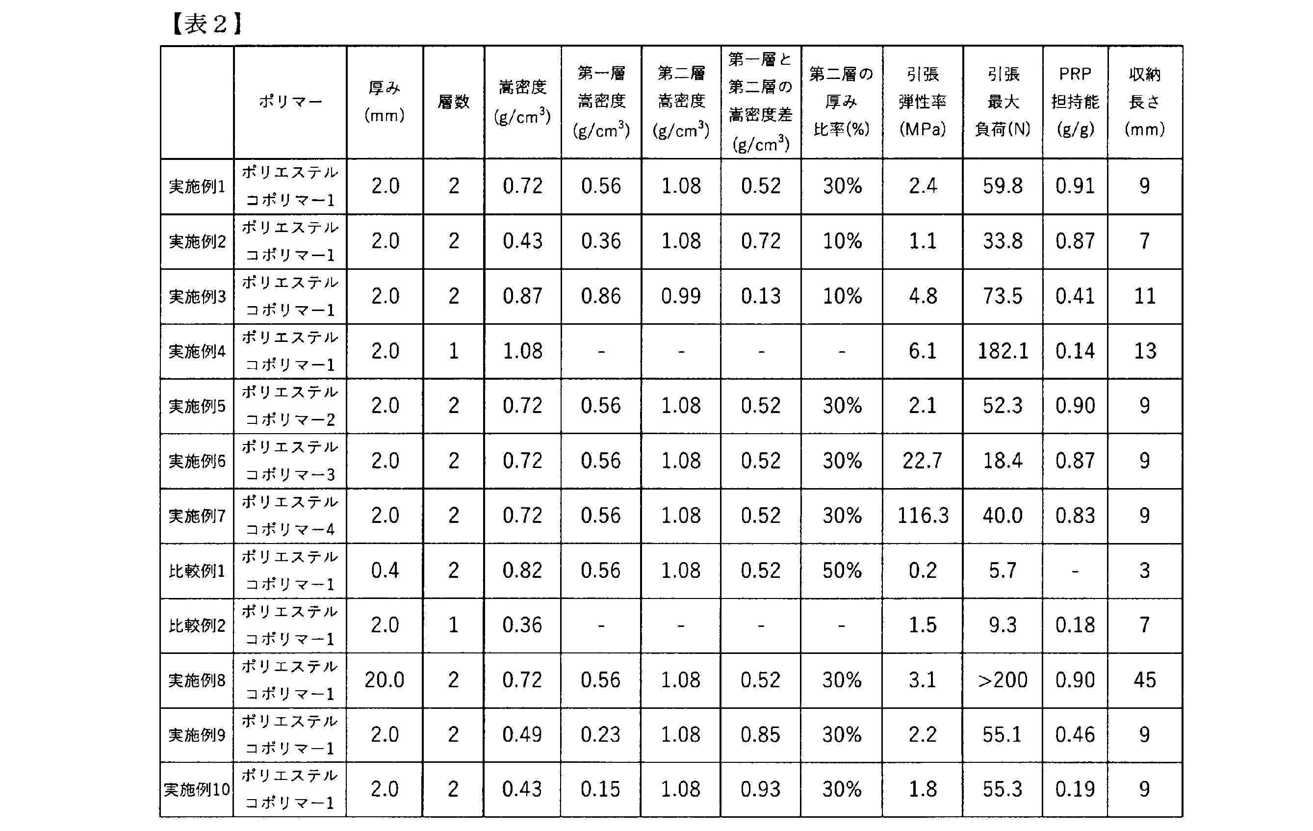
実施例1及び比較例1の結果から、インプラント医療機器の厚みを1.0mm以上とすることで、軟組織修復用途として好適な引張最大負荷が得られることが明らかとなった。また、実施例1及び比較例2の結果から、インプラント医療機器の嵩密度を0.40g/cm3以上とすることで、軟組織の修復用途として好適な引張最大負荷が得られることが明らかとなった。
本発明のインプラント医療機器は、軟組織の修復用途として用いることができ、その中でも特に軟骨、腱、腱板、靭帯の修復材料としての利用が挙げられる。
1・・・ 貼付面、2・・・ 第一層、3・・・ 第二層、4・・・ 最大厚み、5・・・ 最小厚み
Claims (19)
- 2種類のヒドロキシカルボン酸モノマーを主構成単位とするポリヒドロキシカルボン酸を含むポリエステルコポリマーを有し、
厚みが1.0mm以上20.0mm以下、かつ、嵩密度が0.40g/cm3以上1.50g/cm3以下である、軟組織修復用のインプラント医療機器。 - 第一層及び第二層を有し、前記第一層の嵩密度が0.20g/cm3以上0.95g/cm3以下である、請求項1記載のインプラント医療機器。
- 第一層及び第二層を有し、該第一層と該第二層の嵩密度は異なり、
厚みが1.0mm以上20.0mm以下、かつ、全体の嵩密度が0.40g/cm3以上1.50g/cm3以下である、軟組織修復用のインプラント医療機器。 - 2種類のヒドロキシカルボン酸モノマーを主構成単位とするポリヒドロキシカルボン酸を含むポリエステルコポリマーを有する、請求項3記載のインプラント医療機器。
- 前記第一層の嵩密度が0.20g/cm3以上0.95g/cm3以下である、請求項3又は4記載のインプラント医療機器。
- 前記第一層の嵩密度と前記第二層の嵩密度の差は、0.10g/cm3以上0.90g/cm3以下である、請求項2~5のいずれか一項記載のインプラント医療機器。
- 前記第二層の嵩密度が0.95g/cm3より大きく2.00g/cm3以下である、請求項2~6のいずれか一項記載のインプラント医療機器。
- 前記インプラント医療機器の厚みに対する前記第二層の厚みの比率が5%以上40%以下である、請求項2~7のいずれか一項記載のインプラント医療機器。
- 前記第一層と第二層が同じ成分で構成される、請求項2~8のいずれか一項記載のインプラント医療機器。
- 引張最大負荷が20N以上2,000N以下である、請求項1~9のいずれか一項記載のインプラント医療機器。
- 引張弾性率が0.01MPa以上100MPa以下である、請求項1~10のいずれか一項記載のインプラント医療機器。
- 生分解性である、請求項1~11のいずれか一項記載のインプラント医療機器。
- 前記2種類のヒドロキシカルボン酸モノマーをそれぞれ「モノマーA」、「モノマーB」とした場合に、前記ポリヒドロキシカルボン酸の下記の式1で表されるR値が0.25以上0.99以下である、請求項1、2、4~12のいずれか一項記載のインプラント医療機器。
R値 = [AB]/(2[A][B])×100 ・・・式1
[A]:ポリヒドロキシカルボン酸中の、モノマーA残基のモル分率(%)
[B]:ポリヒドロキシカルボン酸中の、モノマーB残基のモル分率(%)
[AB]:ポリヒドロキシカルボン酸中の、モノマーA残基とモノマーB残基が隣り合った構造(A-B、及びB-A)のモル分率(%) - 前記モノマーAは、乳酸、グリコール酸、ジラクチド及びグリコリドからなる群から選択されるモノマーであり、
前記モノマーBは、ヒドロキシ吉草酸、ヒドロキシカプロン酸、バレロラクトン及びカプロラクトンからなる群から選択されるモノマーである、請求項13記載のインプラント医療機器。 - ポリエステルコポリマーのみからなる、請求項1~14のいずれか一項記載のインプラント医療機器。
- 前記軟組織は、腱、腱板又は靭帯である、請求項1~15のいずれか一項記載のインプラント医療機器。
- 筋肉と骨、又は、骨と骨に固定化して使用される、請求項1~16のいずれか一項記載の断裂した軟組織の修復用のインプラント医療機器。
- 請求項1~17のいずれか一項記載のインプラント医療機器を筋肉と骨又は骨と骨に固定化する固定工程を有する、断裂した腱、腱板又は靭帯を修復する方法。
- 請求項1~17のいずれか一項記載のインプラント医療機器の製造方法。
Priority Applications (1)
| Application Number | Priority Date | Filing Date | Title |
|---|---|---|---|
| JP2024540020A JPWO2025005232A1 (ja) | 2023-06-30 | 2024-06-28 |
Applications Claiming Priority (2)
| Application Number | Priority Date | Filing Date | Title |
|---|---|---|---|
| JP2023-108093 | 2023-06-30 | ||
| JP2023108093 | 2023-06-30 |
Publications (1)
| Publication Number | Publication Date |
|---|---|
| WO2025005232A1 true WO2025005232A1 (ja) | 2025-01-02 |
Family
ID=93939106
Family Applications (1)
| Application Number | Title | Priority Date | Filing Date |
|---|---|---|---|
| PCT/JP2024/023482 Ceased WO2025005232A1 (ja) | 2023-06-30 | 2024-06-28 | 軟組織修復用のインプラント医療機器及びその製造方法 |
Country Status (2)
| Country | Link |
|---|---|
| JP (1) | JPWO2025005232A1 (ja) |
| WO (1) | WO2025005232A1 (ja) |
Citations (6)
| Publication number | Priority date | Publication date | Assignee | Title |
|---|---|---|---|---|
| JPH08206191A (ja) * | 1994-10-18 | 1996-08-13 | Ethicon Inc | 軟組織修復および増補用の注射可能微細分散液 |
| CN1396192A (zh) * | 2001-07-13 | 2003-02-12 | 浙江大学 | 生物降解性聚碳酸酯三元共聚物及制备方法 |
| CN1396193A (zh) * | 2001-07-13 | 2003-02-12 | 浙江大学 | 生物降解性聚碳酸酯/聚酯/聚醚三元共聚物及制备方法 |
| WO2006028244A1 (ja) * | 2004-09-07 | 2006-03-16 | Teijin Limited | 生体吸収性多孔体 |
| JP2009506811A (ja) * | 2005-09-01 | 2009-02-19 | ポリメディクス イノベーション ゲーエムベーハー | 創傷を医療処置するための造形体 |
| JP2017527341A (ja) * | 2014-09-07 | 2017-09-21 | オッシオ リミテッド | 異方性バイオ複合材料、異方性バイオ複合材料を含む医療用インプラント、および係る医療用インプラントの治療方法 |
-
2024
- 2024-06-28 WO PCT/JP2024/023482 patent/WO2025005232A1/ja not_active Ceased
- 2024-06-28 JP JP2024540020A patent/JPWO2025005232A1/ja active Pending
Patent Citations (6)
| Publication number | Priority date | Publication date | Assignee | Title |
|---|---|---|---|---|
| JPH08206191A (ja) * | 1994-10-18 | 1996-08-13 | Ethicon Inc | 軟組織修復および増補用の注射可能微細分散液 |
| CN1396192A (zh) * | 2001-07-13 | 2003-02-12 | 浙江大学 | 生物降解性聚碳酸酯三元共聚物及制备方法 |
| CN1396193A (zh) * | 2001-07-13 | 2003-02-12 | 浙江大学 | 生物降解性聚碳酸酯/聚酯/聚醚三元共聚物及制备方法 |
| WO2006028244A1 (ja) * | 2004-09-07 | 2006-03-16 | Teijin Limited | 生体吸収性多孔体 |
| JP2009506811A (ja) * | 2005-09-01 | 2009-02-19 | ポリメディクス イノベーション ゲーエムベーハー | 創傷を医療処置するための造形体 |
| JP2017527341A (ja) * | 2014-09-07 | 2017-09-21 | オッシオ リミテッド | 異方性バイオ複合材料、異方性バイオ複合材料を含む医療用インプラント、および係る医療用インプラントの治療方法 |
Also Published As
| Publication number | Publication date |
|---|---|
| JPWO2025005232A1 (ja) | 2025-01-02 |
Similar Documents
| Publication | Publication Date | Title |
|---|---|---|
| Oh et al. | Hydrophilization of synthetic biodegradable polymer scaffolds for improved cell/tissue compatibility | |
| Ying et al. | Scaffolds from electrospun polyhydroxyalkanoate copolymers: fabrication, characterization, bioabsorption and tissue response | |
| JP2005533148A5 (ja) | ||
| JP2005533148A (ja) | 生分解性の相分離したセグメント化多ブロック共重合体 | |
| CA2636817A1 (en) | Biodegradable elastomers | |
| Hu et al. | In vitro degradation behavior of shape memory PLLA-TMC random copolymers | |
| Oh et al. | Degradation behavior of 3D porous polydioxanone-b-polycaprolactone scaffolds fabricated using the melt-molding particulate-leaching method | |
| US8236350B2 (en) | Polymer for tissue engineering applications and drug delivery | |
| Liu et al. | Novel biodegradable poly (propylene fumarate)-co-poly (l-lactic acid) porous scaffolds fabricated by phase separation for tissue engineering applications | |
| Vacaras et al. | Understanding the basis of medical use of poly-lactide-based resorbable polymers and composites–a review of the clinical and metabolic impact | |
| JP4899152B2 (ja) | 医療用樹脂組成物とその製造方法および成形体 | |
| CN105237714A (zh) | 水响应形状记忆聚氨酯及其制备方法 | |
| HILL | Biomedical polymers | |
| Revati et al. | Biodegradable poly (lactic acid) scaffold for tissue engineering: A brief review | |
| Biswal | Recent progress in PLA‐based composite and their application to biomedical and cosmetic fields | |
| CN1253217C (zh) | 聚l-乳酸在制备做为医用形状记忆材料中的用途 | |
| Lipsa et al. | Poly (α-hydroxyacids) in biomedical applications: synthesis and properties of lactic acid polymers | |
| Bai et al. | In vitro hydrolytic degradation of poly (para-dioxanone)/poly (D, L-lactide) blends | |
| Heiny et al. | Progress in functionalized biodegradable polyesters | |
| Ang et al. | Tailoring the mechanical and biodegradable properties of binary blends of biomedical thermoplastic elastomer | |
| Ferrer et al. | Tailoring bulk and surface composition of polylactides for application in engineering of skeletal tissues | |
| US11370880B1 (en) | Growth factor-loaded elastic poly(xylitol-dodecanedioic acid) polymer for tissue engineering | |
| Mulchandani et al. | Synthesis strategies for biomedical grade polymers | |
| JP5258189B2 (ja) | 柔軟性生分解性ポリマー | |
| US20230181793A1 (en) | Functionalized poly(glycerol sebacate)s and uses thereof |
Legal Events
| Date | Code | Title | Description |
|---|---|---|---|
| WWE | Wipo information: entry into national phase |
Ref document number: 2024540020 Country of ref document: JP |
|
| 121 | Ep: the epo has been informed by wipo that ep was designated in this application |
Ref document number: 24832090 Country of ref document: EP Kind code of ref document: A1 |
|
| NENP | Non-entry into the national phase |
Ref country code: DE |
|
| 122 | Ep: pct application non-entry in european phase |
Ref document number: 24832090 Country of ref document: EP Kind code of ref document: A1 |