WO2024253201A1 - 試料中の標的物質を検出する方法及びそのためのキット - Google Patents
試料中の標的物質を検出する方法及びそのためのキット Download PDFInfo
- Publication number
- WO2024253201A1 WO2024253201A1 PCT/JP2024/020941 JP2024020941W WO2024253201A1 WO 2024253201 A1 WO2024253201 A1 WO 2024253201A1 JP 2024020941 W JP2024020941 W JP 2024020941W WO 2024253201 A1 WO2024253201 A1 WO 2024253201A1
- Authority
- WO
- WIPO (PCT)
- Prior art keywords
- nucleic acid
- stranded nucleic
- specific binding
- acid fragment
- target substance
- Prior art date
- Legal status (The legal status is an assumption and is not a legal conclusion. Google has not performed a legal analysis and makes no representation as to the accuracy of the status listed.)
- Ceased
Links
Images










Classifications
-
- C—CHEMISTRY; METALLURGY
- C07—ORGANIC CHEMISTRY
- C07K—PEPTIDES
- C07K16/00—Immunoglobulins [IG], e.g. monoclonal or polyclonal antibodies
- C07K16/18—Immunoglobulins [IG], e.g. monoclonal or polyclonal antibodies against material from animals or humans
-
- C—CHEMISTRY; METALLURGY
- C12—BIOCHEMISTRY; BEER; SPIRITS; WINE; VINEGAR; MICROBIOLOGY; ENZYMOLOGY; MUTATION OR GENETIC ENGINEERING
- C12M—APPARATUS FOR ENZYMOLOGY OR MICROBIOLOGY; APPARATUS FOR CULTURING MICROORGANISMS FOR PRODUCING BIOMASS, FOR GROWING CELLS OR FOR OBTAINING FERMENTATION OR METABOLIC PRODUCTS, i.e. BIOREACTORS OR FERMENTERS
- C12M1/00—Apparatus for enzymology or microbiology
-
- C—CHEMISTRY; METALLURGY
- C12—BIOCHEMISTRY; BEER; SPIRITS; WINE; VINEGAR; MICROBIOLOGY; ENZYMOLOGY; MUTATION OR GENETIC ENGINEERING
- C12M—APPARATUS FOR ENZYMOLOGY OR MICROBIOLOGY; APPARATUS FOR CULTURING MICROORGANISMS FOR PRODUCING BIOMASS, FOR GROWING CELLS OR FOR OBTAINING FERMENTATION OR METABOLIC PRODUCTS, i.e. BIOREACTORS OR FERMENTERS
- C12M1/00—Apparatus for enzymology or microbiology
- C12M1/34—Measuring or testing with condition measuring or sensing means, e.g. colony counters
-
- C—CHEMISTRY; METALLURGY
- C12—BIOCHEMISTRY; BEER; SPIRITS; WINE; VINEGAR; MICROBIOLOGY; ENZYMOLOGY; MUTATION OR GENETIC ENGINEERING
- C12N—MICROORGANISMS OR ENZYMES; COMPOSITIONS THEREOF; PROPAGATING, PRESERVING, OR MAINTAINING MICROORGANISMS; MUTATION OR GENETIC ENGINEERING; CULTURE MEDIA
- C12N15/00—Mutation or genetic engineering; DNA or RNA concerning genetic engineering, vectors, e.g. plasmids, or their isolation, preparation or purification; Use of hosts therefor
- C12N15/09—Recombinant DNA-technology
- C12N15/11—DNA or RNA fragments; Modified forms thereof; Non-coding nucleic acids having a biological activity
-
- C—CHEMISTRY; METALLURGY
- C12—BIOCHEMISTRY; BEER; SPIRITS; WINE; VINEGAR; MICROBIOLOGY; ENZYMOLOGY; MUTATION OR GENETIC ENGINEERING
- C12Q—MEASURING OR TESTING PROCESSES INVOLVING ENZYMES, NUCLEIC ACIDS OR MICROORGANISMS; COMPOSITIONS OR TEST PAPERS THEREFOR; PROCESSES OF PREPARING SUCH COMPOSITIONS; CONDITION-RESPONSIVE CONTROL IN MICROBIOLOGICAL OR ENZYMOLOGICAL PROCESSES
- C12Q1/00—Measuring or testing processes involving enzymes, nucleic acids or microorganisms; Compositions therefor; Processes of preparing such compositions
- C12Q1/68—Measuring or testing processes involving enzymes, nucleic acids or microorganisms; Compositions therefor; Processes of preparing such compositions involving nucleic acids
- C12Q1/6813—Hybridisation assays
Definitions
- the present invention relates to a method for detecting a target substance in a sample and a kit therefor.
- VUS variable of unknown significance
- VUS VUS protein synthesized in the cells
- This type of evaluation method requires a lot of time, effort, and cost.
- Digital measurement includes digital ELISA and digital PCR.
- the sample solution is divided into an extremely large number of micro-solutions.
- the signal from each micro-solution is then binarized, and the number of molecules of the target substance is measured by determining only whether or not the target substance is present.
- Digital measurement can significantly improve detection sensitivity and quantitation compared to conventional ELISA and real-time PCR methods.
- Non-Patent Document 1 describes a microwell array having microwells and flow channels for supplying reagents, etc., and describes performing digital ELISA using the microwell array.
- Patent Document 1 reports a method for detecting proteins using antibodies modified with oligonucleotides, called Proximity Ligation Assay (PLA). This method uses PCR or RCA for detection.
- PLA Proximity Ligation Assay
- Patent Document 2 describes a method for detecting protein interactions between two molecules using an antibody modified with an oligonucleotide. This method also uses the PCR method or the RCA method for detection.
- Non-Patent Document 3 describes a microwell array having microwells and flow channels for supplying reagents, etc., and describes how cells are used to express signal transduction within the microwell array and how phosphorylated proteins are detected. The state of signal transduction is monitored by detecting phosphorylated proteins.
- Alpha SureFire https://www.perkinelmer.co.jp/assays/tabid/346/Default.aspx, PerkinElmer is an assay system that can detect kinase activity on a cell-based basis without washing procedures. This system detects phosphorylated proteins from cell extracts.
- reaction solutions that effectively suppress non-specific adsorption to various substances are being investigated. These reaction solutions are called blocking agents.
- Blocking agents that have traditionally been used are those based on biological proteins such as bovine serum albumin, casein, and gelatin, highly hydrophilic inorganic particles, and polymers containing phosphorylcholine groups.
- Non-Patent Document 4 2-methacryloyloxyethyl phosphorylcholine copolymer (MPC polymer) is used as a blocking agent to prevent non-specific adsorption.
- MPC polymer 2-methacryloyloxyethyl phosphorylcholine copolymer
- Kan C. W., et al. Isolation and detection of single molecules on paramagnetic beads using sequential fluid flows in microfabricated polymer array assemblies., Lab on a Chip, 12 (5), 977-985, 2012.
- Mohammed H., et al. Approaches for Assessing and Discovering Protein Interactions in Cancer., Mol Cancer Res, 11 (11), 1295-1302, 2013.
- Blazek M., et al. Proximity Ligation Assay for High-content Profiling of Cell Signaling Pathways on A Microfluidic Chip., Molecular & Cellular Proteomics 12: 10.1074/mcp.M113.032821, 3898-3907, 2013.
- Y. Iwasaki, K. Ishihara. Phosphorylcholine-containing polymers for biomedical applications., Anal Bioanal Chem 381, 534-546, 2005.
- FIG. 1 shows a portion of mitogen-activated protein kinase (MAPK) signal transduction as an example.
- MAPK mitogen-activated protein kinase
- the mutant protein is BRAF
- VUS unknown clinical significance
- MEK the substrate of BRAF
- BRAF the substrate of BRAF
- upstream stimulation as shown in the left side of Figure 1.
- BRAF is constitutively active, as shown in the right side of Figure 1, and phosphorylates MEK, the substrate, even in the absence of upstream stimulation.
- Such BRAF causes cancer.
- the present invention aims to provide a method for detecting a target substance in a sample and a kit for the same.
- functional analysis (also called evaluation) of VUS can be performed easily, quickly, and efficiently.
- a method for detecting a target substance in a sample comprising: incubating a mixture containing the sample, a first specific binding substance for the target substance, which is labeled with a first single-stranded nucleic acid fragment, and a second specific binding substance for the target substance, which is labeled with a second single-stranded nucleic acid fragment, so that, if the target substance is present in the sample, a complex containing the target substance, the first specific binding substance, and the second specific binding substance is formed, and at least a portion of the first single-stranded nucleic acid fragment and at least a portion of the second single-stranded nucleic acid fragment hybridize to form a double-stranded nucleic acid; and detecting the formation of the double-stranded nucleic acid.
- the method wherein detection of the formation of the double-stranded nucleic acid indicates the presence of the target substance in the sample, and the mixture further comprises 2-methacryloyloxyethyl phosphorylcholine copolymer (MPC polymer), or the mixture further comprises casein and a nonionic surfactant.
- MPC polymer 2-methacryloyloxyethyl phosphorylcholine copolymer
- the MPC polymer is a copolymer of 2-methacryloyloxyethyl phosphorylcholine and a comonomer having at least one group selected from the group consisting of a hydrophobic group, a hydrophilic group, an anionic group, and a cationic group.
- [3] The method according to [1] or [2], wherein the concentration of the MPC polymer in the mixture is 0.01 to 0.5% (w/v).
- [4] The method according to any one of [1] to [3], wherein the mixture is incubated in a well, and the volume of the well is 10 fL to 100 pL.
- [5] The method according to any one of [1] to [4], wherein the first single-stranded nucleic acid fragment and the second single-stranded nucleic acid fragment each have a base length of 10 to 200 bases.
- [6] The method according to any one of [1] to [5], wherein the step of detecting the formation of the double-stranded nucleic acid is carried out by an invasive cleavage assay.
- a kit for detecting a target substance in a sample comprising: a well array having a plurality of wells; a first specific binding substance for the target substance labeled with a first single-stranded nucleic acid fragment; a second specific binding substance for the target substance labeled with a second single-stranded nucleic acid fragment; and a buffer containing an MPC polymer or containing casein and a nonionic surfactant.
- the kit according to [7] further comprising a sealing liquid for sealing the opening of the well.
- a method for detecting a target substance in a sample comprising: incubating a mixture containing the sample, a first specific binding substance for the target substance labeled with a first single-stranded nucleic acid fragment, a second specific binding substance for the target substance labeled with a second single-stranded nucleic acid fragment, and a 2-methacryloyloxyethyl phosphorylcholine copolymer, so that a complex containing the target substance, the first specific binding substance, and the second specific binding substance is formed, and at least a portion of the first single-stranded nucleic acid fragment and at least a portion of the second single-stranded nucleic acid fragment hybridize to form a double-stranded nucleic acid; detecting the formation of said double stranded nucleic acid; A method for detecting a target substance, wherein detection of the formation of the double-stranded nucleic acid indicates the presence of the target substance in the sample.
- the 2-methacryloyloxyethyl phosphorylcholine copolymer is a copolymer of 2-methacryloyloxyethyl phosphorylcholine and a comonomer having at least one group selected from the group consisting of a hydrophobic group, a hydrophilic group, an anionic group, and a cationic group.
- concentration of the MPC polymer in the mixture is 0.01 to 0.5% (w/v).
- a method for detecting a target substance in a sample comprising: incubating a mixture containing the sample, a first specific binding substance for the target substance, which is labeled with a first single-stranded nucleic acid fragment, a second specific binding substance for the target substance, which is labeled with a second single-stranded nucleic acid fragment, and a buffer containing casein and a nonionic surfactant, so that a complex containing the target substance, the first specific binding substance, and the second specific binding substance is formed, and at least a portion of the first single-stranded nucleic acid fragment and at least a portion of the second single-stranded nucleic acid fragment hybridize to form a double-stranded nucleic acid; detecting the formation of said double stranded nucleic acid; A method for detecting a target substance, wherein detection of the formation of the double-stranded nucleic acid indicates the presence of the target substance in the sample.
- [15] The method according to any one of [11] to [14], wherein the mixture is incubated in a well, and the volume of the well is 10 fL to 100 pL.
- [16] The method according to any one of [11] to [15], wherein the first single-stranded nucleic acid fragment and the second single-stranded nucleic acid fragment each have a base length of 10 to 200 bases.
- [17] The method according to any one of [11] to [16], wherein the step of detecting the formation of the double-stranded nucleic acid is carried out by invasive cleavage assay.
- a kit for detecting a target substance in a sample comprising: a well array having a plurality of wells; a first substance that specifically binds to the target substance and is labeled with a first single-stranded nucleic acid fragment; a second substance that specifically binds to the target substance and is labeled with a second single-stranded nucleic acid fragment; and a 2-methacryloyloxyethyl phosphorylcholine copolymer.
- a kit for detecting a target substance in a sample comprising: a well array having a plurality of wells; a first specific binding substance for the target substance, the first specific binding substance being labeled with a first single-stranded nucleic acid fragment; a second specific binding substance for the target substance, the second specific binding substance being labeled with a second single-stranded nucleic acid fragment; and a buffer containing casein and a nonionic surfactant.
- the kit according to [18] or [19] further comprising a sealing liquid for sealing the opening of the well.
- kit according to any one of [18] to [20], further comprising a reagent for detecting a double-stranded nucleic acid formed by hybridization of at least a portion of the first single-stranded nucleic acid fragment and at least a portion of the second single-stranded nucleic acid fragment.
- the present invention provides a method for detecting a target substance in a sample and a kit for the same.
- the present invention enables functional analysis (evaluation) of VUS to be performed simply, quickly, and efficiently.
- FIG. 1 is a diagram illustrating normal and deleterious mutations.
- FIG. 2 is a schematic diagram illustrating a method for detecting a target substance.
- FIG. 3 is a schematic diagram illustrating a method for detecting a target substance.
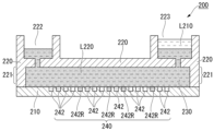
- FIG. 4 is a schematic cross-sectional view showing an example of a fluidic device.
- FIG. 5 is a schematic cross-sectional view illustrating a method for detecting a target substance.
- FIG. 6 is a schematic cross-sectional view illustrating a method for detecting a target substance.
- FIG. 7 is a schematic cross-sectional view showing an example of a fluidic device.
- FIG. 8 is a schematic cross-sectional view illustrating a method for detecting a target substance.
- FIG. 1 is a diagram illustrating normal and deleterious mutations.
- FIG. 2 is a schematic diagram illustrating a method for detecting a target substance.
- FIG. 3 is a schematic diagram illustrating a method for detecting a
- FIG. 9 is a schematic cross-sectional view illustrating a method for detecting a target substance.
- Figure 10 is a schematic diagram illustrating an example of the Invasive Cleavage Assay (ICA) method.
- FIG. 11 is a schematic diagram illustrating a method for evaluating the activity of a target protein.
- FIG. 12 is a graph showing the results of Experimental Example 1.
- FIG. 13 is a graph showing the results of Experimental Example 1.
- FIG. 14 is a graph showing the results of Experimental Example 2.
- FIG. 15 is a graph showing the results of Experimental Example 2.
- FIG. 16 is a graph showing the results of Experimental Example 3.
- FIG. 17 is a graph showing the results of Experimental Example 3.
- FIG. 18 is a graph showing the results of Experimental Example 3.
- FIG. 19 is a graph showing the results of Experimental Example 3.
- FIG. 20 is a graph showing the results of Experimental Example 3.
- FIG. 21 is a graph showing the results of Experimental Example 4.
- the present invention provides a method for detecting a target substance in a sample, the method comprising: incubating a mixture containing the sample, a first specific binding substance for the target substance, which is labeled with a first single-stranded nucleic acid fragment, and a second specific binding substance for the target substance, which is labeled with a second single-stranded nucleic acid fragment, so that, if the target substance is present in the sample, a complex containing the target substance, the first specific binding substance, and the second specific binding substance is formed, and at least a portion of the first single-stranded nucleic acid fragment and at least a portion of the second single-stranded nucleic acid fragment hybridize to form a double-stranded nucleic acid; and detecting the formation of the double-stranded nucleic acid, wherein detection of the formation of the double-stranded nucleic acid indicates the presence of the target substance in the sample
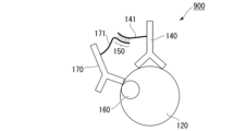
- FIG. 2 is a schematic diagram illustrating the method of this embodiment.
- FIG. 2 shows an example of detecting a phosphorylated protein 120 as a target substance.
- a phosphate group is indicated by the symbol 160.
- step (a) a mixture containing a sample, a first specific binding substance 140 for the target substance 120 labeled with a first single-stranded nucleic acid fragment 141, and a second specific binding substance 170 for the target substance 120 labeled with a second single-stranded nucleic acid fragment 171 is incubated.
- the first specific binding substance 140 and the second specific binding substance 170 recognize different epitopes on the target substance 120.
- the specific binding substance 170 recognizes a region containing a phosphate group 160 of the phosphorylated protein 120.
- a complex 900 is formed that contains the target substance 120, the first specific binding substance 140, and the second specific binding substance 170, and at least a portion of the first single-stranded nucleic acid fragment 141 and at least a portion of the second single-stranded nucleic acid fragment 171 hybridize to form a double-stranded nucleic acid (also called a double-stranded nucleic acid region) 150.
- a double-stranded nucleic acid also called a double-stranded nucleic acid region
- step (b) the formation of double-stranded nucleic acid 150 is detected. Detection of the formation of double-stranded nucleic acid 150 indicates the presence of target substance 120 in the sample. The detection of the formation of double-stranded nucleic acid 150 will be described later.
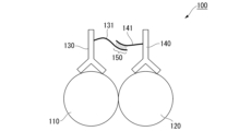
- FIG. 3 is a schematic diagram illustrating a different aspect of this embodiment.
- the target substance is a bond between protein 110 and protein 120.
- step (a) a mixture containing a sample, a first specific binding substance 140 for protein 120 labeled with a first single-stranded nucleic acid fragment 141, and a second specific binding substance 130 for protein 110 labeled with a second single-stranded nucleic acid fragment 131 is incubated.
- a complex 100 is formed that contains the conjugate of protein 110 and protein 120, which is the target substance, a first specific binding substance 140, and a second specific binding substance 130, and at least a portion of the first single-stranded nucleic acid fragment 141 and at least a portion of the second single-stranded nucleic acid fragment 131 hybridize to form a double-stranded nucleic acid (also called a double-stranded nucleic acid region) 150.
- a double-stranded nucleic acid also called a double-stranded nucleic acid region
- step (b) the formation of double-stranded nucleic acid 150 is detected.
- the detection of the formation of double-stranded nucleic acid 150 indicates the presence of a bound body of protein 110 and protein 120, which are the target substances, in the sample. In other words, the detection of the formation of double-stranded nucleic acid 150 indicates that protein 110 and protein 120 interact with each other.
- the method of FIG. 3 may be applied when the protein of interest 110 does not bind (i.e., does not interact) with the target protein 120, in which case the formation of the double-stranded nucleic acid 150 is not detected.
- the detection of the formation of the double-stranded nucleic acid 150 is described below.
- the time required to detect the target substance can be shortened.
- the signal-to-noise ratio (sometimes referred to as S/N) can be increased by further including casein and a nonionic surfactant in the mixed solution.
- the mixed solution contains casein, a nonionic surfactant, and tris(hydroxymethyl)aminomethane, not only can the signal-to-noise ratio (S/N) be increased, but the time required to detect the target substance can also be shortened.
- S/N signal-to-noise ratio
- the method of this embodiment allows the activity of mutant proteins to be evaluated easily and quickly.
- the function of VUS can be analyzed (i.e., evaluated) easily, quickly, and efficiently.
- MPC polymer is a polymer of 2-methacryloyloxyethyl phosphorylcholine (MPC) that has a phospholipid polar group (also called a phosphorylcholine group) and a methacryloyl group in the molecule.
- MPC polymer is a polymer that mimics biological membranes, and is considered to have a high level of biocompatibility, such as extremely low interaction with biological components such as proteins and blood cells, and excellent antithrombotic properties.
- MPC polymer can be copolymerized with various comonomers, and various properties can be imparted depending on the type of comonomer.
- the MPC polymer may be a copolymer with a comonomer having at least one group selected from the group consisting of a hydrophobic group, a hydrophilic group, an anionic group, and a cationic group.
- a comonomer having at least one group selected from the group consisting of a hydrophobic group, a hydrophilic group, an anionic group, and a cationic group Commercially available MPC polymers can be used, and specific examples include the Lipidure (registered trademark)-BL series (NOF Corp.).
- the MPC polymer that can be preferably used is one having a surface tension of 70 to 80 ⁇ 10 ⁇ 3 N/m as measured using a 0.1 wt % aqueous solution at 25° C.
- the MPC polymer that can be preferably used is one having a kinetic viscosity of 3 cSt or less or 20 to 30 cSt as measured using a 1 wt % aqueous solution at 25° C.
- the surface tension of a 0.1 wt % aqueous solution of MPC polymer can be measured using a surface tensiometer (e.g., DY-700, manufactured by Kyowa Interface Science Co., Ltd.).
- the dynamic viscosity of a 0.1 wt % aqueous solution of MPC polymer can be measured using a viscometer (e.g., Canon-Fenske Inverse Particle Size SF No. 150, manufactured by Shibata Scientific Co., Ltd.).
- a viscometer e.g., Canon-Fenske Inverse Particle Size SF No. 150, manufactured by Shibata Scientific Co., Ltd.
- the concentration of the MPC polymer in the mixed solution is preferably 0.01 to 0.5% (w/v), more preferably 0.01 to 0.1% (w/v), and even more preferably 0.01 to 0.05% (w/v).
- Casein is a type of naturally occurring phosphoprotein found in milk and cheese.
- concentration of casein in the mixture is preferably about 0.0025 to 1% by mass, and more preferably about 0.01 to 1% by mass.
- Nonionic surfactants are surfactants that do not contain ionic groups as polar groups, and examples of such surfactants include acetylene glycol surfactants such as ethylene oxide and/or propylene oxide adducts of acetylene glycol (specifically, ethylene oxide and/or propylene oxide adducts such as 2,4,7,9-tetramethyl-5-decyne-4,7-diol, 3,6-dimethyl-4-octyne-3,6-diol, etc.); acetylene alcohol surfactants such as ethylene oxide and/or propylene oxide adducts of acetylene alcohol (specifically, ethylene oxide and/or propylene oxide adducts such as 3,5-dimethyl-1-hexane-3-ol, etc.); polyoxyethylene nonylphenyl ether, polyoxyethylene octylphenyl ether, polyoxyethylene tetrahydrofuran ...
- the surfactant examples include ether surfactants such as diethylene dodecyl phenyl ether, polyoxyethylene alkyl allyl ether, polyoxyethylene oleyl ether, polyoxyethylene lauryl ether, polyoxyethylene alkyl ether, and polyoxyalkylene alkyl ether; ester surfactants such as polyoxyethylene oleic acid, polyoxyethylene oleic acid ester, polyoxyethylene distearate ester, sorbitan laurate, sorbitan monostearate, sorbitan monooleate, sorbitan sesquioleate, polyoxyethylene monooleate, and polyoxyethylene stearate; polyether-modified siloxane surfactants such as dimethylpolysiloxane; and other fluorine-containing surfactants such as fluorine alkyl esters and perfluoroalkyl carboxylates.
- concentration of the nonionic surfactant in the mixture is preferably about 0.0025 to 1% by mass, and
- Tris(hydroxymethyl)aminomethane is a type of buffer. Tris(hydroxymethyl)aminomethane may be in the form of a salt. An example of a salt of tris(hydroxymethyl)aminomethane is tris(hydroxymethyl)aminomethane hydrochloride.
- the concentration of tris(hydroxymethyl)aminomethane in the mixture is preferably about 0.0025 to 1% by mass, and more preferably about 0.01 to 1% by mass.
- the base length of the first single-stranded nucleic acid fragment 141 may be 10 to 200 bases.
- the base length of the second single-stranded nucleic acid fragment 171 (131) may also be 10 to 200 bases.
- the length of the double-stranded nucleic acid 150 formed by hybridization of at least a portion of the first single-stranded nucleic acid fragment 141 and at least a portion of the second single-stranded nucleic acid fragment 171 (131) is preferably about 7 to 30 bases, and may be, for example, 9 bases, 12 bases, or 15 bases.
- protein 110 may be a protein having a genetic variant of unknown clinical significance (VUS).
- VUS unknown clinical significance
- Protein 110 may be, for example, a kinase. More specific target proteins include proteins in intracellular signal transduction pathways, such as BRAF, A-RAF, Raf, MAP3K 4/12, MAP3K11, ASK1, and TAK1.
- BRAF protein having a genetic variant of unknown clinical significance
- A-RAF protein having a genetic variant of unknown clinical significance
- Raf proteins in intracellular signal transduction pathways
- MAP3K 4/12 proteins in intracellular signal transduction pathways
- MAP3K11 MAP3K11
- ASK1 MAP3K11
- TAK1 TAK1
- the detection shown in the example of FIG. 2 can be performed in the presence of a kinase (protein 110) having a VUS, and by detecting phosphorylation of protein 120, it is possible to evaluate whether or not the kinase (protein 110) having a VUS is in a constitutively activated state.
- the method of this embodiment can be carried out by digital measurement.
- the mixed solution is incubated in a well, and the well preferably constitutes a well array in which multiple wells are arranged.
- the well array is arranged within a flow path of the fluidic device.
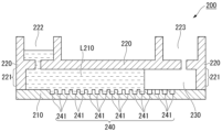
- FIG. 4 is a schematic cross-sectional view showing an example of a fluidic device in which the method of this embodiment can be suitably implemented.
- the fluidic device 200 includes a substrate 210 and a lid member 220 arranged opposite the substrate 210.
- the lid member 220 has a convex portion 221, and the tip of the convex portion 221 contacts the substrate 210.
- the well array 240 is integrally molded with the substrate 210 on one side of the substrate 210 and faces the lid member 220.
- the well array 240 has a plurality of wells 241.
- the lid member 220 may be welded or bonded to the substrate 210.
- the wells 241 open onto the surface of the substrate 210.
- the wells 241 are microwells with a small volume.
- the volume of one well 241 may be approximately 10 fL to 100 pL, or 50 fL to 1200 fL.
- multiple wells 241 of the same shape and size form a well array 240.
- the same shape and size means that the wells have the same shape and capacity to the extent required for digital measurement, and variations within the range of manufacturing errors are acceptable.
- the diameter of the wells 241 may be, for example, about 1 to 10 ⁇ m, and the depth of the wells 241 may be, for example, about 1 to 10 ⁇ m.
- the arrangement of the wells 241 is not particularly limited, and may be, for example, arranged in a triangular lattice pattern, a square lattice pattern, or randomly arranged.
- a space is formed between the well array 240 and the cover member 220 due to the presence of the convex portion 221.
- This space constitutes the flow path 230.
- the flow path 230 functions as a path for transporting a liquid in which the target substance, the protein, the first specific binding substance, the second specific binding substance, etc. are dispersed, and a sealing liquid described below.
- the height of the flow path 230 (the distance between the surface of the substrate 210 and the surface of the cover member 220 facing the substrate 210) may be, for example, 500 ⁇ m or less, for example, 300 ⁇ m or less, for example, 200 ⁇ m or less, or for example, 100 ⁇ m or less.
- the protrusion 221 may be molded integrally with the lid member 220.
- the lid member 220 can be molded into a plate shape having the protrusion 221, for example, by molding a thermoplastic resin fluid using a molding die.
- the lid member 220 may also be formed with an inlet port 222 and an outlet port 223 for the reagent.
- the lid member 220 has a protrusion 221
- the lid member 220 and the substrate 210 are overlapped so that the protrusion 221 contacts the surface of the substrate 210 where the well 241 opens.
- the space between the lid member 220 and the substrate 210 becomes the flow path 230.
- the lid member 220 and the substrate 210 may be welded by laser welding or the like.
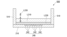
- FIG. 7 is a schematic cross-sectional view showing an example of a fluidic device.
- the fluidic device 500 includes a substrate 210 and a wall member 510.
- the well array 240 is integrally formed with the substrate 210 on one side of the substrate 210.
- the well array 240 has a plurality of wells 241.
- the fluidic device 500 differs mainly from the above-described fluidic device 200 in that the fluidic device 500 does not include a cover member 220.
- the lid member 220 and the protrusion 221 are integrally molded.
- the lid member 220 and the protrusion 221 may be molded as separate bodies.
- the well array 240 is integrally molded with the substrate 210 on one side of the substrate 210.
- the well array does not have to be integrally molded with the substrate 210.
- the well array 240 molded separately from the fluidic device may be disposed on the substrate 210 of the fluidic device.
- a resin layer may be laminated on the surface of the substrate 210, and the well array may be formed in the resin layer by etching or the like.
- the substrate 210 is formed, for example, using a resin.
- a resin there is no particular limit to the type of resin, but it is preferable that the resin is resistant to reagents and sealing liquid. Furthermore, if the signal to be detected is fluorescence, it is preferable that the resin has little autofluorescence.
- resins include, but are not limited to, cycloolefin polymers, cycloolefin copolymers, silicone, polypropylene, polycarbonate, polystyrene, polyethylene, polyvinyl acetate, fluororesins, and amorphous fluororesins.
- a plurality of wells 241 may be formed on one surface of the substrate 210 in the thickness direction.
- Methods for forming wells using resin include injection molding, thermal imprinting, and optical imprinting.
- a fluororesin may be laminated on the substrate 210 and the fluororesin may be processed by etching or the like to form a well array.
- CYTOP registered trademark
- Asahi Glass or the like may be used as the fluororesin.
- the material of the lid member 220 is preferably a resin with low autofluorescence, and may be, for example, a thermoplastic resin such as a cycloolefin polymer or a cycloolefin copolymer.
- the lid member 220 may be made of a material that does not transmit light of wavelengths close to the wavelength detected during fluorescent observation of the signal, or may be made of a material that does not transmit light at all.
- the lid member 220 may be made of a thermoplastic resin to which carbon, metal particles, etc. have been added.
- the method of this embodiment is a method for detecting a target substance 120 in a sample, and includes a step (a) of incubating a mixture containing the sample, a first specific binding substance 140 for the target substance 120 labeled with a first single-stranded nucleic acid fragment 141, and a second specific binding substance 170 for the target substance 120 labeled with a second single-stranded nucleic acid fragment 171, and forming a complex 900 containing the target substance 120, the first specific binding substance 140, and the second specific binding substance 170 when the target substance 120 is present in the sample, and hybridizing at least a portion of the first single-stranded nucleic acid fragment 141 and at least a portion of the second single-stranded nucleic acid fragment 171 to form a double-stranded nucleic acid 150, and a step (b) of detecting the formation of the double-stranded nucleic acid 150, and the detection of the formation of the double-stranded nucleic acid 150 indicates the presence of
- the method of this embodiment is similar to that of the example in Figure 3, in which the target substance is a conjugate of protein 110 and protein 120, except that a second specific binding substance 130 for protein 110 is used as the second specific binding substance.
- the mixture further contains an MPC polymer, or further contains casein and a nonionic surfactant. More preferably, the mixture further contains casein, a nonionic surfactant, and tris(hydroxymethyl)aminomethane.
- the method of this embodiment can shorten the time required to detect a target substance in a sample or increase the signal-to-noise ratio (S/N).
- the present invention can perform functional analysis (also called evaluation) of VUS simply, quickly, and efficiently.
- the mixed liquid L210 is introduced from the inlet port 222 of the fluidic device 200 and sent to the flow path 230.
- the mixed liquid L210 contains an MPC polymer, and is a liquid in which the target substance 120, the first specific binding substance 140, and the second specific binding substance 170 are dispersed, and also contains a reagent for detecting the formation of double-stranded nucleic acid 150.
- the mixed solution L210 sent to the flow path 230 comes into contact with the well array 240. Then, the mixed solution L210 is contained inside the well 241. As a result, the MPC polymer, the target substance 120, the first specific binding substance 140, the second specific binding substance 170, and a reagent for detecting the formation of the double-stranded nucleic acid 150 are introduced into the well 241.
- the mixed solution L210 contains a complex 900 containing the target substance 120, the first specific binding substance 140, and the second specific binding substance 170.
- the number of complexes 900 introduced into one well 241 is not particularly limited, but preferably one or less complexes 900 are introduced into one well 241, i.e., 0 or 1 complex 900. This allows detection of the complexes 900 on a single unit basis, i.e., digital measurement is possible. In addition, it is not necessary for the complexes 900 to be introduced into all wells of the well array.
- the means for introducing the complex 900 into the well is not particularly limited, and examples include a method in which the complex 900 is allowed to settle in the fluid device (specifically, in the flow channel) by its own weight and distributed to the well.
- a substance that captures the complex 900 hereinafter, sometimes referred to as a capture material
- the capture material may be bound to the complex 900 that does not easily settle by its own weight and then delivered. It is also possible to improve the efficiency of introducing the complex 900 into the well by immobilizing the capture material in advance in the well and capturing the delivered complex 900.
- the step of binding the capture substance to the complex 900 can be performed at any point in the method of this embodiment.
- this step can be performed by contacting the complex 900 with the capture substance in a sample tube before the step of introducing the complex 900 into the well 241.
- the capture substance can be introduced into the well 241, and then the complex 900 can be introduced into the well and the capture substance can be contacted with the complex 900 in the well.
- the capture material is a substance capable of capturing the complex 900.
- the capture material may be, for example, a conjugate between a solid phase and a substance that specifically binds to the complex 900.
- the solid phase may be a particle, a membrane, a substrate, or the like.
- the number of specific binding substances to the complex 900 may be one or more. For example, there may be three, four, or five or more types.
- the particles are not particularly limited, and examples thereof include polymer particles, magnetic particles, and glass particles.
- the particles are preferably surface-treated to avoid non-specific adsorption.
- particles having functional groups such as carboxyl groups on the surface are preferable for immobilizing specific binding substances. More specifically, products such as "Magnosphere LC300" manufactured by JSR Corporation can be used.
- Examples of the specific binding substance in the first specific binding substance 140, the second specific binding substance 170, and the captured substance include an antibody, an antibody fragment, and an aptamer.
- Examples of the antibody fragment include Fab, F(ab') 2 , Fab', a single-chain antibody (scFv), a disulfide-stabilized antibody (dsFv), a dimerized V region fragment (diabody), and a peptide containing CDR.
- the antibody may be a monoclonal antibody or a polyclonal antibody. Alternatively, the antibody may be a commercially available antibody.
- a method for labeling a specific binding substance with a single-stranded nucleic acid fragment includes a method using a crosslinking agent.
- the single-stranded nucleic acid fragment may be labeled with a specific binding substance via a linker molecule.
- the linker is not particularly limited, and examples thereof include polyethylene chains, hydrocarbon chains, and peptides.
- the single-stranded nucleic acid fragment may be DNA or RNA. It may also include artificial nucleic acids such as BNA and LNA.
- the method of immobilizing a specific binding substance on the particle surface is not particularly limited, and examples thereof include physical adsorption, chemical bonding, avidin-biotin bonding, and antibody bonding.
- Physical adsorption methods include methods of immobilizing a specific binding substance on the particle surface by hydrophobic or electrostatic interaction.
- Chemical bonding methods include methods using a crosslinking agent.
- the specific binding substance can be immobilized on the particle surface by reacting a crosslinking agent with the carboxyl groups of the specific binding substance to form an active ester, and then reacting the hydroxyl groups with the ester groups. It is also preferable to provide a spacer between the specific binding substance and the particle surface so as not to inhibit the ability of the specific binding substance to recognize target molecules.
- the complex 900 when the complex 900 is introduced into the well 241 using a capture agent, it is preferable to form a conjugate between the capture agent and the complex 900 under conditions where 0 or 1 complex 900 is captured per capture agent. Furthermore, it is preferable to configure each well 241 so that 0 or 1 capture agent is introduced. This makes digital measurement possible.
- a complex 900 containing them is formed, and at least a portion of the first single-stranded nucleic acid fragment 141 and at least a portion of the second single-stranded nucleic acid fragment 171 hybridize to form a double-stranded nucleic acid 150.
- the formation of the complex 900 may be performed in a sample tube or in a well 241.
- a step of sealing the opening of the well 241 may be carried out.
- the method of sealing the opening of the well 241 is not particularly limited as long as it can prevent the liquid contained in one well 241 from mixing with the liquid contained in another well 241, and for example, the opening of the well 241 may be sealed by covering it with a sealing liquid. Alternatively, the opening of the well 241 may be sealed by stacking a plate-like member such as a glass plate on it.
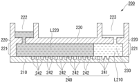
- sealing liquid L220 is sent from the inlet port 222 of the lid member 220 to the flow path 230 between the substrate 210 and the lid member 220.
- the sealing liquid L220 sent to the flow path 230 comes into contact with the well array 240.
- the sealing liquid L220 then pushes away and replaces the reagent liquid L210 sent to the flow path 230 that is not contained in the wells 241.
- the sealing liquid L220 individually seals each of the multiple wells 241 that contain the mixed liquid L210 containing the target substance 120, and the wells 241 become independent reaction spaces (i.e., microcompartments 242).
- FIG. 6 shows the state in which all of the wells 241 in the well array 240 have been sealed with sealing liquid L220, forming sealed wells (i.e., microcompartments) 242.
- lipid bilayer membrane can be formed at the opening of the well 241, and the multiple wells 241 can be individually sealed with the lipid bilayer membrane to form sealed wells 242.
- lipids that form lipid bilayer membranes include, but are not limited to, 1,2-dioleoyl-sn-glycero-3-phosphoethanolamine (DOPE), 1,2-dioleoyl-sn-glycero-3-phosphoglycerol (DOPG), mixtures thereof, etc.
- DOPE 1,2-dioleoyl-sn-glycero-3-phosphoethanolamine
- DOPG 1,2-dioleoyl-sn-glycero-3-phosphoglycerol
- the sealing liquid is a liquid that can form droplets (also called microdroplets) by individually sealing the liquids introduced into the multiple wells 241 so that they do not mix with each other, and is preferably an oil-based solution, and more preferably an oil.
- oil fluorine-based oil, silicone-based oil, hydrocarbon-based oil, or a mixture of these oils can be used. More specifically, Sigma's product name "FC-40" can be used.
- FC-40 (CAS number: 86508-42-1) is a fluorinated aliphatic compound with a specific gravity of 1.85 g/mL at 25°C.
- the formation of the double-stranded nucleic acid 150 is detected.
- the formation of the double-stranded nucleic acid 150 is preferably detected using a signal amplification reaction.
- An example of the signal amplification reaction is the Invasive Cleavage Assay (ICA).
- the ICA reaction is based on the principle that signal amplification proceeds through a cycle of two reactions: (1) complementary binding between nucleic acids, and (2) recognition and cleavage of the triple-stranded structure by an enzyme.
- the ICA reaction is less susceptible to reaction cycle inhibition by impurities. Therefore, by using the ICA reaction, the formation of the double-stranded nucleic acid 150 can be detected with high accuracy.
- the mixed liquid L210 (a liquid containing the MPC polymer, the target substance 120, the first specific binding substance 140, and the second specific binding substance 170) further contains reaction reagents necessary for the ICA reaction.
- the reaction reagents required for the ICA reaction include ICA reaction reagents such as a flap probe, a flap endonuclease (FEN), and a fluorescent substrate.
- the flap probe is a nucleic acid fragment designed to hybridize with the first single-stranded nucleic acid fragment 141 or the second single-stranded nucleic acid fragment 171 to form a flap structure with the double-stranded nucleic acid 150.
- FIG. 10 is a schematic diagram illustrating an example of the ICA method.
- the ICA method detects double-stranded nucleic acid 150 formed by hybridization of at least a part of a first single-stranded nucleic acid fragment 141 and at least a part of a second single-stranded nucleic acid fragment 171.
- a flap probe is hybridized to the first single-stranded nucleic acid fragment 141 or the second single-stranded nucleic acid fragment 171.
- the flap probe 810 hybridizes to the second single-stranded nucleic acid fragment 171.
- a first flap site 811 is formed.
- the first flap site 811 is cleaved to generate a nucleic acid fragment 811.
- the nucleic acid fragment 811 then hybridizes to a fluorescent substrate (i.e., nucleic acid fragment 820) to form a second flap site 821.
- a fluorescent substance F is bound to the 5' end of the nucleic acid fragment 820, and a quencher Q is bound to a few bases 3' from the 5' end of the nucleic acid fragment 820.
- FEN is reacted with the second flap site 821
- the second flap site 821 is cleaved and the nucleic acid fragment 821 is generated.
- the fluorescent substance F is separated from the quencher Q and a fluorescent signal is generated.
- the formation of the double-stranded nucleic acid 150 can be detected.
- the mixed liquid L210 may be a typical liquid used in biochemical analysis performed using a fluid device, and is preferably an aqueous solution.
- the mixed liquid L210 may contain a surfactant or the like to make it easier to seal the liquid in the well.
- ICA reaction When an ICA reaction is used to detect the formation of double-stranded nucleic acid 150, if double-stranded nucleic acid 150 is present, fluorescent substance F is released from quenching substance Q by an isothermal enzyme reaction, and a predetermined fluorescent signal is emitted in response to excitation light.
- the formation of double-stranded nucleic acid 150 can be detected by selecting an appropriate known method depending on the type of signal to be detected. For example, when observing a fluorescent signal, excitation light corresponding to the fluorescent substance is irradiated onto well 242, and the fluorescence emitted by the fluorescent substance is observed. For example, as shown in FIG. 6, a predetermined reaction is carried out within sealed well 242, and the generated signal is observed.
- well 242R is a well in which a signal was detected
- well 242 is a well in which a signal was not detected.
- the mixed liquid L210 is introduced into the fluid device 500.
- the mixed liquid L210 is a liquid in which the target substance 120, the first specific binding substance 140, and the second specific binding substance 170 are dispersed, and also contains a reagent for detecting the formation of the double-stranded nucleic acid 150.
- the mixed liquid L210 contains a complex 900 containing the target substance 120, the first specific binding substance 140, and the second specific binding substance 170.
- the concentration of the complex 900 is preferably adjusted to a concentration such that one or less molecule of the complex 900 is contained per well 241.
- sealing liquid L220 is introduced into the fluidic device 500.
- the specific gravity of the sealing liquid L220 is greater than that of the mixed liquid L210. Therefore, the sealing liquid L220 sinks below the mixed liquid L210 that is not contained in the wells 241, and comes into contact with the well array 240.
- the sealing liquid L220 then individually seals each of the multiple wells 241 that contain the mixed liquid L210 containing the complexes 900, forming independent reaction spaces (i.e., microcompartments 242).
- well 242R is a well in which a signal was detected
- well 242 is a well in which a signal was not detected.
- the method of this embodiment may include a step of synthesizing a kinase (protein 110) having a VUS in a cell-free protein synthesis system, and a step of detecting phosphorylation of protein 120 in the presence of protein 110.
- the method of this embodiment may include a step of synthesizing a kinase (protein 110) having a VUS in a cell-free protein synthesis system, and a step of detecting binding between protein 110 and protein 120.
- the protein 110 may be synthesized in a cell-free protein synthesis system, and the formation of the complex 900 may be performed consecutively with the synthesis of the protein 110. This allows the activity of the protein 110 to be evaluated easily and in a short period of time when the protein 110 is a kinase or the like that contains a VUS.
- a cell-free protein synthesis system is a synthesis system that does not synthesize proteins within cells, but rather synthesizes proteins in vitro from nucleic acid templates using ribosomes, transcription and translation factors, etc., derived from living cells or artificially synthesized.
- the cell-free protein synthesis system may be a synthesis system that includes a transcription process in addition to the translation process.
- the nucleic acid that codes for the protein is DNA
- the cell-free protein synthesis system may contain factors that enable transcription. Examples of factors that enable transcription include, but are not limited to, RNA polymerase and nucleotides, and factors known to those skilled in the art may be used.
- RNA may be synthesized in advance using DNA encoding a protein as a template, and the RNA may be added to a cell-free protein synthesis system.
- artificially chemically synthesized RNA may be used.
- the nucleic acid fragment that serves as a template for cell-free protein synthesis may be a nucleic acid fragment derived from a living organism, a nucleic acid fragment derived from a cultured cell, or a nucleic acid fragment derived from a virus.
- the nucleic acid fragment may be artificially synthesized based on the results of genetic analysis.
- Cell-free protein synthesis systems are not particularly limited, and examples include synthesis systems that utilize cell extracts obtained from wheat germ, yeast, insect cells, cultured mammalian cells, rabbit reticulocytes, Escherichia coli, etc.; and synthesis systems that reconstitute factors necessary for translation. Among these, cell-free protein synthesis using a human expression system is preferred.
- the cell-free protein synthesis system may contain factors involved in translation, such as tRNA, aminoacylated tRNA synthetase, translation initiation factors, translation elongation factors, translation termination factors, etc.
- adenosine triphosphate may be further contacted with the protein 110, the target substance 120, the first specific binding substance 140, and the second specific binding substance 170. This makes it possible to evaluate whether the target protein is phosphorylated. In other words, it becomes possible to perform a kinase assay.
- FIG. 11 is a schematic diagram showing an example of the method of this embodiment.
- the mutant protein is BRAF
- VUS unknown clinical significance
- cell-free protein synthesis In FIG. 11, cell-free protein synthesis, kinase assay, antigen-antibody reaction, and ICA reaction are performed consecutively. This allows the activity of the target protein, protein 110, to be evaluated more easily and in a shorter period of time.
- the effect of an inhibitor can be determined by adding the inhibitor to the reaction system.
- the method of this embodiment preferably does not include a washing step.
- a washing step even when protein 110 is synthesized using a cell-free protein synthesis system in the embodiments of Figures 2 and 3, if it is possible to evaluate whether protein 110 binds to protein 120 without including a washing step, the activity evaluation of protein 110 can be performed more simply and in a shorter period of time.
- the present invention provides a kit for detecting a target substance in a sample, comprising: (i) a well array having a plurality of wells; (ii) a first specific binding substance for the target substance, labeled with a first single-stranded nucleic acid fragment; (iii) a second specific binding substance for the target substance, labeled with a second single-stranded nucleic acid fragment; and (iv) a buffer comprising an MPC polymer or comprising casein and a nonionic surfactant.
- the kit of this embodiment can be used to suitably carry out the method for detecting a target substance in a sample described above.
- the kit of this embodiment includes a step of incubating a mixture containing a sample, a first specific binding substance for a target substance labeled with a first single-stranded nucleic acid fragment, and a second specific binding substance for the target substance labeled with a second single-stranded nucleic acid fragment, so that if the target substance is present in the sample, a complex containing the target substance, the first specific binding substance, and the second specific binding substance is formed, and at least a portion of the first single-stranded nucleic acid fragment and at least a portion of the second single-stranded nucleic acid fragment hybridize to form a double-stranded nucleic acid, and a step of detecting the formation of the double-stranded nucleic acid, and it can be said that the kit is for use in a method in which the detection of the formation of the double-stranded nucleic acid indicates the presence of the target substance in the sample.
- the well array may be disposed inside the fluidic device described above.
- the target substance, the first single-stranded nucleic acid fragment, the first specific binding substance, the second single-stranded nucleic acid fragment, and the second specific binding substance are the same as those described above.
- the time required to detect the target substance can be shortened.
- the signal-to-noise ratio (S/N) can be increased by including casein and a nonionic surfactant in the buffer.
- the buffer contains casein, a nonionic surfactant, and tris(hydroxymethyl)aminomethane, not only can the signal-to-noise ratio (S/N) be increased, but the time required to detect the target substance can also be shortened.
- S/N signal-to-noise ratio
- the MPC polymer, casein, nonionic surfactant, and tris(hydroxymethyl)aminomethane are the same as those described above.
- the kit of this embodiment may further contain ATP. This makes it possible to evaluate whether or not the target protein is phosphorylated. In other words, it makes it possible to perform a kinase assay.
- the kit of this embodiment may further include a sealing liquid for sealing the openings of the wells of the well array.
- the sealing liquid is the same as that described above.
- the kit of this embodiment may further include a reagent for detecting a double-stranded nucleic acid formed by hybridization of at least a portion of the first single-stranded nucleic acid fragment and at least a portion of the second single-stranded nucleic acid fragment.
- Such reagents include the reagents for the ICA reaction described above, specifically, flap probes, flap endonuclease (FEN), fluorescent substrates, etc.
- the oligonucleotide DNA1 (5'-TTTGTCACTGTTCCTCCTTTTGTTTTCCTTTCTGTGAGCAATTTCACCCAA-3', SEQ ID NO: 1) was bound to the anti-phosphorylated MEK rabbit polyclonal antibody to obtain a DNA1-modified anti-phosphorylated MEK rabbit polyclonal antibody.
- an oligonucleotide, DNA2 (5'-GCATGGTTCCAATTTGGGTGAT-3', SEQ ID NO: 2), was bound to the anti-MEK rabbit polyclonal antibody to obtain a DNA2-modified anti-MEK rabbit polyclonal antibody.
- An antigen-antibody reaction solution was prepared by mixing the reagents shown in Table 1 below.
- BSA bovine serum albumin
- TBS Tris-buffered saline
- Antigen-antibody reaction and ICA reaction The above-mentioned antigen-antibody reaction solution and the above-mentioned ICA reaction solution were mixed and incubated for 60 minutes at 30° C. Then, the mixture was incubated for 60 minutes at 66° C. to carry out the ICA reaction.
- Figures 12 and 13 are graphs showing the results of the immunoICA reaction.
- the fluorescence intensity detected by the immunoICA reaction using phosphorylated MEK was taken as the signal (S)
- the fluorescence intensity detected by the immunoICA reaction using non-phosphorylated MEK was taken as the noise (N)
- S the fluorescence intensity detected by the immunoICA reaction using phosphorylated MEK
- N the noise
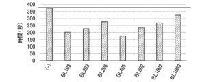
- Figure 12 is a graph showing the results of examining the effect of adding MPC polymer on the reaction rate.
- the vertical axis indicates the time until the S/N ratio reaches its maximum value
- the horizontal axis indicates the type of MPC polymer added.
- BL103”, “BL203”, “BL206”, “BL405", “BL802”, “BL1002”, and “BL1003” respectively refer to Lipidure (registered trademark)-BL103, Lipidure (registered trademark)-BL203, Lipidure (registered trademark)-BL206, Lipidure (registered trademark)-BL405, Lipidure (registered trademark)-BL802, Lipidure (registered trademark)-BL1002, and Lipidure (registered trademark)-BL1003 (all NOF Corporation).
- (-) indicates the results for a sample to which no MPC polymer was added. As a result, it became clear that the time required for the S/N ratio to reach its maximum value could be shortened by adding MPC polymer to the reaction solution.
- Figure 13 is a graph showing the results of examining the effect of adding MPC polymer on S/N.
- the vertical axis shows the maximum S/N.
- the horizontal axis like Figure 12, shows the type of MPC polymer added. As a result, no improvement in S/N was observed even when MPC polymer was added to the reaction solution.
- Figures 14 and 15 are graphs showing the results of the immunoICA reaction.
- the fluorescence intensity detected by the immunoICA reaction using phosphorylated MEK was taken as the signal (S)
- the fluorescence intensity detected by the immunoICA reaction using non-phosphorylated MEK was taken as the noise (N)
- S the fluorescence intensity detected by the immunoICA reaction using phosphorylated MEK
- N the noise
- Figure 14 is a graph showing the results of examining the effect of the type of blocking buffer on the reaction rate.
- the vertical axis indicates the time until the S/N ratio reaches its maximum value
- the horizontal axis indicates the type of blocking buffer used.
- “Solution A” means Can Get Signal buffer solution A (Toyobo)
- “Solution B” means Can Get Signal buffer solution B (Toyobo).
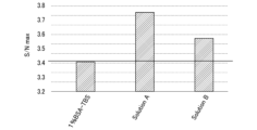
- Figure 15 is a graph showing the results of an investigation into the effect of the type of blocking buffer on the S/N ratio.
- the vertical axis indicates the maximum S/N ratio
- the horizontal axis like Figure 14, indicates the type of blocking buffer used.
- the maximum S/N ratio could be increased by using a blocking buffer containing casein and a nonionic surfactant (Can Get Signal buffer solution A or Can Get Signal buffer solution B, both Toyobo) instead of 1% BSA-TBS as the blocking buffer.
- a blocking buffer containing casein and a nonionic surfactant Can Get Signal buffer solution A or Can Get Signal buffer solution B, both Toyobo
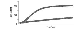
- Figures 16-17 are graphs showing the results of investigating the effect of adding Lipidure (registered trademark)-BL1002 and Lipidure (registered trademark)-BL1003, respectively, on ICA reactivity.
- the vertical axis indicates ICA reaction intensity
- the horizontal axis indicates reaction time.
- Figure 18 shows the results of a sample to which no MPC polymer was added. As a result, an increase in reaction intensity was observed when MPC polymer was added to the reaction solution.
- an ICA reaction was carried out by adding the MPC polymer shown in Table 2 or the blocking buffer shown in Table 3.
- the fluorescence intensity when the target DNA was added was taken as the signal (S), and the fluorescence intensity when the target DNA was not added was taken as the noise (N), and the S/N was calculated.
- Figure 19 shows the time (unit: seconds) required for the S/N to reach a maximum when the MPC polymer shown in Table 2 or the blocking buffer shown in Table 3 was added.
- Figure 20 shows the maximum S/N when the MPC polymer shown in Table 2 or the blocking buffer shown in Table 3 was added.
- PC shows the results of a sample to which the MPC polymer and blocking buffer were not added, for comparison.
- the oligonucleotide DNA11 (5'-TTTGTCACTGTTCCTCCTTTTGTTTTCCTTTCTGTGAGCAATTTCACCCAA-3', SEQ ID NO: 9) was bound to the anti-phosphorylated MEK rabbit polyclonal antibody to obtain a DNA11-modified anti-phosphorylated MEK rabbit polyclonal antibody.
- an oligonucleotide, DNA12 (5'-GCATGGTTCCAATTTGGGTGAT-3', SEQ ID NO: 10), was bound to the anti-MEK rabbit polyclonal antibody to obtain a DNA12-modified anti-MEK rabbit polyclonal antibody.
- Antigen-antibody reaction and ICA reaction The above-mentioned kinase assay and antigen-antibody reaction solution and the above-mentioned ICA reaction solution were mixed and incubated for 60 minutes at 30° C. Then, the mixture was incubated for 60 minutes at 66° C. to carry out the ICA reaction.
- the blocking buffer concentration was set to 1, containing casein, a nonionic surfactant, and tris(hydroxymethyl)aminomethane, and reaction solutions were prepared with concentrations adjusted to 1/2 and 1/4, and measurements were performed using a real-time PCR device (Qiagen, Rotor-Gene Q).
- the fluorescence intensity detected by the immuno-ICA reaction using phosphorylated MEK was taken as the signal (S)
- the fluorescence intensity detected by the immuno-ICA reaction using non-phosphorylated MEK was taken as the noise (N)
- S/N ratio was calculated.
- Figure 21 is a graph showing the results of examining the effect of blocking buffer concentration on S/N.
- the vertical axis shows the maximum S/N
- the horizontal axis shows the time it takes for the S/N to reach the maximum.
- the blocking buffer concentration which contains casein, a nonionic surfactant, and tris(hydroxymethyl)aminomethane
- the maximum S/N ratio (4.3) was reached.
- the time to reach the maximum S/N ratio was twice as long as that when the blocking buffer concentration was 1/2.
- the present invention provides a method for detecting a target substance in a sample and a kit for the same.
- 100,900...complex 110,120...target substance (protein), 160...phosphate group, 130,140,170...specific binding substance, 131,141,171...single-stranded nucleic acid fragment, 150...double-stranded nucleic acid (double-stranded nucleic acid region), 200,500...fluidic device, 210...substrate, 220...cover member, 221...projection, 222...inlet port, 223...exhaust port, 230...channel, 241,242,242R...well (microcompartment), 240...well array, 510...wall member, 810...flap probe, 811,821...flap site (nucleic acid fragment), 820...nucleic acid fragment.
Landscapes
- Chemical & Material Sciences (AREA)
- Life Sciences & Earth Sciences (AREA)
- Health & Medical Sciences (AREA)
- Engineering & Computer Science (AREA)
- Organic Chemistry (AREA)
- Bioinformatics & Cheminformatics (AREA)
- Zoology (AREA)
- Wood Science & Technology (AREA)
- Genetics & Genomics (AREA)
- Biotechnology (AREA)
- General Health & Medical Sciences (AREA)
- Biochemistry (AREA)
- Biomedical Technology (AREA)
- General Engineering & Computer Science (AREA)
- Molecular Biology (AREA)
- Microbiology (AREA)
- Medicinal Chemistry (AREA)
- Biophysics (AREA)
- Proteomics, Peptides & Aminoacids (AREA)
- Immunology (AREA)
- Analytical Chemistry (AREA)
- Physics & Mathematics (AREA)
- Sustainable Development (AREA)
- Plant Pathology (AREA)
- Measuring Or Testing Involving Enzymes Or Micro-Organisms (AREA)
Priority Applications (2)
| Application Number | Priority Date | Filing Date | Title |
|---|---|---|---|
| EP24819428.4A EP4726054A1 (en) | 2023-06-09 | 2024-06-07 | Method for detecting target substance in sample, and kit therefor |
| JP2025526171A JPWO2024253201A1 (https=) | 2023-06-09 | 2024-06-07 |
Applications Claiming Priority (2)
| Application Number | Priority Date | Filing Date | Title |
|---|---|---|---|
| JP2023095436 | 2023-06-09 | ||
| JP2023-095436 | 2023-06-09 |
Publications (1)
| Publication Number | Publication Date |
|---|---|
| WO2024253201A1 true WO2024253201A1 (ja) | 2024-12-12 |
Family
ID=93795495
Family Applications (1)
| Application Number | Title | Priority Date | Filing Date |
|---|---|---|---|
| PCT/JP2024/020941 Ceased WO2024253201A1 (ja) | 2023-06-09 | 2024-06-07 | 試料中の標的物質を検出する方法及びそのためのキット |
Country Status (3)
| Country | Link |
|---|---|
| EP (1) | EP4726054A1 (https=) |
| JP (1) | JPWO2024253201A1 (https=) |
| WO (1) | WO2024253201A1 (https=) |
Citations (6)
| Publication number | Priority date | Publication date | Assignee | Title |
|---|---|---|---|---|
| JP2005514582A (ja) * | 2001-04-30 | 2005-05-19 | ベンタナ・メデイカル・システムズ・インコーポレーテツド | 自動化ハイブリダイゼーション用の試薬および方法 |
| US10465235B2 (en) | 2011-05-24 | 2019-11-05 | Navinci Diagnostics Ab | Multiplexed proximity ligation assay |
| JP2020122799A (ja) | 2014-05-15 | 2020-08-13 | メソ スケール テクノロジーズ エルエルシー | 改善されたアッセイ方法 |
| WO2023080137A1 (ja) * | 2021-11-04 | 2023-05-11 | 凸版印刷株式会社 | 標的物質の検出方法 |
| JP2023095436A (ja) | 2021-12-24 | 2023-07-06 | 京セラ株式会社 | 制御装置及び制御方法 |
| WO2024029511A1 (ja) * | 2022-08-01 | 2024-02-08 | Toppanホールディングス株式会社 | 評価方法、複合体及びキット |
-
2024
- 2024-06-07 JP JP2025526171A patent/JPWO2024253201A1/ja active Pending
- 2024-06-07 EP EP24819428.4A patent/EP4726054A1/en active Pending
- 2024-06-07 WO PCT/JP2024/020941 patent/WO2024253201A1/ja not_active Ceased
Patent Citations (6)
| Publication number | Priority date | Publication date | Assignee | Title |
|---|---|---|---|---|
| JP2005514582A (ja) * | 2001-04-30 | 2005-05-19 | ベンタナ・メデイカル・システムズ・インコーポレーテツド | 自動化ハイブリダイゼーション用の試薬および方法 |
| US10465235B2 (en) | 2011-05-24 | 2019-11-05 | Navinci Diagnostics Ab | Multiplexed proximity ligation assay |
| JP2020122799A (ja) | 2014-05-15 | 2020-08-13 | メソ スケール テクノロジーズ エルエルシー | 改善されたアッセイ方法 |
| WO2023080137A1 (ja) * | 2021-11-04 | 2023-05-11 | 凸版印刷株式会社 | 標的物質の検出方法 |
| JP2023095436A (ja) | 2021-12-24 | 2023-07-06 | 京セラ株式会社 | 制御装置及び制御方法 |
| WO2024029511A1 (ja) * | 2022-08-01 | 2024-02-08 | Toppanホールディングス株式会社 | 評価方法、複合体及びキット |
Non-Patent Citations (7)
Also Published As
| Publication number | Publication date |
|---|---|
| JPWO2024253201A1 (https=) | 2024-12-12 |
| EP4726054A1 (en) | 2026-04-15 |
Similar Documents
| Publication | Publication Date | Title |
|---|---|---|
| US20230213510A1 (en) | Methods relating to improving accuracy of capture object-based assays | |
| US11898194B2 (en) | Method for detecting target molecule | |
| JP5503540B2 (ja) | 溶液中の分析物濃度を決定する方法 | |
| US10054591B2 (en) | Amplifying rare cell surface markers | |
| US12509737B2 (en) | Method for detecting target molecule | |
| US20240280592A1 (en) | METHOD FOR DETECTING AMYLOID-ß BOUND TO BRAIN-NEURONALCELL-DERIVED EXOSOME IN BLOOD, KIT FOR DETECTION OF AMYLOID-ß BOUND TO BRAIN-NEURONAL-CELL-DERIVED EXOSOME IN BLOOD, AND METHOD FOR EVALUATING ACCUMULATION LEVEL OF AMYLOID-ß IN BRAIN | |
| US20240279717A1 (en) | Method of detecting target substance | |
| US20250164470A1 (en) | Evaluation method, complex, and kit | |
| US10914735B2 (en) | Protein detection using FET | |
| JP2025188101A (ja) | 検出方法 | |
| JP7831518B2 (ja) | ウェルに液体を導入する方法 | |
| WO2024253201A1 (ja) | 試料中の標的物質を検出する方法及びそのためのキット | |
| JPWO2006062149A1 (ja) | 生体サンプル分析用プレート | |
| US20260009067A1 (en) | Method and kit for detecting target rna | |
| JP2012189541A (ja) | マイクロ化学チップ | |
| US20250352999A1 (en) | Fluidic device, and method for detecting target molecule | |
| WO2025254035A1 (ja) | 複数種類の標的分子を検出する方法 | |
| JP2010088317A (ja) | 生体試料定量用チップ、生体試料定量用キット、及び生体試料定量方法 | |
| US20230357822A1 (en) | Method for analyzing cell released biomolecules | |
| JP2006220531A (ja) | 生体サンプル判別用プレート |
Legal Events
| Date | Code | Title | Description |
|---|---|---|---|
| 121 | Ep: the epo has been informed by wipo that ep was designated in this application |
Ref document number: 24819428 Country of ref document: EP Kind code of ref document: A1 |
|
| ENP | Entry into the national phase |
Ref document number: 2025526171 Country of ref document: JP Kind code of ref document: A |
|
| WWE | Wipo information: entry into national phase |
Ref document number: 2025526171 Country of ref document: JP |
|
| WWE | Wipo information: entry into national phase |
Ref document number: 2024819428 Country of ref document: EP |
|
| NENP | Non-entry into the national phase |
Ref country code: DE |
|
| ENP | Entry into the national phase |
Ref document number: 2024819428 Country of ref document: EP Effective date: 20260109 |
|
| ENP | Entry into the national phase |
Ref document number: 2024819428 Country of ref document: EP Effective date: 20260109 |
|
| ENP | Entry into the national phase |
Ref document number: 2024819428 Country of ref document: EP Effective date: 20260109 |
|
| ENP | Entry into the national phase |
Ref document number: 2024819428 Country of ref document: EP Effective date: 20260109 |
|
| ENP | Entry into the national phase |
Ref document number: 2024819428 Country of ref document: EP Effective date: 20260109 |
|
| WWP | Wipo information: published in national office |
Ref document number: 2024819428 Country of ref document: EP |