WO2024225488A1 - 抗pd-1抗体療法に応答性である候補がん患者を同定する方法 - Google Patents
抗pd-1抗体療法に応答性である候補がん患者を同定する方法 Download PDFInfo
- Publication number
- WO2024225488A1 WO2024225488A1 PCT/JP2024/016627 JP2024016627W WO2024225488A1 WO 2024225488 A1 WO2024225488 A1 WO 2024225488A1 JP 2024016627 W JP2024016627 W JP 2024016627W WO 2024225488 A1 WO2024225488 A1 WO 2024225488A1
- Authority
- WO
- WIPO (PCT)
- Prior art keywords
- antibody
- fgfbp2
- cancer patient
- administration
- expression level
- Prior art date
- Legal status (The legal status is an assumption and is not a legal conclusion. Google has not performed a legal analysis and makes no representation as to the accuracy of the status listed.)
- Ceased
Links
Images


Classifications
-
- C—CHEMISTRY; METALLURGY
- C07—ORGANIC CHEMISTRY
- C07K—PEPTIDES
- C07K14/00—Peptides having more than 20 amino acids; Gastrins; Somatostatins; Melanotropins; Derivatives thereof
- C07K14/435—Peptides having more than 20 amino acids; Gastrins; Somatostatins; Melanotropins; Derivatives thereof from animals; from humans
-
- C—CHEMISTRY; METALLURGY
- C07—ORGANIC CHEMISTRY
- C07K—PEPTIDES
- C07K16/00—Immunoglobulins [IG], e.g. monoclonal or polyclonal antibodies
- C07K16/18—Immunoglobulins [IG], e.g. monoclonal or polyclonal antibodies against material from animals or humans
- C07K16/28—Immunoglobulins [IG], e.g. monoclonal or polyclonal antibodies against material from animals or humans against receptors, cell surface antigens or cell surface determinants
-
- C—CHEMISTRY; METALLURGY
- C12—BIOCHEMISTRY; BEER; SPIRITS; WINE; VINEGAR; MICROBIOLOGY; ENZYMOLOGY; MUTATION OR GENETIC ENGINEERING
- C12Q—MEASURING OR TESTING PROCESSES INVOLVING ENZYMES, NUCLEIC ACIDS OR MICROORGANISMS; COMPOSITIONS OR TEST PAPERS THEREFOR; PROCESSES OF PREPARING SUCH COMPOSITIONS; CONDITION-RESPONSIVE CONTROL IN MICROBIOLOGICAL OR ENZYMOLOGICAL PROCESSES
- C12Q1/00—Measuring or testing processes involving enzymes, nucleic acids or microorganisms; Compositions therefor; Processes of preparing such compositions
- C12Q1/68—Measuring or testing processes involving enzymes, nucleic acids or microorganisms; Compositions therefor; Processes of preparing such compositions involving nucleic acids
-
- C—CHEMISTRY; METALLURGY
- C12—BIOCHEMISTRY; BEER; SPIRITS; WINE; VINEGAR; MICROBIOLOGY; ENZYMOLOGY; MUTATION OR GENETIC ENGINEERING
- C12Q—MEASURING OR TESTING PROCESSES INVOLVING ENZYMES, NUCLEIC ACIDS OR MICROORGANISMS; COMPOSITIONS OR TEST PAPERS THEREFOR; PROCESSES OF PREPARING SUCH COMPOSITIONS; CONDITION-RESPONSIVE CONTROL IN MICROBIOLOGICAL OR ENZYMOLOGICAL PROCESSES
- C12Q1/00—Measuring or testing processes involving enzymes, nucleic acids or microorganisms; Compositions therefor; Processes of preparing such compositions
- C12Q1/68—Measuring or testing processes involving enzymes, nucleic acids or microorganisms; Compositions therefor; Processes of preparing such compositions involving nucleic acids
- C12Q1/6869—Methods for sequencing
-
- G—PHYSICS
- G01—MEASURING; TESTING
- G01N—INVESTIGATING OR ANALYSING MATERIALS BY DETERMINING THEIR CHEMICAL OR PHYSICAL PROPERTIES
- G01N33/00—Investigating or analysing materials by specific methods not covered by groups G01N1/00 - G01N31/00
- G01N33/48—Biological material, e.g. blood, urine; Haemocytometers
- G01N33/50—Chemical analysis of biological material, e.g. blood, urine; Testing involving biospecific ligand binding methods; Immunological testing
- G01N33/53—Immunoassay; Biospecific binding assay; Materials therefor
- G01N33/575—Immunoassay; Biospecific binding assay; Materials therefor for cancer
Definitions
- the present invention relates to a method for identifying candidate cancer patients who are responsive to anti-PD-1 antibody therapy.
- Vertebrates including humans, have a self-defense system against cancer through immune response.
- dendritic cells recognize mutant antigens expressed on tumor cells and present them to naive CD8 + T cells
- activation of cytotoxic CD8 + T cells is induced.
- Activated cytotoxic CD8 + T cells migrate to tumor tissue, infiltrate the tumor microenvironment, and recognize and bind to tumor cells.
- the bound cytotoxic CD8 + T cells secrete cytotoxins, which induce apoptosis and death of tumor cells.
- T cells In order to prevent the immune system from attacking its own cells, T cells have factors such as PD-1 (Programmed cell death-1) and CTLA-4 (Cytotoxic T-Lymphocyte (associated) Antigen 4) that can only bind to the body's own cells, and immune cells other than T cells and general cells have factors such as PD-L1 (Programmed cell Death Ligand 1)/PD-L2 (Programmed cell Death Ligand 2) and CD80/CD86 that bind to the above factors. When these two factors bind, the immune system is able to avoid attack by T cells, creating a system known as the immune checkpoint.
- PD-1 Programmed cell death-1
- CTLA-4 Cytotoxic T-Lymphocyte (associated) Antigen 4
- CD80/CD86 CD80/CD86
- Cancer cells contain factors such as PD-L1 because they are autologous cells. Therefore, by binding to T cells, cancer cells can avoid attack by T cells via the immune checkpoint system.
- PD-L1 is known to be expressed in various tumor tissues, including malignant melanoma, ovarian cancer, esophageal cancer, and lung cancer.
- cancer immunotherapy has been carried out in recent years using immune checkpoint inhibitors such as anti-PD-1 antibodies, anti-CTLA-4 antibodies, and anti-PD-L1 antibodies.
- immune checkpoint inhibitors such as anti-PD-1 antibodies, anti-CTLA-4 antibodies, and anti-PD-L1 antibodies.
- the anti-PD-1 antibodies nivolumab and pembrolizumab are used in cancer immunotherapy against malignant melanoma, non-small cell lung cancer, and other cancers.
- Non-Patent Document 1 it has been reported that the response rate was 19.4% when pembrolizumab was administered every 2 weeks or every 3 weeks to 495 patients with non-small cell lung cancer. It has also been reported that the response rate was 20% when nivolumab was administered every 2 weeks to 272 patients with non-small cell lung cancer (Non-Patent Document 2).
- Patent Document 1 discloses a method for assisting in the prediction of the effectiveness of cancer treatment by immune checkpoint inhibitors, which includes the steps of: obtaining information on the T cell receptor (TCR) repertoire of tumor-infiltrating T cells in tumor tissue and the TCR repertoire of CD8-positive T cells in peripheral blood, using tumor tissue samples and peripheral blood samples collected from a cancer patient before starting treatment with an immune checkpoint inhibitor; comparing the TCR repertoire of tumor-infiltrating T cells with the TCR repertoire of peripheral blood CD8-positive T cells and identifying T cell clones detected in common between the two as overlapping clones; and calculating the total frequency of overlapping clones in the TCR repertoire of peripheral blood CD8-positive T cells, and assisting in the prediction that the immune checkpoint inhibitor will provide a cancer treatment effect in the cancer patient if the total frequency of the overlapping clones is equal to or greater than a predetermined cutoff value.
- TCR T cell receptor
- Patent Document 2 discloses a method for assisting in determining the efficacy of an immune checkpoint inhibitor, which includes a step of measuring a free protein marker in a liquid sample collected from a subject, the free protein marker including free PD-1, and the measurement result indicating the efficacy of the immune checkpoint inhibitor for the subject.
- Patent document 3 discloses a method for determining the responsiveness of a cancer patient to an immune checkpoint inhibitor, the method including determining the responsiveness of the cancer patient to the immune checkpoint inhibitor based on the level of at least one short-chain fatty acid in a sample collected from the cancer patient.
- Patent Document 1 requires the collection of tumor tissue samples from cancer patients when carrying out a method to assist in predicting the effectiveness of cancer treatment with immune checkpoint inhibitors, which places a heavy burden on patients and leaves room for improvement.
- Patent Document 2 places a small burden on patients by using free protein markers in the blood as indicators to assist in determining the efficacy of immune checkpoint inhibitors.
- the free protein markers include free PD-1, it is difficult to accurately determine the efficacy rate of patients who have received an anti-PD-1 antibody once with subsequent continuous administrations, leaving room for improvement.
- Patent Document 3 places little burden on patients when assessing the responsiveness of cancer patients to immune checkpoint inhibitors, as it uses the level of short-chain fatty acids in the blood as an indicator. However, there is still room for improvement, as the responsiveness of patients who have been administered an anti-PD-1 antibody once to subsequent continuous administrations has not been examined.
- the objective of the present invention is to provide a method for identifying candidate cancer patients who will respond to a first administration of an anti-PD-1 antibody and those who will respond to subsequent administrations from peripheral blood, while placing a minimal burden on cancer patients in anti-PD-1 antibody therapy.
- the inventors conducted extensive research to solve the above problems. As a result, they discovered that the above problems could be solved by using the following configuration that uses a specific blood biomarker, and thus completed the present invention.
- a method for identifying a candidate cancer patient X who is responsive to anti-PD-1 antibody therapy comprising step 1 of measuring an expression level of FGFBP2 mRNA in peripheral blood mononuclear cells of the cancer patient, and step 2 of comparing the expression level of FGFBP2 mRNA with a predetermined cutoff value, wherein a result of the expression level of FGFBP2 mRNA exceeding the predetermined cutoff value indicates that the cancer patient is a candidate cancer patient X to be administered a first dose of an anti-PD-1 antibody.
- a method for identifying a candidate cancer patient Y who is responsive to anti-PD-1 antibody therapy comprising: step A measuring the expression level of FGFBP2 protein in the serum, plasma, or blood of a cancer patient before and after an nth administration of an anti-PD-1 antibody (n is a positive integer); and step B comparing the expression level of FGFBP2 protein before and after the nth administration of the anti-PD-1 antibody, wherein the expression level of FGFBP2 protein after the nth administration of the anti-PD-1 antibody exceeds the expression level of FGFBP2 protein before the nth administration of the anti-PD-1 antibody indicates that the cancer patient is a candidate cancer patient Y to be administered the anti-PD-1 antibody for the n+1th or subsequent time.
- a diagnostic kit for identifying a candidate cancer subject X who is responsive to anti-PD-1 antibody therapy comprising a primer set for detecting FGFBP2 mRNA.
- a diagnostic kit for identifying a candidate cancer subject Y who is responsive to anti-PD-1 antibody therapy comprising an antibody against FGFBP2 or an antigen-binding fragment thereof.
- a method for using the expression level of FGFBP2 mRNA in peripheral blood mononuclear cells of a cancer patient as an index of the responsiveness of a candidate cancer patient X to anti-PD-1 antibody therapy comprising step 1 of measuring the expression level of FGFBP2 mRNA in peripheral blood mononuclear cells of the cancer patient, and step 2 of comparing the expression level of FGFBP2 mRNA with a predetermined cutoff value, wherein the expression level of FGFBP2 mRNA exceeds the predetermined cutoff value, indicating that the patient is a candidate cancer patient X to be administered a first dose of an anti-PD-1 antibody.
- a method for using the expression level of FGFBP2 protein in the serum, plasma, or blood of a cancer patient as an index of the responsiveness of a candidate cancer patient Y to anti-PD-1 antibody therapy comprising: step A measuring the expression level of FGFBP2 protein in the serum, plasma, or blood of the cancer patient before and after an nth (n is a positive integer) administration of an anti-PD-1 antibody; and step B comparing the expression levels of FGFBP2 protein before and after the nth administration of the anti-PD-1 antibody, wherein the expression level of FGFBP2 protein after the nth administration of the anti-PD-1 antibody exceeds the expression level of FGFBP2 protein before the first administration of the anti-PD-1 antibody indicates that the cancer patient is a candidate cancer patient Y to be administered the anti-PD-1 antibody for the n+1th or subsequent times.
- step A measuring the expression level of FGFBP2 protein in the serum, plasma, or blood of the cancer patient before and after an nth (n is a positive integer) administration of an anti-PD
- the present invention provides a method for identifying candidate cancer patients who are responsive to a first administration of anti-PD-1 antibody and to subsequent administrations from peripheral blood, which places a low burden on cancer patients in anti-PD-1 antibody therapy.
- FIG. 1 is a violin plot showing the expression level of FGFBP2 mRNA in each cell type of peripheral blood mononuclear cells.
- the horizontal axis indicates the cell type, and the vertical axis indicates the expression level of FGFBP2 mRNA.
- FIG. 1 is a violin plot showing the expression level of FGFBP2 mRNA in each cell type of peripheral blood mononuclear cells.
- the horizontal axis indicates the cell type, and the vertical axis indicates the expression level of FGFBP2 mRNA.
- the cell types on the horizontal axis are, from the left, CD4n, Th1, Th2, Th17, eTreg, nTreg, CD4Cluster8, CD4Cluster9, CD4CTL, Th1GZMK, CD4TrmL, CD8Tn, CD8Tem, CD8Temra, CD8Teff, CD8Tcm1, CD8Tcm2, CD8MAIT, CD8Trm, MDSCL, MDSCL2, Moth1, Moth2, Moth3, Mapc, MDSC, Mnc, Minfr, MLC, Mfcer1, CD56BrightHigh, CD56BrightLow, MatureNK, AdaptiveNK, ActiveNK, NKCluster3, NKCluster5, NKCluster6, NKCluster7, NKCluster8, B Naive1, BNaive2, BNaive1Mature, Bmemory1, Bmemory2, BIGHV3, BIGHV4, BPlasma, BHemo, BIF, and B
- FIG. 2 is a violin plot showing the expression level of FGFBP2 mRNA in clonal cells of effector CD4 + T cells.
- the horizontal axis indicates the group of clonal cell number (X), and the vertical axis indicates the expression level of FGFBP2 mRNA.
- the groups on the horizontal axis indicate, from the left, Single (0 ⁇ X ⁇ 1), Small (1 ⁇ X ⁇ 5), Medium (5 ⁇ X ⁇ 20), Large (20 ⁇ X ⁇ 100), Hyper (100 ⁇ X ⁇ 1000), and Unknown.
- 3 is a violin plot showing the expression level of FGFBP2 mRNA in clonal cells of effector CD8 + T cells.
- the horizontal axis indicates the group of clonal cell number (X), and the vertical axis indicates the expression level of FGFBP2 mRNA.
- the groups on the horizontal axis indicate, from the left, Single (0 ⁇ X ⁇ 1), Small (1 ⁇ X ⁇ 5), Medium (5 ⁇ X ⁇ 20), Large (20 ⁇ X ⁇ 100), Hyper (100 ⁇ X ⁇ 1000), and Unknown.
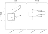
- 4 is a graph showing the amount of FGFBP2 protein in plasma obtained from cancer patients who responded or did not respond to anti-PD-1 antibody therapy before and after the first administration of anti-PD-1 antibody.
- the horizontal axis shows the amount of FGFBP2 protein expression in responding or non-responding samples before and after administration of anti-PD-1 antibody
- the vertical axis shows the amount of FGFBP2 protein expression.
- Pre-treatment indicates the amount before administration of anti-PD-1 antibody
- Post-treatment indicates the amount after administration of anti-PD-1 antibody
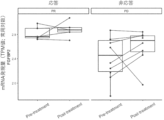
- 5 is a graph showing the expression level of FGFBP2 mRNA in peripheral blood mononuclear cells obtained from cancer patients who responded or did not respond to anti-PD-1 antibody therapy before and after the first administration of anti-PD-1 antibody.
- the horizontal axis shows the expression level of FGFBP2 mRNA in responding or non-responding samples before and after administration of anti-PD-1 antibody, and the vertical axis shows the expression level of FGFBP2 mRNA.
- Pre-treatment indicates before administration of anti-PD-1 antibody
- Post-treatment indicates after administration of anti-PD-1 antibody.
- an anti-PD-1 antibody is an antibody that binds to a PD-1 receptor and inhibits the binding between PD-L1 expressed in cancer cells and PD-1 expressed in immune cells, or inhibits the binding between PD-L1 expressed in cancer cells and PD-1 expressed in immune cells.
- the present invention relates to an antibody that inhibits the binding between PD-L2 expressed in immune cells and PD-1 expressed in immune cells.
- the anti-PD-1 antibody includes a monoclonal antibody or an antigen-binding fragment thereof that specifically binds to PD-1.
- the monoclonal antibody may be a human antibody, a humanized antibody, or a chimeric antibody.
- the anti-PD-1 antibody is not particularly limited as long as it has the above-mentioned functions, but examples thereof include nivolumab, pembrolizumab, spartalizumab, zimblerimab, tislerizumab, dostarlimab, toripalimab, cetrelimab, camrelizumab, genolimusma, and cetrelimab.
- anti-PD-1 antibodies examples include Genolimzumab, Sintilimab, Lodapolimab, Sasanlimab, Retifanlimab, Balstilimab, Serplulimab, Budigalimab, Prolgolimab, Geptanolimab, and Cemiplimab-rwlc.
- the anti-PD-1 antibodies are preferably Nivolumab and Pembrolizumab.
- Anti-PD-1 antibody therapy is a cancer treatment method in which an anti-PD-1 antibody is administered to a cancer patient.
- the responsiveness of anti-PD-1 antibody therapy for example, the therapeutic effect when an effective amount of at least one of the above anti-PD-1 antibodies is administered can be judged according to the criteria of the RECIST (Response Evaluation Criteria in Solid Tumors) guidelines, and a complete response (CR) or partial response (PR) can be considered to be responsive, and a progressive disease (PD) can be considered to be unresponsive.
- RECIST Response Evaluation Criteria in Solid Tumors
- Antibodies include chimeric antibodies, humanized antibodies, and fully human antibodies.
- a chimeric antibody refers to an antibody whose variable region is derived from a non-human animal and whose constant region is at least partially derived from a human.
- a humanized antibody refers to an antibody whose heavy and light chain complementarity determining regions (CDRs) are derived from a non-human animal and whose constant region and framework region are derived from a human.
- CDRs heavy and light chain complementarity determining regions
- a fully human antibody refers to an antibody whose entirety, including the complementarity determining regions, is derived from a human.
- An antigen-binding fragment refers to a protein that contains a portion of an antibody and is capable of binding to an antigen, and examples of the antigen-binding fragment include F(ab') 2 , Fab', Fab, variable fragments of antibodies (Fv), disulfide-linked Fv, single-chain antibodies (scFv), and polymers thereof.
- the cancers targeted in the present invention are not limited to those for which the therapeutic effect of anti-PD-1 antibodies has been reported, or those for which the therapeutic effect can be predicted from the mechanism of action, but are preferably solid cancers.
- solid cancers include breast cancer, and chest cancers including lung cancers such as non-small cell lung cancer and small cell lung cancer; skin cancers including basal cell carcinoma, squamous cell carcinoma, and malignant melanoma; digestive cancers including liver cancer including hepatocellular carcinoma and cholangiocarcinoma, colon cancer including colon cancer and rectal cancer, gallbladder cancer, bile duct cancer, pancreatic cancer, duodenal cancer, esophageal cancer, gastric cancer, and anal cancer; urinary cancers including kidney cancer, ureter cancer, bladder cancer, prostate cancer, penile cancer, and testicular (testicle) cancer; reproductive cancers including uterine cancer, ovarian cancer, vulvar cancer, and vaginal cancer, including cervical cancer and uterine cancer
- FGFBP2 fibroblast growth factor binding protein type 2; NCBI Refseq: NM_031950: NP_114156
- KSP37 Killer-Specific Secretory Protein Of 37KDa
- FGFBP2 also includes variants of FGFBP2.
- One embodiment of the present invention is a method for identifying a candidate cancer patient X who is responsive to anti-PD-1 antibody therapy, comprising step 1 of measuring the expression level of FGFBP2 mRNA in peripheral blood mononuclear cells of the cancer patient, and step 2 of comparing the expression level of FGFBP2 mRNA with a predetermined cutoff value, wherein the expression level of FGFBP2 mRNA exceeds the predetermined cutoff value, indicating that the cancer patient is a candidate cancer patient X to be administered a first dose of an anti-PD-1 antibody.
- Step 1 is a step of measuring the expression level of FGFBP2 mRNA in peripheral blood mononuclear cells (PBMCs) of a cancer patient.
- PBMCs peripheral blood mononuclear cells
- peripheral blood mononuclear cells of a cancer patient can be carried out by a known method.
- peripheral blood mononuclear cells can be separated from blood collected from a cancer patient by density gradient centrifugation using a lymphocyte density separation solution such as Lymphoprep TM (Serumwerk Bernburg).
- Blood of a cancer patient can be collected from the cancer patient by a method known to those skilled in the art.
- Blood (whole blood) can be collected by blood collection using, for example, a syringe or the like.
- RNA can be extracted from peripheral blood mononuclear cells using known methods. For example, it can be extracted using a kit such as RNAiso Blood (manufactured by Takara Bio Inc.). Furthermore, the expression level of FGFBP2 mRNA can be measured by appropriately selecting from known methods. For example, mRNA can be extracted from peripheral blood mononuclear cells using known methods, and then it can be measured using techniques such as RT-PCR, real-time RT-PCR, microarray, Northern blot, dot blot, QuantiGene Plex (QGP), RNase protection assay, and RNA-Sequencing (RNA-Seq) as described later in the Examples.
- RT-PCR real-time RT-PCR
- microarray Northern blot
- dot blot dot blot
- QGP QuantiGene Plex
- RNase protection assay RNA-Sequencing
- primers for PCR commercially available primers such as those manufactured by Nippon Shinyo Biological Co., Ltd. (product number: HP101811) and those manufactured by OriGene Technologies Inc. (product number: HP215686) can be used.
- Step 2 is a step of comparing the expression level of FGFBP2 mRNA obtained in step 1 with a predetermined cutoff value.
- the cutoff value is a benchmark for determining whether a cancer patient will respond to the first administration of anti-PD-1 antibody in anti-PD-1 antibody therapy.
- the cutoff value can be determined by measuring the expression levels of FGFBP2 mRNA in peripheral blood mononuclear cells collected from two or more, preferably four or more, and more preferably six or more cancer patients who are responsive to the first administration of anti-PD-1 antibody in anti-PD-1 antibody therapy and non-responsive patients, calculating the average or median of the expression levels, and based on the average or median.
- the cutoff value may also be set from a Receiver Operating Characteristic (ROC) curve.
- ROC Receiver Operating Characteristic
- One embodiment of the present invention is a method for using the expression level of FGFBP2 mRNA in peripheral blood mononuclear cells of a cancer patient as an indicator of the responsiveness of a candidate cancer patient X to anti-PD-1 antibody therapy, comprising: step 1 of measuring the expression level of FGFBP2 mRNA in peripheral blood mononuclear cells of the cancer patient; and step 2 of comparing the expression level of FGFBP2 mRNA with a predetermined cutoff value.
- the method further comprises: the FGFBP2 mRNA expression level exceeding the predetermined cutoff value indicating that the patient is a candidate cancer patient X for receiving a first dose of an anti-PD-1 antibody.
- One embodiment of the present invention is a method for identifying a candidate cancer patient Y who is responsive to anti-PD-1 antibody therapy, comprising step A measuring the expression level of FGFBP2 protein in the serum, plasma, or blood of a cancer patient before and after an nth administration of an anti-PD-1 antibody (n is a positive integer), and step B comparing the expression level of FGFBP2 protein before and after the nth administration of the anti-PD-1 antibody, wherein the expression level of FGFBP2 protein after the nth administration of the anti-PD-1 antibody exceeds the expression level of FGFBP2 protein before the nth administration of the anti-PD-1 antibody indicates that the cancer patient is a candidate cancer patient Y to be administered the anti-PD-1 antibody for the n+1th or subsequent times.
- Step A is a step of measuring the expression level of FGFBP2 protein in the serum, plasma, or blood of a cancer patient before and after the nth (n is a positive integer) administration of an anti-PD-1 antibody.
- nth administration of an anti-PD-1 antibody refers to the first (initial) administration of an anti-PD-1 antibody to a cancer patient, as well as the second, third, fourth, and subsequent administrations.
- n is preferably 1 to 3, and more preferably 1.
- the period after the nth administration of anti-PD-1 antibody can be set arbitrarily, depending on the administration interval of the anti-PD-1 antibody, as long as it is after the nth administration of the anti-PD-1 antibody. Usually, it is within 105 days, preferably within 73 days, more preferably within 28 days, and even more preferably within 21 days after the nth administration of the anti-PD-1 antibody.
- the administration interval of the anti-PD-1 antibody is not particularly limited, and can be set appropriately depending on the anti-PD-1 antibody used, the symptoms of the cancer patient to be administered, the race, age, sex, weight, etc. Examples include once a week, once every two weeks, once every three weeks, once every four weeks, once a month, once every two months, etc.
- parenteral administration usually refers to administration by local administration or injection other than enteral, and includes, for example, subcutaneous, intradermal, intra-arterial, intravenous, intramuscular, intrathecal, intralymphatic, intralesional, intracapsular, intra-articular, intraframe, intracardiac, intraperitoneal, transtracheal, subcapsular, subarachnoid, intraspinal, intradural, and intrasternal injection and infusion.
- the dosage range of the anti-PD-1 antibody is set appropriately depending on the anti-PD-1 antibody used, but is, for example, 0.01 mg/kg or more, preferably 0.1 mg/kg or more, more preferably 1 mg/kg or more, and 20 mg/kg or less, preferably 10 mg/kg or less, more preferably 5 mg/kg or less.
- it may be a fixed dose such as 100 mg, 120 mg, 200 mg, 240 mg, 300 mg, 360 mg, 400 mg, or 480 mg.
- Serum, plasma or blood of a cancer patient can be collected from the cancer patient by known methods.
- Blood is as described above.
- Serum is a portion of whole blood from which blood cells and specific blood coagulation factors have been removed, and can be obtained, for example, as the supernatant after clotting of whole blood.
- Plasma is a portion of whole blood from which blood cells have been removed, and can be obtained, for example, as the supernatant when whole blood is centrifuged under conditions that do not cause clotting (for example, in the presence of an anticoagulant such as EDTA, heparin, or sodium citrate).
- FGFBP2 protein in serum, plasma or blood can be measured by known methods. For example, it can be measured immunologically by enzyme immunoassay (EIA or ELISA), chemiluminescent immunoassay (CLIA), chemiluminescent enzyme immunoassay (CLEIA), fluorescent antibody technique (FAT), electrochemiluminescent immunoassay (ECLIA), radioimmunoassay (RIA), fluorescent enzyme immunoassay (FEIA), immunochromatography, etc.
- EIA or ELISA enzyme immunoassay
- CLIA chemiluminescent immunoassay
- CLIA chemiluminescent enzyme immunoassay
- CLIA chemiluminescent enzyme immunoassay
- CLIA chemiluminescent enzyme immunoassay
- FET fluorescent antibody technique
- ELIA electrochemiluminescent immunoassay
- RIA radioimmunoassay
- FIA fluorescent enzyme immunoassay
- the expression level of FGFBP2 protein may be measured using commercially available ELISA kits such as the Elabscience ELISA kit (product number: E-EL-H1119) and the amsbio ELISA kit (product number: AMS.E-EL-H1119-24T) or ELISA using an antibody that recognizes FGFBP2 protein such as TDA3. It may also be measured using the Proximity Extension Assay (Olink Proteomics) described later in the Examples.
- the anti-FGFBP2 antibody used in the above measurement method is not particularly limited, but may be, for example, a commercially available antibody such as Proteintech's FGFBP2 antibody (product number: 13254-1), Life Span Sciences' FGFBP2 antibody (product number: LS-C291449), Fujifilm Wako Pure Chemical's FGFBP2 antibody (product number: NBP1-81341), Santa Cruz Biotechnology's FGFBP2 antibody (product number: sc-365803), or BioLegend's FGFBP2 antibody (product number: 346603).
- a commercially available antibody such as Proteintech's FGFBP2 antibody (product number: 13254-1), Life Span Sciences' FGFBP2 antibody (product number: LS-C291449), Fujifilm Wako Pure Chemical's FGFBP2 antibody (product number: NBP1-81341), Santa Cruz Biotechnology's FGFBP2 antibody (product number: sc-365803), or BioLegend's FGFBP2 antibody (product
- Step B is a step of comparing the expression levels of FGFBP2 protein before and after the nth administration of the anti-PD-1 antibody. The comparison is made with the expression levels of FGFBP2 protein in any combination of the time points before and after the nth administration of the anti-PD-1 antibody in step A.
- n 1
- a comparison of the expression level of any FGFBP2 protein from 21 days before to the day of the first administration of anti-PD-1 antibody with the expression level of any FGFBP2 protein within 105 days after the first administration of anti-PD-1 antibody is included.
- a comparison of the expression level of any FGFBP2 protein from 14 days before to the day of the first administration of anti-PD-1 antibody with the expression level of any FGFBP2 protein within 73 days after the first administration of anti-PD-1 antibody is preferred, a comparison of the expression level of any FGFBP2 protein from the day before or the day of the first administration of anti-PD-1 antibody with the expression level of any FGFBP2 protein within 28 days after the first administration of anti-PD-1 antibody is more preferred, and a comparison of the expression level of any FGFBP2 protein from the day before or the day of the first administration of anti-PD-1 antibody with the expression level of any FGFBP2 protein within 21 days after the first administration of anti-PD-1 antibody is even more preferred.
- n is greater than or equal to 2
- a comparison can be made between the expression level of FGFBP2 protein on any day between 21 days before and the day of administration of any of the 1st to (n-1) administrations of anti-PD-1 antibody and the expression level of any of the FGFBP2 protein within 105 days after the nth administration of anti-PD-1 antibody.
- any FGFBP2 protein 14 days before to the day of the first administration of anti-PD-1 antibody with the expression level of any FGFBP2 protein within 73 days after the nth administration of anti-PD-1 antibody it is more preferable to compare the expression level of any FGFBP2 protein on the day before or the day of the first administration of anti-PD-1 antibody with the expression level of any FGFBP2 protein within 28 days after the nth administration of anti-PD-1 antibody, and it is even more preferable to compare the expression level of FGFBP2 protein on the day before or the day of the first administration of anti-PD-1 antibody with the expression level of FGFBP2 protein within 21 days after the nth administration of anti-PD-1 antibody.
- the expression level of FGFBP2 protein after the nth administration of the anti-PD-1 antibody is preferably higher than 1-fold, more preferably 1.04-fold or more, and even more preferably 1.13-fold or more, relative to the expression level of FGFBP2 protein before the nth administration of the anti-PD-1 antibody, the patient is more accurately indicated to be a candidate cancer patient Y.
- the cancer patient is identified as a candidate cancer patient Y for administration of a second or subsequent dose of anti-PD-1 antibody.
- One embodiment of the present invention is a method for using the expression level of FGFBP2 protein in the serum, plasma, or blood of a cancer patient as an index of the responsiveness of a candidate cancer patient Y to anti-PD-1 antibody therapy, the method comprising: step A measuring the expression level of FGFBP2 protein in the serum, plasma, or blood of the cancer patient before and after an nth (n is a positive integer) administration of an anti-PD-1 antibody; and step B comparing the expression levels of FGFBP2 protein before and after the nth administration of the anti-PD-1 antibody, wherein the expression level of FGFBP2 protein after the nth administration of the anti-PD-1 antibody exceeds the expression level of FGFBP2 protein before the nth administration of the anti-PD-1 antibody indicates that the cancer patient is a candidate cancer patient Y to be administered the anti-PD-1 antibody for the n+1th or subsequent times.
- One embodiment of the present invention is a diagnostic kit for identifying a candidate cancer subject X who is responsive to anti-PD-1 antibody therapy, comprising a primer set for detecting FGFBP2 mRNA.
- the meaning of each term in this embodiment is the same as that of each term above.
- the kit may include, in addition to the primer set for detecting mRNA, information on the method of use and cutoff values.
- the kit may include needles, syringes and containers for blood collection, as well as anticoagulants such as EDTA, heparin and sodium citrate.
- This diagnostic kit is used to carry out steps 1 and 2 above, and if the result exceeds a predetermined cutoff value, the cancer patient can be identified as candidate cancer subject X who is responsive to anti-PD-1 antibody therapy.
- One embodiment of the present invention is a diagnostic kit for identifying a candidate cancer subject Y who is responsive to anti-PD-1 antibody therapy, comprising an antibody against FGFBP2 or an antigen-binding fragment thereof.
- the meaning of each term in this embodiment is the same as that of each term above.
- the kit may include a secondary antibody or a higher-order antibody that binds to an antibody against FGFBP2 or an antigen-binding fragment thereof.
- the secondary antibody or a higher-order antibody may be bound to a fluorescent dye that emits fluorescence of a predetermined wavelength (color).
- fluorescent dyes examples include Alexa Fluor (registered trademark, manufactured by Invitrogen) dyes, rhodamine dyes, pyrromethene dyes, fluorescein dyes, BODIPY (registered trademark, manufactured by Invitrogen) dyes, Cascade (registered trademark, manufactured by Invitrogen) dyes, coumarin dyes, NBD (registered trademark) dyes, pyrene dyes, cyanine dyes, perylene dyes, oxazine dyes, and other fluorescent dyes made of low-molecular-weight organic compounds that are not polymers or other high-molecular-weight organic compounds.
- the kit may also include information on how to use the kit, needles, syringes and containers for collecting blood, and anticoagulants such as EDTA, heparin, and sodium citrate.
- the cancer patient can be identified as cancer patient Y, a candidate for administration of anti-PD-1 antibody for the n+1th or subsequent time.
- the antibody against FGFBP2 or its antigen-binding fragment may be immobilized on a predetermined immunochromatography strip.
- the strip may be prepared in advance so that a reaction between the FGFBP2 protein and the antibody or its antigen-binding fragment will cause a color to appear on the strip with a predetermined intensity according to the expression level of the FGFBP2 protein. Then, serum, plasma or blood of a cancer patient may be dropped onto the strip, and the candidate cancer patient Y may be identified based on the difference in color.
- the anti-PD-1 antibody was administered intravenously to lung cancer patients as described below at a dose of 240 mg as nivolumab (product name: Opdivo (registered trademark) Intravenous Infusion 240 mg, manufactured by Ono Pharmaceutical Co., Ltd.) or 200 mg as pembrolizumab (product name: Keytruda (registered trademark) Intravenous Infusion 100 mg, manufactured by MSD K.K.).
- nivolumab product name: Opdivo (registered trademark) Intravenous Infusion 240 mg, manufactured by Ono Pharmaceutical Co., Ltd.
- pembrolizumab product name: Keytruda (registered trademark) Intravenous Infusion 100 mg, manufactured by MSD K.K.
- Pre-treatment samples from 10 patients who were judged to have PR were used as responding samples, and pre-treatment samples from 10 patients who were judged to have PD (5 patients administered nivolumab, 5 patients administered pembrolizumab) were used as non-responding samples.
- post-treatment samples were used from 5 PR patients (1 patient administered nivolumab, 4 patients administered pembrolizumab) and 5 PD patients (3 patients administered nivolumab, 2 patients administered pembrolizumab), all of whom were the same patients as the pre-treatment samples.
- PEA Proximity Extension Assay
- NGS next-generation sequencing
- the obtained plasma protein amounts before and after administration of the anti-PD-1 antibody were compared to search for proteins whose plasma protein amounts differed due to anti-PD-1 antibody administration between responding and non-responding samples.
- a two-way analysis of variance was performed using drug responsiveness information from the RECIST guidelines and information on plasma protein amounts quantified using PEA technology before and after anti-PD-1 antibody administration, and proteins for which the interaction between drug responsiveness information and information before and after drug administration was below the significant level (P value 0.05) were selected. Inter-individual differences were added as random effects.
- PBMCs Peripheral blood mononuclear cells from eight Japanese lung cancer patients before and after administration of an anti-PD-1 antibody were used in the following study.
- Patient drug response was determined according to the RECIST guidelines, with four cases judged to be PR being treated as responding samples and four cases judged to be PD being treated as non-responding samples, and PBMCs from all patients before and after administration were investigated.
- Single cell RNA-Seq was performed on frozen PBMCs to simultaneously obtain gene expression levels, membrane protein expression levels, and TCR sequence information for each cell.
- Chromium Single Cell V(D)J Reagent Kits v1.1 with Feature Barcoding Technology for Cell Surface Protein (10X Genomics) and NovaSeq6000 (Illumina) were used.
- the antibodies used for membrane protein quantification were those listed in Table 1.
- the decoded sequences were converted to gene expression (GEX) count data, membrane protein expression (ADT) count data, and TCR sequence information using Cell Ranger-5.0.0 (10X Genomics) (Zheng et al., Nature Communications volume 8, Article number: 14049 (2017)).
- the references used were GRCh38-2020-A (10X Genomics) for GEX and vdj_GRCh38_alts_ensembl-4.0.0 (10X Genomics) for TCR sequence information.
- the obtained count data was quality controlled and normalized using the Seurat-3.2.0 package (Stuart et al., 2019 Cell 177 (7): 1888-1902.e21.).
- PBMC cells were divided into CD4 + T cells, CD8 + T cells, NK cells, monocytes, and B cells according to the ADT amounts of CD3, CD4, CD8a, CD56, CD14, and CD19, respectively, and clustering was performed for each cell group using the Harmony-0.1.0 package (Korsunsky et al., Nature Methods volume 16, pages1289-1296 2019) and the FindClusters function in Seurat, and detailed cell types were assigned based on the expression level differences between clusters.
- FGFBP2 has a distinct expression pattern, and it was confirmed to be specifically expressed in cell groups with cytotoxic properties, such as CD4 + cytotoxic T cells, CD8 + activated T cells, and cytotoxic NK cells ( Figure 1).
- T cells have the property of proliferating and expanding when the TCR on the cell recognizes an antigen, resulting in a phenomenon in which the frequency of clones with some identical TCRs increases significantly. Therefore, cell groups with the same TCR recognize cancer cells and may play an important role in the immune response to cancer. Based on this concept, we confirmed the amount of FGFBP2 expression in T cells undergoing clonal expansion and explored the relationship between FGFBP2 and cancer immune responses.
- effector CD4 + T cells CD4CTL; CD4+ T cells with cytotoxic activity
- effector CD8 + T cells CD8Tem; effector memory CD8 + T cells, CD8Temra; CD8 + effector memory cells re-expressing CD45RA, CD8Teff; effector CD8 + T cells
- FGFBP2 farnesoid protein
- Figure 2 effector CD4 + cells
- Figure 3 effector CD8 + T cells
- Example 1 The responsiveness to anti-PD-1 antibody therapy was examined using the expression level of FGFBP2 protein in plasma before and after administration of the anti-PD-1 antibody as an indicator.
- Example 2 We investigated whether responsiveness to anti-PD-1 antibody therapy can be determined using the expression level of FGFBP2 mRNA in peripheral blood mononuclear cells before and after administration of anti-PD-1 antibody as an indicator.
- PBMCs Peripheral blood mononuclear cells
- Patients' responsiveness to anti-PD-1 antibodies was evaluated according to the RECIST guidelines, with pre-treatment PBMC samples from 5 patients who were judged to have PR (5 patients who were administered pembrolizumab) as responding samples, and pre-treatment PBMC samples from 10 patients who were judged to have PD (5 patients who were administered nivolumab, 5 patients who were administered pembrolizumab) as non-responding samples.
- RNA-Seq was performed on frozen PBMCs. This process used the NEBNext (registered trademark) Poly(A) mRNA Magnetic Isocation Module (New England BioLabs), the NEB NEXT Directional Ultra RNA Library Prep Kit for Illumina (registered trademark) (New England BioLabs), and the NovaSeq6000 (Illumina).
- GRCh38 from Ensembl98 (Zerbino et al. Nucleic Acids Research 46 (D1): D754-61, 2018) was used as the reference genome sequence and gene information in this series of analyses.
- Gene expression count data was converted to logarithmic TPM (Transcripts Per Million) values using a Python script, and the TPM values were used to compare expression levels.
- Comparison of FGFBP2 mRNA expression levels between responder and non-responder samples showed that FGFBP2 mRNA levels were higher in responder samples than in non-responder samples before administration of anti-PD-1 antibody ( Figure 5).
- the cancer patient is a candidate cancer patient X for the first administration of an anti-PD-1 antibody.
Landscapes
- Chemical & Material Sciences (AREA)
- Health & Medical Sciences (AREA)
- Life Sciences & Earth Sciences (AREA)
- Organic Chemistry (AREA)
- Immunology (AREA)
- Engineering & Computer Science (AREA)
- Proteomics, Peptides & Aminoacids (AREA)
- Molecular Biology (AREA)
- Zoology (AREA)
- General Health & Medical Sciences (AREA)
- Biochemistry (AREA)
- Biophysics (AREA)
- Wood Science & Technology (AREA)
- Genetics & Genomics (AREA)
- Physics & Mathematics (AREA)
- Biotechnology (AREA)
- Medicinal Chemistry (AREA)
- Analytical Chemistry (AREA)
- Microbiology (AREA)
- Biomedical Technology (AREA)
- General Engineering & Computer Science (AREA)
- Urology & Nephrology (AREA)
- Hematology (AREA)
- Bioinformatics & Cheminformatics (AREA)
- Pathology (AREA)
- Cell Biology (AREA)
- General Physics & Mathematics (AREA)
- Food Science & Technology (AREA)
- Toxicology (AREA)
- Gastroenterology & Hepatology (AREA)
- Measuring Or Testing Involving Enzymes Or Micro-Organisms (AREA)
Priority Applications (1)
| Application Number | Priority Date | Filing Date | Title |
|---|---|---|---|
| JP2025516952A JPWO2024225488A1 (https=) | 2023-04-27 | 2024-04-30 |
Applications Claiming Priority (2)
| Application Number | Priority Date | Filing Date | Title |
|---|---|---|---|
| JP2023073492 | 2023-04-27 | ||
| JP2023-073492 | 2023-04-27 |
Publications (1)
| Publication Number | Publication Date |
|---|---|
| WO2024225488A1 true WO2024225488A1 (ja) | 2024-10-31 |
Family
ID=93256949
Family Applications (1)
| Application Number | Title | Priority Date | Filing Date |
|---|---|---|---|
| PCT/JP2024/016627 Ceased WO2024225488A1 (ja) | 2023-04-27 | 2024-04-30 | 抗pd-1抗体療法に応答性である候補がん患者を同定する方法 |
Country Status (2)
| Country | Link |
|---|---|
| JP (1) | JPWO2024225488A1 (https=) |
| WO (1) | WO2024225488A1 (https=) |
Citations (4)
| Publication number | Priority date | Publication date | Assignee | Title |
|---|---|---|---|---|
| JP2018503373A (ja) * | 2014-12-30 | 2018-02-08 | ジェネンテック, インコーポレイテッド | がんの予後診断及び治療のための方法及び組成物 |
| JP2018505658A (ja) * | 2014-12-09 | 2018-03-01 | メルク・シャープ・アンド・ドーム・コーポレーションMerck Sharp & Dohme Corp. | Pd−1アンタゴニストに対する応答の遺伝子シグネチャーバイオマーカーを得るための系および方法 |
| JP2020516253A (ja) * | 2017-04-14 | 2020-06-11 | ジェネンテック, インコーポレイテッド | がんのための診断及び治療方法 |
| JP2022511502A (ja) * | 2018-12-05 | 2022-01-31 | ジェネンテック, インコーポレイテッド | がんの免疫療法のための診断方法及び診断用組成物 |
-
2024
- 2024-04-30 WO PCT/JP2024/016627 patent/WO2024225488A1/ja not_active Ceased
- 2024-04-30 JP JP2025516952A patent/JPWO2024225488A1/ja active Pending
Patent Citations (4)
| Publication number | Priority date | Publication date | Assignee | Title |
|---|---|---|---|---|
| JP2018505658A (ja) * | 2014-12-09 | 2018-03-01 | メルク・シャープ・アンド・ドーム・コーポレーションMerck Sharp & Dohme Corp. | Pd−1アンタゴニストに対する応答の遺伝子シグネチャーバイオマーカーを得るための系および方法 |
| JP2018503373A (ja) * | 2014-12-30 | 2018-02-08 | ジェネンテック, インコーポレイテッド | がんの予後診断及び治療のための方法及び組成物 |
| JP2020516253A (ja) * | 2017-04-14 | 2020-06-11 | ジェネンテック, インコーポレイテッド | がんのための診断及び治療方法 |
| JP2022511502A (ja) * | 2018-12-05 | 2022-01-31 | ジェネンテック, インコーポレイテッド | がんの免疫療法のための診断方法及び診断用組成物 |
Also Published As
| Publication number | Publication date |
|---|---|
| JPWO2024225488A1 (https=) | 2024-10-31 |
Similar Documents
| Publication | Publication Date | Title |
|---|---|---|
| Steele et al. | Multimodal mapping of the tumor and peripheral blood immune landscape in human pancreatic cancer | |
| Song et al. | Spatial multi-omics revealed the impact of tumor ecosystem heterogeneity on immunotherapy efficacy in patients with advanced non-small cell lung cancer treated with bispecific antibody | |
| Li et al. | Distinct CD8+ T cell dynamics associate with response to neoadjuvant cancer immunotherapies | |
| JP2025172069A (ja) | 腫瘍浸潤制御性t細胞において選択的に脱制御されたマーカー | |
| EP3701041A1 (en) | Methods and compositions for treating diseases associated with exhausted t cells | |
| Mohamed et al. | Identification of biomarkers for the early detection of non-small cell lung cancer: a systematic review and meta-analysis | |
| Li et al. | Longitudinal plasma proteome profiling reveals the diversity of biomarkers for diagnosis and cetuximab therapy response of colorectal cancer | |
| US20120329878A1 (en) | Phenotyping tumor-infiltrating leukocytes | |
| Tsuji et al. | Clonality and antigen-specific responses shape the prognostic effects of tumor-infiltrating T cells in ovarian cancer | |
| Moutafi et al. | High-throughput transcriptome profiling indicates ribosomal RNAs to be associated with resistance to immunotherapy in non-small cell lung cancer (NSCLC) | |
| Zeng et al. | CRABP2 regulates infiltration of cancer-associated fibroblasts and immune response in melanoma | |
| WO2024188443A1 (en) | Marker to predict response to treatment of inflammatory bowel disease | |
| US20140100188A1 (en) | Phenotyping tumor-infiltrating leukocytes | |
| El-Sharkawy et al. | Circulating tumor cells in breast cancer: a step toward precision medicine for real-time monitoring of metastasis | |
| WO2024225488A1 (ja) | 抗pd-1抗体療法に応答性である候補がん患者を同定する方法 | |
| WO2017214189A1 (en) | Methods and compositions for detection and diagnosis of bladder cancer | |
| WO2021013891A1 (en) | Methods for diagnosing cancer | |
| EP2457096A2 (en) | Phenotyping tumor-infiltrating leukocytes | |
| US20220235357A1 (en) | Compositions and methods for identifying and inhibiting a pan-cancer cellular transition of adipose-derived stromal cells | |
| Rozowsky et al. | A toolkit for profiling the immune landscape of pediatric central nervous system malignancies | |
| US20230266326A1 (en) | Host signatures for predicting immunotherapy response | |
| CN117795340A (zh) | 生物标志物及其用途 | |
| Machado et al. | Tumor Microenvironment and Its Clinicopathologic and Prognostic Association in Cutaneous and Noncutaneous Angiosarcomas | |
| JP7012363B2 (ja) | がん患者におけるfstl1阻害剤による治療効果を予測するためのバイオマーカー | |
| Khasraw et al. | A Spatial Multi-Omic Framework Identifies Gliomas Permissive to TIL Expansion |
Legal Events
| Date | Code | Title | Description |
|---|---|---|---|
| 121 | Ep: the epo has been informed by wipo that ep was designated in this application |
Ref document number: 24797230 Country of ref document: EP Kind code of ref document: A1 |
|
| ENP | Entry into the national phase |
Ref document number: 2025516952 Country of ref document: JP Kind code of ref document: A |
|
| WWE | Wipo information: entry into national phase |
Ref document number: 2025516952 Country of ref document: JP |
|
| NENP | Non-entry into the national phase |
Ref country code: DE |