WO2024166834A1 - 3次元腫瘍組織モデル、及びその製造方法 - Google Patents
3次元腫瘍組織モデル、及びその製造方法 Download PDFInfo
- Publication number
- WO2024166834A1 WO2024166834A1 PCT/JP2024/003577 JP2024003577W WO2024166834A1 WO 2024166834 A1 WO2024166834 A1 WO 2024166834A1 JP 2024003577 W JP2024003577 W JP 2024003577W WO 2024166834 A1 WO2024166834 A1 WO 2024166834A1
- Authority
- WO
- WIPO (PCT)
- Prior art keywords
- cells
- tumor tissue
- vascular
- dimensional
- layer
- Prior art date
- Legal status (The legal status is an assumption and is not a legal conclusion. Google has not performed a legal analysis and makes no representation as to the accuracy of the status listed.)
- Ceased
Links
Images


Classifications
-
- C—CHEMISTRY; METALLURGY
- C07—ORGANIC CHEMISTRY
- C07K—PEPTIDES
- C07K14/00—Peptides having more than 20 amino acids; Gastrins; Somatostatins; Melanotropins; Derivatives thereof
- C07K14/435—Peptides having more than 20 amino acids; Gastrins; Somatostatins; Melanotropins; Derivatives thereof from animals; from humans
- C07K14/78—Connective tissue peptides, e.g. collagen, elastin, laminin, fibronectin, vitronectin or cold insoluble globulin [CIG]
-
- C—CHEMISTRY; METALLURGY
- C12—BIOCHEMISTRY; BEER; SPIRITS; WINE; VINEGAR; MICROBIOLOGY; ENZYMOLOGY; MUTATION OR GENETIC ENGINEERING
- C12N—MICROORGANISMS OR ENZYMES; COMPOSITIONS THEREOF; PROPAGATING, PRESERVING, OR MAINTAINING MICROORGANISMS; MUTATION OR GENETIC ENGINEERING; CULTURE MEDIA
- C12N5/00—Undifferentiated human, animal or plant cells, e.g. cell lines; Tissues; Cultivation or maintenance thereof; Culture media therefor
- C12N5/06—Animal cells or tissues; Human cells or tissues
-
- C—CHEMISTRY; METALLURGY
- C12—BIOCHEMISTRY; BEER; SPIRITS; WINE; VINEGAR; MICROBIOLOGY; ENZYMOLOGY; MUTATION OR GENETIC ENGINEERING
- C12Q—MEASURING OR TESTING PROCESSES INVOLVING ENZYMES, NUCLEIC ACIDS OR MICROORGANISMS; COMPOSITIONS OR TEST PAPERS THEREFOR; PROCESSES OF PREPARING SUCH COMPOSITIONS; CONDITION-RESPONSIVE CONTROL IN MICROBIOLOGICAL OR ENZYMOLOGICAL PROCESSES
- C12Q1/00—Measuring or testing processes involving enzymes, nucleic acids or microorganisms; Compositions therefor; Processes of preparing such compositions
- C12Q1/02—Measuring or testing processes involving enzymes, nucleic acids or microorganisms; Compositions therefor; Processes of preparing such compositions involving viable microorganisms
Definitions
- This disclosure relates to a three-dimensional tumor tissue model and a method for producing the same.
- Non-Patent Documents 1-2 In the development of new drugs for cancer treatment, it is important to accurately evaluate the efficacy of the drug, and for example, two-dimensionally cultured cancer cells and animal models are used as non-clinical models (e.g., Non-Patent Documents 1-2).
- two-dimensionally cultured cells when two-dimensionally cultured cells are used, the drug sensitivity of cancer cells may be overestimated.
- animal models there are problems such as differences in species, time and cost required for preparation.
- Non-Patent Document 3 spheroids or organoids created by three-dimensionally culturing cancer cells are also used as non-clinical models for drug evaluation for cancer treatment.
- spheroids or organoids made from cancer cells lack blood vessels, it is not possible to evaluate the delivery and distribution of drugs through blood vessels or the behavior of blood cells such as T cells.
- Patent Document 1 discloses a three-dimensional liver tissue model with a main blood vessel-like structure and a sinusoid-like structure.
- a three-dimensional tumor tissue model with a blood vessel-like structure is not known.
- the present disclosure aims to provide a three-dimensional tumor tissue model having a primary blood vessel-like structure and a tumor blood vessel-like structure, and a method for producing the same. It also aims to provide a method for evaluating the effect on the tumor tissue of the evaluation subject using the three-dimensional tumor tissue model.
- Tumor tissue is characterized by high proliferation, vascular attraction and mobility of cancer cells, the presence of interstitium that blocks oxygen and nutrient supply, and a special vascular network structure with blind ends. As such, it differs significantly from non-tumor biological tissues (e.g., liver tissue) in terms of its requirement for nutrients and oxygen, the hardness of its extracellular matrix, and its responsiveness to mechanical conditions such as fluid stimuli. Therefore, it is generally believed that it is difficult to use methods for constructing 3D tumor tissue models of non-tumor biological tissues (e.g., liver tissue) as they are to construct tumor tissue models.
- non-tumor biological tissues e.g., liver tissue
- a three-dimensional tumor tissue model with a blood vessel-like structure can be constructed using the method described in the Examples below, and have completed the present disclosure.
- a three-dimensional tumor tissue model having a main vessel-like structure and a tumor vessel-like structure connected to the main vessel-like structure, comprising vascular cells, cancer cells, and an extracellular matrix.
- a blood vessel layer and a tumor layer in contact with the blood vessel layer, The vascular layer has a main vessel-like structure and a tumor vessel-like structure connected to the main vessel-like structure, and contains vascular cells and extracellular matrix;
- [8] The method for producing a vascular cell culture medium according to [6] or [7], wherein the medium contains 50% by mass or more of a medium for vascular cells based on the total volume of the medium.
- a method for evaluating the effect of an evaluation target on tumor tissue comprising the steps of perfusing a solution containing an evaluation target into a three-dimensional tumor tissue model described in any one of [1] to [5], and evaluating the effect of the evaluation target on the three-dimensional tumor tissue model.
- the evaluation method according to [10] wherein the evaluation subject includes lymphocytes.
- the evaluation method according to [11], wherein the lymphocytes are T cells.
- a three-dimensional tumor tissue model having a main blood vessel-like structure and a tumor blood vessel-like structure, and a method for producing the same.
- a method for evaluating the effect on the tumor tissue of the evaluation subject using the three-dimensional tumor tissue model it is possible to provide a method for evaluating the effect on the tumor tissue of the evaluation subject using the three-dimensional tumor tissue model.
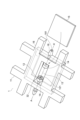
- FIG. 1 is a perspective view showing an example of a perfusion device.
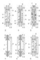
- 1A to 1C are diagrams showing the production steps of a method for producing a single-layered three-dimensional tumor tissue model according to one embodiment, where (i) to (vi) are cross-sectional views along the A-A' axis of the perfusion device in FIG. 1.
- FIG. 1 shows the results of immunostaining of four types of three-dimensional tumor tissues constructed by changing the cell density, medium type, and cell type in Example 1.
- FIG. 1 shows HE staining (A) and immunostaining (B) of a three-dimensional tumor tissue constructed in Example 1 with a constant perfusion flow rate, and FIG.
- FIG. 2 shows HE staining (C) and immunostaining (D) of a three-dimensional tumor tissue constructed in Example 1 with a varying perfusion flow rate.
- FIG. 13 shows the results of immunostaining of three-dimensional tumor tissues constructed using various colon cancer cell lines in Example 2.
- FIG. 1A shows the results of observation with a fluorescence microscope when the efficacy of doxorubicin hydrochloride was evaluated using the three-dimensional tumor tissue constructed in Example 2
- FIG. 1B shows the results of evaluating the number of colon cancer cells after administration of doxorubicin hydrochloride.
- FIG. 13 shows the results of investigating the effect of doxorubicin hydrochloride on capillary blood vessel formation when evaluating the efficacy of doxorubicin hydrochloride using the three-dimensional tumor tissue constructed in Example 2.
- FIG. 13 shows the results of fluorescence microscopy observation when measuring the distribution of doxorubicin hydrochloride and cetuximab using the three-dimensional tumor tissue constructed in Example 3.
- FIG. 1 shows the results of examining the rejection reaction of T cells against HUVEC in Example 4.
- 1 is a graph showing the results of examining the rejection reaction of T cells against HUVEC in Example 4.
- FIG. 13 shows the results of investigating the medium conditions for culturing HUVEC in Example 4 using T cell medium (A), an equal mixture of T cell medium and EGM2 medium (B), and EGM2 medium (C).
- 1 is a graph showing the results of examining medium conditions for culturing T cells in Example 4.
- FIG. 13 shows the results of perfusing T cells into a three-dimensional tumor tissue in Example 5, in which (A) shows the localization of HUVECs and T cells, and (B) shows the localization of DiFi and T cells.
- FIG. 13 shows the results of investigating the effect of T cell perfusion on the appearance of three-dimensional tumor tissue in Example 5.
- FIG. 13 shows the results of investigating the effect of the presence or absence of oscillation when perfusing T cells into three-dimensional tumor tissue in Example 5.
- FIG. 13 shows the results of HE staining and immunostaining of the two-layered three-dimensional tumor tissue constructed in Example 6. 13 shows immunostained images of nuclei and CK18 in three-dimensional tumor tissues in the case where (A) an EGFR-CD3 bispecific antibody (EGFR-CD3 antibody) was not added to the perfusion medium and where (B) an EGFR-CD3 bispecific antibody was added to the perfusion medium in Example 7.
- A an EGFR-CD3 bispecific antibody
- B an EGFR-CD3 bispecific antibody
- FIG. 13 is a graph showing the cell density of CK18-positive cells in three-dimensional tumor tissues under conditions in which an EGFR-CD3 bispecific antibody (EGFR-CD3 antibody) was not added or was added to the perfusion medium in Example 7.
- FIG. 1 is a perspective view showing an example of a perfusion device.
- FIG. 21 is an exploded perspective view of the perfusion device shown in FIG. 20.
- FIG. 13 shows the results of investigating the effect on the appearance of a three-dimensional tumor tissue constructed using the perfusion device used in Example 8.
- 13 shows immunostained images of nuclei (A), CD31 (B), and CD3 (C) of the three-dimensional tumor tissue constructed in Example 8, and a merged image of these (D).
- FIG. 13 shows the results of investigating the effect on the appearance of a three-dimensional tumor tissue constructed using the method of Example 9.
- FIG. 13 shows HE staining of the three-dimensional tumor tissue constructed in Example 9.
- 13 shows immunostained images of nuclei (A), CD31 (B), and CD3 (C) of the three-dimensional tumor tissue constructed in Example 9, and a merged image of these (D).
- 13 shows immunostained images of nuclei (A), CK18 (B), and CD3 (C) of the three-dimensional tumor tissue constructed in Example 9, and a merged image of these (D).
- the three-dimensional tumor tissue model according to this embodiment has a main vessel-like structure and a tumor vessel-like structure connected to the main vessel-like structure, and contains vascular cells, cancer cells, and extracellular matrix.
- three-dimensional tumor tissue refers to a structure in which vascular cells and cancer cells are arranged three-dimensionally in an extracellular matrix, and refers to an artificially constructed three-dimensional tumor-like tissue that mimics tumor tissue.
- the shape of the three-dimensional tumor tissue model is not particularly limited and can vary depending on the shape of the container that holds the extracellular matrix, and examples include tubular, spherical, ellipsoidal, and rectangular parallelepiped shapes.
- vascular-like structure refers to a cylindrical structure that is artificially constructed to mimic blood vessels in the body, and that contains or is composed of vascular cells.
- main vascular-like structure refers to a vascular-like structure that penetrates the three-dimensional tumor tissue model, has an opening that allows perfusion from the outside, and has a larger thickness (diameter) than the tumor vascular-like structure, capillary-like structure, etc. described below.
- An external perfusion device or the like can be connected to the opening of the main vascular-like structure to perfuse the three-dimensional tumor tissue model.
- tumor vascular-like structure refers to a cylindrical vascular-like structure that is artificially constructed to mimic capillaries that carry oxygen, nutrients, etc.
- cancer cells are present in an extracellular matrix, and at least some of the cancer cells may be in contact with each other and have a tumor tissue-like structure, and at least some of the cancer cells may be gathered around the main vascular-like structure and/or the tumor vascular-like structure. Therefore, when perfused by an external perfusion device, oxygen, nutrients, etc. are transported to the cancer cells through the main vascular-like structures and tumor vascular-like structures.
- the three-dimensional tumor tissue model according to this embodiment makes it possible to accurately evaluate the effectiveness of drugs for cancer treatment, for example.
- the distribution and delivery of the drug in the tumor tissue can be evaluated.
- the effect of cells infiltrating into the tumor tissue on the tumor tissue can also be evaluated.
- the thickness (diameter) of the main blood vessel-like structure is not particularly limited as long as it is thick enough to perfuse the three-dimensional tumor tissue model, but may be, for example, more than 50 ⁇ m, 100 ⁇ m or more, 200 ⁇ m or more, and may be 1000 ⁇ m or less, 900 ⁇ m or less, 800 ⁇ m or less, 700 ⁇ m or less, 600 ⁇ m or less, 500 ⁇ m or less, 400 ⁇ m or less, or 300 ⁇ m or less.
- the length of the main blood vessel-like structure of the three-dimensional tumor tissue model is not particularly limited, and may be, for example, 50,000 ⁇ m or less, 40,000 ⁇ m or less, 30,000 ⁇ m or less, 20,000 ⁇ m or less, 10,000 ⁇ m or less, 9,000 ⁇ m or less, 8,000 ⁇ m or less, 7,000 ⁇ m or less, 6,000 ⁇ m or less, 5,000 ⁇ m or less, 4,000 ⁇ m or less, 3,000 ⁇ m or less, 2,000 ⁇ m or less, 1,000 ⁇ m or less, 750 ⁇ m or less, 500 ⁇ m or less, 400 ⁇ m or less, 300 ⁇ m or less, 200 ⁇ m or less, or 50 ⁇ m or more, 100 ⁇ m or more, 200 ⁇ m or more, 300 ⁇ m or more, 400 ⁇ m or more, or 500 ⁇ m or more.
- the number of main blood vessel-like structures in a three-dimensional tumor tissue model may be one, two, or more.
- Tumor vascular-like structures are newly formed vascular-like structures that branch off from the main vascular-like structure, and are also called capillary-like structures. It is preferable that the tumor vascular-like structures have a blind-end structure.
- the thickness (diameter) of the tumor vascular-like structures is not particularly limited as long as it is thick enough to transport oxygen, nutrients, etc. to cancer cells, but may be, for example, 1 ⁇ m or more, 2 ⁇ m or more, 3 ⁇ m or more, 4 ⁇ m or more, 5 ⁇ m or more, or 50 ⁇ m or less, 40 ⁇ m or less, 30 ⁇ m or less, 20 ⁇ m or less, or 10 ⁇ m or less.
- the length of the tumor blood vessel-like structure in the three-dimensional tumor tissue model is not particularly limited, but may be, for example, 5000 ⁇ m or less, 4000 ⁇ m or less, 3000 ⁇ m or less, 2000 ⁇ m or less, 1000 ⁇ m or less, 750 ⁇ m or less, 500 ⁇ m or less, 400 ⁇ m or less, 300 ⁇ m or less, 200 ⁇ m or less, or 50 ⁇ m or more, 100 ⁇ m or more, 200 ⁇ m or more, 300 ⁇ m or more, 400 ⁇ m or more, or 500 ⁇ m or more.
- the tumor blood vessel-like structure in the three-dimensional tumor tissue model may be one (one), and preferably has multiple structures, for example, 3 or more, 5 or more, 7 or more, 10 or more, or 15 or more.
- Vascular cells are not particularly limited as long as they are capable of forming a blood vessel-like structure, and may be, for example, cells derived from mammals, such as humans, monkeys, dogs, cats, pigs, cows, horses, rabbits, mice, and rats.
- Vascular cells include, for example, vascular epithelial cells and vascular endothelial cells, with vascular endothelial cells being preferred, and human-derived vascular endothelial cells being more preferred.
- Vascular cells may also be cell lines, primary cultured cells, or vascular cells obtained by inducing differentiation of pluripotent stem cells.
- Pluripotent stem cells include, for example, embryonic stem cells (ES cells), induced pluripotent stem cells (iPS cells), and the like.
- human-derived vascular endothelial cells include human umbilical vein endothelial cells (HUVEC), human umbilical artery endothelial cells (HUAEC), human coronary artery endothelial cells (HCAEC), human saphenous vein endothelial cells (HSaVEC), human pulmonary artery endothelial cells (HPAEC), human aortic endothelial cells (HAoEC), and human dermal microvascular endothelial cells (Human Dermal Microvascular Endothelial Cells).
- HUVEC human umbilical vein endothelial cells
- HAAEC human coronary artery endothelial cells
- HPAEC human saphenous vein endothelial cells
- HPAEC human pulmonary artery endothelial cells
- HAoEC human aortic endothelial cells
- Human dermal microvascular endothelial cells Human Dermal Microvascular Endothelial Cells
- human-derived vascular endothelial cells examples include human dermal blood endothelial cells (HDMEC), human dermal blood endothelial cells (HDBEC), human dermal lymphatic endothelial cells (HDLEC), human pulmonary microvascular endothelial cells (HPMEC), human cardiac microvascular endothelial cells (HCMEC), human bladder microvascular endothelial cells (HBdMEC), human uterine microvascular endothelial cells (HUtMEC), and human brain microvascular endothelial cells.
- human-derived vascular endothelial cells human umbilical vein endothelial cells are preferred.
- Vascular endothelial cells isolated from human tumors can also be used.
- the vascular cells that form the main vessel-like structures and the vascular cells that exist in areas other than the main vessel-like structures, such as tumor vessel-like structures, may be of the same type or different types.
- Cancer cells isolated from tumor tissues prepared by transplanting cancer cell lines or tumors excised from patients into mice may also be used. In this case, only cancer cells may be isolated from the tumor tissue, or the cancer cells and stromal cells may be used in combination. Cancer cells isolated from tissues cultured as organoids from cancer cell lines or tumors excised from patients may also be used. In this case, too, only the cancer cells may be isolated from the organoids, or the cancer cells may be used in combination with stromal cells.
- Extracellular matrix is an insoluble substance present outside cells in living organisms.
- the extracellular matrix in the three-dimensional tumor tissue model may be dispersed in an aqueous medium as described below, and is preferably solid (gel-like) at cell culture temperatures (e.g., 30-37°C).
- the extracellular matrix used in the three-dimensional tumor tissue model is preferably a solution in which the extracellular matrix is dissolved in an aqueous medium (hereinafter, sometimes simply referred to as "extracellular matrix solution”), which is liquid at temperatures lower than the cell culture temperature (e.g., 0-20°C), but gels at the cell culture temperature (e.g., 30-37°C) (hereinafter, simply referred to as "extracellular matrix gel").
- the concentration of the extracellular matrix in the extracellular matrix solution or extracellular matrix gel is not particularly limited, and may be any concentration that provides a hardness generally suitable for cell culture and cell migration upon gelation, and may be, for example, 0.1 mg/mL or more, 0.5 mg/mL or more, 1 mg/mL or more, 1.5 mg/mL or more, 2 mg/mL or more, 2.5 mg/mL or more, or 3 mg/mL or more, or 50 mg/mL or less, 40 mg/mL or less, 30 mg/mL or less, 20 mg/mL or less, 15 mg/mL or less, 10 mg/mL or less, or 5 mg/mL or less.
- the extracellular matrix examples include collagen, fibrin, elastin, proteoglycan, fibronectin, hyaluronic acid, laminin, vitronectin, tenascin, entactin, fibrillin, glycosaminoglycan, alginic acid, etc.
- Collagen or fibrin is preferable, and collagen is more preferable.
- the extracellular matrix may contain one type alone or two or more types in combination.
- the collagen may be various types of collagen, such as type I, type II, type III, type IV, type V, and type XI.
- Basement membrane extracts such as Matrigel and Geltrex may also be used.
- the extracellular matrix in the three-dimensional tumor tissue model according to this embodiment contains water unless specifically excluded.
- water means an aqueous medium as described below.
- the content (including water) of the extracellular matrix in the three-dimensional tumor tissue model according to this embodiment is not particularly limited, but may be, for example, 10% by mass or more, 15% by mass or more, 20% by mass or more, 25% by mass or more, or 30% by mass or more, and may be 95% by mass or less, 90% by mass or less, 80% by mass or less, 70% by mass or less, 60% by mass or less, 50% by mass or less, 30% by mass or less, 20% by mass or less, or 15% by mass or less, based on the total mass of the three-dimensional tumor tissue model.
- the thickness of the three-dimensional tumor tissue model according to this embodiment is not particularly limited, but may be, for example, 5000 ⁇ m or less, 4000 ⁇ m or less, 3000 ⁇ m or less, 2000 ⁇ m or less, 1000 ⁇ m or less, 750 ⁇ m or less, 500 ⁇ m or less, 400 ⁇ m or less, 300 ⁇ m or less, 200 ⁇ m or less, or 50 ⁇ m or more, 100 ⁇ m or more, 200 ⁇ m or more, 300 ⁇ m or more, 400 ⁇ m or more, or 500 ⁇ m or more.
- the thickness of the three-dimensional tumor tissue model means the distance from the vascular wall surface (the outer wall surface of the blood vessel) of the main blood vessel-like structure to the surface of the three-dimensional tumor tissue model.
- the three-dimensional tumor tissue model according to this embodiment may contain interstitial cells.
- Interstitial cells are cells that constitute the supporting tissue of epithelial cells. Examples of interstitial cells include fibroblasts, immune cells, pericytes, nerve cells, mast cells, epithelial cells, cardiac myocytes, hepatocytes, pancreatic islet cells, tissue stem cells, smooth muscle cells, mesenchymal stem cells, etc.
- Immune cells include, for example, lymphocytes, macrophages, granulocytes, and dendritic cells, with lymphocytes being preferred.
- Lymphocytes include, for example, T cells, B cells, and natural killer cells, with T cells being preferred.
- the three-dimensional tumor tissue model may contain cancer stromal cells as stromal cells.
- cancer stromal cells include fibroblasts, immune cells, vascular endothelial cells, etc. present in the cancer stroma.
- fibroblasts present in the cancer stroma include cancer-associated fibroblasts (CAFs), etc.
- immune cells present in the cancer stroma include tumor-associated macrophages (TAMs), etc.
- vascular endothelial cells present in the cancer stroma include tumor vascular endothelial cells, etc.
- the three-dimensional tumor tissue model according to this embodiment may be a single-layer type three-dimensional tumor tissue model, or a multi-layer type three-dimensional tumor tissue model.
- a single-layer type three-dimensional tumor tissue model is a three-dimensional tumor tissue model having one layer, in which all the cells and extracellular matrix are contained in this single layer.
- a layered three-dimensional tumor tissue model is a three-dimensional tumor tissue model that includes two or more layers, and has two or more layers that are in contact with each other, i.e., stacked, and one of the two or more layers does not need to include all types of cells.
- the layered three-dimensional tumor tissue model is not particularly limited, and may be two, three, four, or five layers, and is preferably a two-layer three-dimensional tumor tissue model.
- the multiple layers are layers that are concentric with the longitudinal axis of the main blood vessel-like structure.
- the three-dimensional tumor tissue model in the case of a two-layer three-dimensional tumor tissue model, includes a blood vessel layer and a tumor layer, the blood vessel layer is a layer that surrounds the main blood vessel-like structure, and the tumor layer is stacked on the main blood vessel-like structure (i.e., on the opposite side to the main blood vessel-like structure).
- the vascular layer is a layer that has main vessel-like structures and tumor vessel-like structures, and also contains vascular cells and extracellular matrix, and has main vessel-like structures and tumor vessel-like structures connected to the main vessel-like structures, which contain or consist of vascular cells, in the extracellular matrix.
- the tumor vessel-like structures in the vascular layer increase in number due to proliferation of vascular cells, and the length of the tumor vessel-like structures also extends.
- the tumor vessel-like structures in the vascular layer may extend into the tumor layer.
- cancer cells that have proliferated in the tumor layer described below may infiltrate into the vascular layer.
- the vascular layer may further contain the above-mentioned stromal cells.
- the tumor layer is a layer that contains cancer cells and extracellular matrix. During the culture of the three-dimensional tumor tissue model, the cancer cells may proliferate and infiltrate into the vascular layer or gather around tumor blood vessel-like structures that extend from the vascular layer.
- the tumor layer may further contain the above-mentioned stromal cells.
- the layered three-dimensional tumor tissue model may have a layer (hereinafter, also referred to as a "coating layer") containing a biocompatible material as the outermost layer.
- the coating layer is a gel layer formed by gelling a biocompatible material.
- the "outermost layer” means the layer furthest from the longitudinal axis of the main vessel-like structure. It is preferable that the coating layer does not contain cells.
- biocompatible material is a material that does not adversely affect cell growth, etc., and does not prevent the formation of a three-dimensional tumor tissue model.
- biocompatible materials include bioderived materials such as extracellular matrix components (e.g., fibrin, Matrigel, collagen, and alginic acid), and synthetic polymers such as polyethylene glycol hydrogel.
- Biocompatible materials are preferably bioderived materials, more preferably extracellular matrix, and even more preferably fibrin.
- the three-dimensional tumor tissue model comprises a tissue layer and a covering layer in contact with the tissue layer, the tissue layer has a main vessel-like structure and a tumor vessel-like structure connected to the main vessel-like structure, and contains vascular cells, cancer cells, and extracellular matrix, and the covering layer contains a biocompatible material and is located as the outermost layer in the three-dimensional tumor tissue model.
- the tissue layer is similar to the blood vessel layer except that it contains cancer cells.
- the three-dimensional tumor tissue model is a three-dimensional tumor tissue model having a blood vessel layer and a tumor layer in contact with the blood vessel layer, and further having a coating layer, the coating layer including a biocompatible material, and being located as the outermost layer in the three-dimensional tumor tissue model.
- the manufacturing method of the three-dimensional tumor tissue model according to this embodiment is a manufacturing method of a three-dimensional tumor tissue model having a blood vessel-like structure and a tumor blood vessel-like structure, and includes a step of forming an extracellular matrix gel containing first vascular cells and cancer cells (gel formation step) having a through flow path for forming the main blood vessel-like structure, a step of forming the main blood vessel-like structure by seeding second vascular cells on the inner wall of the through flow path (main blood vessel-like structure formation step), and a step of perfusing culture medium into the main blood vessel-like structure to form a tumor blood vessel-like structure (tumor blood vessel-like structure formation step).
- the manufacturing method according to an embodiment may be, for example, a manufacturing method using a perfusion device.
- the perfusion device according to an embodiment may be, for example, as shown in FIG. 1.
- the perfusion device 1 shown in FIG. 1 includes a perfusion device main body 11 and a bottom cover 15.
- the perfusion device main body 11 is provided with at least a chamber 13 and tubular connectors 12 and 18.
- the shape, size, etc. of the perfusion device 1 are not particularly limited, and can be appropriately designed so that the three-dimensional tumor tissue model has a desired shape and size.
- the perfusion device 1 can also be manufactured, for example, by 3D printer technology.
- the material of the perfusion device 1 is not particularly limited, but may be, for example, acrylic resin.
- the perfusion device 1 is connected to a perfusion device or the like that sends liquid from the outside of the perfusion device 1 to the inside of the three-dimensional tumor tissue model via the tubular connector 12 or 18 after the three-dimensional tumor tissue model is constructed, and the three-dimensional tumor tissue model can be perfused.
- the chamber 13 is a chamber (space) provided in the perfusion device main body 11 for forming and culturing a three-dimensional tumor tissue model.
- the chamber 13 is surrounded by an inner wall 32 and has openings on both the upper and lower sides. The lower opening can be blocked by a bottom cover 15, allowing the chamber 13 to contain a liquid.
- the perfusion device main body 11 may have a groove 16 corresponding to the lower opening of the chamber 13, and the bottom cover 15 can be fitted into the groove 16. By having the groove 16, the perfusion device main body 11 can more easily block the lower opening of the chamber 13.
- At least one pair of tubular connectors 12 and 18 are provided on each of the opposing sides of the perfusion device main body 11, and have through holes 121 and 181 that communicate from the outside of the perfusion device main body 11 to the inside of the chamber 13.
- the through holes 121 of the tubular connector 12 and the through holes 181 of the tubular connector 18 have the same diameter, and the centers of the through holes are on the same axis. This allows the rod-shaped member 17 described below to pass through the through holes 121 and 181.
- the main blood vessel-like structure in the three-dimensional tumor tissue model communicates with the through holes 121 and 181, it is possible to pass liquid into the main blood vessel-like structure from the outside of the perfusion device main body 11 through the tubular connectors 12 and 18.
- the diameters of the through holes 121 and 181 are not particularly limited and may be set appropriately according to the diameter of the main blood vessel-like structure.
- the tubular connectors 12 and 18 are not particularly limited, but may be one pair (one tubular connector 12 and one tubular connector 18), or may be two or more pairs (two or more tubular connectors 12 and two or more tubular connectors 18).
- the tubular connectors 12 and 18 are preferably provided with an anchor structure 14 on the side exposed to the chamber 13. By providing the anchor structure 14 on the tubular connectors 12 and 18, contraction of the three-dimensional tumor tissue is minimized, making it easier to construct a three-dimensional tumor tissue model of the desired shape, and preventing the three-dimensional tumor tissue model from detaching from the perfusion device 1.
- the surfaces of the tubular connectors 12 and 18 exposed to the chamber 13 may be subjected to atmospheric plasma treatment and/or fibronectin coating treatment. This treatment can further prevent the detachment of the cultured three-dimensional tumor tissue model.
- a perfusion device may be, for example, the perfusion device 1a shown in FIG. 20.
- the perfusion device 1a shown in FIG. 20 includes an upper cover 81, a sealing member 91, and a perfusion device main body 11a.
- the perfusion device main body 11a is an integrated unit of the perfusion device main body 11 and the bottom cover 15 of the perfusion device 1 described above. In other words, the perfusion device main body 11a does not have the lower opening described in the description of the chamber 13 in the perfusion device 1.
- Other specific aspects of the perfusion device main body 11a are as described for the perfusion device main body 11.
- the top lid 81 includes a lid portion 811 and four legs 812 with claw portions 813 at the four corners of the lid portion 811.
- the top lid 81 is fixed to the upper side of the perfusion device main body portion 11a, and is a member for closing the upper opening of the chamber 13 in the perfusion device main body portion 11a with the lid portion 811.
- the structure of the three-dimensional tumor tissue e.g., the main blood vessel-like structure, the overall structure of the three-dimensional tumor tissue, etc.
- the structure of the three-dimensional tumor tissue e.g., the main blood vessel-like structure, the overall structure of the three-dimensional tumor tissue, etc.
- the method for fixing the top cover 81 to the perfusion device main body 11a may be, for example, a method of fixing using a snap-fit mechanism, a clamp, or a screw.
- the top cover 81 shown in Fig. 21 has legs 812, and the legs 812 are provided with claws 813 in order to fix it to the perfusion device 11a using a snap-fit mechanism.
- the claws 813 engage with the protrusions 19 of the perfusion device main body 11a, thereby fixing the top cover 81 to the perfusion device 11a.
- the top cover 81 may have a main body recess 814 on either the tubular connector 12 side or the 18 side of the perfusion device main body 11a, or may have a main body recess 814 on both the tubular connector 12 side and the 18 side.
- the shape of the main body recess 814 may be, for example, a square shape, a triangular shape, a curved shape, or a combination thereof.
- the top cover 81 has a main body recess 814, which improves operability with respect to the tubular connector.
- the shape, size, etc. of the top lid 81 are not particularly limited as long as they can close the upper opening of the chamber 13, and can be designed as appropriate.
- the top lid 81 can also be manufactured, for example, by 3D printer technology.
- the material of the top lid 81 is not particularly limited, but may be, for example, acrylic resin, etc.
- the sealing member 91 is provided with a recess 911.
- the sealing member 91 is a member that is interposed between the perfusion device main body 11a and the top cover 81 and that improves the sealing performance of the top cover 81.
- the sealing member 91 is, for example, a packing.
- the sealing member 91 may have a recess 911 on either the tubular connector 12 side or the 18 side of the perfusion device main body 11a, or may have a recess 911 on both the tubular connector 12 side and the 18 side.
- the shape of the recess 911 may be, for example, a square shape, a triangular shape, a curved shape, or a combination thereof.
- the recess 911 is preferably provided in the same direction.
- the sealing member 91 has the recess 911, which improves the operability of the tubular connector.
- the shape, size, etc. of the sealing member 91 are not particularly limited as long as they can close the upper opening of the chamber 13, and can be designed as appropriate.
- the sealing member 91 can also be manufactured by a method such as 3D printer technology, cutting processing, or transfer using a mold.
- the material of the sealing member 91 is not particularly limited, but may be, for example, rubber, silicone (e.g., dimethylpolysiloxane (PDMS)), etc.
- Fig. 2 is a diagram showing a three-dimensional tumor tissue model produced using a perfusion device 1, and is a cross-sectional view of the perfusion device taken along the line AA' in Fig. 1.
- steps (2-i) may be referred to as "step (2-i)" or the like.
- an extracellular matrix gel is formed that has a through-flow path for forming a main blood vessel-like structure and contains first vascular system cells and cancer cells.
- the main blood vessel-like structure is as described above.
- the first vascular system cells are vascular system cells that form the main blood vessel-like structure, and the type of the first vascular system cells is as described above.
- the gel formation process may be, for example, a process including steps (2-i) to (2-iii) in FIG. 2.
- Step (2-i) is a step of passing a rod-shaped member 17 through the through hole 121 of one tubular connector 12 to the through hole 181 of the other tubular connector 18 in the perfusion device main body 11 provided with the pair of tubular connectors 12 and 18, and is a step for forming a through flow path 31 in the gel (extracellular matrix gel 26) in the step (2-iii) described below.
- the thickness of the rod-shaped member 17 corresponds to the outer diameter of the main vessel-like structure 52 in the three-dimensional tumor tissue 61 described below, so the thickness of the rod-shaped member 17 may be appropriately designed according to the outer diameter of the desired main vessel-like structure.
- the material of the rod-shaped member 17 is also not particularly limited, but may be made of metal, for example, and is preferably made of stainless steel.
- the shape of the rod-shaped member 17 is not particularly limited, and may be, for example, needle-shaped (for example, a syringe needle, etc.) or wire-shaped, as long as it can construct a main vessel-like structure 52 with a specified inner diameter and length.
- Step (2-ii) is a step of forming an extracellular matrix gel 26 containing the first vascular cells 22 and the cancer cells 23. More specifically, in step (2-ii), After (2-i), a cell suspension in which first vascular system cells 22 and cancer cells 23 are suspended in an extracellular matrix solution 21 is poured into the chamber 13 with the lower opening closed. The first vascular cells, cancer cells, and extracellular matrix are filled in the vascular system and gelled. This allows the base of the three-dimensional tumor tissue model according to this embodiment to be constructed. As mentioned above.
- the cell suspension in which vascular cells 22 and cancer cells 23 are suspended in the extracellular matrix solution 21 may further contain cells such as the above-mentioned interstitial cells, if necessary.
- the extracellular matrix solution 21 can be prepared by dissolving, dispersing, etc. the extracellular matrix in an aqueous medium.
- the extracellular matrix to be dissolved, dispersed, etc. in an aqueous medium may be in the form of a solution, a lyophilized product, or a powder.
- the concentration of the extracellular matrix in the extracellular matrix solution 21 may be 0.1 mg/mL or more, 0.5 mg/mL or more, 1 mg/mL or more, 1.5 mg/mL or more, 2 mg/mL or more, 2.5 mg/mL or more, or 3 mg/mL or more, and may be 50 mg/mL or less, 40 mg/mL or less, 30 mg/mL or less, 20 mg/mL or less, 15 mg/mL or less, 10 mg/mL or less, or 5 mg/mL or less.
- an aqueous medium means water or an aqueous solution.
- water include deionized water and distilled water
- aqueous solutions include Tris-HCl buffer, HEPES (4-(2-hydroxyethyl)-1-piperazineethanesulfonic acid) buffer, liquid medium, etc.
- Tris-HCl buffer Tris-HCl buffer
- HEPES 4-(2-hydroxyethyl)-1-piperazineethanesulfonic acid
- the cell density of the vascular cells in the cell suspension is not particularly limited as long as a three-dimensional tumor tissue model can be constructed, but may be, for example, 1 x 10 cells/mL or more, 5 x 10 cells/mL or more, 1 x 10 cells/mL or more, 5 x 10 cells/mL or more, or 1 x 10 cells/mL or more, and may be 5 x 10 cells/mL or less, 1 x 10 cells/mL or less, 5 x 10 cells/mL or less, 3 x 10 cells/mL or less, 1 x 10 cells/mL or less, 5 x 10 cells/mL or less, or 1 x 10 cells/mL or less.
- the cell density of the cancer cells in the cell suspension is not particularly limited as long as a three-dimensional tumor tissue model can be constructed, but may be, for example, 1 x 10 cells/mL or more, 5 x 10 cells/mL or more, 1 x 10 cells/mL or more, 5 x 10 cells/mL or more, or 1 x 10 cells/mL or more, or may be 5 x 10 cells/mL or less, 1 x 10 cells/mL or less, 5 x 10 cells/mL or less, 3 x 10 cells/mL or less, 1 x 10 cells/mL or less, 5 x 10 cells/mL or less, or 1 x 10 cells/mL or less.
- the cell density ratio of vascular cells to cancer cells in the cell suspension may be 1:0.3 to 1:3, 1:0.5 to 1:2.5, 1:0.8 to 1:2, or 1:1 to 1:1.5.
- the cell density of the stromal cells in the cell suspension is not particularly limited as long as a three-dimensional tumor tissue model can be constructed, and may be, for example, 1 x 10⁴ cells/mL or more, 5 x 10⁴ cells/mL or more, 1 x 10⁵ cells/mL or more, 5 x 10⁵ cells/mL or more, or 1 x 10⁶ cells/mL or more, and may be 5 x 10⁷ cells/mL or less, 1 x 10⁷ cells/mL or less, 5 x 10⁶ cells/mL or less, 3 x 10⁶ cells/mL or less, 1 x 10⁶ cells/mL or less, 5 x 10⁵ cells/mL or less, or 1 x 10⁵ cells/mL or less.
- the gelation of the cell suspension can be achieved, for example, by heating the cell suspension.
- the temperature and heating time can be set appropriately depending on the type and concentration of the extracellular matrix components, but for example, the heating temperature may be 25 to 45°C and the heating time may be 1 to 3 hours.
- Step (2-iii) is a step of forming a through-flow channel in the extracellular matrix gel 26 containing the first vascular cells 22 and the cancer cells 23 formed in step (2-ii).
- the rod-shaped member 17 passed through the perfusion device main body 11 in step (2-i) is removed after step (2-ii).
- a through channel 31 is formed in the extracellular matrix gel 26 containing the first vascular cells 22 and the cancer cells 23 formed in the step ii).
- the through channel 31 forms a three-dimensional tumor tissue according to the present embodiment. It serves as the basis for the main vessel-like structures 52 in the model.
- the extracellular matrix gel 26 containing the first vascular cells 22 and the cancer cells 23 in step (2-iii) may or may not have been contracted after the extracellular matrix in the cell suspension is gelled in step (2-ii).
- step (2-iii) the extracellular matrix gel 26 containing the vascular cells 22 and the cancer cells 23 is shown in a contracted state after step (2-ii).
- the extracellular matrix gel 26 containing the vascular cells 22 and the cancer cells 23 is in a contracted state, at least a portion of the inner wall 32 of the perfusion device main body 11 and the extracellular matrix gel 26 are not in contact with each other.
- second vascular system cells are seeded on the inner walls of the through-flow channels in the extracellular matrix gel formed in the gel forming step to form the main vessel-like structure.
- the second vascular system cells are vascular system cells that form the main vessel-like structure, and the type of the second vascular system cells is as described above.
- the second vascular system cells may be the same type as the first vascular system cells, or may be a different type.
- the main vessel-like structure formation process is not particularly limited, but may be, for example, a process including steps (2-iv) to (2-v).
- Step (2-iv) In step (2-iv) in FIG. 2, after the above step (2-ii) or the above step (2-iii), the vascular cells 22 and the cancer cells 23 formed in step (2-ii) are The extracellular matrix gel 26 is immersed in a medium 41 containing a medium for vascular cells.
- the gel 26 may be immersed in the medium by pouring the medium so that the entire extracellular matrix gel 26 is immersed, or by immersing the entire extracellular matrix gel in the medium.
- medium for vascular cells means a medium in which vascular cells can grow.
- Examples of medium for vascular cells include Endothelial Growth Medium, Endothelial Growth Medium 2, Endothelial Growth Medium MV, Endothelial Growth Medium MV2, and EGM Bullet Kit, EGM-2 Bullet Kit, EGM-2MV Bullet Kit, and EGM-Plus Bullet Kit, all of which are manufactured by Lonza.
- the medium 41 used in step (4) may be a medium containing serum or a serum-free medium.
- the medium 41 used in step (4) may contain components used in cell culture, such as growth factors, differentiation-inducing factors, hormones, amino acids, sugars, salts, and antibiotics.
- the medium 41 used in step (2-iv) preferably contains 50% by mass or more of vascular cell medium relative to the total amount of the medium, and more preferably 50% by mass or more, 60% by mass or more, 70% by mass or more, 80% by mass or more, 90% by mass or more, or 100% by mass.
- step (2-v) In step (2-v), after the step (2-iv), second vascular cells 51 are seeded into the through-flow passages 31 formed in the extracellular matrix gel 26 containing the first vascular cells 22 and the cancer cells 23 in the step (2-iii) to form a main blood vessel-like structure 52.
- the second vascular system cells 51 seeded in step (2-v) may be, for example, a cell suspension in which the vascular system cells 51 are suspended in an aqueous medium.
- the method for seeding the second vascular system cells 51 into the through-flow passage 31 is not particularly limited, but may include, for example, a method in which a suspension of the vascular system cells 51 is injected into the through-flow passage 31 using a syringe and a syringe pump, etc.
- step (2-v) the second vascular cells 51 are seeded in the through-flow passage 31, and then incubated for a predetermined time to form a main blood vessel-like structure 52, thereby adhering the vascular cells 51 to the inner wall of the through-flow passage 31.
- the orientation of the perfusion device main body 11 may be changed as necessary to allow the second vascular cells 51 to adhere uniformly to the inner wall of the through-flow passage 31.
- the incubation temperature is not particularly limited as long as the second vascular system cells 51 are adhered to the inner wall of the through-flow channel 31, but may be, for example, 35 to 40°C, preferably 37°C, the incubation time may be, for example, about 15 minutes to 1 hour, and the CO2 condition may be, for example, 5%.
- the second vascular system cells are vascular system cells that form the main vascular-like structure, and the type of the second vascular system cells is as described above.
- the second vascular system cells may be the same type as the first vascular system cells, or may be a different type.
- the tumor vascular-like structure formation process is not particularly limited, but may be, for example, a process including the following step (2-vi).
- Step (2-vi) for example, after the above step (2-v), a vascular cell culture medium is fed from the tubular connector 12 provided on the perfusion device main body 11 to the main blood vessel-like structure 52 in the perfusion direction 71.
- the medium to be perfused in the step (2-vi) may be any medium containing a medium for vascular cells, and the medium ...
- the medium used in step (2-vi) may be the same type of medium as the medium 41 containing the medium for vascular system cells used in step (2-iv), or may be a different type of medium. Examples of the above include those mentioned above.
- the culture medium can be perfused into the main blood vessel-like structure 52 by injecting the culture medium into the tubular connector 12 or 18.
- This provides nutrients from inside the three-dimensional tumor tissue model 61 to be cultured, and the first blood vessel cells 22, the second blood vessel cells 51, the cancer cells 23, and the like in the three-dimensional tumor tissue model 61 are cultured.
- the first blood vessel cells 22 form a tumor blood vessel-like structure 62 connected to the main blood vessel-like structure 52, and the three-dimensional tumor tissue model 61 according to this embodiment is effectively constructed as a whole. It is also preferable to perfuse the culture medium into the three-dimensional tumor tissue model 61 from the tubular connector 12 or 18 using an external pump or the like.
- Examples of external pumps include peristaltic pumps, syringe pumps, and air-driven pumps. From the viewpoint of more easily forming a medium circulation path as described below, an external pump capable of suction and delivery is preferred, and a peristaltic pump is more preferred.
- a culture medium circulation path may be formed for perfusion.
- the culture medium circulation path can be formed, for example, by using a tube and an external pump.
- the perfusion device main body 11 holding the extracellular matrix gel 26 containing the first vascular cells 22 and the cancer cells 23 is immersed in a culture dish containing the culture medium to be perfused, one end of the tube is placed in the culture medium in the culture dish, and the other end of the tube is connected to the tubular connector 12. Furthermore, a peristaltic pump is connected to the tube.
- the culture medium in the culture dish is sucked from the end of the tube by the peristaltic pump, and is sent through the tubular connector 12 into the three-dimensional tumor tissue model 61 for perfusion.
- the culture medium sent into the three-dimensional tumor tissue model 61 is discharged into the culture dish through the tubular connector 18.
- the material of the culture dish is not particularly limited, and examples include plastic and glass, with plastic being preferred, and polystyrene being more preferred.
- As the culture dish it is preferable to use a polystyrene dish that is typically used for cell culture.
- the medium circulation path may be appropriately connected to a bubble trap or the like.
- the bubble trap may be, for example, one in which bubbles mixed in the medium are separated from the flow of the medium by buoyancy and accumulated in the container. More specifically, it can be configured by using a tube for introducing the medium into the sealed container and a tube for discharging the medium from the sealed container, with the end of the outlet tube in the sealed container positioned lower than the end of the inlet tube in the sealed container.
- a bubble removal device such as a membrane separator using an air-permeable membrane such as polytetrafluoroethylene (PTFE) may be used.
- PTFE polytetrafluoroethylene
- the culture dish in which the perfusion device main body 11 is immersed and the culture dish into which the medium delivered to the 3D tumor tissue model 61 is discharged may be separated.
- the peristaltic pump aspirates the medium from the culture dish into which the medium is discharged.
- a system may also be used in which, without circulating the medium, new medium is constantly aspirated, delivered to the tumor tissue, and the waste liquid is discharged into a waste liquid bottle.
- the conditions for the perfusion culture in step (2-vi) are not particularly limited as long as the main vessel-like structures 52 and the tumor vessel-like structures 62 are formed.
- the conditions may be 35 to 40° C., and preferably 37° C.
- the culture period may be, for example, about 2 to 20 days
- the CO2 conditions may be, for example, 5%.
- the flow rate of the perfusion in step (2-vi) is not particularly limited, and may be, for example, 0.5 mL/hour or more, 1 mL/hour or more, 2 mL/hour or more, 3 mL/hour or more, 4 mL/hour or more, 5 mL/hour or more, 6 mL/hour or more, 7 mL/hour or more, 8 mL/hour or more, 9 mL/hour or more, or 10 mL/hour or more, and may be 20 mL/hour or less, 19 mL/hour or less, 18 mL/hour or less, 17 mL/hour or less, 16 mL/hour or less, 15 mL/hour or less, 14 mL/hour or less, 13 mL/hour or less, 12 mL/hour or less, 11 mL/hour or less, or 10 mL/hour or less.
- the perfusion flow rate in step (2-vi) may be constant or may be changed.
- the flow rate may be changed after perfusion at a constant flow rate for 1 to 7 days, 1 to 5 days, 1 to 3 days, 1 to 2 days, or 1 day.
- the number of times the flow rate is changed is not particularly limited, and may be, for example, 5 times or less, 3 times or less, 2 times or less, or once.
- the flow rate may be increased or decreased, but it is preferable to increase the flow rate.
- the flow rate may be increased by 2 to 15 times, or may be increased by 3 to 14 times, 4 to 13 times, 5 to 12 times, 6 to 11 times, 7 to 10 times, 8 to 10 times, or 9 to 10 times.
- the method for producing three-dimensional tumor tissue may include a step of forming the above-mentioned coating layer (coating layer forming step) at any timing after the gel formation step.
- the coating layer is as described above.
- the coating layer forming step is preferably carried out after the main blood vessel-like structure forming step.
- the coating layer forming step can be carried out, for example, by the same procedure as (2-ii) in the gel forming step. More specifically, the chamber 13 with the lower opening blocked is filled with a solution containing a biocompatible material and gelled.
- a solution containing a biocompatible material can be prepared by dissolving or dispersing the biocompatible material in an aqueous medium.
- the biocompatible material to be dissolved or dispersed in an aqueous medium may be in the form of a solution, a freeze-dried product, or a powder.
- the concentration of the biocompatible material in the solution containing the biocompatible material may be 0.1 mg/mL or more, 0.5 mg/mL or more, 1 mg/mL or more, 1.5 mg/mL or more, 2 mg/mL or more, 2.5 mg/mL or more, or 3 mg/mL or more, and may be 150 mg/mL or less, 140 mg/mL or less, 130 mg/mL or less, 120 mg/mL or less, 110 mg/mL or less, 100 mg/mL or less, 90 mg/mL or less, 80 mg/mL or less, 70 mg/mL or less, 60 mg/mL or less, 50 mg/mL or less, 40 mg/mL or less, 30 mg/mL or less, 20 mg/mL or less, 15 mg/mL or less, 10 mg/mL or less, or 5 mg/mL or less.
- the gelation of the above solution can be achieved, for example, by heating the solution.
- the heating temperature and heating time can be set appropriately depending on the type and concentration of the biocompatible material, but for example, the heating temperature may be 25 to 45°C and the heating time may be 1 second to 3 hours.
- vascular layer forming step a step of forming a vascular layer made of an extracellular matrix gel containing first vascular system cells and having through holes for forming a main vascular-like structure (vascular layer forming step) and a step of forming a tumor layer made of an extracellular matrix gel containing cancer cells so as to contact at least a part of the surface of the vascular layer opposite to the through channel side (tumor layer forming step) are included.
- the method for producing a bilayered three-dimensional tumor tissue model further includes a main vascular-like structure forming step and a tumor vascular-like structure forming step similar to the method for producing a monolayered three-dimensional tumor tissue model.
- the method for producing a bilayered three-dimensional tumor tissue may include a coating layer forming step at any timing after the tumor layer forming step.
- the coating layer forming step is preferably performed after the main vascular-like structure forming step is performed.
- FIG. 16 shows the steps of the method for producing a two-layered three-dimensional tumor tissue model according to one embodiment.
- FIG. 16 shows the steps of producing a two-layered three-dimensional tumor tissue model using a perfusion device 1, and (i) to (vii) are cross-sectional views of the perfusion device in FIG. 1 taken along the A-A' axis.
- each step in FIG. 16 will also be referred to as "step (16-i)" or the like.
- Step (2-i) and step (16-i) are similar steps.
- Step (16-iv) is the same step as step (2-iii) except that step (16-iv) is performed on the vascular layer 24 made of the extracellular matrix gel 26 containing the first vascular system cells 22, instead of the extracellular matrix gel 26 containing the first vascular system cells 22 and the cancer cells 23.
- Step (16-v) is the same step as step (2-iv) except that step (16-v) is performed on the extracellular matrix gel made of the vascular layer 24 and the tumor layer 25, instead of the extracellular matrix gel 26 containing the first vascular system cells 22 and the cancer cells 23.
- Step (16-vi) is the same as step (2-v) except that it is performed on the vascular layer 24 instead of the extracellular matrix gel 26 containing the first vascular cells 22 and the cancer cells 23.
- Step (16-vii) is the same as step (2-vi) except that it is performed on the two-layered three-dimensional tumor tissue model 61 instead of the single-layered three-dimensional tumor tissue model 61.
- vascular layer formation step a vascular layer is formed having through-flow channels for forming a main vessel-like structure, and made of an extracellular matrix gel containing first vascular system cells.
- the main vessel-like structure, the through-flow channels, and the first vascular system cells are as described above.
- the chamber 13 with the lower opening blocked is filled with a first liquid and gelled to form the vascular layer 24.
- the first liquid is a cell suspension in which the first vascular cells 22 are suspended in the extracellular matrix solution 21, and can be prepared in the same manner as the cell suspension in step (2-ii) above. Otherwise, the vascular layer 24 can be formed in the same manner as in step (2-ii) above.
- tumor layer formation process In the tumor layer formation step, a tumor layer made of an extracellular matrix gel containing cancer cells is formed so as to contact at least a part of the surface of the blood vessel layer opposite to the through-channel side.
- the cancer cells and tumor blood vessel-like structures are as described above.
- the tumor layer formation step is not particularly limited, but may be, for example, the following step (16-iii).
- step (16-iii) a second liquid is filled onto the blood vessel layer 24 and gelled to form a tumor layer 25.
- the second liquid is a cell suspension in which cancer cells 23 are suspended in an extracellular matrix solution 21, and can be prepared in the same manner as the cell suspension in step (2-ii) above. Otherwise, the tumor layer 25 can be formed in the same manner as in step (2-ii) above.
- the tumor layer 25 may be formed so as to contact at least a portion of the surface of the blood vessel layer 24 opposite the through-flow passage 31 side, and may contact the entire surface of the blood vessel layer 24 opposite the through-flow passage side.
- the degree of contact between the tumor layer 25 and the blood vessel layer 24 can be changed by appropriately adjusting the portion where the second liquid is filled on the blood vessel layer 24 and the amount of the second liquid filled.
- each of the vascular layer 24 and the tumor layer 25 is not particularly limited as long as it is a thickness that allows the formation of a vascular-like structure, but may be, for example, 5000 ⁇ m or less, 4000 ⁇ m or less, 3000 ⁇ m or less, 2000 ⁇ m or less, 1000 ⁇ m or less, 750 ⁇ m or less, 500 ⁇ m or less, 400 ⁇ m or less, 300 ⁇ m or less, 200 ⁇ m or less, or 50 ⁇ m or more, 100 ⁇ m or more, 200 ⁇ m or more, 300 ⁇ m or more, 400 ⁇ m or more, or 500 ⁇ m or more.
- the thickness of the vascular layer means the distance from the vascular wall surface (the outer wall surface of the blood vessel) of the main vascular-like structure to the surface of the vascular layer 24 that contacts the tumor layer 25, and the thickness of the tumor layer 25 means the distance from the surface that contacts the vascular layer 24 to the surface opposite the vascular layer.
- the formation of the main blood vessel-like structures 52 and the tumor blood vessel-like structures 62 in the three-dimensional tumor tissue model 61 can be confirmed, for example, by immunostaining, as shown in the examples described below.
- the method for evaluating the effect of the evaluation target on tumor tissue in this embodiment includes a step of perfusing a solution containing the evaluation target into the three-dimensional tumor tissue model in this embodiment (perfusion step), and a step of evaluating the effect of the evaluation target on the three-dimensional tumor tissue model (evaluation step).
- the perfusion process can be carried out under the same conditions as the tumor vascular-like structure formation process in the manufacturing method for the three-dimensional tumor tissue model according to the present embodiment described above.
- the subject of evaluation may be any, for example, a drug such as an anticancer drug, or a cell such as a lymphocyte.
- anticancer drugs include doxorubicin, cetuximab, cisplatin, rituximab, carboplatin, cyclophosphamide, docetaxel, tamoxifen, tegafur, erlotinib, axitinib, etoposide, dexamethasone, mitomycin C, bleomycin, paclitaxel, doxil, bevacizumab, afatinib, nivolumab, 5-FU, and T-cell engagers.
- T-cell engagers are drugs or compounds that aim to bring immune cells, such as killer T cells, that have the role of eliminating foreign substances, closer to disease-causing cells or pathogens, thereby removing the cause of the disease and treating it.
- the lymphocytes may be, for example, T cells.
- the T cells may also be genetically modified, such as CAR-T cells expressing a chimeric antigen receptor.
- the T cell engager may be, for example, an EGFR-CD3 bispecific antibody.
- the EGFR-CD3 bispecific antibody is a bispecific antibody that binds to both CD3, an antigen of T cells, and EGFR, an antigen of cancer cells (DiFi) (Asano, R., et al., Sci Rep 10, 4913 (2020)).
- the evaluation targets may be one type alone or two or more types in combination.
- the solution may contain a medium for culturing the three-dimensional tumor tissue model according to this embodiment.
- the medium is as described above in the tumor vascular-like structure formation process.
- the evaluation process for example, when the evaluation target is a drug, the efficacy of the drug on the three-dimensional tumor tissue according to this embodiment may be evaluated, the distribution of the drug in the three-dimensional tumor tissue may be evaluated, the delivery of the drug in the three-dimensional tumor tissue may be evaluated, or the effect of the drug on blood vessels in the three-dimensional tumor tissue may be evaluated.
- the evaluation target is a lymphocyte
- the infiltration of lymphocytes in the three-dimensional tumor tissue according to this embodiment may be evaluated.
- the evaluation process can be carried out, for example, by comparing the three-dimensional tumor tissue immediately after or before perfusion of the evaluation target into the three-dimensional tumor tissue model according to this embodiment with the three-dimensional tumor tissue after perfusion of the evaluation target into the three-dimensional tumor tissue model according to this embodiment for a predetermined period of time.
- Specific methods for evaluation include, for example, the methods described in the examples below (measuring the number of cancer cells, immunostaining, etc.).
- Example 1 Conditions for constructing a monolayer-type three-dimensional tumor tissue (1) (1-1. Cell culture) In Example 1, each cell was cultured as follows. Colon cancer cells (SW620, HT-29, DiFi) were cultured in Dulbecco's modified Eagle medium (hereinafter referred to as FBS-DMEM) supplemented with 10% fetal bovine serum, 1% 100-fold non-essential amino acids, and 1% 100-fold concentrated penicillin streptomycin solution. HUVEC were cultured in endothelial cell medium (Endothelial Growth Medium 2; EGM2, Promocell) supplemented with growth factors included in the kit and 1% 100-fold concentrated penicillin streptomycin solution.
- FBS-DMEM Dulbecco's modified Eagle medium
- EGM2 Endothelial Growth Medium 2
- Promocell Endothelial Growth Medium 2
- Immortalized MSC were cultured in DMEM supplemented with 10% fetal bovine serum, 1% 100-fold concentrated non-essential amino acid solution, and 1% 100-fold concentrated penicillin streptomycin solution.
- CD8 positive T cells were cultured in a T cell medium (Stemcell technologies, ImmunoCult-XF T Cell Expansion Medium) supplemented with IL-2 10 ng/mL and ImmunoCult Human CD3/CD28/CD2 T Cell Activator 25 ⁇ L/mL.
- Lung cancer cells (SBC-3) were cultured in RPMI-1640 supplemented with 10% fetal bovine serum and 1% 100-fold concentrated penicillin streptomycin solution.
- T Cell Activator was added only for 3 days after thawing, and only IL-2 was added during subsequent passages. All cells were cultured in a 37°C, 5% CO2 environment.
- the perfusion device 1 shown in FIG. 1 was made of acrylic UV-curable resin (product name: VisiJet M3 Crystal, 3D Systems) using a 3D printer (ProJet MJP 3600 MAX (3D Systems)).
- the perfusion device was cleaned with an ultrasonic cleaner and then sterilized with 70% ethanol. After drying in a clean bench, the perfusion device was treated with vacuum plasma for 5 minutes to improve the adhesiveness of cells and collagen gel, and then immersed in 10 ⁇ g/mL bovine fibronectin and coated at 37° C. for 2 hours.
- the perfusion device was passed through the through holes 121 and 181 of the tubular connectors 12 and 18 as the rod-shaped member 17, and then stored at 4° C. for up to one week until it was used to construct a three-dimensional tumor tissue.
- monolayer type three-dimensional tumor tissues of samples 1 to 4 were constructed under the conditions shown in Table 1 below. Specifically, colon cancer cells (6 ⁇ 10 7 or 3 ⁇ 10 7 cells/mL), HUVEC (3 ⁇ 10 7 cells/mL), and MSC (0 or 5 ⁇ 10 6 cells/mL) cultured under the above condition 1. were collected, and the cells were suspended in a 3 mg/mL neutralized type I collagen solution, which was then filled into the perfusion device.
- the neutralized type I collagen solution was prepared by mixing 5 mg/mL collagen acidic solution I-AC50 (KOKENSHA) with calcium- and magnesium-free 10x concentration Dulbecco's phosphate buffer and each of the media shown in Table 1 in a ratio of 9:1:5. After incubation at 37° C. for gelation of the filled collagen, the needle installed in the perfusion device was removed to form a through-flow channel, and the perfusion device was immersed in tissue culture medium in a culture dish. As the tissue culture medium, a mixture of equal amounts of FBS-DMEM and EGM2, or EGM2 was used.
- HUVEC suspension 4 ⁇ 10 6 cells/mL
- 100 ⁇ L of HUVEC suspension 4 ⁇ 10 6 cells/mL
- the perfusion device was turned upside down and incubated for another 20 minutes to allow the cells to adhere to the upper surface of the through-flow channel.
- the perfusion device was then returned to its original position.
- a tube, a peristaltic pump, and a bubble trap were connected to the perfusion device to form a medium circulation path, and the medium was perfused.
- the perfusion culture of the three-dimensional tumor tissue was carried out for 5 days.
- the 3D tumor tissue model was fixed with 4% paraformaldehyde and embedded in paraffin.
- the 3D tumor tissue model was sliced at 5 ⁇ m and stained with hematoxylin and eosin (HE) and vascular endothelial cells.
- the sections were subjected to fluorescent immunostaining or enzyme antibody immunostaining.
- HE staining the thin sections were deparaffinized and rehydrated, and stained with Mayer's hematoxylin and eosin Y in ethanol.
- fluorescent immunostaining after rehydration, The thin sections were immersed in a citrate buffer solution of pH 6 and treated in an autoclave at 121° C.
- the sections were washed with ultrapure water and then immersed in 4% Block Ace for 20 minutes. The sections were then blocked and incubated overnight with a primary antibody diluted in phosphate buffer. Human anti-CD31 antibody (BBA7, R&D Systems, or ab28364, Abcam) was used as the primary antibody. After washing the sections with PBS, a secondary antibody (Alexa Fluor 488- The sections were then incubated with Alexa Fluor 555-conjugated anti-mouse or Alexa Fluor 555-conjugated anti-rabbit (Thermo Fisher Scientific) for 1 hour. Counterstaining was then performed using Hoechst. For enzyme antibody immunostaining, the sections were rehydrated and then sliced.
- the sections were immersed in a citrate buffer solution of pH 6 and treated in an autoclave at 121° C. for 5 minutes to perform antigen retrieval.
- the sections were immersed in 3% hydrogen peroxide for 10 minutes to inactivate endogenous peroxidase, then washed with ultrapure water and blocked with horse serum for 20 minutes.
- the sections were then incubated overnight with a primary antibody.
- Human anti-CD31 antibody (BBA7, R&D Systems, or ab28364, Abcam) was used as the primary antibody.
- the sections were washed with PBS and then incubated with the corresponding antibody of the animal species.
- the sections were incubated with a secondary antibody (ImmPRESS Reagent, VECTOR LABORATORIES) for 1 hour. After color development with ImmPACT DAB (VECTOR LABORATORIES), counterstaining was performed with hematoxylin.
- the results of enzyme antibody immunostaining are shown in FIG. show.
- a monolayer type 3D tumor tissue was constructed using 3 ⁇ 10 7 cells/mL of colon cancer cells and 3 ⁇ 10 7 cells/mL of HUVECs in order to evaluate in a simple system.
- the perfusion flow rate was compared between 1 mL/h during the 3D tumor tissue culture period and 1 mL/h for the first day and 10 mL/h for the remaining culture period.
- Perfusion in the tissue was visualized by injecting a 2:1 mixture of India ink and PBS using a syringe pump.
- Figure 4(C) and (D) more capillaries were formed in the 3D tumor tissue when perfusion was performed at 1 mL/h for the first day and 10 mL/h for the remaining culture period
- Figures 4(A) and (B) compared with when perfusion was performed at 1 mL/h during the 3D tumor tissue culture period.
- tumor tissue Since tumor tissue has high nutritional requirements, it is considered more preferable to perfuse at 1 mL/h on the first day of culture, followed by perfusion at 10 mL/h, which is a flow rate greater than 1 mL/h, in order to construct a 3D tumor tissue model. Therefore, in the following studies, perfusion was performed at 1 mL/h for the first day and 10 mL/h for the remaining culture period.
- 3D tumor tissue was constructed in a similar manner using colon cancer cells SW620 or DiFi in addition to HT-29.
- the 3D tumor tissue was then fixed and paraffin-embedded sections were prepared in the same manner as in 1-4 above, and enzyme antibody immunostaining was performed using human anti-CD31 antibody as the primary antibody.
- enzyme antibody immunostaining was performed using human anti-CD31 antibody as the primary antibody.
- capillaries were formed regardless of which cancer cell was used, demonstrating that it is possible to construct 3D tumor tissue with main vessel-like structures and capillaries (tumor blood vessels).
- Example 2 Drug efficacy evaluation using three-dimensional tumor tissue
- the circulating tissue culture medium was replaced with a medium containing doxorubicin hydrochloride (doxorubicin), a low molecular weight anticancer drug, at a final concentration of 0 ⁇ M (no addition), 1 ⁇ M, or 10 ⁇ M, and perfusion culture was continued.
- doxorubicin doxorubicin hydrochloride
- the tissue was fixed in the same manner as in 1-4 above, paraffin-embedded sections were prepared, and fluorescent immunostaining of cancer cells was performed and photographed using a fluorescent microscope.
- Fluorescent immunostaining was also performed in the same manner as in 1-4 above, using human anti-CK18 antibody (MA5-12104, Thermo Fisher Scientific) as the primary antibody to stain cancer cells.
- image processing A to F was performed using ImageJ. The results are shown in FIG. 6.
- C Nuclear regions are extracted and listed using the "Analyze Particle" plug-in in the nuclear channel ROI.
- D Apply "Dilate” to the CK18 channel to expand the CK18 signal.
- the CK18 signal is localized in the cell membrane and does not overlap with the nucleus. However, this process expands the signal area, making it possible to detect the overlap with the nucleus in the next step.
- E For each nuclear region listed in C, the sum of the brightness values in the corresponding region of the CK18 channel is calculated, thereby calculating the extent to which the CK18 signal overlaps with each nucleus.
- F Nuclei with CK18 signals above a certain threshold are counted as cancer cells.
- paraffin-embedded sections were prepared from 3D tumor tissues administered with 0 ⁇ M and 1 ⁇ M doxorubicin using the same method as in 1-4 above, and vascular endothelial cells were fluorescently immunostained using human anti-CD31 antibody as the primary antibody. This allowed us to evaluate the effect of anticancer drugs on the formation of capillaries in 3D tumor tissues. The results are shown in Figure 7.
- Example 3 Measurement of drug distribution using three-dimensional tumor tissue
- the circulating tissue culture medium was replaced with a medium containing doxorubicin hydrochloride or cetuximab, and perfusion was continued.
- the tissue was fixed in the same manner as in 1-4 above, embedded in paraffin, and sliced.
- the nuclei were stained with Hoechst and then fluorescent observation was performed under a microscope. Since doxorubicin itself has fluorescence, fluorescent observation was possible without staining other than nuclear staining.
- cetuximab was fluorescently immunostained with a human anti-IgG antibody (ab109489, Abcam) in the same manner as in 1-4 above, and fluorescent observation was performed under a microscope. The results are shown in FIG. 8.
- Example 4 Conditions for co-culturing T cells and 3D tumor tissues An experiment was carried out to examine various conditions for perfusing T cells into 3D tumor tissue. First, since the T cells and HUVEC used were derived from different donors, there was concern about a rejection reaction due to a mismatch of human leukocyte antigens. Therefore, a medium containing suspended T cells was administered to HUVEC cultured in a culture dish, and the culture was carried out for 3 days in an environment in which HUVEC and T cells coexisted under the conditions described in 1-1 above. After that, the T cells were removed by washing with PBS, and the cell count of the HUVEC was compared with that of the group not administered T cells. The results are shown in Figures 9 to 10.
- Example 5 Perfusion of T cells into 3D tumor tissue
- EGM2 which was the medium that had been circulating for three-dimensional tumor tissue culture
- EGM2 was replaced with an equal amount of a mixed medium of T cell medium and EGM2 in which T cells were suspended at a concentration of 1 x 10⁶ cells/mL.
- perfusion was continued.
- the tissue was fixed in the same manner as in 1-4 above to prepare paraffin-embedded sections, and fluorescent immunostaining was performed.
- human anti-CD31, human CD3 antibody (SP7, Abcam), and human anti-CK18 antibody were used as primary antibodies to stain HUVEC, T cells, and cancer cells (DiFi), respectively.
- the nuclei were stained with Hoechst. The results are shown in FIG. 13. As shown in FIG. 14, when observed before tissue fixation, there was no significant difference in the appearance of the three-dimensional tumor tissue depending on whether or not T cells were perfused.
- Example 6 Construction and culture of two-layered three-dimensional tumor tissue
- a two-layered three-dimensional tumor tissue was constructed according to the procedure of FIG. 16. More specifically, a hydrogel solution in which 3 ⁇ 10 7 cells/mL HUVECs were suspended was filled into the perfusion device 1, and the gel solution was incubated at 37° C. to gel, forming a vascular layer.
- a hydrogel a mixture of 2 mg/mL type I collagen and 2.5 mg/mL fibrin was used (both concentrations after mixing).
- vascular endothelial growth factor VEGF vascular endothelial growth factor
- a strain in which no forcible expression (mock) was used.
- the needle that had been embedded in advance was removed to form the through-channel 31, and the device was immersed in tissue culture medium.
- EGM2 was used as the tissue culture medium.
- HUVEC suspension 4 ⁇ 10 6 cells / mL
- 100 ⁇ L of HUVEC suspension 4 ⁇ 10 6 cells / mL
- the device was turned upside down and incubated for another 20 minutes to allow the cells to adhere to the upper surface of the channel.
- the perfusion device 1 was returned to its original position again, and after 80 minutes, the medium was perfused using a peristaltic pump.
- the perfusion culture was carried out for 5 days.
- the tissue was fixed in the same manner as in 1-4 above, and paraffin-embedded sections were prepared and subjected to HE staining and fluorescent immunostaining.
- HUVEC was stained using human anti-CD31 antibody as the primary antibody. The results are shown in FIG. 17.
- HE staining confirmed the formation of two layers, a vascular layer and a tumor layer. Furthermore, fluorescent immunostaining showed that more capillaries were formed between the tumor layer and the main blood vessels in the tissue using the VEGF-forced expression strain compared to the tissue using the mock strain, and cancer cells were also observed to proliferate and invade into the capillaries. This result shows that in the three-dimensional tumor tissue of the present invention, differences in the angiogenic ability of cancer cells, such as the amount of VEGF expression, are reflected in the morphology of the three-dimensional tumor tissue, suggesting that it can be used to evaluate anti-VEGF drugs used in cancer treatment.
- Example 7 Efficacy evaluation of T cell engagers using 3D tumor tissue
- EGM2 which was the medium that had been circulating for three-dimensional tumor tissue culture
- EGM2 was replaced with an equal volume mixed medium of T cell medium and EGM2, in which T cells were suspended at a concentration of 1 x 10⁶ cells/mL and further EGFR-CD3 bispecific antibody (hereinafter, simply referred to as "EGFR-CD3 antibody") was added at a final concentration of 100 ng/mL.
- EGFR-CD3 antibody EGFR-CD3 bispecific antibody
- the tissue was fixed in the same manner as in 1-4 above, paraffin-embedded sections were prepared, and fluorescent immunostaining was performed.
- human anti-CK18 antibody was used as the primary antibody to stain the cancer cells (DiFi).
- the nuclei were stained with Hoechst. The results are shown in Figures 18 and 19.
- Example 8 Conditions for constructing a monolayer-type three-dimensional tumor tissue (2) (8-1. Preparation of Perfusion Device)
- the main body 11a of the perfusion device shown in Figs. 20 to 21 was made of acrylic photocurable resin (product name: VisiJet M3 Crystal, 3D Systems) using a 3D printer (ProJet MJP 3600 MAX, 3D Systems).
- the top cover 81 shown in Figs. 20 to 21 was made of acrylic photocurable resin (product name: Clear Resin, Formlabs) using a 3D printer (Form3, Formlabs).
- a mold was made, silicone (PDMS) was poured into the mold, and the mold was cured to make a seal member 91 (packing).
- the three-dimensional tumor tissue was constructed and cultured in the same manner as in Example 5, except that the perfusion device main body 11a prepared in 8-1 above was used to construct the three-dimensional tumor tissue, and the top cover 81 and the seal member 91 were attached to the perfusion device main body 11a to seal the tissue culture space (chamber).
- the tissue was fixed in the same manner as in Example 5 to prepare paraffin-embedded sections, and fluorescent immunostaining was performed. The results are shown in FIG. 23. As shown in FIG.
- Example 9 Conditions for constructing a monolayer-type three-dimensional tumor tissue (3) Three-dimensional tumor tissue was constructed and cultured in the same manner as in Example 5, except that a perfusion device main body 11a was prepared and used as the perfusion device, and fibrin glue was injected into the culture space (chamber) of the three-dimensional tumor tissue in the perfusion device prior to perfusion culture to form a fibrin gel layer covering the three-dimensional tumor tissue.
- the fibrin gel layer was formed as follows. Bolheal (registered trademark) (KM Biologics) was used as the fibrin glue. First, 30 ⁇ L of solution A, whose main component is fibrinogen, was injected into the chamber (around the tissue) of the perfusion device and stirred well to blend with the tissue. Next, 30 ⁇ L of solution B, whose main component is thrombin, was injected into the chamber (around the tissue) of the perfusion device and mixed well with the injected solution A. The solution was then heated to form a fibrin gel layer that covers the tissue. At that time, the concentration of fibrinogen, the precursor of fibrin, was 80 mg/mL in the solution immediately after mixing solutions B and A.
- the tissue was fixed in the same manner as in Example 5, paraffin-embedded sections were prepared, and fluorescent immunostaining was performed.
- the tissue was fixed in the same manner as in 1-4 above, paraffin-embedded sections were prepared, and HE staining was performed.
- the results are shown in Figures 25-26. As shown in Figure 24, when the three-dimensional tumor tissue was observed before fixation, the appearance of the three-dimensional tumor tissue did not differ significantly from the single-layer type three-dimensional tumor tissue constructed in other Examples, except for the formation of a fibrin gel layer.
- the three-dimensional tumor tissue was covered with fibrin gel, and a fibrin gel coating layer was formed. Also, as shown in FIG. 26, even when a fibrin gel coating layer was formed around the three-dimensional tumor tissue, the formation of a main blood vessel-like structure (FIG. 26(B)) and the presence of T cells near the capillaries (FIGS. 26(C) and (D)) were observed. Also, as in Example 8, it was shown that it is possible to evaluate the perfusion and the effect of the evaluation target on the tissue while maintaining, for example, the main blood vessel-like structure or the structure of the three-dimensional tumor tissue itself more stably. This is thought to be because the fibrin gel coating layer played a role similar to that of the top cover 81 and sealing member 91 used in Example 8.
- Example 9 the three-dimensional tumor tissue constructed in Example 9 was perfusion cultured by adding EGFR-CD3 antibody and T cells to the medium in the same manner as in Example 7.
- T cell infiltration was observed, and the cancer cell density was similar to that of the EGFR-CD3 antibody addition condition shown in Figure 18. This shows that even when the tissue is covered with a fibrin gel layer, it is possible to use the tissue to perform evaluation tests of T cell engagers.
Landscapes
- Health & Medical Sciences (AREA)
- Chemical & Material Sciences (AREA)
- Life Sciences & Earth Sciences (AREA)
- Organic Chemistry (AREA)
- Engineering & Computer Science (AREA)
- Zoology (AREA)
- Wood Science & Technology (AREA)
- Genetics & Genomics (AREA)
- Biochemistry (AREA)
- General Health & Medical Sciences (AREA)
- Bioinformatics & Cheminformatics (AREA)
- Proteomics, Peptides & Aminoacids (AREA)
- Biotechnology (AREA)
- Biomedical Technology (AREA)
- General Engineering & Computer Science (AREA)
- Microbiology (AREA)
- Biophysics (AREA)
- Molecular Biology (AREA)
- Toxicology (AREA)
- Gastroenterology & Hepatology (AREA)
- Medicinal Chemistry (AREA)
- Cell Biology (AREA)
- Physics & Mathematics (AREA)
- Analytical Chemistry (AREA)
- Immunology (AREA)
- Apparatus Associated With Microorganisms And Enzymes (AREA)
Priority Applications (1)
| Application Number | Priority Date | Filing Date | Title |
|---|---|---|---|
| JP2024576311A JPWO2024166834A1 (https=) | 2023-02-07 | 2024-02-02 |
Applications Claiming Priority (2)
| Application Number | Priority Date | Filing Date | Title |
|---|---|---|---|
| JP2023016949 | 2023-02-07 | ||
| JP2023-016949 | 2023-02-07 |
Publications (1)
| Publication Number | Publication Date |
|---|---|
| WO2024166834A1 true WO2024166834A1 (ja) | 2024-08-15 |
Family
ID=92262687
Family Applications (1)
| Application Number | Title | Priority Date | Filing Date |
|---|---|---|---|
| PCT/JP2024/003577 Ceased WO2024166834A1 (ja) | 2023-02-07 | 2024-02-02 | 3次元腫瘍組織モデル、及びその製造方法 |
Country Status (2)
| Country | Link |
|---|---|
| JP (1) | JPWO2024166834A1 (https=) |
| WO (1) | WO2024166834A1 (https=) |
Cited By (1)
| Publication number | Priority date | Publication date | Assignee | Title |
|---|---|---|---|---|
| CN118895248A (zh) * | 2024-10-09 | 2024-11-05 | 吉林大学 | 基于基质硬度建立结肠癌肿瘤静止细胞三维模型的方法 |
Citations (4)
| Publication number | Priority date | Publication date | Assignee | Title |
|---|---|---|---|---|
| JP2009531067A (ja) * | 2006-03-24 | 2009-09-03 | ノーティス,インク. | 灌流可能な微小血管システムを作製する方法 |
| JP2015100334A (ja) * | 2013-11-27 | 2015-06-04 | 国立大学法人大阪大学 | 三次元組織体及びその製造方法 |
| US20200115667A1 (en) * | 2017-06-21 | 2020-04-16 | Board Of Regents, The University Of Texas System | Vascularized microfluidic platforms |
| WO2021066196A1 (ja) * | 2019-10-04 | 2021-04-08 | 国立研究開発法人産業技術総合研究所 | 3次元肝組織モデル |
-
2024
- 2024-02-02 JP JP2024576311A patent/JPWO2024166834A1/ja active Pending
- 2024-02-02 WO PCT/JP2024/003577 patent/WO2024166834A1/ja not_active Ceased
Patent Citations (4)
| Publication number | Priority date | Publication date | Assignee | Title |
|---|---|---|---|---|
| JP2009531067A (ja) * | 2006-03-24 | 2009-09-03 | ノーティス,インク. | 灌流可能な微小血管システムを作製する方法 |
| JP2015100334A (ja) * | 2013-11-27 | 2015-06-04 | 国立大学法人大阪大学 | 三次元組織体及びその製造方法 |
| US20200115667A1 (en) * | 2017-06-21 | 2020-04-16 | Board Of Regents, The University Of Texas System | Vascularized microfluidic platforms |
| WO2021066196A1 (ja) * | 2019-10-04 | 2021-04-08 | 国立研究開発法人産業技術総合研究所 | 3次元肝組織モデル |
Non-Patent Citations (13)
| Title |
|---|
| ANONYMOUS: "Developing a technology to create blood vessels in artificial tissue", 14 April 2020 (2020-04-14), XP093200315, Retrieved from the Internet <URL:https://www.aist.go.jp/aist_j/press_release/pr2020/pr20200414/pr20200414.html> * |
| ANONYMOUS: "Stem Cell Biotechnology Research Group", 16 May 2022 (2022-05-16), XP093200330, Retrieved from the Internet <URL:https://unit.aist.go.jp/cmb5/en/group/3-9Group.html> * |
| CHANNEL NEW TECHNOLOGY BRIEFING: ""Creating artificial vascularized tissue" Nobuhito Mori, Researcher, Stem Cell Biotechnology Research Group, Cell and Molecular Engineering Research Division, Biotechnology Field, National Institute of Advanced Industrial Science and Technology", YOUTUBE, 28 September 2022 (2022-09-28), XP093205504, Retrieved from the Internet <URL:https://www.youtube.com/watch?v=50D0q8rT4q8> * |
| IKEDA YUKINORI , YUKIKO T. MATSUNAGA: "Introduction of In Vitro Microvessel Model for Understanding Cellular Dynamics on Cancer Metastasis", SEISAN KENKYU, vol. 72, no. 3, 1 January 2020 (2020-01-01), pages 261 - 267, XP093200327, DOI: 10.11188/seisankenkyu.72.261 * |
| KIM DASOM, HWANG KYEONG SEOB, SEO EUN U, SEO SUYEONG, LEE BYUNG CHUL, CHOI NAKWON, CHOI JONGHOON, KIM HONG NAM: "Vascularized Lung Cancer Model for Evaluating the Promoted Transport of Anticancer Drugs and Immune Cells in an Engineered Tumor Microenvironment", ADVANCED HEALTHCARE MATERIALS, WILEY - V C H VERLAG GMBH & CO. KGAA, DE, vol. 11, no. 12, 1 June 2022 (2022-06-01), DE , XP093200149, ISSN: 2192-2640, DOI: 10.1002/adhm.202102581 * |
| MORI NOBUHITO, AKAGI YUKA, IMAI YUKIKO, TAKAYAMA YUZO, KIDA YASUYUKI S.: "Fabrication of Perfusable Vascular Channels and Capillaries in 3D Liver-like Tissue", SCIENTIFIC REPORTS, NATURE PUBLISHING GROUP, US, vol. 10, no. 1, 1 January 2020 (2020-01-01), US , XP093200162, ISSN: 2045-2322, DOI: 10.1038/s41598-020-62286-3 * |
| MORI NOBUHITO, KIDA YASUYUKI S.: "Expression of genes involved in drug metabolism differs between perfusable 3D liver tissue and conventional 2D‐cultured hepatocellular carcinoma cells", FEBS OPEN BIO, ELSEVIER, US, vol. 10, no. 10, 1 October 2020 (2020-10-01), US , pages 1985 - 2002, XP093200153, ISSN: 2211-5463, DOI: 10.1002/2211-5463.12948 * |
| MORI NOBUHITO: "10) Creating artificial vascularized tissue", 8 September 2022 (2022-09-08), XP093200342, Retrieved from the Internet <URL:https://shingi.jst.go.jp/list/list_2022/2022_aist.html#20220908X-010> * |
| MORI NOBUHITO: "Creating artificially vascularized tissue", 8 September 2022 (2022-09-08), XP093200348, Retrieved from the Internet <URL:https://shingi.jst.go.jp/pdf/2022/2022_aist_010.pdf> * |
| MORI, NOBUHITO: "P-3 Development of 3D tumor device with perfusable tumor vessels", PROGRAMS AND ABSTRACTS OF 29TH ACADEMIC ANNUAL MEETING OF HUMAN & ANIMAL BRIDGING RESEARCH ORGANIZATION; MAY 19-20, 2022, 19 May 2022 (2022-05-19), JP, pages 61, XP009558503 * |
| NOBUHITO MORI, RYO TSUMURA, NOBUKATSU KOGA, YASUYUKI KIDA: "P2-6 Development of 3D tumor device with perfusable tumor vessels", ABSTRACTS OF 20TH JOINT RESEARCH PRESENTATION OF DEPARTMENT OF LIFE SCIENCE AND BIOTECHNOLOGY; JUNE 28TH - JUNE 29TH, 2022, 24 June 2022 (2022-06-24), JP, pages 37, XP009558504 * |
| NOBUHITO MORI, RYO TSUMURA, YOSHIKATSU KOGA, YASUYUKI S. KIDA: "P-68: 3D tumor device with perfusable blood vessels", AATEX - ALTERNATIVES TO ANIMAL TESTING AND EXPERIMENTATION, JAPANESE SOCIETY FOR ALTERNATIVE TO ANIMAL EXPERIMENTS, JP, vol. 27, no. Suppl., 31 December 2022 (2022-12-31) - 20 November 2022 (2022-11-20), JP , pages 185, XP009558507, ISSN: 1344-0411 * |
| RINKI SINGH, NOBUHITO MORI, RYO TSUMURA, YOSHIKATSU KOGA, YASUYUKI S. KIDA: "P-30: 3D Tumor Model with Perfusable Blood Vessels", PROGRAMS AND PREPRINTS OF TSUKUBA BIOMEDICAL ENGINEERING FORUM 2023, 15 January 2023 (2023-01-15), JP, pages 80, XP009558508 * |
Cited By (1)
| Publication number | Priority date | Publication date | Assignee | Title |
|---|---|---|---|---|
| CN118895248A (zh) * | 2024-10-09 | 2024-11-05 | 吉林大学 | 基于基质硬度建立结肠癌肿瘤静止细胞三维模型的方法 |
Also Published As
| Publication number | Publication date |
|---|---|
| JPWO2024166834A1 (https=) | 2024-08-15 |
Similar Documents
| Publication | Publication Date | Title |
|---|---|---|
| Bray et al. | Addressing patient specificity in the engineering of tumor models | |
| Lee et al. | Cell-printed 3D liver-on-a-chip possessing a liver microenvironment and biliary system | |
| JP7763450B2 (ja) | 免疫療法に関するオルガノイドならびにそれを調製および使用する方法 | |
| Maharjan et al. | 3D immunocompetent organ‐on‐a‐chip models | |
| Kennedy et al. | Microvascular experimentation in the chick chorioallantoic membrane as a model for screening angiogenic agents including from gene-modified cells | |
| Sargenti et al. | Physical characterization of colorectal cancer spheroids and evaluation of NK cell infiltration through a flow-based analysis | |
| JP7723468B2 (ja) | 腫瘍組織を用いた初代がん細胞の3次元培養 | |
| KR20180108789A (ko) | 간 섬유화의 모델 시스템 및 그의 제조 및 사용 방법 | |
| AU2016331081A1 (en) | Methods and apparatus for modeling cancer metastasis in vitro | |
| EP3097178A1 (en) | Method and apparatus for isolating invasive and metastatic cells for evaluating therapeutics and prediction of metastatic capacity | |
| US20210341462A1 (en) | Artificial human pulmonary airway and methods of preparation | |
| Seaman et al. | Recent advances in cancer-on-a-chip tissue models to dissect the tumour microenvironment | |
| Onbas et al. | Fabrication of tunable 3D cellular structures in high volume using magnetic levitation guided assembly | |
| CN115747164A (zh) | 一种肾癌类器官的培养方法、培养装置及其应用 | |
| Bogseth et al. | In vitro models of blood and lymphatic vessels—connecting tissues and immunity | |
| Liu et al. | A tumor-on-a-chip for in vitro study of CAR-T cell immunotherapy in solid tumors | |
| Rengaraj et al. | Engineering of a microscale niche for pancreatic tumor cells using bioactive film coatings combined with 3D-architectured scaffolds | |
| WO2019200034A1 (en) | In vitro, multi-niche, bone marrow-on-a-chip | |
| WO2024166834A1 (ja) | 3次元腫瘍組織モデル、及びその製造方法 | |
| Kim et al. | 3D vascularized microphysiological system for investigation of tumor-endothelial crosstalk in anti-cancer drug resistance | |
| Sun et al. | Prediction of CAR‐T Therapy In Vitro: Development of Biomimetic Models for CAR‐T Killing Effect Assessment | |
| JP7265291B2 (ja) | 3次元肝組織モデル | |
| Stefanek et al. | A bioengineering method for modeling alveolar Rhabdomyosarcoma and assessing chemotherapy responses | |
| Liu et al. | Breast tumor-on-chip: from the tumor microenvironment to medical applications | |
| EP4403640A1 (en) | Method for evaluating migratory ability of migratory cells |
Legal Events
| Date | Code | Title | Description |
|---|---|---|---|
| 121 | Ep: the epo has been informed by wipo that ep was designated in this application |
Ref document number: 24753277 Country of ref document: EP Kind code of ref document: A1 |
|
| ENP | Entry into the national phase |
Ref document number: 2024576311 Country of ref document: JP Kind code of ref document: A |
|
| WWE | Wipo information: entry into national phase |
Ref document number: 2024576311 Country of ref document: JP |
|
| NENP | Non-entry into the national phase |
Ref country code: DE |
|
| 122 | Ep: pct application non-entry in european phase |
Ref document number: 24753277 Country of ref document: EP Kind code of ref document: A1 |