WO2024029511A1 - 評価方法、複合体及びキット - Google Patents
評価方法、複合体及びキット Download PDFInfo
- Publication number
- WO2024029511A1 WO2024029511A1 PCT/JP2023/028056 JP2023028056W WO2024029511A1 WO 2024029511 A1 WO2024029511 A1 WO 2024029511A1 JP 2023028056 W JP2023028056 W JP 2023028056W WO 2024029511 A1 WO2024029511 A1 WO 2024029511A1
- Authority
- WO
- WIPO (PCT)
- Prior art keywords
- target protein
- nucleic acid
- stranded nucleic
- specific binding
- acid fragment
- Prior art date
- Legal status (The legal status is an assumption and is not a legal conclusion. Google has not performed a legal analysis and makes no representation as to the accuracy of the status listed.)
- Ceased
Links
Images




Classifications
-
- G—PHYSICS
- G01—MEASURING; TESTING
- G01N—INVESTIGATING OR ANALYSING MATERIALS BY DETERMINING THEIR CHEMICAL OR PHYSICAL PROPERTIES
- G01N33/00—Investigating or analysing materials by specific methods not covered by groups G01N1/00 - G01N31/00
- G01N33/48—Biological material, e.g. blood, urine; Haemocytometers
- G01N33/50—Chemical analysis of biological material, e.g. blood, urine; Testing involving biospecific ligand binding methods; Immunological testing
- G01N33/53—Immunoassay; Biospecific binding assay; Materials therefor
- G01N33/5308—Immunoassay; Biospecific binding assay; Materials therefor for analytes not provided for elsewhere, e.g. nucleic acids, uric acid, worms, mites
-
- C—CHEMISTRY; METALLURGY
- C12—BIOCHEMISTRY; BEER; SPIRITS; WINE; VINEGAR; MICROBIOLOGY; ENZYMOLOGY; MUTATION OR GENETIC ENGINEERING
- C12Q—MEASURING OR TESTING PROCESSES INVOLVING ENZYMES, NUCLEIC ACIDS OR MICROORGANISMS; COMPOSITIONS OR TEST PAPERS THEREFOR; PROCESSES OF PREPARING SUCH COMPOSITIONS; CONDITION-RESPONSIVE CONTROL IN MICROBIOLOGICAL OR ENZYMOLOGICAL PROCESSES
- C12Q1/00—Measuring or testing processes involving enzymes, nucleic acids or microorganisms; Compositions therefor; Processes of preparing such compositions
- C12Q1/68—Measuring or testing processes involving enzymes, nucleic acids or microorganisms; Compositions therefor; Processes of preparing such compositions involving nucleic acids
- C12Q1/6804—Nucleic acid analysis using immunogens
-
- C—CHEMISTRY; METALLURGY
- C12—BIOCHEMISTRY; BEER; SPIRITS; WINE; VINEGAR; MICROBIOLOGY; ENZYMOLOGY; MUTATION OR GENETIC ENGINEERING
- C12Q—MEASURING OR TESTING PROCESSES INVOLVING ENZYMES, NUCLEIC ACIDS OR MICROORGANISMS; COMPOSITIONS OR TEST PAPERS THEREFOR; PROCESSES OF PREPARING SUCH COMPOSITIONS; CONDITION-RESPONSIVE CONTROL IN MICROBIOLOGICAL OR ENZYMOLOGICAL PROCESSES
- C12Q1/00—Measuring or testing processes involving enzymes, nucleic acids or microorganisms; Compositions therefor; Processes of preparing such compositions
- C12Q1/68—Measuring or testing processes involving enzymes, nucleic acids or microorganisms; Compositions therefor; Processes of preparing such compositions involving nucleic acids
- C12Q1/6813—Hybridisation assays
- C12Q1/6816—Hybridisation assays characterised by the detection means
-
- G—PHYSICS
- G01—MEASURING; TESTING
- G01N—INVESTIGATING OR ANALYSING MATERIALS BY DETERMINING THEIR CHEMICAL OR PHYSICAL PROPERTIES
- G01N33/00—Investigating or analysing materials by specific methods not covered by groups G01N1/00 - G01N31/00
- G01N33/48—Biological material, e.g. blood, urine; Haemocytometers
- G01N33/50—Chemical analysis of biological material, e.g. blood, urine; Testing involving biospecific ligand binding methods; Immunological testing
- G01N33/53—Immunoassay; Biospecific binding assay; Materials therefor
-
- G—PHYSICS
- G01—MEASURING; TESTING
- G01N—INVESTIGATING OR ANALYSING MATERIALS BY DETERMINING THEIR CHEMICAL OR PHYSICAL PROPERTIES
- G01N33/00—Investigating or analysing materials by specific methods not covered by groups G01N1/00 - G01N31/00
- G01N33/48—Biological material, e.g. blood, urine; Haemocytometers
- G01N33/50—Chemical analysis of biological material, e.g. blood, urine; Testing involving biospecific ligand binding methods; Immunological testing
- G01N33/68—Chemical analysis of biological material, e.g. blood, urine; Testing involving biospecific ligand binding methods; Immunological testing involving proteins, peptides or amino acids
-
- G—PHYSICS
- G01—MEASURING; TESTING
- G01N—INVESTIGATING OR ANALYSING MATERIALS BY DETERMINING THEIR CHEMICAL OR PHYSICAL PROPERTIES
- G01N2458/00—Labels used in chemical analysis of biological material
- G01N2458/10—Oligonucleotides as tagging agents for labelling antibodies
Definitions
- the present invention relates to a method for evaluating the activity of a mutant protein. More specifically, the present invention provides a method for evaluating whether a target protein binds to a target protein, a complex, a kit for evaluating whether a target protein binds to a target protein, and a method for evaluating whether a target protein binds to a target protein.
- the present invention relates to a method for evaluating whether or not a protein is phosphorylated.
- VUS unknown clinical significance
- VUS VUS protein-binding protein
- a method has been established for qualitative evaluation by detecting the cell extract containing the cells by Western blotting or the like.
- evaluation methods require a lot of time, effort, and cost.
- Digital measurement includes digital ELISA, digital PCR, and the like.
- the sample solution is divided into an extremely large number of microsolutions. Then, the signals from each microsolution are binarized, only the presence or absence of the target substance is determined, and the number of molecules of the target substance is measured. Digital measurement can significantly improve detection sensitivity and quantitative performance compared to conventional ELISA, real-time PCR, and the like.
- Non-Patent Document 1 describes a microwell array having microwells and channels for supplying reagents, etc., and describes that digital ELISA was performed using the microwell array.
- Patent Document 1 reports Proximity Ligation Assay (also referred to as PLA), which is a method for detecting proteins using antibodies modified with oligonucleotides. This method utilizes the PCR method or the RCA method for detection.
- PLA Proximity Ligation Assay
- Patent Document 2 describes a method for detecting protein interactions between two molecules using antibodies modified with oligonucleotides. This method also utilizes the PCR method or RCA method for detection.
- Non-Patent Document 3 describes a microwell array having microwells and flow channels for supplying reagents, etc., in which cells are used to express signal transduction and detection of phosphorylated proteins. It says what happened. Signal transduction is monitored by detecting phosphorylated proteins.
- Alpha SureFire https://www.perkinelmer.co.jp/assays/tabid/346/Default.aspx, PerkinElmer is an assay system that can detect kinase activity on a cell basis without washing. This system detects phosphorylated proteins from cell extracts.
- Kan C. W., et al. Isolation and detection of single molecules on paramagnetic beads using sequential fluid flows in microfabricated polymer array assemblies., Lab on a Chip, 12 (5), 977-985, 2012.
- Mohammed H., et al. Approaches for Assessing and Discovering Protein Interactions in Cancer., Mol Cancer Res, 11 (11), 1295-1302, 2013.
- Blazek M., et al. Proximity Ligation Assay for High-content Profiling of Cell Signaling Pathways on a Microfluidic Chip., Molecular & Cellular Proteomics 12: 10.1074/mcp.M113.032821, 3898-3907, 2013 .
- FIG. 1 shows, by way of example, part of mitogen-activated protein kinase (MAPK) signaling.
- MAPK mitogen-activated protein kinase
- An object of the present invention is to provide a technique for easily and quickly evaluating the activity of mutant proteins.
- a method for evaluating whether or not a target protein binds to a target protein which comprises, in a container, labeling the target protein with the target protein, the target protein, and a first single-stranded nucleic acid fragment. a first specific binding substance and a second specific binding substance for the target protein labeled with a second single-stranded nucleic acid fragment, and as a result, the target protein binds to the target protein.
- a complex containing the target protein, the target protein, the first specific binding substance, and the second specific binding substance is formed, and at least a portion of the first single-stranded nucleic acid fragment is and a step (a) of hybridizing at least a portion of the second single-stranded nucleic acid fragment to form a double-stranded nucleic acid, and a step (b) of detecting the formation of the double-stranded nucleic acid. , wherein the detection of the formation of the double-stranded nucleic acid indicates that the protein of interest binds to the target protein.
- the step (a) includes a step (a1) of synthesizing the target protein using a cell-free protein synthesis system in the container, and a step (a1) of synthesizing the target protein, the target protein, and the first protein in the container.
- a specific binding substance and the second specific binding substance are brought into contact, and as a result, the target protein binds to the target protein, the target protein, the target protein, and the first specific binding A complex containing the substance and the second specific binding substance is formed, and at least a portion of the first single-stranded nucleic acid fragment and at least a portion of the second single-stranded nucleic acid fragment hybridize.
- the method according to [1], comprising the step (a2) of forming a double-stranded nucleic acid.
- the target protein, the target protein, the first specific binding substance, and the second specific binding substance are further contacted with adenosine triphosphate (ATP). , the method described in [2].
- ATP adenosine triphosphate
- [4] The method according to any one of [1] to [3], which does not include a washing step.
- [5] The method according to any one of [1] to [4], wherein the container is a well, and the well has a volume of 10 fL to 100 pL.
- [6] The method according to any one of [1] to [5], wherein the first single-stranded nucleic acid fragment and the second single-stranded nucleic acid fragment each have a base length of 10 to 200 bases.
- the step of detecting the formation of double-stranded nucleic acid is performed by Invasive Cleavage Assay.
- a kit for evaluating whether a target protein binds to a target protein comprising a well array having a plurality of wells, a first well array for the target protein labeled with a first single-stranded nucleic acid fragment, and a well array having a plurality of wells. and a second specific binding substance for the target protein labeled with a second single-stranded nucleic acid fragment.
- a reagent for detecting a double-stranded nucleic acid formed by hybridization of at least a portion of the first single-stranded nucleic acid fragment and at least a portion of the second single-stranded nucleic acid fragment is provided.
- a second specific binding substance for the protein, a third specific binding substance for the phosphorylated target protein labeled with a third single-stranded nucleic acid fragment, and ATP are brought into contact with each other, so that the When the target protein is phosphorylated, a complex containing the target protein, the second specific binding substance, and the third specific binding substance is formed, and the second single-stranded nucleic acid fragment is phosphorylated.
- VUS can be easily and efficiently functionally analyzed (that is, evaluated) in a short period of time.
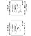
- FIG. 1 is a diagram illustrating normal mutations and deleterious mutations.
- FIG. 2 is a schematic diagram illustrating a method for evaluating whether a target protein binds to a target protein.
- FIG. 3 is a schematic cross-sectional view showing an example of a fluidic device.
- FIG. 4 is a schematic cross-sectional view illustrating a method for evaluating whether a target protein binds to a target protein.
- FIG. 5 is a schematic cross-sectional view illustrating a method for evaluating whether a target protein binds to a target protein.
- FIG. 6 is a schematic cross-sectional view showing an example of a fluidic device.
- FIG. 1 is a diagram illustrating normal mutations and deleterious mutations.
- FIG. 2 is a schematic diagram illustrating a method for evaluating whether a target protein binds to a target protein.
- FIG. 3 is a schematic cross-sectional view showing an example of a fluidic device.
- FIG. 4 is
- FIG. 7 is a schematic cross-sectional view illustrating a method for evaluating whether a target protein binds to a target protein.
- FIG. 8 is a schematic cross-sectional view illustrating a method for evaluating whether a target protein binds to a target protein.
- FIG. 9 is a schematic diagram illustrating an example of the Invasive Cleavage Assay (ICA) method.
- FIG. 10 is a schematic diagram illustrating a method for evaluating the activity of a target protein.
- FIG. 11 is a schematic diagram illustrating a method for evaluating whether a target protein phosphorylates a target protein.
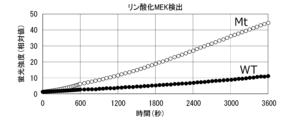
- FIG. 12 is a graph showing the results of Experimental Example 1.
- FIG. 13 is a graph showing the results of Experimental Example 1.
- FIG. 14 is a graph showing the results of Experimental Example 1.
- FIG. 15 is a graph showing the results of Experimental Example 2.
- FIG. 16 is a graph showing the results of Experimental Example 3.
- FIG. 17 is a graph showing the results of Pattern 1 of Experimental Example 4.
- FIG. 18 is a graph showing the results of Pattern 2 of Experimental Example 4.
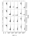
- FIG. 19 is a histogram of fluorescence intensity measured over time in the immuno-ICA reaction of Experimental Example 5.
- the present invention provides a method for evaluating whether or not a target protein binds to a target protein, wherein the target protein, the target protein, and a first single-stranded nucleic acid fragment are labeled in a container.
- a first specific binding substance for the target protein and a second specific binding substance for the target protein labeled with a second single-stranded nucleic acid fragment are brought into contact with each other, and as a result, the target protein binds to the target protein, a complex containing the target protein, the target protein, the first specific binding substance, and the second specific binding substance is formed, and the first single-stranded a step (a) of hybridizing at least a portion of the nucleic acid fragment and at least a portion of the second single-stranded nucleic acid fragment to form a double-stranded nucleic acid; and a step (a) of detecting the formation of the double-stranded nucleic acid. b), wherein the detection of the formation of the double-stranded nucleic acid indicates that the protein of interest binds to the target protein.
- FIG. 2 is a schematic diagram illustrating the method of this embodiment.
- target protein 110, target protein 120, and first single-stranded nucleic acid fragment 131 are labeled with target protein 110, target protein 120, and first single-stranded nucleic acid fragment 131.
- a first specific binding substance 130 for target protein 110 and a second specific binding substance 140 for target protein 120 labeled with a second single-stranded nucleic acid fragment 141 are contacted.
- the target protein 110 binds to the target protein 120, a complex containing the target protein 110, the target protein 120, the first specific binding substance 130, and the second specific binding substance 140 is formed, and the At least a portion of the first single-stranded nucleic acid fragment 131 and at least a portion of the second single-stranded nucleic acid fragment 141 hybridize to form a double-stranded nucleic acid (also referred to as a double-stranded nucleic acid region) 150.
- a double-stranded nucleic acid also referred to as a double-stranded nucleic acid region
- step (b) the formation of double-stranded nucleic acid 150 is detected. If formation of double-stranded nucleic acid 150 is detected, it can be said that target protein 110 binds (that is, interacts) with target protein 120.
- the method of this embodiment may be applied when the target protein 110 does not bind (that is, does not interact) with the target protein 120, and in that case, the formation of the double-stranded nucleic acid 150 is not detected. Detection of the formation of double-stranded nucleic acid 150 will be described later.
- activity evaluation of mutant proteins can be carried out easily and in a short period of time.
- VUS can be easily and efficiently functionally analyzed (ie, evaluated) in a short period of time.
- the first single-stranded nucleic acid fragment 131 is The base length may be 10 to 200 bases.
- the base length of the second single-stranded nucleic acid fragment 141 may also be 10 to 200 bases.
- the length of the double-stranded nucleic acid 150 formed by hybridization of at least a portion of the first single-stranded nucleic acid fragment 121 and at least a portion of the second single-stranded nucleic acid fragment 131 is 7 to 30 mm. It is preferable that the number of bases is about a base, and for example, it may be 9 bases, 12 bases, or 15 bases.
- the target protein 110 may be a protein having a genetic mutation of unknown clinical significance (ie, VUS).
- VUS a genetic mutation of unknown clinical significance
- Target protein 110 may be, for example, a kinase. More specific target proteins include proteins in intracellular signal transduction pathways, such as BRAF, A-RAF, Raf, MAP3K 4/12, MAP3K11, ASK1, and TAK1.
- BRAF protein having a genetic mutation of unknown clinical significance
- the container is preferably a well, and the well preferably constitutes a well array in which a plurality of wells are arranged. Further, the well array is preferably arranged within the flow path of the fluidic device.
- FIG. 3 is a schematic cross-sectional view showing an example of a fluidic device that can suitably implement the method of this embodiment.
- the fluidic device 200 includes a substrate 210 and a lid member 220 disposed opposite to the substrate 210.
- the lid member 220 has a convex portion 221.
- the tip of the protrusion 221 is in contact with the substrate 210.
- the well array 240 is integrally molded with the substrate 210 on one side of the substrate 210, and faces the lid member 220.
- Well array 240 has a plurality of wells 241.
- the lid member 220 may be welded or bonded to the substrate 210.
- the well 241 is open to the surface of the substrate 210. Although the shape, dimensions, and arrangement of the well 241 are not particularly limited, it is preferable that the well 241 be a microwell with a small volume. For example, the volume of one well 241 may be about 10 fL to 100 pL.
- a plurality of wells 241 having the same shape and size constitute a well array 240.
- the same shape and same size may be defined as having the same shape and the same capacity to the extent required for digital measurement, and variations to the extent of manufacturing errors are acceptable.
- the diameter of the well 241 may be, for example, about 1 to 10 ⁇ m.
- the depth of the well 241 may be, for example, about 1 to 10 ⁇ m.
- the arrangement of the wells 241 is not particularly limited; for example, they may be arranged in a triangular lattice shape, a square lattice shape, or randomly arranged.
- a space is formed between the well array 240 and the lid member 220 due to the presence of the convex portion 221.
- the space constitutes a flow path 230.
- the channel 230 functions as a path for transporting a liquid in which the target protein, target protein, first specific binding substance, second specific binding substance, etc. are dispersed, and a sealing liquid to be described later.
- the shape, structure, capacity, etc. of the flow path 230 are not particularly limited, the height of the flow path 230 (distance between the surface of the substrate 210 and the surface of the lid member 220 facing the substrate 210) is, for example, 500 ⁇ m or less. It may be, for example, 300 ⁇ m or less, for example, 200 ⁇ m or less, or, for example, 100 ⁇ m or less.
- the convex portion 221 may be integrally formed with the lid member 220.
- the lid member 220 can be formed into a plate shape having a convex portion 221 by, for example, molding a fluid of thermoplastic resin using a mold. Further, the lid member 220 may be formed with a reagent introduction port 222 and a reagent discharge port 223.
- the lid member 220 When the lid member 220 has the convex portion 221, the lid member 220 and the substrate 210 are stacked so that the convex portion 221 contacts the surface of the substrate 210 where the well 241 opens. As a result, the space between the lid member 220 and the substrate 210 becomes a flow path 230.
- the lid member 220 and the substrate 210 may be welded together by laser welding or the like.
- FIG. 6 is a schematic cross-sectional view showing an example of a fluidic device.
- the fluidic device 500 includes a substrate 210 and a wall member 510.
- the well array 240 is integrally molded with the substrate 210 on one side of the substrate 210.
- Well array 240 has a plurality of wells 241.
- Fluid device 500 differs from fluid device 200 described above primarily in that it does not include lid member 220.
- the lid member 220 and the convex portion 221 are integrally molded.
- the lid member 220 and the convex portion 221 may be molded separately.
- the well array 240 is integrally molded with the substrate 210 on one side of the substrate 210.
- the well array does not have to be integrally molded with the substrate 210.
- a well array 240 molded separately from the fluidic device, may be placed on the substrate 210 of the fluidic device.
- a resin layer may be laminated on the surface of the substrate 210, and a well array may be formed on the resin layer by etching or the like.
- the substrate 210 is formed using resin, for example.
- the type of resin is not particularly limited, it is preferably a resin that is resistant to reagents and sealing liquid.
- a resin with little autofluorescence is preferable.
- resins with low autofluorescence include, but are not limited to, cycloolefin polymers, cycloolefin copolymers, silicones, polypropylene, polycarbonates, polystyrene, polyethylene, polyvinyl acetate, fluororesins, and amorphous fluororesins.
- a plurality of wells 241 may be formed on one surface of the substrate 210 in the thickness direction.
- Methods for forming wells using resin include injection molding, thermal imprinting, optical imprinting, and the like.
- a well array may be formed by laminating a fluororesin on the substrate 210 and processing the fluororesin by etching or the like.
- a fluororesin for example, CYTOP (registered trademark) (Asahi Glass) or the like can be used.
- the material of the lid member 220 is preferably a resin with low autofluorescence, and may be, for example, a thermoplastic resin such as a cycloolefin polymer or a cycloolefin copolymer.
- the lid member 220 may be made of a material that does not transmit light of a wavelength near the wavelength detected when observing signals by fluorescence, or may be made of a material that does not transmit light completely.
- the lid member 220 may be made of thermoplastic resin to which carbon or metal particles are added.
- the method of this embodiment is a method for evaluating whether or not a target protein binds to a target protein, in which a well 241 is labeled with a target protein 110, a target protein 120, and a first single-stranded nucleic acid fragment 131.
- a first specific binding substance 130 for the target protein 110 and a second specific binding substance 140 for the target protein 120 labeled with a second single-stranded nucleic acid fragment 141 are brought into contact with each other, and as a result, the target protein 110 binds to the target protein 120, a complex 100 containing the target protein 110, the target protein 120, the first specific binding substance 130, and the second specific binding substance 140 is formed, and the first one Step (a) in which at least a portion of the stranded nucleic acid fragment 131 and at least a portion of the second single-stranded nucleic acid fragment 141 hybridize to form a double-stranded nucleic acid 150; and detecting the formation of the double-stranded nucleic acid 150. (b), and the detection of the formation of double-stranded nucleic acid 150 indicates that the target protein 110 binds to the target protein 120.
- the evaluation method may be the following detection method.
- the detection method of the present embodiment includes, in the well 241, the target protein 110, the target protein 120, the first specific binding substance 130 for the target protein 110 labeled with the first single-stranded nucleic acid fragment 131, and , contacting a second specific binding substance 140 for the target protein 120 labeled with a second single-stranded nucleic acid fragment 141; binding the target protein 110 to the target protein 120;
- a complex 100 is formed that includes a target protein 120, a first specific binding substance 130, and a second specific binding substance 140, and includes at least a portion of the first single-stranded nucleic acid fragment 131 and the second single-stranded nucleic acid fragment 131.
- the method may include hybridizing at least a portion of the nucleic acid fragment 141 to form a double-stranded nucleic acid 150, and detecting the formation of the double-stranded nucleic acid 150.
- mutant proteins include proteins with VUS.
- the reagent liquid L210 is introduced from the introduction port 222 of the fluidic device 200 and sent to the channel 230.
- Reagent liquid L210 is a liquid in which target protein 110, target protein 120, first specific binding substance 130, and second specific binding substance 140 are dispersed, and is used to detect the formation of double-stranded nucleic acid 150. Also includes reagents.
- the reagent solution L210 sent to the channel 230 comes into contact with the well array 240. Then, the reagent solution L210 is accommodated inside the well 241. As a result, the target protein 110, the target protein 120, the first specific binding substance 130, the second specific binding substance 140, and a reagent for detecting the formation of the double-stranded nucleic acid 150 are introduced into the well 241. be done.
- the reagent solution L210 contains the target protein 110, the target protein 120, the first specific binding substance 130, and the second specific binding substance 140. A complex 100 is included.
- the number of complexes 100 introduced into one well 241 is not particularly limited, but preferably one or less, that is, zero or one complex 100, is introduced into one well 241. Thereby, detection of the complex 100 can be performed one by one, that is, digital measurement becomes possible. Furthermore, it is not necessary to introduce the complex 100 into all wells of the well array.
- the means for introducing the complex 100 into the well is not particularly limited, and examples thereof include a method of allowing the complex 100 to settle within the fluid device (specifically, within the channel 230) by its own weight and then distributing it to the well 241. .
- a substance that captures the complex 100 also referred to as a captured substance
- the efficiency of introducing the complex 100 into the well can also be improved by trapping the complex 100 that has been sent.
- the step of binding the capture substance to the complex 100 can be performed at any point in the method of this embodiment.
- this step may be performed by bringing the complex 100 into contact with the capture object in the sample tube before the step of introducing the complex 100 into the well 241.
- the complex 100 may be introduced into the well, and the captured substance and the complex 100 may be brought into contact within the well.
- a capture substance is a substance that can capture the complex 100.
- the capture material may be, for example, a combination of a solid phase and a substance that specifically binds to the complex 100.
- Examples of the solid phase include particles, films, and substrates.
- the number of specific binding substances for the complex 100 may be one or two or more.
- the number of specific binding substances may be three, four, or five or more.
- the particles are not particularly limited, and include polymer particles, magnetic particles, glass particles, and the like.
- the particles are surface-treated to avoid non-specific adsorption.
- particles having a functional group such as a carboxyl group on the surface are preferable. More specifically, a product such as "Magnosphere LC300" manufactured by JSR Corporation can be used.
- Specific binding substances in the first specific binding substance 130, second specific binding substance 140, and capture material include antibodies, antibody fragments, aptamers, and the like.
- antibody fragments include Fab, F(ab') 2 , Fab', single chain antibodies (scFv), disulfide stabilized antibodies (dsFv), dimer V region fragments (diabodies), and peptides containing CDRs. It will be done.
- the antibody may be a monoclonal antibody or a polyclonal antibody. Alternatively, a commercially available antibody may be used.
- a method for labeling a specific binding substance with a single-stranded nucleic acid fragment includes a method using a crosslinking agent.
- a single-stranded nucleic acid fragment may be labeled with a specific binding substance via a linker molecule.
- the linker is not particularly limited, and examples thereof include polyethylene chains, hydrocarbon chains, peptides, and the like.
- a single-stranded nucleic acid fragment may be DNA or RNA. Furthermore, it may contain artificial nucleic acids such as BNA and LNA.
- Methods for immobilizing specific binding substances on particle surfaces are not particularly limited, and include physical adsorption, chemical bonding, avidin-biotin bonding, and protein G or protein A bonding with antibodies.
- a method using examples include methods in which a specific binding substance is immobilized on the particle surface by hydrophobic interaction or electrostatic interaction.
- Examples of the method using chemical bonding include a method using a crosslinking agent. For example, when the surface of the particle has a hydroxyl group, the carboxyl group of the specific binding substance is reacted with a crosslinking agent to form an active ester, and then the hydroxyl group and this ester group are reacted to form a specific binding substance on the particle. It can be immobilized on a surface. Furthermore, it is preferable to provide a spacer between the specific binding substance and the particle surface so as not to inhibit the ability of the specific binding substance to recognize the target molecule.
- the combination of the captured substance and the complex 100 is formed under the condition that 0 or 1 complex 100 is captured in one captured substance. It is preferable to form Furthermore, it is preferable that one well 241 be configured so that zero or one captured substance is introduced. This allows digital measurement.
- a complex 100 containing them is formed. is formed, and at least a portion of the first single-stranded nucleic acid fragment 131 and at least a portion of the second single-stranded nucleic acid fragment 141 hybridize to form a double-stranded nucleic acid 150. Formation of the complex 100 may be performed within the sample tube or within the well 241.
- a step of sealing the opening of the well 241 may be performed.
- the method of sealing the opening of the well 241 is not particularly limited as long as it can prevent the liquid contained in one well 241 from mixing with the liquid contained in another well 241.
- the opening of the well 241 may be sealed by covering it with a sealing liquid.
- the opening of the well 241 may be sealed by laminating a plate-like member such as a glass plate.
- the sealing liquid L220 is sent from the introduction port 222 of the lid member 220 to the flow path 230 between the substrate 210 and the lid member 220.
- the sealing liquid L220 sent to the channel 230 contacts the well array 240.
- the sealing liquid L220 sweeps away and replaces the reagent liquid L210 that is not accommodated in the well 241 among the reagent liquid L210 sent to the flow path 230.
- the sealing liquid L220 individually seals the plurality of wells 241 containing the reagent liquid L210 containing the target substance 110, and the wells 241 become independent reaction spaces (also referred to as microcompartments 242).
- FIG. 5 shows a state in which all the wells 241 of the well array 240 are sealed with the sealing liquid L220, and sealed wells (that is, microcompartments) 242 are formed.
- lipid bilayer membrane is formed at the opening of the well 241. It is also possible to form a sealed well 242 by forming a plurality of wells 241 and individually sealing each of the plurality of wells 241 with the lipid bilayer membrane.
- lipids that form a lipid bilayer include 1,2-dioleoyl-sn-glycero-3-phosphoethanolamine (also referred to as DOPE) and 1,2-dioleoyl-sn-glycero-3-phosphoglycerol (DOPG). ) and mixtures thereof, but are not limited to these.
- the sealing liquid is a liquid that can individually seal the liquids introduced into the plurality of wells 241 so that they do not mix with each other to form droplets (also referred to as microdroplets), and is preferably an oil-based solution. and more preferably oil.
- oil fluorine oil, silicone oil, hydrocarbon oil, or a mixture thereof can be used. More specifically, a product such as "FC-40” manufactured by Sigma Corporation can be used.
- FC-40 (CAS number: 86508-42-1) is a fluorinated aliphatic compound with a specific gravity of 1.85 g/mL at 25°C.
- Detection of the formation of double-stranded nucleic acid 150 is preferably performed using a signal amplification reaction.
- signal amplification reactions include Invasive Cleavage Assay (also referred to as ICA).
- the ICA reaction is related to the principle that signal amplification proceeds through a cycle of two reactions: (1) complementary binding between nucleic acids, and (2) recognition and cleavage of a triplex structure by an enzyme.
- the ICA reaction is less affected by reaction cycle inhibition caused by impurities. Therefore, by using the ICA reaction, the formation of the double-stranded nucleic acid 150 can be detected with high accuracy.
- the reagent solution L210 that is, the liquid containing the target protein 110, the target protein 120, the first specific binding substance 130, and the second specific binding substance 140
- the reagent solution L210 is used for the ICA reaction. Contains the reaction reagents necessary for
- Reaction reagents necessary for the ICA reaction include ICA reaction reagents such as flap probes, flap endonucleases (also referred to as FEN), and fluorescent substrates.
- the flap probe is a nucleic acid fragment designed to hybridize to the first single-stranded nucleic acid fragment 131 or the second single-stranded nucleic acid fragment 141 to form a flap structure with the double-stranded nucleic acid 150.
- FIG. 9 is a schematic diagram illustrating an example of the ICA method.
- a double-stranded nucleic acid 150 is formed by hybridizing at least a portion of the first single-stranded nucleic acid fragment 131 and at least a portion of the second single-stranded nucleic acid fragment 141 by the ICA method. Detect.
- a flap probe is hybridized to the first single-stranded nucleic acid fragment 131 or the second single-stranded nucleic acid fragment 141.
- flap probe 810 hybridizes to first single-stranded nucleic acid 131.
- a first flap portion 811 is formed.
- the first flap site 811 when the first flap site 811 is reacted with FEN, the first flap site 811 is cleaved and a nucleic acid fragment 811 is generated. Subsequently, the nucleic acid fragment 811 hybridizes to a fluorescent substrate (ie, the nucleic acid fragment 820) to form a second flap region 821.
- a fluorescent substrate ie, the nucleic acid fragment 820
- a fluorescent substance F is bound to the 5' end of the nucleic acid fragment 820, and a quenching substance Q is bound to the 3' side of the 5' end of the nucleic acid fragment 820.
- the second flap site 821 is reacted with FEN, the second flap site 821 is cleaved and a nucleic acid fragment 821 is generated.
- the fluorescent substance F separates from the quencher Q and generates a fluorescent signal. By detecting this fluorescent signal, the formation of double-stranded nucleic acid 150 can be detected.
- reagent liquid L210 a general liquid used in biochemical analysis performed using a fluidic device can be used, and preferably an aqueous solution. Further, by including a surfactant or the like in the reagent liquid L210, it may be possible to easily seal the liquid in the well.
- fluorescent substance F is liberated from quencher Q by an enzymatic reaction based on an isothermal reaction, and corresponds to excitation light. to emit a predetermined fluorescent signal.
- a known appropriate method can be selected depending on the type of signal to be detected. For example, when observing a fluorescent signal, the sealed well 242 is irradiated with excitation light corresponding to the fluorescent substance, and the fluorescence emitted by the fluorescent substance is observed. For example, as shown in FIG. 5, a predetermined reaction is performed in a sealed well 242, and the generated signal is observed.
- a sealed well 242R is a well in which a signal was detected
- a sealed well 242 is a well in which no signal was detected.
- the reagent liquid L210 is introduced into the fluidic device 500.
- Reagent liquid L210 is a liquid in which target protein 110, target protein 120, first specific binding substance 130, and second specific binding substance 140 are dispersed, and is used to detect the formation of double-stranded nucleic acid 150.
- the reagent solution L210 contains the target protein 110, the target protein 120, the first specific binding substance 130, and the second specific binding substance 140.
- a complex 100 is included.
- the concentration of the complex 100 is preferably adjusted to a concentration such that one molecule or less of the complex 100 enters the well 241 per well.
- the sealing liquid L220 is introduced into the fluidic device 500.
- the specific gravity of the sealing liquid L220 is greater than that of the reagent liquid L210. Therefore, the sealing liquid L220 sinks below the reagent liquid L210 that is not accommodated in the well 241 among the reagent liquids L210 and comes into contact with the well array 240. Then, the sealing liquid L220 individually seals the plurality of wells 241 containing the reagent liquid L210 containing the complex 100, thereby forming independent reaction spaces (also referred to as microcompartments) 242.
- microcompartment 242R is a well in which a signal was detected
- microcompartment 242 is a well in which no signal was detected.
- step (a) the target protein 110, the target protein 120, the first specific binding substance 130, and the second specific binding substance 140 are brought into contact with each other in a container.
- the target protein 110 binds to the target protein 120, a complex 100 containing the target protein 110, the target protein 120, the first specific binding substance 130, and the second specific binding substance 140 is formed.
- Step (a) of hybridizing at least a portion of the first single-stranded nucleic acid fragment 131 and at least a portion of the second single-stranded nucleic acid fragment 141 to form the double-stranded nucleic acid 150 is performed in the container, Step (a1) of synthesizing the target protein 110 using a cell-free protein synthesis system, and combining the target protein 110, the target protein 120, the first specific binding substance 130, and the second specific binding substance 140 in a container. contact, and as a result, when the target protein 110 binds to the target protein 120, the complex 100 containing the target protein 110, the target protein 120, the first specific binding substance 130, and the second specific binding substance 140 is formed.
- the target protein 110 may be synthesized using a cell-free protein synthesis system, and the formation of the complex 100 may be performed continuously with the synthesis of the target protein 110.
- the target protein is a kinase or the like containing a genetic mutation, the activity evaluation of the target protein can be carried out easily and in a short period of time.
- a cell-free protein synthesis system does not synthesize proteins within cells, but rather uses ribosomes, transcription and translation factors, etc. derived from living cells or artificially synthesized to synthesize proteins from nucleic acid templates in vitro. Refers to the synthetic system to be synthesized.
- the cell-free protein synthesis system may include a transcription process in addition to the translation process.
- the nucleic acid that encodes a protein is DNA
- the cell-free protein synthesis system may contain factors that enable transcription. Examples of factors that enable transcription include, but are not limited to, RNA polymerase and nucleotides, and factors known to those skilled in the art can be used.
- RNA may be synthesized in advance using DNA encoding a protein as a template, and the RNA may be added to a cell-free protein synthesis system.
- artificially chemically synthesized RNA may be used.
- the nucleic acid fragment that serves as a template for cell-free protein synthesis may be a biologically derived nucleic acid fragment, a cultured cell-derived nucleic acid fragment, or a virus-derived nucleic acid fragment.
- the nucleic acid fragment may be artificially synthesized based on the results of genetic analysis.
- Cell-free protein synthesis systems are not particularly limited, and include, for example, synthetic systems that utilize cell extracts obtained from wheat germ, yeast, insect cells, cultured mammalian cells, rabbit reticulocytes, Escherichia coli, etc.; Examples include synthetic systems in which factors are reconstituted. Among these, human expression cell-free protein synthesis is preferred.
- the cell-free protein synthesis system may contain at least one of factors involved in translation, such as tRNA, aminoacylated tRNA synthetase, translation initiation factor, translation elongation factor, and translation termination factor.
- factors involved in translation such as tRNA, aminoacylated tRNA synthetase, translation initiation factor, translation elongation factor, and translation termination factor.
- the target protein, the target protein, the first specific binding substance, and the second specific binding substance may be further contacted with adenosine triphosphate (ATP). good. This makes it possible to evaluate whether the target protein is phosphorylated. That is, it becomes possible to perform a kinase assay.
- ATP adenosine triphosphate
- FIG. 10 is a schematic diagram showing an example of the method of this embodiment.
- the mutant protein is BRAF
- cell-free protein synthesis kinase assay, antigen-antibody reaction, and ICA reaction are performed sequentially. This makes it possible to evaluate the activity of the target protein more easily and in a shorter period of time. Furthermore, for example, by adding an inhibitor to the reaction system, the effect of the inhibitor can be determined.
- the timing of adding the reagents necessary for each reaction and the timing of introducing the reagent solution into the fluidic device can be appropriately selected. For example, after performing cell-free protein synthesis, reagents necessary for a kinase assay and antigen-antibody reaction are added, and the kinase assay and antigen-antibody reaction are performed.
- An ICA reaction reagent may be added to the mixture after the antigen-antibody reaction and introduced into a fluidic device to perform the ICA reaction.
- reagents necessary for the kinase assay, antigen-antibody reaction, and ICA reaction may be added and introduced into a fluidic device to perform the kinase assay, antigen-antibody reaction, and ICA reaction.
- reagents necessary for the kinase assay, antigen-antibody reaction, and ICA reaction are added, and after performing the kinase assay and antigen-antibody reaction, this is introduced into a fluidic device to perform the ICA reaction. You may go.
- reagents necessary for the kinase assay and antigen-antibody reaction are added, and the kinase assay and antigen-antibody reaction are performed. Thereafter, reagents necessary for the ICA reaction may be added and introduced into the fluidic device to perform the ICA reaction. Further, after performing cell-free protein synthesis, reagents necessary for the kinase assay are added to perform the kinase assay. Thereafter, reagents necessary for antigen-antibody reaction and ICA reaction may be added to perform antigen-antibody reaction, and this may be introduced into a fluidic device to perform ICA reaction. Since the detection sensitivity is good, it is preferable to add the reagents necessary for the ICA reaction after performing the kinase assay and introduce the reagents into the fluidic device.
- the method of this embodiment preferably does not include a washing step.
- a washing step even when the target protein 110 is synthesized using a cell-free protein synthesis system, if it is possible to evaluate whether or not the target protein binds to the target protein without including a washing step, it will be easier to evaluate the activity of the target protein. And it can be implemented in a short period of time.
- the present invention provides a target protein 110, a target protein 120, a first specific binding substance 130 for the target protein 110 labeled with a first single-stranded nucleic acid fragment 131, and a second target protein 130. It includes a second specific binding substance 140 for the target protein 120 labeled with a full-stranded nucleic acid fragment 141, and includes at least a portion of the first single-stranded nucleic acid fragment 131 and at least a portion of the second single-stranded nucleic acid fragment 141.
- a complex 100 is provided, a portion of which has hybridized to form a double-stranded nucleic acid 150.
- a flap probe may further hybridize to the first single-stranded nucleic acid fragment 131 or the second single-stranded nucleic acid fragment 141.
- the activity evaluation of a mutant protein can be carried out easily and in a short period of time.
- the present invention is a kit for evaluating whether or not a target protein 110 binds to a target protein 120, which comprises a well array 240 having a plurality of wells 241, a first single-stranded nucleic acid fragment 131
- a kit comprising a first specific binding substance 130 for the target protein 110 labeled with , and a second specific binding substance 140 for the target protein 120 labeled with a second single-stranded nucleic acid fragment 141 I will provide a.
- the kit of this embodiment it is possible to suitably evaluate whether or not the target protein 110 binds to the target protein 120.
- the kit of this embodiment includes, in the well 241, the target protein 110, the target protein 120, the first specific binding substance 130 for the target protein 110 labeled with the first single-stranded nucleic acid fragment 131, and When a second specific binding substance 140 labeled with a second single-stranded nucleic acid fragment 141 for the target protein 120 is introduced, and as a result, the target protein 110 binds to the target protein 120, the target protein 110, A complex 100 is formed that includes a target protein 120, a first specific binding substance 130, and a second specific binding substance 140, and includes at least a portion of the first single-stranded nucleic acid fragment 131 and the second single-stranded nucleic acid fragment 131.
- the method includes a step of hybridizing at least a portion of the nucleic acid fragment 141 to form a double-stranded nucleic acid 150, and a step of detecting the formation of the double-stranded nucleic acid 150, wherein the formation of the double-stranded nucleic acid 150 is detected. It can also be said that this is for use in a method of showing that the target protein 110 binds to the target protein 120.
- the well array may be placed inside the fluidic device described above.
- the target protein, target protein, first single-stranded nucleic acid fragment, first specific binding substance, second single-stranded nucleic acid fragment, and second specific binding substance are This is the same as described above.
- the kit of this embodiment may further contain ATP. This makes it possible to evaluate whether the target protein is phosphorylated. That is, it becomes possible to perform a kinase assay.
- the kit of this embodiment may further include a sealing liquid L220 that seals the opening of the well 241.
- the sealing liquid L220 is the same as that described above.
- the kit of this embodiment detects a double-stranded nucleic acid 150 formed by hybridization of at least a portion of the first single-stranded nucleic acid fragment 131 and at least a portion of the second single-stranded nucleic acid fragment 141. It may further contain a reagent.
- reagents include the above-mentioned reagents for the ICA reaction, and specific examples include flap probes, flap endonucleases, fluorescent substrates, and the like.
- the present invention provides a method for evaluating whether a target protein phosphorylates a target protein, the method comprising: a target protein, a second single-stranded nucleic acid fragment, a target protein, and a second single-stranded nucleic acid fragment in a container; contacting a labeled second specific binding substance for the target protein, a third specific binding substance for the phosphorylated target protein labeled with a third single-stranded nucleic acid fragment, and ATP;
- the target protein is phosphorylated, a complex containing the target protein, the second specific binding substance, and the third specific binding substance is formed, and the second specific binding substance a step of hybridizing at least a portion of the single-stranded nucleic acid fragment and at least a portion of the third single-stranded nucleic acid fragment to form a double-stranded nucleic acid; and a step of
- the evaluation method may be the following detection method.
- the detection method of the present embodiment includes, in a container, a target protein, a target protein, a second specific binding substance for the target protein labeled with a second single-stranded nucleic acid fragment, a third single-stranded contacting a third specific binding substance for the phosphorylated target protein labeled with a nucleic acid fragment with ATP, the target protein, the second specific binding substance, and the third specific binding substance;
- a complex containing a target binding substance is formed, and at least a portion of the second single-stranded nucleic acid fragment and at least a portion of the third single-stranded nucleic acid fragment hybridize to form a double-stranded nucleic acid. and detecting the formation of the double-stranded nucleic acid.
- FIG. 11 is a schematic diagram illustrating the method of this embodiment.
- target protein 110, target protein 120, and second single-stranded nucleic acid fragment 141 are labeled with target protein 110, target protein 120, and second single-stranded nucleic acid fragment 141.
- a third specific binding substance 170 and ATP are contacted.
- a complex 900 containing the target protein 120, the second specific binding substance 140, and the third specific binding substance 170 is formed.
- At least a portion of the second single-stranded nucleic acid fragment 141 and at least a portion of the third single-stranded nucleic acid fragment 171 hybridize to form a double-stranded nucleic acid (also referred to as a double-stranded nucleic acid region) 150. do.
- Step (a') is similar to step (a) in the method for evaluating whether a target protein binds to a target protein described above, but a third specific binding substance 130 is used instead of the first specific binding substance 130.
- the main difference is that a specific binding substance 170 is brought into contact.
- step (b) the formation of double-stranded nucleic acid 150 is detected. If formation of double-stranded nucleic acid 150 is detected, it can be determined that target protein 120 is phosphorylated.
- Step (b) is similar to step (b) in the above-described method for evaluating whether a target protein binds to a target protein. In the method of this embodiment, the amount of double-stranded nucleic acid 150 formed corresponds to the amount of phosphorylated target protein 120 present.
- the method of this embodiment may be applied when the target protein 110 does not phosphorylate the target protein 120, and in that case, the formation of the double-stranded nucleic acid 150 is not detected.
- activity evaluation of mutant proteins can be carried out easily and in a short period of time.
- VUS can be easily and efficiently functionally analyzed (ie, evaluated) in a short period of time.
- the target protein 110 exerts a kinase activity on the target protein 120, first, the target protein 110 and the target protein 120 bind to form a complex 900, and then the target protein 120 is activated by the kinase activity of the target protein 110. is thought to be phosphorylated.
- the function of the target protein 110 can be analyzed (ie, evaluated) by the above-described method of evaluating whether the target protein binds to the target protein or the method of this embodiment.
- the second single-stranded nucleic acid fragment 141 is The base length may be 10 to 200 bases.
- the base length of the third single-stranded nucleic acid fragment 171 may also be 10 to 200 bases.
- the length of the double-stranded nucleic acid 150 formed by hybridization of at least a portion of the second single-stranded nucleic acid fragment 141 and at least a portion of the third single-stranded nucleic acid fragment 171 is 7 to 30 mm. It is preferable that the number of bases is about a base, and for example, it may be 9 bases, 12 bases, or 15 bases.
- the target protein 110 may be a kinase having a genetic mutation of unknown clinical significance. More specific target proteins include proteins in intracellular signal transduction pathways, such as BRAF, A-RAF, Raf, MAP3K 4/12, MAP3K11, ASK1, TAK1, and the like. In this case, whether a protein with VUS is in a constitutively activated state can be evaluated by whether or not it phosphorylates a target protein.
- the present invention provides a kit for evaluating whether a target protein 110 phosphorylates a target protein 120, the kit comprising: a well array 240 having a plurality of wells 241; a second single-stranded nucleic acid; a second specific binding substance 140 for the target protein 120 labeled with a fragment 141; a third specific binding substance 170 for the phosphorylated target protein 120 labeled with a third single-stranded nucleic acid fragment 171; and ATP.
- the kit of this embodiment it is possible to suitably evaluate whether or not the target protein 110 phosphorylates the target protein 120.
- the target protein 110, the target protein 120, the second specific binding substance 140 for the target protein 120 labeled with the second single-stranded nucleic acid fragment 141, the third A third specific binding substance 170 for the phosphorylated target protein 120 labeled with a single-stranded nucleic acid fragment 171 and ATP were introduced, and as a result, the target protein 110 phosphorylated the target protein 120.
- a complex 900 is formed that includes the target protein 120, the second specific binding substance 140, and the third specific binding substance 170, and includes at least a portion of the second single-stranded nucleic acid fragment 141 and the third specific binding substance 140.
- the formation of the double-stranded nucleic acid 150 includes a step of hybridizing at least a portion of the single-stranded nucleic acid fragment 171 to form a double-stranded nucleic acid 150, and a step of detecting the formation of the double-stranded nucleic acid 150. It can also be said that what is detected is for use in a method showing that the target protein 110 phosphorylates the target protein 120.
- the well array may be placed inside the fluidic device described above.
- the target protein, target protein, second single-stranded nucleic acid fragment, second specific binding substance, third single-stranded nucleic acid fragment, and third specific binding substance This is the same as described above.
- the kit of this embodiment may further include a sealing liquid L220 that seals the opening of the well 241.
- the sealing liquid L220 is the same as that described above.
- the kit of this embodiment detects a double-stranded nucleic acid 150 formed by hybridization of at least a portion of the second single-stranded nucleic acid fragment 141 and at least a portion of the third single-stranded nucleic acid fragment 171. It may further contain a reagent.
- reagents include the above-mentioned ICA reaction reagents, and specific examples include flap probes, flap endonucleases, fluorescent substrates, and the like.
- the present invention includes the following aspects.
- a target protein In a container, a target protein, a target protein, a first specific binding substance for the target protein labeled with a first single-stranded nucleic acid fragment, and a second single-stranded nucleic acid fragment labeled contacting a second specific binding substance for the target protein,
- the target protein binds to the target protein to form a complex containing the target protein, the target protein, the first specific binding substance, and the second specific binding substance; At least a portion of the first single-stranded nucleic acid fragment and at least a portion of the second single-stranded nucleic acid fragment hybridize to form a double-stranded nucleic acid; Detecting the formation of the double-stranded nucleic acid.
- Forming the double-stranded nucleic acid comprises further contacting the target protein, the target protein, the first specific binding substance, and the second specific binding substance with adenosine triphosphate.
- [6] The detection method according to any one of [1] to [5], wherein the first single-stranded nucleic acid fragment and the second single-stranded nucleic acid fragment each have a base length of 10 to 200 bases. . [7] Further comprising adding a reagent for detecting the formation of the double-stranded nucleic acid into the container after the formation of the complex and before the formation of the double-stranded nucleic acid, [1] to The detection method according to any one of [6]. [8] The detection method according to any one of [1] to [7], wherein detecting the formation of the double-stranded nucleic acid is performed by Invasive Cleavage Assay.
- a target protein In a container, a target protein, a target protein, labeled with a second single-stranded nucleic acid fragment, a second specific binding substance for the target protein, labeled with a third single-stranded nucleic acid fragment. , a third specific binding substance for the phosphorylated target protein, and ATP;
- the target protein is phosphorylated to form a complex containing the target protein, the second specific binding substance, and the third specific binding substance; At least a portion of the second single-stranded nucleic acid fragment and at least a portion of the third single-stranded nucleic acid fragment hybridize to form a double-stranded nucleic acid; Detecting the formation of the double-stranded nucleic acid.
- oligonucleotides DNA1 (5'-TTTGTCACTGTTCCTCCTTTTGTTTTCCTTTCTGTGAGCAATTTCACCCAA-3', SEQ ID NO: 1) and DNA2 (5'-GCATGGTTCCAATTTGGGTGAT) were added to the above anti-phosphorylated MEK rabbit polyclonal antibody and anti-BRAF rabbit polyclonal antibody. -3' and SEQ ID NO: 2) were linked to each other.
- Anti-phosphorylated MEK rabbit polyclonal antibody (pMEK1/2 (S217/221) rabbit antibody, Cell Signaling Technology Co., Ltd.) was prepared using an antibody-oligonucleotide conjugation tool (product name "oYo-Link antibody labeling reagent", Funakoshi Co., Ltd.).
- anti-MEK rabbit monoclonal antibody (MEK1 Detector Antibody, Abcam) was conjugated with different oligonucleotides (Integrated DNA Technologies).
- oligonucleotides DNA1 (5'-TTTGTCACTGTTCCTCCTTTTGTTTTCCTTTCTGTGAGCAATTTCACCCAA-3', SEQ ID NO: 1) and DNA2 (5'-GCATGGTTCCAATTTGGGTGAT) were added to the above anti-phosphorylated MEK rabbit polyclonal antibody and anti-MEK rabbit polyclonal antibody. -3' and SEQ ID NO: 2) were linked to each other.
- a mixed solution was prepared by mixing the target protein (mutant BRAF and wild type BRAF), the target protein (inactive MEK), the above two types of oligonucleotide-modified antibodies, ATP, and blocking buffer. The volume of each mixture was 10 ⁇ L.
- the target proteins were prepared to have final concentrations of 0 pM, 2.82 pM, 28.2 pM, and 2821 pM, respectively.
- the target protein was prepared to a final concentration of 28210 pM.
- the two types of oligonucleotide-modified antibodies were prepared to have a final concentration of 8.56 nM.
- Tris-buffered saline also referred to as TBS
- BSA bovine serum albumin
- an ICA reaction reagent for detection was prepared.
- the ICA reaction reagents in this experiment included 2 ⁇ M allele probe (5'-CGCGCCGAGGAATTGCTCACAGAAAGGA-3') (Fasmac, SEQ ID NO: 3), 4 ⁇ M FRET cassette 1 (fluorescent substrate, Alexa488-BHQ:5'-X-TTCT- Y-AGCCGGTTTTCCGGCTGAGACCTCGGCGCG-3', , 50mM Tris-HCl (pH 8.5), 20mM MgCl2 , and 0.05% Tween20. Note that the concentrations of each component in these ICA reaction reagents are the final concentrations in the experimental examples.
- FIG. 12 to 14 are graphs showing the results of Experimental Example 1.
- Figure 12 shows the results of immuno-ICA reaction using 2821 pM mutant BRAF (Mt) and wild type BRAF (WT) as target proteins to detect interaction (i.e. binding) between BRAF and phosphorylated MEK. be.
- the vertical axis shows the fluorescence intensity (relative value) of Alexa488, and the horizontal axis shows the time (seconds) after the start of the ICA reaction.
- FIG. 13 shows the results of detecting phosphorylated MEK in an immuno-ICA reaction using 2821 pM of mutant BRAF (Mt) and wild type BRAF (WT) as target proteins.
- the vertical axis shows the fluorescence intensity (relative value) of Alexa488, and the horizontal axis shows the time (seconds) after the start of the ICA reaction.
- the fluorescence intensity when phosphorylated MEK is detected when wild type BRAF is reacted is N (noise)
- the fluorescence intensity when phosphorylated MEK is detected when mutant BRAF is reacted is S (signal).
- the signal-to-noise ratio (S/N ratio) at the end point was calculated to be 3.9.
- Figure 14 shows the results of Figures 12 and 13, where the fluorescence intensity when the antigen concentration is 0 nM is N (noise), the fluorescence intensity at each other concentration is S (signal), and the signal-to-noise ratio (S /N ratio) is a graph showing the results of calculation.
- the vertical axis shows the maximum value of the S/N ratio in each reaction
- the horizontal axis shows the time (seconds) or enzyme concentration after the start of the ICA reaction.
- Example 2 Kermantine activity evaluation of target proteins (mutant BRAF and wild type BRAF) using fluidic device 1) The interaction (that is, binding) between mutant BRAF and phosphorylated MEK and the interaction between wild type BRAF and phosphorylated MEK were conducted in the same manner as in Experimental Example 1 except that fluidic devices as shown in FIGS. 3 to 5 were used. The effect was detected.
- the fluidic device was set in an aluminum block constant temperature bath (model "DTU-Mini”, Taitec Co., Ltd.), heated at 66°C for 7 minutes or 30 minutes, and then placed in a microscope (product name "All-in-one Fluorescence Microscope", model "BZ"). -X810'' (Keyence Corporation). Subsequently, the brightness of each well was calculated based on the microscopic observation image.
- FIG. 15 is a graph showing the calculation results of the brightness of each well.
- the immuno-ICA reaction was performed using mutant BRAF (Mt) and wild type BRAF (WT) as target proteins, and the results of detecting the interaction between BRAF and phosphorylated MEK are shown.
- the vertical axis in FIG. 15 indicates the number of wells, and the horizontal axis indicates the brightness of the wells.
- the left side of FIG. 15 shows the results when the ICA reaction time was 7 minutes, and the right side of FIG. 15 shows the results when the ICA reaction time was 30 minutes.
- the upper row of FIG. 15 shows the results of detecting the interaction between mutant BRAF (Mt) and phosphorylated MEK
- the lower row of FIG. 15 shows the results of detecting the interaction between wild type BRAF (WT) and phosphorylated MEK.
- Table 1 below is a table summarizing the median brightness values of the wells shown in FIG. 15.
- S/N is the fluorescence intensity when detecting the interaction between wild-type BRAF and phosphorylated MEK, which is N (noise), and the interaction between mutant BRAF and phosphorylated MEK was detected.
- S/N ratio signal-to-noise ratio
- the fluidic device was set in an aluminum block constant temperature bath (model "DTU-Mini”, Taitec Co., Ltd.), heated at 66°C for 7 minutes or 30 minutes, and then placed in a microscope (product name "All-in-one Fluorescence Microscope", model "BZ"). -X810'' (Keyence Corporation). Subsequently, the brightness of each well was calculated based on the microscopic observation image.
- FIG. 16 is a graph showing the calculation results of the brightness of each well.
- the results of immuno-ICA reaction performed using mutant BRAF (Mt) and wild type BRAF (WT) as target proteins and detection of phosphorylated MEK are shown.
- the vertical axis in FIG. 16 indicates the number of wells, and the horizontal axis indicates the brightness of the wells.
- the left side of FIG. 16 shows the results when the ICA reaction time was 7 minutes, and the right side of FIG. 16 shows the results when the ICA reaction time was 30 minutes.
- the upper row of FIG. 16 shows the results of detecting phosphorylated MEK when reacting with mutant BRAF (Mt)
- the lower row of FIG. 16 shows the results of detecting phosphorylated MEK when reacting with wild-type BRAF (WT). Show the results.
- Table 2 below is a table summarizing the median brightness values of the wells shown in FIG. 16.
- S/N is the fluorescence intensity when reacting with wild type BRAF, N (noise), and the fluorescence intensity when reacting with mutant BRAF, S (signal), and at the end point. The results of calculating the signal-to-noise ratio (S/N ratio) are shown.
- Pattern 1 is a case in which phosphorylated MEK is detected by mixing all reagents for the kinase assay reaction, antigen-antibody reaction, and immuno-ICA reaction before performing the kinase assay. After performing the kinase assay, reagents for antigen-antibody reaction and immuno-ICA reaction are mixed and introduced into a tube, and pattern 2 is when phosphorylated MEK is detected.
- a mixed solution was prepared by mixing target proteins (mutant BRAF and wild type BRAF), target proteins (inactive MEK and active MEK), the above two types of oligonucleotide-modified antibodies, ATP, and blocking buffer.
- the volume of each mixture was 10 ⁇ L.
- the target proteins were each prepared at a final concentration of 2821 pM.
- the target proteins were each prepared at a final concentration of 7 nM.
- the two types of oligonucleotide-modified antibodies were prepared so that the final concentration was 1 nM when DNA1 was bound, and the final concentration was 4 nM when DNA2 was bound.
- Tris-buffered saline also referred to as TBS
- casein Tris-buffered saline
- ATP Tris-buffered saline
- the ICA reaction reagent in Pattern 1 was prepared with the same composition and final concentration as in Experimental Examples 1 to 3, except that it was 0.108 ⁇ M flap endonuclease (FEN)-1. Note that the concentrations of each component in these ICA reaction reagents are the final concentrations in the experimental examples.
- a mixed solution was prepared by mixing the target protein (mutant BRAF and wild type BRAF), target protein (inactive MEK and active MEK), ATP, 20mM MgCl2, and blocking buffer. The volume of each mixture was 7.5 ⁇ L. The target proteins were each prepared at a final concentration of 2821 pM. The target proteins were each prepared at a final concentration of 7 nM. Further, as a blocking buffer, Tris-buffered saline (also referred to as TBS) containing casein and a nonionic surfactant was used. As shown in Table 3, six patterns of combinations of BRAF, MEK, and ATP were used as reaction compositions. In Table 3, ⁇ indicates that the corresponding component is included, and ⁇ indicates that the corresponding component is not included.
- the mixed solution of the antigen-antibody reaction reagent and the ICA reaction reagent in pattern 2 contained the above two types of oligonucleotide-modified antibodies, 2 ⁇ M allele probe (5'-CGCGCCGAGGAATTGCTCACAGAAAGGA-3') (Fasmac, SEQ ID NO: 3), and 4 ⁇ M.
- FRET cassette 1 fluorescent substrate, Alexa488-BHQ:5'-X-TTCT-Y-AGCCGGTTTTCCGGCTGAGACCTCGGCGCG-3', X: Alexa488+AminoC6, Y: black hole quencher (BHQ) 1-dT) (Japan Bioservices, SEQ ID NO: 4), 0.108 ⁇ M flap endonuclease (FEN)-1, 50 mM Tris-HCl (pH 8.5), and 0.05% Tween20. Furthermore, the two types of oligonucleotide-modified antibodies were prepared so that the final concentration was 1 nM when DNA1 was bound, and the final concentration was 4 nM when DNA2 was bound.
- each reaction reagent was 2.5 ⁇ L for the antigen-antibody reaction reagent and 10 ⁇ L for the ICA reaction reagent. Note that the concentrations of each component in these ICA reaction reagents are the final concentrations in the experimental examples.
- the kinase assay solution was introduced into the tube, the kinase assay was allowed to react at 30° C. for 60 minutes, and then the antigen-antibody reaction reagent and the ICA reaction reagent were introduced into the tube.
- Immuno-ICA reaction was performed in the same manner as in Experimental Example 1 by reacting at 37°C for 60 minutes and at 66°C for 60 to 120 minutes. Mutant BRAF and wild type BRAF were used as target proteins.
- FIG. 17 is a graph showing the results of Pattern 1 of Experimental Example 4.
- FIG. 18 is a graph showing the results of Pattern 2 of Experimental Example 4. Labels 1 to 6 in FIGS. 17 and 18 correspond to reaction compositions 1 to 6 in Table 3, respectively.
- FIG. 17 and FIG. 18 show the results of detecting phosphorylation by performing an immuno-ICA reaction for 60 minutes using 2821 pM of mutant BRAF (Mt) or wild type BRAF (WT) as the target protein.
- Mt mutant BRAF
- WT wild type BRAF
- Example 5 Kininase activity evaluation of target protein (mutant BRAF and wild type BRAF) in fluidic device 3) Phosphorylated MEK was detected in the same manner as in Experimental Example 4, except that the immuno-ICA reaction was performed using a fluidic device as shown in FIGS. 3 to 5. Mutant BRAF and wild type BRAF were used as target proteins.
- kinase assays and antigen-antibody reactions were performed in tubes, and ICA reactions were performed in fluidic devices.
- pattern 1 the mixed solution and the ICA reaction reagent were mixed, a kinase assay and an antigen-antibody reaction were performed, and then the mixture was introduced into a fluidic device. After reacting at 30° C. for 60 minutes, phosphorylated MEK was detected in the same manner as in Experimental Examples 2 and 3.
- the reaction time of the immuno-ICA reaction was from 60 minutes to 180 minutes.
- pattern 2 after the kinase assay was reacted at 30° C. for 60 minutes, the antigen-antibody reaction reagent and the ICA reaction reagent were mixed, reacted at 37° C. for 60 minutes, and introduced into the fluidic device. The reaction was carried out at 66° C. for 60 to 120 minutes, and phosphorylated MEK was detected in the same manner as in Experimental Examples 2 and 3 (Pattern 2).
- FIG. 19 is a histogram of the fluorescence intensity measured over time in the immuno-ICA reaction. From the left side of Figure 19, fluorescence intensity 120 minutes after the start of the immuno-ICA reaction in pattern 1, fluorescence intensity 180 minutes after the start of the immuno-ica reaction in pattern 1, fluorescence intensity 90 minutes after the start of the immuno-ICA reaction in pattern 2 and the fluorescence intensity 120 minutes after the start of the immuno-ICA reaction in pattern 2 are shown. 1 to 6 in FIG. 19 correspond to reaction compositions 1 to 6 in Table 3, respectively.
- pattern 2 that is, adding the antigen-antibody reaction reagent and the ICA reaction reagent after the kinase assay, resulted in better reactivity.
- the presence of antibodies during kinase assays may inhibit the phosphorylation reaction.
- Sealing liquid 242R... Well in which signal was detected, 510... Wall member, 810... Flap probe, 811... First flap site (nucleic acid fragment), 821... Second flap site (nucleic acid fragment), 820, 820' ...Nucleic acid fragment, F...fluorescent substance, Q...quencher.
Landscapes
- Health & Medical Sciences (AREA)
- Life Sciences & Earth Sciences (AREA)
- Chemical & Material Sciences (AREA)
- Engineering & Computer Science (AREA)
- Immunology (AREA)
- Molecular Biology (AREA)
- Analytical Chemistry (AREA)
- Organic Chemistry (AREA)
- Biomedical Technology (AREA)
- Urology & Nephrology (AREA)
- Hematology (AREA)
- General Health & Medical Sciences (AREA)
- Microbiology (AREA)
- Proteomics, Peptides & Aminoacids (AREA)
- Biotechnology (AREA)
- Biochemistry (AREA)
- Physics & Mathematics (AREA)
- Zoology (AREA)
- Pathology (AREA)
- Wood Science & Technology (AREA)
- Cell Biology (AREA)
- Food Science & Technology (AREA)
- Medicinal Chemistry (AREA)
- General Physics & Mathematics (AREA)
- General Engineering & Computer Science (AREA)
- Genetics & Genomics (AREA)
- Bioinformatics & Cheminformatics (AREA)
- Biophysics (AREA)
- Tropical Medicine & Parasitology (AREA)
- Measuring Or Testing Involving Enzymes Or Micro-Organisms (AREA)
Priority Applications (4)
| Application Number | Priority Date | Filing Date | Title |
|---|---|---|---|
| JP2024539157A JPWO2024029511A1 (https=) | 2022-08-01 | 2023-08-01 | |
| CN202380054883.1A CN119585441A (zh) | 2022-08-01 | 2023-08-01 | 评价方法、复合物及试剂盒 |
| EP23850065.6A EP4567126A4 (en) | 2022-08-01 | 2023-08-01 | EVALUATION PROCESS, COMPLEX AND KIT |
| US19/041,116 US20250164470A1 (en) | 2022-08-01 | 2025-01-30 | Evaluation method, complex, and kit |
Applications Claiming Priority (2)
| Application Number | Priority Date | Filing Date | Title |
|---|---|---|---|
| JP2022-122819 | 2022-08-01 | ||
| JP2022122819 | 2022-08-01 |
Related Child Applications (1)
| Application Number | Title | Priority Date | Filing Date |
|---|---|---|---|
| US19/041,116 Continuation US20250164470A1 (en) | 2022-08-01 | 2025-01-30 | Evaluation method, complex, and kit |
Publications (1)
| Publication Number | Publication Date |
|---|---|
| WO2024029511A1 true WO2024029511A1 (ja) | 2024-02-08 |
Family
ID=89849333
Family Applications (1)
| Application Number | Title | Priority Date | Filing Date |
|---|---|---|---|
| PCT/JP2023/028056 Ceased WO2024029511A1 (ja) | 2022-08-01 | 2023-08-01 | 評価方法、複合体及びキット |
Country Status (5)
| Country | Link |
|---|---|
| US (1) | US20250164470A1 (https=) |
| EP (1) | EP4567126A4 (https=) |
| JP (1) | JPWO2024029511A1 (https=) |
| CN (1) | CN119585441A (https=) |
| WO (1) | WO2024029511A1 (https=) |
Cited By (1)
| Publication number | Priority date | Publication date | Assignee | Title |
|---|---|---|---|---|
| WO2024253201A1 (ja) * | 2023-06-09 | 2024-12-12 | Toppanホールディングス株式会社 | 試料中の標的物質を検出する方法及びそのためのキット |
Citations (4)
| Publication number | Priority date | Publication date | Assignee | Title |
|---|---|---|---|---|
| JP2007020526A (ja) * | 2005-07-20 | 2007-02-01 | Japan Science & Technology Agency | 特異的結合を利用した標的分子の高感度検出法、そのキット |
| JP2020122799A (ja) * | 2014-05-15 | 2020-08-13 | メソ スケール テクノロジーズ エルエルシー | 改善されたアッセイ方法 |
| JP2022122819A (ja) | 2021-02-10 | 2022-08-23 | 和正 宮岸 | 可動型前照灯カウル |
| WO2023080137A1 (ja) * | 2021-11-04 | 2023-05-11 | 凸版印刷株式会社 | 標的物質の検出方法 |
Family Cites Families (3)
| Publication number | Priority date | Publication date | Assignee | Title |
|---|---|---|---|---|
| WO2011100561A1 (en) * | 2010-02-12 | 2011-08-18 | Saint Louis University | Molecular biosensors capable of signal amplification |
| JP6183471B2 (ja) * | 2014-01-31 | 2017-08-23 | 凸版印刷株式会社 | 生体分子解析キット及び生体分子解析方法 |
| KR102557419B1 (ko) * | 2015-08-07 | 2023-07-19 | 프레지던트 앤드 펠로우즈 오브 하바드 칼리지 | 단백질-단백질 상호작용의 초해상도 영상화 |
-
2023
- 2023-08-01 CN CN202380054883.1A patent/CN119585441A/zh active Pending
- 2023-08-01 EP EP23850065.6A patent/EP4567126A4/en active Pending
- 2023-08-01 WO PCT/JP2023/028056 patent/WO2024029511A1/ja not_active Ceased
- 2023-08-01 JP JP2024539157A patent/JPWO2024029511A1/ja active Pending
-
2025
- 2025-01-30 US US19/041,116 patent/US20250164470A1/en active Pending
Patent Citations (4)
| Publication number | Priority date | Publication date | Assignee | Title |
|---|---|---|---|---|
| JP2007020526A (ja) * | 2005-07-20 | 2007-02-01 | Japan Science & Technology Agency | 特異的結合を利用した標的分子の高感度検出法、そのキット |
| JP2020122799A (ja) * | 2014-05-15 | 2020-08-13 | メソ スケール テクノロジーズ エルエルシー | 改善されたアッセイ方法 |
| JP2022122819A (ja) | 2021-02-10 | 2022-08-23 | 和正 宮岸 | 可動型前照灯カウル |
| WO2023080137A1 (ja) * | 2021-11-04 | 2023-05-11 | 凸版印刷株式会社 | 標的物質の検出方法 |
Non-Patent Citations (6)
| Title |
|---|
| BLAZEK M. ET AL.: "Proximity Ligation Assay for High-content Profiling of Cell Signaling Pathways on a Microfluidic Chip.", MOLECULAR & CELLULAR PROTEOMICS, vol. 12, 2013, pages 3898 - 3907, XP055611025, DOI: 10.1074/mcp.M113.032821 |
| KAN C. W. ET AL.: "Isolation and detection of single molecules on paramagnetic beads using sequential fluid flows in microfabricated polymer array assemblies", LAB ON A CHIP, vol. 12, no. 5, 2012, pages 977 - 985, XP055073933, DOI: 10.1039/C2LC20744C |
| MOHAMMED H. ET AL.: "Approaches for Assessing and Discovering Protein Interactions in Cancer.", MOL CANCER RES, vol. 11, no. 11, 2013, pages 1295 - 1302, XP093135026, DOI: 10.1158/1541-7786.MCR-13-0454 |
| MOHAMMED HISHAM, CARROLL JASON S.: "Approaches for Assessing and Discovering Protein Interactions in Cancer", MOLECULAR CANCER RESEARCH, AMERICAN ASSOCIATION FOR CANCER RESEARCH, US, vol. 11, no. 11, 1 November 2013 (2013-11-01), US , pages 1295 - 1302, XP093135026, ISSN: 1541-7786, DOI: 10.1158/1541-7786.MCR-13-0454 * |
| no. 86508-42-1 |
| See also references of EP4567126A4 |
Cited By (1)
| Publication number | Priority date | Publication date | Assignee | Title |
|---|---|---|---|---|
| WO2024253201A1 (ja) * | 2023-06-09 | 2024-12-12 | Toppanホールディングス株式会社 | 試料中の標的物質を検出する方法及びそのためのキット |
Also Published As
| Publication number | Publication date |
|---|---|
| JPWO2024029511A1 (https=) | 2024-02-08 |
| US20250164470A1 (en) | 2025-05-22 |
| CN119585441A (zh) | 2025-03-07 |
| EP4567126A4 (en) | 2025-12-10 |
| EP4567126A1 (en) | 2025-06-11 |
Similar Documents
| Publication | Publication Date | Title |
|---|---|---|
| EP3712275B1 (en) | Target molecule detection method | |
| US20190241948A1 (en) | Biomolecule analysis method | |
| US12153013B2 (en) | Detection methods for epitachophoresis workflow automation | |
| CN108290158A (zh) | 测定板及其用途 | |
| WO2019084458A1 (en) | HIGHLY SPECIFIC DOSINGS | |
| EP3974844A1 (en) | Method for detecting target molecule | |
| US20250164470A1 (en) | Evaluation method, complex, and kit | |
| US20240279717A1 (en) | Method of detecting target substance | |
| US20210285937A1 (en) | Multiplexed colocalization-by-linkage assays for the detection and analysis of analytes | |
| US10914735B2 (en) | Protein detection using FET | |
| US20200309773A1 (en) | Disease proteome protein arrays and uses thereof | |
| JP2025188101A (ja) | 検出方法 | |
| JP7831518B2 (ja) | ウェルに液体を導入する方法 | |
| WO2024253201A1 (ja) | 試料中の標的物質を検出する方法及びそのためのキット | |
| US20260009067A1 (en) | Method and kit for detecting target rna | |
| WO2016141040A1 (en) | Homogenous entropy-driven biomolecular assay (heba) | |
| WO2025254035A1 (ja) | 複数種類の標的分子を検出する方法 | |
| EP4607192A1 (en) | Fluid device, and method for detecting target molecule | |
| US20250354209A1 (en) | In situ proximity detection of target biomolecules | |
| EP4623102A1 (en) | Heat-based transfer of reaction products made in situ to a planar support |
Legal Events
| Date | Code | Title | Description |
|---|---|---|---|
| 121 | Ep: the epo has been informed by wipo that ep was designated in this application |
Ref document number: 23850065 Country of ref document: EP Kind code of ref document: A1 |
|
| WWE | Wipo information: entry into national phase |
Ref document number: 2024539157 Country of ref document: JP |
|
| WWE | Wipo information: entry into national phase |
Ref document number: 202380054883.1 Country of ref document: CN |
|
| WWE | Wipo information: entry into national phase |
Ref document number: 2023850065 Country of ref document: EP |
|
| NENP | Non-entry into the national phase |
Ref country code: DE |
|
| WWP | Wipo information: published in national office |
Ref document number: 202380054883.1 Country of ref document: CN |
|
| ENP | Entry into the national phase |
Ref document number: 2023850065 Country of ref document: EP Effective date: 20250303 |
|
| WWP | Wipo information: published in national office |
Ref document number: 2023850065 Country of ref document: EP |