WO2023145831A1 - 網膜色素変性症を治療するための医薬組成物 - Google Patents
網膜色素変性症を治療するための医薬組成物 Download PDFInfo
- Publication number
- WO2023145831A1 WO2023145831A1 PCT/JP2023/002470 JP2023002470W WO2023145831A1 WO 2023145831 A1 WO2023145831 A1 WO 2023145831A1 JP 2023002470 W JP2023002470 W JP 2023002470W WO 2023145831 A1 WO2023145831 A1 WO 2023145831A1
- Authority
- WO
- WIPO (PCT)
- Prior art keywords
- statin
- pvs
- pharmaceutical composition
- mice
- nanoparticles
- Prior art date
- Legal status (The legal status is an assumption and is not a legal conclusion. Google has not performed a legal analysis and makes no representation as to the accuracy of the status listed.)
- Ceased
Links
Images


Classifications
-
- A—HUMAN NECESSITIES
- A61—MEDICAL OR VETERINARY SCIENCE; HYGIENE
- A61K—PREPARATIONS FOR MEDICAL, DENTAL OR TOILETRY PURPOSES
- A61K9/00—Medicinal preparations characterised by special physical form
- A61K9/48—Preparations in capsules, e.g. of gelatin, of chocolate
- A61K9/50—Microcapsules having a gas, liquid or semi-solid filling; Solid microparticles or pellets surrounded by a distinct coating layer, e.g. coated microspheres, coated drug crystals
- A61K9/51—Nanocapsules; Nanoparticles
- A61K9/5107—Excipients; Inactive ingredients
- A61K9/513—Organic macromolecular compounds; Dendrimers
- A61K9/5146—Organic macromolecular compounds; Dendrimers obtained otherwise than by reactions only involving carbon-to-carbon unsaturated bonds, e.g. polyethylene glycol, polyamines, polyanhydrides
- A61K9/5153—Polyesters, e.g. poly(lactide-co-glycolide)
-
- A—HUMAN NECESSITIES
- A61—MEDICAL OR VETERINARY SCIENCE; HYGIENE
- A61K—PREPARATIONS FOR MEDICAL, DENTAL OR TOILETRY PURPOSES
- A61K31/00—Medicinal preparations containing organic active ingredients
- A61K31/33—Heterocyclic compounds
- A61K31/395—Heterocyclic compounds having nitrogen as a ring hetero atom, e.g. guanethidine or rifamycins
- A61K31/435—Heterocyclic compounds having nitrogen as a ring hetero atom, e.g. guanethidine or rifamycins having six-membered rings with one nitrogen as the only ring hetero atom
- A61K31/47—Quinolines; Isoquinolines
-
- A—HUMAN NECESSITIES
- A61—MEDICAL OR VETERINARY SCIENCE; HYGIENE
- A61K—PREPARATIONS FOR MEDICAL, DENTAL OR TOILETRY PURPOSES
- A61K9/00—Medicinal preparations characterised by special physical form
- A61K9/0012—Galenical forms characterised by the site of application
- A61K9/0019—Injectable compositions; Intramuscular, intravenous, arterial, subcutaneous administration; Compositions to be administered through the skin in an invasive manner
-
- A—HUMAN NECESSITIES
- A61—MEDICAL OR VETERINARY SCIENCE; HYGIENE
- A61K—PREPARATIONS FOR MEDICAL, DENTAL OR TOILETRY PURPOSES
- A61K9/00—Medicinal preparations characterised by special physical form
- A61K9/14—Particulate form, e.g. powders, Processes for size reducing of pure drugs or the resulting products, Pure drug nanoparticles
-
- A—HUMAN NECESSITIES
- A61—MEDICAL OR VETERINARY SCIENCE; HYGIENE
- A61P—SPECIFIC THERAPEUTIC ACTIVITY OF CHEMICAL COMPOUNDS OR MEDICINAL PREPARATIONS
- A61P27/00—Drugs for disorders of the senses
- A61P27/02—Ophthalmic agents
-
- B—PERFORMING OPERATIONS; TRANSPORTING
- B82—NANOTECHNOLOGY
- B82Y—SPECIFIC USES OR APPLICATIONS OF NANOSTRUCTURES; MEASUREMENT OR ANALYSIS OF NANOSTRUCTURES; MANUFACTURE OR TREATMENT OF NANOSTRUCTURES
- B82Y5/00—Nanobiotechnology or nanomedicine, e.g. protein engineering or drug delivery
Definitions
- Retinitis pigmentosa is a disease in which photoreceptor cells and pigment epithelial cells in the retina degenerate due to genetic mutations, and typical symptoms include night blindness and visual field constriction associated with photoreceptor damage.
- the frequency of retinitis pigmentosa is said to be 1 in 4,000 to 8,000 people, but at present there is no established treatment that restores retinal function or suppresses the progression of the disease. Remains symptomatic.
- Statins have the effect of lowering blood cholesterol by inhibiting HMG-CoA reductase, and are widely used as therapeutic agents for hypercholesterolemia.
- Statins are also known to have pro-angiogenic effects. This effect was initially observed only at high doses that greatly exceeded clinical doses in model animals, but PLGA nanoparticles encapsulating pitavastatin were selectively delivered to vascular endothelial cells in a lower extremity ischemia model and at low doses. It has been reported that it induces angiogenesis and improves the symptoms of pulmonary hypertension at a low dose in a pulmonary hypertension model. Pitavastatin-encapsulated PLGA nanoparticles have also been reported to inhibit tissue damage in atherosclerosis models.
- the present disclosure aims to provide a pharmaceutical composition for treating retinitis pigmentosa.
- the present inventors found that peripheral blood inflammatory monocytes and peripherally-derived macrophages in the retinitis increased in retinitis pigmentosa, and statin-encapsulated nanoparticles increased peripheral blood inflammatory monocytes and peripherally-derived macrophages in retinitis pigmentosa model animals.
- the present invention was completed based on the discovery that retinal macrophages are reduced and cone cell death is suppressed.
- compositions comprising statin-encapsulated nanoparticles for treating retinitis pigmentosa.
- the present disclosure provides pharmaceutical compositions containing statin-encapsulated nanoparticles for treating retinitis pigmentosa.
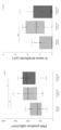
- the middle horizontal bar indicates the median, the box indicates the 25th to 75th percentile, and the whiskers indicate 1.5 times the interquartile range from the bottom and top of the box.
- Outliers are displayed as dots. *P ⁇ 0.05, **P ⁇ 0.01 by Wilcoxon rank sum test. (The same applies to the following figures.)
- FIG. 3 shows the correlation between the percentage of monocyte subsets and the rate of decline in retinal sensitivity (MD slope) in retinitis pigmentosa (RP) patients.
- FIG. 7 shows TUNEL staining of the retinas of rd10 (rd10;Ccl2 +/+ ) and rd10;Ccl2 ⁇ / ⁇ mice (P21) (left) and of the retinas of the nasal and temporal hemispheres.
- Scale bar 50 ⁇ m.
- FIG. 9 shows peanut agglutinin (PNA) staining of retinas of rd10 (rd10;Ccl2 +/+ ) and rd10;Ccl2 ⁇ / ⁇ mice (P42) (left) and PNA in regions 200 ⁇ m and 500 ⁇ m from the optic disc.
- Scale bar 50 ⁇ m.
- FIG. 14 shows PNA staining of the retina of rd10 mice (P52) intravenously administered with PBS, FITC-NP, or PVS-NP (left), and the number of PNA-positive pyramidal cells in areas 200 ⁇ m and 500 ⁇ m from the optic disc (Fig. 14).
- Scale bar 50 ⁇ m.
- FIG. 17 shows rd10 intravenously administered PBS (100 ⁇ l PBS), PVS-NP low (0.1 mg PVS/kg), PVS-NP middle (0.3 mg PVS/kg), PVS-NP high (1.0 mg PVS/kg)
- the left shows the measurement results at 250 ⁇ m from the optic disc, and the right shows the measurement results at 750 ⁇ m from the optic disc.
- FIG. 18 shows the results of averaging the numbers of PNA-positive pyramidal cells at the two measurement points in FIG. 17 for each mouse.
- the present disclosure relates to pharmaceutical compositions comprising statin-loaded nanoparticles for treating retinitis pigmentosa.
- statin means a compound having HMG-CoA (3-hydroxy-3-methylglutaryl-coenzyme A) reductase inhibitory activity.
- Statins include, for example, pitavastatin, atorvastatin, pravastatin, simvastatin, fluvastatin and rosuvastatin.
- the statin is pitavastatin.
- statin when statin is referred to, the term is used in the sense of including the free form, pharmaceutically acceptable salts thereof, and solvates thereof.
- Pharmaceutically acceptable salts include alkali metal salts such as sodium salts and potassium salts, alkaline earth metal salts such as calcium salts and magnesium salts, organic amine salts such as phenethylamine salts, and ammonium salts.
- Products include solvates with water or alcohols.
- pharmaceutically acceptable salts of pitavastatin include pitavastatin calcium, and solvates of pitavastatin or a pharmaceutically acceptable salt thereof include pitavastatin calcium hydrate (e.g. pentahydrate).
- the statin-encapsulated nanoparticles may encapsulate one or more statins, or may encapsulate other drugs in addition to the statin.
- the statin-encapsulated nanoparticles of the present disclosure contain statin inside nanoparticles composed of a biocompatible polymer.
- Biocompatible polymers are, for example, D,L-lactide, D-lactide, L-lactide, D,L-lactic acid, D-lactic acid, L-lactic acid, glycolide, glycolic acid, ⁇ -caprolactone, ⁇ -hydroxyhexanoic acid , ⁇ -butyrolactone, ⁇ -hydroxybutyric acid, ⁇ -valerolactone, ⁇ -hydroxyvaleric acid, hydroxybutyric acid, and malic acid.
- Biocompatible polymers include, for example, polylactic acid, polyglycolic acid, polylactide-co-glycolide (also referred to as PLGA), and lactic acid-aspartic acid copolymer.
- the biocompatible polymer is PLGA.
- PLGA includes polymers containing varying proportions of lactic acid or lactide and glycolic acid or glycolide.
- the ratio of lactic acid or lactide to glycolic acid or glycolide may be, for example, 1:99 to 99:1, preferably 3:1.
- the weight average molecular weight of PLGA can be, for example, 5,000-200,000, or 15,000-25,000.
- PLGA can be synthesized by a known method. Alternatively, commercially available PLGA may be used.
- the particle size of statin-loaded nanoparticles can be, for example, 1-1000 nm, 2-500 nm, 3-300 nm, 10-300 nm, or 50-300 nm.
- the particle size may be 100-300 nm or 200-300 nm. In one embodiment, the particle size is 50-300 nm.
- the particle size in this specification means the spherical equivalent diameter measured by the dynamic light scattering method, and is represented by the median diameter (D 50 ).
- the median diameter (D 50 ) is the particle diameter (50% diameter) at the 50% point in a cumulative curve with the total volume of the population of particles being 100%.
- the cumulative curve and D50 can be determined using a particle size distribution analyzer such as Nanotrac Wave-EX150 (Mictotrac BEL Corp.).
- the surface of statin-encapsulated nanoparticles may be modified with polyethylene glycol (PEG).
- PEG polyethylene glycol
- the surface of statin-encapsulated nanoparticles may be modified with polyethylene glycol (PEG).
- PEG polyethylene glycol
- the statin-encapsulated nanoparticles may be produced by any production method.
- Statin-encapsulated nanoparticles can be produced, for example, by an emulsion-in-water method.
- the emulsion-in-water method uses two kinds of solvents: a good solvent in which the biocompatible polymer dissolves and a poor solvent in which the biocompatible polymer does not dissolve.
- a good solvent and a poor solvent can be appropriately selected by those skilled in the art according to the nanoparticles to be produced.
- Water is an example of a poor solvent.
- a surfactant may be added to the water.
- Surfactants include, for example, polyvinyl alcohol (PVA), lecithin, hydroxymethylcellulose, and hydroxypropylcellulose.
- the antisolvent is water and the surfactant is PVA.
- good solvents include halogenated alkanes, acetone, methanol, ethanol, ethyl acetate, diethyl ether, cyclohexane, benzene, toluene, and mixtures thereof, which are low-boiling and poorly water-soluble organic solvents.
- the good solvent is acetone or a 2:1 mixture of acetone and ethanol.
- a biocompatible polymer is first dissolved in a good solvent, and a drug solution is added and mixed.
- the good solvent containing the polymer and the drug is dropped into the poor solvent under stirring, the good solvent rapidly diffuses into the poor solvent.
- the good solvent is emulsified in the poor solvent to form emulsion droplets of the good solvent.
- the organic solvent continuously diffuses from within the emulsion into the poor solvent, reducing the solubility of the biocompatible polymer and the drug within the emulsion droplets, resulting in spherical crystals encapsulating the drug.
- Particle nanoparticles are produced.
- the organic solvent which is a good solvent, is centrifuged or distilled off under reduced pressure. The obtained nanoparticles can be used as they are or after being pulverized by an operation such as freeze-drying.
- Statin-encapsulated nanoparticles may be produced using a forced thin-film microreactor.
- a forced thin film microreactor When a forced thin film microreactor is used, first, a good solvent containing a polymer and a drug and a poor solvent are placed between processing surfaces which are arranged to face each other and at least one of which rotates relative to the other. to introduce A good solvent and a poor solvent are mixed in the thin film fluid thus formed, and nanoparticles encapsulating the drug are deposited in the thin film fluid.
- ULREA SS-11 M Technique Co. Ltd.
- a statin-loaded nanoparticle can comprise, for example, 0.01-99%, 0.1-30%, 0.5-20%, or 1-15% by weight of a statin.
- the statin-encapsulated nanoparticles comprise 1-15% by weight of statin.
- statin content is expressed as the ratio of the weight of statin to the weight of statin-encapsulated nanoparticles. The statin content can be determined by measuring the weight of statin extracted from a given weight of statin-encapsulated nanoparticles and calculating the ratio of the weight of statin to the weight of statin-encapsulated nanoparticles.
- Statin-encapsulated nanoparticles can be combined into redispersible aggregated particles (nanocomposites) when pulverized by an operation such as freeze-drying.
- nanoparticles can be redispersible complexed by drying with organic or inorganic materials. Due to this compositing, the nanoparticles are aggregated into aggregated particles that are easy to handle, and when they come into contact with water during use, they can be dispersed and exhibit their properties.
- the statin-loaded nanoparticles are conjugated with a sugar alcohol or sucrose. By using substances such as sugar alcohols, variations in the encapsulation rate can be reduced, and the substances can act as excipients to improve the handling properties of the nanoparticles.
- Sugar alcohols include mannitol, trehalose, sorbitol, erythritol, maltitose, xylitose and the like.
- the sugar alcohol is trehalose.
- a fluidized bed drying granulation method can be used for compositing.
- peripheral blood inflammatory monocytes and retinal periphery-derived macrophages are increased in retinitis pigmentosa. It was shown to reduce peripherally-derived retinal macrophages and suppress cone cell death. Therefore, statin-loaded nanoparticles can be used to treat retinitis pigmentosa.
- Retinitis pigmentosa is a disease in which photoreceptor cells and/or pigment epithelial cells in the retina degenerate due to genetic mutations.
- Retinitis pigmentosa includes disease states in which rod cells, cone cells, pigment epithelial cells, or two or more types of cells selected from these cells are degenerated.
- Retinitis pigmentosa is often preceded by degeneration of rod cells, followed by gradual degeneration of cone cells.
- a condition in which only rod cells are degenerated is called rod dystrophy, and a condition in which both rod cells and cone cells are degenerated is called rod-cone dystrophy, both of which are included in the retinitis pigmentosa of the present disclosure.
- Retinitis pigmentosa may be caused by any gene mutation, and may be caused by one or more gene mutations.
- treatment of retinitis pigmentosa includes amelioration of one or more symptoms or findings of retinitis pigmentosa, suppression and delay of exacerbation, and suppression and delay of disease progression.
- Symptoms of retinitis pigmentosa include night blindness, narrow vision, decreased vision, photophobia, day blindness, color blindness, and photopsia.
- the findings of retinitis pigmentosa include (1) ocular fundus findings (retinal vascular narrowing, rough retinal tone, bone body-like pigmentation, multiple white spots, optic nerve atrophy, macular degeneration), (2) electroretinogram (3) hyperfluorescence or hypofluorescence due to retinal pigment epithelial atrophy in fundus autofluorescence findings, (4) ellipsoid zone in the fovea by optical coherence tomography (IS/OS) ) abnormalities (discontinuity or disappearance).
- the statin-encapsulated nanoparticles can be included in pharmaceutical compositions.
- Pharmaceutical compositions are, for example, 0.000001-99.9% by weight, 0.00001-99.8% by weight, 0.0001-99.7% by weight, 0.001-99.6% by weight, 0.01% by weight ⁇ 99.5%, 0.1-99%, 1-50%, 1-40%, 1-30%, 1-20%, or 1-15% statin-loaded nanoparticles by weight can include
- the pharmaceutical composition may further contain pharmaceutically acceptable additives as needed.
- Pharmaceutically acceptable additives include, for example, excipients, lubricants, binders, disintegrants, solubilizers, suspending agents, tonicity agents, buffers, soothing agents, preservatives, Antioxidants, coloring agents and sweetening agents are included.
- compositions can be in dosage forms such as tablets, capsules, powders, granules, solutions, suspensions, emulsions, inhalants, injections, eye drops, eye ointments, and the like. Injections include solution injections, suspension injections, emulsion injections, and extemporaneous injections. In one embodiment, the pharmaceutical composition is an injectable. These formulations can be prepared by conventional methods.
- a statin-encapsulated nanoparticle or a pharmaceutical composition containing the same is administered to a subject in an amount capable of exhibiting a desired effect (herein referred to as an effective amount).
- the dose and administration period can be appropriately determined by those skilled in the art according to conditions such as the age, body weight and health condition of the subject.
- a statin-encapsulated nanoparticle or a pharmaceutical composition containing the same can be used, for example, in the amount of statin per day of 0.001 mg/kg to 100 mg/kg, 0.003 mg/kg to 10 mg/kg, 0.005 mg/kg to 5 mg/kg, 0.01 mg/kg to 3 mg/kg, 0.01 mg/kg to 1 mg/kg, 0.01 mg/kg to 0.75 mg/kg, 0.01 mg/kg to 0.5 mg/kg, 0.01 mg/kg to 0.5 mg/kg. 0.01 mg/kg to 0.3 mg, 0.01 mg/kg to 0.25 mg, 0.01 mg/kg to 0.1 mg, 0.01 mg/kg to 0.09 mg, 0.01 mg/kg to 0.08 mg, 0.01 mg/kg to 0.08 mg.
- statin-encapsulated nanoparticles or the pharmaceutical composition comprising the same contain 0.01 mg/kg to 0.3 mg/kg or 0.03 mg/kg of statin (eg, pitavastatin calcium) per day. Dosed at ⁇ 0.1 mg/kg.
- statin eg, pitavastatin calcium
- a statin-encapsulated nanoparticle or a pharmaceutical composition containing the statin is 1 to 10 mg/body (eg, 1, 2, 4, 8, or 10 mg/body) per day for an adult.
- 1-8 mg/body eg, 1, 2, 4, or 8 mg/body
- Administration may be single or multiple doses. In the case of multiple doses, administration may be on consecutive days, several days (eg 2, 3, 4, 5 or 6 days), a week or weeks (eg 2, 3, 4, 5 or 6 weeks), It may be once a month or several months (eg 2, 3, 4, 5 or 6 months).
- administration is twice a week (eg, once every 3 or 4 days), or once every 1-4 weeks (eg, 1, 2, 3, or 4 weeks).
- the administration period can be, for example, one day or several days (e.g. 2, 3, 4, 5 or 6 days), one week or several weeks (e.g. 2, 3, 4, 5 or 6 weeks), one month or several months (e.g. for example 2, 3, 4, 5 or 6 months), or longer.
- Administration may be systemic or local, and may be oral or parenteral (e.g., intravenous, intramuscular, intrabronchial, intranasal, intraocular). good.
- the statin-loaded nanoparticles or pharmaceutical compositions comprising the same are administered intravenously.
- the subject is a mammal (eg, human, mouse, rat, hamster, rabbit, cat, dog, cow, sheep, monkey, etc.), preferably human.
- the subject is a human subject with retinitis pigmentosa (also referred to as a retinitis pigmentosa patient).
- the present disclosure provides, in one aspect, a method of treating retinitis pigmentosa comprising administering to a subject in need of treatment an effective amount of statin-loaded nanoparticles.
- the present disclosure in one aspect, provides statin-loaded nanoparticles for treating retinitis pigmentosa.
- the disclosure provides, in one aspect, the use of statin-loaded nanoparticles for the manufacture of a medicament for treating retinitis pigmentosa.
- the disclosure provides, in one aspect, the use of statin-loaded nanoparticles to treat retinitis pigmentosa.
- a pharmaceutical composition comprising statin-loaded nanoparticles for treating retinitis pigmentosa.
- the statin is pitavastatin or a pharmaceutically acceptable salt thereof.
- the statin is pitavastatin calcium.
- the statin-encapsulated nanoparticles contain 1 to 15% by weight of statin.
- the statin-loaded nanoparticles comprise PLGA.
- a method of treating retinitis pigmentosa comprising administering to a subject in need of treatment an effective amount of statin-loaded nanoparticles.
- RP retinitis pigmentosa
- characteristic fundus findings e.g., corpuscle-like pigmentation in the mid-peripheral and peripheral retinas
- Diagnosis was based on ring scotomas and decreased or absent a-wave and b-wave amplitudes on electroretinograms.
- cone dystrophy, cone-rod dystrophy, vietticrystalline retinopathy, uveitis, or systemic disease were excluded.
- BCVA visual acuity
- Flow Cytometry Immunolabeled cells were analyzed by the BD FACSVerse system (BD Biosciences, Franklin Lakes, NJ) using FlowJo software. Samples were prepared and analyzed as follows.
- mice Peripheral blood was collected from mice by cardiac puncture and erythrocytes were lysed with VersaLyse Lysing solution (Becton Dickinson Biosciences, San Jose, Calif.) for 10 minutes at room temperature. Cells were washed twice with ice-cold FACS buffer (phosphate-buffered saline (PBS) containing 2% fetal bovine serum (FBS)).
- FACS buffer phosphate-buffered saline (PBS) containing 2% fetal bovine serum (FBS)
- Fc receptors were blocked with anti-mouse CD16/CD32 (eBioscience) for 5 minutes at 4°C and then incubated for 20 minutes on ice with antibodies against: mouse CD192 (CCR2) (Alexa Fluor 647-conjugated, clone SA203G11; Biolegend, San Diego, CA), CD11b (BV421-conjugated, clone M1/70; Biolegend), Ly-6C (Alexa fluor 488-conjugated, clone HK1.4; Biolegend), Ly-6G (allophycocyanin [APC]-cy7-conjugated , clone 1A8; Biolegend) and CX3CR1 (phycoerythrin [PE]-conjugated, clone SA011F11, Biolegend). Dead cells were excluded by the fluorescent marker 7-AAD (BD Pharmingen, San Diego, Calif.). Inflammatory monocytes were identified as CD11b + Ly-6C hi Ly-6G lo
- Mouse microglia and macrophages Mouse retinas were harvested from excised eyeballs, minced and digested in a water bath at 37°C for 30 minutes (1.2 mg/ml collagenase D [Roche Diagnostics, Indianapolis, Ind.] and 40 ⁇ g/ml DNase I [Sigma-Aldrich, St. Louis, Mo.]). After digestion, the tissue was separated by pipetting into a single cell suspension. Cells were washed twice with ice-cold FACS buffer (PBS containing 2% FBS).
- Fc receptors were blocked with anti-mouse CD16/CD32 (eBioscience) for 10 minutes at 4°C and then incubated for 20 minutes on ice with antibodies against: mouse CD11c (PE-Cy7-conjugated, clone N418; Biolegend), CD45 ( APC-conjugated, clone 30-F11; Biolegend), Ly-6C (APC-cy7-conjugated, clone HK1.4; Biolegend), Ly-6G (APC-cy7-conjugated, clone 1A8; Biolegend), CD11b (PE- conjugated, clone M1/70; Biolegend), CD192 (CCR2) (FITC-conjugated, clone SA203G11; Biolegend) and CX3CR1 (BV421-conjugated, clone SA011F11; Biolegend).
- mouse CD11c PE-Cy7-conjugated, clone N418
- Microglia were defined as CD11b hi CD11c mid CD45 mid Ly-6G lo Ly-6C lo cells and macrophages were defined as CD11b hi CD11c hi CD45 hi Ly-6G lo Ly-6C lo cells. Wild-type retina was used as a control for gating microglia and macrophages in each experiment.
- Fc receptors were blocked with anti-mouse CD16/CD32 (eBioscience) for 5 minutes at 4°C and then incubated for 20 minutes on ice with antibodies against: human CD56 (NCAM) (PerCP/Cy5.5-conjugated, clone HCD56; Biolegend), CD19 (PerCP / Cy5.5-conjugated, clone HIB19; Biolegend), HLA-DR (APC-conjugated, clone L243; Biolegend), CD14 (PE-conjugated, clone M5E2; Biolegend), CD16 (BV421-conjugated , clone 3G8; Biolegend), CD192 (CCR2) (FITC-conjugated, clone K036C2; Biolegend) and CX3CR1 (PEcy7-conjugated, clone 2A9-1, Biolegend). Dead cells were excluded by 7-AAD (BD Pharmingen). Monocytes
- TUNEL Staining TUNEL staining was performed using the ApopTag Fluorescein In Situ Apoptosis Detection Kit (Merck Millipore, Darmstadt, Germany) according to the manufacturer's instructions. Immunofluorescence images were acquired using a fluorescence microscope (BZ-X700; Keyence). The number of TUNEL-positive cells in an area of 10,000 ⁇ m2 in the central (200 ⁇ m from the optic nerve), mid-peripheral (500 ⁇ m from the optic nerve) and peripheral (1000 ⁇ m from the optic nerve) regions of the retina of the nasal and temporal hemispheres was determined using Image J software . , ver. 1.52a. The ONL area of each square area was measured and the density of TUNEL-positive cells within the ONL was calculated and expressed as cells/ mm2 . Observers were analyzed blindly without telling the names and conditions of the samples.
- Electroretinogram (ERG) Photopic ERGs were recorded through LED contact lenses using the PuREC system (PC-100; Mayo Corporation, Aichi, Japan). Mice were anesthetized with an intraperitoneal injection of ketamine (100 mg/kg) and xylazine (10 mg/kg) and body temperature was maintained at 37° C. with a heating pad. Pupils were dilated with 0.5% tropicamide and 0.5% phenylephrine hydrochloride. After topical application of oxbuprocaine, LED contact lenses were attached to the mouse cornea. A reference electrode was placed on the tongue and a ground electrode was clipped to the tail. Mice were adapted to a background of white light with an intensity of 30 cd/ m2 for 10 minutes. Sixteen photopic flashes were taken at 3.0 cd ⁇ s/m 2 and averaged.
- PLGA nanoparticles PLGA polymer (Wako Pure Chemical Industries, Osaka, Japan) with an average molecular weight of 20,000 and a ratio of lactide to glycolide of 75:25 was used for the preparation of nanoparticles (NPs).
- FITC D Dojindo Laboratories, Kumamoto, Japan
- pitavastatin calcium Wako, Osaka, Japan
- PVS pitavastatin
- FITC-NP For FITC-NP, first, a tank containing liquid A (aqueous solution containing 2.0% polyvinyl alcohol (PVA)) was pressurized to 0.3 MPa, set value 43 ° C. (measured value about 40 ° C.), speed 120 ml / min. moved with Liquid B (a solution containing PLGA, FITC, acetone, and ethanol in a weight ratio of 4.04:0.20:63.84:31.92) was then added to a set point of 41°C (measured at about 30°C). ) at 100 ml/min. Liquids A and B were reacted on a rotating disk rotating at 1000 rpm with a back pressure of 0.02 MPa.
- liquid A aqueous solution containing 2.0% polyvinyl alcohol (PVA)
- PVA polyvinyl alcohol
- FITC-NP and PVS-NP contained 6.8 ⁇ 0.4% (w/v) FITC and 2.79 ⁇ 0.05% (w/v) PVS, respectively.
- the particle size was measured with a Nanotrac Wave-EX150 (manufactured by MicrotracBEL Corp.). The particle size was 252 nm for FITC-NP and 202 nm for PVS-NP.
- FITC-NP 0.5 mg FITC-NP/100 ⁇ l PBS
- blood samples were taken 2 hours later and administered to IMo.
- FITC uptake was analyzed by flow cytometry.
- retinas were harvested 24 hours after intravenous injection of FITC-NPs and FITC uptake into macrophages and microglia was analyzed.
- Blood cells were labeled with mouse Ly-6C (APC-cy7-conjugated, clone HK1.4; Biolegend) and Ly-6G (PerCP/Cy5.5-conjugated, clone 1A8; Biolegend) with the following antibodies: CD192 (CCR2) (Alexa Fluor 647-conjugated, clone SA203G11; Biolegend), CD11b (BV421-conjugated, clone M1/70; Biolegend), and CX3CR1 (PE-conjugated, clone SA011F11, Biolegend). FITC expression was assessed in CD11b + Ly-6C hi Ly-6G lo-neg IMo.
- CD192 (CCR2) (FITC-conjugated, clone SA203G11; Biolegend) was not used to assess cellular uptake of FITC-NP.
- Retinal cells were stained with the following antibodies: CD11c (PE-Cy7-conjugated, clone N418; Biolegend), CD45 (APC-conjugated, clone 30-F11; Biolegend), Ly-6C (APC-cy7-conjugated, clone HK1.4; Biolegend), Ly-6G (APC-cy7-conjugated, clone 1A8; Biolegend), CD11b (PE-conjugated, clone M1/70, Biolegend), and CX3CR1 (BV421-conjugated, clone SA011F11; Biolegend). FITC fluorescence in microglia and macrophages was then measured.
- NP treatment of rd10 mice rd10 mice were divided into three groups at P21: PBS group (100 ⁇ l PBS), FITC-NP group (0.5 mg FITC-NP/100 ⁇ l PBS), and PVS-NP group (0.0065 mg PVS/0.5 mg PVS).
- PBS, FITC-NP, or PVS-NP were administered intravenously via the tail vein twice weekly (once every 3 or 4 days) from P21 until the end of each experiment.
- PBS group 100 ⁇ l PBS
- PVS-NP low group 0.1 mg PVS/ kg
- PVS-NP middle group 0.3 mg PVS/kg
- PVS-NP high group 1.0 mg PVS/kg
- Dose-finding study To determine the optimal dosing regimen for PVS-NP, a dose-finding study was performed on rd10 mice divided into three groups at P21: PBS (100 ⁇ l PBS) every 2 weeks, PVS-NP (0. 75 mg PVS/kg) administration group every 4 weeks, PVS-NP (0.5 mg PVS/kg) administration group every 2 weeks. Doses were administered intravenously via the tail vein at respective dosing intervals from P21 until the end of each experiment. Photopic ERG and PNA-positive pyramidal cell number analysis were performed at P49.
- TUNEL staining at P21 when rod cell death peaked, showed a significant difference in the number of TUNEL-positive cells in the outer nuclear layer (ONL) between rd10;Ccl2 ⁇ / ⁇ and rd10;Ccl2 +/+ mice. was not observed (Fig. 7). Consistent with this, HE staining at P26 showed no significant difference in ONL thickness in the presence or absence of Ccl2 (Fig. 8), suggesting that loss of Ccl2 affects rod degeneration in rd10 mice. suggested that it might not.
- PVS-NP a dose-finding study was performed by dividing rd10 mice into four groups at P21: PBS group (100 ⁇ l PBS), PVS-NP low group (0.1 mg PVS/kg), PVS-NP middle group (0.3 mg PVS/kg), PVS-NP high group (1.0 mg PVS/kg). Administered intravenously via the tail vein once a week from P21 until the end of each experiment. Photopic ERG and PNA-positive pyramidal cell number analysis were performed at P49. Photopic ERGb waves were significantly higher in the PVS-NP middle group and PVS-NP high group compared to the PBS group (p ⁇ 0.05) (Fig. 16). Similarly, regarding the number of PNA-positive pyramidal cells, cone degeneration was significantly suppressed in the PVS-NP middle group and PVS-NP high group compared to the PBS group (p ⁇ 0.01) (Figs. 17 and 18). ).
- a regimen setting test was performed by dividing into the following three groups, calculated from the concentration of the PVS-NP middle group (0.3 mg PVS/kg): PBS (100 ⁇ l PBS ) A group administered every 2 weeks, a group administered PVS-NP (0.75 mg PVS/kg) every 4 weeks, and a group administered PVS-NP (0.5 mg PVS/kg) every 2 weeks. Doses were administered intravenously via the tail vein at respective dosing intervals from P21 until the end of each experiment. Photopic ERG and PNA-positive pyramidal cell number analysis were performed at P49.
- Photopic ERGb waves were significantly higher in the PVS-NP (0.75 mg PVS/kg) every 4 weeks group compared to the PBS group (p ⁇ 0.01) (FIG. 19).
- cone degeneration was observed in the PVS-NP (0.75 mg PVS/kg) every 4 weeks administration group and the PVS-NP (0.5 mg PVS/kg) every 2 weeks administration group compared with the PBS group. was significantly suppressed (p ⁇ 0.05) (FIG. 20).
- statin-encapsulated nanoparticles are a promising treatment option for retinitis pigmentosa.
Landscapes
- Health & Medical Sciences (AREA)
- Chemical & Material Sciences (AREA)
- Medicinal Chemistry (AREA)
- Pharmacology & Pharmacy (AREA)
- Life Sciences & Earth Sciences (AREA)
- Animal Behavior & Ethology (AREA)
- General Health & Medical Sciences (AREA)
- Public Health (AREA)
- Veterinary Medicine (AREA)
- Engineering & Computer Science (AREA)
- Epidemiology (AREA)
- Bioinformatics & Cheminformatics (AREA)
- Dermatology (AREA)
- Ophthalmology & Optometry (AREA)
- Chemical Kinetics & Catalysis (AREA)
- General Chemical & Material Sciences (AREA)
- Nuclear Medicine, Radiotherapy & Molecular Imaging (AREA)
- Organic Chemistry (AREA)
- Physics & Mathematics (AREA)
- Biomedical Technology (AREA)
- Nanotechnology (AREA)
- Optics & Photonics (AREA)
- Pharmaceuticals Containing Other Organic And Inorganic Compounds (AREA)
Priority Applications (4)
| Application Number | Priority Date | Filing Date | Title |
|---|---|---|---|
| EP23747041.4A EP4470531A4 (en) | 2022-01-28 | 2023-01-26 | Pharmaceutical composition for the treatment of retinitis pigmentosa |
| CN202380030920.5A CN119031907A (zh) | 2022-01-28 | 2023-01-26 | 用于治疗视网膜色素变性的药物组合物 |
| JP2023576985A JPWO2023145831A1 (https=) | 2022-01-28 | 2023-01-26 | |
| US18/832,713 US20250144041A1 (en) | 2022-01-28 | 2023-01-26 | Pharmaceutical composition for treating retinitis pigmentosa |
Applications Claiming Priority (2)
| Application Number | Priority Date | Filing Date | Title |
|---|---|---|---|
| JP2022012146 | 2022-01-28 | ||
| JP2022-012146 | 2022-01-28 |
Publications (1)
| Publication Number | Publication Date |
|---|---|
| WO2023145831A1 true WO2023145831A1 (ja) | 2023-08-03 |
Family
ID=87471563
Family Applications (1)
| Application Number | Title | Priority Date | Filing Date |
|---|---|---|---|
| PCT/JP2023/002470 Ceased WO2023145831A1 (ja) | 2022-01-28 | 2023-01-26 | 網膜色素変性症を治療するための医薬組成物 |
Country Status (5)
| Country | Link |
|---|---|
| US (1) | US20250144041A1 (https=) |
| EP (1) | EP4470531A4 (https=) |
| JP (1) | JPWO2023145831A1 (https=) |
| CN (1) | CN119031907A (https=) |
| WO (1) | WO2023145831A1 (https=) |
Citations (2)
| Publication number | Priority date | Publication date | Assignee | Title |
|---|---|---|---|---|
| WO2008026702A1 (fr) | 2006-08-30 | 2008-03-06 | Kyushu University, National University Corporation | Composition pharmaceutique contenant une nanoparticule de statine encapsulée |
| JP2022012146A (ja) | 2020-07-01 | 2022-01-17 | 株式会社システムフォワード | 来場者管理システム及び来場者管理方法 |
Family Cites Families (5)
| Publication number | Priority date | Publication date | Assignee | Title |
|---|---|---|---|---|
| JP2004149480A (ja) * | 2002-10-31 | 2004-05-27 | Saisentan Igaku Kenkyusho:Kk | 経口又は経皮投与用の眼科疾患治療剤 |
| WO2004103960A2 (en) * | 2003-05-16 | 2004-12-02 | Ambit Biosciences Corporation | Compounds and uses thereof |
| WO2008044339A1 (en) * | 2006-10-13 | 2008-04-17 | Aqumen Biopharmaceuticals K.K. | Therapeutic/preventive agent for intraocular disease containing statin compound |
| CN101959542B (zh) * | 2007-12-25 | 2014-11-12 | 国立大学法人山口大学 | 药物传递系统 |
| JP2023517582A (ja) * | 2020-03-11 | 2023-04-26 | ウニヴェルジテート レーゲンスブルク | 眼疾患の処置における使用のためのナノ粒子 |
-
2023
- 2023-01-26 JP JP2023576985A patent/JPWO2023145831A1/ja active Pending
- 2023-01-26 WO PCT/JP2023/002470 patent/WO2023145831A1/ja not_active Ceased
- 2023-01-26 EP EP23747041.4A patent/EP4470531A4/en active Pending
- 2023-01-26 US US18/832,713 patent/US20250144041A1/en active Pending
- 2023-01-26 CN CN202380030920.5A patent/CN119031907A/zh active Pending
Patent Citations (2)
| Publication number | Priority date | Publication date | Assignee | Title |
|---|---|---|---|---|
| WO2008026702A1 (fr) | 2006-08-30 | 2008-03-06 | Kyushu University, National University Corporation | Composition pharmaceutique contenant une nanoparticule de statine encapsulée |
| JP2022012146A (ja) | 2020-07-01 | 2022-01-17 | 株式会社システムフォワード | 来場者管理システム及び来場者管理方法 |
Non-Patent Citations (4)
| Title |
|---|
| K. ICHIMURA ET AL., LNT HEART J, vol. 59, 2018, pages 1432 - 1444 |
| ONO MAYUMI, JUN-ICHI FUKUSHI, DAISUKE GOTO, HITOE TORISU, SHIGEO YOSHIDA, MICHIHIKO KUWANO: "Mechanism of Angiogenesis and Angiogenic Diseases", KAGAKU TO SEIBUTSU, vol. 37, no. 1, 1 January 1999 (1999-01-01), pages 14 - 19, XP093081057 * |
| S. KATSUKI ET AL., CIRCULATION, vol. 129, 2014, pages 896 - 906 |
| See also references of EP4470531A4 |
Also Published As
| Publication number | Publication date |
|---|---|
| EP4470531A1 (en) | 2024-12-04 |
| CN119031907A (zh) | 2024-11-26 |
| US20250144041A1 (en) | 2025-05-08 |
| EP4470531A4 (en) | 2026-01-14 |
| JPWO2023145831A1 (https=) | 2023-08-03 |
Similar Documents
| Publication | Publication Date | Title |
|---|---|---|
| Hayreh | Central retinal artery occlusion | |
| Weleber et al. | Results at 2 years after gene therapy for RPE65-deficient Leber congenital amaurosis and severe early-childhood–onset retinal dystrophy | |
| TWI658828B (zh) | 用於舒緩及減輕近視之醫藥組合物及其製備方法與用途 | |
| US20210353770A1 (en) | Delivery of Urea to Cells of the Macula and Retina Using Liposome Constructs | |
| Schneier et al. | Ocular toxocariasis: advances in diagnosis and treatment | |
| KR102713615B1 (ko) | 인간에서의 신경퇴화 및 녹내장에 대한 진단적 마커로서의 망막 및 시신경 내 지방 알갱이 | |
| Eraghi et al. | The role of age in ocular toxoplasmosis: clinical signs of immunosenescence and inflammaging | |
| Fischer et al. | Acute posterior multifocal placoid pigment epitheliopathy following COVID-19 infection | |
| CN121285391A (zh) | 聚唾液酸-聚合物缀合物和纳米颗粒 | |
| KR20200043401A (ko) | 신경 질환을 치료하기 위한 방법 및 조성물 | |
| WO2023145831A1 (ja) | 網膜色素変性症を治療するための医薬組成物 | |
| Szamier et al. | Retinal histopathology of a carrier of X-chromosome-linked retinitis pigmentosa | |
| Cordeiro et al. | T cell immunoregulation in active ocular toxoplasmosis | |
| Grover et al. | Atypical presentation of pattern dystrophy in two families with peripherin/RDS mutations | |
| RU2362172C1 (ru) | Способ прогнозирования прогрессирования пролиферативных осложнений при периферических увеитах у детей | |
| Zhang et al. | Case report: Ultrasound biomicroscopy as a guide for the selection of injection sites for dexamethasone intravitreal implant (Ozurdex) for peripheral granulomatous ocular toxocariasis in children | |
| Rorke-Adams et al. | Head trauma | |
| Wang et al. | Chronic mucocutaneous candidiasis presenting as Candida endophthalmitis | |
| Delgado et al. | CASE REPORT: Amelanotic uveal cyst in a Yorkshire terrier dog | |
| Patel et al. | Postinfectious epilepsy | |
| Hellman et al. | Age-related macular degeneration | |
| Jiang et al. | Central Retinal Artery Occlusion Following Delayed Acute Retinal Necrosis Post-SARS-CoV-2 Recovery: Clinical Practice and Literature Review | |
| CA3034081C (en) | Delivery of urea to cells of the macula and retina using liposome constructs | |
| Müller | Ocular toxoplasmosis-literature review and case report | |
| Das et al. | Bilateral nongranulomatous uveitis with transposition of the great arteries: A rare case report |
Legal Events
| Date | Code | Title | Description |
|---|---|---|---|
| 121 | Ep: the epo has been informed by wipo that ep was designated in this application |
Ref document number: 23747041 Country of ref document: EP Kind code of ref document: A1 |
|
| WWE | Wipo information: entry into national phase |
Ref document number: 18832713 Country of ref document: US |
|
| ENP | Entry into the national phase |
Ref document number: 2023576985 Country of ref document: JP Kind code of ref document: A |
|
| NENP | Non-entry into the national phase |
Ref country code: DE |
|
| ENP | Entry into the national phase |
Ref document number: 2023747041 Country of ref document: EP Effective date: 20240828 |
|
| WWE | Wipo information: entry into national phase |
Ref document number: 202380030920.5 Country of ref document: CN |
|
| WWP | Wipo information: published in national office |
Ref document number: 18832713 Country of ref document: US |