WO2023085441A1 - マクロポーラス構造体 - Google Patents
マクロポーラス構造体 Download PDFInfo
- Publication number
- WO2023085441A1 WO2023085441A1 PCT/JP2022/042460 JP2022042460W WO2023085441A1 WO 2023085441 A1 WO2023085441 A1 WO 2023085441A1 JP 2022042460 W JP2022042460 W JP 2022042460W WO 2023085441 A1 WO2023085441 A1 WO 2023085441A1
- Authority
- WO
- WIPO (PCT)
- Prior art keywords
- poly
- mixture
- block copolymer
- cells
- polysaccharide
- Prior art date
- Legal status (The legal status is an assumption and is not a legal conclusion. Google has not performed a legal analysis and makes no representation as to the accuracy of the status listed.)
- Ceased
Links
Images


Classifications
-
- A—HUMAN NECESSITIES
- A61—MEDICAL OR VETERINARY SCIENCE; HYGIENE
- A61K—PREPARATIONS FOR MEDICAL, DENTAL OR TOILETRY PURPOSES
- A61K47/00—Medicinal preparations characterised by the non-active ingredients used, e.g. carriers or inert additives; Targeting or modifying agents chemically bound to the active ingredient
- A61K47/30—Macromolecular organic or inorganic compounds, e.g. inorganic polyphosphates
- A61K47/32—Macromolecular compounds obtained by reactions only involving carbon-to-carbon unsaturated bonds, e.g. carbomers, poly(meth)acrylates, or polyvinyl pyrrolidone
-
- A—HUMAN NECESSITIES
- A61—MEDICAL OR VETERINARY SCIENCE; HYGIENE
- A61K—PREPARATIONS FOR MEDICAL, DENTAL OR TOILETRY PURPOSES
- A61K47/00—Medicinal preparations characterised by the non-active ingredients used, e.g. carriers or inert additives; Targeting or modifying agents chemically bound to the active ingredient
- A61K47/30—Macromolecular organic or inorganic compounds, e.g. inorganic polyphosphates
- A61K47/36—Polysaccharides; Derivatives thereof, e.g. gums, starch, alginate, dextrin, hyaluronic acid, chitosan, inulin, agar or pectin
-
- A—HUMAN NECESSITIES
- A61—MEDICAL OR VETERINARY SCIENCE; HYGIENE
- A61K—PREPARATIONS FOR MEDICAL, DENTAL OR TOILETRY PURPOSES
- A61K47/00—Medicinal preparations characterised by the non-active ingredients used, e.g. carriers or inert additives; Targeting or modifying agents chemically bound to the active ingredient
- A61K47/30—Macromolecular organic or inorganic compounds, e.g. inorganic polyphosphates
- A61K47/36—Polysaccharides; Derivatives thereof, e.g. gums, starch, alginate, dextrin, hyaluronic acid, chitosan, inulin, agar or pectin
- A61K47/38—Cellulose; Derivatives thereof
-
- A—HUMAN NECESSITIES
- A61—MEDICAL OR VETERINARY SCIENCE; HYGIENE
- A61K—PREPARATIONS FOR MEDICAL, DENTAL OR TOILETRY PURPOSES
- A61K9/00—Medicinal preparations characterised by special physical form
- A61K9/06—Ointments; Bases therefor; Other semi-solid forms, e.g. creams, sticks, gels
-
- B—PERFORMING OPERATIONS; TRANSPORTING
- B01—PHYSICAL OR CHEMICAL PROCESSES OR APPARATUS IN GENERAL
- B01D—SEPARATION
- B01D61/00—Processes of separation using semi-permeable membranes, e.g. dialysis, osmosis or ultrafiltration; Apparatus, accessories or auxiliary operations specially adapted therefor
- B01D61/24—Dialysis ; Membrane extraction
-
- B—PERFORMING OPERATIONS; TRANSPORTING
- B01—PHYSICAL OR CHEMICAL PROCESSES OR APPARATUS IN GENERAL
- B01D—SEPARATION
- B01D71/00—Semi-permeable membranes for separation processes or apparatus characterised by the material; Manufacturing processes specially adapted therefor
- B01D71/06—Organic material
- B01D71/08—Polysaccharides
-
- B—PERFORMING OPERATIONS; TRANSPORTING
- B01—PHYSICAL OR CHEMICAL PROCESSES OR APPARATUS IN GENERAL
- B01D—SEPARATION
- B01D71/00—Semi-permeable membranes for separation processes or apparatus characterised by the material; Manufacturing processes specially adapted therefor
- B01D71/06—Organic material
- B01D71/08—Polysaccharides
- B01D71/10—Cellulose; Modified cellulose
-
- C—CHEMISTRY; METALLURGY
- C08—ORGANIC MACROMOLECULAR COMPOUNDS; THEIR PREPARATION OR CHEMICAL WORKING-UP; COMPOSITIONS BASED THEREON
- C08G—MACROMOLECULAR COMPOUNDS OBTAINED OTHERWISE THAN BY REACTIONS ONLY INVOLVING UNSATURATED CARBON-TO-CARBON BONDS
- C08G65/00—Macromolecular compounds obtained by reactions forming an ether link in the main chain of the macromolecule
- C08G65/02—Macromolecular compounds obtained by reactions forming an ether link in the main chain of the macromolecule from cyclic ethers by opening of the heterocyclic ring
- C08G65/32—Polymers modified by chemical after-treatment
-
- C—CHEMISTRY; METALLURGY
- C08—ORGANIC MACROMOLECULAR COMPOUNDS; THEIR PREPARATION OR CHEMICAL WORKING-UP; COMPOSITIONS BASED THEREON
- C08G—MACROMOLECULAR COMPOUNDS OBTAINED OTHERWISE THAN BY REACTIONS ONLY INVOLVING UNSATURATED CARBON-TO-CARBON BONDS
- C08G81/00—Macromolecular compounds obtained by interreacting polymers in the absence of monomers, e.g. block polymers
-
- C—CHEMISTRY; METALLURGY
- C08—ORGANIC MACROMOLECULAR COMPOUNDS; THEIR PREPARATION OR CHEMICAL WORKING-UP; COMPOSITIONS BASED THEREON
- C08J—WORKING-UP; GENERAL PROCESSES OF COMPOUNDING; AFTER-TREATMENT NOT COVERED BY SUBCLASSES C08B, C08C, C08F, C08G or C08H
- C08J9/00—Working-up of macromolecular substances to porous or cellular articles or materials; After-treatment thereof
- C08J9/26—Working-up of macromolecular substances to porous or cellular articles or materials; After-treatment thereof by elimination of a solid phase from a macromolecular composition or article, e.g. leaching out
-
- C—CHEMISTRY; METALLURGY
- C08—ORGANIC MACROMOLECULAR COMPOUNDS; THEIR PREPARATION OR CHEMICAL WORKING-UP; COMPOSITIONS BASED THEREON
- C08L—COMPOSITIONS OF MACROMOLECULAR COMPOUNDS
- C08L101/00—Compositions of unspecified macromolecular compounds
- C08L101/02—Compositions of unspecified macromolecular compounds characterised by the presence of specified groups, e.g. terminal or pendant functional groups
-
- C—CHEMISTRY; METALLURGY
- C08—ORGANIC MACROMOLECULAR COMPOUNDS; THEIR PREPARATION OR CHEMICAL WORKING-UP; COMPOSITIONS BASED THEREON
- C08L—COMPOSITIONS OF MACROMOLECULAR COMPOUNDS
- C08L5/00—Compositions of polysaccharides or of their derivatives not provided for in groups C08L1/00 or C08L3/00
Definitions
- the present invention relates to a mixture containing a polysaccharide, a block copolymer of a polymer having a lower critical solution temperature and a hydrophilic polymer, and a macroporous structure.
- N-isopropylacrylamide polymer poly (N-isopropylacrylamide)
- PNIPAM poly (N-isopropylacrylamide)
- 1 A. Halperin, M. Kroger, FM Winnik, Angew Chem Int Ed Engl 2015, 54, 15342-15367).
- PNIPAM has both a hydrophilic group and a hydrophobic group in the side chain, and when it is a linear polymer, it has a lower critical solution temperature (LCST: lower critical solution temperature) at 32 ° C.
- LCST lower critical solution temperature
- Non-Patent Document 1 indicates the volume phase transition temperature (VPTT).
- PNIPAM has the property that the gel swells when the temperature of the aqueous solution is below VPTT, whereas the gel shrinks when the temperature is above VPTT. Therefore, PNIPAM is expected to be applied to various fields such as drug delivery systems (DDS), actuators, immobilized enzymes, and biosensors.
- DDS drug delivery systems
- actuators immobilized enzymes
- biosensors biosensors.
- alginate gel is characterized by its high biocompatibility, mild gelation conditions, and fast gelation rate, and is widely used as a material for foodstuffs and grafts for transplantation.
- alginate gels it is difficult to change the pore size of the gel significantly, and problems remain, such as the impediment of permeation and release of secretions, bacteria, viruses, etc., and the inability of cell bodies to pass through the gel. It is
- the present invention is as follows. [1] A mixture containing a polysaccharide and a block copolymer of a polymer having a lower critical solution temperature and a hydrophilic polymer. [2] The mixture according to [1], wherein the polysaccharide is at least one selected from the group consisting of alginic acid, starch, glycogen, cellulose, xanthan gum, hyaluronic acid, carrageenan, pectin and pullulan, and salts thereof.
- the polymer having a lower critical solution temperature is at least one selected from the group consisting of poly(N-alkylacrylamide), poly(N-vinylalkylamide) and polyvinylalkyl ether.
- the hydrophilic polymer is polyethylene glycol, polyethyleneimine, polyvinyl alcohol, polyacrylic acid, polyacrylamide, polyvinyl alcohol, polyvinylpyrrolidone, polyvinylacetamide, polyamine, poly(4-styrenesulfonic acid), poly(allylamine hydrochloride) ), poly(vinylsulfonic acid, sodium salt), poly(diallyldimethylammonium chloride), and poly(2-methacryloyloxyethylphosphorylcholine), the mixture according to [1].
- a polysaccharide and a block copolymer of a polymer having a lower critical solution temperature and a hydrophilic polymer are mixed to cause phase separation, and the block copolymer is removed from the phase-separated mixture.
- a method for producing a macroporous structure comprising the step of removing.
- the method of [11] further comprising mixing the cells before causing phase separation.
- the polysaccharide is at least one selected from the group consisting of alginic acid, starch, glycogen, cellulose, xanthan gum, hyaluronic acid, carrageenan, pectin and pullulan, and salts thereof.
- the polymer having a lower critical solution temperature is at least one selected from the group consisting of poly(N-alkylacrylamide), poly(N-vinylalkylamide) and polyvinylalkyl ether. the method of. [15]
- the hydrophilic polymer is polyethylene glycol, polyethyleneimine, polyvinyl alcohol, polyacrylic acid, polyacrylamide, polyvinyl alcohol, polyvinylpyrrolidone, polyvinylacetamide, polyamine, poly(4-styrenesulfonic acid), poly(allylamine hydrochloride).
- the macroporous structure of the present invention has a controlled pore size and is a penetrating type, it has become possible to permeate and/or release structures of sizes that conventional alginate gels could not penetrate.
- FIG. 1 is a conceptual diagram of a manufacturing process of a macroporous structure of the present invention
- FIG. 1 is a schematic diagram of a synthetic method for synthesizing a block copolymer of 4-branched polyethylene glycol and PNIPAM.
- FIG. FIG. 2 shows a dual coaxial laminar flow apparatus for producing fibrous cell-containing macroporous structures.
- FIG. 2 is a diagram showing a summary of pore sizes of macroporous alginate gels.
- Fig. 3 shows macroporous alginate gel fibers at 37°C;
- FIG. FIG. 2 shows culture of mouse neural stem cell fibers.
- FIG. 10 is a view of VERO cells infected with an adeno-associated virus vector expressing EGFP, observed by a confocal microscope. Left: uninfected macroporous cell fibersRight: infected macroporous cell fibers with EGFP expression
- FIG. 4 is a view of VERO cells infected with a lentiviral vector expressing EGFP observed by a confocal microscope. Left: uninfected macroporous cell fibersRight: infected macroporous cell fibers with EGFP expression
- FIG. 4 shows cells infected with high concentrations of lentivirus.
- Macroporous alginate gel fibers after induced differentiation of neural stem cells were observed with a confocal microscope.
- Macroporous alginate gel fibers after induced differentiation of neural stem cells were observed with a confocal microscope.
- Cell fibers cultured for 20 days were immunostained and observed with a confocal microscope.
- the present invention relates to a mixture comprising (i) a polysaccharide and (ii) a block copolymer of a polymer having a lower critical solution temperature (LCST) and a hydrophilic polymer.
- a structure having a macroporous structure can be obtained by mixing the polysaccharide and the block copolymer to cause phase separation in the mixture, and then removing the block copolymer. can.
- Fig. 1 is a conceptual diagram for forming a macroporous structure from a mixture of block copolymers and polysaccharides.
- phase separation can be caused by mixing below the LCST or above the LCST (FIG. 1a).
- the polymer with the LCST becomes hydrophilic and the block copolymer dissolves in water.
- a phase-separated structure spontaneously forms (Fig. 1b).
- the polymer having the LCST becomes hydrophobic, so that it precipitates in water and becomes cloudy. That is, the block copolymer has a phase-separated structure forming an emulsion.
- a macroporous structure formed by polysaccharides can be obtained (Fig. 1c).
- Block copolymer of polymer having lower critical solution temperature (LCST) and hydrophilic polymer 2.1 Polymer Having LCST
- the polymer having LCST is not particularly limited, and examples thereof include poly(N-alkylacrylamide), poly(N-vinylalkylamide) and polyvinyl alkyl ether. can be used alone or in combination of two or more. Examples of polymers with LCST are shown below.
- Poly(N-alkylacrylamide) Poly(N-alkylacrylamides) have the following formula I: (R 10 represents a hydrogen atom or an alkyl group having 1 to 4 carbon atoms (C 1-4 ) (referred to as a “C 1-4 alkyl group”; the same shall apply hereinafter), and n is an integer of 1 or more represents.) is indicated by
- poly(N-alkylacrylamide) examples include poly(N-methylacrylamide), poly(N-ethylacrylamide), and poly(N-isopropylacrylamide) (PNIPAM).
- Poly(N-vinylalkylamides) have the following formula II: ( R20 represents a hydrogen atom or a C1-4 alkyl group, and n represents an integer of 1 or more.) is indicated by Examples of poly(N-vinylalkylamides) include poly(N-vinylacetamide), poly(N-vinylisopropylamide), poly(N-vinylisobutyramide), and the like.
- Polyvinyl alkyl ethers have the following formula III: ( R30 represents a hydrogen atom or a C1-4 alkyl group, and n represents an integer of 1 or more.) is indicated by Examples of polyvinyl alkyl ethers include polyvinyl methyl ether and polyvinyl ethyl ether.
- the phase transition temperature can be controlled or the stability of the phase separation structure can be controlled by variously changing the terminal alkyl group.
- PNIPAM has both a hydrophilic group and a hydrophobic group in its side chain, has a lower critical solution temperature of 32° C. when it is a linear polymer, and exhibits a volume phase transition temperature (VPTT) in water.
- VPTT volume phase transition temperature
- PNIPAM has the property that the gel swells when the temperature of the aqueous solution is below VPTT, whereas the gel contracts when the temperature is above VPTT. can be done.
- Functional groups include maleimides, thiols, alkynes, azides, amines, activated carboxylic acids, and the like.
- a thiol group SH group
- the maleimide group reacts with the SH group to form a stable thioether group.
- the hydrophilic polymer includes polyethylene glycol, polyethyleneimine, polyvinyl alcohol, polyacrylic acid, polyacrylamide, polyvinyl alcohol, polyvinylpyrrolidone, polyvinylacetamide, polyamine, poly(4-styrenesulfonic acid), Poly(allylamine hydrochloride), poly(vinylsulfonic acid, sodium salt), poly(diallyldimethylammonium chloride), poly(2-methacryloyloxyethylphosphorylcholine) and the like are used alone or in combination of two or more.
- the hydrophilic polymer is polyethylene glycol (PEG).
- PEG may be linear or multi-branched, and the number of branches of the multi-branched type is not particularly limited. type, 8-antennary type, etc.
- PEG can also be a star polymer or dendrimer. It is preferable to add functional groups to both ends or one end of PEG. Functional groups include, but are not limited to, thiol, acrylate, amine, biotin, aldehyde, maleimide, succinimidyl carboxymethyl ester, succinimidyl glutarate ester, and the like.
- a hyperbranched PEG can be represented, for example, by Formula IV: represents a structure represented by
- R 1 is: (X represents a functional group).
- n can be arbitrarily set according to the purpose, for example, n is 25 to 1000.
- R 2 , R 3 and R 4 are each independently a hydrogen atom or (X represents a functional group).
- X represents a functional group, the types of which are the same as described above, and include, for example, thiol, acrylate, amine, biotin, aldehyde, maleimide, succinimidyl carboxymethyl ester, succinimidyl glutarate ester and the like. It is not limited. Also, the functional groups are independent in each branched chain and may be the same or different. In addition, n in R 1 , R 2 , R 3 and R 4 is also independent in each branched chain and may be the same number or different numbers.
- m represents an integer of 1 to 4 when the PEG is bi-antennary to octa-antennary.
- R 3 and R 4 are hydrogen atoms
- the PEG is bi-antennary
- m is 1 and one of R 3 and R 4 is a hydrogen atom
- PEG is tri-antennary
- m is 1 and none of R 2 , R 3 and R 4 is a hydrogen atom
- PEG is tetra-antennary.
- the PEG is octa-branched.
- the multi-branched PEG used in the present invention is preferably 4-branched or 8-branched.
- the multi-branched PEG can be obtained by a known method, but can also be purchased as a commercial product (NOF).
- a star polymer is a polymer in which three or more polymers are radially branched from the center, and the branch points are concentrated in one place.
- Methods for synthesizing star polymers are well known in the art. For example, a method of chain-polymerizing monomers starting from a core, a method of synthesizing an arm portion first, and a method of bonding the end of the arm to the functional group portion of the core. , a method of synthesizing a linear polymer by living chain polymerization and cross-linking the core site using a cross-linking agent.
- a dendrimer is a dendritic polymer with a structure that is regularly branched from the center.
- Commercially available multifunctional PEG (Merck) can also be used.
- a functional group can be added to one or both ends of the PEG to link a polymer having an LCST, and in the branched type, a functional group can be added to the end of the branched PEG to form an LCST can be linked.
- functional groups include, but are not limited to, thiol, acrylate, amine, biotin, aldehyde, maleimide, succinimidyl carboxymethyl ester, succinimidyl glutarate ester, and the like.
- Block copolymer In the present invention, a block copolymer of a polymer having an LCST and a hydrophilic polymer is synthesized by adding a functional group to each polymer and reacting the functional groups with each other. can do. For example, a maleimide group reacts with a thiol group (SH group) to form a stable thioether group. good.
- FIG. 2 is a schematic diagram of a synthesis example of synthesizing a block copolymer of tetra-branched PEG with SH groups added as a hydrophilic polymer and PNIPAM as a polymer having an LCST.
- the block copolymer may be labeled with a fluorescent substance.
- the fluorescent substance is not particularly limited, and includes chemically synthesized substances such as fluorescein, rhodamine, Cy, Alexa Fluor TM , and HiLyte Fluor TM , or fluorescent proteins such as phycoerythrin (PE) and allophycocyanin (APC). is mentioned. These fluorescent substances are commercially available.
- Polysaccharides used in the present invention are exemplified by alginic acid, starch, glycogen, cellulose, xanthan gum, hyaluronic acid, carrageenan, pectin and pullulan, and can be used singly or in combination of two or more.
- the present invention also includes salts of these polysaccharides. Salts include sodium salts, potassium salts, calcium salts, barium salts, and the like.
- the polysaccharide used is preferably alginic acid.
- Alginic acid may be naturally derived or synthetic.
- Alginic acids that are preferably used are bioabsorbable polysaccharides extracted from brown algae such as sculpin, arame, and kelp. It is a polymerized polymer. More specifically, a block copolymer in which a homopolymer fraction of D-mannuronic acid, a homopolymer fraction of L-guluronic acid, and a fraction in which D-mannuronic acid and L-guluronic acid are randomly arranged are arbitrarily bound. It is a polymer.
- Alginic acid is also commercially available (Mochida Pharmaceutical).
- a polysaccharide and a block copolymer of a polymer having an LCST and a hydrophilic polymer are mixed to cause phase separation, and after the phase separation,
- a method for making a macroporous structure comprising the step of removing said block copolymer from a mixture.
- block copolymers of polymers having an LCST and hydrophilic polymers dissolve in water when the LCST is less than the LCST.
- polysaccharides alginic acid, etc.
- the mixing ratio of the polysaccharide and the block copolymer is the mixing ratio of the polysaccharide and the block copolymer (polysaccharide: block copolymer) in the present invention.
- ) can take various ratios between 9:1 and 1:9, in one embodiment for example 6:4 to 8:2, and in another embodiment for example 7:3 to 8:2. be.
- the macroporous structure can be maintained by gelling the polysaccharide to reduce fluidity.
- the block copolymer and the polysaccharide are mixed below the LCST, the mixture is raised above the LCST to fix the structure by gelation, and then lowered below the LCST again.
- Gelation can be performed, for example, by treating the polysaccharide with a calcium ion-containing solution (eg, an aqueous solution of calcium chloride), specifically by mixing a mixture of the block copolymer and the polysaccharide with an aqueous solution of calcium chloride. can.
- a calcium ion-containing solution eg, an aqueous solution of calcium chloride
- a block copolymer of a polymer having an LCST and a hydrophilic polymer the polymer becomes insoluble in water above the LCST and forms an emulsion.
- a phase-separated structure is formed also by mixing with polysaccharides (alginic acid etc.) in that state. After gelling the polysaccharide, the block copolymer dissolves in water when the temperature is lowered below the LCST, so the block copolymer can be removed by simply stirring, shaking, or standing still in water.
- the mixture can contain cells. In this case, it is included before phase separation of the mixture occurs. After gelling the polysaccharide as necessary, the block copolymer is removed by setting the mixture to LCST or less and stirring in water. Thereby, a cell-containing macroporous structure can be obtained.
- Macroporous structures containing cells are used as tools for trapping viruses and bacteria, antibody production tools introduced with antibody-producing hybridomas, etc. Since the gel has a pore size of a predetermined size, it can be used as a semipermeable membrane. In that case, it can be used for dialysis or blood purification.
- a polymer having an LCST a polymer having a high-temperature LCST may be used by appropriately changing the types of substituents.
- Cells are not particularly limited, and pluripotent ES cells, iPS cells, endocrine cells, exocrine cells, stem cells (hematopoietic stem cells, neural stem cells, mesenchymal stem cells), cells in various tissues (e.g., skeletal muscle cells). , cardiomyocytes, nervous system cells, fibroblasts, endothelial cells, epithelial cells, liver cells, pancreatic cells, skin cells, blood cells, stromal cells, etc.).
- pluripotent ES cells e.g., iPS cells, endocrine cells, exocrine cells, stem cells (hematopoietic stem cells, neural stem cells, mesenchymal stem cells), cells in various tissues (e.g., skeletal muscle cells). , cardiomyocytes, nervous system cells, fibroblasts, endothelial cells, epithelial cells, liver cells, pancreatic cells, skin cells, blood cells, stromal cells, etc.).
- Endocrine cells neurocrine cells, pituitary cells, thyroid cells, parathyroid cells, pancreatic islet cells, gastrointestinal endocrine cells, cardiomyocytes, hepatocytes, kidney cells, adipocytes, adrenal cells, gonadal cells, etc.
- Exocrine cells gastrointestinal epithelial cells , cells that release physiologically active substances (hepatocytes that release albumin, enzyme-producing cells that release enzymes, etc.)
- Nervous system cells Neural stem cells, central nerve cells, peripheral nerve cells, glial cells, etc.
- Skeletal muscle system cells Osteocytes, chondrocytes, etc.
- Blood cells Hematopoietic stem cells, white blood cells, red blood cells, platelets, etc.
- Stromal cells Fibroblasts, blood vessels cells, etc.
- affinity cells expressing membrane proteins having affinity for specific substances contained in blood (for example, viruses and cytokines) can be used.
- bio-related substances such as proteins, peptides, antibodies (including antibody fragments), lipids, sugars, and nucleic acids can be introduced into the mixture. do not have.
- Fibrous structure provides a method for producing a fibrous structure by applying the method for producing a macroporous structure.
- a method using a dual coaxial microfluidic device (WO 2011/046105) can be adopted to fabricate fibrous structures.
- a coaxial microfluidic device has a configuration in which two fluids are coaxially divided into a core portion and a shell portion and ejected.
- the coaxial microfluidic device 10 comprises a cell suspension injection tube 101, a polysaccharide and block copolymer mixture injection tube 102, and a calcium ion-containing aqueous solution injection tube 103.
- a mixture of a polysaccharide and a block copolymer is also simply referred to as a "mixture”.
- An injection port 111 for injecting the cell suspension is provided at the top of the cell suspension injection tube 101, and the cell suspension 121 is injected from the top.
- the cell suspension injection tube 101 and the mixture injection tube 102 are tapered at the bottom, and the mixture injection tube 101 and the mixture injection tube 102 are provided between the cell suspension injection tube 101 and the mixture injection tube 102. Opening 112 is provided. An opening 113 for injecting a calcium ion-containing aqueous solution is provided between the mixture injecting tube 102 and the calcium ion-containing aqueous solution injecting tube 103 .
- the cell suspension 121 is injected from the lower injection port of the cell suspension injection tube 101 toward the mixture injection tube 102 to form the core of the fiber.
- the mixture 122 is injected from the opening 112 toward the mixture injection tube 102, the cell suspension occupies the core portion of the fiber, the mixture covers the cell periphery to form a shell portion, and a coaxial core-shell shape is formed. form a fluid.
- the fluid is injected toward the calcium ion-containing aqueous solution injection tube 103 and the calcium ion-containing aqueous solution (for example, CaCl 2 -containing aqueous solution) is injected from the opening 113, the shell of the fluid is gelled.
- FIG. 3b A partially enlarged view of the gelled fiber 123 is shown in FIG. 3b.
- the block copolymer becomes water-soluble by adjusting the temperature below the LCST. Block copolymers can be removed.
- a fibrous macroporous structure hereinafter simply referred to as "macroporous structure” 130 having a cell core and a macroporous shell (porous) can be obtained (Fig. 3c). .
- the injection speed of each of the cell suspension and the mixture is not limited, but in the case of a coaxial microfluidic device with a pore size of about 50 ⁇ m to 2 mm, the speed at the injection port is, for example, 10 to 500 ⁇ L/min. be. Also, the rate of introduction into the calcium ion-containing aqueous solution is not particularly limited, and is, for example, 1 to 10 mL/min.
- the pore diameters of the core and shell are arbitrary and can be appropriately adjusted according to the purpose.
- the outer diameter of the shell portion of the macroporous structure 130 can be about 10 ⁇ m to 2 mm, 200 ⁇ m to 2 mm, and 50 ⁇ m to 1 mm.
- the length of the macroporous structure 130 is not particularly limited, and can be about several millimeters to several meters. Examples of the cross-sectional shape include circles, ellipses, and polygons such as quadrilaterals and pentagons.
- the macroporous structure obtained as described above can freely change the pore size, so that cells can easily pass through it and have a high affinity with cells. Therefore, the cells contained in the structure of the present invention enter into the porous material and aggregate to form a cell structure such as spheroids.
- the cells encapsulated in the macroporous structure 130 can be grown.
- the macroporous structure 130 can also be cultured for several months by appropriately exchanging the culture medium.
- the cells are differentiated and a part of the differentiated cells or tissue is formed outside the macroporous structure 130. is formed.
- the macroporous structure 130 containing cells in this way can be used as various regenerative medical materials depending on the type of cells. For example, after encapsulating neural stem cells in an undifferentiated state in the macroporous structure 130, culturing for a predetermined period of time, and then substituting with a differentiation-inducing medium from which the cell growth factors FGF and EGF have been removed and culturing, the cells are undifferentiated. Neural stem cells differentiate into nerve cells, and part of the tissue extends outside the macroporous structure 130 . The macroporous structure 130 thus formed can be used as a nerve regenerative medical material.
- the macroporous structure 130 contains virus-capturing cells or the like, the macroporous structure 130 is useful for purifying virus-infected biomaterials (eg, virus-infected blood).
- virus-infected biomaterials eg, virus-infected blood
- the novel coronavirus SARS-CoV-2
- S protein spike protein
- ACE2 angiotensin-converting enzyme 2
- various ACE2-expressing cells VeroE6 cells, VeroE6/TMPRSS cells, Hela cells, Hela/ACE2 cells
- these cells are encapsulated in the macroporous structure 130 .
- the cells encapsulated in the macroporous structure 130 express ACE2, so the macroporous structure 130 encapsulating the ACE2-expressing cells can be used as a virus trapping material for purification of virus-infected blood. can be done.
- the beads S protein beads
- the beads can be used as a SARS-CoV-2 cell infection model. can be done.
- AAV adeno-associated virus
- PEG-b-PNIPAM 10000-5500
- PEG-b-PNIPAM was synthesized by introducing PNIPAM to the ends of four functional groups of tetra-PEG by thio-ene reaction.
- tetra PEG-SH (2 g, 200 ⁇ mol,) with a molecular weight of 10,000
- PNIPAM-MA 4.4 g, 800 ⁇ mol
- PEG-b-PNIPAM-Alexa Fluor 488 was synthesized by introducing Alexa Fluor 488-MA into one of the four functional group ends of PEG-b-PNIPAM to enable observation with a fluorescence microscope.
- tetra PEG-SH (15.26 mg)/100 ⁇ L of PBS and Alexa Fluor488-MA (1 mg)/100 ⁇ L of PBS are mixed and stirred at 4° C. for 1 hour. Then, the mixture was cooled to 4°C, PNIPAM-MA (25.81 mg)/220 ⁇ L of PBS was added, and the mixture was stirred overnight at 4°C to prepare a PEG-b-PNIPAM-Alexa Fluor 488 solution.
- PEG-b-PNIPAM 10000-2000
- PEG-b-PNIPAM was synthesized by introducing PNIPAM to the ends of four functional groups of tetra-PEG by thio-ene reaction.
- tetra PEG-SH 500 mg, 50 ⁇ mol
- PNIPAM-MA 400 mg, 200 ⁇ mol
- PEG A -b-PNIPAM solution was prepared.
- PEG-b-PNIPAM-Alexa Fluor 488 was synthesized by introducing Alexa Fluor 488-MA into one of the four functional group ends of PEG-b-PNIPAM to enable observation with a fluorescence microscope.
- tetra PEG-SH (15.26 mg)/100 ⁇ L of PBS and Alexa Fluor488-MA (1 mg)/100 ⁇ L of PBS are mixed and stirred at 4° C. for 1 hour. After that, the mixture was cooled to 4°C, PNIPAM-MA (9.38 mg)/220 ⁇ L of PBS was added, and the mixture was stirred overnight at 4°C to prepare a PEG-b-PNIPAM-Alexa Fluor 488 solution.
- PEG-b-PNIPAM 20000-5500
- PEG-b-PNIPAM was synthesized by introducing PNIPAM to the ends of four functional groups of tetra-PEG by thio-ene reaction.
- tetra PEG-SH 500 mg, 25 ⁇ mol
- PNIPAM-MA 550 mg, 100 ⁇ mol
- a -b-PNIPAM solution was prepared.
- PEG-b-PNIPAM-Alexa Fluor 488 was synthesized by introducing Alexa Fluor 488-MA into one of the four functional group ends of PEG-b-PNIPAM to enable observation with a fluorescence microscope.
- tetra PEG-SH (15.26 mg)/100 ⁇ L of PBS and Alexa Fluor488-MA (0.5 mg)/100 ⁇ L of PBS are mixed and stirred at 4° C. for 1 hour. Then, the mixture was cooled to 4°C, PNIPAM-MA (12.90 mg)/220 ⁇ L of PBS was added, and the mixture was stirred overnight at 4°C to prepare a PEG-b-PNIPAM-Alexa Fluor 488 solution.
- PEG-b-PNIPAM 20000-2000
- PEG-b-PNIPAM was synthesized by introducing PNIPAM to the ends of four functional groups of tetra-PEG by thio-ene reaction.
- tetra PEG-SH 500 mg, 25 ⁇ mol
- PNIPAM-MA 200 mg, 100 ⁇ mol
- a -b-PNIPAM solution was prepared.
- PEG-b-PNIPAM-Alexa Fluor 488 was synthesized by introducing Alexa Fluor 488-MA to one of the four functional group ends of PEG-b-PNTPAM to enable observation with a fluorescence microscope.
- tetra PEG-SH (15.26 mg)/100 ⁇ L of PBS and Alexa Fluor488-MA (0.5 mg)/100 ⁇ L of PBS are mixed and stirred at 4° C. for 1 hour. After that, the mixture was cooled to 4°C, PNIPAM-MA (4.69 mg)/220 ⁇ L of PBS was added, and the mixture was stirred overnight at 4°C to prepare a PEG-b-PNIPAM-Alexa Fluor 488 solution.
- phase-separated network structure was observed in both 7.5:2.5 and 8:2, and it was found from the z-stack image that the phase-separated structure was a penetrating structure. It was also confirmed that the network structure becomes smaller as the proportion of the sodium alginate solution increases.
- a summary of the combinations of sodium alginate and block copolymers and confocal microscopy images is shown in FIG.
- ⁇ Alginate + PEG-b-PNIPAM (macroporous alginate gel) (synthesis method or acquisition method)
- a 1.8% sodium alginate solution and a 10% PEG-b-PNIPAM solution cooled to 4° C. were mixed and allowed to stand for one day for phase separation.
- the mixed solution is transferred into a constant temperature bath at 37° C. and allowed to stand for 1 hour. While being careful not to lower the temperature, the mixed solution was dropped into a calcium chloride solution at 37° C. to prepare a gel of cloudy alginic acid. After that, the mixture was cooled to 4° C. and stirred to remove PEG-b-PNIPAM to prepare a macroporous alginate gel.
- mNSCs Neural stem cells obtained from the midbrain of ICR mice (gestational day: 13.5 days) were mixed with 2 mM L-glutamine (25030-081, Gibco, Life Technologies), 1 % penicillin-streptomycin solution (P4333, Sigma-Aldrich), 20 ng/mL bFGF (AF-100-18B, Peprotech), 20 ng mL/L hEGF (AF-100-15, Peprotech), B-27 TM Supplement (50X) , in Neurobasal-A (10888-022, Gibco, Life Technologies) growth medium supplemented with Minus vitamin A (12587010, ThermoFisher). mNSCs were passaged 2-3 times using TrypLE Select (12563-011, Gibco, Life Technologies) before use. The mNSCs cell aggregates were cultured in a 10 cm dish for cell culture and collected.
- FIG. 11 shows an outline of the production method of mNSCs fiber.
- a 1.8% sodium alginate solution and a 10% PEG-b-PNIPAM solution cooled to 4°C were mixed at a ratio of 7:3 and separated for 10 hours. was injected into a 100 mM calcium chloride solution at 37° C. at a rate of 100 ⁇ L/min. Thereafter, the obtained mNSCs gel fibers were stirred in a TrypLE Select cooled to 4° C. to remove PEG-b-PNIPAM and produce mNSCs macroporous alginate gel fibers.
- the produced cell fibers were cultured in TrypLE Select, and 2 mM L-glutamine (25030-081, Gibco, Life Technologies), 1% penicillin-streptomycin solution (P4333, Sigma-Aldrich), and B-27 TM Supplement were used for differentiation induction. (50X), and cultured in Neurobasal-A (10888-022, Gibco, Life Technologies) differentiation induction medium supplemented with serum free (17504044, ThermoFisher).
- mNSCs cell aggregates only proliferate within the fibers before induction of differentiation, but after induction of differentiation, some highly migratory cells move outward within the fibers, and once they exit the shell, they aggregate. It was found that the cells inside the aggregate followed and formed a large extracellular cell aggregate (Figs. 12 and 13).
- Example 2 Viral infection of fibers (1) AAV virus infection Using an adeno-associated virus (AAV) vector expressing EGFP, VERO cells within macroporous cell fibers were examined using confocal microscopy. .
- AAV adeno-associated virus
- macroporous cell fibers made of VERO cells were cultured for 7 days in a 10 cm cell culture dish, then the medium was removed and 4.29 ⁇ 10 11
- 10 mL of medium was added and allowed to stand at 37° C. for 48 hours in an incubator.
- the cells were washed with PBS, 10 mL of medium was added, and the cells were allowed to stand in an incubator at 37° C. for 48 hours. It was transferred to a glass bottom dish and observed with a confocal microscope.
- macroporous cell fibers made of VERO cells (1.0 ⁇ 10 8 cells/ml) were cultured for 5 days in a 10 cm cell culture dish. After culturing, the medium was removed, and 100 ⁇ L of a 1.40 ⁇ 10 9 copies/mL CMV-EGFP lentiviral solution was added. After standing at 37°C for 30 minutes, 10 mL of medium was added and left at 37°C for 48 hours in an incubator.
- the cells in the cell aggregates disintegrate (Fig. 16, left).
- the risk of cells coming out of the fiber can be reduced (Fig. 16, middle and right).
- the infection titer was 10 7 transduction units (TU)
- the cells did not disaggregate even after the infection progressed. It was observed to be scattered and spread over the entire core portion.
- Macroporous cell fibers encapsulating mouse neural stem cells (2.0 ⁇ 10 8 cells/ml) were diluted with 2 mM L-glutamine, 1% penicillin-streptomycin solution, 20 ng/ml bFGF, 20 ng/ml hEGF, B27 without vitamin. It was cultured in Neurobasal-A medium containing A for 5 to 10 days. After culturing, differentiation was induced by substituting with a differentiation-inducing medium from which bFGF and hEGF were removed.
- Coaxial microfluidic device 101 Tube for injecting cell suspension 102: Tube for injecting mixture of polysaccharide and block copolymer 103: Tube for injecting aqueous solution containing calcium ions 111: Inlet 112: Opening for injecting mixture Part 113: Opening for injecting aqueous solution containing calcium ions 121: Cell suspension 122: Mixture 123: Fiber 130: Macroporous structure
Landscapes
- Chemical & Material Sciences (AREA)
- Health & Medical Sciences (AREA)
- Medicinal Chemistry (AREA)
- Chemical Kinetics & Catalysis (AREA)
- Polymers & Plastics (AREA)
- Organic Chemistry (AREA)
- Animal Behavior & Ethology (AREA)
- General Health & Medical Sciences (AREA)
- Veterinary Medicine (AREA)
- Public Health (AREA)
- Life Sciences & Earth Sciences (AREA)
- Epidemiology (AREA)
- Pharmacology & Pharmacy (AREA)
- Inorganic Chemistry (AREA)
- Engineering & Computer Science (AREA)
- Water Supply & Treatment (AREA)
- Materials Engineering (AREA)
- Urology & Nephrology (AREA)
- General Chemical & Material Sciences (AREA)
- Micro-Organisms Or Cultivation Processes Thereof (AREA)
Abstract
多糖類と、下限臨界溶液温度を有する高分子と親水性高分子とのブロック共重合体とを含む混合物、及び当該混合物からブロック共重合体が除去された構造体、並びに、多糖類と、下限臨界溶液温度を有する高分子と親水性高分子とのブロック共重合体とを混合して相分離を起こさせ、当該相分離後の混合物から前記ブロック共重合体を除去する工程を含む、マクロポーラス構造体の製造方法。
Description
本発明は、多糖類と、下限臨界溶液温度を有する高分子と親水性高分子とのブロック共重合体とを含む混合物、及びマクロポーラス構造体に関する。
近年、外部環境(温度、pH、光、電場など)に応答して構造及び性質が変化する機能性高分子が注目されており、この高分子を用いて種々のインテリジェント材料を開発する試みが行われている。その中でも、不連続な体積変化(体積相転移現象)を起こすゲルとして、温度応答性高分子であるN−イソプロピルアクリルアミドポリマー(PNIPAM:poly(N−isopropylacrylamide))が知られている(非特許文献1:A.Halperin,M.Kroger,F.M.Winnik,Angew Chem Int Ed Engl 2015,54,15342−15367)。
PNIPAMは、側鎖に親水基及び疎水基をともに有しており、直鎖ポリマーのときは32℃に下限臨界溶液温度(LCST:lower critical solution temperature)を持つとともに(非特許文献1)、水中では体積相転移温度(VPTT:volume phase transition temperature)を示す。PNIPAMは、水溶液温度がVPTT以下ではゲルは膨潤しているのに対し、VPTT以上ではゲルは収縮するという性質を持つ。このため、PNIPAMは、ドラッグデリバリーシステム(DDS)、アクチュエータ、固定化酵素、バイオセンサーなど様々な分野への応用が期待されている。
他方、アルギン酸ゲルは、生体適合性が高く、ゲル化条件がマイルドであり、そしてゲル化速度が速いという特徴を有しており、食料品や移植用グラフトの材料等に幅広く使用されている。しかし、アルギン酸ゲルではゲルの孔径のサイズを大きく変えることが困難であり、分泌物、細菌、ウイルスなどの透過及び放出が妨げられたり、細胞体構造体がゲル内を通過できないなど、課題が残されている。
A.Halperin,M.Kroger,F.M.Winnik,Angew Chem Int Ed Engl 2015,54,15342−15367)
上記事情により、ゲルの孔径を制御し、貫通型のマクロポーラス構造体を開発することが求められていた。
本発明者は、上記課題を解決するために鋭意検討を行った結果、多糖類と、下限臨界溶液温度を有する高分子と親水性高分子とのブロック共重合体とを含む混合物により、上記課題を解決し得ることに成功し、本発明を完成するに至った。
本発明者は、上記課題を解決するために鋭意検討を行った結果、多糖類と、下限臨界溶液温度を有する高分子と親水性高分子とのブロック共重合体とを含む混合物により、上記課題を解決し得ることに成功し、本発明を完成するに至った。
すなわち、本発明は以下の通りである。
[1]多糖類と、下限臨界溶液温度を有する高分子と親水性高分子とのブロック共重合体とを含む混合物。
[2]多糖類が、アルギン酸、デンプン、グリコーゲン、セルロース、キサンタンガム、ヒアルロン酸、カラギーナン、ペクチン及びプルラン、並びにこれらの塩からなる群から選ばれる少なくとも1つである、[1]に記載の混合物。
[3]下限臨界溶液温度を有する高分子が、ポリ(N−アルキルアクリルアミド)、ポリ(N−ビニルアルキルアミド)及びポリビニルアルキルエーテルからなる群から選ばれる少なくとも1つである、[1]に記載の混合物。
[4]親水性高分子が、ポリエチレングリコール、ポリエチレンイミン、ポリビニルアルコール、ポリアクリル酸、ポリアクリルアミド、ポリビニルアルコール、ポリビニルピロリドン、ポリビニルアセトアミド、ポリアミン、ポリ(4−スチレンスルホン酸)、ポリ(アリルアミン塩酸塩)、ポリ(ビニルスルホン酸、ナトリウム塩)、ポリ(ジアリルジメチルアンモニウムクロリド)、及びポリ(2−メタクリロイルオキシエチルホスホリルコリン)からなる群から選ばれる少なくとも1つである、[1]に記載の混合物。
[5]多糖類とブロック共重合体との混合比(多糖類:ブロック共重合体)が、6:4~8:2である、[1]に記載の混合物。
[6]ファイバー状に形成してなる、[1]に記載の混合物。
[7][1]に記載の混合物と生体関連物質とを含む、生体関連物質含有混合物。
[8]生体関連物質が、細胞、抗体又は核酸である[7]に記載の混合物。
[9][1]に記載の混合物、又は[7]に記載の生体関連物質含有混合物からブロック共重合体が除去された構造体。
[10]生体関連物質が、細胞、抗体又は核酸である[9]に記載の構造体。
[11]多糖類と、下限臨界溶液温度を有する高分子と親水性高分子とのブロック共重合体とを混合して相分離を起こさせ、当該相分離後の混合物から前記ブロック共重合体を除去する工程を含む、マクロポーラス構造体の製造方法。
[12]相分離を起こさせる前に細胞を混合する工程をさらに含む、[11]に記載の方法。
[13]多糖類が、アルギン酸、デンプン、グリコーゲン、セルロース、キサンタンガム、ヒアルロン酸、カラギーナン、ペクチン及びプルラン、並びにこれらの塩からなる群から選ばれる少なくとも1つである、[11]に記載の方法。
[14]下限臨界溶液温度を有する高分子が、ポリ(N−アルキルアクリルアミド)、ポリ(N−ビニルアルキルアミド)及びポリビニルアルキルエーテルからなる群から選ばれる少なくとも1つである、[11]に記載の方法。
[15]親水性高分子が、ポリエチレングリコール、ポリエチレンイミン、ポリビニルアルコール、ポリアクリル酸、ポリアクリルアミド、ポリビニルアルコール、ポリビニルピロリドン、ポリビニルアセトアミド、ポリアミン、ポリ(4−スチレンスルホン酸)、ポリ(アリルアミン塩酸塩)、ポリ(ビニルスルホン酸、ナトリウム塩)、ポリ(ジアリルジメチルアンモニウムクロリド)、及びポリ(2−メタクリロイルオキシエチルホスホリルコリン)からなる群から選ばれる少なくとも1つである、[11]に記載の方法。
[16]多糖類とブロック共重合体との混合比(多糖類:ブロック共重合体)を7:3~8:2で混合する、[11]に記載の方法。
[17]混合物をファイバー状に形成する、[11]に記載の方法。
[18]細胞が、アンジオテンシン変換酵素2を発現する細胞である[10]に記載の構造体。
[19][18]に記載の構造体を含む、ウイルス捕捉材。
[20]細胞が神経幹細胞である[10]に記載の構造体。
[21][20]に記載の構造体を含む、神経再生医療材。
[1]多糖類と、下限臨界溶液温度を有する高分子と親水性高分子とのブロック共重合体とを含む混合物。
[2]多糖類が、アルギン酸、デンプン、グリコーゲン、セルロース、キサンタンガム、ヒアルロン酸、カラギーナン、ペクチン及びプルラン、並びにこれらの塩からなる群から選ばれる少なくとも1つである、[1]に記載の混合物。
[3]下限臨界溶液温度を有する高分子が、ポリ(N−アルキルアクリルアミド)、ポリ(N−ビニルアルキルアミド)及びポリビニルアルキルエーテルからなる群から選ばれる少なくとも1つである、[1]に記載の混合物。
[4]親水性高分子が、ポリエチレングリコール、ポリエチレンイミン、ポリビニルアルコール、ポリアクリル酸、ポリアクリルアミド、ポリビニルアルコール、ポリビニルピロリドン、ポリビニルアセトアミド、ポリアミン、ポリ(4−スチレンスルホン酸)、ポリ(アリルアミン塩酸塩)、ポリ(ビニルスルホン酸、ナトリウム塩)、ポリ(ジアリルジメチルアンモニウムクロリド)、及びポリ(2−メタクリロイルオキシエチルホスホリルコリン)からなる群から選ばれる少なくとも1つである、[1]に記載の混合物。
[5]多糖類とブロック共重合体との混合比(多糖類:ブロック共重合体)が、6:4~8:2である、[1]に記載の混合物。
[6]ファイバー状に形成してなる、[1]に記載の混合物。
[7][1]に記載の混合物と生体関連物質とを含む、生体関連物質含有混合物。
[8]生体関連物質が、細胞、抗体又は核酸である[7]に記載の混合物。
[9][1]に記載の混合物、又は[7]に記載の生体関連物質含有混合物からブロック共重合体が除去された構造体。
[10]生体関連物質が、細胞、抗体又は核酸である[9]に記載の構造体。
[11]多糖類と、下限臨界溶液温度を有する高分子と親水性高分子とのブロック共重合体とを混合して相分離を起こさせ、当該相分離後の混合物から前記ブロック共重合体を除去する工程を含む、マクロポーラス構造体の製造方法。
[12]相分離を起こさせる前に細胞を混合する工程をさらに含む、[11]に記載の方法。
[13]多糖類が、アルギン酸、デンプン、グリコーゲン、セルロース、キサンタンガム、ヒアルロン酸、カラギーナン、ペクチン及びプルラン、並びにこれらの塩からなる群から選ばれる少なくとも1つである、[11]に記載の方法。
[14]下限臨界溶液温度を有する高分子が、ポリ(N−アルキルアクリルアミド)、ポリ(N−ビニルアルキルアミド)及びポリビニルアルキルエーテルからなる群から選ばれる少なくとも1つである、[11]に記載の方法。
[15]親水性高分子が、ポリエチレングリコール、ポリエチレンイミン、ポリビニルアルコール、ポリアクリル酸、ポリアクリルアミド、ポリビニルアルコール、ポリビニルピロリドン、ポリビニルアセトアミド、ポリアミン、ポリ(4−スチレンスルホン酸)、ポリ(アリルアミン塩酸塩)、ポリ(ビニルスルホン酸、ナトリウム塩)、ポリ(ジアリルジメチルアンモニウムクロリド)、及びポリ(2−メタクリロイルオキシエチルホスホリルコリン)からなる群から選ばれる少なくとも1つである、[11]に記載の方法。
[16]多糖類とブロック共重合体との混合比(多糖類:ブロック共重合体)を7:3~8:2で混合する、[11]に記載の方法。
[17]混合物をファイバー状に形成する、[11]に記載の方法。
[18]細胞が、アンジオテンシン変換酵素2を発現する細胞である[10]に記載の構造体。
[19][18]に記載の構造体を含む、ウイルス捕捉材。
[20]細胞が神経幹細胞である[10]に記載の構造体。
[21][20]に記載の構造体を含む、神経再生医療材。
本発明のマクロポーラス構造体は、孔径が制御されており、かつ貫通型であるため、従来のアルギン酸ゲルでは透過できなかったサイズの構造体を透過及び/又は放出することが可能となった。
1.概要
本発明は、(i)多糖類、及び(ii)下限臨界溶液温度(LCST)を有する高分子と親水性高分子とのブロック共重合体を含む混合物に関する。本発明においては、上記多糖類とブロック共重合体とを混合することによりその混合物において相分離を起こさせ、その後ブロック共重合体を除去することにより、マクロポーラス構造を有する構造体を得ることができる。
本発明は、(i)多糖類、及び(ii)下限臨界溶液温度(LCST)を有する高分子と親水性高分子とのブロック共重合体を含む混合物に関する。本発明においては、上記多糖類とブロック共重合体とを混合することによりその混合物において相分離を起こさせ、その後ブロック共重合体を除去することにより、マクロポーラス構造を有する構造体を得ることができる。
図1は、ブロック共重合体と多糖類との混合物からマクロポーラス構造体を形成させるための概念図である。図1において、相分離を起こさせるには、混合をLCST未満で行う場合とLCST以上で行う態様がある(図1a)。例えば、LCST未満では、LCSTを有する高分子が親水性となってブロック共重合体は水に溶解する。その状態で多糖類と混合すると、自発的に相分離構造が形成される(図1b)。またLCST以上では、LCSTを有する高分子が疎水性になるため水中では析出して白濁する。すなわち、ブロック共重合体がエマルジョンを形成した相分離構造となる。
相分離構造を形成した後にブロック共重合体を除去すれば、多糖類により形成されたマクロポーラス構造体を得ることができる(図1c)。本発明においては、上記相分離構造を形成した段階で多糖類をゲル化すると、流動性が抑制されたゲル状のマクロポーラス構造体が得られる点で好ましい。
2.下限臨界溶液温度(LCST)を有する高分子と親水性高分子とのブロック共重合体
2.1.LCSTを有する高分子
本発明において、LCSTを有する高分子は特に限定されるものではなく、ポリ(N−アルキルアクリルアミド)、ポリ(N−ビニルアルキルアミド)及びポリビニルアルキルエーテルなどが挙げられ、1種を単独で、又は2種以上を組み合わせて使用することができる。LCSTを有する高分子の例を以下に示す。
2.1.LCSTを有する高分子
本発明において、LCSTを有する高分子は特に限定されるものではなく、ポリ(N−アルキルアクリルアミド)、ポリ(N−ビニルアルキルアミド)及びポリビニルアルキルエーテルなどが挙げられ、1種を単独で、又は2種以上を組み合わせて使用することができる。LCSTを有する高分子の例を以下に示す。
(1)ポリ(N−アルキルアクリルアミド)
ポリ(N−アルキルアクリルアミド)は、次式I:

(R10は、水素原子、又は炭素数1~4(C1−4)のアルキル基(「C1−4アルキル基」という。以下同様に表示する。)を表し、nは1以上の整数を表す。)
で示される。
ポリ(N−アルキルアクリルアミド)は、次式I:
で示される。
ポリ(N−アルキルアクリルアミド)の一例として、ポリ(N−メチルアクリルアミド)、ポリ(N−エチルアクリルアミド)、ポリ(N−イソプロピルアクリルアミド)(PNIPAM)などが挙げられる。
(2)ポリ(N−ビニルアルキルアミド)
ポリ(N−ビニルアルキルアミド)は、次式II:
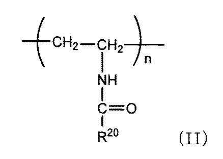
(R20は、水素原子、又はC1−4アルキル基を表し、nは1以上の整数を表す。)
で示される。
ポリ(N−ビニルアルキルアミド)の一例として、ポリ(N−ビニルアセトアミド)、ポリ(N−ビニルイソプロピルアミド)、ポリ(N−ビニルイソブチルアミド)などが挙げられる。
ポリ(N−ビニルアルキルアミド)は、次式II:
で示される。
ポリ(N−ビニルアルキルアミド)の一例として、ポリ(N−ビニルアセトアミド)、ポリ(N−ビニルイソプロピルアミド)、ポリ(N−ビニルイソブチルアミド)などが挙げられる。
(3)ポリビニルアルキルエーテル
ポリビニルアルキルエーテルは、次式III:

(R30は、水素原子、又はC1−4アルキル基を表し、nは1以上の整数を表す。)
で示される。
ポリビニルアルキルエーテルの一例として、ポリビニルメチルエーテル、ポリビニルエチルエーテルなどが挙げられる。
ポリビニルアルキルエーテルは、次式III:
で示される。
ポリビニルアルキルエーテルの一例として、ポリビニルメチルエーテル、ポリビニルエチルエーテルなどが挙げられる。
(4)分子設計
上記高分子において、末端のアルキル基を種々変換することで、相転移温度を制御し、あるいは相分離構造の安定性を制御することができる。例えばPNIPAMは、側鎖に親水基及び疎水基をともに有しており、直鎖ポリマーのときは32℃に下限臨界溶液温度を持つとともに水中では体積相転移温度(VPTT)を示す。PNIPAMは、水溶液温度がVPTT以下ではゲルは膨潤しているのに対し、VPTT以上ではゲルは収縮するという性質を持つことから、本発明のマクロポーラス構造体の製造に際し、この性質を利用することができる。
上記高分子において、末端のアルキル基を種々変換することで、相転移温度を制御し、あるいは相分離構造の安定性を制御することができる。例えばPNIPAMは、側鎖に親水基及び疎水基をともに有しており、直鎖ポリマーのときは32℃に下限臨界溶液温度を持つとともに水中では体積相転移温度(VPTT)を示す。PNIPAMは、水溶液温度がVPTT以下ではゲルは膨潤しているのに対し、VPTT以上ではゲルは収縮するという性質を持つことから、本発明のマクロポーラス構造体の製造に際し、この性質を利用することができる。
末端のアルキル基に官能基を付与することで、親水性高分子と連結することが可能となる。官能基としては、マレイミド、チオール、アルキン、アジド、アミン、活性カルボン酸などを含む。例えば、後述のように親水性高分子にチオール基(SH基)を付加しておくと、マレイミド基はSH基と反応し、安定なチオエーテル基を形成する。
2.2.親水性高分子
本発明において、親水性高分子は、ポリエチレングリコール、ポリエチレンイミン、ポリビニルアルコール、ポリアクリル酸、ポリアクリルアミド、ポリビニルアルコール、ポリビニルピロリドン、ポリビニルアセトアミド、ポリアミン、ポリ(4−スチレンスルホン酸)、ポリ(アリルアミン塩酸塩)、ポリ(ビニルスルホン酸、ナトリウム塩)、ポリ(ジアリルジメチルアンモニウムクロリド)、ポリ(2−メタクリロイルオキシエチルホスホリルコリン)などを、単独で又は2種以上を組み合わせて使用される。
本発明において、親水性高分子は、ポリエチレングリコール、ポリエチレンイミン、ポリビニルアルコール、ポリアクリル酸、ポリアクリルアミド、ポリビニルアルコール、ポリビニルピロリドン、ポリビニルアセトアミド、ポリアミン、ポリ(4−スチレンスルホン酸)、ポリ(アリルアミン塩酸塩)、ポリ(ビニルスルホン酸、ナトリウム塩)、ポリ(ジアリルジメチルアンモニウムクロリド)、ポリ(2−メタクリロイルオキシエチルホスホリルコリン)などを、単独で又は2種以上を組み合わせて使用される。
本発明の好ましい態様において、親水性高分子はポリエチレングリコール(PEG)である。
PEGは、直鎖型でも多分岐型でもよく、多分岐型の分岐数は特に限定されるものではなく、2分岐型、3分岐型、4分岐型、5分岐型、6分岐型、7分岐型、8分岐型などを含む。また、PEGはスターポリマーやデンドリマーとすることもできる。PEGには両端に又は一端に官能基を付加しておくことが好ましい。官能基としては、チオール、アクリレート、アミン、ビオチン、アルデヒド、マレイミド、スクシンイミジルカルボキシメチルエステル、スクシンイミジルグルタレートエステル等が挙げられるが、これらに限定されるものではない。
PEGは、直鎖型でも多分岐型でもよく、多分岐型の分岐数は特に限定されるものではなく、2分岐型、3分岐型、4分岐型、5分岐型、6分岐型、7分岐型、8分岐型などを含む。また、PEGはスターポリマーやデンドリマーとすることもできる。PEGには両端に又は一端に官能基を付加しておくことが好ましい。官能基としては、チオール、アクリレート、アミン、ビオチン、アルデヒド、マレイミド、スクシンイミジルカルボキシメチルエステル、スクシンイミジルグルタレートエステル等が挙げられるが、これらに限定されるものではない。
多分岐型PEGは、例えば、次式IV:

で示される構造を表す。
式IVにおいて、R1は、次式:

(Xは官能基を表す。)で示される。nは、目的に応じて任意に設定することができ、例えばnは25~1000である。
R2、R3及びR4は、それぞれ独立して、水素原子、又は次式:

(Xは官能基を表す。)で示される。
R2、R3及びR4は、それぞれ独立して、水素原子、又は次式:
Xは官能基を表し、その種類は前記と同様、例えばチオール、アクリレート、アミン、ビオチン、アルデヒド、マレイミド、スクシンイミジルカルボキシメチルエステル、スクシンイミジルグルタレートエステル等が挙げられるが、これらに限定されるものではない。
また、官能基はそれぞれの分岐鎖において独立しており、同一であっても異なってもよい。また、R1、R2、R3及びR4におけるnも、それぞれの分岐鎖において独立しており、同一数であっても異なる数であってもよい。
また、官能基はそれぞれの分岐鎖において独立しており、同一であっても異なってもよい。また、R1、R2、R3及びR4におけるnも、それぞれの分岐鎖において独立しており、同一数であっても異なる数であってもよい。
PEGが2分岐型から8分岐型の場合、mは、1~4の整数を表す。
mが1であり、かつR3及びR4がいずれも水素原子のときは、PEGは2分岐型となり、mが1であり、R3及びR4のいずれか一方が水素原子のときは、PEGは3分岐型となり、mが1であり、かつR2、R3及びR4がいずれも水素原子でないときは、PEGは4分岐型となる。さらに、例えばmが4であってR2及びR3がいずれも水素原子ではなくR4が水素原子であるときは、PEGは8分岐型となる。
本発明において使用する多分岐型PEGは4分岐型又は8分岐型であることが好ましい。
mが1であり、かつR3及びR4がいずれも水素原子のときは、PEGは2分岐型となり、mが1であり、R3及びR4のいずれか一方が水素原子のときは、PEGは3分岐型となり、mが1であり、かつR2、R3及びR4がいずれも水素原子でないときは、PEGは4分岐型となる。さらに、例えばmが4であってR2及びR3がいずれも水素原子ではなくR4が水素原子であるときは、PEGは8分岐型となる。
本発明において使用する多分岐型PEGは4分岐型又は8分岐型であることが好ましい。
4分岐型及び8分岐型のPEGの一例を以下に示す。

8分岐型:
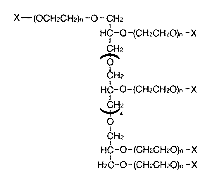
本発明において、上記多分岐型PEGは、公知方法により得ることができるが、また市販品(日油)として購入することも可能である。
スターポリマーは、中心より3本以上のポリマーが放射状に分岐した高分子であり、分岐点が一箇所に集中している。スターポリマーの合成法は当分野において周知であり、例えばコアを開始点としてモノマーを連鎖的に重合させる方法、アーム部分を先に合成して、アームの末端とコアの官能基部分を結合させる方法、リビング的な連鎖重合により直鎖状高分子を合成し、架橋剤を用いてコア部位を架橋させる方法などが採用される。
デンドリマーは、中心から規則的に分枝した構造を持つ樹状高分子である。デンドリマーの合成法には、コアとなる分子にジェネレーションごとに分子を結合させ枝分かれさせていくダイバージェント法、及び予め枝の部分を合成して最後にコア分子と反応させるコンバージェント法の2つがある。市販の多官能性PEG(Merck社)を使用することもできる。
さらに、直鎖型では、PEGの一端又は両端に官能基を付加して、LCSTを有する高分子を連結させることができ、分岐型では、分岐したPEGの末端に官能基を付加して、LCSTを有する高分子を連結させることができる。
官能基としては、例えばチオール、アクリレート、アミン、ビオチン、アルデヒド、マレイミド、スクシンイミジルカルボキシメチルエステル、スクシンイミジルグルタレートエステル等が挙げられるが、これらに限定されるものではない。
官能基としては、例えばチオール、アクリレート、アミン、ビオチン、アルデヒド、マレイミド、スクシンイミジルカルボキシメチルエステル、スクシンイミジルグルタレートエステル等が挙げられるが、これらに限定されるものではない。
2.3.ブロック共重合体
本発明において、LCSTを有する高分子と親水性高分子とのブロック共重合体は、それぞれの高分子に官能基を付加しておいて、官能基同士を反応させることにより、合成することができる。
例えば、マレイミド基はチオール基(SH基)と反応して安定なチオエーテル基を形成するため、親水性高分子にSH基、LCSTを有する高分子にマレイミド基を付加して両者を反応させればよい。図2は、親水性高分子としてSH基を付加した4分岐型のPEGと、LCSTを有する高分子としてPNIPAMとのブロック共重合体を合成する合成例の模式図である。
本発明において、LCSTを有する高分子と親水性高分子とのブロック共重合体は、それぞれの高分子に官能基を付加しておいて、官能基同士を反応させることにより、合成することができる。
例えば、マレイミド基はチオール基(SH基)と反応して安定なチオエーテル基を形成するため、親水性高分子にSH基、LCSTを有する高分子にマレイミド基を付加して両者を反応させればよい。図2は、親水性高分子としてSH基を付加した4分岐型のPEGと、LCSTを有する高分子としてPNIPAMとのブロック共重合体を合成する合成例の模式図である。
なお、本発明においては、ブロック共重合体を蛍光物質で標識しておいてもよい。蛍光物質は、特に限定されるものではなく、例えばフルオレセイン、ローダミン、Cy、Alexa FluorTM、HiLyte FluorTM等の化学合成物質、あるいはフィコエリスリン(PE)、アロフィコシアニン(APC)等の蛍光タンパク質などが挙げられる。これらの蛍光物質は、市販品として入手可能である。蛍光物質でブロック共重合体を標識することにより、その後のステップにおいて、混合物から相分離構造が形成されたか否か、あるいはブロック共重合体を除去できたか否かの確認をすることができる。
3.多糖類
本発明において使用される多糖類は、アルギン酸、デンプン、グリコーゲン、セルロース、キサンタンガム、ヒアルロン酸、カラギーナン、ペクチン及びプルランが例示され、1種を単独で、又は2種以上を適宜組み合わせて使用できる。また本発明においては、これらの多糖類の塩も含む。
塩としては、ナトリウム塩、カリウム塩、カルシウム塩、バリウム塩などを含む。
本発明において使用される多糖類は、アルギン酸、デンプン、グリコーゲン、セルロース、キサンタンガム、ヒアルロン酸、カラギーナン、ペクチン及びプルランが例示され、1種を単独で、又は2種以上を適宜組み合わせて使用できる。また本発明においては、これらの多糖類の塩も含む。
塩としては、ナトリウム塩、カリウム塩、カルシウム塩、バリウム塩などを含む。
本発明の一態様において、使用される多糖類は、好ましくはアルギン酸である。アルギン酸は、天然由来でも合成物であってもよい。好ましく用いられるアルギン酸類は、カジカ、アラメ、コンブなどの褐藻類から抽出される生体内吸収性の多糖類であり、D−マンヌロン酸とL−グルロン酸という2種類のウロン酸が直鎖状に重合したポリマーである。より具体的には、D−マンヌロン酸のホモポリマー画分、L−グルロン酸のホモポリマー画分、及びD−マンヌロン酸とL−グルロン酸がランダムに配列した画分が任意に結合したブロック共重合体である。アルギン酸は、市販品として入手することもできる(持田製薬)。
4.マクロポーラス構造体の製造
本発明の一態様においては、多糖類と、LCSTを有する高分子と親水性高分子とのブロック共重合体とを混合して相分離を起こさせ、当該相分離後の混合物から前記ブロック共重合体を除去する工程を含む、マクロポーラス構造体の製造方法が提供される。
本発明の一態様においては、多糖類と、LCSTを有する高分子と親水性高分子とのブロック共重合体とを混合して相分離を起こさせ、当該相分離後の混合物から前記ブロック共重合体を除去する工程を含む、マクロポーラス構造体の製造方法が提供される。
前記の通り、LCSTを有する高分子と親水性高分子とのブロック共重合体は、LCST未満では当該高分子が水に溶解する。その状態で多糖類(アルギン酸等)と混合することにより、自発的に相分離構造を形成する。LCST未満に下げるとブロック共重合体は水に溶解するため、単に水中で攪拌、震盪又は静置することにより、ブロック共重合体を除去することができる。
ここで、本発明において多糖類とブロック共重合体との混合比(多糖類:ブロック共重合体)は、本発明において多糖類とブロック共重合体との混合比(多糖類:ブロック共重合体)は、9:1から1:9の間で様々な比をとることができ、一つの態様では例えば6:4~8:2であり、別の態様では例えば7:3~8:2である。
本発明においては、多糖類をゲル化して流動性を落とすことにより、マクロポーラス構造を保持することができる。この場合は、LCST未満でブロック共重合体と多糖類とを混合し、混合物をLCST以上に上げて構造をゲル化により固定化した後、再度LCST未満に下げればよい。ゲル化は、例えば多糖類をカルシウムイオン含有溶液(例えば塩化カルシウム水溶液)で処理すること、具体的にはブロック共重合体と多糖類との混合物に塩化カルシウム水溶液を混合することにより、行うことができる。
本発明においては、多糖類をゲル化して流動性を落とすことにより、マクロポーラス構造を保持することができる。この場合は、LCST未満でブロック共重合体と多糖類とを混合し、混合物をLCST以上に上げて構造をゲル化により固定化した後、再度LCST未満に下げればよい。ゲル化は、例えば多糖類をカルシウムイオン含有溶液(例えば塩化カルシウム水溶液)で処理すること、具体的にはブロック共重合体と多糖類との混合物に塩化カルシウム水溶液を混合することにより、行うことができる。
他方、LCSTを有する高分子と親水性高分子とのブロック共重合体において、LCST以上では当該高分子が水に不溶となり、エマルジョンを形成する。その状態で多糖類(アルギン酸等)と混合することによっても相分離構造を形成する。その後多糖類をゲル化した後にLCST未満に下げるとブロック共重合体は水に溶解するため、単に水中で攪拌、震盪又は静置することにより、ブロック共重合体を除去することができる。
さらに、本発明においては、混合物に細胞を含有させることができる。この場合、混合物の相分離を起こさせる前に含有させておく。そして、多糖類を必要によりゲル化した後、混合物をLCST以下に設定して水中で攪拌等を行うことでブロック共重合体を除去する。これにより、細胞含有のマクロポーラス構造体を得ることができる。
細胞を含有するマクロポーラス構造体は、ウイルスや細菌の捕獲用ツール、抗体産生ハイブリドーマを導入した抗体製造ツール等に利用される。ゲルは所定の大きさの孔径を有するため、半透膜としての利用が可能である。その場合は、透析や血液浄化に使用することができる。
マクロポーラス構造体に細胞等の生体関連物質を導入する場合は、4℃~37℃の温度範囲に調節することが必要であるが、細胞以外他の物質を導入することも可能である。例えば酵母を導入する場合は、0℃から最大100℃程度までの温度範囲内で行うことができる。この場合は、LCSTを有する高分子において、置換基の種類を適宜変えて高温のLCSTを有する高分子を使用すればよい。
細胞としては特に限定されるものではなく、万能性を有するES細胞、iPS細胞、内分泌細胞、外分泌細胞、幹細胞(造血幹細胞、神経幹細胞、間葉系幹細胞)、各種組織における細胞(例えば骨格筋細胞、心筋細胞、神経系細胞、線維芽細胞、内皮細胞、上皮細胞、肝細胞、膵細胞、皮膚細胞、血液細胞、間質細胞等)などが挙げられる。
これらの細胞の具体例として、以下の細胞を例示することができる。
内分泌細胞:神経分泌細胞、下垂体細胞、甲状腺細胞、副甲状腺細胞、膵島細胞、胃腸内分泌細胞、心筋細胞、肝細胞、腎臓細胞、脂肪細胞、副腎細胞、性腺細胞等
外分泌細胞:消化管上皮細胞、生理活性物質を放出する細胞(アルブミンを放出する肝細胞、酵素を放出する酵素産生細胞等)
神経系細胞:神経幹細胞、中枢神経細胞、末梢神経細胞、グリア細胞等
骨格筋系細胞:骨細胞、軟骨細胞等
血液細胞:造血幹細胞、白血球、赤血球、血小板等
間質細胞:繊維芽細胞、血管細胞等
内分泌細胞:神経分泌細胞、下垂体細胞、甲状腺細胞、副甲状腺細胞、膵島細胞、胃腸内分泌細胞、心筋細胞、肝細胞、腎臓細胞、脂肪細胞、副腎細胞、性腺細胞等
外分泌細胞:消化管上皮細胞、生理活性物質を放出する細胞(アルブミンを放出する肝細胞、酵素を放出する酵素産生細胞等)
神経系細胞:神経幹細胞、中枢神経細胞、末梢神経細胞、グリア細胞等
骨格筋系細胞:骨細胞、軟骨細胞等
血液細胞:造血幹細胞、白血球、赤血球、血小板等
間質細胞:繊維芽細胞、血管細胞等
また、血液に含まれる特定物質(例えばウイルスやサイトカイン)と親和性を有する膜タンパク質を発現した親和性細胞を用いることができる。
さらに、細胞の他にも、タンパク質、ペプチド、抗体(抗体断片を含む)、脂質、糖類、核酸類などの生体関連物質を混合物に導入することが可能であり、生体関連物質の種類に限定はない。
さらに、細胞の他にも、タンパク質、ペプチド、抗体(抗体断片を含む)、脂質、糖類、核酸類などの生体関連物質を混合物に導入することが可能であり、生体関連物質の種類に限定はない。
5.ファイバー状構造体
本発明においては、上記マクロポーラス構造体の製造法を応用して、ファイバー状の構造体を製造する方法を提供する。
ファイバー状構造体を製造するには、二重の同軸マイクロ流体装置(WO2011/046105号)を用いる方法を採用することができる。同軸マイクロ流体装置は、2つの流体を同軸となるようにコア部及びシェル部に分けて射出する構成となっている。
本発明においては、上記マクロポーラス構造体の製造法を応用して、ファイバー状の構造体を製造する方法を提供する。
ファイバー状構造体を製造するには、二重の同軸マイクロ流体装置(WO2011/046105号)を用いる方法を採用することができる。同軸マイクロ流体装置は、2つの流体を同軸となるようにコア部及びシェル部に分けて射出する構成となっている。
同軸マイクロ流体装置装置の一例を図3に示す。図3aにおいて、同軸マイクロ流体装置10は、細胞懸濁液射出用チューブ101、多糖類とブロック共重合体との混合物射出用チューブ102、カルシウムイオン含有水溶液射出用チューブ103を備える。なお、多糖類とブロック共重合体との混合物を、単に「混合物」ともいう。細胞懸濁液射出用チューブ101の上部は、細胞懸濁液を注入するための注入口111が設けられ、上部から細胞懸濁液121が注入される。細胞懸濁液射出用チューブ101及び混合物射出用チューブ102は下部がテーパ状の先細り形状に形成されており、細胞懸濁液射出用チューブ101と混合物射出用チューブ102との間には混合物射出用の開口部112が設けられる。また、混合物射出用チューブ102とカルシウムイオン含有水溶液射出用チューブ103との間にはカルシウムイオン含有水溶液射出用の開口部113が設けられる。
細胞懸濁液121は、細胞懸濁液射出用チューブ101の下部の射出口から混合物射出用チューブ102に向けて射出され、ファイバーのコア部を構成する。開口部112から混合物射出用チューブ102に向けて混合物122を射出すると、ファイバーのコア部を細胞懸濁液が占め、混合物が細胞周囲を覆ってシェル部を形成し、同軸のコア・シェル状の流体を形成する。その流体をカルシウムイオン含有水溶液射出用チューブ103に向けて射出するとともに、開口部113からカルシウムイオン含有水溶液(例えばCaCl2含有水溶液)を射出すると、流体のシェル部がゲル化される。その結果、コア部が細胞で占められ、シェル部がゲル化されたファイバー123を得ることができる。ゲル化されたファイバー123の一部拡大図を図3bに示す。この段階では、ファイバー123のシェル部にはブロック共重合体が含まれるため、これをLCST以下の温度に調節することで、ブロック共重合体は水溶性となり、ファイバーを水中で攪拌することにより当該ブロック共重合体を除去することができる。これにより、コア部が細胞、シェル部がマクロポーラス状(多孔質)となったファイバー状のマクロポーラス構造体(以下、単に「マクロポーラス構造体」という)130を得ることができる(図3c)。
細胞懸濁液及び混合物のそれぞれの射出速度は限定されるものではないが、孔径が50μm~2mm程度のサイズの同軸マイクロ流体装置を有する場合は、射出口における速度は例えば10~500μL/分である。また、カルシウムイオン含有水溶液への導入速度も特に限定されるものではなく、例えば1~10mL/分である。
マクロポーラス構造体130において、コア及びシェルの孔径は任意であり、目的に応じて適宜調節することができる。例えば、マクロポーラス構造体130のシェル部の外径は10μm~2mm、200μm~2mm、50μm~1mm程度とすることができる。マクロポーラス構造体130の長さは特に限定されず、数mm~数m程度とすることができる。その断面形状としては、円形、楕円系、四角形や五角形等の多角形等が挙げられる。
上記のようにして得られたマクロポーラス構造体は、孔径を自由に変えることができるため細胞が通り易く、細胞との親和性が高い。従って、本発明の構造体に含まれる細胞は多孔質の中に入り込んで細胞が凝集し、スフェロイド等の細胞構造体が形成される。
マクロポーラス構造体130を培養液中で培養することにより、マクロポーラス構造体130に内包された細胞を増殖させることができる。マクロポーラス構造体130は、培養液を適切に交換することにより、数か月培養することもできる。
また、マクロポーラス構造体130に含まれる細胞の種類によっては、分化誘導培地に置換して培養することにより、細胞が分化してマクロポーラス構造体130の外側に分化後の細胞又は組織の一部が形成される。
このようにして細胞が内包されたマクロポーラス構造体130は、細胞の種類によって、各種再生医療材として使用することができる。例えば、マクロポーラス構造体130中に神経幹細胞を未分化の状態で内包させた後、所定期間培養し、その後細胞増殖因子のFGFやEGFを除去した分化誘導培地に置換して培養すると、未分化の神経幹細胞は神経細胞に分化し、マクロポーラス構造体130の外に組織の一部が伸長する。このようにして形成されたマクロポーラス構造体130は、神経再生医療材として使用することができる。
このようにして細胞が内包されたマクロポーラス構造体130は、細胞の種類によって、各種再生医療材として使用することができる。例えば、マクロポーラス構造体130中に神経幹細胞を未分化の状態で内包させた後、所定期間培養し、その後細胞増殖因子のFGFやEGFを除去した分化誘導培地に置換して培養すると、未分化の神経幹細胞は神経細胞に分化し、マクロポーラス構造体130の外に組織の一部が伸長する。このようにして形成されたマクロポーラス構造体130は、神経再生医療材として使用することができる。
あるいは、マクロポーラス構造体130に、ウイルスを捕捉する細胞等を内包すると、当該マクロポーラス構造体130はウイルスに感染した生体関連物質(例えばウイルス感染血液)を浄化するのに有用である。
例えば、新型コロナウイルス(SARS−CoV−2)は、その表面のスパイクタンパク質(Sタンパク質)が細胞表面のアンジオテンシン変換酵素2(ACE2)をレセプターとして結合して感染する。そこで、レンチウイルスベクターを用いた遺伝子導入による各種ACE2発現細胞(VeroE6細胞、VeroE6/TMPRSS細胞、Hela細胞、Hela/ACE2細胞)を作製し、この細胞をマクロポーラス構造体130に内包させる。
例えば、新型コロナウイルス(SARS−CoV−2)は、その表面のスパイクタンパク質(Sタンパク質)が細胞表面のアンジオテンシン変換酵素2(ACE2)をレセプターとして結合して感染する。そこで、レンチウイルスベクターを用いた遺伝子導入による各種ACE2発現細胞(VeroE6細胞、VeroE6/TMPRSS細胞、Hela細胞、Hela/ACE2細胞)を作製し、この細胞をマクロポーラス構造体130に内包させる。
これにより、マクロポーラス構造体130に内包された細胞はACE2を発現するため、ACE2発現細胞を内包したマクロポーラス構造体130は、ウイルス捕捉材として、ウイルスに感染した血液の浄化等に使用することができる。
なお、SARS−CoV−2の細胞感染機構を模倣することができるSタンパク質を表面に有するビーズ(Sタンパク質ビーズ)を作製すると、当該ビーズは、SARS−CoV−2の細胞感染モデルとして使用することができる。
なお、SARS−CoV−2の細胞感染機構を模倣することができるSタンパク質を表面に有するビーズ(Sタンパク質ビーズ)を作製すると、当該ビーズは、SARS−CoV−2の細胞感染モデルとして使用することができる。
さらに、アデノ随伴ウイルス(AAV)は、遺伝子導入ウイルスとして知られているため、AAVに目的遺伝子を導入し、当該AAVを感染させた細胞をマクロポーラス構造体130に注入することで、遺伝子治療用マクロポーラス構造体130ができる。
実施例
以下、実施例により本発明をさらに具体的に説明する。但し、本発明の範囲はこれらの実施例により限定されるものではない。
[実施例1]
以下、実施例により本発明をさらに具体的に説明する。但し、本発明の範囲はこれらの実施例により限定されるものではない。
[実施例1]
・材料
アルギン酸ナトリウム(Pronova UP MVG alginate,Nova Matrix)、tetra PEG−SH(SUNBRIGHT PTE−50SH,PTE−100SH,PTE−200SH,油化産業株式会社)、Alexa FluorTM 488 C5 Maleimide(A10254,Thermo Fischer)、ポリ(N−イソプロピルアクリルアミド)マレイミド末端(731048,728632,Merck)は市販品を購入して実験に使用した。
アルギン酸ナトリウム(Pronova UP MVG alginate,Nova Matrix)、tetra PEG−SH(SUNBRIGHT PTE−50SH,PTE−100SH,PTE−200SH,油化産業株式会社)、Alexa FluorTM 488 C5 Maleimide(A10254,Thermo Fischer)、ポリ(N−イソプロピルアクリルアミド)マレイミド末端(731048,728632,Merck)は市販品を購入して実験に使用した。
・PEG−b−PNIPAM(10000−5500)の合成法
tetra−PEGの4官能基末端にPNIPAMをthio−ene反応によって導入して、PEG−b−PNIPAMを合成した。合成法としては、分子量10000のtetra PEG−SH(2g,200μmol,)と分子量5500のPNIPAM−MA(4.4g,800μmol)を4℃に冷やしたPBS 64mLに溶解させ、4℃下で終夜撹拌し、PEG−b−PNIPAM溶液を調製した。さらに、蛍光顕微鏡での観察を可能にするためにPEG−b−PNIPAMの4官能基末端のうちの一つをAlexa Fluor 488−MAを導入したPEG−b−PNIPAM−Alexa Fluor 488も合成した。合成法としては、tetra PEG−SH(15.26mg)/PBS 100μLとAlexa Fluor488−MA(1mg)/PBS 100μLを混合し、4℃下で1時間で撹拌する。その後、4℃に冷却しPNIPAM−MA(25.81mg)/PBS 220μLを添加し、4℃下で終夜撹拌しPEG−b−PNIPAM−Alexa Fluor 488溶液を調製した。
tetra−PEGの4官能基末端にPNIPAMをthio−ene反応によって導入して、PEG−b−PNIPAMを合成した。合成法としては、分子量10000のtetra PEG−SH(2g,200μmol,)と分子量5500のPNIPAM−MA(4.4g,800μmol)を4℃に冷やしたPBS 64mLに溶解させ、4℃下で終夜撹拌し、PEG−b−PNIPAM溶液を調製した。さらに、蛍光顕微鏡での観察を可能にするためにPEG−b−PNIPAMの4官能基末端のうちの一つをAlexa Fluor 488−MAを導入したPEG−b−PNIPAM−Alexa Fluor 488も合成した。合成法としては、tetra PEG−SH(15.26mg)/PBS 100μLとAlexa Fluor488−MA(1mg)/PBS 100μLを混合し、4℃下で1時間で撹拌する。その後、4℃に冷却しPNIPAM−MA(25.81mg)/PBS 220μLを添加し、4℃下で終夜撹拌しPEG−b−PNIPAM−Alexa Fluor 488溶液を調製した。
・PEG−b−PNIPAM(10000−2000)の合成法
tetra−PEGの4官能基末端にPNIPAMをthio−ene反応によって導入して、PEG−b−PNIPAMを合成した。合成法としては、分子量20000のtetra PEG−SH(500mg,50μmol)と分子量2000のPNIPAM−MA(400mg,200μmol)を4℃に冷やしたPBS 16mLに溶解させ、4℃下で終夜撹拌し、PEG−b−PNIPAM溶液を調製した。さらに、蛍光顕微鏡での観察を可能にするためにPEG−b−PNIPAMの4官能基末端のうちの一つをAlexa Fluor 488−MAを導入したPEG−b−PNIPAM−Alexa Fluor 488も合成した。合成法としては、tetra PEG−SH(15.26mg)/PBS 100μLとAlexa Fluor488−MA(1mg)/PBS 100μLを混合し、4℃下で1時間で撹拌する。その後、4℃に冷却しPNIPAM−MA(9.38mg)/PBS 220μLを添加し、4℃下で終夜撹拌しPEG−b−PNIPAM−Alexa Fluor 488溶液を調製した。
tetra−PEGの4官能基末端にPNIPAMをthio−ene反応によって導入して、PEG−b−PNIPAMを合成した。合成法としては、分子量20000のtetra PEG−SH(500mg,50μmol)と分子量2000のPNIPAM−MA(400mg,200μmol)を4℃に冷やしたPBS 16mLに溶解させ、4℃下で終夜撹拌し、PEG−b−PNIPAM溶液を調製した。さらに、蛍光顕微鏡での観察を可能にするためにPEG−b−PNIPAMの4官能基末端のうちの一つをAlexa Fluor 488−MAを導入したPEG−b−PNIPAM−Alexa Fluor 488も合成した。合成法としては、tetra PEG−SH(15.26mg)/PBS 100μLとAlexa Fluor488−MA(1mg)/PBS 100μLを混合し、4℃下で1時間で撹拌する。その後、4℃に冷却しPNIPAM−MA(9.38mg)/PBS 220μLを添加し、4℃下で終夜撹拌しPEG−b−PNIPAM−Alexa Fluor 488溶液を調製した。
・PEG−b−PNIPAM(20000−5500)の合成法
tetra−PEGの4官能基末端にPNIPAMをthio−ene反応によって導入して、PEG−b−PNIPAMを合成した。合成法としては、分子量20000のtetra PEG−SH(500mg,25μmol)と分子量5500のPNIPAM−MA(550mg,100μmol)を4℃に冷やしたPBS 16mLに溶解させ、4℃下で終夜撹拌し、PEG−b−PNIPAM溶液を調製した。さらに、蛍光顕微鏡での観察を可能にするためにPEG−b−PNIPAMの4官能基末端のうちの一つをAlexa Fluor 488−MAを導入したPEG−b−PNIPAM−Alexa Fluor 488も合成した。合成法としては、tetra PEG−SH(15.26mg)/PBS 100μLとAlexa Fluor488−MA(0.5mg)/PBS 100μLを混合し、4℃下で1時間で撹拌する。その後、4℃に冷却しPNIPAM−MA(12.90mg)/PBS 220μLを添加し、4℃下で終夜撹拌しPEG−b−PNIPAM−Alexa Fluor 488溶液を調製した。
tetra−PEGの4官能基末端にPNIPAMをthio−ene反応によって導入して、PEG−b−PNIPAMを合成した。合成法としては、分子量20000のtetra PEG−SH(500mg,25μmol)と分子量5500のPNIPAM−MA(550mg,100μmol)を4℃に冷やしたPBS 16mLに溶解させ、4℃下で終夜撹拌し、PEG−b−PNIPAM溶液を調製した。さらに、蛍光顕微鏡での観察を可能にするためにPEG−b−PNIPAMの4官能基末端のうちの一つをAlexa Fluor 488−MAを導入したPEG−b−PNIPAM−Alexa Fluor 488も合成した。合成法としては、tetra PEG−SH(15.26mg)/PBS 100μLとAlexa Fluor488−MA(0.5mg)/PBS 100μLを混合し、4℃下で1時間で撹拌する。その後、4℃に冷却しPNIPAM−MA(12.90mg)/PBS 220μLを添加し、4℃下で終夜撹拌しPEG−b−PNIPAM−Alexa Fluor 488溶液を調製した。
・PEG−b−PNIPAM(20000−2000)の合成法
tetra−PEGの4官能基末端にPNIPAMをthio−ene反応によって導入して、PEG−b−PNIPAMを合成した。合成法としては、分子量20000のtetra PEG−SH(500mg,25μmol)と分子量2000のPNIPAM−MA(200mg,100μmol)を4℃に冷やしたPBS 16mLに溶解させ、4℃下で終夜撹拌し、PEG−b−PNIPAM溶液を調製した。さらに、蛍光顕微鏡での観察を可能にするためにPEG−b−PNTPAMの4官能基末端のうちの一つをAlexa Fluor 488−MAを導入したPEG−b−PNIPAM−Alexa Fluor 488も合成した。合成法としては、tetra PEG−SH(15.26mg)/PBS 100μLとAlexa Fluor488−MA(0.5mg)/PBS 100μLを混合し、4℃下で1時間で撹拌する。その後、4℃に冷却しPNIPAM−MA(4.69mg)/PBS 220μLを添加し、4℃下で終夜撹拌しPEG−b−PNIPAM−Alexa Fluor 488溶液を調製した。
tetra−PEGの4官能基末端にPNIPAMをthio−ene反応によって導入して、PEG−b−PNIPAMを合成した。合成法としては、分子量20000のtetra PEG−SH(500mg,25μmol)と分子量2000のPNIPAM−MA(200mg,100μmol)を4℃に冷やしたPBS 16mLに溶解させ、4℃下で終夜撹拌し、PEG−b−PNIPAM溶液を調製した。さらに、蛍光顕微鏡での観察を可能にするためにPEG−b−PNTPAMの4官能基末端のうちの一つをAlexa Fluor 488−MAを導入したPEG−b−PNIPAM−Alexa Fluor 488も合成した。合成法としては、tetra PEG−SH(15.26mg)/PBS 100μLとAlexa Fluor488−MA(0.5mg)/PBS 100μLを混合し、4℃下で1時間で撹拌する。その後、4℃に冷却しPNIPAM−MA(4.69mg)/PBS 220μLを添加し、4℃下で終夜撹拌しPEG−b−PNIPAM−Alexa Fluor 488溶液を調製した。
・結果
PEG−b−PNIPAM溶液2.4mlとPEG−b−PNIPAM−Alexa Fluor 488溶液0.1μlを混合し、蛍光ブロックコポリマー溶液を調製した。アルギン酸ナトリウムは生理食塩水に溶解させ、1.8%のアルギン酸ナトリウム溶液を調製した。アルギン酸ナトリウム溶液と蛍光ブロックコポリマー溶液を6.5:3.5(図4)、7:3(図5)、7.5:2.5(図6)、8:2(図7)の割合で混合し、厚さ100μmのセルに移し、4℃下で0、1、2、3、4、5時間後の共焦点顕微鏡観察を実施した(緑色:蛍光ブロックコポリマー、黒色:アルギン酸ナトリウム)。7.5:2.5、8:2ではいずれも、相分離した網目構造が観察され、zスタック画像より相分離構造は貫通構造であることが分かった。また、アルギン酸ナトリウム溶液の割合が増えるほど、網目構造が小さくなることも確認された。
アルギン酸ナトリウムとブロックコポリマーとのとの組み合わせ及び及び共焦点顕微鏡観察像のまとめを図8に示す。
PEG−b−PNIPAM溶液2.4mlとPEG−b−PNIPAM−Alexa Fluor 488溶液0.1μlを混合し、蛍光ブロックコポリマー溶液を調製した。アルギン酸ナトリウムは生理食塩水に溶解させ、1.8%のアルギン酸ナトリウム溶液を調製した。アルギン酸ナトリウム溶液と蛍光ブロックコポリマー溶液を6.5:3.5(図4)、7:3(図5)、7.5:2.5(図6)、8:2(図7)の割合で混合し、厚さ100μmのセルに移し、4℃下で0、1、2、3、4、5時間後の共焦点顕微鏡観察を実施した(緑色:蛍光ブロックコポリマー、黒色:アルギン酸ナトリウム)。7.5:2.5、8:2ではいずれも、相分離した網目構造が観察され、zスタック画像より相分離構造は貫通構造であることが分かった。また、アルギン酸ナトリウム溶液の割合が増えるほど、網目構造が小さくなることも確認された。
アルギン酸ナトリウムとブロックコポリマーとのとの組み合わせ及び及び共焦点顕微鏡観察像のまとめを図8に示す。
・Alginate+PEG−b−PNIPAM(マクロポーラスアルギン酸ゲル)(合成法又は取得法)
マクロポーラスアルギン酸ゲルの調製方法としては、4℃に冷却した1.8% アルギン酸ナトリウム溶液と10% PEG−b−PNIPAM溶液を混合し、1日静置し相分離させた。混合溶液を37℃の恒温槽内に移し、1時間静置する。温度が下がらないように気をつけながら、混合溶液を37℃の塩化カルシウム溶液中に滴下し、白濁したアルギン酸をゲルを作製した。その後、4℃に冷却し撹拌する事で、PEG−b−PNIPAMを除去し、マクロポーラスのアルギン酸ゲルを作製した。
マクロポーラスアルギン酸ゲルの調製方法としては、4℃に冷却した1.8% アルギン酸ナトリウム溶液と10% PEG−b−PNIPAM溶液を混合し、1日静置し相分離させた。混合溶液を37℃の恒温槽内に移し、1時間静置する。温度が下がらないように気をつけながら、混合溶液を37℃の塩化カルシウム溶液中に滴下し、白濁したアルギン酸をゲルを作製した。その後、4℃に冷却し撹拌する事で、PEG−b−PNIPAMを除去し、マクロポーラスのアルギン酸ゲルを作製した。
・Alginate/PEG−b−PNIPAM core/shell fiberの製造法、測定法、特徴
内包したい細胞懸濁液、蛍光ビーズなどを含む温度37℃のコア部分を流速25μL/min、多糖類とブロック共重合体との混合物を含む温度37℃のシェル部分を流速75μL/min、カルシウムイオンを含有する水溶液を温度37℃のシース部分を流速3600μL/minで送液することで、多糖類とブロック共重合体との混合物をシェルとするファイバー構造を作製した。その後、4℃に冷却し撹拌する事で、PEG−b−PNIPAMを除去し、マクロポーラスのアルギン酸core/shell fiberを作製した。直径100nmの蛍光ビーズを内包した際は、4℃に冷却前にはファイバー内に蛍光が確認されたが、4℃に冷却撹拌した後は完全に抜けていることが確認された(図9、10)。
内包したい細胞懸濁液、蛍光ビーズなどを含む温度37℃のコア部分を流速25μL/min、多糖類とブロック共重合体との混合物を含む温度37℃のシェル部分を流速75μL/min、カルシウムイオンを含有する水溶液を温度37℃のシース部分を流速3600μL/minで送液することで、多糖類とブロック共重合体との混合物をシェルとするファイバー構造を作製した。その後、4℃に冷却し撹拌する事で、PEG−b−PNIPAMを除去し、マクロポーラスのアルギン酸core/shell fiberを作製した。直径100nmの蛍光ビーズを内包した際は、4℃に冷却前にはファイバー内に蛍光が確認されたが、4℃に冷却撹拌した後は完全に抜けていることが確認された(図9、10)。
・mNSCsの細胞凝集体の作製方法
ICRマウスの中脳(胎生日数:13.5日)から取得した神経幹細胞(mNSCs)を、2mMのL−グルタミン(25030−081,Gibco,Life Technologies)、1%ペニシリン−ストレプトマイシン溶液(P4333,Sigma−Aldrich)、20ng/mL bFGF(AF−100−18B,Peprotech)、20ng mL/L hEGF(AF−100−15,Peprotech)、B−27TM Supplement(50X),minus vitamin A(12587010,ThermoFisher)を加えたNeurobasal−A(10888−022,Gibco,Life Technologies)増殖培地で培養した。mNSCsは、TrypLE Select(12563−011、Gibco,Life Technologies)を用いて2−3回継代してから使用した。mNSCs細胞凝集体は、細胞培養用10cm dishで培養し、回収した。
ICRマウスの中脳(胎生日数:13.5日)から取得した神経幹細胞(mNSCs)を、2mMのL−グルタミン(25030−081,Gibco,Life Technologies)、1%ペニシリン−ストレプトマイシン溶液(P4333,Sigma−Aldrich)、20ng/mL bFGF(AF−100−18B,Peprotech)、20ng mL/L hEGF(AF−100−15,Peprotech)、B−27TM Supplement(50X),minus vitamin A(12587010,ThermoFisher)を加えたNeurobasal−A(10888−022,Gibco,Life Technologies)増殖培地で培養した。mNSCsは、TrypLE Select(12563−011、Gibco,Life Technologies)を用いて2−3回継代してから使用した。mNSCs細胞凝集体は、細胞培養用10cm dishで培養し、回収した。
・mNSCsファイバーの製造法、培養法、測定法、特徴
mNSCsファイバーの製造法の概要を図11に示す。
4℃に冷却した1.8% アルギン酸ナトリウム溶液と10% PEG−b−PNIPAM溶液を7:3の割合で混合し10時間層分離した後、mNSCs細胞凝集体と混合し、1mlのガラスキャプラリーから37℃の100mM塩化カルシウム溶液中に100μL/minの速度で注入した。その後、得られたmNSCsゲルファイバーを、4℃に冷却したTrypLE Select内で撹拌する事で、PEG−b−PNIPAMを除去し、mNSCsマクロポーラスアルギン酸ゲルファイバーを作製した。
mNSCsファイバーの製造法の概要を図11に示す。
4℃に冷却した1.8% アルギン酸ナトリウム溶液と10% PEG−b−PNIPAM溶液を7:3の割合で混合し10時間層分離した後、mNSCs細胞凝集体と混合し、1mlのガラスキャプラリーから37℃の100mM塩化カルシウム溶液中に100μL/minの速度で注入した。その後、得られたmNSCsゲルファイバーを、4℃に冷却したTrypLE Select内で撹拌する事で、PEG−b−PNIPAMを除去し、mNSCsマクロポーラスアルギン酸ゲルファイバーを作製した。
作製した細胞ファイバーはTrypLE Selectで培養し、分化誘導時には、2mMのL−グルタミン(25030−081,Gibco,Life Technologies)、1%ペニシリン−ストレプトマイシン溶液(P4333,Sigma−Aldrich)、B−27TM Supplement(50X),serum free(17504044,ThermoFisher)を加えたNeurobasal−A(10888−022,Gibco,Life Technologies)分化誘導培地で培養した。mNSCs細胞凝集体は分化誘導前はファイバー内で増殖するだけであるが、分化誘導かける事によって遊走性の高い一部の細胞がファイバー内を外側に向かって動き、一度シェルの外に出ると凝集体の内部の細胞が追随し、細胞外に大きな細胞凝集体を形成する事がわかった(図12、13)。
[実施例2]
ファイバーへのウイルス感染
(1)AAVウイルスの感染
EGFPを発現するアデノ随伴ウイルス(AAV:adeno−associated virus)ベクターを用いて、マクロポーラス細胞ファイバ内のVERO細胞への共焦点顕微鏡を用いて調べた。
ファイバーへのウイルス感染
(1)AAVウイルスの感染
EGFPを発現するアデノ随伴ウイルス(AAV:adeno−associated virus)ベクターを用いて、マクロポーラス細胞ファイバ内のVERO細胞への共焦点顕微鏡を用いて調べた。
具体的には、10cm細胞培養ディッシュ中で、VERO細胞(1.0×10⁸ cells/ml)で作成したマクロポーラス細胞ファイバを7日間培養した後、培地を除去し、4.29 x 1011 copies/mLのCMV−EGFPのAAVウイルス溶液を100μL添加し、30分間37℃で静置したのち、培地を10mL添加し37℃で48時間インキュベーター内に静置した。その後、PBSで洗浄し培地を10mL添加し、37℃で48時間インキュベーター内に静置した。ガラスボトムディッシュに移して、共焦点顕微鏡で観察した。
結果を図14に示す。
青色の蛍光はDAPIでの核染色、緑色の蛍光はAAVに感染した細胞を示している。controlはAAVウイルスを添加していないマクロポーラス構造体の共焦点顕微鏡での観察像になる。
図14より、AAVを添加していない、controlは緑色の蛍光をほとんど示していないが、AAVを添加したマクロポーラス構造体では、ほとんどの細胞が緑色の蛍光を示していることから、マクロポーラス構造体をAAVが膜を透過し、VERO細胞が感染していることが示された。
青色の蛍光はDAPIでの核染色、緑色の蛍光はAAVに感染した細胞を示している。controlはAAVウイルスを添加していないマクロポーラス構造体の共焦点顕微鏡での観察像になる。
図14より、AAVを添加していない、controlは緑色の蛍光をほとんど示していないが、AAVを添加したマクロポーラス構造体では、ほとんどの細胞が緑色の蛍光を示していることから、マクロポーラス構造体をAAVが膜を透過し、VERO細胞が感染していることが示された。
(2)レンチウイルスの感染
<材料と方法>
EGFPを発現するLentiベクターをもちいて、マクロポーラス細胞ファイバ内のVERO細胞への感染を共焦点顕微鏡を用いて調べた。
<材料と方法>
EGFPを発現するLentiベクターをもちいて、マクロポーラス細胞ファイバ内のVERO細胞への感染を共焦点顕微鏡を用いて調べた。
具体的には、10cm細胞培養ディッシュ中で、VERO細胞(1.0×10⁸ cells/ml)で作成したマクロポーラス細胞ファイバを5日間培養した。培養後、培地を除去し、1.40 x 109 copies/mLのCMV−EGFPのレンチウイルス溶液を100μL添加した。30分間37℃で静置したのち、培地を10mL添加し37℃で48時間インキュベーター内に静置した。
その後、PBSで洗浄し培地を10mL添加し、37℃で48時間インキュベーター内に静置した。ガラスボトムディッシュに移して、共焦点顕微鏡で観察した。
結果を図15に示す。
青色の蛍光はDAPIでの核染色、緑色の蛍光はレンチウイルスに感染した細胞を示している。controlはレンチウイルスを添加していないマクロポーラス構造体の共焦点顕微鏡での観察像になる。
結果を図15に示す。
青色の蛍光はDAPIでの核染色、緑色の蛍光はレンチウイルスに感染した細胞を示している。controlはレンチウイルスを添加していないマクロポーラス構造体の共焦点顕微鏡での観察像になる。
図15より、レンチウイルスを添加していない、controlは緑色の蛍光をほとんど観察されなかったが、レンチウイルスを添加したマクロポーラス構造体では、ほとんどの細胞が緑色の蛍光を示していることから、マクロポーラス構造体をレンチウイルスが膜を透過し、VERO細胞が感染していることが観察された。
また、より高濃度のレンチウイルスに感染すると、細胞凝集体の細胞がバラバラになる(図16、左)。しかし、マクロポーラス細胞ファイバでは、アルギン酸のシェルが細胞を被覆しているため、ファイバーの外に細胞が出るリスクを軽減できる(図16、中及び右)。感染タイターが107トランスダクションユニット(TU)の場合は感染がすすんでも、細胞がそこまでバラバラになっていないが、108TUの場合は感染により、マクロポーラス構造体内で凝集していた細胞がバラバラになっており、コア部分全体に拡がって観察された。
[実施例3]
マウスの神経幹細胞(2.0×10⁸ cells/ml)を内包したマクロポーラスな細胞ファイバを、2mM L−glutamine、1% penicillin−streptomycin solution、20ng/ml bFGF、20ng/ml hEGF、B27 without vitamin Aを含むNeurobasal−A培地で5~10日間培養した。培養後、bFGF、hEGFを除去した分化誘導培地に置換することで分化誘導をかけた。
マウスの神経幹細胞(2.0×10⁸ cells/ml)を内包したマクロポーラスな細胞ファイバを、2mM L−glutamine、1% penicillin−streptomycin solution、20ng/ml bFGF、20ng/ml hEGF、B27 without vitamin Aを含むNeurobasal−A培地で5~10日間培養した。培養後、bFGF、hEGFを除去した分化誘導培地に置換することで分化誘導をかけた。
カルシウム指示薬のFlou−8 AMで処理したところ、マクロポーラスなハイドロゲル部分の貫通孔を軸索が外側に伸びていることが確認できた(図17)。また、マクロポーラスハイドロゲルファイバの表面部分を観察すると、軸索が、ファイバーの向きと並行して、ファイバーの外側を走っている事が確認できた(図18)。
分化誘導培地に置換後、7~10日培養した細胞ファイバを、神経細胞のマーカーであるTuj1抗体を用いて免疫染色を行った。
その結果、一般的なアルギン酸ハイドロゲルファイバではほとんど抗体が透過しないのに対して、マクロポーラスファイバでは抗体が透過し、マクロポーラスなハイドロゲル部分に内包された組織も免疫染色されることが確認できた(図19)。マクロポーラスファイバ作製後、20日以上の長期培養サンプルについても抗体のような分子量の大きなタンパク質も透過可能であり、組織と反応性を有していた。
その結果、一般的なアルギン酸ハイドロゲルファイバではほとんど抗体が透過しないのに対して、マクロポーラスファイバでは抗体が透過し、マクロポーラスなハイドロゲル部分に内包された組織も免疫染色されることが確認できた(図19)。マクロポーラスファイバ作製後、20日以上の長期培養サンプルについても抗体のような分子量の大きなタンパク質も透過可能であり、組織と反応性を有していた。
10:同軸マイクロ流体装置
101:細胞懸濁液射出用チューブ
102:多糖類とブロック共重合体との混合物射出用チューブ
103:カルシウムイオン含有水溶液射出用チューブ
111:注入口
112:混合物射出用の開口部
113:カルシウムイオン含有水溶液射出用の開口部
121:細胞懸濁液
122:混合物
123:ファイバー
130:マクロポーラス構造体
101:細胞懸濁液射出用チューブ
102:多糖類とブロック共重合体との混合物射出用チューブ
103:カルシウムイオン含有水溶液射出用チューブ
111:注入口
112:混合物射出用の開口部
113:カルシウムイオン含有水溶液射出用の開口部
121:細胞懸濁液
122:混合物
123:ファイバー
130:マクロポーラス構造体
Claims (15)
- 多糖類と、下限臨界溶液温度を有する高分子と親水性高分子とのブロック共重合体とを含む混合物。
- 多糖類が、アルギン酸、デンプン、グリコーゲン、セルロース、キサンタンガム、ヒアルロン酸、カラギーナン、ペクチン及びプルラン、並びにこれらの塩からなる群から選ばれる少なくとも1つである、請求項1に記載の混合物。
- 下限臨界溶液温度を有する高分子が、ポリ(N−アルキルアクリルアミド)、ポリ(N−ビニルアルキルアミド)及びポリビニルアルキルエーテルからなる群から選ばれる少なくとも1つである、請求項1に記載の混合物。
- 親水性高分子が、ポリエチレングリコール、ポリエチレンイミン、ポリビニルアルコール、ポリアクリル酸、ポリアクリルアミド、ポリビニルアルコール、ポリビニルピロリドン、ポリビニルアセトアミド、ポリアミン、ポリ(4−スチレンスルホン酸)、ポリ(アリルアミン塩酸塩)、ポリ(ビニルスルホン酸、ナトリウム塩)、ポリ(ジアリルジメチルアンモニウムクロリド)、及びポリ(2−メタクリロイルオキシエチルホスホリルコリン)からなる群から選ばれる少なくとも1つである、請求項1に記載の混合物。
- 多糖類とブロック共重合体との混合比(多糖類:ブロック共重合体)が、6:4~8:2である、請求項1に記載の混合物。
- ファイバー状に形成してなる、請求項1に記載の混合物。
- 請求項1に記載の混合物と生体関連物質とを含む、生体関連物質含有混合物。
- 請求項1に記載の混合物、又は請求項7に記載の生体関連物質含有混合物からブロック共重合体が除去された構造体。
- 多糖類と、下限臨界溶液温度を有する高分子と親水性高分子とのブロック共重合体とを混合して相分離を起こさせ、当該相分離後の混合物から前記ブロック共重合体を除去する工程を含む、マクロポーラス構造体の製造方法。
- 相分離を起こさせる前に細胞を混合する工程をさらに含む、請求項9に記載の方法。
- 多糖類が、アルギン酸、デンプン、グリコーゲン、セルロース、キサンタンガム、ヒアルロン酸、カラギーナン、ペクチン及びプルラン、並びにこれらの塩からなる群から選ばれる少なくとも1つである、請求項9に記載の方法。
- 下限臨界溶液温度を有する高分子が、ポリ(N−アルキルアクリルアミド)、ポリ(N−ビニルアルキルアミド)及びポリビニルアルキルエーテルからなる群から選ばれる少なくとも1つである、請求項9に記載の方法。
- 親水性高分子が、ポリエチレングリコール、ポリエチレンイミン、ポリビニルアルコール、ポリアクリル酸、ポリアクリルアミド、ポリビニルアルコール、ポリビニルピロリドン、ポリビニルアセトアミド、ポリアミン、ポリ(4−スチレンスルホン酸)、ポリ(アリルアミン塩酸塩)、ポリ(ビニルスルホン酸、ナトリウム塩)、ポリ(ジアリルジメチルアンモニウムクロリド)、及びポリ(2−メタクリロイルオキシエチルホスホリルコリン)からなる群から選ばれる少なくとも1つである、請求項9に記載の方法。
- 多糖類とブロック共重合体との混合比(多糖類:ブロック共重合体)を7:3~8:2で混合する、請求項9に記載の方法。
- 混合物をファイバー状に形成する、請求項9に記載の方法。
Priority Applications (1)
| Application Number | Priority Date | Filing Date | Title |
|---|---|---|---|
| JP2023559955A JPWO2023085441A1 (ja) | 2021-11-10 | 2022-11-09 |
Applications Claiming Priority (2)
| Application Number | Priority Date | Filing Date | Title |
|---|---|---|---|
| JP2021-183114 | 2021-11-10 | ||
| JP2021183114 | 2021-11-10 |
Publications (1)
| Publication Number | Publication Date |
|---|---|
| WO2023085441A1 true WO2023085441A1 (ja) | 2023-05-19 |
Family
ID=86335937
Family Applications (1)
| Application Number | Title | Priority Date | Filing Date |
|---|---|---|---|
| PCT/JP2022/042460 Ceased WO2023085441A1 (ja) | 2021-11-10 | 2022-11-09 | マクロポーラス構造体 |
Country Status (2)
| Country | Link |
|---|---|
| JP (1) | JPWO2023085441A1 (ja) |
| WO (1) | WO2023085441A1 (ja) |
Cited By (1)
| Publication number | Priority date | Publication date | Assignee | Title |
|---|---|---|---|---|
| WO2023210691A1 (ja) * | 2022-04-27 | 2023-11-02 | 国立大学法人東京大学 | 活性成分含有溶液処理装置および体外活性成分含有溶液循環システム |
Citations (7)
| Publication number | Priority date | Publication date | Assignee | Title |
|---|---|---|---|---|
| JPS6144935A (ja) * | 1984-08-09 | 1986-03-04 | Kuraray Co Ltd | 高分子組成物 |
| JP2008245747A (ja) * | 2007-03-29 | 2008-10-16 | National Cardiovascular Center | 表皮親和性コーティング剤及び医療用具 |
| JP2008267822A (ja) * | 2007-04-16 | 2008-11-06 | National Cardiovascular Center | Cd44認識性コーティング剤及び多孔体 |
| WO2011046105A1 (ja) * | 2009-10-14 | 2011-04-21 | 国立大学法人 東京大学 | 被覆されたマイクロゲルファイバ |
| JP2014167179A (ja) * | 2013-02-28 | 2014-09-11 | Univ Of Tokyo | 束状構造を有するゲルファイバー集合体の製造方法 |
| JP2018059248A (ja) * | 2016-10-07 | 2018-04-12 | 学校法人慶應義塾 | 刺激応答性ファイバ、刺激応答性ファイバの製造方法、及び刺激応答性ファイバの製造装置 |
| JP2019075993A (ja) * | 2017-10-20 | 2019-05-23 | 国立大学法人 東京大学 | 細胞の三次元培養方法、細胞構造体、細胞構造体の製造方法及び細胞の運搬方法 |
-
2022
- 2022-11-09 WO PCT/JP2022/042460 patent/WO2023085441A1/ja not_active Ceased
- 2022-11-09 JP JP2023559955A patent/JPWO2023085441A1/ja active Pending
Patent Citations (7)
| Publication number | Priority date | Publication date | Assignee | Title |
|---|---|---|---|---|
| JPS6144935A (ja) * | 1984-08-09 | 1986-03-04 | Kuraray Co Ltd | 高分子組成物 |
| JP2008245747A (ja) * | 2007-03-29 | 2008-10-16 | National Cardiovascular Center | 表皮親和性コーティング剤及び医療用具 |
| JP2008267822A (ja) * | 2007-04-16 | 2008-11-06 | National Cardiovascular Center | Cd44認識性コーティング剤及び多孔体 |
| WO2011046105A1 (ja) * | 2009-10-14 | 2011-04-21 | 国立大学法人 東京大学 | 被覆されたマイクロゲルファイバ |
| JP2014167179A (ja) * | 2013-02-28 | 2014-09-11 | Univ Of Tokyo | 束状構造を有するゲルファイバー集合体の製造方法 |
| JP2018059248A (ja) * | 2016-10-07 | 2018-04-12 | 学校法人慶應義塾 | 刺激応答性ファイバ、刺激応答性ファイバの製造方法、及び刺激応答性ファイバの製造装置 |
| JP2019075993A (ja) * | 2017-10-20 | 2019-05-23 | 国立大学法人 東京大学 | 細胞の三次元培養方法、細胞構造体、細胞構造体の製造方法及び細胞の運搬方法 |
Non-Patent Citations (1)
| Title |
|---|
| ANDREI MARIA; STǎNESCU PAUL O.; DRǎGHICI CONSTANTIN; TEODORESCU MIRCEA: "Degradable thermosensitive injectable hydrogels with two-phase composite structure from aqueous solutions of poly(N-isopropylacrylamide-co-5,6-benzo-2-methylene-1,3-dioxepane)—poly(ethylene glycol) triblock copolymers and biopolymers", COLLOID & POLYMER SCIENCE, SPRINGER VERLAG, HEIDELBERG, DE, vol. 295, no. 10, 21 July 2017 (2017-07-21), DE , pages 1805 - 1816, XP036321074, ISSN: 0303-402X, DOI: 10.1007/s00396-017-4161-2 * |
Cited By (1)
| Publication number | Priority date | Publication date | Assignee | Title |
|---|---|---|---|---|
| WO2023210691A1 (ja) * | 2022-04-27 | 2023-11-02 | 国立大学法人東京大学 | 活性成分含有溶液処理装置および体外活性成分含有溶液循環システム |
Also Published As
| Publication number | Publication date |
|---|---|
| JPWO2023085441A1 (ja) | 2023-05-19 |
Similar Documents
| Publication | Publication Date | Title |
|---|---|---|
| JP6829898B2 (ja) | 注入可能なマクロ多孔質ハイドロゲル | |
| US8512693B2 (en) | Self-assembling membranes and related methods thereof | |
| US6897064B2 (en) | Cell or tissue-culturing carrier, and culturing method | |
| Tsai et al. | Fabrication of UV-crosslinked chitosan scaffolds with conjugation of RGD peptides for bone tissue engineering | |
| JPWO2016208777A1 (ja) | 細胞培養器 | |
| CN110049789B (zh) | 生物表面的共形涂层 | |
| Tian et al. | Preparation and characterization of galactosylated alginate–chitosan oligomer microcapsule for hepatocytes microencapsulation | |
| Li et al. | Engineering superstable islets-laden chitosan microgels with carboxymethyl cellulose coating for long-term blood glucose regulation in vivo | |
| TW202015801A (zh) | 可注射型自組裝微球凝膠、其用途及可注射型自組裝微球凝膠的製備方法 | |
| He et al. | An injectable and retrievable bioartificial pancreas fabricated by time-sequentially assembling islets-laden microgels for minimally invasive treatment of type 1 diabetes | |
| Yang et al. | PTMAc-PEG-PTMAc hydrogel modified by RGDC and hyaluronic acid promotes neural stem cells' survival and differentiation in vitro | |
| JP2017070303A (ja) | 細胞培養用組成物及び細胞培養器 | |
| CN104119451B (zh) | 一种阳离子化丝素蛋白及其制备方法 | |
| US20140170224A1 (en) | Gelatin-based microgels | |
| CN104099372B (zh) | 一种阳离子化丝素蛋白/基因复合物、制备方法及其应用 | |
| US10150946B2 (en) | Media for stem cells | |
| CN116120453A (zh) | 一种基于蛋白多聚体的间充质干细胞工程化改造方法及其应用 | |
| CN107937443A (zh) | 一种适用于核酸转染的自组装纳米制剂及其制备与应用 | |
| US11648320B2 (en) | Non-covalently assembled biomatrix layer | |
| CN105802251A (zh) | 一种自组装胶原模板组织工程材料及其制备方法与应用 | |
| CN121079396A (zh) | 组合物 | |
| Fu et al. | Development of ZmT-PEG hydrogels through Michael addition reaction and protein self-assembly for 3D cell culture | |
| EP4628528A1 (en) | Alginate hydrogel particles for cell culture applications | |
| WO2025260203A1 (en) | Click chemistry polymer hydrogels | |
| CAMANA et al. | Synthesis and characterisation of Pluronic hydrogels for biomedical applications |
Legal Events
| Date | Code | Title | Description |
|---|---|---|---|
| 121 | Ep: the epo has been informed by wipo that ep was designated in this application |
Ref document number: 22892939 Country of ref document: EP Kind code of ref document: A1 |
|
| ENP | Entry into the national phase |
Ref document number: 2023559955 Country of ref document: JP Kind code of ref document: A |
|
| 122 | Ep: pct application non-entry in european phase |
Ref document number: 22892939 Country of ref document: EP Kind code of ref document: A1 |