WO2023067922A1 - Dispositif de traitement d'image d'endoscope, procédé de traitement d'image d'endoscope, et système d'endoscope - Google Patents
Dispositif de traitement d'image d'endoscope, procédé de traitement d'image d'endoscope, et système d'endoscope Download PDFInfo
- Publication number
- WO2023067922A1 WO2023067922A1 PCT/JP2022/033262 JP2022033262W WO2023067922A1 WO 2023067922 A1 WO2023067922 A1 WO 2023067922A1 JP 2022033262 W JP2022033262 W JP 2022033262W WO 2023067922 A1 WO2023067922 A1 WO 2023067922A1
- Authority
- WO
- WIPO (PCT)
- Prior art keywords
- endoscopic image
- state
- image processing
- organ
- processor
- Prior art date
Links
- 238000003672 processing method Methods 0.000 title claims abstract description 11
- 238000001514 detection method Methods 0.000 claims abstract description 94
- 238000000034 method Methods 0.000 claims abstract description 39
- 230000008569 process Effects 0.000 claims abstract description 20
- 210000000056 organ Anatomy 0.000 claims description 69
- 208000015181 infectious disease Diseases 0.000 claims description 61
- 210000002784 stomach Anatomy 0.000 claims description 61
- 210000004400 mucous membrane Anatomy 0.000 claims description 24
- 210000003238 esophagus Anatomy 0.000 claims description 17
- 210000002429 large intestine Anatomy 0.000 claims description 13
- 230000005856 abnormality Effects 0.000 claims description 11
- 208000022559 Inflammatory bowel disease Diseases 0.000 claims description 9
- 208000023514 Barrett esophagus Diseases 0.000 claims description 4
- 208000023665 Barrett oesophagus Diseases 0.000 claims description 4
- 230000004054 inflammatory process Effects 0.000 claims description 4
- 206010003694 Atrophy Diseases 0.000 claims description 3
- 206010061218 Inflammation Diseases 0.000 claims description 3
- 230000037444 atrophy Effects 0.000 claims description 3
- 210000001835 viscera Anatomy 0.000 abstract 2
- 230000002496 gastric effect Effects 0.000 description 25
- 230000006870 function Effects 0.000 description 23
- 238000010586 diagram Methods 0.000 description 14
- 210000000981 epithelium Anatomy 0.000 description 13
- 230000008029 eradication Effects 0.000 description 9
- 206010028980 Neoplasm Diseases 0.000 description 8
- 201000011510 cancer Diseases 0.000 description 7
- 230000008859 change Effects 0.000 description 6
- 238000001839 endoscopy Methods 0.000 description 6
- 238000003384 imaging method Methods 0.000 description 6
- 230000003902 lesion Effects 0.000 description 6
- 208000037062 Polyps Diseases 0.000 description 5
- 238000007689 inspection Methods 0.000 description 5
- 238000005516 engineering process Methods 0.000 description 4
- 230000004048 modification Effects 0.000 description 4
- 238000012986 modification Methods 0.000 description 4
- 238000013528 artificial neural network Methods 0.000 description 3
- 238000013527 convolutional neural network Methods 0.000 description 3
- 206010019375 Helicobacter infections Diseases 0.000 description 2
- 230000008901 benefit Effects 0.000 description 2
- 210000004204 blood vessel Anatomy 0.000 description 2
- 238000004422 calculation algorithm Methods 0.000 description 2
- 238000013135 deep learning Methods 0.000 description 2
- 239000000284 extract Substances 0.000 description 2
- 238000005286 illumination Methods 0.000 description 2
- 238000003780 insertion Methods 0.000 description 2
- 230000037431 insertion Effects 0.000 description 2
- 238000010801 machine learning Methods 0.000 description 2
- 210000004798 organs belonging to the digestive system Anatomy 0.000 description 2
- 238000007637 random forest analysis Methods 0.000 description 2
- 230000035945 sensitivity Effects 0.000 description 2
- XLYOFNOQVPJJNP-UHFFFAOYSA-N water Substances O XLYOFNOQVPJJNP-UHFFFAOYSA-N 0.000 description 2
- 208000003200 Adenoma Diseases 0.000 description 1
- 206010009900 Colitis ulcerative Diseases 0.000 description 1
- 208000011231 Crohn disease Diseases 0.000 description 1
- 206010054949 Metaplasia Diseases 0.000 description 1
- 206010067994 Mucosal atrophy Diseases 0.000 description 1
- 206010028116 Mucosal inflammation Diseases 0.000 description 1
- 208000005718 Stomach Neoplasms Diseases 0.000 description 1
- 201000006704 Ulcerative Colitis Diseases 0.000 description 1
- 238000003491 array Methods 0.000 description 1
- 230000009901 attention process Effects 0.000 description 1
- 230000004397 blinking Effects 0.000 description 1
- 230000000295 complement effect Effects 0.000 description 1
- 238000005401 electroluminescence Methods 0.000 description 1
- 206010017758 gastric cancer Diseases 0.000 description 1
- 208000017819 hyperplastic polyp Diseases 0.000 description 1
- 230000002757 inflammatory effect Effects 0.000 description 1
- 239000004973 liquid crystal related substance Substances 0.000 description 1
- 230000036244 malformation Effects 0.000 description 1
- 229910044991 metal oxide Inorganic materials 0.000 description 1
- 150000004706 metal oxides Chemical class 0.000 description 1
- 210000004877 mucosa Anatomy 0.000 description 1
- 210000003097 mucus Anatomy 0.000 description 1
- 239000004065 semiconductor Substances 0.000 description 1
- 239000007787 solid Substances 0.000 description 1
- 201000011549 stomach cancer Diseases 0.000 description 1
- 239000000126 substance Substances 0.000 description 1
Images


Classifications
-
- G—PHYSICS
- G06—COMPUTING; CALCULATING OR COUNTING
- G06T—IMAGE DATA PROCESSING OR GENERATION, IN GENERAL
- G06T7/00—Image analysis
-
- A—HUMAN NECESSITIES
- A61—MEDICAL OR VETERINARY SCIENCE; HYGIENE
- A61B—DIAGNOSIS; SURGERY; IDENTIFICATION
- A61B1/00—Instruments for performing medical examinations of the interior of cavities or tubes of the body by visual or photographical inspection, e.g. endoscopes; Illuminating arrangements therefor
- A61B1/00002—Operational features of endoscopes
- A61B1/00004—Operational features of endoscopes characterised by electronic signal processing
- A61B1/00009—Operational features of endoscopes characterised by electronic signal processing of image signals during a use of endoscope
- A61B1/000094—Operational features of endoscopes characterised by electronic signal processing of image signals during a use of endoscope extracting biological structures
-
- A—HUMAN NECESSITIES
- A61—MEDICAL OR VETERINARY SCIENCE; HYGIENE
- A61B—DIAGNOSIS; SURGERY; IDENTIFICATION
- A61B1/00—Instruments for performing medical examinations of the interior of cavities or tubes of the body by visual or photographical inspection, e.g. endoscopes; Illuminating arrangements therefor
- A61B1/00002—Operational features of endoscopes
- A61B1/00004—Operational features of endoscopes characterised by electronic signal processing
- A61B1/00009—Operational features of endoscopes characterised by electronic signal processing of image signals during a use of endoscope
- A61B1/000096—Operational features of endoscopes characterised by electronic signal processing of image signals during a use of endoscope using artificial intelligence
-
- A—HUMAN NECESSITIES
- A61—MEDICAL OR VETERINARY SCIENCE; HYGIENE
- A61B—DIAGNOSIS; SURGERY; IDENTIFICATION
- A61B1/00—Instruments for performing medical examinations of the interior of cavities or tubes of the body by visual or photographical inspection, e.g. endoscopes; Illuminating arrangements therefor
- A61B1/00002—Operational features of endoscopes
- A61B1/00043—Operational features of endoscopes provided with output arrangements
- A61B1/00045—Display arrangement
- A61B1/0005—Display arrangement combining images e.g. side-by-side, superimposed or tiled
-
- A—HUMAN NECESSITIES
- A61—MEDICAL OR VETERINARY SCIENCE; HYGIENE
- A61B—DIAGNOSIS; SURGERY; IDENTIFICATION
- A61B1/00—Instruments for performing medical examinations of the interior of cavities or tubes of the body by visual or photographical inspection, e.g. endoscopes; Illuminating arrangements therefor
- A61B1/04—Instruments for performing medical examinations of the interior of cavities or tubes of the body by visual or photographical inspection, e.g. endoscopes; Illuminating arrangements therefor combined with photographic or television appliances
- A61B1/045—Control thereof
-
- G—PHYSICS
- G06—COMPUTING; CALCULATING OR COUNTING
- G06T—IMAGE DATA PROCESSING OR GENERATION, IN GENERAL
- G06T7/00—Image analysis
- G06T7/0002—Inspection of images, e.g. flaw detection
- G06T7/0012—Biomedical image inspection
-
- G—PHYSICS
- G06—COMPUTING; CALCULATING OR COUNTING
- G06T—IMAGE DATA PROCESSING OR GENERATION, IN GENERAL
- G06T2207/00—Indexing scheme for image analysis or image enhancement
- G06T2207/10—Image acquisition modality
- G06T2207/10068—Endoscopic image
-
- G—PHYSICS
- G06—COMPUTING; CALCULATING OR COUNTING
- G06T—IMAGE DATA PROCESSING OR GENERATION, IN GENERAL
- G06T2207/00—Indexing scheme for image analysis or image enhancement
- G06T2207/20—Special algorithmic details
- G06T2207/20081—Training; Learning
-
- G—PHYSICS
- G06—COMPUTING; CALCULATING OR COUNTING
- G06T—IMAGE DATA PROCESSING OR GENERATION, IN GENERAL
- G06T2207/00—Indexing scheme for image analysis or image enhancement
- G06T2207/30—Subject of image; Context of image processing
- G06T2207/30004—Biomedical image processing
- G06T2207/30092—Stomach; Gastric
Definitions
- the present invention relates to an endoscope image processing apparatus, an endoscope image processing method, and an endoscope system, and more particularly, to an endoscope image processing apparatus for processing an image (endoscopic image) captured by an endoscope. , an endoscope image processing method and an endoscope system.
- Patent Document 1 discloses a technology for detecting a polyp as an elevated lesion from an endoscopic image. It is described that the detection criteria for polyps are changed depending on the type.
- Patent Document 1 has the drawback that it is necessary to detect a color tone change for each detected upheaval change area and set a detection criterion individually.
- One embodiment according to the technology of the present disclosure provides an endoscopic image processing device, an endoscopic image processing method, and an endoscopic system that can appropriately detect a region of interest from an endoscopic image.
- An endoscopic image processing apparatus for processing an endoscopic image comprising a processor, the processor acquires an endoscopic image, and recognizes the state of an organ to be inspected from the acquired endoscopic image.
- An endoscopic image processing apparatus for setting a detection criterion for a region of interest according to a recognition result of the state of an organ, and detecting the region of interest from an endoscopic image based on the set detection criterion.
- a processor acquires a plurality of endoscopic images captured in time series, recognizes the state of an organ from a first endoscopic image of the plurality of endoscopic images,
- the endoscopic image processing device according to any one of (1) to (5), which detects a region of interest from a second endoscopic image that is different from the first endoscopic image.
- the processor determines whether or not the recognition results of the state of the organ recognized from the plurality of first endoscopic images satisfy a specific condition, and determines the state of the organ recognized from the plurality of first endoscopic images. If the recognition result of (6) satisfies a specific condition, the recognition result of the state of the organ is determined, and the setting of the detection criterion based on the determined recognition result of the state of the organ is fixed. Device.
- the processor notifies the endoscopic image to be displayed on the display device by surrounding the detected region of interest with a frame, and displays the frame in a display mode according to the setting of the detection criteria.
- Endoscope image processing device notifies the endoscopic image to be displayed on the display device by surrounding the detected region of interest with a frame, and displays the frame in a display mode according to the setting of the detection criteria.
- the processor detects a region of interest from the endoscopic image using the learned model, and sets the learned model used for detecting the region of interest according to the recognition result of the state of the organ, from (1) The endoscope image processing device according to any one of (11).
- the processor calculates a degree of certainty indicating likelihood, detects an attention area candidate from the endoscopic image, and selects an attention area candidate having a degree of certainty greater than or equal to a threshold among the detected attention area candidates as an attention area.
- the endoscopic image processing apparatus according to any one of (1) to (11), which detects and sets a threshold according to the recognition result of the state of the organ.
- (23) A process of calculating a degree of certainty indicating certainty, detecting an attention area candidate from an endoscopic image, and detecting an attention area candidate having a degree of certainty greater than or equal to a threshold as an attention area from among the detected attention area candidates. , wherein information on the state of an organ to be inspected is obtained, and a threshold value is set according to the state of the organ.
- An endoscope system comprising an endoscope and an endoscope image processing device according to any one of (1) to (21) for processing an endoscope image captured by the endoscope.
- a region of interest can be appropriately detected from an endoscopic image.
- Block diagram of main functions possessed by the processor device Block diagram of the main functions of the endoscope image processing device Block diagram of functions possessed by the state recognition unit Block diagram of functions possessed by the attention area detection unit
- a diagram showing an example of display on a display device A diagram showing an example of display on a display device
- Block diagram of functions possessed by the attention area detection unit Flowchart showing a procedure of processing for detecting a region of interest from an endoscopic image
- H. pylori infection findings such as redness and malformation may occur even if the cancer is not cancer. Therefore, in the case of current H. pylori infection, these are no longer targets for detection. In this way, if a region of interest is detected based on the same detection criteria regardless of the state of an organ to be inspected, a situation may arise in which the region of interest cannot be detected appropriately.
- the detection criteria are switched according to the state of the organ to be inspected.
- the region of interest can be appropriately detected from the endoscopic image regardless of the state of the organ to be inspected.
- FIG. 1 is a diagram showing an outline of the system configuration of an endoscope system.
- the endoscope system 1 of the present embodiment includes an endoscope 10, a light source device 20, a processor device 30, an input device 40, a display device 50, an endoscope image processing device 100, and the like.
- the endoscope 10 is connected to a light source device 20 and a processor device 30 .
- the light source device 20 , the input device 40 and the endoscope image processing device 100 are connected to the processor device 30 .
- the display device 50 is connected to the endoscope image processing device 100 .
- the endoscope system 1 of the present embodiment is configured as a system capable of observation using special light (special light observation) in addition to observation using normal white light (white light observation).
- Special light viewing includes narrowband light viewing.
- Narrowband light observation includes BLI observation (Blue laser imaging observation), NBI observation (Narrowband imaging observation), LCI observation (Linked Color Imaging observation), and the like. Note that the special light observation itself is a well-known technique, so detailed description thereof will be omitted.
- the endoscope 10 of the present embodiment is an electronic endoscope (flexible endoscope), particularly an electronic endoscope for upper digestive organs.
- the electronic endoscope includes an operation section, an insertion section, a connection section, and the like, and images an object with an imaging device incorporated in the distal end of the insertion section.
- a color image pickup device for example, a color image pickup device using a CMOS (Complementary Metal Oxide Semiconductor), a CCD (Charge Coupled Device), etc.
- CMOS Complementary Metal Oxide Semiconductor
- CCD Charge Coupled Device
- the endoscope 10 is connected to the light source device 20 and the processor device 30 via the connecting portion.
- the light source device 20 generates illumination light to be supplied to the endoscope 10 .
- the endoscope system 1 of the present embodiment is configured as a system capable of special light observation in addition to normal white light observation. Therefore, the light source device 20 has a function of generating light corresponding to special light observation (for example, narrow band light) in addition to normal white light. Note that, as described above, special light observation itself is a known technique, and therefore the description of the generation of the illumination light will be omitted. Switching of the light source type is performed by, for example, a light source type switching button provided for operating the endoscope 10 .
- the processor device 30 centrally controls the operation of the entire endoscope system.
- the processor device 30 includes a processor, a main memory, an auxiliary memory, etc. as its hardware configuration.
- a processor is comprised by CPU(Central Processing Unit) etc., for example.
- the main storage unit is composed of, for example, a RAM (Random Access Memory) or the like.
- the auxiliary storage unit is composed of, for example, a hard disk drive (HDD), a flash memory including an SSD (Solid State Drive), or the like.
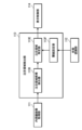
- FIG. 2 is a block diagram of the main functions of the processor device.
- the processor device 30 has functions such as an endoscope control section 31, a light source control section 32, an image processing section 33, an input control section 34, an output control section 35, and the like. Each function is realized by the processor executing a predetermined program.
- the auxiliary storage stores various programs executed by the processor, various data required for control and the like.
- the endoscope control unit 31 controls the endoscope 10.
- the control of the endoscope 10 includes drive control of the imaging device, air/water supply control, suction control, and the like.
- the light source controller 32 controls the light source device 20 .
- the control of the light source device 20 includes light emission control of the light source, switching control of the light source type, and the like.
- the image processing unit 33 performs various signal processing on the signal output from the imaging device of the endoscope 10 to generate a captured image.
- the input control unit 34 performs processing for accepting input of operations via the input device 40 and input of various types of information.
- the output control unit 35 controls output of information to the endoscope image processing apparatus 100 .
- Information to be output to the endoscope image processing apparatus 100 includes an image captured by an endoscope (endoscopic image), information input via the input device 40, various operation information, and the like.
- the various operation information includes operation information of the operation unit of the endoscope 10 (for example, information on switching the light source type, etc.).
- the input device 40 constitutes a user interface in the endoscope system 1 together with the display device 50 .
- the input device 40 is composed of, for example, a keyboard, mouse, foot switch, and the like.
- the input device 40 can also include a touch panel, a voice input device, a line-of-sight input device, and the like.
- the display device 50 is used not only for displaying endoscopic images, but also for displaying various kinds of information.
- the display device 50 is configured by, for example, a liquid crystal display (LCD), an organic electroluminescence display (OELD), or the like.
- the display device 50 can also be configured with a projector, a head-mounted display, or the like.
- the endoscopic image processing apparatus 100 performs processing for detecting a region of interest such as a lesion from an endoscopic image. In addition, processing for outputting an endoscopic image to the display device 50 including the detection result of the attention area is performed.
- the endoscopic image processing apparatus 100 includes a processor, a main storage section, an auxiliary storage section, etc. as its hardware configuration. That is, the endoscope image processing apparatus 100 is configured by a computer.
- a processor is comprised by CPU etc., for example.
- the main storage unit is composed of, for example, a RAM or the like.
- the auxiliary storage unit is composed of, for example, a flash memory including an SSD, a hard disk drive, or the like.
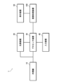
- Fig. 3 is a block diagram of the main functions of the endoscope image processing apparatus.
- the endoscopic image processing apparatus 100 has functions such as an endoscopic image acquisition section 111, a state recognition section 112, an attention area detection section 113, a display control section 114, and the like. Each function is realized by the processor executing a predetermined program.
- the auxiliary memory stores programs executed by the processor, various data required for image processing, and the like.
- the endoscopic image acquisition unit 111 acquires endoscopic images output from the processor device 30 .
- the images captured by the endoscope 10 (endoscopic images) are output from the processor device 30 in chronological order.
- the endoscopic image acquisition unit 111 sequentially acquires endoscopic images output from the processor device 30 in chronological order.
- the state recognition unit 112 performs processing for recognizing the state of the organ to be inspected from the endoscopic image acquired by the endoscopic image acquisition unit 111 .
- processing for recognizing the state of the organ to be inspected from the endoscopic image acquired by the endoscopic image acquisition unit 111 is performed.
- FIG. 4 is a block diagram of functions possessed by the state recognition unit.
- the state recognition unit 112 has the functions of a state recognizer 112A that performs recognition processing and a state determination unit 112B that performs processing to determine the recognition result.
- the state recognizer 112A is composed of, for example, a trained model trained to recognize the state of an organ from an endoscopic image. Specifically, learning learned using machine learning algorithms such as Neural Network (NN), Convolutional Neural Network (CNN), AdaBoost, Random Forest, or deep learning Consists of ready-made models. As described above, in the present embodiment, in order to recognize the state of gastric H. pylori infection, model learning is performed using pylori-positive and pylori-negative endoscopic images as a learning data set.
- NN Neural Network
- CNN Convolutional Neural Network
- AdaBoost AdaBoost
- Random Forest Random Forest
- the recognition of gastric H. pylori infection from endoscopic images can be determined by the presence or absence of mucosal atrophy, inflammation, and intestinal metaplasia.
- the presence or absence of these substances can be recognized not only by changes in color tone, but also by observation of blood vessels, irregularities in surface structures, and the like.
- the histological abnormalities of the mucous membrane due to H. pylori infection are not localized but spread over a wide area of the stomach. For this reason, it is preferable to recognize from an image that captures a wide range of the stomach.
- even H. pylori-positive stomachs may have structures resembling locally pylori-negative mucosa.
- the state determination unit 112B determines the recognition result when the recognition result by the state recognizer 112A satisfies a specific condition. That is, the state of the organ to be inspected is determined. In this embodiment, the pylori infection state of the stomach is determined. Normally, the gastric H. pylori infection status does not fluctuate during the examination of the same subject. Therefore, when a specific condition is satisfied, the recognition result is fixed and the subsequent recognition processing is stopped. The state determination unit 112B determines whether or not the recognition result can be determined based on a plurality of recognition results, and determines the recognition result when it determines that the recognition result can be determined.
- the recognition result is fixed when the same recognition result is obtained consecutively a specified number of times. Therefore, state determination unit 112B of the present embodiment counts the number of times the same recognition result is obtained consecutively. Further, it is determined whether or not the number of times has reached a specified number of times to determine whether or not the recognition result can be finalized.
- the region-of-interest detection unit 113 performs processing for detecting a region of interest such as a lesion from the endoscopic image acquired by the endoscopic image acquisition unit 111 .
- a process of detecting a cancer suspected region (a region suspected of having cancer) from an endoscopic image of the stomach is performed.
- a process of detecting a region of interest from an endoscopic image is performed using a detector configured with a trained model.
- FIG. 5 is a block diagram of the functions of the attention area detection unit.
- the attention area detection unit 113 has a first detector 113A and a second detector 113B, and uses the detector set by the detector setting unit 113C to perform processing for detecting an attention area.
- the first detector 113A is a detector that corresponds to examination of a pylori-positive stomach. Specifically, it is a detector composed of a trained model trained using endoscopic images of the stomach positive for pylori. Therefore, good detection results are obtained for H. pylori-positive stomachs.
- the second detector 113B is a detector corresponding to examination of a pylori-negative stomach. Specifically, it is a detector composed of a trained model trained using pylori-negative gastric endoscopic images. Therefore, good detection results are obtained for pylori-negative stomachs.
- the first detector 113A and the second detector 113B are neural networks, convolutional neural networks, AdaBoost, random forest, etc. Machine learning algorithms or deep learning Consists of pre-trained models that have been trained.
- the detector setting unit 113C sets the detector to be used according to the recognition result of the H. pylori infection state by the state recognition unit 112. Specifically, when the recognition result by the state recognition unit 112 is positive for pylori, the first detector 113A is selected and set. On the other hand, when the recognition result by the state recognition unit 112 is negative for pylori, the second detector 113B is selected.
- the setting of the detector by the detector setting unit 113C is an example of setting the detection criteria for the attention area.
- the display control unit 114 controls the display of the display device 50.
- the display control unit 114 causes the display device 50 to display an endoscopic image. Further, when the attention area is detected, the display control unit 114 causes the display device 50 to display the detection result. Furthermore, the display control unit 114 causes the display device 50 to display the recognition result of the state of the organ when detecting the region of interest.
- FIGS. 6 and 7 are diagrams showing an example of the display of the display device. This figure shows an example of display in the case of a so-called wide monitor.
- FIG. 6 shows an example of display in the case of H. pylori positive.
- FIG. 7 shows an example of display in the case of H. pylori negative.
- a main display area A1 and a sub-display area A2 are set within the screen.
- the main display area A1 is an area in which the endoscopic image Im is displayed.
- the endoscopic image Im is displayed in the main display area A1 in a predetermined display mode.
- the endoscopic image Im is displayed within a circle that is notched at the top and bottom.
- the sub-display area A2 is a so-called blank area, and is used to display various information such as setting information and captured still images.
- the display control unit 114 displays the detection box B superimposed on the endoscopic image Im.
- the detection box B is composed of a rectangular frame with only the corners displayed, and is displayed so as to surround the attention area X. FIG. Therefore, it is displayed in a size corresponding to the size of the attention area X. FIG. Also, the detection box B is displayed in a predetermined color (for example, green). Detection box B is an example of a frame.
- the display control unit 114 displays the information IP of the recognition result in the sub-display area A2.
- the recognition result of the gastric H. pylori infection state is displayed.
- “H. pylori : infected” is displayed in the case of positive H. pylori.
- H. pylori negative is displayed in the case of H. pylori negative.
- the recognition result information IP is an example of information about the state of an organ recognized from an endoscopic image.
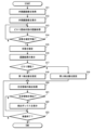
- FIG. 8 is a flowchart showing the procedure of processing for detecting a region of interest from an endoscopic image.
- the images (endoscopic images) captured in time series by the endoscope 10 are sequentially output from the processor device 30 and taken into the endoscope image processing device 100 . That is, time-series endoscopic images are sequentially acquired (step S11).
- the actual examination (observation) is started after the stomach is washed in a predetermined manner.
- the acquired endoscopic image is displayed on the display device 50 (step S12).
- the endoscopic image processing apparatus 100 displays the endoscopic image Im in the main display area A1 set on the screen (see FIGS. 6 and 7).
- recognition processing of the H. pylori infection state is performed on the acquired endoscopic image (step S13). Recognition processing is sequentially performed on each endoscopic image acquired in time series.
- step S14 it is determined whether or not the subject's pylori infection status can be determined from the result of the recognition process. This determination is made based on a plurality of recognition results. In this embodiment, determination is made based on whether or not the same recognition result has been obtained continuously for a specified number of times. If the same recognition result is obtained continuously for a specified number of times, it is determined that the state of H. pylori infection can be determined.
- the pylori infection status is determined (step S15). That is, pylori-positive or pylori-negative is determined.
- the recognition result is displayed on the display device 50 (step S16). That is, the recognized H. pylori infection state is displayed (see FIGS. 6 and 7). The recognition result is displayed in the sub-display area A2 of the screen.
- step S17 it is determined whether or not the confirmed recognition result is positive for pylori (step S17). Then, based on the determination result, a detector to be used for detection of the attention area is set. Specifically, when the confirmed recognition result is positive for pylori, the detector to be used is set to the first detector 113A (step S18). On the other hand, when the confirmed recognition result is negative for pylori, the detector to be used is set to the second detector 113B (step S19).
- the detection process of the attention process is performed using the set detector (step S20). Specifically, in the case of positive H. pylori, the first detector 113A is used to detect a region of interest from an endoscopic image. In addition, in the case of negative H. pylori, the second detector 113B is used to perform detection processing of the region of interest from the endoscopic image. The detection process is sequentially performed on each endoscopic image acquired in time series. Then, it is determined whether or not the attention area is detected for each detection process (step S21).
- the detection box B is displayed superimposed on the endoscopic image Im being displayed on the display device 50 (step S22).
- a detection box B is displayed so as to surround the detected attention area X (see FIGS. 6 and 7).
- step S23 it is determined whether or not the inspection has ended. When the inspection ends, the process ends. If the examination is continuing, the process of detecting the region of interest from the endoscopic image is continued (step S20).
- the detector to be used when detecting a region of interest from an endoscopic image, the detector to be used is switched according to the pylori infection state of the stomach. . Thereby, the attention area can be appropriately detected.
- [Modification] [Modified example of recognition of gastric pylori infection status]
- the state after eradication of H. pylori can also be recognized.
- detectors corresponding to respective recognition results are prepared. That is, the detector (first detector) used in the case of pylori positive (current infection), the detector (second detector) used in the case of pylori negative (uninfected), and after eradication of pylori (existing infection) is provided (third detector).
- the detector to be used is set according to the recognition result of the gastric H. pylori infection state, and detection processing of the region of interest is performed. That is, when the recognition result is positive for H. pylori, the first detector is selected, and detection processing of the region of interest is performed. Further, when the recognition result is negative for pylori, the second detector is selected, and detection processing of the region of interest is performed. Further, when the recognition result is after eradication of H. pylori, the third detector is selected, and detection processing of the region of interest is performed. As a result, the attention area can be determined more appropriately.
- the state after eradication of H. pylori can be included in one of the states for recognition.
- the gastric endoscopy is performed by recognizing the pylori infection state of the stomach and switching the detector to be used according to the recognition result.
- the gastric conditions to be recognized are not limited to pylori infection conditions. It is possible to adopt a configuration in which the state of the stomach is widely recognized and the detector to be used is switched according to the result. In particular, histological abnormalities in mucous membranes have a great influence on the detection of lesions. Therefore, it is possible to recognize the state of the histological abnormality of the mucous membrane and switch the detector to be used according to the recognition result.
- Conditions related to histological abnormalities of mucous membranes are exemplified by conditions related to inflammation and/or atrophy of mucous membranes.
- the state can be recognized by dividing it into three or more.
- the model that constitutes the detector is trained with an image group corresponding to the object to be recognized.
- the machine-learned model is used to recognize the stomach pylori infection state from the endoscopic image of the stomach, but the stomach state including the pylori infection state is recognized.
- the method is not limited to this. Other techniques may be used to recognize stomach conditions.
- the endoscopic image processing apparatus 100 may acquire information on the state of the stomach by inputting the information on the state of the stomach from the outside.
- the patient information it is also possible to input and acquire information on pylori infection in the stomach of the person to be examined from the outside.
- This information may also be manually entered by the user via input device 40 .
- it may be configured such that it is included in other input information and automatically input.
- the stomach When recognizing the state of the stomach from an endoscopic image, it can be configured to recognize from an endoscopic image of a specific region of the stomach. That is, it is possible to adopt a configuration in which an area in which a target state can be easily recognized can be recognized from a photographed image.
- the pylori infection state can be recognized from an image of the lesser curvature of the stomach or the upper region when the stomach is divided into upper and lower parts.
- the histological abnormality of the mucous membrane due to pylori infection spreads over a wide range of the stomach, it is preferable to recognize it from an image of a wide range of the stomach.
- the recognition result is determined and the subsequent recognition processing is stopped.
- the attention area can be appropriately detected by constantly executing recognition processing and dynamically switching the detectors to be used.
- the mucous membrane cannot be imaged due to factors such as water fog or blurring, it is difficult to determine the state of the mucous membrane. Therefore, when recognizing a state that does not change locally, it is possible to effectively prevent subsequent erroneous recognition by determining the state at an early stage. In addition, this makes it possible to appropriately detect the attention area.
- the conditions for determining the recognition result are not limited to those shown in the above embodiment. It can be appropriately set according to the object to be recognized. For example, when the same detection result is obtained at a certain rate or more, the recognition result may be determined.
- the processing for detecting the processing of interest is started after the recognition result of the state of the stomach is confirmed.
- detection of the region of interest is performed using a predetermined detector until the recognition result of the state of the stomach is determined.
- a detector may be prepared to be used when the state of the organ is undetermined, and this detector may be used to detect the region of interest while the state of the organ is undetermined.
- the endoscopic image for recognizing the state of the stomach is preferably an image obtained by photographing a relatively wider range than the endoscopic image for detecting the region of interest.
- An image of a wide range includes an image obtained by capturing a wide range with a single image, and an image obtained by dividing a wide range into a plurality of images.
- the endoscopic image for detecting the region of interest is preferably an image captured temporally after the endoscopic image for recognizing the state of the stomach.
- the endoscopic image for recognizing the state of the stomach is an example of the first endoscopic image
- the endoscopic image for detecting the region of interest is an example of the second endoscopic image.
- the information of the recognition result of the stomach pylori infection state is displayed on the display device 50, but the method of notifying the recognition result is not limited to this.
- an icon corresponding to the recognition result may be displayed on the display device to notify the recognition result.
- the recognition result can be notified by changing the display mode of the detection box according to the recognition result. good.
- the recognition result may be notified by changing the color of the detection box according to the recognition result.
- the color of the detection box As an example, in the case of H. pylori positive (currently infected), the color of the detection box is displayed in green, and in the case of H. pylori negative (uninfected), the color of the detection box is displayed in red.
- the color of the detection box is displayed in blue.
- the recognition result may be reported by changing the shape of the detection box or changing the blinking state of the display according to the recognition result.
- the display of the information on the recognition result in the sub-display area A2 can be omitted.
- notification it is possible to have a configuration in which notification is performed in combination with a sound such as a notification sound.
- the user can grasp which detector is operating. This makes it possible to check whether the wrong detector is being used.
- the detectors to be used are preferably configured to be manually switched. Thereby, when it becomes clear from the notification result that the wrong detector has been selected, it is possible to switch to the correct detector.
- the appearance of a region of interest varies depending on the state of the esophagus, particularly the state of structural abnormality of the mucous membrane. Therefore, in the examination of the esophagus as well, it is preferable to recognize the state of the esophagus when detecting the region of interest, and switch the detection criteria according to the recognition result.
- the state of the mucous membrane of the esophagus is recognized as squamous epithelium or columnar epithelium, and the detector to be used is switched according to the recognition result.
- a detector corresponding to squamous epithelium is used to detect the region of interest. If the mucous membrane of the esophagus is columnar epithelium, detection processing of the region of interest is performed using a detector corresponding to columnar epithelium. Alternatively, it recognizes whether the mucous membrane of the esophagus is columnar epithelium or not, and switches the detector to be used according to the recognition result. That is, when the state of the mucous membrane of the esophagus is a columnar epithelium, detection processing of the region of interest is performed using a detector corresponding to the columnar epithelium.
- the region of interest is detected as a normal esophagus using a detector corresponding to the normal esophagus.
- the region of interest can be appropriately detected from the endoscopic image of the esophagus.
- the detector As an example, it recognizes whether or not the condition of the large intestine has inflammatory bowel disease, and switches the detector to be used according to the recognition result. That is, in the case of the large intestine with inflammatory bowel disease, detection processing of the region of interest is performed using a detector corresponding to the case of having inflammatory bowel disease. On the other hand, in the case of the large intestine that does not have inflammatory bowel disease, detection processing of the region of interest is performed as a normal large intestine using a detector corresponding to the normal large intestine. As a result, the region of interest can be appropriately detected from the endoscopic image of the large intestine.
- the recognition result can be determined when a specific condition is satisfied in the recognition process.
- the light source type when observation with different light source types is possible, information on the currently selected light source type may be acquired, and detectors may be switched for each light source type.
- a detector corresponding to the recognition result of the organ is prepared for each observation mode.
- white light observation, BLI observation and LCI observation are selectively possible.
- the detector to be used in the case of positive observation by white light observation the detector to be used in the case of negative observation by white light observation
- the detector to be used in the case of positive observation by BLI the detector to be used in case of positive observation by BLI.
- a detector to be used in the case of H. pylori negative, a detector to be used in the case of LCI observation to be H. pylori positive, and a detector to be used in the case of LCI observation to be pylori negative are prepared.
- the detector to be used is switched according to the recognition result of the pylori infection state of the stomach. As a result, the region of interest can be appropriately detected from the endoscopic image regardless of the type of light source.
- the detection criteria are switched by switching the detectors to be used according to the recognition result, but the method of changing the detection criteria is not limited to this.
- a method of changing the detection standard without changing the detector will be described.
- the configuration is the same as that of the endoscopic image processing apparatus of the first embodiment, except for changing the detection criteria for the attention area. Therefore, only the function of the region-of-interest detection unit included in the endoscope image processing apparatus according to the present embodiment will be described below.
- FIG. 9 is a block diagram of the functions of the attention area detection unit.
- the attention area detection unit 113 of the present embodiment has the functions of an attention area candidate detector 113D, an attention area specifying unit 113E, and a threshold value setting unit 113F.
- the attention area candidate detector 113D performs processing for detecting attention area candidates from the endoscopic image.
- the region-of-interest candidate detector 113D is configured with a trained model, and detects region-of-interest candidates with certainty.
- the degree of certainty is a degree indicating the certainty of the attention area. Therefore, the attention area candidate detector 113D calculates the reliability of all the attention area candidates to be detected, and performs the detection processing.
- the attention area specifying unit 113E extracts the attention area candidates detected by the attention area candidate detector 113D, and extracts the attention area candidates whose degrees of certainty are greater than or equal to the threshold value, and identifies the attention areas.
- the threshold setting unit 113F sets the threshold used by the attention area specifying unit 113E.
- the threshold setting unit 113 ⁇ /b>F sets a threshold according to the state recognition result of the inspection target organ by the state recognition unit 112 .
- the threshold is set according to the recognition result of the gastric pylori infection state. Specifically, in the case of pylori positive (currently infected), the first threshold is set, and in the case of pylori negative (uninfected), the second threshold is set. If the state after eradication of H. pylori (already infected) is also recognized, a threshold (third threshold) corresponding to the state after eradication of H. pylori is prepared.
- the second threshold set in the case of negative H. pylori is set to a value relatively lower than the first threshold set in the case of positive H. pylori (first threshold>second threshold). That is, the detection sensitivity in the case of pylori-negative is made relatively higher than the detection sensitivity in the case of pylori-positive, so that the region of interest is easily detected.
- pylori-negative stomach is generally characterized by its mucosal surface being smooth, glossy and lustrous.
- pylori-positive stomachs are characterized by reddened mucous membranes and white turbid mucus.
- the region of interest in the pylori-positive stomach is easier to detect than in the pylori-negative stomach, and there is a greater risk of erroneous detection. That is, there is a greater concern that the normal area will be recognized as the attention area. For this reason, the second threshold value set in the case of negative H. pylori is set to a relatively lower value than the first threshold value set in the case of positive H. pylori to suppress erroneous recognition.
- the third threshold value set in the case after eradication of pylori is the same value as the first threshold value, or higher than the second threshold value, and the first Set to a value lower than the threshold. That is, it is set so as to satisfy the relationships of first threshold ⁇ third threshold and third threshold>second threshold.
- the process of setting the threshold is an example of the process of setting the detection criteria for the attention area.
- the operation of the endoscopic image processing apparatus 100 of the present embodiment will be described below, taking as an example the case of conducting an endoscopy of the stomach.
- the threshold value is set depending on whether the gastric pylori infection status is positive or negative.
- FIG. 10 is a flowchart showing the procedure of processing for detecting a region of interest from an endoscopic image.
- the images (endoscopic images) captured in time series by the endoscope 10 are sequentially output from the processor device 30 and taken into the endoscope image processing device 100 . That is, time-series endoscopic images are sequentially acquired (step S31). The acquired endoscopic image is displayed on the display device 50 (step S32).
- recognition processing of the H. pylori infection state is performed on the acquired endoscopic image (step S33). Recognition processing is sequentially performed on each endoscopic image acquired in time series.
- step S34 it is determined whether or not the subject's pylori infection status can be determined from the recognition processing results. If it is determined that it can be determined, the state of H. pylori infection is determined (step S35). That is, pylori-positive or pylori-negative is confirmed. When the pylori infection status is determined, the recognition result is displayed on the display device 50 (step S36).
- step S37 it is determined whether or not the confirmed recognition result is positive for pylori. Then, based on the determination result, a threshold value to be used when detecting the attention area is set. Specifically, when the confirmed recognition result is pylori-positive, the first threshold is set (step S38). On the other hand, if the confirmed recognition result is negative for pylori, the second threshold is set (step S39).
- detection processing of the process of interest is performed based on the set threshold (step S40). Specifically, in the case of positive H. pylori, those with a degree of certainty greater than or equal to the first threshold are extracted from the detected attention area candidates and output as the attention area detection result. On the other hand, in the case of H. pylori-negative, the detected attention area candidates whose certainty is equal to or higher than the second threshold are extracted and output as the attention area detection result.
- step S41 It is determined whether or not the attention area is detected as a result of the attention area detection processing (step S41).
- the detection box B is displayed superimposed on the endoscopic image Im being displayed on the display device 50 (step S42).
- step S43 it is determined whether or not the inspection has ended.
- the process ends. If the examination is continuing, the process of detecting the region of interest from the endoscopic image is continued (step S40).
- the endoscopic image processing apparatus of the present embodiment when a region of interest is detected from an endoscopic image using a single detector (region-of-interest candidate detector), the stomach pylori infection status, the threshold used for detection is switched. As a result, the attention area can be appropriately detected even when the attention area is detected with a single detector.
- the endoscopic image processing apparatus of the first embodiment has the advantage of being able to optimize the detector according to the state of the organ, so that more accurate detection can be achieved.
- the endoscopic image processing apparatus of this embodiment is a single detector, it has the advantage of reducing the cost of collecting learning data for building a model.
- processors include CPUs and/or GPUs (Graphic Processing Units), FPGAs (Field Programmable Gate Arrays), etc., which are general-purpose processors that execute programs and function as various processing units.
- Programmable Logic Device PLD
- ASIC Application Specific Integrated Circuit
- a dedicated electric circuit which is a processor having a circuit configuration specially designed to execute specific processing, etc. included.
- a program is synonymous with software.
- a single processing unit may be composed of one of these various processors, or may be composed of two or more processors of the same type or different types.
- one processing unit may be composed of a plurality of FPGAs or a combination of a CPU and an FPGA.
- a plurality of processing units may be configured by one processor.
- a single processor is configured with a combination of one or more CPUs and software, as typified by computers used for clients and servers. , in which the processor functions as a plurality of processing units.
- SoC System on Chip
- the various processing units are configured using one or more of the above various processors as a hardware structure.
- endoscope system 10 endoscope 20 light source device 30 processor device 31 endoscope control section 32 light source control section 33 image processing section 34 input control section 35 output control section 40 input device 50 display device 100 endoscope image processing device 111 endoscopic image acquisition unit 112 state recognition unit 112A state recognition unit 112B state determination unit 113 attention area detection unit 113A first detector 113B second detector 113C detector setting unit 113D attention area candidate detector 113E attention area specifying unit 113F Threshold setting unit 114 Display control unit A1 Main display area A2 Sub-display area B Detection box IP Recognition result information Im Endoscopic image X Attention area S11 to S23 Procedures S31 to 31 for detecting an attention area from an endoscope image S43 Procedure of processing for detecting a region of interest from an endoscopic image
Landscapes
- Health & Medical Sciences (AREA)
- Life Sciences & Earth Sciences (AREA)
- Engineering & Computer Science (AREA)
- Surgery (AREA)
- Medical Informatics (AREA)
- Physics & Mathematics (AREA)
- General Health & Medical Sciences (AREA)
- Nuclear Medicine, Radiotherapy & Molecular Imaging (AREA)
- Radiology & Medical Imaging (AREA)
- Pathology (AREA)
- Biomedical Technology (AREA)
- Heart & Thoracic Surgery (AREA)
- Optics & Photonics (AREA)
- Molecular Biology (AREA)
- Animal Behavior & Ethology (AREA)
- Biophysics (AREA)
- Public Health (AREA)
- Veterinary Medicine (AREA)
- Signal Processing (AREA)
- Computer Vision & Pattern Recognition (AREA)
- General Physics & Mathematics (AREA)
- Theoretical Computer Science (AREA)
- Quality & Reliability (AREA)
- Artificial Intelligence (AREA)
- Evolutionary Computation (AREA)
- Endoscopes (AREA)
Abstract
La présente invention concerne un dispositif de traitement d'image d'endoscope, un procédé de traitement d'image d'endoscope, et un système d'endoscope, pouvant détecter de manière appropriée une région d'intérêt à partir d'une image d'endoscope. Un dispositif de traitement d'image d'endoscope selon la présente invention qui traite des images d'endoscope acquiert une image d'endoscope, reconnaît un état d'un organe interne qui est un objet d'examen à partir de l'image d'endoscope acquise, définit une référence de détection pour une région d'intérêt en fonction d'un résultat de reconnaissance de l'état de l'organe interne, et détecte la région d'intérêt à partir de l'image d'endoscope sur la base de la référence de détection qui est définie.
Priority Applications (2)
| Application Number | Priority Date | Filing Date | Title |
|---|---|---|---|
| JP2023554990A JPWO2023067922A1 (fr) | 2021-10-20 | 2022-09-05 | |
| US18/633,487 US20240257348A1 (en) | 2021-10-20 | 2024-04-11 | Endoscopic image processing device, endoscopic image processing method, and endoscope system |
Applications Claiming Priority (2)
| Application Number | Priority Date | Filing Date | Title |
|---|---|---|---|
| JP2021171784 | 2021-10-20 | ||
| JP2021-171784 | 2021-10-20 |
Related Child Applications (1)
| Application Number | Title | Priority Date | Filing Date |
|---|---|---|---|
| US18/633,487 Continuation US20240257348A1 (en) | 2021-10-20 | 2024-04-11 | Endoscopic image processing device, endoscopic image processing method, and endoscope system |
Publications (1)
| Publication Number | Publication Date |
|---|---|
| WO2023067922A1 true WO2023067922A1 (fr) | 2023-04-27 |
Family
ID=86058971
Family Applications (1)
| Application Number | Title | Priority Date | Filing Date |
|---|---|---|---|
| PCT/JP2022/033262 WO2023067922A1 (fr) | 2021-10-20 | 2022-09-05 | Dispositif de traitement d'image d'endoscope, procédé de traitement d'image d'endoscope, et système d'endoscope |
Country Status (3)
| Country | Link |
|---|---|
| US (1) | US20240257348A1 (fr) |
| JP (1) | JPWO2023067922A1 (fr) |
| WO (1) | WO2023067922A1 (fr) |
Citations (5)
| Publication number | Priority date | Publication date | Assignee | Title |
|---|---|---|---|---|
| WO2007119297A1 (fr) * | 2006-03-16 | 2007-10-25 | Olympus Medical Systems Corp. | dispositif de traitement d'image pour usage médical et procédé de traitement d'image pour usage médical |
| JP2008036243A (ja) * | 2006-08-08 | 2008-02-21 | Olympus Medical Systems Corp | 医療用画像処理装置及び医療用画像処理方法 |
| JP2017113184A (ja) * | 2015-12-22 | 2017-06-29 | 富士フイルム株式会社 | 内視鏡システム及び内視鏡システムの作動方法 |
| WO2020162275A1 (fr) * | 2019-02-08 | 2020-08-13 | 富士フイルム株式会社 | Dispositif de traitement d'image médicale, système d'endoscope et procédé de traitement d'image médicale |
| JP2021100555A (ja) * | 2019-12-24 | 2021-07-08 | 富士フイルム株式会社 | 医療画像処理装置、内視鏡システム、診断支援方法及びプログラム |
-
2022
- 2022-09-05 JP JP2023554990A patent/JPWO2023067922A1/ja active Pending
- 2022-09-05 WO PCT/JP2022/033262 patent/WO2023067922A1/fr unknown
-
2024
- 2024-04-11 US US18/633,487 patent/US20240257348A1/en active Pending
Patent Citations (5)
| Publication number | Priority date | Publication date | Assignee | Title |
|---|---|---|---|---|
| WO2007119297A1 (fr) * | 2006-03-16 | 2007-10-25 | Olympus Medical Systems Corp. | dispositif de traitement d'image pour usage médical et procédé de traitement d'image pour usage médical |
| JP2008036243A (ja) * | 2006-08-08 | 2008-02-21 | Olympus Medical Systems Corp | 医療用画像処理装置及び医療用画像処理方法 |
| JP2017113184A (ja) * | 2015-12-22 | 2017-06-29 | 富士フイルム株式会社 | 内視鏡システム及び内視鏡システムの作動方法 |
| WO2020162275A1 (fr) * | 2019-02-08 | 2020-08-13 | 富士フイルム株式会社 | Dispositif de traitement d'image médicale, système d'endoscope et procédé de traitement d'image médicale |
| JP2021100555A (ja) * | 2019-12-24 | 2021-07-08 | 富士フイルム株式会社 | 医療画像処理装置、内視鏡システム、診断支援方法及びプログラム |
Also Published As
| Publication number | Publication date |
|---|---|
| US20240257348A1 (en) | 2024-08-01 |
| JPWO2023067922A1 (fr) | 2023-04-27 |
Similar Documents
| Publication | Publication Date | Title |
|---|---|---|
| JP7346285B2 (ja) | 医療画像処理装置、内視鏡システム、医療画像処理装置の作動方法及びプログラム | |
| CN113573654B (zh) | 用于检测并测定病灶尺寸的ai系统、方法和存储介质 | |
| JP2022186872A (ja) | 医用画像処理装置、プロセッサ装置、内視鏡システム、医用画像処理装置の作動方法、及びプログラム | |
| CN111970955A (zh) | 内窥镜观察辅助装置、内窥镜观察辅助方法及程序 | |
| WO2007119297A1 (fr) | dispositif de traitement d'image pour usage médical et procédé de traitement d'image pour usage médical | |
| CN113613543B (zh) | 诊断辅助装置、诊断辅助方法以及记录介质 | |
| WO2020054543A1 (fr) | Dispositif et procédé de traitement d'image médicale, système d'endoscope, dispositif de processeur, dispositif d'aide au diagnostic et programme | |
| US20230255467A1 (en) | Diagnostic imaging device, diagnostic imaging method, diagnostic imaging program, and learned model | |
| US20240304311A1 (en) | Medical image processing apparatus, medical image proces sing method, program, and diagnosis support apparatus | |
| JP4749732B2 (ja) | 医用画像処理装置 | |
| US11862327B2 (en) | Medical image processing system | |
| WO2020170809A1 (fr) | Dispositif de traitement d'image médicale, système d'endoscope et procédé de traitement d'image médicale | |
| WO2021029292A1 (fr) | Système d'aide au diagnostic en imagerie, système d'endoscope, procédé d'aide au diagnostic en imagerie et programme d'aide au diagnostic en imagerie | |
| WO2023067922A1 (fr) | Dispositif de traitement d'image d'endoscope, procédé de traitement d'image d'endoscope, et système d'endoscope | |
| WO2022230607A1 (fr) | Dispositif de traitement d'image médicale, système d'endoscope, et procédé de fonctionnement de dispositif de traitement d'image médicale | |
| US11830185B2 (en) | Medical image processing system and learning method | |
| JP7264407B2 (ja) | 訓練用の大腸内視鏡観察支援装置、作動方法、及びプログラム | |
| WO2021241735A1 (fr) | Dispositif processeur d'endoscope | |
| WO2023228659A1 (fr) | Dispositif de traitement d'image et système d'endoscope | |
| WO2023162216A1 (fr) | Dispositif de traitement d'image, procédé de traitement d'image et support de stockage | |
| JPWO2023067922A5 (fr) | ||
| US20240161288A1 (en) | Endoscope system, operation method of endoscope system, and processor | |
| JP7561382B2 (ja) | 大腸内視鏡観察支援装置、作動方法、及びプログラム | |
| US20230410304A1 (en) | Medical image processing apparatus, medical image processing method, and program | |
| US20230091729A1 (en) | Endoscopic image processing apparatus |
Legal Events
| Date | Code | Title | Description |
|---|---|---|---|
| 121 | Ep: the epo has been informed by wipo that ep was designated in this application |
Ref document number: 22883233 Country of ref document: EP Kind code of ref document: A1 |
|
| ENP | Entry into the national phase |
Ref document number: 2023554990 Country of ref document: JP Kind code of ref document: A |
|
| NENP | Non-entry into the national phase |
Ref country code: DE |