WO2023067922A1 - Endoscope image processing device, endoscope image processing method, and endoscope system - Google Patents
Endoscope image processing device, endoscope image processing method, and endoscope system Download PDFInfo
- Publication number
- WO2023067922A1 WO2023067922A1 PCT/JP2022/033262 JP2022033262W WO2023067922A1 WO 2023067922 A1 WO2023067922 A1 WO 2023067922A1 JP 2022033262 W JP2022033262 W JP 2022033262W WO 2023067922 A1 WO2023067922 A1 WO 2023067922A1
- Authority
- WO
- WIPO (PCT)
- Prior art keywords
- endoscopic image
- state
- image processing
- organ
- processor
- Prior art date
Links
Images


Classifications
-
- A—HUMAN NECESSITIES
- A61—MEDICAL OR VETERINARY SCIENCE; HYGIENE
- A61B—DIAGNOSIS; SURGERY; IDENTIFICATION
- A61B1/00—Instruments for performing medical examinations of the interior of cavities or tubes of the body by visual or photographical inspection, e.g. endoscopes; Illuminating arrangements therefor
- A61B1/04—Instruments for performing medical examinations of the interior of cavities or tubes of the body by visual or photographical inspection, e.g. endoscopes; Illuminating arrangements therefor combined with photographic or television appliances
- A61B1/045—Control thereof
-
- G—PHYSICS
- G06—COMPUTING; CALCULATING OR COUNTING
- G06T—IMAGE DATA PROCESSING OR GENERATION, IN GENERAL
- G06T7/00—Image analysis
Definitions
- the present invention relates to an endoscope image processing apparatus, an endoscope image processing method, and an endoscope system, and more particularly, to an endoscope image processing apparatus for processing an image (endoscopic image) captured by an endoscope. , an endoscope image processing method and an endoscope system.
- Patent Document 1 discloses a technology for detecting a polyp as an elevated lesion from an endoscopic image. It is described that the detection criteria for polyps are changed depending on the type.
- Patent Document 1 has the drawback that it is necessary to detect a color tone change for each detected upheaval change area and set a detection criterion individually.
- One embodiment according to the technology of the present disclosure provides an endoscopic image processing device, an endoscopic image processing method, and an endoscopic system that can appropriately detect a region of interest from an endoscopic image.
- An endoscopic image processing apparatus for processing an endoscopic image comprising a processor, the processor acquires an endoscopic image, and recognizes the state of an organ to be inspected from the acquired endoscopic image.
- An endoscopic image processing apparatus for setting a detection criterion for a region of interest according to a recognition result of the state of an organ, and detecting the region of interest from an endoscopic image based on the set detection criterion.
- a processor acquires a plurality of endoscopic images captured in time series, recognizes the state of an organ from a first endoscopic image of the plurality of endoscopic images,
- the endoscopic image processing device according to any one of (1) to (5), which detects a region of interest from a second endoscopic image that is different from the first endoscopic image.
- the processor determines whether or not the recognition results of the state of the organ recognized from the plurality of first endoscopic images satisfy a specific condition, and determines the state of the organ recognized from the plurality of first endoscopic images. If the recognition result of (6) satisfies a specific condition, the recognition result of the state of the organ is determined, and the setting of the detection criterion based on the determined recognition result of the state of the organ is fixed. Device.
- the processor notifies the endoscopic image to be displayed on the display device by surrounding the detected region of interest with a frame, and displays the frame in a display mode according to the setting of the detection criteria.
- Endoscope image processing device notifies the endoscopic image to be displayed on the display device by surrounding the detected region of interest with a frame, and displays the frame in a display mode according to the setting of the detection criteria.
- the processor detects a region of interest from the endoscopic image using the learned model, and sets the learned model used for detecting the region of interest according to the recognition result of the state of the organ, from (1) The endoscope image processing device according to any one of (11).
- the processor calculates a degree of certainty indicating likelihood, detects an attention area candidate from the endoscopic image, and selects an attention area candidate having a degree of certainty greater than or equal to a threshold among the detected attention area candidates as an attention area.
- the endoscopic image processing apparatus according to any one of (1) to (11), which detects and sets a threshold according to the recognition result of the state of the organ.
- (23) A process of calculating a degree of certainty indicating certainty, detecting an attention area candidate from an endoscopic image, and detecting an attention area candidate having a degree of certainty greater than or equal to a threshold as an attention area from among the detected attention area candidates. , wherein information on the state of an organ to be inspected is obtained, and a threshold value is set according to the state of the organ.
- An endoscope system comprising an endoscope and an endoscope image processing device according to any one of (1) to (21) for processing an endoscope image captured by the endoscope.
- a region of interest can be appropriately detected from an endoscopic image.
- Block diagram of main functions possessed by the processor device Block diagram of the main functions of the endoscope image processing device Block diagram of functions possessed by the state recognition unit Block diagram of functions possessed by the attention area detection unit
- a diagram showing an example of display on a display device A diagram showing an example of display on a display device
- Block diagram of functions possessed by the attention area detection unit Flowchart showing a procedure of processing for detecting a region of interest from an endoscopic image
- H. pylori infection findings such as redness and malformation may occur even if the cancer is not cancer. Therefore, in the case of current H. pylori infection, these are no longer targets for detection. In this way, if a region of interest is detected based on the same detection criteria regardless of the state of an organ to be inspected, a situation may arise in which the region of interest cannot be detected appropriately.
- the detection criteria are switched according to the state of the organ to be inspected.
- the region of interest can be appropriately detected from the endoscopic image regardless of the state of the organ to be inspected.
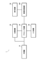
- FIG. 1 is a diagram showing an outline of the system configuration of an endoscope system.
- the endoscope system 1 of the present embodiment includes an endoscope 10, a light source device 20, a processor device 30, an input device 40, a display device 50, an endoscope image processing device 100, and the like.
- the endoscope 10 is connected to a light source device 20 and a processor device 30 .
- the light source device 20 , the input device 40 and the endoscope image processing device 100 are connected to the processor device 30 .
- the display device 50 is connected to the endoscope image processing device 100 .
- the endoscope system 1 of the present embodiment is configured as a system capable of observation using special light (special light observation) in addition to observation using normal white light (white light observation).
- Special light viewing includes narrowband light viewing.
- Narrowband light observation includes BLI observation (Blue laser imaging observation), NBI observation (Narrowband imaging observation), LCI observation (Linked Color Imaging observation), and the like. Note that the special light observation itself is a well-known technique, so detailed description thereof will be omitted.
- the endoscope 10 of the present embodiment is an electronic endoscope (flexible endoscope), particularly an electronic endoscope for upper digestive organs.
- the electronic endoscope includes an operation section, an insertion section, a connection section, and the like, and images an object with an imaging device incorporated in the distal end of the insertion section.
- a color image pickup device for example, a color image pickup device using a CMOS (Complementary Metal Oxide Semiconductor), a CCD (Charge Coupled Device), etc.
- CMOS Complementary Metal Oxide Semiconductor
- CCD Charge Coupled Device
- the endoscope 10 is connected to the light source device 20 and the processor device 30 via the connecting portion.
- the light source device 20 generates illumination light to be supplied to the endoscope 10 .
- the endoscope system 1 of the present embodiment is configured as a system capable of special light observation in addition to normal white light observation. Therefore, the light source device 20 has a function of generating light corresponding to special light observation (for example, narrow band light) in addition to normal white light. Note that, as described above, special light observation itself is a known technique, and therefore the description of the generation of the illumination light will be omitted. Switching of the light source type is performed by, for example, a light source type switching button provided for operating the endoscope 10 .
- the processor device 30 centrally controls the operation of the entire endoscope system.
- the processor device 30 includes a processor, a main memory, an auxiliary memory, etc. as its hardware configuration.
- a processor is comprised by CPU(Central Processing Unit) etc., for example.
- the main storage unit is composed of, for example, a RAM (Random Access Memory) or the like.
- the auxiliary storage unit is composed of, for example, a hard disk drive (HDD), a flash memory including an SSD (Solid State Drive), or the like.
- FIG. 2 is a block diagram of the main functions of the processor device.
- the processor device 30 has functions such as an endoscope control section 31, a light source control section 32, an image processing section 33, an input control section 34, an output control section 35, and the like. Each function is realized by the processor executing a predetermined program.
- the auxiliary storage stores various programs executed by the processor, various data required for control and the like.
- the endoscope control unit 31 controls the endoscope 10.
- the control of the endoscope 10 includes drive control of the imaging device, air/water supply control, suction control, and the like.
- the light source controller 32 controls the light source device 20 .
- the control of the light source device 20 includes light emission control of the light source, switching control of the light source type, and the like.
- the image processing unit 33 performs various signal processing on the signal output from the imaging device of the endoscope 10 to generate a captured image.
- the input control unit 34 performs processing for accepting input of operations via the input device 40 and input of various types of information.
- the output control unit 35 controls output of information to the endoscope image processing apparatus 100 .
- Information to be output to the endoscope image processing apparatus 100 includes an image captured by an endoscope (endoscopic image), information input via the input device 40, various operation information, and the like.
- the various operation information includes operation information of the operation unit of the endoscope 10 (for example, information on switching the light source type, etc.).
- the input device 40 constitutes a user interface in the endoscope system 1 together with the display device 50 .
- the input device 40 is composed of, for example, a keyboard, mouse, foot switch, and the like.
- the input device 40 can also include a touch panel, a voice input device, a line-of-sight input device, and the like.
- the display device 50 is used not only for displaying endoscopic images, but also for displaying various kinds of information.
- the display device 50 is configured by, for example, a liquid crystal display (LCD), an organic electroluminescence display (OELD), or the like.
- the display device 50 can also be configured with a projector, a head-mounted display, or the like.
- the endoscopic image processing apparatus 100 performs processing for detecting a region of interest such as a lesion from an endoscopic image. In addition, processing for outputting an endoscopic image to the display device 50 including the detection result of the attention area is performed.
- the endoscopic image processing apparatus 100 includes a processor, a main storage section, an auxiliary storage section, etc. as its hardware configuration. That is, the endoscope image processing apparatus 100 is configured by a computer.
- a processor is comprised by CPU etc., for example.
- the main storage unit is composed of, for example, a RAM or the like.
- the auxiliary storage unit is composed of, for example, a flash memory including an SSD, a hard disk drive, or the like.
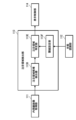
- Fig. 3 is a block diagram of the main functions of the endoscope image processing apparatus.
- the endoscopic image processing apparatus 100 has functions such as an endoscopic image acquisition section 111, a state recognition section 112, an attention area detection section 113, a display control section 114, and the like. Each function is realized by the processor executing a predetermined program.
- the auxiliary memory stores programs executed by the processor, various data required for image processing, and the like.
- the endoscopic image acquisition unit 111 acquires endoscopic images output from the processor device 30 .
- the images captured by the endoscope 10 (endoscopic images) are output from the processor device 30 in chronological order.
- the endoscopic image acquisition unit 111 sequentially acquires endoscopic images output from the processor device 30 in chronological order.
- the state recognition unit 112 performs processing for recognizing the state of the organ to be inspected from the endoscopic image acquired by the endoscopic image acquisition unit 111 .
- processing for recognizing the state of the organ to be inspected from the endoscopic image acquired by the endoscopic image acquisition unit 111 is performed.
- FIG. 4 is a block diagram of functions possessed by the state recognition unit.
- the state recognition unit 112 has the functions of a state recognizer 112A that performs recognition processing and a state determination unit 112B that performs processing to determine the recognition result.
- the state recognizer 112A is composed of, for example, a trained model trained to recognize the state of an organ from an endoscopic image. Specifically, learning learned using machine learning algorithms such as Neural Network (NN), Convolutional Neural Network (CNN), AdaBoost, Random Forest, or deep learning Consists of ready-made models. As described above, in the present embodiment, in order to recognize the state of gastric H. pylori infection, model learning is performed using pylori-positive and pylori-negative endoscopic images as a learning data set.
- NN Neural Network
- CNN Convolutional Neural Network
- AdaBoost AdaBoost
- Random Forest Random Forest
- the recognition of gastric H. pylori infection from endoscopic images can be determined by the presence or absence of mucosal atrophy, inflammation, and intestinal metaplasia.
- the presence or absence of these substances can be recognized not only by changes in color tone, but also by observation of blood vessels, irregularities in surface structures, and the like.
- the histological abnormalities of the mucous membrane due to H. pylori infection are not localized but spread over a wide area of the stomach. For this reason, it is preferable to recognize from an image that captures a wide range of the stomach.
- even H. pylori-positive stomachs may have structures resembling locally pylori-negative mucosa.
- the state determination unit 112B determines the recognition result when the recognition result by the state recognizer 112A satisfies a specific condition. That is, the state of the organ to be inspected is determined. In this embodiment, the pylori infection state of the stomach is determined. Normally, the gastric H. pylori infection status does not fluctuate during the examination of the same subject. Therefore, when a specific condition is satisfied, the recognition result is fixed and the subsequent recognition processing is stopped. The state determination unit 112B determines whether or not the recognition result can be determined based on a plurality of recognition results, and determines the recognition result when it determines that the recognition result can be determined.
- the recognition result is fixed when the same recognition result is obtained consecutively a specified number of times. Therefore, state determination unit 112B of the present embodiment counts the number of times the same recognition result is obtained consecutively. Further, it is determined whether or not the number of times has reached a specified number of times to determine whether or not the recognition result can be finalized.
- the region-of-interest detection unit 113 performs processing for detecting a region of interest such as a lesion from the endoscopic image acquired by the endoscopic image acquisition unit 111 .
- a process of detecting a cancer suspected region (a region suspected of having cancer) from an endoscopic image of the stomach is performed.
- a process of detecting a region of interest from an endoscopic image is performed using a detector configured with a trained model.
- FIG. 5 is a block diagram of the functions of the attention area detection unit.
- the attention area detection unit 113 has a first detector 113A and a second detector 113B, and uses the detector set by the detector setting unit 113C to perform processing for detecting an attention area.
- the first detector 113A is a detector that corresponds to examination of a pylori-positive stomach. Specifically, it is a detector composed of a trained model trained using endoscopic images of the stomach positive for pylori. Therefore, good detection results are obtained for H. pylori-positive stomachs.
- the second detector 113B is a detector corresponding to examination of a pylori-negative stomach. Specifically, it is a detector composed of a trained model trained using pylori-negative gastric endoscopic images. Therefore, good detection results are obtained for pylori-negative stomachs.
- the first detector 113A and the second detector 113B are neural networks, convolutional neural networks, AdaBoost, random forest, etc. Machine learning algorithms or deep learning Consists of pre-trained models that have been trained.
- the detector setting unit 113C sets the detector to be used according to the recognition result of the H. pylori infection state by the state recognition unit 112. Specifically, when the recognition result by the state recognition unit 112 is positive for pylori, the first detector 113A is selected and set. On the other hand, when the recognition result by the state recognition unit 112 is negative for pylori, the second detector 113B is selected.
- the setting of the detector by the detector setting unit 113C is an example of setting the detection criteria for the attention area.
- the display control unit 114 controls the display of the display device 50.
- the display control unit 114 causes the display device 50 to display an endoscopic image. Further, when the attention area is detected, the display control unit 114 causes the display device 50 to display the detection result. Furthermore, the display control unit 114 causes the display device 50 to display the recognition result of the state of the organ when detecting the region of interest.
- FIGS. 6 and 7 are diagrams showing an example of the display of the display device. This figure shows an example of display in the case of a so-called wide monitor.
- FIG. 6 shows an example of display in the case of H. pylori positive.
- FIG. 7 shows an example of display in the case of H. pylori negative.
- a main display area A1 and a sub-display area A2 are set within the screen.
- the main display area A1 is an area in which the endoscopic image Im is displayed.
- the endoscopic image Im is displayed in the main display area A1 in a predetermined display mode.
- the endoscopic image Im is displayed within a circle that is notched at the top and bottom.
- the sub-display area A2 is a so-called blank area, and is used to display various information such as setting information and captured still images.
- the display control unit 114 displays the detection box B superimposed on the endoscopic image Im.
- the detection box B is composed of a rectangular frame with only the corners displayed, and is displayed so as to surround the attention area X. FIG. Therefore, it is displayed in a size corresponding to the size of the attention area X. FIG. Also, the detection box B is displayed in a predetermined color (for example, green). Detection box B is an example of a frame.
- the display control unit 114 displays the information IP of the recognition result in the sub-display area A2.
- the recognition result of the gastric H. pylori infection state is displayed.
- “H. pylori : infected” is displayed in the case of positive H. pylori.
- H. pylori negative is displayed in the case of H. pylori negative.
- the recognition result information IP is an example of information about the state of an organ recognized from an endoscopic image.
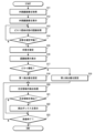
- FIG. 8 is a flowchart showing the procedure of processing for detecting a region of interest from an endoscopic image.
- the images (endoscopic images) captured in time series by the endoscope 10 are sequentially output from the processor device 30 and taken into the endoscope image processing device 100 . That is, time-series endoscopic images are sequentially acquired (step S11).
- the actual examination (observation) is started after the stomach is washed in a predetermined manner.
- the acquired endoscopic image is displayed on the display device 50 (step S12).
- the endoscopic image processing apparatus 100 displays the endoscopic image Im in the main display area A1 set on the screen (see FIGS. 6 and 7).
- recognition processing of the H. pylori infection state is performed on the acquired endoscopic image (step S13). Recognition processing is sequentially performed on each endoscopic image acquired in time series.
- step S14 it is determined whether or not the subject's pylori infection status can be determined from the result of the recognition process. This determination is made based on a plurality of recognition results. In this embodiment, determination is made based on whether or not the same recognition result has been obtained continuously for a specified number of times. If the same recognition result is obtained continuously for a specified number of times, it is determined that the state of H. pylori infection can be determined.
- the pylori infection status is determined (step S15). That is, pylori-positive or pylori-negative is determined.
- the recognition result is displayed on the display device 50 (step S16). That is, the recognized H. pylori infection state is displayed (see FIGS. 6 and 7). The recognition result is displayed in the sub-display area A2 of the screen.
- step S17 it is determined whether or not the confirmed recognition result is positive for pylori (step S17). Then, based on the determination result, a detector to be used for detection of the attention area is set. Specifically, when the confirmed recognition result is positive for pylori, the detector to be used is set to the first detector 113A (step S18). On the other hand, when the confirmed recognition result is negative for pylori, the detector to be used is set to the second detector 113B (step S19).
- the detection process of the attention process is performed using the set detector (step S20). Specifically, in the case of positive H. pylori, the first detector 113A is used to detect a region of interest from an endoscopic image. In addition, in the case of negative H. pylori, the second detector 113B is used to perform detection processing of the region of interest from the endoscopic image. The detection process is sequentially performed on each endoscopic image acquired in time series. Then, it is determined whether or not the attention area is detected for each detection process (step S21).
- the detection box B is displayed superimposed on the endoscopic image Im being displayed on the display device 50 (step S22).
- a detection box B is displayed so as to surround the detected attention area X (see FIGS. 6 and 7).
- step S23 it is determined whether or not the inspection has ended. When the inspection ends, the process ends. If the examination is continuing, the process of detecting the region of interest from the endoscopic image is continued (step S20).
- the detector to be used when detecting a region of interest from an endoscopic image, the detector to be used is switched according to the pylori infection state of the stomach. . Thereby, the attention area can be appropriately detected.
- [Modification] [Modified example of recognition of gastric pylori infection status]
- the state after eradication of H. pylori can also be recognized.
- detectors corresponding to respective recognition results are prepared. That is, the detector (first detector) used in the case of pylori positive (current infection), the detector (second detector) used in the case of pylori negative (uninfected), and after eradication of pylori (existing infection) is provided (third detector).
- the detector to be used is set according to the recognition result of the gastric H. pylori infection state, and detection processing of the region of interest is performed. That is, when the recognition result is positive for H. pylori, the first detector is selected, and detection processing of the region of interest is performed. Further, when the recognition result is negative for pylori, the second detector is selected, and detection processing of the region of interest is performed. Further, when the recognition result is after eradication of H. pylori, the third detector is selected, and detection processing of the region of interest is performed. As a result, the attention area can be determined more appropriately.
- the state after eradication of H. pylori can be included in one of the states for recognition.
- the gastric endoscopy is performed by recognizing the pylori infection state of the stomach and switching the detector to be used according to the recognition result.
- the gastric conditions to be recognized are not limited to pylori infection conditions. It is possible to adopt a configuration in which the state of the stomach is widely recognized and the detector to be used is switched according to the result. In particular, histological abnormalities in mucous membranes have a great influence on the detection of lesions. Therefore, it is possible to recognize the state of the histological abnormality of the mucous membrane and switch the detector to be used according to the recognition result.
- Conditions related to histological abnormalities of mucous membranes are exemplified by conditions related to inflammation and/or atrophy of mucous membranes.
- the state can be recognized by dividing it into three or more.
- the model that constitutes the detector is trained with an image group corresponding to the object to be recognized.
- the machine-learned model is used to recognize the stomach pylori infection state from the endoscopic image of the stomach, but the stomach state including the pylori infection state is recognized.
- the method is not limited to this. Other techniques may be used to recognize stomach conditions.
- the endoscopic image processing apparatus 100 may acquire information on the state of the stomach by inputting the information on the state of the stomach from the outside.
- the patient information it is also possible to input and acquire information on pylori infection in the stomach of the person to be examined from the outside.
- This information may also be manually entered by the user via input device 40 .
- it may be configured such that it is included in other input information and automatically input.
- the stomach When recognizing the state of the stomach from an endoscopic image, it can be configured to recognize from an endoscopic image of a specific region of the stomach. That is, it is possible to adopt a configuration in which an area in which a target state can be easily recognized can be recognized from a photographed image.
- the pylori infection state can be recognized from an image of the lesser curvature of the stomach or the upper region when the stomach is divided into upper and lower parts.
- the histological abnormality of the mucous membrane due to pylori infection spreads over a wide range of the stomach, it is preferable to recognize it from an image of a wide range of the stomach.
- the recognition result is determined and the subsequent recognition processing is stopped.
- the attention area can be appropriately detected by constantly executing recognition processing and dynamically switching the detectors to be used.
- the mucous membrane cannot be imaged due to factors such as water fog or blurring, it is difficult to determine the state of the mucous membrane. Therefore, when recognizing a state that does not change locally, it is possible to effectively prevent subsequent erroneous recognition by determining the state at an early stage. In addition, this makes it possible to appropriately detect the attention area.
- the conditions for determining the recognition result are not limited to those shown in the above embodiment. It can be appropriately set according to the object to be recognized. For example, when the same detection result is obtained at a certain rate or more, the recognition result may be determined.
- the processing for detecting the processing of interest is started after the recognition result of the state of the stomach is confirmed.
- detection of the region of interest is performed using a predetermined detector until the recognition result of the state of the stomach is determined.
- a detector may be prepared to be used when the state of the organ is undetermined, and this detector may be used to detect the region of interest while the state of the organ is undetermined.
- the endoscopic image for recognizing the state of the stomach is preferably an image obtained by photographing a relatively wider range than the endoscopic image for detecting the region of interest.
- An image of a wide range includes an image obtained by capturing a wide range with a single image, and an image obtained by dividing a wide range into a plurality of images.
- the endoscopic image for detecting the region of interest is preferably an image captured temporally after the endoscopic image for recognizing the state of the stomach.
- the endoscopic image for recognizing the state of the stomach is an example of the first endoscopic image
- the endoscopic image for detecting the region of interest is an example of the second endoscopic image.
- the information of the recognition result of the stomach pylori infection state is displayed on the display device 50, but the method of notifying the recognition result is not limited to this.
- an icon corresponding to the recognition result may be displayed on the display device to notify the recognition result.
- the recognition result can be notified by changing the display mode of the detection box according to the recognition result. good.
- the recognition result may be notified by changing the color of the detection box according to the recognition result.
- the color of the detection box As an example, in the case of H. pylori positive (currently infected), the color of the detection box is displayed in green, and in the case of H. pylori negative (uninfected), the color of the detection box is displayed in red.
- the color of the detection box is displayed in blue.
- the recognition result may be reported by changing the shape of the detection box or changing the blinking state of the display according to the recognition result.
- the display of the information on the recognition result in the sub-display area A2 can be omitted.
- notification it is possible to have a configuration in which notification is performed in combination with a sound such as a notification sound.
- the user can grasp which detector is operating. This makes it possible to check whether the wrong detector is being used.
- the detectors to be used are preferably configured to be manually switched. Thereby, when it becomes clear from the notification result that the wrong detector has been selected, it is possible to switch to the correct detector.
- the appearance of a region of interest varies depending on the state of the esophagus, particularly the state of structural abnormality of the mucous membrane. Therefore, in the examination of the esophagus as well, it is preferable to recognize the state of the esophagus when detecting the region of interest, and switch the detection criteria according to the recognition result.
- the state of the mucous membrane of the esophagus is recognized as squamous epithelium or columnar epithelium, and the detector to be used is switched according to the recognition result.
- a detector corresponding to squamous epithelium is used to detect the region of interest. If the mucous membrane of the esophagus is columnar epithelium, detection processing of the region of interest is performed using a detector corresponding to columnar epithelium. Alternatively, it recognizes whether the mucous membrane of the esophagus is columnar epithelium or not, and switches the detector to be used according to the recognition result. That is, when the state of the mucous membrane of the esophagus is a columnar epithelium, detection processing of the region of interest is performed using a detector corresponding to the columnar epithelium.
- the region of interest is detected as a normal esophagus using a detector corresponding to the normal esophagus.
- the region of interest can be appropriately detected from the endoscopic image of the esophagus.
- the detector As an example, it recognizes whether or not the condition of the large intestine has inflammatory bowel disease, and switches the detector to be used according to the recognition result. That is, in the case of the large intestine with inflammatory bowel disease, detection processing of the region of interest is performed using a detector corresponding to the case of having inflammatory bowel disease. On the other hand, in the case of the large intestine that does not have inflammatory bowel disease, detection processing of the region of interest is performed as a normal large intestine using a detector corresponding to the normal large intestine. As a result, the region of interest can be appropriately detected from the endoscopic image of the large intestine.
- the recognition result can be determined when a specific condition is satisfied in the recognition process.
- the light source type when observation with different light source types is possible, information on the currently selected light source type may be acquired, and detectors may be switched for each light source type.
- a detector corresponding to the recognition result of the organ is prepared for each observation mode.
- white light observation, BLI observation and LCI observation are selectively possible.
- the detector to be used in the case of positive observation by white light observation the detector to be used in the case of negative observation by white light observation
- the detector to be used in the case of positive observation by BLI the detector to be used in case of positive observation by BLI.
- a detector to be used in the case of H. pylori negative, a detector to be used in the case of LCI observation to be H. pylori positive, and a detector to be used in the case of LCI observation to be pylori negative are prepared.
- the detector to be used is switched according to the recognition result of the pylori infection state of the stomach. As a result, the region of interest can be appropriately detected from the endoscopic image regardless of the type of light source.
- the detection criteria are switched by switching the detectors to be used according to the recognition result, but the method of changing the detection criteria is not limited to this.
- a method of changing the detection standard without changing the detector will be described.
- the configuration is the same as that of the endoscopic image processing apparatus of the first embodiment, except for changing the detection criteria for the attention area. Therefore, only the function of the region-of-interest detection unit included in the endoscope image processing apparatus according to the present embodiment will be described below.
- FIG. 9 is a block diagram of the functions of the attention area detection unit.
- the attention area detection unit 113 of the present embodiment has the functions of an attention area candidate detector 113D, an attention area specifying unit 113E, and a threshold value setting unit 113F.
- the attention area candidate detector 113D performs processing for detecting attention area candidates from the endoscopic image.
- the region-of-interest candidate detector 113D is configured with a trained model, and detects region-of-interest candidates with certainty.
- the degree of certainty is a degree indicating the certainty of the attention area. Therefore, the attention area candidate detector 113D calculates the reliability of all the attention area candidates to be detected, and performs the detection processing.
- the attention area specifying unit 113E extracts the attention area candidates detected by the attention area candidate detector 113D, and extracts the attention area candidates whose degrees of certainty are greater than or equal to the threshold value, and identifies the attention areas.
- the threshold setting unit 113F sets the threshold used by the attention area specifying unit 113E.
- the threshold setting unit 113 ⁇ /b>F sets a threshold according to the state recognition result of the inspection target organ by the state recognition unit 112 .
- the threshold is set according to the recognition result of the gastric pylori infection state. Specifically, in the case of pylori positive (currently infected), the first threshold is set, and in the case of pylori negative (uninfected), the second threshold is set. If the state after eradication of H. pylori (already infected) is also recognized, a threshold (third threshold) corresponding to the state after eradication of H. pylori is prepared.
- the second threshold set in the case of negative H. pylori is set to a value relatively lower than the first threshold set in the case of positive H. pylori (first threshold>second threshold). That is, the detection sensitivity in the case of pylori-negative is made relatively higher than the detection sensitivity in the case of pylori-positive, so that the region of interest is easily detected.
- pylori-negative stomach is generally characterized by its mucosal surface being smooth, glossy and lustrous.
- pylori-positive stomachs are characterized by reddened mucous membranes and white turbid mucus.
- the region of interest in the pylori-positive stomach is easier to detect than in the pylori-negative stomach, and there is a greater risk of erroneous detection. That is, there is a greater concern that the normal area will be recognized as the attention area. For this reason, the second threshold value set in the case of negative H. pylori is set to a relatively lower value than the first threshold value set in the case of positive H. pylori to suppress erroneous recognition.
- the third threshold value set in the case after eradication of pylori is the same value as the first threshold value, or higher than the second threshold value, and the first Set to a value lower than the threshold. That is, it is set so as to satisfy the relationships of first threshold ⁇ third threshold and third threshold>second threshold.
- the process of setting the threshold is an example of the process of setting the detection criteria for the attention area.
- the operation of the endoscopic image processing apparatus 100 of the present embodiment will be described below, taking as an example the case of conducting an endoscopy of the stomach.
- the threshold value is set depending on whether the gastric pylori infection status is positive or negative.
- FIG. 10 is a flowchart showing the procedure of processing for detecting a region of interest from an endoscopic image.
- the images (endoscopic images) captured in time series by the endoscope 10 are sequentially output from the processor device 30 and taken into the endoscope image processing device 100 . That is, time-series endoscopic images are sequentially acquired (step S31). The acquired endoscopic image is displayed on the display device 50 (step S32).
- recognition processing of the H. pylori infection state is performed on the acquired endoscopic image (step S33). Recognition processing is sequentially performed on each endoscopic image acquired in time series.
- step S34 it is determined whether or not the subject's pylori infection status can be determined from the recognition processing results. If it is determined that it can be determined, the state of H. pylori infection is determined (step S35). That is, pylori-positive or pylori-negative is confirmed. When the pylori infection status is determined, the recognition result is displayed on the display device 50 (step S36).
- step S37 it is determined whether or not the confirmed recognition result is positive for pylori. Then, based on the determination result, a threshold value to be used when detecting the attention area is set. Specifically, when the confirmed recognition result is pylori-positive, the first threshold is set (step S38). On the other hand, if the confirmed recognition result is negative for pylori, the second threshold is set (step S39).
- detection processing of the process of interest is performed based on the set threshold (step S40). Specifically, in the case of positive H. pylori, those with a degree of certainty greater than or equal to the first threshold are extracted from the detected attention area candidates and output as the attention area detection result. On the other hand, in the case of H. pylori-negative, the detected attention area candidates whose certainty is equal to or higher than the second threshold are extracted and output as the attention area detection result.
- step S41 It is determined whether or not the attention area is detected as a result of the attention area detection processing (step S41).
- the detection box B is displayed superimposed on the endoscopic image Im being displayed on the display device 50 (step S42).
- step S43 it is determined whether or not the inspection has ended.
- the process ends. If the examination is continuing, the process of detecting the region of interest from the endoscopic image is continued (step S40).
- the endoscopic image processing apparatus of the present embodiment when a region of interest is detected from an endoscopic image using a single detector (region-of-interest candidate detector), the stomach pylori infection status, the threshold used for detection is switched. As a result, the attention area can be appropriately detected even when the attention area is detected with a single detector.
- the endoscopic image processing apparatus of the first embodiment has the advantage of being able to optimize the detector according to the state of the organ, so that more accurate detection can be achieved.
- the endoscopic image processing apparatus of this embodiment is a single detector, it has the advantage of reducing the cost of collecting learning data for building a model.
- processors include CPUs and/or GPUs (Graphic Processing Units), FPGAs (Field Programmable Gate Arrays), etc., which are general-purpose processors that execute programs and function as various processing units.
- Programmable Logic Device PLD
- ASIC Application Specific Integrated Circuit
- a dedicated electric circuit which is a processor having a circuit configuration specially designed to execute specific processing, etc. included.
- a program is synonymous with software.
- a single processing unit may be composed of one of these various processors, or may be composed of two or more processors of the same type or different types.
- one processing unit may be composed of a plurality of FPGAs or a combination of a CPU and an FPGA.
- a plurality of processing units may be configured by one processor.
- a single processor is configured with a combination of one or more CPUs and software, as typified by computers used for clients and servers. , in which the processor functions as a plurality of processing units.
- SoC System on Chip
- the various processing units are configured using one or more of the above various processors as a hardware structure.
- endoscope system 10 endoscope 20 light source device 30 processor device 31 endoscope control section 32 light source control section 33 image processing section 34 input control section 35 output control section 40 input device 50 display device 100 endoscope image processing device 111 endoscopic image acquisition unit 112 state recognition unit 112A state recognition unit 112B state determination unit 113 attention area detection unit 113A first detector 113B second detector 113C detector setting unit 113D attention area candidate detector 113E attention area specifying unit 113F Threshold setting unit 114 Display control unit A1 Main display area A2 Sub-display area B Detection box IP Recognition result information Im Endoscopic image X Attention area S11 to S23 Procedures S31 to 31 for detecting an attention area from an endoscope image S43 Procedure of processing for detecting a region of interest from an endoscopic image
Abstract
To provide an endoscope image processing device, an endoscope image processing method, and an endoscope system, capable of appropriately detecting a region of interest from an endoscope image. An endoscope image processing device according to the present invention that processes endoscope images acquires an endoscope image, recognizes a state of an internal organ that is an object of examination from the acquired endoscope image, sets a detection reference for a region of interest in accordance with a recognition result of the state of the internal organ, and detects the region of interest from the endoscope image on the basis of the detection reference that is set.
Description
本発明は、内視鏡画像処理装置、内視鏡画像処理方法及び内視鏡システムに係り、特に、内視鏡で撮影された画像(内視鏡画像)を処理する内視鏡画像処理装置、内視鏡画像処理方法及び内視鏡システムに関する。
The present invention relates to an endoscope image processing apparatus, an endoscope image processing method, and an endoscope system, and more particularly, to an endoscope image processing apparatus for processing an image (endoscopic image) captured by an endoscope. , an endoscope image processing method and an endoscope system.
内視鏡による検査を支援する技術として、内視鏡画像から病変等の注目領域を自動で検出し、報知する技術が知られている。
As a technology that supports endoscopic examinations, a technology that automatically detects and notifies regions of interest such as lesions from endoscopic images is known.
特許文献1には、内視鏡画像から隆起性病変としてのポリープを検出する技術に関して、内視鏡画像から隆起性変化領域を検出し、検出した隆起性変化領域が色調変化を伴う領域か否かによって、ポリープの検出基準を変えることが記載されている。
Patent Document 1 discloses a technology for detecting a polyp as an elevated lesion from an endoscopic image. It is described that the detection criteria for polyps are changed depending on the type.
しかしながら、特許文献1では、検出された隆起性変化領域ごとに色調変化を検出し、かつ、個別に検出基準を設定しなければならないという欠点がある。
However, Patent Document 1 has the drawback that it is necessary to detect a color tone change for each detected upheaval change area and set a detection criterion individually.
本開示の技術に係る一つの実施形態は、内視鏡画像から適切に注目領域を検出できる内視鏡画像処理装置、内視鏡画像処理方法及び内視鏡システムを提供する。
One embodiment according to the technology of the present disclosure provides an endoscopic image processing device, an endoscopic image processing method, and an endoscopic system that can appropriately detect a region of interest from an endoscopic image.
(1)内視鏡画像を処理する内視鏡画像処理装置であって、プロセッサを備え、プロセッサは、内視鏡画像を取得し、取得した内視鏡画像から検査対象の臓器の状態を認識し、臓器の状態の認識結果に応じて、注目領域の検出基準を設定し、設定された検出基準に基づいて、内視鏡画像から注目領域を検出する、内視鏡画像処理装置。
(1) An endoscopic image processing apparatus for processing an endoscopic image, comprising a processor, the processor acquires an endoscopic image, and recognizes the state of an organ to be inspected from the acquired endoscopic image. An endoscopic image processing apparatus for setting a detection criterion for a region of interest according to a recognition result of the state of an organ, and detecting the region of interest from an endoscopic image based on the set detection criterion.
(2)プロセッサは、臓器の特定の領域を撮影した内視鏡画像から臓器の状態を認識する、(1)の内視鏡画像処理装置。
(2) The endoscopic image processing device of (1), wherein the processor recognizes the state of the organ from an endoscopic image of a specific region of the organ.
(3)プロセッサは、臓器の異なる領域を撮影した複数の内視鏡画像から臓器の状態を認識する、(1)の内視鏡画像処理装置。
(3) The endoscopic image processing device of (1), wherein the processor recognizes the state of the organ from a plurality of endoscopic images of different regions of the organ.
(4)臓器の状態の認識に用いる内視鏡画像が、注目領域の検出に用いる内視鏡画像よりも相対的に広い範囲を撮影した内視鏡画像である、(1)の内視鏡画像処理装置。
(4) The endoscope of (1), wherein the endoscopic image used for recognizing the state of the organ is an endoscopic image obtained by photographing a relatively wider range than the endoscopic image used for detecting the region of interest. Image processing device.
(5)プロセッサは、内視鏡画像から粘膜の組織学的な異常に関する状態を認識して、臓器の状態を認識する、(1)から(4)のいずれか一の内視鏡画像処理装置。
(5) The endoscopic image processing apparatus according to any one of (1) to (4), wherein the processor recognizes a state of histological abnormalities in the mucous membrane from the endoscopic image and recognizes the state of the organ. .
(6)プロセッサは、時系列に撮影された複数の内視鏡画像を取得し、複数の内視鏡画像のうちの第1内視鏡画像から臓器の状態を認識し、 複数の内視鏡画像のうち第1内視鏡画像と異なる第2内視鏡画像から注目領域を検出する、(1)から(5)のいずれか一の内視鏡画像処理装置。
(6) a processor acquires a plurality of endoscopic images captured in time series, recognizes the state of an organ from a first endoscopic image of the plurality of endoscopic images, The endoscopic image processing device according to any one of (1) to (5), which detects a region of interest from a second endoscopic image that is different from the first endoscopic image.
(7)プロセッサは、複数の第1内視鏡画像から認識した臓器の状態の認識結果が、特定の条件を満たすか否か判定し、複数の第1内視鏡画像から認識した臓器の状態の認識結果が、特定の条件を満たす場合に、臓器の状態の認識結果を確定し、確定した臓器の状態の認識結果に基づく検出基準の設定を固定する、(6)の内視鏡画像処理装置。
(7) The processor determines whether or not the recognition results of the state of the organ recognized from the plurality of first endoscopic images satisfy a specific condition, and determines the state of the organ recognized from the plurality of first endoscopic images. If the recognition result of (6) satisfies a specific condition, the recognition result of the state of the organ is determined, and the setting of the detection criterion based on the determined recognition result of the state of the organ is fixed. Device.
(8)第2内視鏡画像は、第1内視鏡画像よりも時間的に後に撮影された内視鏡画像である、(6)又は(7)の内視鏡画像処理装置。
(8) The endoscopic image processing apparatus according to (6) or (7), wherein the second endoscopic image is an endoscopic image captured temporally later than the first endoscopic image.
(9)プロセッサは、内視鏡画像及び内視鏡画像から認識した臓器の状態に関する情報を表示装置に表示させる、(1)から(8)のいずれか一の内視鏡画像処理装置。
(9) The endoscopic image processing device according to any one of (1) to (8), wherein the processor causes the display device to display the endoscopic image and information about the state of the organ recognized from the endoscopic image.
(10)プロセッサは、検出基準の設定に応じて、異なる態様で注目領域の検出結果を報知する、(1)から(9)のいずれか一の内視鏡画像処理装置。
(10) The endoscopic image processing apparatus according to any one of (1) to (9), wherein the processor notifies the detection result of the attention area in different modes according to the setting of the detection criteria.
(11)プロセッサは、表示装置に表示させる内視鏡画像に対し、検出された注目領域を枠で囲って報知し、検出基準の設定に応じた表示態様で枠を表示させる、(10)の内視鏡画像処理装置。
(11) The processor notifies the endoscopic image to be displayed on the display device by surrounding the detected region of interest with a frame, and displays the frame in a display mode according to the setting of the detection criteria. Endoscope image processing device.
(12)プロセッサは、学習済みモデルを用いて内視鏡画像から注目領域を検出し、臓器の状態の認識結果に応じて、注目領域の検出に用いる学習済みモデルを設定する、(1)から(11)のいずれか一の内視鏡画像処理装置。
(12) The processor detects a region of interest from the endoscopic image using the learned model, and sets the learned model used for detecting the region of interest according to the recognition result of the state of the organ, from (1) The endoscope image processing device according to any one of (11).
(13)プロセッサは、確からしさを示す確信度を算出して、内視鏡画像から注目領域候補を検出し、検出した注目領域候補の中から確信度が閾値以上の注目領域候補を注目領域として検出し、臓器の状態の認識結果に応じて、閾値を設定する、(1)から(11)のいずれか一の内視鏡画像処理装置。
(13) The processor calculates a degree of certainty indicating likelihood, detects an attention area candidate from the endoscopic image, and selects an attention area candidate having a degree of certainty greater than or equal to a threshold among the detected attention area candidates as an attention area. The endoscopic image processing apparatus according to any one of (1) to (11), which detects and sets a threshold according to the recognition result of the state of the organ.
(14)プロセッサは、粘膜の組織学的な異常に関する状態として、粘膜の炎症及び/又は萎縮に関する状態を認識して、臓器の状態を認識する、(5)の内視鏡画像処理装置。
(14) The endoscopic image processing apparatus of (5), wherein the processor recognizes the state of inflammation and/or atrophy of the mucous membrane as the state of histological abnormality of the mucous membrane, and recognizes the state of the organ.
(15)プロセッサは、胃のピロリ感染に関する状態を認識する、(14)の内視鏡画像処理装置。
(15) The endoscopic image processing device of (14), wherein the processor recognizes conditions related to gastric H. pylori infection.
(16)プロセッサは、胃のピロリ感染に関する状態として、未感染、現感染、既感染の状態を認識する、(15)の内視鏡画像処理装置。
(16) The endoscopic image processing apparatus of (15), wherein the processor recognizes non-infected, current infected, and pre-infected states as states related to gastric H. pylori infection.
(17)プロセッサは、胃のピロリ感染に関する状態の認識結果が未感染の場合、検出基準を現感染及び/又は既感染の場合よりも相対的に低く設定する、(16)の内視鏡画像処理装置。
(17) The endoscopic image of (16), wherein the processor sets the detection criteria relatively lower than the case of current infection and/or past infection when the recognition result of the state related to gastric pylori infection is uninfected. processing equipment.
(18)プロセッサは、食道のバレット食道に関する状態を認識する、(14)の内視鏡画像処理装置。
(18) The endoscopic image processing device of (14), wherein the processor recognizes a condition relating to Barrett's esophagus of the esophagus.
(19)プロセッサは、大腸の炎症性腸疾患に関する状態を認識する、(14)の内視鏡画像処理装置。
(19) The endoscopic image processing device of (14), wherein the processor recognizes conditions related to inflammatory bowel disease of the large intestine.
(20)プロセッサは、臓器の状態を3つ以上の状態に分けて認識し、認識した臓器の状態に応じた検出基準を設定する、(1)から(18)のいずれか一の内視鏡画像処理装置。
(20) The endoscope according to any one of (1) to (18), wherein the processor recognizes the state of the organ by dividing it into three or more states, and sets detection criteria according to the recognized state of the organ. Image processing device.
(21)プロセッサは、光源種の情報を取得し、臓器の状態の認識結果及び光源種に応じて、検出基準を設定する、(1)から(20)のいずれか一の内視鏡画像処理装置。
(21) The endoscopic image processing according to any one of (1) to (20), wherein the processor acquires information on the light source type and sets a detection criterion according to the organ state recognition result and the light source type. Device.
(22)学習済みモデルを用いて内視鏡画像から注目領域を検出する処理を行う内視鏡画像処理方法であって、検査対象の臓器の状態の情報を取得し、臓器の状態に応じて、使用する学習済みモデルを設定する、内視鏡画像処理方法。
(22) An endoscopic image processing method for detecting a region of interest from an endoscopic image using a trained model, wherein information on the state of an organ to be inspected is obtained, and according to the state of the organ, , which sets the trained model to use, the endoscopic image processing method.
(23)確からしさを示す確信度を算出して、内視鏡画像から注目領域候補を検出し、検出した注目領域候補の中から確信度が閾値以上の注目領域候補を注目領域として検出する処理を行う内視鏡画像処理方法であって、検査対象の臓器の状態の情報を取得し、臓器の状態に応じて、閾値を設定する、内視鏡画像処理方法。
(23) A process of calculating a degree of certainty indicating certainty, detecting an attention area candidate from an endoscopic image, and detecting an attention area candidate having a degree of certainty greater than or equal to a threshold as an attention area from among the detected attention area candidates. , wherein information on the state of an organ to be inspected is obtained, and a threshold value is set according to the state of the organ.
(24)内視鏡と、内視鏡で撮影された内視鏡画像を処理する(1)から(21)のいずれか一の内視鏡画像処理装置と、を備えた内視鏡システム。
(24) An endoscope system comprising an endoscope and an endoscope image processing device according to any one of (1) to (21) for processing an endoscope image captured by the endoscope.
本発明によれば、内視鏡画像から適切に注目領域を検出できる。
According to the present invention, a region of interest can be appropriately detected from an endoscopic image.
以下、添付図面に従って本発明の好ましい実施形態について詳説する。
Hereinafter, preferred embodiments of the present invention will be described in detail with reference to the accompanying drawings.
[第1の実施の形態]
[概要]
内視鏡検査では、検査対象とする臓器の状態によって、病変等の注目領域の見え方が大きく異なる場合がある。たとえば、胃の内視鏡検査では、胃のヘリコバクター・ピロリ(Helicobacter pylori)感染(以下「ピロリ感染」と称する)の状態によって、注目領域(たとえば、胃癌領域等)の見え方が大きく異なる。このため、注目領域を自動検出する場合に、同じ検出基準で検出すると、適切に注目領域を検出できない事態が生じ得る。たとえば、ピロリ未感染の場合、発赤、異形等を伴う領域は、癌を疑うべき領域であり、注目領域として検出対象となる。一方、ピロリ現感染の場合は、癌でなくとも発赤、異形等といった所見を有することがある。このため、ピロリ現感染の場合、これらは検出対象ではなくなる。このように、検査対象とする臓器の状態によらずに、同じ検出基準で注目領域を検出すると、適切に注目領域を検出ができない事態が生じ得る。 [First embodiment]
[overview]
In endoscopy, the appearance of a region of interest such as a lesion may vary greatly depending on the state of an organ to be inspected. For example, in gastric endoscopic examination, the appearance of a region of interest (for example, a gastric cancer region, etc.) greatly varies depending on the state of gastric Helicobacter pylori infection (hereinafter referred to as “pylori infection”). Therefore, if the same detection criteria are used to automatically detect the attention area, a situation may arise in which the attention area cannot be detected appropriately. For example, in the case of non-infection with H. pylori, a region with redness, dysmorphia, etc. is a suspected region of cancer, and is detected as a region of interest. On the other hand, in the case of H. pylori infection, findings such as redness and malformation may occur even if the cancer is not cancer. Therefore, in the case of current H. pylori infection, these are no longer targets for detection. In this way, if a region of interest is detected based on the same detection criteria regardless of the state of an organ to be inspected, a situation may arise in which the region of interest cannot be detected appropriately.
そこで、本実施の形態の内視鏡システムでは、内視鏡画像から注目領域を検出する際、検査対象とする臓器の状態に応じて、検出基準を切り替える。これにより、検査対象とする臓器の状態に依らずに、内視鏡画像から適切に注目領域を検出できる。
Therefore, in the endoscope system of the present embodiment, when detecting a region of interest from an endoscopic image, the detection criteria are switched according to the state of the organ to be inspected. As a result, the region of interest can be appropriately detected from the endoscopic image regardless of the state of the organ to be inspected.
[内視鏡システムの構成]
ここでは、上部消化器官、特に胃の内視鏡検査を行う内視鏡システムに本発明を適用した場合を例に説明する。上記のように、胃の内視鏡検査では、胃のピロリ感染の状態によって、注目領域の見え方が大きく異なる。このため、本実施の形態の内視鏡システムでは、胃のピロリ感染の状態に応じて検出基準を切り替えて、内視鏡画像から注目領域の検出を行う。 [Configuration of endoscope system]
Here, a case where the present invention is applied to an endoscope system for performing endoscopy of the upper digestive organs, particularly the stomach, will be described as an example. As described above, in gastric endoscopic examination, the appearance of the region of interest varies greatly depending on the state of pylori infection in the stomach. For this reason, in the endoscope system of the present embodiment, detection criteria are switched according to the state of H. pylori infection in the stomach, and the region of interest is detected from the endoscopic image.
[システム構成]
図1は、内視鏡システムのシステム構成の概略を示す図である。 [System configuration]
FIG. 1 is a diagram showing an outline of the system configuration of an endoscope system.
図1は、内視鏡システムのシステム構成の概略を示す図である。 [System configuration]
FIG. 1 is a diagram showing an outline of the system configuration of an endoscope system.
同図に示すように、本実施の形態の内視鏡システム1は、内視鏡10、光源装置20、プロセッサ装置30、入力装置40、表示装置50及び内視鏡画像処理装置100等を備える。内視鏡10は、光源装置20及びプロセッサ装置30に接続される。光源装置20、入力装置40及び内視鏡画像処理装置100は、プロセッサ装置30に接続される。表示装置50は、内視鏡画像処理装置100に接続される。
As shown in the figure, the endoscope system 1 of the present embodiment includes an endoscope 10, a light source device 20, a processor device 30, an input device 40, a display device 50, an endoscope image processing device 100, and the like. . The endoscope 10 is connected to a light source device 20 and a processor device 30 . The light source device 20 , the input device 40 and the endoscope image processing device 100 are connected to the processor device 30 . The display device 50 is connected to the endoscope image processing device 100 .
本実施の形態の内視鏡システム1は、通常の白色光による観察(白色光観察)の他に、特殊光を用いた観察(特殊光観察)が可能なシステムとして構成される。特殊光観察には、狭帯域光観察が含まれる。狭帯域光観察には、BLI観察(Blue laser imaging観察)、NBI観察(Narrow band imaging観察)、LCI観察(Linked Color Imaging観察)等が含まれる。なお、特殊光観察自体は、公知の技術であるので、その詳細についての説明は省略する。
The endoscope system 1 of the present embodiment is configured as a system capable of observation using special light (special light observation) in addition to observation using normal white light (white light observation). Special light viewing includes narrowband light viewing. Narrowband light observation includes BLI observation (Blue laser imaging observation), NBI observation (Narrowband imaging observation), LCI observation (Linked Color Imaging observation), and the like. Note that the special light observation itself is a well-known technique, so detailed description thereof will be omitted.
[内視鏡]
本実施の形態の内視鏡10は、電子内視鏡(軟性鏡)、特に、上部消化器官用の電子内視鏡である。電子内視鏡は、操作部、挿入部及び接続部等を備え、挿入部の先端に組み込まれた撮像素子で被写体を撮影する。撮像素子には、所定のフィルタ配列(たとえば、ベイヤ配列)を有するカラー撮像素子(たとえば、CMOS(Complementary Metal Oxide Semiconductor)、CCD(Charge Coupled Device)等を用いたカラー撮像素子)が使用される。なお、内視鏡自体は公知であるので、その詳細についての説明は省略する。内視鏡10は、接続部を介して光源装置20及びプロセッサ装置30に接続される。 [Endoscope]
Theendoscope 10 of the present embodiment is an electronic endoscope (flexible endoscope), particularly an electronic endoscope for upper digestive organs. The electronic endoscope includes an operation section, an insertion section, a connection section, and the like, and images an object with an imaging device incorporated in the distal end of the insertion section. A color image pickup device (for example, a color image pickup device using a CMOS (Complementary Metal Oxide Semiconductor), a CCD (Charge Coupled Device), etc.) having a predetermined filter array (eg, Bayer array) is used as the image pickup device. Since the endoscope itself is publicly known, a detailed description thereof will be omitted. The endoscope 10 is connected to the light source device 20 and the processor device 30 via the connecting portion.
本実施の形態の内視鏡10は、電子内視鏡(軟性鏡)、特に、上部消化器官用の電子内視鏡である。電子内視鏡は、操作部、挿入部及び接続部等を備え、挿入部の先端に組み込まれた撮像素子で被写体を撮影する。撮像素子には、所定のフィルタ配列(たとえば、ベイヤ配列)を有するカラー撮像素子(たとえば、CMOS(Complementary Metal Oxide Semiconductor)、CCD(Charge Coupled Device)等を用いたカラー撮像素子)が使用される。なお、内視鏡自体は公知であるので、その詳細についての説明は省略する。内視鏡10は、接続部を介して光源装置20及びプロセッサ装置30に接続される。 [Endoscope]
The
[光源装置]
光源装置20は、内視鏡10に供給する照明光を生成する。上記のように、本実施の形態の内視鏡システム1は、通常の白色光観察の他に、特殊光観察が可能なシステムとして構成される。このため、光源装置20は、通常の白色光の他、特殊光観察に対応した光(たとえば、狭帯域光)を生成する機能を有する。なお、上記のように、特殊光観察自体は、公知の技術であるので、その照明光の生成についての説明は省略する。光源種の切り替えは、たとえば、内視鏡10の操作に備えられる光源種切替ボタン等で行われる。 [Light source device]
Thelight source device 20 generates illumination light to be supplied to the endoscope 10 . As described above, the endoscope system 1 of the present embodiment is configured as a system capable of special light observation in addition to normal white light observation. Therefore, the light source device 20 has a function of generating light corresponding to special light observation (for example, narrow band light) in addition to normal white light. Note that, as described above, special light observation itself is a known technique, and therefore the description of the generation of the illumination light will be omitted. Switching of the light source type is performed by, for example, a light source type switching button provided for operating the endoscope 10 .
光源装置20は、内視鏡10に供給する照明光を生成する。上記のように、本実施の形態の内視鏡システム1は、通常の白色光観察の他に、特殊光観察が可能なシステムとして構成される。このため、光源装置20は、通常の白色光の他、特殊光観察に対応した光(たとえば、狭帯域光)を生成する機能を有する。なお、上記のように、特殊光観察自体は、公知の技術であるので、その照明光の生成についての説明は省略する。光源種の切り替えは、たとえば、内視鏡10の操作に備えられる光源種切替ボタン等で行われる。 [Light source device]
The
[プロセッサ装置]
プロセッサ装置30は、内視鏡システム全体の動作を統括制御する。プロセッサ装置30は、そのハードウェア構成として、プロセッサ、主記憶部、補助記憶部等を備える。プロセッサは、たとえば、CPU(Central Processing Unit)等で構成される。主記憶部は、たとえば、RAM(Random Access Memory)等で構成される。補助記憶部は、たとえば、ハードディスクドライブ(Hard Disk Drive:HDD)、SSD(Solid State Drive)を含むフラッシュメモリ等で構成される。 [Processor device]
Theprocessor device 30 centrally controls the operation of the entire endoscope system. The processor device 30 includes a processor, a main memory, an auxiliary memory, etc. as its hardware configuration. A processor is comprised by CPU(Central Processing Unit) etc., for example. The main storage unit is composed of, for example, a RAM (Random Access Memory) or the like. The auxiliary storage unit is composed of, for example, a hard disk drive (HDD), a flash memory including an SSD (Solid State Drive), or the like.
プロセッサ装置30は、内視鏡システム全体の動作を統括制御する。プロセッサ装置30は、そのハードウェア構成として、プロセッサ、主記憶部、補助記憶部等を備える。プロセッサは、たとえば、CPU(Central Processing Unit)等で構成される。主記憶部は、たとえば、RAM(Random Access Memory)等で構成される。補助記憶部は、たとえば、ハードディスクドライブ(Hard Disk Drive:HDD)、SSD(Solid State Drive)を含むフラッシュメモリ等で構成される。 [Processor device]
The
図2は、プロセッサ装置が有する主な機能のブロック図である。
FIG. 2 is a block diagram of the main functions of the processor device.
同図に示すように、プロセッサ装置30は、内視鏡制御部31、光源制御部32、画像処理部33、入力制御部34及び出力制御部35等の機能を有する。各機能は、プロセッサが所定のプログラムを実行することにより実現される。補助記憶部には、プロセッサが実行する各種プログラム、及び、制御等に必要な各種データ等が格納される。
As shown in the figure, the processor device 30 has functions such as an endoscope control section 31, a light source control section 32, an image processing section 33, an input control section 34, an output control section 35, and the like. Each function is realized by the processor executing a predetermined program. The auxiliary storage stores various programs executed by the processor, various data required for control and the like.
内視鏡制御部31は、内視鏡10を制御する。内視鏡10の制御には、撮像素子の駆動制御、送気送水の制御及び吸引の制御等が含まれる。
The endoscope control unit 31 controls the endoscope 10. The control of the endoscope 10 includes drive control of the imaging device, air/water supply control, suction control, and the like.
光源制御部32は、光源装置20を制御する。光源装置20の制御には、光源の発光制御、光源種の切り替え制御等が含まれる。
The light source controller 32 controls the light source device 20 . The control of the light source device 20 includes light emission control of the light source, switching control of the light source type, and the like.
画像処理部33は、内視鏡10の撮像素子から出力される信号に各種信号処理を施して、撮影画像を生成する処理を行う。
The image processing unit 33 performs various signal processing on the signal output from the imaging device of the endoscope 10 to generate a captured image.
入力制御部34は、入力装置40を介した操作の入力、及び、各種情報の入力を受け付ける処理を行う。
The input control unit 34 performs processing for accepting input of operations via the input device 40 and input of various types of information.
出力制御部35は、内視鏡画像処理装置100への情報の出力を制御する。内視鏡画像処理装置100に出力する情報には、内視鏡で撮影された画像(内視鏡画像)の他、入力装置40を介して入力された情報、及び、各種操作情報等が含まれる。各種操作情報には、内視鏡10の操作部の操作情報(たとえば、光源種の切り替え情報等)が含まれる。
The output control unit 35 controls output of information to the endoscope image processing apparatus 100 . Information to be output to the endoscope image processing apparatus 100 includes an image captured by an endoscope (endoscopic image), information input via the input device 40, various operation information, and the like. be The various operation information includes operation information of the operation unit of the endoscope 10 (for example, information on switching the light source type, etc.).
[入力装置]
入力装置40は、表示装置50と共に内視鏡システム1におけるユーザーインターフェースを構成する。入力装置40は、たとえば、キーボード、マウス及びフットスイッチ等で構成される。この他、入力装置40には、タッチパネル、音声入力装置及び視線入力装置等も含めることができる。 [Input device]
Theinput device 40 constitutes a user interface in the endoscope system 1 together with the display device 50 . The input device 40 is composed of, for example, a keyboard, mouse, foot switch, and the like. In addition, the input device 40 can also include a touch panel, a voice input device, a line-of-sight input device, and the like.
入力装置40は、表示装置50と共に内視鏡システム1におけるユーザーインターフェースを構成する。入力装置40は、たとえば、キーボード、マウス及びフットスイッチ等で構成される。この他、入力装置40には、タッチパネル、音声入力装置及び視線入力装置等も含めることができる。 [Input device]
The
[表示装置]
表示装置50は、内視鏡画像の表示に使用される他、各種情報の表示に使用される。表示装置50は、たとえば、液晶ディスプレイ(Liquid Crystal Display:LCD)、有機ELディスプレイ(Organic Electroluminescence Display:OELD)等で構成される。この他、表示装置50は、プロジェクタ、ヘッドマウントディスプレイ等で構成することもできる。 [Display device]
Thedisplay device 50 is used not only for displaying endoscopic images, but also for displaying various kinds of information. The display device 50 is configured by, for example, a liquid crystal display (LCD), an organic electroluminescence display (OELD), or the like. In addition, the display device 50 can also be configured with a projector, a head-mounted display, or the like.
表示装置50は、内視鏡画像の表示に使用される他、各種情報の表示に使用される。表示装置50は、たとえば、液晶ディスプレイ(Liquid Crystal Display:LCD)、有機ELディスプレイ(Organic Electroluminescence Display:OELD)等で構成される。この他、表示装置50は、プロジェクタ、ヘッドマウントディスプレイ等で構成することもできる。 [Display device]
The
[内視鏡画像処理装置]
内視鏡画像処理装置100は、内視鏡画像から病変部等の注目領域を検出する処理を行う。また、その注目領域の検出結果を含めて、内視鏡画像を表示装置50に出力する処理を行う。 [Endoscope image processing device]
The endoscopicimage processing apparatus 100 performs processing for detecting a region of interest such as a lesion from an endoscopic image. In addition, processing for outputting an endoscopic image to the display device 50 including the detection result of the attention area is performed.
内視鏡画像処理装置100は、内視鏡画像から病変部等の注目領域を検出する処理を行う。また、その注目領域の検出結果を含めて、内視鏡画像を表示装置50に出力する処理を行う。 [Endoscope image processing device]
The endoscopic
内視鏡画像処理装置100は、そのハードウェア構成として、プロセッサ、主記憶部、補助記憶部等を備える。すなわち、内視鏡画像処理装置100は、コンピュータで構成される。プロセッサは、たとえば、CPU等で構成される。主記憶部は、たとえば、RAM等で構成される。補助記憶部は、たとえば、SSDを含むフラッシュメモリ、ハードディスクドライブ等で構成される。
The endoscopic image processing apparatus 100 includes a processor, a main storage section, an auxiliary storage section, etc. as its hardware configuration. That is, the endoscope image processing apparatus 100 is configured by a computer. A processor is comprised by CPU etc., for example. The main storage unit is composed of, for example, a RAM or the like. The auxiliary storage unit is composed of, for example, a flash memory including an SSD, a hard disk drive, or the like.
図3は、内視鏡画像処理装置が有する主な機能のブロック図である。
Fig. 3 is a block diagram of the main functions of the endoscope image processing apparatus.
同図に示すように、内視鏡画像処理装置100は、内視鏡画像取得部111、状態認識部112、注目領域検出部113及び表示制御部114等の機能を有する。各機能は、プロセッサが、所定のプログラムを実行することにより実現される。補助記憶部には、プロセッサが実行するプログラム、及び、画像処理等に必要な各種データ等が格納される。
As shown in the figure, the endoscopic image processing apparatus 100 has functions such as an endoscopic image acquisition section 111, a state recognition section 112, an attention area detection section 113, a display control section 114, and the like. Each function is realized by the processor executing a predetermined program. The auxiliary memory stores programs executed by the processor, various data required for image processing, and the like.
内視鏡画像取得部111は、プロセッサ装置30から出力される内視鏡画像を取得する。ここで、プロセッサ装置30からは、内視鏡10で撮影された画像(内視鏡画像)が、時系列順に出力される。内視鏡画像取得部111は、プロセッサ装置30から時系列順に出力される内視鏡画像を順次取得する。
The endoscopic image acquisition unit 111 acquires endoscopic images output from the processor device 30 . Here, the images captured by the endoscope 10 (endoscopic images) are output from the processor device 30 in chronological order. The endoscopic image acquisition unit 111 sequentially acquires endoscopic images output from the processor device 30 in chronological order.
状態認識部112は、内視鏡画像取得部111で取得した内視鏡画像から検査対象の臓器の状態を認識する処理を行う。一例として、本実施の形態では、胃の内視鏡画像から胃のピロリ感染状態、すなわち、ピロリ陽性(現感染)及びピロリ陰性(未感染)を認識する処理を行う。
The state recognition unit 112 performs processing for recognizing the state of the organ to be inspected from the endoscopic image acquired by the endoscopic image acquisition unit 111 . As an example, in the present embodiment, a process of recognizing the H. pylori infection state of the stomach, that is, Pylori positive (currently infected) and Pylori negative (uninfected) from an endoscopic image of the stomach is performed.
図4は、状態認識部が有する機能のブロック図である。
FIG. 4 is a block diagram of functions possessed by the state recognition unit.
状態認識部112は、認識処理を行う状態認識器112A、及び、認識結果を確定させる処理を行う状態確定部112Bの機能を有する。
The state recognition unit 112 has the functions of a state recognizer 112A that performs recognition processing and a state determination unit 112B that performs processing to determine the recognition result.
状態認識器112Aは、たとえば、内視鏡画像から臓器の状態を認識するように学習された学習済みモデルで構成される。具体的には、ニューラルネットワーク(Neural Network:NN)、畳み込みニューラルネットワーク(Convolutional Neural Network:CNN)、アダブースト(AdaBoost)、ランダムフォレスト(Random Forest)等の機械学習アルゴリズム又は深層学習を用いて学習した学習済みモデルで構成される。上記のように、本実施の形態では、胃のピロリ感染状態を認識するため、学習用のデータセットとして、ピロリ陽性及びピロリ陰性の内視鏡画像を用いてモデルの学習が行われる。
The state recognizer 112A is composed of, for example, a trained model trained to recognize the state of an organ from an endoscopic image. Specifically, learning learned using machine learning algorithms such as Neural Network (NN), Convolutional Neural Network (CNN), AdaBoost, Random Forest, or deep learning Consists of ready-made models. As described above, in the present embodiment, in order to recognize the state of gastric H. pylori infection, model learning is performed using pylori-positive and pylori-negative endoscopic images as a learning data set.
一般に、内視鏡画像による胃のピロリ感染状況の認識は、粘膜の萎縮、炎症、腸上皮化生の有無等で判定できる。これらの有無は、色調変化だけではなく、血管透見、表面構造の異形などから認識される。また、ピロリ感染による粘膜の組織学的な異常は局所的ではなく、胃の広い範囲に広がっている。このため、胃の広い範囲を撮影している画像から認識することが好ましい。また、ピロリ陽性の胃であっても、局所的にピロリ陰性の粘膜に類似した構造を有する場合がある。このため、粘膜に近づいた画像で認識すると誤った認識結果を出力する可能性が高くなる。よって、胃のピロリ感染状態を認識するモデルを構築する際は、これらの知見を考慮して、モデルを構築することが好ましい。
In general, the recognition of gastric H. pylori infection from endoscopic images can be determined by the presence or absence of mucosal atrophy, inflammation, and intestinal metaplasia. The presence or absence of these substances can be recognized not only by changes in color tone, but also by observation of blood vessels, irregularities in surface structures, and the like. Moreover, the histological abnormalities of the mucous membrane due to H. pylori infection are not localized but spread over a wide area of the stomach. For this reason, it is preferable to recognize from an image that captures a wide range of the stomach. In addition, even H. pylori-positive stomachs may have structures resembling locally pylori-negative mucosa. For this reason, if an image close to the mucous membrane is used for recognition, there is a high possibility that an erroneous recognition result is output. Therefore, when constructing a model for recognizing the state of gastric pylori infection, it is preferable to construct the model in consideration of these findings.
状態確定部112Bは、状態認識器112Aによる認識結果が、特定の条件を満たす場合に、認識結果を確定させる。すなわち、検査対象の臓器の状態を確定させる。本実施の形態では、胃のピロリ感染状態を確定させる。通常、同じ被検者を検査している間、胃のピロリ感染状態が変動することはない。そこで、特定の条件を満たす場合に、認識結果を確定させて、以後の認識処理を停止させる。状態確定部112Bは、複数の認識結果に基づいて、認識結果の確定の可否を判定し、確定可能と判定した場合に、認識結果を確定させる。一例として、本実施の形態では、連続して規定回数、同じ認識結果が得られた場合に、認識結果を確定させる。したがって、本実施の形態の状態確定部112Bは、連続して同じ認識結果が得られた回数をカウントする。また、その回数が規定回数に達したか否かを判定して、認識結果の確定の可否を判定する。
The state determination unit 112B determines the recognition result when the recognition result by the state recognizer 112A satisfies a specific condition. That is, the state of the organ to be inspected is determined. In this embodiment, the pylori infection state of the stomach is determined. Normally, the gastric H. pylori infection status does not fluctuate during the examination of the same subject. Therefore, when a specific condition is satisfied, the recognition result is fixed and the subsequent recognition processing is stopped. The state determination unit 112B determines whether or not the recognition result can be determined based on a plurality of recognition results, and determines the recognition result when it determines that the recognition result can be determined. As an example, in the present embodiment, the recognition result is fixed when the same recognition result is obtained consecutively a specified number of times. Therefore, state determination unit 112B of the present embodiment counts the number of times the same recognition result is obtained consecutively. Further, it is determined whether or not the number of times has reached a specified number of times to determine whether or not the recognition result can be finalized.
注目領域検出部113は、内視鏡画像取得部111で取得した内視鏡画像から病変等の注目領域を検出する処理を行う。一例として、本実施の形態では、胃の内視鏡画像から癌疑い領域(癌が疑われる領域)を検出する処理を行う。本実施の形態では、学習済みモデルで構成される検出器を用いて、内視鏡画像から注目領域を検出する処理を行う。
The region-of-interest detection unit 113 performs processing for detecting a region of interest such as a lesion from the endoscopic image acquired by the endoscopic image acquisition unit 111 . As an example, in the present embodiment, a process of detecting a cancer suspected region (a region suspected of having cancer) from an endoscopic image of the stomach is performed. In the present embodiment, a process of detecting a region of interest from an endoscopic image is performed using a detector configured with a trained model.
図5は、注目領域検出部が有する機能のブロック図である。
FIG. 5 is a block diagram of the functions of the attention area detection unit.
注目領域検出部113は、第1検出器113A及び第2検出器113Bを有し、検出器設定部113Cで設定された検出器を用いて、注目領域を検出する処理を行う。
The attention area detection unit 113 has a first detector 113A and a second detector 113B, and uses the detector set by the detector setting unit 113C to perform processing for detecting an attention area.
第1検出器113Aは、ピロリ陽性の胃の検査に対応した検出器である。具体的には、ピロリ陽性の胃の内視鏡画像を用いて学習した学習済みモデルで構成される検出器である。よって、ピロリ陽性の胃に対し、良好な検出結果が得られる。
The first detector 113A is a detector that corresponds to examination of a pylori-positive stomach. Specifically, it is a detector composed of a trained model trained using endoscopic images of the stomach positive for pylori. Therefore, good detection results are obtained for H. pylori-positive stomachs.
一方、第2検出器113Bは、ピロリ陰性の胃の検査に対応した検出器である。具体的には、ピロリ陰性の胃の内視鏡画像を用いて学習した学習済みモデルで構成される検出器である。よって、ピロリ陰性の胃に対し、良好な検出結果が得られる。
On the other hand, the second detector 113B is a detector corresponding to examination of a pylori-negative stomach. Specifically, it is a detector composed of a trained model trained using pylori-negative gastric endoscopic images. Therefore, good detection results are obtained for pylori-negative stomachs.
なお、状態認識部112の状態認識器112Aと同様に、第1検出器113A及び第2検出器113Bは、ニューラルネットワーク、畳み込みニューラルネットワーク、アダブースト、ランダムフォレスト等の機械学習アルゴリズム又は深層学習を用いて学習した学習済みモデルで構成される。
In addition, like the state recognizer 112A of the state recognition unit 112, the first detector 113A and the second detector 113B are neural networks, convolutional neural networks, AdaBoost, random forest, etc. Machine learning algorithms or deep learning Consists of pre-trained models that have been trained.
検出器設定部113Cは、状態認識部112によるピロリ感染状態の認識結果に応じて、使用する検出器を設定する。具体的には、状態認識部112による認識結果がピロリ陽性の場合、第1検出器113Aを選択して設定する。一方、状態認識部112による認識結果がピロリ陰性の場合、第2検出器113Bを選択して選択する。本実施の形態において、検出器設定部113Cによる検出器の設定は、注目領域の検出基準を設定の一例である。
The detector setting unit 113C sets the detector to be used according to the recognition result of the H. pylori infection state by the state recognition unit 112. Specifically, when the recognition result by the state recognition unit 112 is positive for pylori, the first detector 113A is selected and set. On the other hand, when the recognition result by the state recognition unit 112 is negative for pylori, the second detector 113B is selected. In the present embodiment, the setting of the detector by the detector setting unit 113C is an example of setting the detection criteria for the attention area.
表示制御部114は、表示装置50の表示を制御する。表示制御部114は、内視鏡画像を表示装置50に表示させる。また、表示制御部114は、注目領域の検出を行った場合に、その検出結果を表示装置50に表示させる。更に、表示制御部114は、注目領域の検出を行う場合に、臓器の状態の認識結果を表示装置50に表示させる。
The display control unit 114 controls the display of the display device 50. The display control unit 114 causes the display device 50 to display an endoscopic image. Further, when the attention area is detected, the display control unit 114 causes the display device 50 to display the detection result. Furthermore, the display control unit 114 causes the display device 50 to display the recognition result of the state of the organ when detecting the region of interest.
図6及び図7は、表示装置の表示の一例を示す図である。同図は、いわゆるワイドモニターの場合の表示の一例を示している。図6は、ピロリ陽性の場合の表示の例を示している。図7は、ピロリ陰性の場合の表示の例を示している。
6 and 7 are diagrams showing an example of the display of the display device. This figure shows an example of display in the case of a so-called wide monitor. FIG. 6 shows an example of display in the case of H. pylori positive. FIG. 7 shows an example of display in the case of H. pylori negative.
図6及び図7に示すように、画面内には、主表示領域A1及び副表示領域A2が設定される。主表示領域A1は、内視鏡画像Imが表示される領域である。内視鏡画像Imは、所定の表示態様で主表示領域A1に表示される。本実施の形態では、上下を切り欠いた円内に内視鏡画像Imが表示される。副表示領域A2は、いわゆる余白領域であり、設定情報、撮影した静止画等の各種情報の表示に用いられる。
As shown in FIGS. 6 and 7, a main display area A1 and a sub-display area A2 are set within the screen. The main display area A1 is an area in which the endoscopic image Im is displayed. The endoscopic image Im is displayed in the main display area A1 in a predetermined display mode. In the present embodiment, the endoscopic image Im is displayed within a circle that is notched at the top and bottom. The sub-display area A2 is a so-called blank area, and is used to display various information such as setting information and captured still images.
注目領域の検出を行う場合において、主表示領域A1に表示させる内視鏡画像Imから注目領域Xが検出されると、表示制御部114は、内視鏡画像Imに重ねて検出ボックスBを表示させる。検出ボックスBは、コーナー部のみを表示した矩形の枠で構成され、注目領域Xを囲うように表示される。したがって、注目領域Xのサイズに応じたサイズで表示される。また、検出ボックスBは、所定の色(たとえば、緑色)で表示される。検出ボックスBは枠の一例である。
When the attention area is detected, when the attention area X is detected from the endoscopic image Im displayed in the main display area A1, the display control unit 114 displays the detection box B superimposed on the endoscopic image Im. Let The detection box B is composed of a rectangular frame with only the corners displayed, and is displayed so as to surround the attention area X. FIG. Therefore, it is displayed in a size corresponding to the size of the attention area X. FIG. Also, the detection box B is displayed in a predetermined color (for example, green). Detection box B is an example of a frame.
また、表示制御部114は、状態認識部112が、検査対象の臓器の状態を認識すると、その認識結果の情報IPを副表示領域A2に表示させる。本実施の形態では、胃のピロリ感染状態の認識結果が表示される。一例として、図6に示すように、ピロリ陽性の場合、「H.pylori : infected」と表示される。一方、ピロリ陰性の場合、図7に示すように、「H.pylori : not infected」と表示される。この表示を確認することにより、胃のピロリ感染状態を確認できると同時に、いずれの検出器を用いて、検出処理を行っているのかを確認できる。本実施の形態において、認識結果の情報IPは、内視鏡画像から認識した臓器の状態に関する情報の一例である。
Also, when the state recognition unit 112 recognizes the state of the organ to be examined, the display control unit 114 displays the information IP of the recognition result in the sub-display area A2. In this embodiment, the recognition result of the gastric H. pylori infection state is displayed. As an example, as shown in FIG. 6, "H. pylori : infected" is displayed in the case of positive H. pylori. On the other hand, in the case of H. pylori negative, as shown in FIG. 7, "H. pylori: not infected" is displayed. By checking this display, it is possible to check the pylori infection state of the stomach and at the same time to check which detector is being used for detection processing. In the present embodiment, the recognition result information IP is an example of information about the state of an organ recognized from an endoscopic image.
[内視画像処理装置の動作]
以下、胃の内視鏡検査を行う場合を例に、内視鏡画像処理装置100の動作について説明する。 [Operation of the endoscopic image processing device]
The operation of the endoscopicimage processing apparatus 100 will be described below, taking the case of conducting an endoscopy of the stomach as an example.
以下、胃の内視鏡検査を行う場合を例に、内視鏡画像処理装置100の動作について説明する。 [Operation of the endoscopic image processing device]
The operation of the endoscopic
図8は、内視鏡画像から注目領域を検出する処理の手順を示すフローチャートである。
FIG. 8 is a flowchart showing the procedure of processing for detecting a region of interest from an endoscopic image.
検査が開始されると、内視鏡10で時系列に撮影された画像(内視鏡画像)が、プロセッサ装置30から順次出力され、内視鏡画像処理装置100に取り込まれる。すなわち、時系列の内視鏡画像が順次取得される(ステップS11)。なお、実際の検査(観察)は、胃に対し所定の洗浄が行われた後、開始される。
When the examination starts, the images (endoscopic images) captured in time series by the endoscope 10 are sequentially output from the processor device 30 and taken into the endoscope image processing device 100 . That is, time-series endoscopic images are sequentially acquired (step S11). The actual examination (observation) is started after the stomach is washed in a predetermined manner.
取得された内視鏡画像は、表示装置50に表示される(ステップS12)。内視鏡画像処理装置100は、画面に設定された主表示領域A1に内視鏡画像Imを表示させる(図6及び図7照)。
The acquired endoscopic image is displayed on the display device 50 (step S12). The endoscopic image processing apparatus 100 displays the endoscopic image Im in the main display area A1 set on the screen (see FIGS. 6 and 7).
内視鏡画像が取得されると、取得された内視鏡画像に対し、ピロリ感染状態の認識処理が行われる(ステップS13)。認識処理は、時系列に取得される各内視鏡画像に対し順次行われる。
When the endoscopic image is acquired, recognition processing of the H. pylori infection state is performed on the acquired endoscopic image (step S13). Recognition processing is sequentially performed on each endoscopic image acquired in time series.
認識処理が行われると、その認識処理結果から被検者のピロリ感染状態を確定できるか否かが判定される(ステップS14)。この判定は、複数の認識結果に基づいて行われる。本実施の形態では、連続して規定回数、同じ認識結果が得られたか否かによって判定される。連続して規定回数、同じ認識結果が得られた場合、ピロリ感染状態を確定可能と判定する。
When the recognition process is performed, it is determined whether or not the subject's pylori infection status can be determined from the result of the recognition process (step S14). This determination is made based on a plurality of recognition results. In this embodiment, determination is made based on whether or not the same recognition result has been obtained continuously for a specified number of times. If the same recognition result is obtained continuously for a specified number of times, it is determined that the state of H. pylori infection can be determined.
確定可能と判定されると、ピロリ感染状態が確定される(ステップS15)。すなわち、ピロリ陽性又はピロリ陰性が確定される。
When it is determined that it can be determined, the pylori infection status is determined (step S15). That is, pylori-positive or pylori-negative is determined.
ピロリ感染状態が確定されると、認識結果が表示装置50に表示される(ステップS16)。すなわち、認識したピロリ感染状態が表示される(図6及び図7照)。認識結果は、画面の副表示領域A2に表示される。
When the pylori infection status is confirmed, the recognition result is displayed on the display device 50 (step S16). That is, the recognized H. pylori infection state is displayed (see FIGS. 6 and 7). The recognition result is displayed in the sub-display area A2 of the screen.
また、ピロリ感染状態が確定すると、確定した認識結果が、ピロリ陽性か否かが判定される(ステップS17)。そして、その判定結果に基づいて、注目領域の検出に使用する検出器が設定される。具体的には、確定した認識結果がピロリ陽性の場合、使用する検出器が第1検出器113Aに設定される(ステップS18)。一方、確定した認識結果がピロリ陰性の場合、使用する検出器が第2検出器113Bに設定される(ステップS19)。
Also, when the pylori infection status is confirmed, it is determined whether or not the confirmed recognition result is positive for pylori (step S17). Then, based on the determination result, a detector to be used for detection of the attention area is set. Specifically, when the confirmed recognition result is positive for pylori, the detector to be used is set to the first detector 113A (step S18). On the other hand, when the confirmed recognition result is negative for pylori, the detector to be used is set to the second detector 113B (step S19).
使用する検出器の設定後、設定された検出器を用いて、注目処理の検出処理が行われる(ステップS20)。具体的には、ピロリ陽性の場合、第1検出器113Aを用いて、内視鏡画像から注目領域の検出処理が行われる。また、ピロリ陰性の場合、第2検出器113Bを用いて、内視鏡画像から注目領域の検出処理が行われる。検出処理は、時系列に取得される各内視鏡画像に対し順次行われる。そして、検出処理ごとに注目領域が検出されたか否かが判定される(ステップS21)。
After setting the detector to be used, the detection process of the attention process is performed using the set detector (step S20). Specifically, in the case of positive H. pylori, the first detector 113A is used to detect a region of interest from an endoscopic image. In addition, in the case of negative H. pylori, the second detector 113B is used to perform detection processing of the region of interest from the endoscopic image. The detection process is sequentially performed on each endoscopic image acquired in time series. Then, it is determined whether or not the attention area is detected for each detection process (step S21).
注目領域が検出されると、表示装置50に表示中の内視鏡画像Imに重ねて、検出ボックスBが表示される(ステップS22)。検出ボックスBは、検出された注目領域Xを囲うように表示される(図6及び図7参照)。
When the attention area is detected, the detection box B is displayed superimposed on the endoscopic image Im being displayed on the display device 50 (step S22). A detection box B is displayed so as to surround the detected attention area X (see FIGS. 6 and 7).
この後、検査が終了されたか否かが判定される(ステップS23)。検査が終了すると、処理を終了する。検査が継続している場合は、引き続き内視鏡画像から注目領域の検出処理が行われる(ステップS20)。
After that, it is determined whether or not the inspection has ended (step S23). When the inspection ends, the process ends. If the examination is continuing, the process of detecting the region of interest from the endoscopic image is continued (step S20).
以上説明したように、本実施の形態の内視鏡画像処理装置100によれば、内視鏡画像から注目領域を検出する際、胃のピロリ感染状態に応じて、使用する検出器が切り替えられる。これにより、適切に注目領域を検出できる。
As described above, according to the endoscopic image processing apparatus 100 of the present embodiment, when detecting a region of interest from an endoscopic image, the detector to be used is switched according to the pylori infection state of the stomach. . Thereby, the attention area can be appropriately detected.
[変形例]
[胃のピロリ感染状態の認識についての変形例]
内視鏡画像から認識する胃のピロリ感染状態については、ピロリ陽性及びピロリ陰性に加えて、ピロリ除菌後の状態も認識する構成とすることもできる。この場合、それぞれの認識結果に応じた検出器が用意される。すなわち、ピロリ陽性(現感染)の場合に使用する検出器(第1検出器)、ピロリ陰性(未感染)の場合に使用する検出器(第2検出器)、及び、ピロリ除菌後(既感染)の場合に使用する検出器(第3検出器)が用意される。検査の際は、胃のピロリ感染状態の認識結果に応じて、使用する検出器が設定され、注目領域の検出処理が行われる。すなわち、認識結果がピロリ陽性の場合は、第1検出器が選択されて、注目領域の検出処理が行われる。また、認識結果がピロリ陰性の場合は、第2検出器が選択されて、注目領域の検出処理が行われる。また、認識結果がピロリ除菌後の場合は、第3検出器が選択されて、注目領域の検出処理が行われる。これにより、より適切に注目領域をできる。 [Modification]
[Modified example of recognition of gastric pylori infection status]
As for the H. pylori infection state of the stomach to be recognized from the endoscopic image, in addition to H. pylori-positive and pylori-negative conditions, the state after eradication of H. pylori can also be recognized. In this case, detectors corresponding to respective recognition results are prepared. That is, the detector (first detector) used in the case of pylori positive (current infection), the detector (second detector) used in the case of pylori negative (uninfected), and after eradication of pylori (existing infection) is provided (third detector). During the examination, the detector to be used is set according to the recognition result of the gastric H. pylori infection state, and detection processing of the region of interest is performed. That is, when the recognition result is positive for H. pylori, the first detector is selected, and detection processing of the region of interest is performed. Further, when the recognition result is negative for pylori, the second detector is selected, and detection processing of the region of interest is performed. Further, when the recognition result is after eradication of H. pylori, the third detector is selected, and detection processing of the region of interest is performed. As a result, the attention area can be determined more appropriately.
なお、胃のピロリ感染状態として、ピロリ陽性及びピロリ陰性の2つの状態を認識する場合、ピロリ除菌後の状態は、いずれか一方の状態に含めて認識する構成とすることができる。
In addition, when recognizing two states of H. pylori-positive and pylori-negative as gastric H. pylori infection states, the state after eradication of H. pylori can be included in one of the states for recognition.
[胃の状態の認識についての変形例]
上記実施の形態では、胃の内視鏡検査において、胃のピロリ感染状態を認識し、その認識結果に応じて、使用する検出器を切り替える場合を例に説明した。認識する胃の状態は、ピロリ感染状態に限定されるものではない。胃の状態を広く認識し、その結果に応じて、使用する検出器を切り替える構成とすることができる。特に、粘膜の組織学的な異常は、病変部の検出に及ぼす影響が大きい。よって、粘膜の組織学的な異常に関する状態を認識し、その認識結果に応じて、使用する検出器を切り替える構成とすることができる。粘膜の組織学的な異常に関する状態としては、粘膜の炎症及び/又は萎縮に関する状態が例示される。これらの状態を認識し、その認識結果に応じて、使用する検出器を切り替えることで、適切に注目領域を検出することが可能になる。この場合、状態を3つ以上に分けて認識することもできる。また、この場合、検出器を構成するモデルは、認識する対象に応じた画像群で学習する。 [Modified Example of Stomach State Recognition]
In the above embodiment, the gastric endoscopy is performed by recognizing the pylori infection state of the stomach and switching the detector to be used according to the recognition result. The gastric conditions to be recognized are not limited to pylori infection conditions. It is possible to adopt a configuration in which the state of the stomach is widely recognized and the detector to be used is switched according to the result. In particular, histological abnormalities in mucous membranes have a great influence on the detection of lesions. Therefore, it is possible to recognize the state of the histological abnormality of the mucous membrane and switch the detector to be used according to the recognition result. Conditions related to histological abnormalities of mucous membranes are exemplified by conditions related to inflammation and/or atrophy of mucous membranes. By recognizing these states and switching the detector to be used according to the recognition result, it is possible to appropriately detect the attention area. In this case, the state can be recognized by dividing it into three or more. Also, in this case, the model that constitutes the detector is trained with an image group corresponding to the object to be recognized.
[胃の状態の認識手法についての変形例]
上記実施の形態では、機械学習された学習済みモデルを用いて、胃の内視鏡画像から胃のピロリ感染状態を認識する構成としているが、ピロリ感染状態を含めて、胃の状態を認識する手法は、これに限定されるものではない。その他の手法を用いて、胃の状態を認識してもよい。 [Modification of Stomach Condition Recognition Method]
In the above embodiment, the machine-learned model is used to recognize the stomach pylori infection state from the endoscopic image of the stomach, but the stomach state including the pylori infection state is recognized. The method is not limited to this. Other techniques may be used to recognize stomach conditions.
また、内視鏡画像処理装置100に対し、外部から胃の状態の情報を入力して、胃の状態の情報を取得する構成とすることもできる。たとえば、患者情報として、検査対象者の胃のピロリ感染情報の情報を外部から入力して、取得する構成とすることもできる。この情報は、ユーザが入力装置40を介して手動で入力することもできる。また、他の入力情報に含めて自動で入力する構成とすることもできる。
Further, it is also possible to configure the endoscopic image processing apparatus 100 to acquire information on the state of the stomach by inputting the information on the state of the stomach from the outside. For example, as the patient information, it is also possible to input and acquire information on pylori infection in the stomach of the person to be examined from the outside. This information may also be manually entered by the user via input device 40 . Alternatively, it may be configured such that it is included in other input information and automatically input.
内視鏡画像から胃の状態を認識する場合は、胃の特定の領域を撮影した内視鏡画像から認識する構成とすることができる。すなわち、目的とする状態を認識しやすい領域を撮影した画像から認識する構成とすることができる。たとえば、ピロリ感染状態については、胃の小弯部ないし胃を上下に区分した場合の上側の領域を撮影した画像から認識する構成とすることができる。なお、上記のように、ピロリ感染については、ピロリ感染による粘膜の組織学的な異常が胃の広い範囲に広がっているため、胃の広い範囲を撮影している画像から認識することが好ましい。この場合、広範囲を撮影した1枚の画像から認識する方法の他、異なる領域を撮影した複数の内視鏡画像から認識する方法を採用できる。すなわち、対象とする領域を分割して撮影した複数枚の画像から認識する方法を採用できる。検出器を構成するモデルは、認識する手法に合わせた画像群で学習させる。
When recognizing the state of the stomach from an endoscopic image, it can be configured to recognize from an endoscopic image of a specific region of the stomach. That is, it is possible to adopt a configuration in which an area in which a target state can be easily recognized can be recognized from a photographed image. For example, the pylori infection state can be recognized from an image of the lesser curvature of the stomach or the upper region when the stomach is divided into upper and lower parts. As described above, with respect to pylori infection, since the histological abnormality of the mucous membrane due to pylori infection spreads over a wide range of the stomach, it is preferable to recognize it from an image of a wide range of the stomach. In this case, it is possible to adopt a method of recognizing from a single image photographed over a wide range, or a method of recognizing from a plurality of endoscopic images photographed in different regions. That is, it is possible to adopt a method of recognizing from a plurality of images obtained by dividing a target area. The model that constitutes the detector is trained with an image group that matches the recognition method.
特定の範囲を撮影した画像から胃の状態の認識する場合、更に、内視鏡画像から特定の範囲を認識して、状態の認識処理を行う構成とすることもできる。たとえば、胃体下部小弯部から胃の状態を認識する場合、胃体下部小弯部を認識し、認識された胃体下部小弯部の画像から胃の状態を認識する構成とすることができる。
When recognizing the state of the stomach from an image of a specific range, it is also possible to recognize the specific range from the endoscopic image and perform state recognition processing. For example, when recognizing the state of the stomach from the lesser curvature of the lower gastric corpus, it is possible to recognize the lesser curvature of the lower gastric corpus and recognize the state of the stomach from the recognized image of the lesser curvature of the lower gastric corpus. can.
[認識結果の確定処理について]
上記実施の形態では、特定の条件を満たす場合に、認識結果を確定させ、以後の認識処理を停止する構成としているが、常時認識処理を実行し、使用する検出器を動的に変化させる構成とすることもできる。たとえば、局所的に変化する状態に応じて、検出器を切り替える場合には、常時認識処理を実行し、使用する検出器を動的に切り替えることにより、適切に注目領域を検出できる。一方、ピロリ感染状態のように、局所的に変化しない状態を認識する場合には、認識結果を確定させる処理を行い、以後、認識処理を停止して、使用する検出器を固定することが好ましい。画像によっては、適切に状態を認識できない場合があるからである。たとえば、水かぶり、ボケなどの要因で粘膜を撮影できていない場合は、粘膜の状態判定は困難である。したがって、局所的に変化しない状態を認識する場合は、早期に状態を確定させることで、以後の誤認識を有効に阻止できる。また、これにより、適切に注目領域を検出できる。 [Concerning finalization of recognition results]
In the above-described embodiment, when a specific condition is satisfied, the recognition result is determined and the subsequent recognition processing is stopped. can also be For example, when switching detectors according to locally changing conditions, the attention area can be appropriately detected by constantly executing recognition processing and dynamically switching the detectors to be used. On the other hand, when recognizing a state that does not change locally, such as the state of Helicobacter pylori infection, it is preferable to perform processing to determine the recognition result, stop the recognition processing, and fix the detector to be used. . This is because depending on the image, the state may not be properly recognized. For example, if the mucous membrane cannot be imaged due to factors such as water fog or blurring, it is difficult to determine the state of the mucous membrane. Therefore, when recognizing a state that does not change locally, it is possible to effectively prevent subsequent erroneous recognition by determining the state at an early stage. In addition, this makes it possible to appropriately detect the attention area.
また、認識結果を確定させる条件については、上記実施の形態で示したものに限定されるものではない。認識する対象等に応じて、適宜設定できる。たとえば、一定以上の割合で同じ検出結果が得られた場合に、認識結果を確定する構成としてもよい。
Also, the conditions for determining the recognition result are not limited to those shown in the above embodiment. It can be appropriately set according to the object to be recognized. For example, when the same detection result is obtained at a certain rate or more, the recognition result may be determined.
また、上記実施の形態では、胃の状態の認識結果を確定させた後に、注目処理を検出する処理を開始する構成としているが、胃の状態の認識と平行して、注目領域を検出する構成とすることもできる。この場合、胃の状態の認識結果が確定するまでの間、注目領域の検出は、あらかじめ定めた検出器を使用して行われる。また、臓器の状態が未確定の際に使用する検出器を用意し、未確定の間、この検出器を使用して、注目領域を検出してもよい。
Further, in the above-described embodiment, the processing for detecting the processing of interest is started after the recognition result of the state of the stomach is confirmed. can also be In this case, detection of the region of interest is performed using a predetermined detector until the recognition result of the state of the stomach is determined. Alternatively, a detector may be prepared to be used when the state of the organ is undetermined, and this detector may be used to detect the region of interest while the state of the organ is undetermined.
[胃の状態の認識と注目領域の検出の関係]
上記のように、ピロリ感染については、胃の広い範囲を撮影している画像から認識することが好ましい。よって、胃の状態を認識する内視鏡画像は、注目領域を検出する内視鏡画像よりも相対的に広い範囲を撮影した画像であることが好ましい。広い範囲の画像には、1枚で広範囲を撮影した画像の他、広い範囲を複数枚に分けて撮影した画像が含まれる。 [Relationship between recognition of stomach condition and detection of attention area]
As described above, it is preferable to recognize H. pylori infection from images of a wide area of the stomach. Therefore, the endoscopic image for recognizing the state of the stomach is preferably an image obtained by photographing a relatively wider range than the endoscopic image for detecting the region of interest. An image of a wide range includes an image obtained by capturing a wide range with a single image, and an image obtained by dividing a wide range into a plurality of images.
また、時系列において、注目領域の検出は、胃の状態を認識し、使用する検出器を決定した後に行うことが好ましい。よって、注目領域を検出する内視鏡画像は、胃の状態を認識する内視鏡画像よりも時間的に後に撮影された画像であることが好ましい。この場合において、胃の状態を認識する内視鏡画像は、第1内視鏡画像の一例であり、また、注目領域を検出する内視鏡画像は、第2内視鏡画像の一例である。
In addition, in time series, it is preferable to detect the region of interest after recognizing the state of the stomach and determining the detector to be used. Therefore, the endoscopic image for detecting the region of interest is preferably an image captured temporally after the endoscopic image for recognizing the state of the stomach. In this case, the endoscopic image for recognizing the state of the stomach is an example of the first endoscopic image, and the endoscopic image for detecting the region of interest is an example of the second endoscopic image. .
[認識結果の報知方法の変形例]
上記実施の形態では、胃のピロリ感染状態の認識結果の情報を表示装置50に表示する構成としているが、認識結果を報知する方法は、これに限定されるものではない。たとえば、認識結果に応じたアイコンを表示装置に表示させて、認識結果を報知する構成とすることもできる。 [Modification of Recognition Result Reporting Method]
In the above-described embodiment, the information of the recognition result of the stomach pylori infection state is displayed on thedisplay device 50, but the method of notifying the recognition result is not limited to this. For example, an icon corresponding to the recognition result may be displayed on the display device to notify the recognition result.
上記実施の形態では、胃のピロリ感染状態の認識結果の情報を表示装置50に表示する構成としているが、認識結果を報知する方法は、これに限定されるものではない。たとえば、認識結果に応じたアイコンを表示装置に表示させて、認識結果を報知する構成とすることもできる。 [Modification of Recognition Result Reporting Method]
In the above-described embodiment, the information of the recognition result of the stomach pylori infection state is displayed on the
また、上記実施の形態のように、注目領域を検出した場合に、検出結果を検出ボックスで報知する場合、検出ボックスの表示態様を認識結果に応じて変えることで、認識結果を報知してもよい。たとえば、認識結果に応じて、検出ボックスの色を変えて、認識結果を報知してもよい。一例として、ピロリ陽性(現感染)の場合、検出ボックスの色を緑で表示させ、ピロリ陰性(未感染)の場合、検出ボックスの色を赤色で表示させる。ピロリ除菌後(既感染)の状態も認識する場合は、たとえば、検出ボックスの色を青で表示させる。この他、認識結果に応じて、検出ボックスの形状を変えたり、表示の点滅状態を変えたりして、認識結果を報知してもよい。なお、このように検出ボックスの色等で認識結果を報知する場合、副表示領域A2への認識結果の情報の表示は省略することができる。
Further, when the detection result is notified by the detection box when the attention area is detected as in the above embodiment, the recognition result can be notified by changing the display mode of the detection box according to the recognition result. good. For example, the recognition result may be notified by changing the color of the detection box according to the recognition result. As an example, in the case of H. pylori positive (currently infected), the color of the detection box is displayed in green, and in the case of H. pylori negative (uninfected), the color of the detection box is displayed in red. When recognizing the state after eradication of H. pylori (already infected), for example, the color of the detection box is displayed in blue. Alternatively, the recognition result may be reported by changing the shape of the detection box or changing the blinking state of the display according to the recognition result. When notifying the recognition result by the color of the detection box or the like in this way, the display of the information on the recognition result in the sub-display area A2 can be omitted.
また、報知の際は、報知音などの音声と組み合わせて報知する構成とすることもできる。
Also, at the time of notification, it is possible to have a configuration in which notification is performed in combination with a sound such as a notification sound.
認識結果を報知することで、ユーザは、いずれの検出器が動作しているかを把握できる。これにより、誤った検出器が用いられていないか確認できる。
By announcing the recognition results, the user can grasp which detector is operating. This makes it possible to check whether the wrong detector is being used.
なお、使用する検出器については、手動で切り替えられる構成とすることが好ましい。これにより、報知結果から誤った検出器が選択されていたことが判明した場合に、正しい検出器に切り替えることができる。
It should be noted that the detectors to be used are preferably configured to be manually switched. Thereby, when it becomes clear from the notification result that the wrong detector has been selected, it is possible to switch to the correct detector.
[他の臓器の検査への適用について]
上記実施の形態では、胃の検査を行う場合を例に説明したが、本発明の適用は、これ人限定されるものではない。以下、他の臓器を検査する場合について概説する。 [About application to examination of other organs]
In the above embodiment, the case of examining the stomach has been described as an example, but the application of the present invention is not limited to this person. The case of examining other organs will be outlined below.
(1)食道の検査
食道については、粘膜が、扁平上皮であるか円柱上皮(バレット食道)であるかによって、発現する癌の種別が異なる。正常食道である扁平上皮の場合、退色調の扁平上皮の中から発赤(NBI、BLIといった画像強調観察(Image Enhanced Endoscopy:IEE)では、brownishに見える)を伴う領域を拾い上げることが求められる。一方、バレット食道の場合、画像強調観察において全体がbrownishになっている中から血管、表面構造の異形を拾い上げることが必要となる。このように、食道についても、その状態、特に粘膜の組織的な異常の状態によって、注目領域(たとえば、癌疑い領域等)の見え方が異なる。よって、食道の検査についても、注目領域を検出する際に、食道の状態を認識し、その認識結果に応じて、検出基準を切り替えることが好ましい。一例として、食道の粘膜の状態が扁平上皮であるか円柱上皮であるかを認識し、その認識結果に応じて、使用する検出器を切り替える。すなわち、食道の粘膜の状態が扁平上皮である場合は、扁平上皮に対応した検出器を使用して、注目領域の検出処理を行う。また、食道の粘膜の状態が円柱上皮である場合は、円柱上皮に対応した検出器を使用して、注目領域の検出処理を行う。あるいは、食道の粘膜の状態が円柱上皮であるかを否か認識し、その認識結果に応じて、使用する検出器を切り替える。すなわち、食道の粘膜の状態が円柱上皮である場合は、円柱上皮に対応した検出器を使用して、注目領域の検出処理を行う。一方、食道の粘膜の状態が円柱上皮でない場合は、正常食道として、正常食道に対応した検出器を使用して、注目領域の検出処理を行う。これにより、食道の内視鏡画像から適切に注目領域を検出できる。 (1) Examination of Esophagus As for the esophagus, the type of cancer that develops differs depending on whether the mucous membrane is squamous epithelium or columnar epithelium (Barrett's esophagus). In the case of normal esophageal squamous epithelium, it is required to pick up a region with redness (appears brownish in Image Enhanced Endoscopy (IEE) such as NBI and BLI) from the faded squamous epithelium. On the other hand, in the case of Barrett's esophagus, it is necessary to pick up abnormalities of the blood vessels and surface structures from among the brownish whole by image-enhanced observation. As described above, in the esophagus as well, the appearance of a region of interest (for example, a suspected cancer region, etc.) varies depending on the state of the esophagus, particularly the state of structural abnormality of the mucous membrane. Therefore, in the examination of the esophagus as well, it is preferable to recognize the state of the esophagus when detecting the region of interest, and switch the detection criteria according to the recognition result. As an example, the state of the mucous membrane of the esophagus is recognized as squamous epithelium or columnar epithelium, and the detector to be used is switched according to the recognition result. That is, when the mucous membrane of the esophagus is squamous, a detector corresponding to squamous epithelium is used to detect the region of interest. If the mucous membrane of the esophagus is columnar epithelium, detection processing of the region of interest is performed using a detector corresponding to columnar epithelium. Alternatively, it recognizes whether the mucous membrane of the esophagus is columnar epithelium or not, and switches the detector to be used according to the recognition result. That is, when the state of the mucous membrane of the esophagus is a columnar epithelium, detection processing of the region of interest is performed using a detector corresponding to the columnar epithelium. On the other hand, when the state of the mucous membrane of the esophagus is not the columnar epithelium, the region of interest is detected as a normal esophagus using a detector corresponding to the normal esophagus. As a result, the region of interest can be appropriately detected from the endoscopic image of the esophagus.
食道については、粘膜が、扁平上皮であるか円柱上皮(バレット食道)であるかによって、発現する癌の種別が異なる。正常食道である扁平上皮の場合、退色調の扁平上皮の中から発赤(NBI、BLIといった画像強調観察(Image Enhanced Endoscopy:IEE)では、brownishに見える)を伴う領域を拾い上げることが求められる。一方、バレット食道の場合、画像強調観察において全体がbrownishになっている中から血管、表面構造の異形を拾い上げることが必要となる。このように、食道についても、その状態、特に粘膜の組織的な異常の状態によって、注目領域(たとえば、癌疑い領域等)の見え方が異なる。よって、食道の検査についても、注目領域を検出する際に、食道の状態を認識し、その認識結果に応じて、検出基準を切り替えることが好ましい。一例として、食道の粘膜の状態が扁平上皮であるか円柱上皮であるかを認識し、その認識結果に応じて、使用する検出器を切り替える。すなわち、食道の粘膜の状態が扁平上皮である場合は、扁平上皮に対応した検出器を使用して、注目領域の検出処理を行う。また、食道の粘膜の状態が円柱上皮である場合は、円柱上皮に対応した検出器を使用して、注目領域の検出処理を行う。あるいは、食道の粘膜の状態が円柱上皮であるかを否か認識し、その認識結果に応じて、使用する検出器を切り替える。すなわち、食道の粘膜の状態が円柱上皮である場合は、円柱上皮に対応した検出器を使用して、注目領域の検出処理を行う。一方、食道の粘膜の状態が円柱上皮でない場合は、正常食道として、正常食道に対応した検出器を使用して、注目領域の検出処理を行う。これにより、食道の内視鏡画像から適切に注目領域を検出できる。 (1) Examination of Esophagus As for the esophagus, the type of cancer that develops differs depending on whether the mucous membrane is squamous epithelium or columnar epithelium (Barrett's esophagus). In the case of normal esophageal squamous epithelium, it is required to pick up a region with redness (appears brownish in Image Enhanced Endoscopy (IEE) such as NBI and BLI) from the faded squamous epithelium. On the other hand, in the case of Barrett's esophagus, it is necessary to pick up abnormalities of the blood vessels and surface structures from among the brownish whole by image-enhanced observation. As described above, in the esophagus as well, the appearance of a region of interest (for example, a suspected cancer region, etc.) varies depending on the state of the esophagus, particularly the state of structural abnormality of the mucous membrane. Therefore, in the examination of the esophagus as well, it is preferable to recognize the state of the esophagus when detecting the region of interest, and switch the detection criteria according to the recognition result. As an example, the state of the mucous membrane of the esophagus is recognized as squamous epithelium or columnar epithelium, and the detector to be used is switched according to the recognition result. That is, when the mucous membrane of the esophagus is squamous, a detector corresponding to squamous epithelium is used to detect the region of interest. If the mucous membrane of the esophagus is columnar epithelium, detection processing of the region of interest is performed using a detector corresponding to columnar epithelium. Alternatively, it recognizes whether the mucous membrane of the esophagus is columnar epithelium or not, and switches the detector to be used according to the recognition result. That is, when the state of the mucous membrane of the esophagus is a columnar epithelium, detection processing of the region of interest is performed using a detector corresponding to the columnar epithelium. On the other hand, when the state of the mucous membrane of the esophagus is not the columnar epithelium, the region of interest is detected as a normal esophagus using a detector corresponding to the normal esophagus. As a result, the region of interest can be appropriately detected from the endoscopic image of the esophagus.
(2)大腸の検査
大腸については、正常な大腸からポリープを検出する場合、多くの例では、過形成ポリープ、腺腫等が検出対象となる。一方、潰瘍性大腸炎、クローン病等の炎症性腸疾患(Inflammatory Bowel Disease:IBD)を有する場合、炎症性ポリープ、粘膜下腫瘍等が多発する。これらは、いずれも隆起構造ではあるが、表面の画像的特徴は大きく異なる。よって、大腸の検査についても、注目領域を検出する際に、大腸の状態を認識し、その認識結果に応じて、検出基準を切り替えることが好ましい。一例として、大腸の状態が炎症性腸疾患を有するか否かを認識し、その認識結果に応じて、使用する検出器を切り替える。すなわち、炎症性腸疾患を有する大腸の場合は、炎症性腸疾患を有する場合に対応した検出器を使用して、注目領域の検出処理を行う。一方、炎症性腸疾患を有していない大腸の場合は、正常大腸として、正常大腸に対応した検出器を使用して、注目領域の検出処理を行う。これにより、大腸の内視鏡画像から適切に注目領域を検出できる。 (2) Examination of Large Intestine As for the large intestine, when polyps are detected from a normal large intestine, hyperplastic polyps, adenomas, and the like are often detected. On the other hand, when having inflammatory bowel disease (IBD) such as ulcerative colitis and Crohn's disease, inflammatory polyps, submucosal tumors and the like occur frequently. Although these are all protuberant structures, their surface image features are very different. Therefore, it is preferable to recognize the state of the large intestine and switch the detection criteria according to the recognition result when detecting the region of interest for the examination of the large intestine as well. As an example, it recognizes whether or not the condition of the large intestine has inflammatory bowel disease, and switches the detector to be used according to the recognition result. That is, in the case of the large intestine with inflammatory bowel disease, detection processing of the region of interest is performed using a detector corresponding to the case of having inflammatory bowel disease. On the other hand, in the case of the large intestine that does not have inflammatory bowel disease, detection processing of the region of interest is performed as a normal large intestine using a detector corresponding to the normal large intestine. As a result, the region of interest can be appropriately detected from the endoscopic image of the large intestine.
大腸については、正常な大腸からポリープを検出する場合、多くの例では、過形成ポリープ、腺腫等が検出対象となる。一方、潰瘍性大腸炎、クローン病等の炎症性腸疾患(Inflammatory Bowel Disease:IBD)を有する場合、炎症性ポリープ、粘膜下腫瘍等が多発する。これらは、いずれも隆起構造ではあるが、表面の画像的特徴は大きく異なる。よって、大腸の検査についても、注目領域を検出する際に、大腸の状態を認識し、その認識結果に応じて、検出基準を切り替えることが好ましい。一例として、大腸の状態が炎症性腸疾患を有するか否かを認識し、その認識結果に応じて、使用する検出器を切り替える。すなわち、炎症性腸疾患を有する大腸の場合は、炎症性腸疾患を有する場合に対応した検出器を使用して、注目領域の検出処理を行う。一方、炎症性腸疾患を有していない大腸の場合は、正常大腸として、正常大腸に対応した検出器を使用して、注目領域の検出処理を行う。これにより、大腸の内視鏡画像から適切に注目領域を検出できる。 (2) Examination of Large Intestine As for the large intestine, when polyps are detected from a normal large intestine, hyperplastic polyps, adenomas, and the like are often detected. On the other hand, when having inflammatory bowel disease (IBD) such as ulcerative colitis and Crohn's disease, inflammatory polyps, submucosal tumors and the like occur frequently. Although these are all protuberant structures, their surface image features are very different. Therefore, it is preferable to recognize the state of the large intestine and switch the detection criteria according to the recognition result when detecting the region of interest for the examination of the large intestine as well. As an example, it recognizes whether or not the condition of the large intestine has inflammatory bowel disease, and switches the detector to be used according to the recognition result. That is, in the case of the large intestine with inflammatory bowel disease, detection processing of the region of interest is performed using a detector corresponding to the case of having inflammatory bowel disease. On the other hand, in the case of the large intestine that does not have inflammatory bowel disease, detection processing of the region of interest is performed as a normal large intestine using a detector corresponding to the normal large intestine. As a result, the region of interest can be appropriately detected from the endoscopic image of the large intestine.
なお、胃のピロリ感染状態と同様に、炎症性腸疾患の状態は、同じ被検者を検査している間、変動することはない。したがって、その認識処理において、特定の条件を満たす場合に、認識結果を確定させることができる。
It should be noted that, like the gastric H. pylori infection status, the inflammatory bowel disease status does not change while the same subject is examined. Therefore, the recognition result can be determined when a specific condition is satisfied in the recognition process.
(3)その他の臓器の検査
その他の臓器を検査する場合も同様に、その状態に応じて、注目領域の画像的特徴が変わる場合は、臓器の状態を認識し、その認識結果に応じて、使用する検出器を切り替えることで、適切に注目領域を検出する機能を実現できる。 (3) Examination of Other Organs Similarly, when examining other organs, if the image characteristics of the region of interest change according to the condition, the condition of the organ is recognized, and according to the recognition result, By switching the detector to be used, a function of appropriately detecting the attention area can be realized.
その他の臓器を検査する場合も同様に、その状態に応じて、注目領域の画像的特徴が変わる場合は、臓器の状態を認識し、その認識結果に応じて、使用する検出器を切り替えることで、適切に注目領域を検出する機能を実現できる。 (3) Examination of Other Organs Similarly, when examining other organs, if the image characteristics of the region of interest change according to the condition, the condition of the organ is recognized, and according to the recognition result, By switching the detector to be used, a function of appropriately detecting the attention area can be realized.
なお、胃の検査について説明した各変形例については、他の臓器を検査する場合についても適宜適用できる。
It should be noted that each modified example described for examination of the stomach can also be appropriately applied to the case of examining other organs.
[光源種の切り替えが可能な場合]
本実施の形態の内視鏡システム1のように、光源種を変えた観察が可能な場合、更に、選択中の光源種の情報を取得し、光源種ごとに検出器を切り替える構成とすることもできる。具体的には、白色光観察及び特殊光観察が可能な場合に、観察モードごとに臓器の認識結果に対応した検出器を用意する。たとえば、内視鏡システムにおいて、選択的に白色光観察、BLI観察及びLCI観察が可能であるとする。この場合、白色光観察でピロリ陽性の場合に使用する検出器と、白色光観察でピロリ陰性の場合に使用する検出器と、BLI観察でピロリ陽性の場合に使用する検出器と、BLI観察でピロリ陰性の場合に使用する検出器と、LCI観察でピロリ陽性の場合に使用する検出器と、LCI観察でピロリ陰性の場合に使用する検出器と、を用意する。そして、各各観察モードにおいて、胃のピロリ感染状態の認識結果に応じて、使用する検出器を切り替える。これにより、光源種によらずに、適切に内視鏡画像から注目領域を検出できる。 [When the light source type can be switched]
As in the endoscope system 1 of the present embodiment, when observation with different light source types is possible, information on the currently selected light source type may be acquired, and detectors may be switched for each light source type. can also Specifically, when white light observation and special light observation are possible, a detector corresponding to the recognition result of the organ is prepared for each observation mode. For example, in an endoscope system, it is assumed that white light observation, BLI observation and LCI observation are selectively possible. In this case, the detector to be used in the case of positive observation by white light observation, the detector to be used in the case of negative observation by white light observation, the detector to be used in the case of positive observation by BLI, and the detector to be used in case of positive observation by BLI. A detector to be used in the case of H. pylori negative, a detector to be used in the case of LCI observation to be H. pylori positive, and a detector to be used in the case of LCI observation to be pylori negative are prepared. In each observation mode, the detector to be used is switched according to the recognition result of the pylori infection state of the stomach. As a result, the region of interest can be appropriately detected from the endoscopic image regardless of the type of light source.
[第2の実施の形態]
上記実施の形態では、認識結果に応じて、使用する検出器を切り替えることで、検出基準を切り替える構成としているが、検出基準を変える方法は、これに限定されるものではない。本実施の形態では、検出器を変えることなく検出基準を変える方法について説明する。 [Second embodiment]
In the above embodiment, the detection criteria are switched by switching the detectors to be used according to the recognition result, but the method of changing the detection criteria is not limited to this. In this embodiment, a method of changing the detection standard without changing the detector will be described.
なお、注目領域の検出基準を変える点以外の構成は、上記第1の実施の形態の内視鏡画像処理装置と同じである。したがって、以下においては、本実施の形態の内視鏡画像処理装置が有する注目領域検出部の機能についてのみ説明する。
The configuration is the same as that of the endoscopic image processing apparatus of the first embodiment, except for changing the detection criteria for the attention area. Therefore, only the function of the region-of-interest detection unit included in the endoscope image processing apparatus according to the present embodiment will be described below.
図9は、注目領域検出部が有する機能のブロック図である。
FIG. 9 is a block diagram of the functions of the attention area detection unit.
同図に示すように、本実施の形態の注目領域検出部113は、注目領域候補検出器113D、注目領域特定部113E及び閾値設定部113Fの機能を有する。
As shown in the figure, the attention area detection unit 113 of the present embodiment has the functions of an attention area candidate detector 113D, an attention area specifying unit 113E, and a threshold value setting unit 113F.
注目領域候補検出器113Dは、内視鏡画像から注目領域候補を検出する処理を行う。注目領域候補検出器113Dは、学習済みモデルで構成され、確信度を伴って注目領域候補を検出する。確信度は、注目領域としての確からしさを示す度合いである。よって、注目領域候補検出器113Dは、検出するすべての注目領域候補について確信度を算出して、その検出処理を行う。
The attention area candidate detector 113D performs processing for detecting attention area candidates from the endoscopic image. The region-of-interest candidate detector 113D is configured with a trained model, and detects region-of-interest candidates with certainty. The degree of certainty is a degree indicating the certainty of the attention area. Therefore, the attention area candidate detector 113D calculates the reliability of all the attention area candidates to be detected, and performs the detection processing.
注目領域特定部113Eは、注目領域候補検出器113Dで検出された注目領域候補の中から確信度が閾値以上のものを抽出して、注目領域を特定する。
The attention area specifying unit 113E extracts the attention area candidates detected by the attention area candidate detector 113D, and extracts the attention area candidates whose degrees of certainty are greater than or equal to the threshold value, and identifies the attention areas.
閾値設定部113Fは、注目領域特定部113Eで使用する閾値を設定する。閾値設定部113Fは、状態認識部112による検査対象臓器の状態の認識結果に応じて閾値を設定する。
The threshold setting unit 113F sets the threshold used by the attention area specifying unit 113E. The threshold setting unit 113</b>F sets a threshold according to the state recognition result of the inspection target organ by the state recognition unit 112 .
たとえば、胃の内視鏡検査において、胃のピロリ感染状態を認識し、その認識結果に応じて、検査基準を設定する場合、胃のピロリ感染状態の認識結果に応じて、閾値を設定する。具体的には、ピロリ陽性(現感染)の場合、第1閾値に設定し、ピロリ陰性(未感染)の場合、第2閾値に設定する。ピロリ除菌後(既感染)の状態も認識する場合は、ピロリ除菌後の状態に対応した閾値(第3閾値)を用意する。
For example, in gastric endoscopic examination, if the gastric H. pylori infection state is recognized and the examination criteria are set according to the recognition result, the threshold is set according to the recognition result of the gastric pylori infection state. Specifically, in the case of pylori positive (currently infected), the first threshold is set, and in the case of pylori negative (uninfected), the second threshold is set. If the state after eradication of H. pylori (already infected) is also recognized, a threshold (third threshold) corresponding to the state after eradication of H. pylori is prepared.
ここで、ピロリ陰性の場合に設定される第2閾値は、ピロリ陽性の場合に設定される第1閾値よりも相対的に低い値に設定される(第1閾値>第2閾値)。すなわち、ピロリ陰性の場合の検出感度をピロリ陽性の場合の検出感度よりも相対的に高くして、注目領域を検出されやすくする。一般に、ピロリ陰性の胃は、その粘膜の表面が平滑で光沢、艶があるという特徴を有する。一方、ピロリ陽性の胃は、その粘膜が全体的に赤くなり、粘液は白く濁って粘調性を有するという特徴がある。したがって、同一基準で検出した場合、ピロリ陽性の胃の方が、ピロリ陰性の胃よりも注目領域が検出されやすくなり、誤検出される懸念が大きくなる。すなわち、正常な領域が注目領域と認識される懸念が大きくなる。このため、ピロリ陰性の場合に設定される第2閾値を、ピロリ陽性の場合に設定される第1閾値よりも相対的に低い値に設定し、誤認識を抑制する。
Here, the second threshold set in the case of negative H. pylori is set to a value relatively lower than the first threshold set in the case of positive H. pylori (first threshold>second threshold). That is, the detection sensitivity in the case of pylori-negative is made relatively higher than the detection sensitivity in the case of pylori-positive, so that the region of interest is easily detected. pylori-negative stomach is generally characterized by its mucosal surface being smooth, glossy and lustrous. On the other hand, pylori-positive stomachs are characterized by reddened mucous membranes and white turbid mucus. Therefore, when detected by the same standard, the region of interest in the pylori-positive stomach is easier to detect than in the pylori-negative stomach, and there is a greater risk of erroneous detection. That is, there is a greater concern that the normal area will be recognized as the attention area. For this reason, the second threshold value set in the case of negative H. pylori is set to a relatively lower value than the first threshold value set in the case of positive H. pylori to suppress erroneous recognition.
なお、ピロリ除菌後の状態も認識する場合は、ピロリ除菌後の場合に設定される第3閾値を、第1閾値と同じ値か、あるいは、第2閾値よりも高く、かつ、第1閾値よりも低い値に設定する。すなわち、第1閾値≧第3閾値かつ第3閾値>第2閾値の関係を満たすように設定する。
In addition, when the state after eradication of pylori is also recognized, the third threshold value set in the case after eradication of pylori is the same value as the first threshold value, or higher than the second threshold value, and the first Set to a value lower than the threshold. That is, it is set so as to satisfy the relationships of first threshold≧third threshold and third threshold>second threshold.
本実施の形態において、閾値を設定する処理は、注目領域の検出基準を設定する処理の一例である。
In the present embodiment, the process of setting the threshold is an example of the process of setting the detection criteria for the attention area.
以下、胃の内視鏡検査を行う場合を例に、本実施の形態の内視鏡画像処理装置100の動作について説明する。なお、ここでは、胃のピロリ感染状態が陽性か陰性かによって閾値を設定する場合を例に説明する。
The operation of the endoscopic image processing apparatus 100 of the present embodiment will be described below, taking as an example the case of conducting an endoscopy of the stomach. Here, an example will be described in which the threshold value is set depending on whether the gastric pylori infection status is positive or negative.
図10は、内視鏡画像から注目領域を検出する処理の手順を示すフローチャートである。
FIG. 10 is a flowchart showing the procedure of processing for detecting a region of interest from an endoscopic image.
検査が開始されると、内視鏡10で時系列に撮影された画像(内視鏡画像)が、プロセッサ装置30から順次出力され、内視鏡画像処理装置100に取り込まれる。すなわち、時系列の内視鏡画像が順次取得される(ステップS31)。取得された内視鏡画像は、表示装置50に表示される(ステップS32)。
When the examination starts, the images (endoscopic images) captured in time series by the endoscope 10 are sequentially output from the processor device 30 and taken into the endoscope image processing device 100 . That is, time-series endoscopic images are sequentially acquired (step S31). The acquired endoscopic image is displayed on the display device 50 (step S32).
内視鏡画像が取得されると、取得された内視鏡画像に対し、ピロリ感染状態の認識処理が行われる(ステップS33)。認識処理は、時系列に取得される各内視鏡画像に対し順次行われる。
When the endoscopic image is acquired, recognition processing of the H. pylori infection state is performed on the acquired endoscopic image (step S33). Recognition processing is sequentially performed on each endoscopic image acquired in time series.
認識処理が行われると、その認識処理結果から被検者のピロリ感染状態を確定できるか否かが判定される(ステップS34)。確定可能と判定されると、ピロリ感染状態が確定される(ステップS35)。すなわち、ピロリ陽性又はピロリ陰性が確定される。ピロリ感染状態が確定されると、認識結果が表示装置50に表示される(ステップS36)。
When the recognition processing is performed, it is determined whether or not the subject's pylori infection status can be determined from the recognition processing results (step S34). If it is determined that it can be determined, the state of H. pylori infection is determined (step S35). That is, pylori-positive or pylori-negative is confirmed. When the pylori infection status is determined, the recognition result is displayed on the display device 50 (step S36).
また、ピロリ感染状態が確定すると、確定した認識結果が、ピロリ陽性か否かが判定される(ステップS37)。そして、その判定結果に基づいて、注目領域の検出の際に使用する閾値が設定される。具体的には、確定した認識結果がピロリ陽性の場合、第1閾値に設定される(ステップS38)。一方、確定した認識結果がピロリ陰性の場合、第2閾値に設定される(ステップS39)。
Also, when the pylori infection status is confirmed, it is determined whether or not the confirmed recognition result is positive for pylori (step S37). Then, based on the determination result, a threshold value to be used when detecting the attention area is set. Specifically, when the confirmed recognition result is pylori-positive, the first threshold is set (step S38). On the other hand, if the confirmed recognition result is negative for pylori, the second threshold is set (step S39).
閾値の設定後、設定された閾値に基づいて、注目処理の検出処理が行われる(ステップS40)。具体的には、ピロリ陽性の場合、検出された注目領域候補の中から確信度が第1閾値以上のものが抽出され、注目領域の検出結果として出力される。一方、ピロリ陰性の場合、検出された注目領域候補の中から確信度が第2閾値以上のものが抽出され、注目領域の検出結果として出力される。
After setting the threshold, detection processing of the process of interest is performed based on the set threshold (step S40). Specifically, in the case of positive H. pylori, those with a degree of certainty greater than or equal to the first threshold are extracted from the detected attention area candidates and output as the attention area detection result. On the other hand, in the case of H. pylori-negative, the detected attention area candidates whose certainty is equal to or higher than the second threshold are extracted and output as the attention area detection result.
注目領域の検出処理の結果、注目領域が検出されたか否かが判定される(ステップS41)。注目領域が検出されると、表示装置50に表示中の内視鏡画像Imに重ねて、検出ボックスBが表示される(ステップS42)。
It is determined whether or not the attention area is detected as a result of the attention area detection processing (step S41). When the attention area is detected, the detection box B is displayed superimposed on the endoscopic image Im being displayed on the display device 50 (step S42).
この後、検査が終了されたか否かが判定される(ステップS43)。検査が終了すると、処理を終了する。検査が継続している場合は、引き続き内視鏡画像から注目領域の検出処理が行われる(ステップS40)。
After that, it is determined whether or not the inspection has ended (step S43). When the inspection ends, the process ends. If the examination is continuing, the process of detecting the region of interest from the endoscopic image is continued (step S40).
以上説明したように、本実施の形態の内視鏡画像処理装置によれば、単一の検出器(注目領域候補検出器)を用いて内視鏡画像から注目領域を検出する場合において、胃のピロリ感染状態に応じて、検出に使用する閾値が切り替えられる。これにより、単一の検出器で注目領域を検出する場合であっても、適切に注目領域を検出できる。
As described above, according to the endoscopic image processing apparatus of the present embodiment, when a region of interest is detected from an endoscopic image using a single detector (region-of-interest candidate detector), the stomach pylori infection status, the threshold used for detection is switched. As a result, the attention area can be appropriately detected even when the attention area is detected with a single detector.
第1の実施の形態の内視鏡画像処理装置は、臓器の状態に応じて検出器を最適化できため、より高精度な検出を実現できる利点がある。
The endoscopic image processing apparatus of the first embodiment has the advantage of being able to optimize the detector according to the state of the organ, so that more accurate detection can be achieved.
一方、本実施の形態の内視鏡画像処理装置は、単一の検出器であるため、モデル構築のための学習データの収集コストを削減できる利点がある。
On the other hand, since the endoscopic image processing apparatus of this embodiment is a single detector, it has the advantage of reducing the cost of collecting learning data for building a model.
なお、上記第1の実施の形態で説明した各変形例については、本実施の形態についても適宜適用できる。
It should be noted that each modification described in the first embodiment can also be appropriately applied to the present embodiment.
[その他の実施の形態]
[ハードウェア構成]
内視鏡画像処理装置が有する機能は、各種のプロセッサ(Processor)で実現される。各種のプロセッサには、プログラムを実行して各種の処理部として機能する汎用的なプロセッサであるCPU及び/又はGPU(Graphic Processing Unit)、FPGA(Field Programmable Gate Array)などの製造後に回路構成を変更可能なプロセッサであるプログラマブルロジックデバイス(Programmable Logic Device:PLD)、ASIC(Application Specific Integrated Circuit)などの特定の処理を実行させるために専用に設計された回路構成を有するプロセッサである専用電気回路などが含まれる。プログラムは、ソフトウェアと同義である。 [Other embodiments]
[Hardware configuration]
Functions of the endoscope image processing apparatus are realized by various processors. Various processors include CPUs and/or GPUs (Graphic Processing Units), FPGAs (Field Programmable Gate Arrays), etc., which are general-purpose processors that execute programs and function as various processing units. Programmable Logic Device (PLD), which is a programmable processor, ASIC (Application Specific Integrated Circuit), etc. A dedicated electric circuit, which is a processor having a circuit configuration specially designed to execute specific processing, etc. included. A program is synonymous with software.
1つの処理部は、これら各種のプロセッサのうちの1つで構成されていてもよいし、同種又は異種の2つ以上のプロセッサで構成されてもよい。たとえば、1つの処理部は、複数のFPGA、或いは、CPUとFPGAの組み合わせによって構成されてもよい。また、複数の処理部を1つのプロセッサで構成してもよい。複数の処理部を1つのプロセッサで構成する例としては、第1に、クライアントやサーバなどに用いられるコンピュータに代表されるように、1つ以上のCPUとソフトウェアの組合せで1つのプロセッサを構成し、このプロセッサが複数の処理部として機能する形態がある。第2に、システムオンチップ(System on Chip:SoC)などに代表されるように、複数の処理部を含むシステム全体の機能を1つのIC(Integrated Circuit)チップで実現するプロセッサを使用する形態がある。このように、各種の処理部は、ハードウェア的な構造として、上記各種のプロセッサを1つ以上用いて構成される。
A single processing unit may be composed of one of these various processors, or may be composed of two or more processors of the same type or different types. For example, one processing unit may be composed of a plurality of FPGAs or a combination of a CPU and an FPGA. Also, a plurality of processing units may be configured by one processor. As an example of configuring a plurality of processing units with a single processor, firstly, a single processor is configured with a combination of one or more CPUs and software, as typified by computers used for clients and servers. , in which the processor functions as a plurality of processing units. Second, as typified by System on Chip (SoC), etc., there is a form of using a processor that realizes the function of the entire system including multiple processing units with a single IC (Integrated Circuit) chip. be. In this way, the various processing units are configured using one or more of the above various processors as a hardware structure.
1 内視鏡システム
10 内視鏡
20 光源装置
30 プロセッサ装置
31 内視鏡制御部
32 光源制御部
33 画像処理部
34 入力制御部
35 出力制御部
40 入力装置
50 表示装置
100 内視鏡画像処理装置
111 内視鏡画像取得部
112 状態認識部
112A 状態認識器
112B 状態確定部
113 注目領域検出部
113A 第1検出器
113B 第2検出器
113C 検出器設定部
113D 注目領域候補検出器
113E 注目領域特定部
113F 閾値設定部
114 表示制御部
A1 主表示領域
A2 副表示領域
B 検出ボックス
IP 認識結果の情報
Im 内視鏡画像
X 注目領域
S11~S23 内視鏡画像から注目領域を検出する処理の手順
S31~S43 内視鏡画像から注目領域を検出する処理の手順 1endoscope system 10 endoscope 20 light source device 30 processor device 31 endoscope control section 32 light source control section 33 image processing section 34 input control section 35 output control section 40 input device 50 display device 100 endoscope image processing device 111 endoscopic image acquisition unit 112 state recognition unit 112A state recognition unit 112B state determination unit 113 attention area detection unit 113A first detector 113B second detector 113C detector setting unit 113D attention area candidate detector 113E attention area specifying unit 113F Threshold setting unit 114 Display control unit A1 Main display area A2 Sub-display area B Detection box IP Recognition result information Im Endoscopic image X Attention area S11 to S23 Procedures S31 to 31 for detecting an attention area from an endoscope image S43 Procedure of processing for detecting a region of interest from an endoscopic image
10 内視鏡
20 光源装置
30 プロセッサ装置
31 内視鏡制御部
32 光源制御部
33 画像処理部
34 入力制御部
35 出力制御部
40 入力装置
50 表示装置
100 内視鏡画像処理装置
111 内視鏡画像取得部
112 状態認識部
112A 状態認識器
112B 状態確定部
113 注目領域検出部
113A 第1検出器
113B 第2検出器
113C 検出器設定部
113D 注目領域候補検出器
113E 注目領域特定部
113F 閾値設定部
114 表示制御部
A1 主表示領域
A2 副表示領域
B 検出ボックス
IP 認識結果の情報
Im 内視鏡画像
X 注目領域
S11~S23 内視鏡画像から注目領域を検出する処理の手順
S31~S43 内視鏡画像から注目領域を検出する処理の手順 1
Claims (24)
- 内視鏡画像を処理する内視鏡画像処理装置であって、
プロセッサを備え、
前記プロセッサは、
前記内視鏡画像を取得し、
取得した前記内視鏡画像から検査対象の臓器の状態を認識し、
前記臓器の状態の認識結果に応じて、注目領域の検出基準を設定し、
設定された前記検出基準に基づいて、前記内視鏡画像から前記注目領域を検出する、
内視鏡画像処理装置。 An endoscopic image processing device for processing an endoscopic image,
with a processor
The processor
obtaining the endoscopic image;
recognizing the state of an organ to be inspected from the acquired endoscopic image;
setting a detection criterion for the region of interest according to the recognition result of the state of the organ;
detecting the region of interest from the endoscopic image based on the set detection criteria;
Endoscope image processing device. - 前記プロセッサは、前記臓器の特定の領域を撮影した前記内視鏡画像から前記臓器の状態を認識する、
請求項1に記載の内視鏡画像処理装置。 the processor recognizes the state of the organ from the endoscopic image of a specific region of the organ;
The endoscopic image processing apparatus according to claim 1. - 前記プロセッサは、前記臓器の異なる領域を撮影した複数の前記内視鏡画像から前記臓器の状態を認識する、
請求項1に記載の内視鏡画像処理装置。 The processor recognizes the state of the organ from a plurality of endoscopic images of different regions of the organ.
The endoscopic image processing apparatus according to claim 1. - 前記臓器の状態の認識に用いる前記内視鏡画像が、前記注目領域の検出に用いる前記内視鏡画像よりも相対的に広い範囲を撮影した前記内視鏡画像である、
請求項1に記載の内視鏡画像処理装置。 The endoscopic image used for recognizing the state of the organ is an endoscopic image obtained by photographing a relatively wider range than the endoscopic image used for detecting the attention area.
The endoscopic image processing apparatus according to claim 1. - 前記プロセッサは、前記内視鏡画像から粘膜の組織学的な異常に関する状態を認識して、前記臓器の状態を認識する、
請求項1から4のいずれか1項に記載の内視鏡画像処理装置。 The processor recognizes the state of the histological abnormality of the mucous membrane from the endoscopic image, and recognizes the state of the organ.
The endoscope image processing apparatus according to any one of claims 1 to 4. - 前記プロセッサは、
時系列に撮影された複数の前記内視鏡画像を取得し、
複数の前記内視鏡画像のうちの第1内視鏡画像から前記臓器の状態を認識し、
複数の前記内視鏡画像のうち前記第1内視鏡画像と異なる第2内視鏡画像から前記注目領域を検出する、
請求項1から5のいずれか1項に記載の内視鏡画像処理装置。 The processor
Acquiring a plurality of endoscopic images captured in time series,
recognizing the state of the organ from a first endoscopic image among the plurality of endoscopic images;
detecting the region of interest from a second endoscopic image different from the first endoscopic image among the plurality of endoscopic images;
The endoscope image processing apparatus according to any one of claims 1 to 5. - 前記プロセッサは、
複数の前記第1内視鏡画像から認識した前記臓器の状態の認識結果が、特定の条件を満たすか否か判定し、
複数の前記第1内視鏡画像から認識した前記臓器の状態の認識結果が、前記特定の条件を満たす場合に、前記臓器の状態の認識結果を確定し、
確定した前記臓器の状態の認識結果に基づく前記検出基準の設定を固定する、
請求項6に記載の内視鏡画像処理装置。 The processor
determining whether or not the recognition result of the state of the organ recognized from the plurality of first endoscopic images satisfies a specific condition;
determining the recognition result of the state of the organ when the recognition result of the state of the organ recognized from the plurality of first endoscopic images satisfies the specific condition;
fixing the setting of the detection criteria based on the determined recognition result of the state of the organ;
The endoscope image processing apparatus according to claim 6. - 前記第2内視鏡画像は、前記第1内視鏡画像よりも時間的に後に撮影された前記内視鏡画像である、
請求項6又は7に記載の内視鏡画像処理装置。 The second endoscopic image is the endoscopic image captured temporally after the first endoscopic image,
The endoscope image processing apparatus according to claim 6 or 7. - 前記プロセッサは、前記内視鏡画像及び前記内視鏡画像から認識した前記臓器の状態に関する情報を表示装置に表示させる、
請求項1から8のいずれか1項に記載の内視鏡画像処理装置。 The processor causes a display device to display the endoscopic image and information about the state of the organ recognized from the endoscopic image.
The endoscope image processing apparatus according to any one of claims 1 to 8. - 前記プロセッサは、前記検出基準の設定に応じて、異なる態様で前記注目領域の検出結果を報知する、
請求項1から9のいずれか1項に記載の内視鏡画像処理装置。 The processor notifies the detection result of the attention area in different manners according to the setting of the detection criteria.
The endoscope image processing apparatus according to any one of claims 1 to 9. - 前記プロセッサは、表示装置に表示させる前記内視鏡画像に対し、検出された前記注目領域を枠で囲って報知し、前記検出基準の設定に応じた表示態様で前記枠を表示させる、
請求項10に記載の内視鏡画像処理装置。 The processor notifies the endoscopic image to be displayed on the display device by enclosing the detected region of interest with a frame, and displaying the frame in a display mode according to the setting of the detection criteria.
The endoscopic image processing apparatus according to claim 10. - 前記プロセッサは、
学習済みモデルを用いて前記内視鏡画像から前記注目領域を検出し、
前記臓器の状態の認識結果に応じて、前記注目領域の検出に用いる前記学習済みモデルを設定する、
請求項1から11のいずれか1項に記載の内視鏡画像処理装置。 The processor
detecting the region of interest from the endoscopic image using the trained model;
setting the learned model to be used for detecting the attention area according to the recognition result of the state of the organ;
The endoscopic image processing apparatus according to any one of claims 1 to 11. - 前記プロセッサは、
確からしさを示す確信度を算出して、前記内視鏡画像から注目領域候補を検出し、
検出した前記注目領域候補の中から前記確信度が閾値以上の前記注目領域候補を前記注目領域として検出し、
前記臓器の状態の認識結果に応じて、前記閾値を設定する、
請求項1から11のいずれか1項に記載の内視鏡画像処理装置。 The processor
Calculating a degree of certainty indicating likelihood, detecting an attention area candidate from the endoscopic image,
detecting, as the attention area, the attention area candidate having the certainty factor greater than or equal to a threshold from the detected attention area candidates;
setting the threshold according to the recognition result of the state of the organ;
The endoscopic image processing apparatus according to any one of claims 1 to 11. - 前記プロセッサは、前記粘膜の組織学的な異常に関する状態として、前記粘膜の炎症及び/又は萎縮に関する状態を認識して、前記臓器の状態を認識する、
請求項5に記載の内視鏡画像処理装置。 The processor recognizes the state of the organ by recognizing a state of inflammation and/or atrophy of the mucous membrane as a state of histological abnormality of the mucous membrane.
The endoscopic image processing apparatus according to claim 5. - 前記プロセッサは、胃のピロリ感染に関する状態を認識する、
請求項14に記載の内視鏡画像処理装置。 the processor recognizes a condition related to pylori infection of the stomach;
The endoscopic image processing apparatus according to claim 14. - 前記プロセッサは、前記胃のピロリ感染に関する状態として、未感染、現感染、既感染の状態を認識する、
請求項15に記載の内視鏡画像処理装置。 The processor recognizes uninfected, current infected, and pre-infected states as states related to pylori infection of the stomach.
The endoscopic image processing apparatus according to claim 15. - 前記プロセッサは、前記胃のピロリ感染に関する状態の認識結果が未感染の場合、前記検出基準を現感染及び/又は既感染の場合よりも相対的に低く設定する、
請求項16に記載の内視鏡画像処理装置。 The processor sets the detection criteria to be relatively lower than current infection and/or past infection when the recognition result of the state related to pylori infection of the stomach is uninfected.
The endoscopic image processing apparatus according to claim 16. - 前記プロセッサは、食道のバレット食道に関する状態を認識する、
請求項14に記載の内視鏡画像処理装置。 The processor recognizes a state of Barrett's esophagus of the esophagus.
The endoscopic image processing apparatus according to claim 14. - 前記プロセッサは、大腸の炎症性腸疾患に関する状態を認識する、
請求項14に記載の内視鏡画像処理装置。 the processor recognizes a condition related to inflammatory bowel disease of the large intestine;
The endoscopic image processing apparatus according to claim 14. - 前記プロセッサは、
前記臓器の状態を3つ以上の状態に分けて認識し、
認識した前記臓器の状態に応じた前記検出基準を設定する、
請求項1から18のいずれか1項に記載の内視鏡画像処理装置。 The processor
recognizing the state of the organ by dividing it into three or more states;
setting the detection criteria according to the recognized state of the organ;
An endoscopic image processing apparatus according to any one of claims 1 to 18. - 前記プロセッサは、
光源種の情報を取得し、
前記臓器の状態の認識結果及び前記光源種に応じて、前記検出基準を設定する、
請求項1から20のいずれか1項に記載の内視鏡画像処理装置。 The processor
Get the information of the light source type,
setting the detection criteria according to the recognition result of the state of the organ and the light source type;
An endoscopic image processing apparatus according to any one of claims 1 to 20. - 学習済みモデルを用いて内視鏡画像から注目領域を検出する処理を行う内視鏡画像処理方法であって、
検査対象の臓器の状態の情報を取得し、
前記臓器の状態に応じて、使用する前記学習済みモデルを設定する、
内視鏡画像処理方法。 An endoscopic image processing method for detecting a region of interest from an endoscopic image using a trained model,
Acquiring information on the state of the organ to be inspected,
setting the learned model to be used according to the state of the organ;
Endoscopic image processing method. - 確からしさを示す確信度を算出して、内視鏡画像から注目領域候補を検出し、検出した前記注目領域候補の中から前記確信度が閾値以上の前記注目領域候補を注目領域として検出する処理を行う内視鏡画像処理方法であって、
検査対象の臓器の状態の情報を取得し、
前記臓器の状態に応じて、前記閾値を設定する、
内視鏡画像処理方法。 A process of calculating a certainty factor indicating likelihood, detecting an attention area candidate from an endoscopic image, and detecting the attention area candidate having the certainty factor equal to or higher than a threshold as an attention area from the detected attention area candidates. An endoscopic image processing method for performing
Acquiring information on the state of the organ to be inspected,
setting the threshold according to the state of the organ;
Endoscopic image processing method. - 内視鏡と、
前記内視鏡で撮影された内視鏡画像を処理する請求項1から21のいずれか1項に記載の内視鏡画像処理装置と、
を備えた内視鏡システム。 an endoscope;
The endoscopic image processing device according to any one of claims 1 to 21, which processes an endoscopic image captured by the endoscope;
An endoscope system with a
Applications Claiming Priority (2)
| Application Number | Priority Date | Filing Date | Title |
|---|---|---|---|
| JP2021171784 | 2021-10-20 | ||
| JP2021-171784 | 2021-10-20 |
Publications (1)
| Publication Number | Publication Date |
|---|---|
| WO2023067922A1 true WO2023067922A1 (en) | 2023-04-27 |
Family
ID=86058971
Family Applications (1)
| Application Number | Title | Priority Date | Filing Date |
|---|---|---|---|
| PCT/JP2022/033262 WO2023067922A1 (en) | 2021-10-20 | 2022-09-05 | Endoscope image processing device, endoscope image processing method, and endoscope system |
Country Status (1)
| Country | Link |
|---|---|
| WO (1) | WO2023067922A1 (en) |
Citations (5)
| Publication number | Priority date | Publication date | Assignee | Title |
|---|---|---|---|---|
| WO2007119297A1 (en) * | 2006-03-16 | 2007-10-25 | Olympus Medical Systems Corp. | Image processing device for medical use and image processing method for medical use |
| JP2008036243A (en) * | 2006-08-08 | 2008-02-21 | Olympus Medical Systems Corp | Medical image processing apparatus and method |
| JP2017113184A (en) * | 2015-12-22 | 2017-06-29 | 富士フイルム株式会社 | Endoscope system and operation method for endoscope system |
| WO2020162275A1 (en) * | 2019-02-08 | 2020-08-13 | 富士フイルム株式会社 | Medical image processing device, endoscope system, and medical image processing method |
| JP2021100555A (en) * | 2019-12-24 | 2021-07-08 | 富士フイルム株式会社 | Medical image processing device, endoscope system, diagnosis support method and program |
-
2022
- 2022-09-05 WO PCT/JP2022/033262 patent/WO2023067922A1/en unknown
Patent Citations (5)
| Publication number | Priority date | Publication date | Assignee | Title |
|---|---|---|---|---|
| WO2007119297A1 (en) * | 2006-03-16 | 2007-10-25 | Olympus Medical Systems Corp. | Image processing device for medical use and image processing method for medical use |
| JP2008036243A (en) * | 2006-08-08 | 2008-02-21 | Olympus Medical Systems Corp | Medical image processing apparatus and method |
| JP2017113184A (en) * | 2015-12-22 | 2017-06-29 | 富士フイルム株式会社 | Endoscope system and operation method for endoscope system |
| WO2020162275A1 (en) * | 2019-02-08 | 2020-08-13 | 富士フイルム株式会社 | Medical image processing device, endoscope system, and medical image processing method |
| JP2021100555A (en) * | 2019-12-24 | 2021-07-08 | 富士フイルム株式会社 | Medical image processing device, endoscope system, diagnosis support method and program |
Similar Documents
| Publication | Publication Date | Title |
|---|---|---|
| JP7346285B2 (en) | Medical image processing device, endoscope system, operating method and program for medical image processing device | |
| US11690494B2 (en) | Endoscope observation assistance apparatus and endoscope observation assistance method | |
| CN113573654A (en) | AI system for detecting and determining lesion size | |
| US20070078335A1 (en) | System and method for in-vivo feature detection | |
| WO2007119297A1 (en) | Image processing device for medical use and image processing method for medical use | |
| JP7166430B2 (en) | Medical image processing device, processor device, endoscope system, operating method and program for medical image processing device | |
| WO2020162275A1 (en) | Medical image processing device, endoscope system, and medical image processing method | |
| US20210406737A1 (en) | System and methods for aggregating features in video frames to improve accuracy of ai detection algorithms | |
| WO2020054543A1 (en) | Medical image processing device and method, endoscope system, processor device, diagnosis assistance device and program | |
| US20210233648A1 (en) | Medical image processing apparatus, medical image processing method, program, and diagnosis support apparatus | |
| JP4749732B2 (en) | Medical image processing device | |
| US11862327B2 (en) | Medical image processing system | |
| WO2021029292A1 (en) | Diagnostic imaging support device, endoscope system, diagnostic imaging support method, and diagnostic imaging support program | |
| WO2020170809A1 (en) | Medical image processing device, endoscope system, and medical image processing method | |
| US20230255467A1 (en) | Diagnostic imaging device, diagnostic imaging method, diagnostic imaging program, and learned model | |
| WO2023067922A1 (en) | Endoscope image processing device, endoscope image processing method, and endoscope system | |
| JP7314394B2 (en) | Endoscopy support device, endoscopy support method, and endoscopy support program | |
| JP7264407B2 (en) | Colonoscopy observation support device for training, operation method, and program | |
| WO2021241735A1 (en) | Endoscope processor device | |
| WO2023228659A1 (en) | Image processing device and endoscope system | |
| WO2023162216A1 (en) | Image processing device, image processing method, and storage medium | |
| WO2022230607A1 (en) | Medical image processing device, endoscope system, and operation method for medical image processing device | |
| WO2022186111A1 (en) | Medical image processing device, medical image processing method, and program | |
| US20230091729A1 (en) | Endoscopic image processing apparatus | |
| US11830185B2 (en) | Medical image processing system and learning method |
Legal Events
| Date | Code | Title | Description |
|---|---|---|---|
| 121 | Ep: the epo has been informed by wipo that ep was designated in this application |
Ref document number: 22883233 Country of ref document: EP Kind code of ref document: A1 |