WO2023063099A1 - 卵子評価方法、卵子評価装置、及び卵子評価用プログラム - Google Patents
卵子評価方法、卵子評価装置、及び卵子評価用プログラム Download PDFInfo
- Publication number
- WO2023063099A1 WO2023063099A1 PCT/JP2022/036466 JP2022036466W WO2023063099A1 WO 2023063099 A1 WO2023063099 A1 WO 2023063099A1 JP 2022036466 W JP2022036466 W JP 2022036466W WO 2023063099 A1 WO2023063099 A1 WO 2023063099A1
- Authority
- WO
- WIPO (PCT)
- Prior art keywords
- ovum
- evaluation
- egg
- target
- quality
- Prior art date
- Legal status (The legal status is an assumption and is not a legal conclusion. Google has not performed a legal analysis and makes no representation as to the accuracy of the status listed.)
- Ceased
Links
Images




Classifications
-
- G—PHYSICS
- G01—MEASURING; TESTING
- G01N—INVESTIGATING OR ANALYSING MATERIALS BY DETERMINING THEIR CHEMICAL OR PHYSICAL PROPERTIES
- G01N33/00—Investigating or analysing materials by specific methods not covered by groups G01N1/00 - G01N31/00
- G01N33/48—Biological material, e.g. blood, urine; Haemocytometers
- G01N33/483—Physical analysis of biological material
- G01N33/4833—Physical analysis of biological material of solid biological material, e.g. tissue samples, cell cultures
-
- G—PHYSICS
- G06—COMPUTING OR CALCULATING; COUNTING
- G06T—IMAGE DATA PROCESSING OR GENERATION, IN GENERAL
- G06T7/00—Image analysis
- G06T7/0002—Inspection of images, e.g. flaw detection
- G06T7/0012—Biomedical image inspection
- G06T7/0014—Biomedical image inspection using an image reference approach
- G06T7/0016—Biomedical image inspection using an image reference approach involving temporal comparison
-
- A—HUMAN NECESSITIES
- A61—MEDICAL OR VETERINARY SCIENCE; HYGIENE
- A61B—DIAGNOSIS; SURGERY; IDENTIFICATION
- A61B17/00—Surgical instruments, devices or methods
- A61B17/42—Gynaecological or obstetrical instruments or methods
- A61B17/425—Gynaecological or obstetrical instruments or methods for reproduction or fertilisation
- A61B17/435—Gynaecological or obstetrical instruments or methods for reproduction or fertilisation for embryo or ova transplantation
-
- C—CHEMISTRY; METALLURGY
- C12—BIOCHEMISTRY; BEER; SPIRITS; WINE; VINEGAR; MICROBIOLOGY; ENZYMOLOGY; MUTATION OR GENETIC ENGINEERING
- C12M—APPARATUS FOR ENZYMOLOGY OR MICROBIOLOGY; APPARATUS FOR CULTURING MICROORGANISMS FOR PRODUCING BIOMASS, FOR GROWING CELLS OR FOR OBTAINING FERMENTATION OR METABOLIC PRODUCTS, i.e. BIOREACTORS OR FERMENTERS
- C12M21/00—Bioreactors or fermenters specially adapted for specific uses
- C12M21/06—Bioreactors or fermenters specially adapted for specific uses for in vitro fertilization
-
- C—CHEMISTRY; METALLURGY
- C12—BIOCHEMISTRY; BEER; SPIRITS; WINE; VINEGAR; MICROBIOLOGY; ENZYMOLOGY; MUTATION OR GENETIC ENGINEERING
- C12M—APPARATUS FOR ENZYMOLOGY OR MICROBIOLOGY; APPARATUS FOR CULTURING MICROORGANISMS FOR PRODUCING BIOMASS, FOR GROWING CELLS OR FOR OBTAINING FERMENTATION OR METABOLIC PRODUCTS, i.e. BIOREACTORS OR FERMENTERS
- C12M41/00—Means for regulation, monitoring, measurement or control, e.g. flow regulation
- C12M41/30—Means for regulation, monitoring, measurement or control, e.g. flow regulation of concentration
- C12M41/36—Means for regulation, monitoring, measurement or control, e.g. flow regulation of concentration of biomass, e.g. colony counters or by turbidity measurements
-
- C—CHEMISTRY; METALLURGY
- C12—BIOCHEMISTRY; BEER; SPIRITS; WINE; VINEGAR; MICROBIOLOGY; ENZYMOLOGY; MUTATION OR GENETIC ENGINEERING
- C12N—MICROORGANISMS OR ENZYMES; COMPOSITIONS THEREOF; PROPAGATING, PRESERVING, OR MAINTAINING MICROORGANISMS; MUTATION OR GENETIC ENGINEERING; CULTURE MEDIA
- C12N5/00—Undifferentiated human, animal or plant cells, e.g. cell lines; Tissues; Cultivation or maintenance thereof; Culture media therefor
- C12N5/06—Animal cells or tissues; Human cells or tissues
- C12N5/0602—Vertebrate cells
- C12N5/0608—Germ cells
- C12N5/0609—Oocytes, oogonia
-
- G—PHYSICS
- G01—MEASURING; TESTING
- G01N—INVESTIGATING OR ANALYSING MATERIALS BY DETERMINING THEIR CHEMICAL OR PHYSICAL PROPERTIES
- G01N15/00—Investigating characteristics of particles; Investigating permeability, pore-volume or surface-area of porous materials
- G01N15/10—Investigating individual particles
- G01N15/14—Optical investigation techniques, e.g. flow cytometry
- G01N15/1429—Signal processing
- G01N15/1433—Signal processing using image recognition
-
- G—PHYSICS
- G06—COMPUTING OR CALCULATING; COUNTING
- G06T—IMAGE DATA PROCESSING OR GENERATION, IN GENERAL
- G06T7/00—Image analysis
- G06T7/0002—Inspection of images, e.g. flaw detection
- G06T7/0012—Biomedical image inspection
-
- G—PHYSICS
- G06—COMPUTING OR CALCULATING; COUNTING
- G06T—IMAGE DATA PROCESSING OR GENERATION, IN GENERAL
- G06T7/00—Image analysis
- G06T7/20—Analysis of motion
- G06T7/246—Analysis of motion using feature-based methods, e.g. the tracking of corners or segments
-
- G—PHYSICS
- G16—INFORMATION AND COMMUNICATION TECHNOLOGY [ICT] SPECIALLY ADAPTED FOR SPECIFIC APPLICATION FIELDS
- G16H—HEALTHCARE INFORMATICS, i.e. INFORMATION AND COMMUNICATION TECHNOLOGY [ICT] SPECIALLY ADAPTED FOR THE HANDLING OR PROCESSING OF MEDICAL OR HEALTHCARE DATA
- G16H10/00—ICT specially adapted for the handling or processing of patient-related medical or healthcare data
- G16H10/60—ICT specially adapted for the handling or processing of patient-related medical or healthcare data for patient-specific data, e.g. for electronic patient records
-
- G—PHYSICS
- G01—MEASURING; TESTING
- G01N—INVESTIGATING OR ANALYSING MATERIALS BY DETERMINING THEIR CHEMICAL OR PHYSICAL PROPERTIES
- G01N15/00—Investigating characteristics of particles; Investigating permeability, pore-volume or surface-area of porous materials
- G01N15/10—Investigating individual particles
- G01N2015/1006—Investigating individual particles for cytology
-
- G—PHYSICS
- G01—MEASURING; TESTING
- G01N—INVESTIGATING OR ANALYSING MATERIALS BY DETERMINING THEIR CHEMICAL OR PHYSICAL PROPERTIES
- G01N15/00—Investigating characteristics of particles; Investigating permeability, pore-volume or surface-area of porous materials
- G01N15/10—Investigating individual particles
- G01N15/14—Optical investigation techniques, e.g. flow cytometry
- G01N2015/1493—Particle size
- G01N2015/1495—Deformation of particles
-
- G—PHYSICS
- G06—COMPUTING OR CALCULATING; COUNTING
- G06T—IMAGE DATA PROCESSING OR GENERATION, IN GENERAL
- G06T2207/00—Indexing scheme for image analysis or image enhancement
- G06T2207/10—Image acquisition modality
- G06T2207/10056—Microscopic image
-
- G—PHYSICS
- G06—COMPUTING OR CALCULATING; COUNTING
- G06T—IMAGE DATA PROCESSING OR GENERATION, IN GENERAL
- G06T2207/00—Indexing scheme for image analysis or image enhancement
- G06T2207/20—Special algorithmic details
- G06T2207/20081—Training; Learning
-
- G—PHYSICS
- G06—COMPUTING OR CALCULATING; COUNTING
- G06T—IMAGE DATA PROCESSING OR GENERATION, IN GENERAL
- G06T2207/00—Indexing scheme for image analysis or image enhancement
- G06T2207/30—Subject of image; Context of image processing
- G06T2207/30004—Biomedical image processing
- G06T2207/30024—Cell structures in vitro; Tissue sections in vitro
-
- G—PHYSICS
- G06—COMPUTING OR CALCULATING; COUNTING
- G06T—IMAGE DATA PROCESSING OR GENERATION, IN GENERAL
- G06T2207/00—Indexing scheme for image analysis or image enhancement
- G06T2207/30—Subject of image; Context of image processing
- G06T2207/30004—Biomedical image processing
- G06T2207/30044—Fetus; Embryo
Definitions
- the present invention relates to a method, apparatus, and computer program for evaluating egg quality.
- In vitro fertilization and microinsemination are well known clinical treatments for human assisted reproductive technology.
- In vitro fertilization is a method in which eggs taken out from a patient are allowed to coexist with sperm (insemination) to promote fertilization, and the fertilized eggs are cultured for a predetermined period and then transplanted into the mother's body (embryo transfer).
- microinsemination is a method in which a fine glass needle called an injection pipette is inserted into an ovum extracted from a patient, and one sperm is injected into the ovum using the injection pipette to fertilize the ovum. This micro-insemination is performed by a professional technician (generally called an "embryologist") under microscope observation.
- Patent Document 1 the deformation responsiveness, strain state, or deformation state of the outer membrane based on the local deformation of the ovum observed in an image of the ovum that has been pinched by a microprobe. discloses a technique for quantitatively measuring hardness, which is one of the mechanical feature quantities of eggs.
- Non-Patent Document 1 shows that when a human embryo is aspirated with a manipulator, if it is too hard or too soft, the developmental potential will be low.
- Non-Patent Document 2 a micro force sensor using a strain gauge is attached to the tip of a two-finger micro hand, and the reaction force generated in the end effector portion when grasping a cell with the two finger micro hand is measured as a micro force.
- a technique for estimating the stiffness of the cell by measuring it with a sensor is disclosed.
- Non-Patent Document 3 describes specific changes in the elasticity of the zona pellucida of the egg at each stage of egg maturation, fertilization, and early embryonic development using a micro tactile sensor system with a strain gauge attached to the base of the needle. is stated to have been verified.
- the ovum is roughly spherical in shape, in a method where the ovum is sandwiched between sensors, if the contact position of the sensor deviates from the position facing the center of the ovum, force is applied obliquely, resulting in an accurate measurement. elasticity, etc. cannot be measured. In addition, the sensor does not always hit the same position on the egg, so reproducibility is low when performing repeated measurements. Furthermore, since the ovum is usually a distorted sphere rather than a perfect sphere, it is rather difficult to bring the sensor into contact on an axis passing through the center point of the ovum.
- the present invention was made to solve these problems, and its main purpose is to evaluate the quality of oocytes accurately and according to theoretical standards based on the results of noninvasive measurements and observations.
- An object of the present invention is to provide an ovum evaluation method, an ovum evaluation apparatus, and an ovum evaluation program that can
- One aspect of the oocyte evaluation method according to the present invention is a method for evaluating oocytes, an analysis step of non-invasively obtaining an index value that quantifies the mechanical properties of the target egg by analyzing the state of deformation of the target egg using a photographed image of the target egg; an evaluation step of evaluating the target egg quality based on the index value obtained by the analysis step; to implement.
- one aspect of the ovum evaluation apparatus which has been made to solve the above problems, is an apparatus for implementing the ovum evaluation method according to the above aspect of the present invention, An image obtained by photographing a target ovum is received, and the state of deformation of the target ovum is analyzed using the image to noninvasively obtain an index value that quantifies the mechanical characteristics of the target ovum. an analysis unit; an evaluation unit that evaluates the quality of the target egg based on the index value obtained by the analysis unit and outputs an evaluation result; Prepare.
- one aspect of the oocyte evaluation program according to the present invention is a program for evaluating oocytes using a computer, to the computer, an analysis step of non-invasively obtaining an index value that quantifies the mechanical properties of the target egg by analyzing the state of deformation of the target egg using a photographed image of the target egg; an evaluation step of evaluating the target egg quality based on the index value obtained by the analysis step; is executed.
- the "desired egg” is the object to be fertilized by in vitro fertilization or micro-insemination, and also for embryo transfer after fertilization through embryo culture. Therefore, the operations and manipulations that are necessarily performed on the ovum in such assisted reproductive techniques may not be performed directly on the ovum, such as holding the ovum for fertilization or inserting an injection pipette as it is performed in microinsemination, for example. Any task or operation that touches the skin does not qualify as invasive for the purposes of this specification and the present invention.
- the "image" obtained by photographing the target ovum may be an image at a certain point in time, that is, a still image, or may be a moving image or a time-lapse image consisting of a plurality of temporally consecutive images. and can be. Therefore, the "target deformation state of the ovum” can be the state of the ovum at a certain point in time when the ovum is deformed, or the behavior of the ovum during the process of deformation of the ovum.
- the evaluation results by "evaluation of egg quality” include the results of the quality of one egg, the results of ranking the quality of a plurality of eggs, and the results of known fertilized eggs such as the Veeck classification and the Gardner classification. It can include multi-stage evaluation results corresponding to the grading used for evaluation, as well as determination results of suitability for embryo transfer or suitability for cryopreservation. In the present invention, such evaluation results are output as a result of processing by a device (which may include a computer), and the evaluation results may correspond to judgment results by a highly skilled embryologist. .
- the oocyte evaluation program is stored in a non-temporary computer-readable recording medium such as a CD-ROM, a DVD-ROM, a memory card, a USB memory (dongle), etc. shall be provided to the user at the
- the program can also be provided to the user in the form of data transfer via a communication line such as the Internet.
- the program can be pre-installed in a computer that is part of the system (strictly speaking, a storage device that is part of the computer) when the user purchases the system.
- the desired oocyte quality can be obtained noninvasively and accurately, and based on the index value that quantifies the mechanical properties, that is, the theoretical It can be evaluated with a rationale.
- the ovum is not damaged during evaluation of the quality of the ovum, and the ovum after evaluation can be used favorably for its original purpose, for example.
- the work burden of the embryologist is reduced, and the variation in the quality evaluation depending on the skill of the embryologist in charge is eliminated, improving the reliability of the evaluation of the oocyte quality. do.
- the data obtained when evaluating the quality of the oocytes remains as numerical values, it is possible for an embryologist or the like to easily and objectively verify later whether the evaluation was appropriate or not. Become.
- FIG. 4 is a flow chart showing an example of work/processing procedures in microinsemination using one embodiment of the oocyte evaluation method according to the present invention.
- FIG. 2 is a flowchart showing detailed processing procedures of pre-insemination egg evaluation processing in the work and processing procedures of microinsemination shown in FIG. 1 ;
- FIG. 1 is a schematic configuration diagram of an example of an ovum evaluation apparatus for performing pre-fertilization ovum evaluation processing and fertilization ovum evaluation processing;
- FIG. 5 is a diagram showing the result of actual measurement of the luminance profile on the line U shown in FIG. 4;
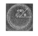
- FIG. 4 is an explanatory diagram of a process of averaging luminance values of points on a line U; The figure which shows the relationship between the angle (phi) of an ovum, and radius r (phi).
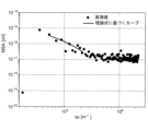
- FIG. 4 is a diagram showing values of mean square amplitude (MSA) of radius calculated from measured data and a fitting curve using a theoretical formula;
- FIG. 4 is an explanatory diagram of a method for calculating the aspect ratio of a deformed ovum when inserted with an injection pipette. The figure which shows the deformation
- FIG. 4 is an explanatory diagram of how to obtain an index value for relaxation of deformation of an ovum after opening a hole by inserting an injection pipette.
- FIG. 10 is a diagram showing the temporal change in deformation relaxation of the ovum after opening the hole by inserting the injection pipette. Explanatory drawing of how to determine the internal area of the ovum at the time of inserting the injection pipette.
- FIG. 1 is a flow chart showing a series of operations and processing procedures in microinsemination using one embodiment of the oocyte evaluation method according to the present invention.
- FIG. 2 is a flowchart showing a detailed processing procedure of pre-insemination egg evaluation processing in the work/processing procedure of microinsemination shown in FIG.
- FIG. 3 is a schematic configuration diagram of an embodiment of an egg evaluation apparatus according to the present invention for performing the pre-fertilization egg evaluation process and the post-fertilization egg evaluation process shown in FIG.
- the ovum evaluation method and ovum evaluation apparatus of the present embodiment are mainly for evaluating the quality of ova and selecting high-quality ova when performing microinsemination, which is one of assisted reproductive medicines. .
- some of the oocyte evaluation methods of the present embodiment can also be used to evaluate and select oocytes for in vitro fertilization instead of intracytoplasmic insemination.
- the doctor When performing microinsemination, the doctor first collects eggs from the patient. Usually, a plurality of ova are collected from a patient (step S1).
- the embryologist sets the collected ova either collectively or individually in the ovum evaluation device described later, and performs a predetermined operation on the device.
- the oocyte evaluation apparatus performs pre-fertilization oocyte evaluation processing as the first stage oocyte evaluation on the plurality of oocytes (step S2).
- the embryologist selects one or a plurality of ova to be subjected to microinsemination based on the evaluation result of this ovum evaluation device (step S3).
- Embryologists carry out insemination while observing the selected eggs under a microscope.
- the state of the ovum during the insemination operation is photographed as a moving image by the ovum evaluation apparatus (step S4).
- the embryologist then performs the prescribed operations on the egg evaluation device.
- the ovum evaluation apparatus performs the ova evaluation process at the time of fertilization based on the image taken during the fertilization work as the second stage ovum evaluation (step S5).
- the embryologist selects one or a plurality of eggs (fertilized eggs) to be cultured (or cryopreserved) based on the evaluation result by the egg evaluation device (step S6).
- the embryologist cultures the selected fertilized egg for a predetermined period, and after the culture, the doctor transplants the cultured embryo into the patient (step S7).
- the embryologist freezes and preserves the fertilized egg after culturing for a predetermined period.
- the oocyte quality is evaluated in two stages, before fertilization and at the time of fertilization, using an oocyte evaluation apparatus, thereby determining the quality of the fertilized egg to be transplanted to the patient. Guaranteed.
- Both of these two stages of oocyte evaluation are evaluations at the stage before the fertilized oocytes are accommodated in the incubator, and oocytes with a high fertilization success rate can be selected at an early stage.
- culture loss can be reduced by efficiently selecting fertilized eggs to be accommodated in an incubator with a limited capacity.
- the number of waiting patients for infertility treatment can be reduced by increasing the turnover rate of incubators.
- This egg evaluation apparatus includes a microscopic observation unit 1 including an imaging unit 10 , an imaging control unit 2 , a data processing unit 3 , a main control unit 4 , an input unit 5 and a display unit 6 .
- the data processing unit 3 includes, as functional blocks, an image data storage unit 30, a contour extraction unit 31, a radius calculation unit 32, a radial displacement amount calculation unit 33, a mechanical parameter calculation unit 34, a first evaluation unit 35, an aspect ratio calculation unit. 36, and a second evaluator 37.
- the first evaluation unit 35 performs classification or regression by machine learning. have.
- the microscopic observation unit 1 may be either a bright field microscope or a phase contrast microscope.
- the imaging unit 10 may acquire either a color image or a monochrome image.
- the imaging unit 10 may be a video camera capable of capturing moving images at a general frame rate (60 frames/second), or may be a camera with a moderately reduced frame rate that performs time-lapse photography at predetermined time intervals. good.
- At least some of the functions of the data processing unit 3 and the main control unit 4 are performed using a computer such as a personal computer as a hardware resource, and control/processing software (programs) pre-installed in the computer is executed by the computer.
- control/processing software programs pre-installed in the computer is executed by the computer.
- the input unit 5 and the display unit 6 are respectively a keyboard, a pointing device (mouse, etc.) and a monitor attached to the personal computer.
- This computer program is one embodiment of the oocyte evaluation program according to the present invention.
- the embryologist (or other person in charge) sets the target egg 100 to be evaluated on the stage of the microscopic observation unit 1 so that the polar body does not appear in the image, and starts analysis from the input unit 5.
- Perform an operation to instruct Upon receiving this instruction, the imaging control unit 2 operates the imaging unit 10 to capture a moving image of the ovum 100 for a predetermined time (for example, about 30 seconds) (step S20).
- the moving image data obtained by the imaging unit 10 is transferred to the data processing unit 3 and temporarily stored in the image data storage unit 30 .
- the contour extraction section 31 in the data processing section 3 executes processing for extracting the contour of the ovum in each frame image of the moving image (step S21). Specifically, the following processing is executed.
- the contour extraction unit 31 first performs noise removal processing using a Gaussian filter, median filter, or the like as preprocessing on each frame image.
- FIG. 4 is an example of a photographed image of an ovum. An egg (egg cell) is covered with a cell membrane, but there is a kind of protective layer called the zona pellucida on the outside of the cell membrane.
- the noise removal process is a process for removing fine sesame-salt noise that occurs in the vicinity of this clear zone or cell membrane on the image.
- the contour extracting unit 31 then obtains a luminance profile showing changes in luminance values of pixels (or pixels at predetermined intervals) along a straight line extending outward from the center point of the ovum for each frame image.
- FIG. 5 is a luminance profile on straight line U shown in FIG.
- the center point of the egg can be obtained by approximating the center coordinates of a circle using, for example, the method of least squares (calculating the center of the circle from the contour coordinates).
- the contour extraction unit 31 obtains the luminance value of each point (pixel) on the luminance profile by averaging the luminance values of a total of five points including two points in front and two points in the rear. Executes a smoothing process that replaces the
- FIG. 6 is an explanatory diagram of this smoothing process.
- the luminance value of the center point of the five points is , as shown in FIG . )/5.
- the contour extraction unit 31 extracts the maximum value (or minimum value) of the differential values in the luminance profile after smoothing along a plurality of straight lines extending radially in different directions from the center point of the ovum. Select as a point. Concerning the 360° angle range around the central point of the ovum, points on the contour are obtained on each straight line drawn at predetermined angular intervals, and by connecting these points, the contour of the target ovum is extracted with high accuracy. be able to.
- Non-Patent Document 4 authored by some of the present inventors, discloses extracting the contour from an image of red blood cells, which have a shape close to a circle, and determining the radius (the distance from the center point to the contour). It is The present inventors examined whether a similar method can be applied to ova, but it is difficult to apply it as it is. The reason for this is that in the case of erythrocytes, the shape of the curve of the luminance profile is smooth and the contour appears quite clearly, whereas in the case of ova, due to the thick zona pellucida existing outside the cell membrane, especially This is because there is a lot of noise in the vicinity of the cell membrane, which makes contour extraction difficult.
- the contour extracting unit 31 first calculates a brightness profile along radial straight lines in 64 directions at (360/64)° angular intervals around the center point of the ovum. Roughly extract the range. After that, while gradually narrowing the calculation range of the brightness profile in the radial direction to the range that is estimated to include the contour, radial straight lines in 256 directions at (360/256)° angular intervals around the center point of the oocyte By repeating the same calculation three times along the contour, a highly accurate contour is finally obtained.
- the contour extraction unit 31 similarly extracts the contours of the ovum for all the frame images that make up the moving image over a predetermined period of time.
- the radius calculation unit 32 calculates the radius (strictly speaking, the distance from the center point to the contour) in each direction at each predetermined angular interval ⁇ around the center point of the egg for each image of each frame. (step S22). That is, as shown in FIG. 7, the radius r( ⁇ ) is calculated for a plurality of predetermined angles ⁇ .
- ⁇ 1°
- the center point for calculating the radius should be the position of the center of gravity calculated from the contour.
- the shape of an egg which is a type of cell, changes over time due to fluctuations.
- the radial displacement amount calculator 33 calculates the radial displacement amount reflecting the shape fluctuation of the ovum, based on a huge amount of information on the radius for each image of each frame (step S23). Therefore, the method disclosed in Non-Patent Document 4 is used here. Specifically, the radial displacement amount calculator 33 calculates the mean square amplitude (MSA), which is a function of the wave number q, by performing a Fourier transform operation using the following equation (1).
- MSA mean square amplitude
- ⁇ > indicates the temporal average over all frames.
- Formula (1) obtains the difference between the radius and the temporal average of the radius for each same direction, and the temporal change of the value obtained by adding the difference for all directions, is converted into a function of the wave number by Fourier transform.
- FIG. 8 plots the values of the mean square amplitude calculated by the above procedure based on the measured data.
- q x is the continuous wave number corresponding to the experimental q.
- L is the length in one dimension of the cell (ovum in this case).
- k B is the Boltzmann constant
- T is the absolute temperature during the experiment.
- the other ⁇ , ⁇ , and ⁇ are unknown mechanical parameters, ⁇ is the spring constant, ⁇ is the surface tension, and ⁇ is the flexural elasticity.
- the dynamic parameter calculation unit 34 calculates three unknown dynamic parameters by fitting the equation (2) to the mean square amplitude calculated from the measured data as described above (step S24). In FIG. 8, the solid line shows the curve when this fitting is performed.
- These three mechanical parameters are index values that reflect the mechanical properties of eggs, that is, hardness and softness. Thus, it quantifies the mechanical properties of the egg.
- the first evaluation unit 35 obtains information on the quality of the egg from the values of the mechanical parameters of the egg calculated as described above (step S25).
- the egg evaluation apparatus of this embodiment uses a machine learning technique for this quality evaluation. That is, the first evaluation unit 35 receives the values of the three mechanical parameters as input, and outputs an evaluation result in which a good-quality egg is "1" and a poor-quality egg is "0". By performing classification using a trained model based on the algorithm, the quality of the egg is determined.
- a trained model is created in advance (for example, before the manufacturer ships the device) as follows. That is, the mechanical parameters are determined according to the procedure as described above for one or a plurality of ova collected from each of a large number of patients.
- a skilled embryologist evaluates the quality of each egg in the process of normal insemination and subsequent culture of those eggs, and leaves the evaluation results.
- the evaluation result by the embryologist can be binary information indicating whether the quality is good, such as suitability for transplantation, suitability for cryopreservation, or the like. Alternatively, it may be multivalued information according to a known multi-stage evaluation method such as Veeck classification or Gardner classification.
- the evaluation result can be that the quality is poor without the embryologist's judgment.
- this training data can be learned by a neural network or the like. can create a trained model, or egg quality classifier.
- This learned model is stored in the learned model storage unit 350 .
- the work of creating a trained model based on teacher data as described above may be performed by the user, but is generally performed by the manufacturer of the device or the software provider.
- the quality of the egg is output as an output. You can get results.
- This evaluation result is output from the display section 6 through the main control section 4 .
- the first evaluation unit 35 may use a binary classification machine learning algorithm. Even if the correct data is binary, the probability of good quality can be calculated as a numerical value by using a machine learning algorithm of regression analysis such as logistic regression. Therefore, in that case, for example, by comparing the calculated probability with a threshold value, the quality of the egg can be determined.
- the first evaluation unit 35 evaluates the evaluation results of egg quality. , fertilization rate and implantation rate. In this case also, the first evaluation unit 35 determines whether the numerical value of the probability output as the evaluation result is equal to or greater than a predetermined threshold, thereby obtaining and displaying the quality result of the egg. It can be displayed in part 6. Of course, such a numerical value of probability may be displayed together with the quality judgment result, or only the numerical value of probability may be displayed.
- infertility treatment involves collecting multiple eggs from a patient, but it is necessary to narrow down to one of them in the end. Therefore, when the probability that the egg quality is good is obtained as a numerical value as described above, the first evaluation unit 35 ranks a plurality of eggs collected from one patient based on the numerical value, and ranks the eggs. The ranking result can be displayed on the display unit 6 . After confirming this, the embryologist (doctor) refers to the ranking, selects the target egg for microinsemination from among the multiple eggs, and selects the appropriate egg to return to the patient's body. can be elected.
- multistage evaluation methods such as the above-mentioned Veeck classification and Gardner classification are well known as methods for evaluating the quality of fertilized eggs by embryologists.
- the Veeck classification is an index for evaluating the quality of early embryos on the 2nd to 4th day of culture with fertilized oocytes, and is classified into 5 grades.
- the Gardner classification is an index for evaluating the state of blastocysts on the 5th to 6th day of culture, and is classified according to 6 grades.
- These are methods for evaluating eggs after fertilization, but a trained model is created by learning using teacher data with the results of grading in Veeck classification and Gardner classification as correct data, and this trained model is used. It may be used to obtain grading results in the Veeck classification or the Gardner classification from the mechanical parameter values of the ovum before fertilization.
- the mechanical parameter of the egg is used as an evaluation index, but also various information other than the mechanical parameter of the egg, such as the size of the egg. It may be added as an evaluation index.
- patient-specific information such as the patient's age, past results of artificial insemination, and past medical history may be added as an evaluation index.
- the patient's age since it is known that the patient's age has a significant effect on egg quality, it is highly relevant to use information on the patient's age as one of the evaluation indicators. In that case, it is possible to treat the age as an input of a trained model equivalent to a mechanical parameter, but instead of the results obtained by machine learning, e.g. The threshold for judging that the quality is good, and the threshold for judging that the quality is good when the probability of expressing good quality is changed according to the age of the patient. can be
- the present inventors used a trained model created to evaluate the quality of ova from the standpoint of suitability for transplantation into the mother's body to determine whether ova collected from actual patients can be accurately evaluated. It was verified experimentally. In this verification, the quality of ova collected from 14 patients was evaluated based on measured mechanical parameters. As a result, we were able to confirm that the results of the evaluation using the trained model were almost equivalent to the evaluation of oocyte quality by a skilled embryologist.
- the embryologist refers to the results of the evaluation and selects the best quality oocytes out of multiple oocytes collected from a single patient. select one or more and discard the others. Then, the embryologist performs the microinsemination operation on the selected oocytes with good quality.
- the method of microinsemination is exactly the same as before, and the embryologist inserts an injection pipette into the egg while observing the egg under a microscope to inject sperm.
- this microscopic insemination operation is performed under observation by the microscopic observation unit 1 of the oocyte evaluation apparatus shown in FIG.
- the imaging unit 10 captures the state of the ovum during the operation, particularly the state of the ovum from immediately before the injection pipette is inserted into the ovum until a predetermined time has passed after the injection pipette is removed. do.
- the obtained moving image data is transferred to the data processing section 3 and temporarily stored in the image data storage section 30 .
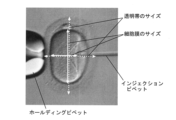
- FIG. 9 is an image showing the state of an egg when an injection pipette is inserted into the egg for microinsemination.
- the zona pellucida is greatly deformed as the injection pipette is moved, and when a hole is formed in the zona pellucida, the deformation is relaxed and the zona returns to its original state.
- the cell membrane of the ovum As the injection pipette moves, the cell membrane is greatly deformed so as to be depressed inward.
- the aspect ratio calculation unit 36 selects, from the stored moving image, the clear zone after the clear zone is greatly deformed at the time of inserting the injection pipette as described above, and immediately before the deformation starts to be relaxed. Detects the image in which is deformed the most.
- the aspect ratio calculator 36 calculates the length in the short axis direction and the length in the long axis direction of the transparencies from the image, and obtains the ratio of the lengths as the aspect ratio.
- the length in the minor axis direction is the width of the zona pellucida at a position passing through substantially the center of the ovum along the advance and retreat direction of the injection pipette (horizontal direction in FIG. 9).
- the length in the long axis direction is the width of the zona pellucida at a position passing through substantially the center of the ovum along the direction perpendicular to the advancing/retreating direction of the injection pipette (the vertical direction in FIG. 9).
- the above aspect ratio of the zona pellucida which is part of the egg, is affected by the mechanical properties of the zona pellucida, specifically its elasticity.
- the aspect ratio is a useful index for measuring the mechanical properties of the egg, and since the mechanical properties of the egg influence the quality of the egg, the quality of the egg can be evaluated based on the aspect ratio. can. Therefore, the second evaluation unit 37 receives the value of the aspect ratio and compares it with a predetermined threshold to determine the quality of the egg. Then, the determination result of the egg quality is displayed on the display unit 6 through the main control unit 4 .
- an index other than the aspect ratio for example, personal information such as the patient's age may be used to evaluate the quality of the ovum.
- the aspect ratio of the cell membrane instead of the zona pellucida, that is, the aspect ratio in the most deformed state immediately before the deformation began to be relaxed after the cell membrane was greatly deformed when the injection pipette was inserted, and this aspect ratio was calculated. may be used to determine the quality of the egg.
- both the aspect ratio of the zona pellucida and the aspect ratio of the cell membrane may be used to determine the quality of the egg.
- the fertilization rate and implantation rate may be poor. Therefore, by excluding fertilized eggs that are evaluated to be of poor quality at the time of insemination from being cultured, loss in culture of fertilized eggs can be reduced and the incubator can be used effectively.
- the aspect ratio at the time of maximum deformation of the zona pellucida and/or the cell membrane was used to evaluate the ovum at the time of insemination. can also be used to assess egg quality.
- Fig. 10 is an example of an image showing the state of an ovum held by a holding pipette.
- the cell membrane is deformed so as to protrude clearly due to suction by the holding pipette.
- the cell membrane hardly protrudes despite the suction by the holding pipette.
- This difference in deformation reflects the difference in elasticity of the ovum. Therefore, the second evaluation unit 37 can determine the quality of the ovum by determining the presence or absence of deformation through image processing, for example.
- the quality of the egg may be determined by quantifying the amount of deformation (the amount of protrusion) and comparing the quantified value with a threshold value.
- the second evaluation unit 37 determines the time required for the deformation of the cell membrane to relax (return to its original state) from the moment the injection pipette penetrates the cell membrane (hereinafter referred to as "relaxation time") from the moving image acquired during the insemination work. ), and the quality of the egg can be determined based on this relaxation time.
- FIG. 11 is a diagram showing a site where changes in the cell membrane are observed when obtaining an index value for relaxation of deformation of the ovum after the hole is opened by inserting the injection pipette.
- FIG. 12 is a diagram showing an example of temporal changes in cell membrane deformation (egg deformation relaxation) after injection pipette penetration.
- FIG. 11 is an image showing the state just before the tip of the injection pipette penetrates the cell membrane.
- the deformation of the cell membrane is focused on the three sizes x 1 , x 2 and x 3 shown in the figure. do.
- x 1 is the distance from the position of the tip of the injection pipette just before the cell membrane opens to the position of the cell membrane facing in front of it.
- x2 is the depth of the cell membrane depression formed by being pushed by the injection pipette.
- x 3 is the edge diameter of the cell membrane depression formed by being pushed by the injection pipette. All of these show maximum values just before the tip of the injection pipette penetrates the cell membrane, and the values decrease with the passage of time after penetration.
- the above-described machine learning technique may be used to input the three relaxation times ⁇ i and derive the evaluation result of the ovum.
- the relaxation time of at least one of x 1 , x 2 , and x 3 shown in FIG. 11 may be used to evaluate egg quality.
- the quality of the egg may be evaluated using at least one size of x1 , x2 , and x3 at the time when the cell membrane is maximally deformed, as an evaluation index.
- FIG. 13 shows a portion (A) surrounded by a zona pellucida and a portion (B) surrounded by cell membranes on an image of an ovum.
- the change in the area of the part also reflects the elasticity of the ovum. Therefore, by using such information as an evaluation index, it is also possible to evaluate the quality of ova.
- the evaluation of egg quality by the first evaluation unit 35 and the evaluation of egg quality by the second evaluation unit 37 are completely independent evaluations, it is obvious that only one of them may be performed. be.
- the combined use of both methods makes it possible to narrow down the eggs to be fertilized and the eggs to be cultured (fertilized eggs).
- the evaluation of egg quality by the first evaluation unit 35 is also useful when selecting eggs to be subjected to in vitro fertilization instead of microinsemination.
- the oocyte evaluation apparatus of the above-described embodiment was able to perform both oocyte evaluation before fertilization and oocyte evaluation during insemination, they can be performed by separate apparatuses.
- the egg evaluation apparatus outputs the evaluation result or determination result of the set egg quality, and the embryologist selects the egg by referring to this.
- One aspect of the ovum evaluation method is a method for evaluating an ovum, an analysis step of non-invasively obtaining an index value that quantifies the mechanical properties of the target egg by analyzing the state of deformation of the target egg using a photographed image of the target egg; an evaluation step of evaluating the target egg quality based on the index value obtained by the analysis step; to implement.
- one aspect of the ovum evaluation apparatus is An image obtained by photographing a target ovum is received, and the state of deformation of the target ovum is analyzed using the image to noninvasively obtain an index value that quantifies the mechanical characteristics of the target ovum. an analysis unit; an evaluation unit that evaluates the target egg quality based on the index value obtained by the analysis unit; Prepare.
- one aspect of the oocyte evaluation program is a program for evaluating oocytes using a computer, comprising: an analysis step of non-invasively obtaining an index value that quantifies the mechanical properties of the target egg by analyzing the state of deformation of the target egg using a photographed image of the target egg; an evaluation step of evaluating the target egg quality based on the index value obtained by the analysis step; is executed.
- the quality of the target ovum can be accurately non-invasively evaluated based on the index value that quantifies the mechanical properties. , that is, it can be evaluated with theoretical backing.
- the ovum is not damaged during evaluation of the quality of the ovum, and the ovum after evaluation can be used favorably for its original purpose, for example.
- the work burden on the embryologist is reduced, and variations in the quality evaluation depending on the skills of the embryologist in charge are eliminated, improving the reliability of the evaluation of oocyte quality.
- the data obtained when evaluating the quality of the ovum remains as numerical values, it is easy for an embryologist or the like to later verify whether or not the evaluation of the quality was appropriate.
- the target oocyte is an oocyte before fertilization
- the image is a moving image or a time-lapse image of the target oocyte
- the analyzing step includes a contour extracting step of extracting a contour of a target ovum from a plurality of images constituting a moving image or a time-lapse image, a displacement obtaining step of obtaining a displacement of the contour of the target ovum, and and a mechanical parameter calculation step of calculating a mechanical parameter of the egg as the index value from the amount of displacement of the contour of the egg.
- the target egg is an egg before fertilization
- the image is a moving image or a time-lapse image of the target egg
- the analysis unit includes a contour extraction processing unit that extracts a contour of a target ovum from a plurality of images constituting a moving image or a time-lapse image, and a displacement amount acquisition processing unit that obtains a displacement amount of the contour of the target ovum.
- a mechanical parameter calculation processing unit that calculates a mechanical parameter of the egg as the index value from the amount of displacement of the outline of the target egg.
- the shape of an egg changes over time due to fluctuations.
- the change in the shape of the ovum due to this fluctuation is affected by the elasticity reflecting the hardness of the ovum.
- changes in the shape of the egg are obtained as contour displacement amount information.
- the mechanical parameter calculation step the mechanical parameter of the egg is calculated from information on the amount of displacement of the contour.
- the mechanical parameter in the mechanical parameter calculation step, the mechanical parameter may be obtained by performing parameter fitting using a theoretical formula representing cell elasticity. can.
- the mechanical parameter calculation processing unit performs parameter fitting using a theoretical formula representing the elasticity of the cell with respect to the measured value representing the displacement amount of the outline of the target ovum.
- the dynamic parameter can be obtained by performing
- the usual way is to apply some force to the object and measure the deformation rate of the object against that force.
- information regarding the elasticity of the ovum is obtained with high accuracy without applying any force to the ovum or restricting its movement, even indirectly. be able to.
- the evaluation results of the oocytes by an embryologist in at least one stage from fertilization to implantation are evaluated.
- the oocyte quality can be evaluated using a discriminator obtained by machine learning using training data including training data or training data in which information on the probability of success or failure in any of the stages is known. .
- the evaluation unit includes teacher data including the evaluation results of the oocytes by the embryologist at at least one stage from fertilization to implantation,
- the oocyte quality can be evaluated using a discriminator created in advance by machine learning using teacher data in which information on the probability of success or failure in any of the stages is known.
- the machine learning algorithm used does not matter.
- the ovum evaluation method and ovum evaluation apparatus of the fourth aspect the ovum before culture is accurately evaluated by reflecting the judgment result of the adequacy of transplantation of the fertilized egg by a highly skilled embryologist. be able to.
- the egg evaluation method and the egg evaluation apparatus of the fourth aspect it is possible to accurately evaluate the quality of the egg by reflecting past success or failure results such as fertilization rate and implantation rate.
- the evaluation step evaluates the quality of the egg using patient-specific information including age in addition to the index value.
- the evaluation unit evaluates the quality of the ovum using patient-specific information including age input in advance in addition to the index value.
- the evaluation unit ranks the plurality of ova using the information regarding the quality of the ova obtained for each of the plurality of ova. can do.
- one of the plurality of ova collected from the patient which is estimated to have the highest probability of pregnancy, or has a relatively high possibility of pregnancy.
- a small number of eggs presumed to be fertilized can be subjected to microinsemination.
- the image is an image taken when a needle is inserted into the ovum for the purpose of microinsemination
- An index value may be determined.
- the image is an image captured when the needle is inserted into the ovum for the purpose of microinsemination
- the analysis part reflects the index value reflecting the degree of deformation of the zona pellucida or cell membrane at the time of maximum deformation due to needle insertion, or the degree of relaxation of the deformation of the zona pellucida or cell membrane due to needle insertion. An index value may be determined.
- the quality of the egg is evaluated from the index values calculated based on the images taken when microinsemination is performed.
- this assessment of egg quality can also be said to be non-invasive in that no special invasive measurements or observations are made to assess egg quality.
- the sensory and qualitative evaluation such as elasticity or lack of elasticity, which has been conventionally performed by embryologists when performing microinsemination Quantitative evaluation using specific numerical values can be performed instead. This improves the accuracy and reliability of egg quality assessment. Furthermore, the data obtained when evaluating the quality of the ovum remains as a numerical value, making it easy to verify the accuracy of the evaluation.
- the index value reflecting the degree of deformation at the time of maximum deformation is obtained by inserting a needle at the time of maximum deformation of the zona pellucida or cell membrane of the target oocyte. It can be a ratio of widths in two directions, ie, a direction and a direction orthogonal thereto.
- the index value reflecting the degree of deformation at the time of maximum deformation is the direction of insertion of the needle at the time of maximum deformation of the zona pellucida or cell membrane of the target egg, and the direction orthogonal to it. It can be the ratio of the widths in the two directions with respect to the direction in which the
- the ovum evaluation method and ovum evaluation apparatus of the eighth aspect it is possible to accurately evaluate the quality of the ovum based on the index value that accurately reflects the degree of elasticity of the ovum during microinsemination.
- a selection step of selecting eggs using the evaluation results in the evaluation step can be further implemented.
- the burden of the ovum selection work by the embryologist is reduced.
- ova can be selected based on evaluation results with theoretical backing rather than sensory evaluation, which improves the reliability of selection.
- the quality of the egg before fertilization is evaluated by the analysis step and the evaluation step,
- the deformation of the ova is analyzed using images taken when a needle is inserted into the ova for the purpose of microinsemination, and the mechanical properties are numerically evaluated.
- a second evaluation step of evaluating the quality of the ovum subjected to microinsemination based on the index value obtained by the second analysis step; shall be further implemented.
- a second analysis unit that non-invasively obtains an index value that quantifies the mechanical properties of the egg by analyzing the deformation mode of the egg using the a second evaluation unit that evaluates the quality of an egg subjected to microinsemination based on the index value obtained by the second analysis unit; can be further provided.
- the sorting is performed.
- the quality of the ovum can be evaluated again by the ovum evaluation method of the seventh aspect.
Landscapes
- Engineering & Computer Science (AREA)
- Health & Medical Sciences (AREA)
- Life Sciences & Earth Sciences (AREA)
- Chemical & Material Sciences (AREA)
- General Health & Medical Sciences (AREA)
- Biomedical Technology (AREA)
- Physics & Mathematics (AREA)
- Bioinformatics & Cheminformatics (AREA)
- Organic Chemistry (AREA)
- Zoology (AREA)
- Wood Science & Technology (AREA)
- General Physics & Mathematics (AREA)
- Biochemistry (AREA)
- Genetics & Genomics (AREA)
- Medical Informatics (AREA)
- Biotechnology (AREA)
- Theoretical Computer Science (AREA)
- Analytical Chemistry (AREA)
- Computer Vision & Pattern Recognition (AREA)
- Molecular Biology (AREA)
- Nuclear Medicine, Radiotherapy & Molecular Imaging (AREA)
- General Engineering & Computer Science (AREA)
- Microbiology (AREA)
- Immunology (AREA)
- Pathology (AREA)
- Public Health (AREA)
- Quality & Reliability (AREA)
- Radiology & Medical Imaging (AREA)
- Surgery (AREA)
- Sustainable Development (AREA)
- Primary Health Care (AREA)
- Epidemiology (AREA)
- Medicinal Chemistry (AREA)
- Food Science & Technology (AREA)
- Urology & Nephrology (AREA)
- Hematology (AREA)
- Biophysics (AREA)
- Optics & Photonics (AREA)
- Reproductive Health (AREA)
- Cell Biology (AREA)
Priority Applications (4)
| Application Number | Priority Date | Filing Date | Title |
|---|---|---|---|
| EP22880795.4A EP4417700A4 (en) | 2021-10-14 | 2022-09-29 | Ovum evaluation method, ovum evaluation device, and program for evaluating ovum |
| JP2023555104A JP7807018B2 (ja) | 2021-10-14 | 2022-09-29 | 卵子評価方法、卵子評価装置、及び卵子評価用プログラム |
| CN202280069267.9A CN118176304A (zh) | 2021-10-14 | 2022-09-29 | 卵子评价方法、卵子评价装置以及卵子评价用程序 |
| US18/701,534 US20240319163A1 (en) | 2021-10-14 | 2022-09-29 | Ovum Evaluation Method, Ovum Evaluation Apparatus, and Ovum Evaluation Program |
Applications Claiming Priority (2)
| Application Number | Priority Date | Filing Date | Title |
|---|---|---|---|
| JP2021168634 | 2021-10-14 | ||
| JP2021-168634 | 2021-10-14 |
Publications (1)
| Publication Number | Publication Date |
|---|---|
| WO2023063099A1 true WO2023063099A1 (ja) | 2023-04-20 |
Family
ID=85988302
Family Applications (1)
| Application Number | Title | Priority Date | Filing Date |
|---|---|---|---|
| PCT/JP2022/036466 Ceased WO2023063099A1 (ja) | 2021-10-14 | 2022-09-29 | 卵子評価方法、卵子評価装置、及び卵子評価用プログラム |
Country Status (5)
| Country | Link |
|---|---|
| US (1) | US20240319163A1 (https=) |
| EP (1) | EP4417700A4 (https=) |
| JP (1) | JP7807018B2 (https=) |
| CN (1) | CN118176304A (https=) |
| WO (1) | WO2023063099A1 (https=) |
Families Citing this family (1)
| Publication number | Priority date | Publication date | Assignee | Title |
|---|---|---|---|---|
| US12354267B1 (en) * | 2024-06-24 | 2025-07-08 | Inti Taiwan, Inc. | Oocyte quality analysis system |
Citations (2)
| Publication number | Priority date | Publication date | Assignee | Title |
|---|---|---|---|---|
| JP2012531584A (ja) * | 2009-06-25 | 2012-12-10 | フェイズ ホログラフィック イメージング ペーホーイー アーベー | デジタルホログラフィック撮像による卵又は胚の分析 |
| JP2016054684A (ja) | 2014-09-09 | 2016-04-21 | 国立大学法人名古屋大学 | マイクロ流体チップ及びマイクロ流体チップ内のマイクロプローブの駆動方法、弾性粒子の駆動方法、および装置 |
Family Cites Families (1)
| Publication number | Priority date | Publication date | Assignee | Title |
|---|---|---|---|---|
| WO2021014461A1 (en) * | 2019-07-19 | 2021-01-28 | Payeli Sravan Kumar | Intracytoplasmic sperm injection |
-
2022
- 2022-09-29 WO PCT/JP2022/036466 patent/WO2023063099A1/ja not_active Ceased
- 2022-09-29 US US18/701,534 patent/US20240319163A1/en active Pending
- 2022-09-29 CN CN202280069267.9A patent/CN118176304A/zh active Pending
- 2022-09-29 EP EP22880795.4A patent/EP4417700A4/en active Pending
- 2022-09-29 JP JP2023555104A patent/JP7807018B2/ja active Active
Patent Citations (2)
| Publication number | Priority date | Publication date | Assignee | Title |
|---|---|---|---|---|
| JP2012531584A (ja) * | 2009-06-25 | 2012-12-10 | フェイズ ホログラフィック イメージング ペーホーイー アーベー | デジタルホログラフィック撮像による卵又は胚の分析 |
| JP2016054684A (ja) | 2014-09-09 | 2016-04-21 | 国立大学法人名古屋大学 | マイクロ流体チップ及びマイクロ流体チップ内のマイクロプローブの駆動方法、弾性粒子の駆動方法、および装置 |
Non-Patent Citations (10)
| Title |
|---|
| BARNETT-ITZHAKI ZOHAR; ELBAZ MIRIAM; BUTTERMAN RACHELY; AMAR DEVORA; AMITAY MOSHE; RACOWSKY CATHERINE; ORVIETO RAOUL; HAUSER RUSS;: "Machine learning vs. classic statistics for the prediction of IVF outcomes", JOURNAL OF ASSISTED REPRODUCTION AND GENETICS., PLENUM PUBLISHING., US, vol. 37, no. 10, 11 August 2020 (2020-08-11), US , pages 2405 - 2412, XP037267365, ISSN: 1058-0468, DOI: 10.1007/s10815-020-01908-1 * |
| FORCE MEASUREMENT IN MICROENVIRONMENT USING TWO-FINGERED MICRO HAND (ARAI LABORATORY, OSAKA UNIVERSITY, 22 January 2021 (2021-01-22), Retrieved from the Internet <URL:<URL:http://www.arailab.sys.es.osaka-u.ac.jp/index.php?option=com-content&view=article&id=75&Itemid=97&lang=ja>> |
| ITO HIROAKI, KUSS NAVINA, RAPP BASTIAN E., ICHIKAWA MASATOSHI, GUTSMANN THOMAS, BRANDENBURG KLAUS, PÖSCHL JOHANNES M. B., TANAKA M: "Quantification of the Influence of Endotoxins on the Mechanics of Adult and Neonatal Red Blood Cells", JOURNAL OF PHYSICAL CHEMISTRY PART B, AMERICAN CHEMICAL SOCIETY, US, vol. 119, no. 25, 25 June 2015 (2015-06-25), US , pages 7837 - 7845, XP093057313, ISSN: 1520-6106, DOI: 10.1021/acs.jpcb.5b01544 * |
| ITO HIROAKI7 OTHER PERSONS: "Quantification of the influence of endotoxins on the mechanics of adult and neonatal red blood cells", THE JOURNAL OF PHYSICAL CHEMISTRY B, vol. 119, no. 25, 2015, pages 7837 - 7845, XP093057313, DOI: 10.1021/acs.jpcb.5b01544 |
| LIVIA Z. YANEZ4 OTHER PERSONS: "Human oocyte developmental potential is predicted by mechanical properties within hours after fertilization", NATURE COMMUNICATIONS, vol. 7, no. 1, 2016, pages 1 - 12 |
| MURAYAMA YOSHINOBU: "Evaluation of the Embryo Quality for Elective Single Embryo Transfer", FINAL RESEARCH REPORT OF GRANTS-IN-AID FOR SCIENTIFIC RESEARCH (SCIENTIFIC RESEARCH GRANTS), 31 July 2013 (2013-07-31), XP093057319, Retrieved from the Internet <URL:https://kaken.nii.ac.jp/ja/file/KAKENHI-PROJECT-20680026/20680026seika.pdf> [retrieved on 20230623] * |
| MURAYAMA YOSHINOBU9 OTHER PERSONS: "Elasticity measurement of zona pellucida using a micro tactile sensor to evaluate embryo quality", JOURNAL OF MAMMALIAN OVA RESEARCH, vol. 25, no. 1, 2008, pages 8 - 16, XP055206006, DOI: 10.1274/jmor.25.8 |
| See also references of EP4417700A4 |
| TARGOSZ ANNA, PRZYSTAŁKA PIOTR, WIADERKIEWICZ RYSZARD, MRUGACZ GRZEGORZ: "Semantic segmentation of human oocyte images using deep neural networks", BIOMEDICAL ENGINEERING ONLINE, vol. 20, no. 1, 1 December 2021 (2021-12-01), XP093057316, DOI: 10.1186/s12938-021-00864-w * |
| YAMAMOTO AKIHISA: "The hardness of eggs to assist sterility treatment/Softness quantitative evaluation technology of softness.", NEW TECHNOLOGY PRESENTATION MEETINGS! TLO-KYOTO CO., LTD. DEPARTMENT OF TECHNOLOGY TRANSFER, KYOTO UNIVERSITY, 28 June 2022 (2022-06-28), XP093057312, Retrieved from the Internet <URL:https://shingi.jst.go.jp/pdf/2022/2022_kyoto-u_001.pdf> [retrieved on 20230623] * |
Also Published As
| Publication number | Publication date |
|---|---|
| CN118176304A (zh) | 2024-06-11 |
| EP4417700A1 (en) | 2024-08-21 |
| EP4417700A4 (en) | 2025-04-23 |
| JPWO2023063099A1 (https=) | 2023-04-20 |
| US20240319163A1 (en) | 2024-09-26 |
| JP7807018B2 (ja) | 2026-01-27 |
Similar Documents
| Publication | Publication Date | Title |
|---|---|---|
| Bormann et al. | Consistency and objectivity of automated embryo assessments using deep neural networks | |
| Cherouveim et al. | Artificial intelligence for sperm selection—A systematic review | |
| Jiang et al. | Artificial intelligence in the in vitro fertilization laboratory: a review of advancements over the last decade | |
| US8265357B2 (en) | Determination of a change in a cell population | |
| US20250095150A1 (en) | Image Processing for Real-Time Ranking and Selection of Spermatozoa | |
| Arav et al. | Prediction of embryonic developmental competence by time-lapse observation and ‘shortest-half’analysis | |
| JP7692232B1 (ja) | 精子選別補助装置、精子選別補助システム、及び精子選別補助プログラム | |
| WO2024051016A1 (zh) | 一种用于模型小动物的肌肉超声图像分析方法 | |
| WO2023063099A1 (ja) | 卵子評価方法、卵子評価装置、及び卵子評価用プログラム | |
| Schoolcraft et al. | Paving the way for a gold standard of care for infertility treatment: improving outcomes through standardization of laboratory procedures | |
| Srivastava et al. | Unsupervised deep learning based longitudinal follicular growth tracking during IVF cycle using 3D transvaginal ultrasound in assisted reproduction | |
| JP2022547900A (ja) | 生殖補助手順に用いられる品質保証の評価指標の自動評価 | |
| Wong et al. | A method for analyzing AFM force mapping data obtained from soft tissue cryosections | |
| TWI859981B (zh) | 由計算機的系統實施的方法以及卵母細胞品質分析系統 | |
| CN104531827A (zh) | 细胞质量评价的方法 | |
| Citarella et al. | EmbryoVision AI: An explainable deep learning framework for enhanced blastocyst selection in assisted reproductive technologies | |
| Allahbadia et al. | In contemporary reproductive medicine human beings are not yet dispensable | |
| CN115919367B (zh) | 超声图像处理方法及装置、电子设备和存储介质 | |
| CN111938671A (zh) | 一种基于多维度内感知特征的焦虑特质量化方法 | |
| JP7630131B2 (ja) | 細胞機能の評価方法及び細胞解析装置 | |
| Mittal et al. | Revolutionizing Retinal Health: Transfer Learning for Early Detection of Pathologies in Limited OCT Image Datasets | |
| Zeng et al. | Future perspectives of non-invasive techniques for evaluating oocyte and embryo quality | |
| US12354267B1 (en) | Oocyte quality analysis system | |
| Kevin et al. | Automatic image analysis of zebrafish embryo development for lab-on-a-chip | |
| JP7840606B1 (ja) | 精子を追跡するための情報処理装置、方法、及びプログラム |
Legal Events
| Date | Code | Title | Description |
|---|---|---|---|
| 121 | Ep: the epo has been informed by wipo that ep was designated in this application |
Ref document number: 22880795 Country of ref document: EP Kind code of ref document: A1 |
|
| ENP | Entry into the national phase |
Ref document number: 2023555104 Country of ref document: JP Kind code of ref document: A |
|
| WWE | Wipo information: entry into national phase |
Ref document number: 202280069267.9 Country of ref document: CN |
|
| WWE | Wipo information: entry into national phase |
Ref document number: 18701534 Country of ref document: US |
|
| WWE | Wipo information: entry into national phase |
Ref document number: 2022880795 Country of ref document: EP |
|
| NENP | Non-entry into the national phase |
Ref country code: DE |
|
| ENP | Entry into the national phase |
Ref document number: 2022880795 Country of ref document: EP Effective date: 20240514 |