WO2023053586A1 - イムノクロマトグラフキット及び検査装置 - Google Patents
イムノクロマトグラフキット及び検査装置 Download PDFInfo
- Publication number
- WO2023053586A1 WO2023053586A1 PCT/JP2022/023441 JP2022023441W WO2023053586A1 WO 2023053586 A1 WO2023053586 A1 WO 2023053586A1 JP 2022023441 W JP2022023441 W JP 2022023441W WO 2023053586 A1 WO2023053586 A1 WO 2023053586A1
- Authority
- WO
- WIPO (PCT)
- Prior art keywords
- substance
- test
- inspection
- luminescent substrate
- labeled
- Prior art date
- Legal status (The legal status is an assumption and is not a legal conclusion. Google has not performed a legal analysis and makes no representation as to the accuracy of the status listed.)
- Ceased
Links
Images




Classifications
-
- G—PHYSICS
- G01—MEASURING; TESTING
- G01N—INVESTIGATING OR ANALYSING MATERIALS BY DETERMINING THEIR CHEMICAL OR PHYSICAL PROPERTIES
- G01N21/00—Investigating or analysing materials by the use of optical means, i.e. using sub-millimetre waves, infrared, visible or ultraviolet light
- G01N21/75—Systems in which material is subjected to a chemical reaction, the progress or the result of the reaction being investigated
- G01N21/76—Chemiluminescence; Bioluminescence
-
- B—PERFORMING OPERATIONS; TRANSPORTING
- B01—PHYSICAL OR CHEMICAL PROCESSES OR APPARATUS IN GENERAL
- B01L—CHEMICAL OR PHYSICAL LABORATORY APPARATUS FOR GENERAL USE
- B01L3/00—Containers or dishes for laboratory use, e.g. laboratory glassware; Droppers
- B01L3/50—Containers for the purpose of retaining a material to be analysed, e.g. test tubes
- B01L3/502—Containers for the purpose of retaining a material to be analysed, e.g. test tubes with fluid transport, e.g. in multi-compartment structures
-
- B—PERFORMING OPERATIONS; TRANSPORTING
- B01—PHYSICAL OR CHEMICAL PROCESSES OR APPARATUS IN GENERAL
- B01L—CHEMICAL OR PHYSICAL LABORATORY APPARATUS FOR GENERAL USE
- B01L3/00—Containers or dishes for laboratory use, e.g. laboratory glassware; Droppers
- B01L3/50—Containers for the purpose of retaining a material to be analysed, e.g. test tubes
- B01L3/502—Containers for the purpose of retaining a material to be analysed, e.g. test tubes with fluid transport, e.g. in multi-compartment structures
- B01L3/5023—Containers for the purpose of retaining a material to be analysed, e.g. test tubes with fluid transport, e.g. in multi-compartment structures with a sample being transported to, and subsequently stored in an absorbent for analysis
-
- B—PERFORMING OPERATIONS; TRANSPORTING
- B01—PHYSICAL OR CHEMICAL PROCESSES OR APPARATUS IN GENERAL
- B01L—CHEMICAL OR PHYSICAL LABORATORY APPARATUS FOR GENERAL USE
- B01L3/00—Containers or dishes for laboratory use, e.g. laboratory glassware; Droppers
- B01L3/50—Containers for the purpose of retaining a material to be analysed, e.g. test tubes
- B01L3/502—Containers for the purpose of retaining a material to be analysed, e.g. test tubes with fluid transport, e.g. in multi-compartment structures
- B01L3/5027—Containers for the purpose of retaining a material to be analysed, e.g. test tubes with fluid transport, e.g. in multi-compartment structures by integrated microfluidic structures, i.e. dimensions of channels and chambers are such that surface tension forces are important, e.g. lab-on-a-chip
- B01L3/502715—Containers for the purpose of retaining a material to be analysed, e.g. test tubes with fluid transport, e.g. in multi-compartment structures by integrated microfluidic structures, i.e. dimensions of channels and chambers are such that surface tension forces are important, e.g. lab-on-a-chip characterised by interfacing components, e.g. fluidic, electrical, optical or mechanical interfaces
-
- B—PERFORMING OPERATIONS; TRANSPORTING
- B01—PHYSICAL OR CHEMICAL PROCESSES OR APPARATUS IN GENERAL
- B01L—CHEMICAL OR PHYSICAL LABORATORY APPARATUS FOR GENERAL USE
- B01L3/00—Containers or dishes for laboratory use, e.g. laboratory glassware; Droppers
- B01L3/50—Containers for the purpose of retaining a material to be analysed, e.g. test tubes
- B01L3/502—Containers for the purpose of retaining a material to be analysed, e.g. test tubes with fluid transport, e.g. in multi-compartment structures
- B01L3/5027—Containers for the purpose of retaining a material to be analysed, e.g. test tubes with fluid transport, e.g. in multi-compartment structures by integrated microfluidic structures, i.e. dimensions of channels and chambers are such that surface tension forces are important, e.g. lab-on-a-chip
- B01L3/50273—Containers for the purpose of retaining a material to be analysed, e.g. test tubes with fluid transport, e.g. in multi-compartment structures by integrated microfluidic structures, i.e. dimensions of channels and chambers are such that surface tension forces are important, e.g. lab-on-a-chip characterised by the means or forces applied to move the fluids
-
- G—PHYSICS
- G01—MEASURING; TESTING
- G01N—INVESTIGATING OR ANALYSING MATERIALS BY DETERMINING THEIR CHEMICAL OR PHYSICAL PROPERTIES
- G01N21/00—Investigating or analysing materials by the use of optical means, i.e. using sub-millimetre waves, infrared, visible or ultraviolet light
- G01N21/62—Systems in which the material investigated is excited whereby it emits light or causes a change in wavelength of the incident light
- G01N21/63—Systems in which the material investigated is excited whereby it emits light or causes a change in wavelength of the incident light optically excited
- G01N21/64—Fluorescence; Phosphorescence
-
- G—PHYSICS
- G01—MEASURING; TESTING
- G01N—INVESTIGATING OR ANALYSING MATERIALS BY DETERMINING THEIR CHEMICAL OR PHYSICAL PROPERTIES
- G01N21/00—Investigating or analysing materials by the use of optical means, i.e. using sub-millimetre waves, infrared, visible or ultraviolet light
- G01N21/62—Systems in which the material investigated is excited whereby it emits light or causes a change in wavelength of the incident light
- G01N21/63—Systems in which the material investigated is excited whereby it emits light or causes a change in wavelength of the incident light optically excited
- G01N21/64—Fluorescence; Phosphorescence
- G01N21/6428—Measuring fluorescence of fluorescent products of reactions or of fluorochrome labelled reactive substances, e.g. measuring quenching effects, using measuring "optrodes"
-
- G—PHYSICS
- G01—MEASURING; TESTING
- G01N—INVESTIGATING OR ANALYSING MATERIALS BY DETERMINING THEIR CHEMICAL OR PHYSICAL PROPERTIES
- G01N21/00—Investigating or analysing materials by the use of optical means, i.e. using sub-millimetre waves, infrared, visible or ultraviolet light
- G01N21/75—Systems in which material is subjected to a chemical reaction, the progress or the result of the reaction being investigated
- G01N21/76—Chemiluminescence; Bioluminescence
- G01N21/763—Bioluminescence
-
- G—PHYSICS
- G01—MEASURING; TESTING
- G01N—INVESTIGATING OR ANALYSING MATERIALS BY DETERMINING THEIR CHEMICAL OR PHYSICAL PROPERTIES
- G01N33/00—Investigating or analysing materials by specific methods not covered by groups G01N1/00 - G01N31/00
- G01N33/48—Biological material, e.g. blood, urine; Haemocytometers
- G01N33/50—Chemical analysis of biological material, e.g. blood, urine; Testing involving biospecific ligand binding methods; Immunological testing
- G01N33/53—Immunoassay; Biospecific binding assay; Materials therefor
- G01N33/536—Immunoassay; Biospecific binding assay; Materials therefor with immune complex formed in liquid phase
- G01N33/542—Immunoassay; Biospecific binding assay; Materials therefor with immune complex formed in liquid phase with steric inhibition or signal modification, e.g. fluorescent quenching
-
- G—PHYSICS
- G01—MEASURING; TESTING
- G01N—INVESTIGATING OR ANALYSING MATERIALS BY DETERMINING THEIR CHEMICAL OR PHYSICAL PROPERTIES
- G01N33/00—Investigating or analysing materials by specific methods not covered by groups G01N1/00 - G01N31/00
- G01N33/48—Biological material, e.g. blood, urine; Haemocytometers
- G01N33/50—Chemical analysis of biological material, e.g. blood, urine; Testing involving biospecific ligand binding methods; Immunological testing
- G01N33/53—Immunoassay; Biospecific binding assay; Materials therefor
- G01N33/543—Immunoassay; Biospecific binding assay; Materials therefor with an insoluble carrier for immobilising immunochemicals
-
- G—PHYSICS
- G01—MEASURING; TESTING
- G01N—INVESTIGATING OR ANALYSING MATERIALS BY DETERMINING THEIR CHEMICAL OR PHYSICAL PROPERTIES
- G01N33/00—Investigating or analysing materials by specific methods not covered by groups G01N1/00 - G01N31/00
- G01N33/48—Biological material, e.g. blood, urine; Haemocytometers
- G01N33/50—Chemical analysis of biological material, e.g. blood, urine; Testing involving biospecific ligand binding methods; Immunological testing
- G01N33/53—Immunoassay; Biospecific binding assay; Materials therefor
- G01N33/543—Immunoassay; Biospecific binding assay; Materials therefor with an insoluble carrier for immobilising immunochemicals
- G01N33/54366—Apparatus specially adapted for solid-phase testing
- G01N33/54386—Analytical elements
- G01N33/54387—Immunochromatographic test strips
- G01N33/54388—Immunochromatographic test strips based on lateral flow
-
- G—PHYSICS
- G01—MEASURING; TESTING
- G01N—INVESTIGATING OR ANALYSING MATERIALS BY DETERMINING THEIR CHEMICAL OR PHYSICAL PROPERTIES
- G01N33/00—Investigating or analysing materials by specific methods not covered by groups G01N1/00 - G01N31/00
- G01N33/48—Biological material, e.g. blood, urine; Haemocytometers
- G01N33/50—Chemical analysis of biological material, e.g. blood, urine; Testing involving biospecific ligand binding methods; Immunological testing
- G01N33/53—Immunoassay; Biospecific binding assay; Materials therefor
- G01N33/558—Immunoassay; Biospecific binding assay; Materials therefor using diffusion or migration of antigen or antibody
-
- G—PHYSICS
- G01—MEASURING; TESTING
- G01N—INVESTIGATING OR ANALYSING MATERIALS BY DETERMINING THEIR CHEMICAL OR PHYSICAL PROPERTIES
- G01N33/00—Investigating or analysing materials by specific methods not covered by groups G01N1/00 - G01N31/00
- G01N33/48—Biological material, e.g. blood, urine; Haemocytometers
- G01N33/50—Chemical analysis of biological material, e.g. blood, urine; Testing involving biospecific ligand binding methods; Immunological testing
- G01N33/58—Chemical analysis of biological material, e.g. blood, urine; Testing involving biospecific ligand binding methods; Immunological testing involving labelled substances
- G01N33/582—Chemical analysis of biological material, e.g. blood, urine; Testing involving biospecific ligand binding methods; Immunological testing involving labelled substances with fluorescent label
-
- B—PERFORMING OPERATIONS; TRANSPORTING
- B01—PHYSICAL OR CHEMICAL PROCESSES OR APPARATUS IN GENERAL
- B01L—CHEMICAL OR PHYSICAL LABORATORY APPARATUS FOR GENERAL USE
- B01L2200/00—Solutions for specific problems relating to chemical or physical laboratory apparatus
- B01L2200/04—Exchange or ejection of cartridges, containers or reservoirs
-
- B—PERFORMING OPERATIONS; TRANSPORTING
- B01—PHYSICAL OR CHEMICAL PROCESSES OR APPARATUS IN GENERAL
- B01L—CHEMICAL OR PHYSICAL LABORATORY APPARATUS FOR GENERAL USE
- B01L2200/00—Solutions for specific problems relating to chemical or physical laboratory apparatus
- B01L2200/16—Reagents, handling or storing thereof
-
- B—PERFORMING OPERATIONS; TRANSPORTING
- B01—PHYSICAL OR CHEMICAL PROCESSES OR APPARATUS IN GENERAL
- B01L—CHEMICAL OR PHYSICAL LABORATORY APPARATUS FOR GENERAL USE
- B01L2300/00—Additional constructional details
- B01L2300/04—Closures and closing means
- B01L2300/046—Function or devices integrated in the closure
- B01L2300/047—Additional chamber, reservoir
-
- B—PERFORMING OPERATIONS; TRANSPORTING
- B01—PHYSICAL OR CHEMICAL PROCESSES OR APPARATUS IN GENERAL
- B01L—CHEMICAL OR PHYSICAL LABORATORY APPARATUS FOR GENERAL USE
- B01L2300/00—Additional constructional details
- B01L2300/06—Auxiliary integrated devices, integrated components
- B01L2300/0627—Sensor or part of a sensor is integrated
- B01L2300/0663—Whole sensors
-
- B—PERFORMING OPERATIONS; TRANSPORTING
- B01—PHYSICAL OR CHEMICAL PROCESSES OR APPARATUS IN GENERAL
- B01L—CHEMICAL OR PHYSICAL LABORATORY APPARATUS FOR GENERAL USE
- B01L2300/00—Additional constructional details
- B01L2300/06—Auxiliary integrated devices, integrated components
- B01L2300/0672—Integrated piercing tool
-
- B—PERFORMING OPERATIONS; TRANSPORTING
- B01—PHYSICAL OR CHEMICAL PROCESSES OR APPARATUS IN GENERAL
- B01L—CHEMICAL OR PHYSICAL LABORATORY APPARATUS FOR GENERAL USE
- B01L2300/00—Additional constructional details
- B01L2300/08—Geometry, shape and general structure
- B01L2300/0809—Geometry, shape and general structure rectangular shaped
- B01L2300/0825—Test strips
-
- B—PERFORMING OPERATIONS; TRANSPORTING
- B01—PHYSICAL OR CHEMICAL PROCESSES OR APPARATUS IN GENERAL
- B01L—CHEMICAL OR PHYSICAL LABORATORY APPARATUS FOR GENERAL USE
- B01L2400/00—Moving or stopping fluids
- B01L2400/04—Moving fluids with specific forces or mechanical means
- B01L2400/0403—Moving fluids with specific forces or mechanical means specific forces
- B01L2400/0406—Moving fluids with specific forces or mechanical means specific forces capillary forces
-
- B—PERFORMING OPERATIONS; TRANSPORTING
- B01—PHYSICAL OR CHEMICAL PROCESSES OR APPARATUS IN GENERAL
- B01L—CHEMICAL OR PHYSICAL LABORATORY APPARATUS FOR GENERAL USE
- B01L2400/00—Moving or stopping fluids
- B01L2400/04—Moving fluids with specific forces or mechanical means
- B01L2400/0475—Moving fluids with specific forces or mechanical means specific mechanical means and fluid pressure
- B01L2400/0481—Moving fluids with specific forces or mechanical means specific mechanical means and fluid pressure squeezing of channels or chambers
-
- G—PHYSICS
- G01—MEASURING; TESTING
- G01N—INVESTIGATING OR ANALYSING MATERIALS BY DETERMINING THEIR CHEMICAL OR PHYSICAL PROPERTIES
- G01N21/00—Investigating or analysing materials by the use of optical means, i.e. using sub-millimetre waves, infrared, visible or ultraviolet light
- G01N21/62—Systems in which the material investigated is excited whereby it emits light or causes a change in wavelength of the incident light
- G01N21/63—Systems in which the material investigated is excited whereby it emits light or causes a change in wavelength of the incident light optically excited
- G01N21/64—Fluorescence; Phosphorescence
- G01N21/6428—Measuring fluorescence of fluorescent products of reactions or of fluorochrome labelled reactive substances, e.g. measuring quenching effects, using measuring "optrodes"
- G01N2021/6439—Measuring fluorescence of fluorescent products of reactions or of fluorochrome labelled reactive substances, e.g. measuring quenching effects, using measuring "optrodes" with indicators, stains, dyes, tags, labels, marks
Definitions
- the present disclosure relates to immunochromatography kits and inspection devices.
- Immunological measurement methods are widely used as methods for qualitatively or quantitatively measuring test substances present in biological samples such as urine and blood.
- the immunochromatography method is highly convenient because the operation is simple and the measurement can be performed in a short time.
- Immunochromatography uses a test strip in which an antibody (capture antibody) that specifically binds to a test substance (eg, antigen) is immobilized on the test area.
- a test substance eg, antigen
- the sandwich method and the competitive method are widely used as assay methods used in immunochromatography.
- a labeled antibody that specifically binds to the antigen (labeled antibody) and the sample are developed on the test strip, and if the sample contains the antigen, the capture antibody - forming an antigen-labeled antibody complex.
- the antigen which is the test substance, is qualitatively or quantitatively measured by detecting a signal such as color development due to the labeling of the labeled antibody.
- the competitive method a labeled pseudo-antigen (labeled pseudo-antigen) and a sample liquid are developed on the test strip, and the antigen in the sample liquid and the labeled pseudo-antigen are competitively captured by the capture antibody in the test area.
- the antigen which is the test substance, is qualitatively or quantitatively measured by detecting a signal such as coloring caused by the label of the labeled pseudo-antigen captured in the test area.
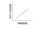
- the signal intensity due to labeling increases as the amount of antigen in the sample increases, whereas in the competitive method, the signal intensity due to labeling decreases as the amount of antigen in the sample increases.
- Chemiluminescence immunochromatography is known in which a luminescent protein is used as a label in the immunochromatographic method described above, and luminescence from the luminescent protein is detected by chemically reacting the luminescent protein with a luminescent substrate (e.g., Japanese Patent Laid-Open No. 2015-105858).
- a luminescent substrate e.g., Japanese Patent Laid-Open No. 2015-105858.
- a specimen and a labeled antibody labeled with a luminescent protein are developed on a test strip on which a capture antibody is immobilized in the test area.
- the test strip is then developed with a luminescent substrate that reacts with the label photoprotein to produce light.
- the antigen which is the substance to be tested, is qualitatively or quantitatively measured.
- Japanese Patent Publication No. 2018-522221 discloses a chemiluminescence immunochromatography method using a competitive method, in which bioluminescence resonance energy transfer (BRET: Bioluminescence Resonance Energy Transfer) occurs between a photoprotein and a fluorescent substance. It is disclosed to use the technique of In Japanese Patent Publication No. 2018-522221, a test strip in which a capturing antibody labeled with a luminescent protein is immobilized in the test area is used, and a specimen, a prepared fluorescent substance-labeled pseudoantigen, and a luminescent protein are used. and a luminescent substrate to react with are developed on the test strip. Then, the antigen in the specimen and the labeled pseudo-antigen are competitively captured by the capture antibody.
- BRET Bioluminescence Resonance Energy Transfer
- the inspection area when the luminescent protein and the fluorescent substance are close to each other, the energy generated by the reaction between the luminescent substrate and the luminescent protein is transferred to the fluorescent substance to generate fluorescence.
- the chemiluminescence immunochromatography method described in Japanese Patent Application Publication No. 2018-522221 the larger the amount of antigen captured in the specimen, the less the amount of labeled pseudoantigen captured in the test area. becomes smaller.
- the antigen, which is the test substance is qualitatively or quantitatively measured by detecting fluorescence from this BRET.
- a labeled antibody labeled with a luminescent protein is developed over the entire area of the test strip, including the test area.
- some of the labeled antibodies specifically bind to the antigen through antigen-antibody reaction, but some of the remaining labeled antibodies remain around the test area even though they do not specifically bind to the antigen.
- the luminescent substrate is developed on the test strip, not only the label of the labeled antibody specifically adsorbed to the antigen but also the luminescent protein, which is the label of the remaining labeled antibody, reacts with the luminescent substrate.
- the area surrounding the inspection area is used as the background, luminescence due to the remaining labeled antibody will occur even in the background. Since the light signal in the background becomes noise with respect to the light signal generated by the antigen-antibody reaction, the S (Signal)/N (Noise) ratio is lowered, resulting in lower measurement accuracy.
- JP-A-2018-522221 the photoprotein is immobilized only in the inspection area as a label for the capture antibody. Therefore, fluorescence is generated by BRET only in the examination region where the photoprotein of the capture antibody and the fluorescent material of the labeled antibody are in close proximity, so that no fluorescence is generated in the background in the absence of the capture antibody.
- the amount of labeled pseudo-antigen that binds to the capture antibody immobilized in the test area is maximized in the absence of antigen in the specimen. The signal intensity is highest in the absence of , and the signal intensity decreases as the amount of antigen increases.
- the signal intensity becomes maximum in the absence of antigen, and this becomes the baseline signal intensity for measuring the amount of antigen, but there is a problem that the baseline signal intensity varies due to various factors.
- the amount of labeled pseudo-antigen that binds to the capture antibody in the absence of antigen in the sample depends on the fluid velocity caused by factors such as the momentum with which the sample is spotted onto the test strip and the amount of sample spotted. changes due to fluctuations in
- BRET the fluorescence signal intensity changes according to the distance between the photoprotein and the fluorescent substance. Therefore, if the binding state of the labeled pseudo-antigen that binds to the capture antibody fluctuates, the baseline will also fluctuate. If the baseline fluctuates, the measurement accuracy will decrease. In particular, the smaller the amount of antigen in the specimen, that is, the lower the concentration of the antigen in the specimen, the greater the influence of variations in signal intensity between baseline measurements on measurement accuracy.
- the present disclosure has been made in view of the above circumstances, and aims to provide an immunochromatography kit and a testing apparatus that are capable of highly accurate measurement even when the concentration of a test substance in a sample is low compared to conventional methods. aim.
- the immunochromatography kit of the present disclosure includes a cartridge containing a test strip having a test area on which a sample is developed and which captures a test substance contained in the sample; a capture binding substance that specifically binds to the test substance, the capture binding substance labeled with a photoprotein and immobilized in the test region; a labeled binding substance that specifically binds to the test substance, the labeled binding substance labeled with a fluorescent substance and supplied to the test region;
- the luminescent substrate supplied to the inspection region generates energy by reacting with the luminescent protein, and in a state in which the labeled binding substance is captured by the binding substance for capture via the test substance, the energy is transferred to the fluorescent substance.
- a luminescent substrate that produces a translocating bioluminescence energy resonance transfer phenomenon.
- the luminescent protein may be a luminescent protein that emits blue light
- the fluorescent substance may be a green fluorescent dye or green fluorescent protein that emits green fluorescence with a quantum yield of 0.7 or more.
- the photoprotein is a blue photoprotein that emits blue light, and the fluorescent substance may have a peak wavelength of 530 nm or more.
- It may include a specimen collection tool for collecting specimens.
- An instruction manual may be included that describes the test procedure for supplying the luminescent substrate to the test area after the sample and the labeled binding substance are supplied to the test area.
- a label holding pad provided with a label binding substance may be arranged on the test strip at the sample supply position.
- a luminescent substrate solution holding part containing a substrate solution containing a luminescent substrate may be provided in the cartridge.
- the washing liquid supplied to the inspection region is used to wash the labeled binding substance non-specifically adsorbed to the inspection region.
- a cleaning liquid may also be provided.
- a cleaning liquid holding portion containing cleaning liquid may be provided in the cartridge.
- the inspection device of the present disclosure includes a loading unit in which the cartridge in the immunochromatograph kit of the present disclosure is detachably loaded, a detection unit that detects the intensity of luminescence from the fluorescent substance in the inspection area; and a processor that determines whether the sample is positive or negative based on the intensity of luminescence obtained from the detection unit.
- the inspection device of the present disclosure includes a luminescent substrate supply mechanism that supplies the luminescent substrate to the inspection area,
- the processor may supply the luminescent substrate to the test area by means of a luminescent substrate supply mechanism after the analyte and labeled binding substance have been supplied to the test area.
- the loading unit is loaded with a cartridge having a cleaning liquid holding unit inside, further comprising a cleaning liquid supply mechanism for supplying the cleaning liquid to the inspection area
- the processor may supply washing liquid to the examination area by the washing liquid supply mechanism after the specimen and the labeled antibody are supplied to the examination area and before supplying the luminescent substrate to the examination area.
- the detection unit detects the intensity of luminescence from the fluorescent substance in the inspection region (luminescence intensity at the peak wavelength of the fluorescent substance) as the first luminescence intensity, and the intensity of luminescence from the photoprotein ( The luminescence intensity at the peak wavelength of the photoprotein) is detected as the second luminescence intensity
- the processor acquires the first luminescence intensity and the second luminescence intensity from the detection unit, and divides the first luminescence intensity by the second luminescence intensity. A value may be used to make the determination.
- the detection unit detects the intensity of luminescence from the fluorescent substance in the inspection area as the first luminescence intensity, and the peak wavelength of luminescence from the fluorescent substance and the peak wavelength of luminescence from the photoprotein.
- the processor detects, as a third emission intensity, the intensity of emission at an isoluminescence point existing in the wavelength range between A determination may be made using the value divided by the emission intensity.
- the immunochromatography kit and testing device of the present disclosure highly accurate measurement is possible even when the concentration of the test substance in the sample is low compared to conventional methods.
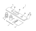
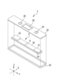
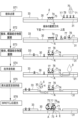
- FIG. 4 is a diagram showing the whole immunochromatography kit composition. 4 is an exploded perspective view of the cartridge; FIG. It is a figure which shows the procedure of an immunochromatography examination.
- (a) is a diagram for explaining luminescence from the test area when the test is negative, and
- (b) is a diagram for explaining the luminescence from the test area when the test is positive.
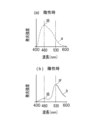
- (a) is a diagram showing an example of the emission spectrum detected from the test area when negative, and
- (b) is a diagram showing an example of the emission spectrum detected from the test area when positive.
- FIG. 4 is a diagram showing the relationship between the intensity of light emitted from a fluorescent substance by BRET and the concentration of a test substance; It is a figure which shows the isoluminous intensity point in negative time and positive time.
- FIG. 3 is a diagram for explaining luminescence from an inspection region in a chemiluminescence immunochromatographic inspection (prior art 1) without using BRET.
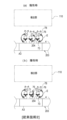
- Competitive method and with respect to chemiluminescence immunochromatographic test using BRET (prior art 2), (a) is a diagram explaining luminescence from the test area when negative, and (b) is a diagram illustrating luminescence from the test area when positive. It is a figure explaining light emission.
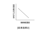
- FIG. 10 is a diagram showing the relationship between the emission intensity of light emitted from a fluorescent substance and the concentration of a test substance by BRET in Prior Art 2;
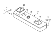
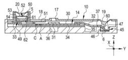
- FIG. 11 is a perspective view of a cartridge of a modified design;
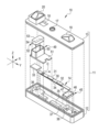
- FIG. 11 is an exploded perspective view of a cartridge of a modified design;
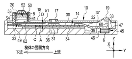
- FIG. 11 is a cutaway side view of the cartridge of the modified design
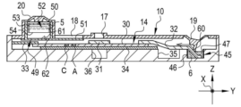
- FIG. 4 is a cutaway side view of the cartridge showing a state in which the first pressing operation portion is pushed
- FIG. 10 is a cutaway side view of the cartridge showing a state in which the second pressing portion is pushed in in addition to the first pressing portion
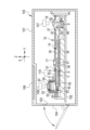
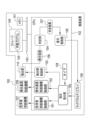
- It is a perspective view which shows an inspection apparatus. It is a sectional view showing the inside of an inspection device. It is a block diagram which shows the processing part of CPU of an inspection apparatus. It is a flowchart which shows the processing procedure of an inspection apparatus.
- an immunochromatography kit 1 of one embodiment includes a cartridge 3 containing a test strip 2, a luminescent substrate solution 5 containing a luminescent substrate 4, a washing solution 6, and a specimen collecting tool 7. and an instruction manual 8.
- the cartridge 3 is a single-use type that is used one by one for each sample, and has a case 21 in the shape of an elongated rectangular parallelepiped.
- the case 21 is composed of a case body 22 and a cover member 23 .
- the case body 22 is shaped like an elongated plate and has a recess for accommodating the test strip 2 .
- the cover member 23 includes a specimen dropping port 24 , a luminescent substrate dropping port 26 and an observation window 28 .
- both the X direction and the Y direction in FIG. 2 are directions along the horizontal plane and are orthogonal to each other.
- the X direction is the direction along the short side of the case 21
- the Y direction is the direction along the long side of the case 21 .
- the Z direction is a direction along the vertical direction and is orthogonal to the X and Y directions. The same applies to subsequent drawings
- the sample dropping port 24 is a round hole into which a sample liquid 70 (see FIG. 3) containing a sample is dropped.
- the specimen is not particularly limited as long as it can contain the test substance 71 .
- Specimens are biological samples of individuals to be immunochromatographically tested, particularly animals, more specifically human biological samples, such as blood, serum, plasma, cerebrospinal fluid, tears, sweat, urine, feces, pus, nasal discharge, nasal swabs. Liquids, throat swabs, nasal aspirates, sputum, etc., as well as organs, tissues, mucous membranes, and skin, or swabs containing them, or animals and plants themselves or their dried bodies.
- the test substance 71 includes antigens, antibodies, proteins, low-molecular-weight compounds, and the like. Note that the washing liquid 6 is also dropped from the specimen dropping port 24 .
- the luminescent substrate dropping port 26 is a round hole into which the luminescent substrate solution 5 containing the luminescent substrate 4 is dropped.
- dropping the luminescent substrate solution 5 is synonymous with dropping the luminescent substrate 4 .
- the luminescent substrate 4 will be described later.
- the observation window 28 is a rectangular opening for observing the inspection area A of the inspection strip 2 from the outside.
- An observation window 28 is formed between the sample dropping port 24 and the luminescent substrate dropping port 26 .
- the specimen collecting tool 7 is a tool for collecting a specimen, and in this example, a swab for collecting nasal swab is used.
- a sample-collecting tool 7 an appropriate sample-collecting tool can be provided according to the above-described sample.
- the swab with the sample collected is immersed in a developing solution (also referred to as a sample treatment solution) (not shown) to extract the collected sample to prepare a sample solution 70 (see FIG. 3).
- the sample liquid 70 containing the specimen thus obtained is dropped from the specimen dropping port 24 . That is, dropping the sample liquid 70 is synonymous with dropping the sample.
- the instruction manual 8 describes how to use the test strip 2, the luminescent substrate solution 5, the washing liquid 6, the sample collection tool 7, etc., and the test procedure.
- the instruction manual 8 describes an inspection procedure in which the luminescent substrate 4 is supplied to the inspection area A after the sample and the labeled binding substance are supplied to the inspection area A. The details of the inspection procedure will be described later. .
- a communication line is provided in the examination procedure manual provided by uploading it to the server in the form of electronic data.
- FIG. 2 is a diagram showing the configuration of the cartridge 3
- FIG. 3 is a diagram showing an inspection procedure using the cartridge 3.
- the test strip 2 has an overall elongated rectangular plate shape and has a carrier 30, a label holding pad 31, an absorbent pad 33, and a back adhesive sheet .
- the carrier 30 is made of a porous, insoluble material such as a nitrocellulose membrane. Near the center of the carrier 30, there is provided an inspection area A for capturing the test substance 71 (see FIG. 3) contained in the sample. A capture binding substance 74 (see FIG. 3) labeled with a photoprotein 73 (see FIG. 3) is immobilized in the inspection region A.
- FIG. 3 A capture binding substance 74 (see FIG. 3) labeled with a photoprotein 73 (see FIG. 3) is immobilized in the inspection region A.
- the capturing binding substance 74 is a binding substance that specifically binds to the test substance 71 .
- the capturing binding substance 74 is an antibody against the antigen, and when the test substance 71 is an antibody, it is an antigen against the antibody.
- the binding substance for capture 74 is an aptamer for the protein, a low-molecular-weight compound, or the like.
- the luminescent protein 73 labeled on the capturing binding substance 74 is a protein that reacts with the luminescent substrate 4 to generate luminescence.
- the label holding pad 31 is attached at a position of the carrier 30 facing the specimen dropping port 24 .
- a sample liquid 70 dropped into the specimen dropping port 24 is spotted on the label holding pad 31 . That is, the label holding pad 31 is arranged on the test strip 2 at the sample supply position.
- the label holding pad 31 contains a label binding substance 77 labeled with a fluorescent substance 76 (see FIG. 3).
- the labeled binding substance 77 is a binding substance that specifically binds to the test substance 71 .
- the labeled binding substance 77 is an antibody against the antigen, and when the test substance 71 is an antibody, it is a secondary antibody capable of binding to the antibody.
- the test substance 71 is a protein, a low-molecular-weight compound, or the like
- the labeled binding substance 77 is an aptamer for the protein, a low-molecular-weight compound, or the like.
- the capturing binding substance 74 and the labeling binding substance 77 may be the same or different.
- the test substance 71 is an influenza A virus or its biomarker
- an anti-influenza A monoclonal antibody manufactured by Medix Biochemica, product name: Anti-Influenza A SPT N-5 7307
- Medix Biochemica product name: Anti-Influenza A SPT N-5 7307
- the fluorescent substance 76 is a fluorescent dye or fluorescent protein that emits fluorescence when excited energy is applied.
- the luminescent protein 73 and the fluorescent substance 76 are selected so that the wavelength range of the emission spectrum of the luminescent protein 73 and the wavelength range of the excitation spectrum of the fluorescent substance 76 overlap. If there is a wavelength range in which the emission spectrum of the photoprotein 73 overlaps with the excitation spectrum of the fluorescent material 76, energy transfer can occur.
- the luminescent protein 73 is, for example, a luminescent protein that emits blue light.
- the fluorescent substance 76 having a quantum yield of 0.7 or more may be a green fluorescent dye or a green fluorescent protein that emits green fluorescence.
- blue light is light having a maximum emission wavelength (hereinafter referred to as peak wavelength) within a wavelength range of approximately 430 nm to approximately 490 nm
- green light is light having a peak within a wavelength range of approximately 490 nm to approximately 550 nm. It is light with a wavelength.
- the peak wavelength of the photoprotein 73 and the peak wavelength of the fluorescent substance 76 differ by 40 nm or more.
- the fluorescent substance 76 can be a fluorescent dye or fluorescent protein having a peak wavelength of 530 nm or more. The further apart the peak wavelengths are, the easier it is to identify the respective light peaks, which is preferable. For example, when photoprotein 73 has a peak wavelength within the blue light wavelength range of approximately 430 nm to approximately 490 nm, fluorescent substance 76 has a peak wavelength within the red light wavelength range of approximately 640 nm to 770 nm, and so on. is.
- the luminescent substrate 4 generates energy by reacting with the photoprotein 73 and being oxidized, becomes an excited state, and emits light when transitioning to the ground state.
- a bioluminescence energy resonance transfer phenomenon BRET
- the fluorescent substance 76 is excited by the luminescence energy E generated by the reaction between the luminescent substrate 4 and the luminescent protein 73, and the fluorescent substance 76 emits fluorescence.
- the immunochromatography kit 1 of the present disclosure it is determined whether the specimen is positive or negative by detecting luminescence from the fluorescent substance 76 by this BRET. Details of the inspection procedure will be described later.
- luminescent protein 73 examples include Blue luminescent protein NanoLuc (registered trademark, Promega), Green fluorescent protein mNeongreen (Allele Biotechnology), and Furimazine ( manufactured by Promega).
- Blue luminescent protein NanoLuc registered trademark, Promega
- Green fluorescent protein mNeongreen Allele Biotechnology
- Furimazine manufactured by Promega
- the absorbent pad 33 is attached to the other end opposite to the one end of the carrier 30 to which the label holding pad 31 is attached. Like the carrier 30, the absorbent pad 33 is made of a porous material. The absorbent pad 33 absorbs the sample liquid 70 spread on the carrier 30 , the washing liquid 6 and the luminescent substrate solution 5 .
- the back adhesive sheet 34 is a base material with an adhesive surface to which the carrier 30 is adhesively fixed.
- the washing liquid 6 is supplied to the inspection area A after supplying the specimen and the labeled binding substance 77 to the inspection area A and before supplying the luminescent substrate 4 to the inspection area A.
- the washing liquid 6 washes the labeled binding substance 77 non-specifically adsorbed to the inspection region A.
- non-specific adsorption refers to a state in which the labeled binding substance 77 is adsorbed onto the carrier without specific binding between the test substance 71 and the capturing binding substance 74 .
- the sample liquid 70 is spotted on the label holding pad 31 as shown in step ST1.
- the test substance 71 in the sample liquid 70 spotted on the label holding pad 31 specifically binds to the label binding substance 77 of the label holding pad 31 .
- step ST2 the sample liquid 70 permeates from the label holding pad 31 to the carrier 30 and spreads toward the inspection area A side (downstream side) by capillary action.
- the test substance 71 that has reached the inspection area A is captured by the capturing binding substance 74 .
- the fluorescent substance 76 is captured by the capturing binding substance 74 in the inspection area A via the test substance 71 and the labeling binding substance 77 .
- the labeled binding substance 77 that has not been captured in the inspection area A is further developed to the absorbent pad 33 on the downstream side. Note that part of the labeled binding substance 77 may not spread to the absorbent pad 33 and be non-specifically adsorbed onto the carrier 30 .
- the cleaning liquid 6 is supplied.
- the washing liquid 6 is supplied to the carrier 30 through the label holding pad 31 and spreads toward the inspection area A side (downstream side) by capillary action.
- the labeled binding substance 77 non-specifically adsorbed on the carrier is washed.
- the labeled binding substance 77 non-specifically adsorbed to the inspection area A is flowed to the absorbent pad 33 together with the washing liquid 6 .
- the luminescent substrate solution 5 is supplied.
- the luminescent substrate solution 5 is supplied to the carrier 30 from the downstream side of the inspection area A and spreads toward the inspection area A side (upstream side).
- step ST6 the luminescent substrate 4 contained in the luminescent substrate solution 5 that has reached the inspection area A reacts with the luminescent protein 73 immobilized on the inspection area A.
- the energy E generated by this reaction is transferred to the captured fluorescent substance 76 to generate BRET, and fluorescence 78 is generated from the fluorescent substance 76 .
- the light emitted from the test area A can be used to determine whether the specimen is positive or negative.
- the light emitted from the inspection area A is detected by a detection unit 110 such as a spectrophotometer in an inspection apparatus 100 (see FIGS. 16 and 17), which will be described later.
- a detection unit 110 such as a spectrophotometer in an inspection apparatus 100 (see FIGS. 16 and 17), which will be described later.
- FIG. 4 the luminescence from the inspection area A in each case of positive and negative will be described with reference to FIGS. 4 and 5.
- FIG. 4 the luminescence from the inspection area A in each case of positive and negative will be described with reference to FIGS. 4 and 5.
- FIG. 4 illustrates (a) luminescence from test area A when the specimen is negative (negative) and (b) luminescence from test area A when the specimen is positive (positive), respectively.
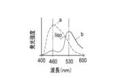
- FIG. 5 shows (a) the emission spectrum from test area A when negative and (b) the emission spectrum from test area A when positive.
- the light 75 emitted by the photoprotein 73 is blue light with a peak wavelength of 460 nm
- the fluorescence emitted by the fluorescent substance 76 is green light with a peak wavelength of 530 nm.
- the sample does not contain the test substance 71, so as shown in FIG. As a result, the fluorescent material 76 is also not captured.
- the luminescent substrate 4 when the luminescent substrate 4 is supplied to the inspection area A, the luminescent substrate 4 reacts with the luminescent protein 73 to generate luminous energy and emit light. At this time, light 75 of photoprotein 73 is generated.
- an emission spectrum a having a peak at 460 nm is obtained, as shown in FIG. 5(a).
- An emission spectrum a shown in FIG. 5A is an example of an emission spectrum when the light 75 is blue light having a peak wavelength of 460 nm.
- the specimen 71 is captured by the binding substance for capture 74 in the inspection region A, and the fluorescent substance 76 is captured via the specimen 71. be.
- the luminescent substrate 4 reacts with the luminescent protein 73 to generate luminescence energy E, and fluorescence 78 is generated from the fluorescent substance 76 by BRET.
- Fluorescence 78 is green light with a peak wavelength of 530 nm. Note that even in the case of positive, in the inspection region A, there is also a capturing binding substance 74 that does not capture the test substance 71 .
- the more the test substance 71 in the sample that is, the higher the concentration of the test substance 71, the higher the intensity of the emitted light from the fluorescent substance 76 by BRET.
- the luminescent substrate 4 and the luminescent protein 73 react relatively and the light 75 generated from the luminescent substrate 4 becomes smaller. That is, when the test substance 71 does not exist in the specimen and is negative, an emission spectrum of light 75 having a peak wavelength of 460 nm is obtained as shown in FIG. 5A, and the amount of the test substance 71 in the specimen increases. As it increases, the 460 nm peak in the detected emission spectrum becomes smaller and the 530 nm peak becomes relatively larger.
- the emission spectrum a or b as shown in FIG. ) and the emission intensity IB of the light 75 emitted without causing BRET (hereinafter referred to as the second emission intensity IB) are obtained, and the value IF/ IB (hereinafter referred to as an emission intensity ratio IF/IB) can be obtained to determine whether the test is positive or negative. For example, if the emission intensity ratio IF/IB is greater than a predetermined threshold, it is determined as positive, and if it is equal to or less than the threshold, it is determined as negative.
- the emission intensity ratio used for determination as shown in FIG. 7, the emission intensity Iiso (hereinafter referred to as , using the third emission intensity Iiso), the emission intensity IF of the fluorescence 78 generated by BRET is divided by the emission intensity Iiso at the isoluminescence point to obtain a ratio IF/Iiso (emission intensity ratio IF/Iiso), A positive or negative determination may be made. For example, if the emission intensity ratio IF/Iiso is greater than a predetermined threshold, it is determined to be positive, and if it is equal to or less than the threshold, it is determined to be negative.
- FIG. A photoprotein 73 is used as a label for the labeled antibody 207 . If the specimen is positive, the labeled antibody 207 labeled with the photoprotein 73 is captured via the specimen 71 . Since the luminescent substrate 4 is supplied to the inspection area A2 in this state, the luminescent substrate 4 reacts with the luminescent protein 73 to emit light. At this time, the luminescent substrate 4 reacts with the luminescent protein 73 to generate light 75 having the peak wavelength of the luminescent protein 73 .
- the luminescent protein 73 that is the label of the non-specifically adsorbed labeled antibody 207 and the luminescent substrate 4 reacts with each other to emit light.
- the luminescence caused by the photoprotein 73 of the non-specifically adsorbed labeled antibody 207 in the region surrounding the inspection region A2 increases the background, lowering the S/N ratio and lowering the measurement accuracy. there were.
- the immunochromatography kit 1 of the present embodiment includes a cartridge 3 containing a test strip 2 having a test area A in which a sample is developed and which captures a test substance 71 contained in the sample, and a photoprotein 73. It comprises a labeled binding substance 74 immobilized in the inspection area A, a labeled binding substance 77 labeled with a fluorescent substance 76 and supplied to the inspection area A, and a luminescent substrate 4 .
- the energy E generated by the reaction between the luminescent substrate 4 and the luminescent protein 73 moves to the fluorescent substance 76 in a state where the labeled binding substance 77 is captured via the test substance 71, Fluorescence 78 is produced from the fluorescent material 76 .
- This energy transfer phenomenon called BRET occurs when the photoprotein 73 and the fluorescent substance 76 are in close proximity (for example, when they are positioned at a distance of about 10 nm or less). Therefore, when the labeling binding substance 77 is not captured by the capturing binding substance 74, BRET does not occur because the photoprotein 73 and the fluorescent substance 76 are not close to each other.
- the immunochromatography kit 1 of the present embodiment which can be determined by measurement using BRET, can suppress the background compared to the conventional chemiluminescence immunochromatography method that does not use BRET, so measurement accuracy is high. .
- test area A3 of the test strip 203 A capture antibody 204 labeled with a photoprotein 73 is immobilized.
- a labeled pseudoantigen 72 labeled with a fluorescent substance 76 and a luminescent substrate 4 are developed on a test strip 203 together with a specimen.
- FIG. 9 illustrates (a) luminescence from the test area A3 when the specimen is negative (negative) and (b) luminescence from the test area A3 when the specimen is positive (positive). It is schematic for As shown in FIG. 9(a), when negative, the labeled pseudoantigen 72 is captured by the capture antibody 204 in the test area A3, and the luminescence energy E generated by the reaction between the luminescent substrate 4 and the luminescent protein 73 is converted into a fluorescent substance. 76 and fluorescence 78 is generated from the fluorescent material 76 . Further, as shown in FIG. 9(b), when positive, the labeled pseudoantigen 72 and the test substance 71 are competitively captured by the capture antibody 204 in the test area A3.
- the luminous energy E generated by the reaction between the luminescent protein 73 that labels the capture antibody 204 that captures the test substance 71 and the luminescent substrate 4 does not transfer to the fluorescent substance 76 . , becomes light 75 and emits light. Therefore, in the positive case, the fluorescence 78 from the fluorescent substance 76 due to BRET decreases, and the light 75 generated by the reaction between the photoprotein 73 and the luminescent substrate 4 increases.
- the emission intensity of fluorescence 78 generated from the fluorescent substance 76 by BRET is the highest when the test substance 71 is not present in the specimen, and the amount of the test substance 71 is the highest.
- the signal intensity of the fluorescence 78 generated from the fluorescent substance 76 by BRET decreases as the concentration increases (the test substance concentration increases). If the antigen, which is the analyte 71 , is not present in the specimen, it is the labeled pseudo-antigen 72 that binds to the capture antibody 204 .
- the amount of the antigen that binds to the capture antibody 204 and the manner of binding fluctuate due to fluctuations in fluid velocity and reaction temperature that accompany fluctuations in the amount of specimen, resulting in fluctuations in signal intensity. likely to occur.
- the difference between the case where the test substance 71 is extremely small and the baseline where the test substance 71 is not present becomes difficult, and the measurement accuracy is low when the test substance 71 is very small.
- the immunochromatography kit 1 of the present embodiment uses a sandwich method in which the test substance 71 is sandwiched between the capturing binding substance 74 and the labeling binding substance 77 in the test region A of the test strip 2 . Therefore, compared to conventional BRET using a competitive method, measurement accuracy is higher when the amount of test substance in the sample is small.
- the sandwich method when the test substance 71 does not exist, the signal intensity of fluorescence generated from the fluorescent substance by BRET is zero, and the signal intensity increases as the amount of the test substance 71 increases (FIG. 6). reference). Therefore, it is easy to detect a change in signal intensity when the amount of the test substance 71 is very small, and the measurement accuracy is high when the amount of the test substance 71 is very small.
- the washing liquid 6 is provided, and is supplied to the inspection area A after the sample and the labeled binding substance 77 are supplied to the inspection area A and before the luminescent substrate 4 is supplied to the inspection area A. Since the washing liquid 6 can remove the non-specifically adsorbed labeled antibody 207, the measurement accuracy can be improved.
- the immunochromatography kit 1 of the above-described embodiment includes a washing liquid 6 and a luminescent substrate solution 5 containing a luminescent substrate 4 separately from the cartridge.
- the cartridge includes not only the test strip but also at least the luminescent substrate solution holding portion 52 containing the luminescent substrate solution 5 containing the luminescent substrate 4 and the washing solution holding portion 45 containing the washing solution 6. It may include one side.
- the modified design cartridge 10 shown in FIGS. 11 and 12 has an elongated rectangular parallelepiped case 11 .
- the case 11 is composed of a case body 12 and a cover member 13 .
- the case main body 12 is a box having a storage space surrounded by an elongated rectangular plate-shaped bottom plate and four side plates erected vertically from four sides of the bottom plate.
- An inspection strip 14 having an inspection area A is accommodated in the accommodation space of the case body 12 .
- the cover member 13 has an elongated rectangular plate shape like the bottom plate of the case body 12 and functions as a lid that covers the housing space of the case body 12 .
- a specimen dropping port 17 and an observation window 18 are formed in the cover member 13 . Further, the cover member 13 is provided with a first pressing operation portion 19 and a second pressing operation portion 20 .
- the specimen dropping port 17, the observation window 18, the first pressing operation part 19, and the second pressing operation part 20 are integrally molded.
- the sample dropping port 17 is a round hole into which the sample liquid 70 is dropped, and has a boss shape with an edge protruding from the surface.
- a sample dropping port 17 is formed in the central portion of the cover member 13 .
- the observation window 18 is a rectangular opening for observing the inspection area A and the like of the inspection strip 14 from the outside.
- the observation window 18 is formed between the sample dropping port 17 and the second pressing operation section 20 .
- the first pressing operation portion 19 is provided at one end of the cover member 13 in the Y direction.
- the first pressing operation part 19 is pressed by the user when supplying the cleaning liquid 6 (see FIG. 14) to the test strip 14 .
- the second pressing operation portion 20 is provided at the other Y-direction end portion of the cover member 13 opposite to the first pressing operation portion 19 .
- the second pressing operation part 20 is pressed by the user when supplying the luminescent substrate solution 5 (see FIG. 15) to the test strip 14 .
- the test strip 14 has a liquid transfer pad 32 in addition to the carrier 30, label holding pad 31, absorbent pad 33, and back adhesive sheet 34.
- a control area C is provided on one end side of the carrier 30 in the Y direction.
- the inspection area A is located upstream of the control area C in the development direction.
- the inspection area A and the control area C are hatched, but this hatching is for convenience of explanation and indicates that the areas A and C are colored. isn't it.
- FIGS. 13 to 15, FIG. 17, and the like The same applies to FIGS. 13 to 15, FIG. 17, and the like.
- the liquid transfer pad 32 is attached to the other end of the carrier 30 opposite to the one end of the carrier 30 where the test area A and the like are provided.
- the liquid-sending pad 32 is made of a porous material like the carrier 30 and the like, and sends the cleaning liquid 6 to the carrier 30 by capillary action.
- the back adhesive sheet 34 and, by extension, the test strip 14 are placed on convex supports 35 and 36 formed in the housing space of the case body 12 .
- the support base 35 is provided at the center of the case body 12 .
- the support base 36 is provided at one end of the case main body 12 on the downstream side in the developing direction of the sample liquid 70 .
- the supports 35 and 36 are of the same height.
- a convex support base 37 is also formed at the other end of the case body 12 on the upstream side in the developing direction of the sample liquid 70 .
- Support base 37 is the same height as support bases 35 and 36 .
- a liquid transfer pad 32 is placed on the support table 37 .
- a recessed first accommodating portion 38 is formed in the support base 37 .
- the first housing portion 38 is provided at a position facing the other end of the liquid transfer pad 32 opposite to the one end attached to the carrier 30 .
- a cleaning liquid holding portion 45 is accommodated in the first accommodation portion 38 .
- the cleaning liquid holding part 45 is composed of a container 46 having an opening on one side and a seal 47 that liquid-tightly covers the opening of the container 46 .
- the container 46 is made of, for example, a resin material.
- the cleaning liquid 6 is stored inside the container 46 .
- the seal 47 is an aluminum sheet, for example, and can be easily broken with a sharp knife or the like.
- the cleaning liquid holding portion 45 is housed in the first housing portion 38 with the seal 47 facing upward so that the seal 47 faces the other end of the liquid feeding pad 32 .
- a multifunctional member 49 is arranged on the top of the support base 36 .
- the multifunctional member 49 is made of a transparent resin material such as acrylic resin.
- the multifunctional member 49 is a member in which the second housing portion 50 and the flow path forming portion 51 are integrally provided.
- the second storage part 50 is a box with an open top, and a luminescent substrate solution holding part 52 is stored therein.
- the flow path forming portion 51 is a flat plate extending in the Y direction from the bottom portion of the second housing portion 50 .
- the flow path forming part 51 extends to the front of the label holding pad 31 and covers the upper part of the inspection area A and the control area C of the carrier 30 .
- a gap D is provided between the carrier 30 and the channel forming portion 51 (see FIG. 13).
- the distance D is, for example, in the range of 0.01 mm to 1 mm.
- the luminescent substrate solution holding part 52 is composed of a container 53 having an opening on one side and a seal 54 that liquid-tightly covers the opening of the container 53 .
- the container 53 is made of, for example, a resin material.
- a luminescent substrate solution 5 is stored inside the container 53 .
- the seal 54 is, for example, an aluminum sheet and can be easily broken with a sharp knife or the like.
- the luminescent substrate solution holding part 52 is accommodated in the second accommodating part 50 with the seal 54 facing downward so that the seal 54 faces the test strip 2 .
- the first pressing operation portion 19 is provided with a first breaking protrusion 60 .
- a luminescent substrate solution holding part 52 is attached to the inner surface of the second pressing operation part 20 .
- a second breaking projection 61 and a supply port 62 are provided at the bottom of the second housing portion 50 .
- the first breaking projection 60 and the second breaking projection 61 have sharp tips.
- the first breaking protrusion 60 abuts the other end of the liquid feeding pad 32 when the first pressing operation portion 19 is pressed, and the other end of the liquid feeding pad 32 holds the cleaning liquid.
- Push against seal 47 of portion 45 The seal 47 is broken by pushing the other end of the liquid feeding pad 32 by the first breaking protrusion 60 , and the other end of the liquid feeding pad 32 is dropped into the container 46 and immersed in the cleaning liquid 6 .
- the cleaning liquid 6 is spread from the other end of the liquid transfer pad 32 toward the carrier 30 by capillary action.
- the first pressing operation part 19 maintains the crushed state even after being crushed by the pressing operation. Therefore, the development of the cleaning liquid 6 is continued until almost all of the cleaning liquid 6 is sucked up by the liquid feeding pad 32 .
- the luminescent substrate solution holding part 52 moves downward in the second containing part 50, and the second breaking projection 61 having the second breaking projection 61 moves downward. It reaches the bottom of the housing portion 50 . Then, the seal 54 of the luminescent substrate solution holding portion 52 is broken by the second breaking projection 61 .
- the luminescent substrate solution 5 stored in the luminescent substrate solution holding part 52 flows from the broken portion of the seal 54 to the supply port 62, and further flows through the flow path secured by the interval D (see FIG. 13) to be supplied to the carrier 30. be done.
- Control region C contains control binding substance 79 labeled with photoprotein 73 .
- Control binding substance 79 specifically binds to labeled binding substance 77 .
- the fluorescent substance 76 is trapped in the control area C.
- FIG. Some labeled binding substances 77 do not bind to the analyte 71 .
- Such a labeled binding substance 77 reaches the control area C without being captured in the inspection area A, and is captured in the control area C.
- the fluorescent substance 76 is captured via the label binding substance 77, in the control region C, the photoprotein 73 and the fluorescent substance 76 are positioned close to each other.
- BRET occurs in the control region C when the luminescent substrate 4 is developed. It can be confirmed that the sample liquid 70 has been fully developed on the carrier 30 and that the development of the sample liquid 70 has been completed by the light emitted from the fluorescent substance 76 by BRET.
- the control binding substance 79 may be the test substance 71 itself or a compound having a site recognized by the labeled binding substance 77 .
- a compound having a site recognized by the labeled binding substance 77 includes, for example, a compound obtained by binding a derivative of the test substance 71 to a protein.
- an anti-mouse IgG antibody manufactured by Fujifilm Wako Pure Chemical Industries, Ltd., product name: anti-mouse IgG (H+L), Rabbit F(ab')2, product code: 566-70621
- an anti-mouse IgG antibody manufactured by Fujifilm Wako Pure Chemical Industries, Ltd., product name: anti-mouse IgG (H+L), Rabbit F(ab')2, product code: 566-70621
- the cartridge 10 of the design modification described above includes a luminescent substrate solution holding portion 52 containing the luminescent substrate solution 5 containing the luminescent substrate 4 in the cartridge 10 .
- the immunochromatography kit 1 since the luminescent substrate solution 5 is provided in the cartridge 10, the work of measuring the luminescent substrate solution 5 from a container such as an ampoule or a vial and supplying it to the inspection area A is reduced. .
- the cartridge 10 is provided with the second pressing operation portion 20, and by pressing the second pressing operation portion 20, the luminescent substrate solution 5 can be supplied to the test strip 14, thereby realizing a simple test. can.
- the cartridge 10 includes a cleaning liquid holding portion 45 containing the cleaning liquid 6 inside the cartridge 10 .
- the washing liquid 6 is provided in the cartridge 10, the work of measuring the luminescent substrate solution 5 from a container such as an ampoule or a vial and supplying it to the inspection area A is reduced.
- the cartridge 10 is provided with the first pressing operation portion 19, and by pressing the first pressing operation portion 19, the cleaning liquid 6 can be supplied to the test strip 14, so that simple inspection can be realized.
- the first pressing operation portion 19 and the second pressing operation portion 20 of the cartridge 10 can be operated by the user. 19 and the second pressing operation part 20 can be pressed inside the inspection device 100 .
- the inspection device 100 includes a rectangular parallelepiped housing 101 .
- An L-shaped tray-shaped loading section 102 into which the cartridge 10 is loaded is provided in the housing 101 .
- a loading port 103 for the cartridge 10 is formed on the front surface of the housing 101 .
- a lid 104 that can be opened and closed to cover the loading port 103 is attached to the front surface of the housing 101 .
- a power switch 105 of the inspection apparatus 100 is arranged on the front surface of the housing 101 .
- a touch panel display 106 is attached to the upper surface of the housing 101 .
- the touch panel display 106 accepts an operation instruction such as an instruction to start an immunochromatographic examination (hereinafter referred to as an instruction to start an examination) from a user. Further, the touch panel display 106 displays information such as determination results.
- the user opens the lid 104 to expose the loading port 103 , and loads the cartridge 10 spotted with the sample liquid 70 into the loading section 102 through the loading port 103 . After that, the user closes the lid 104 and operates the touch panel display 106 to input an inspection start instruction.
- the inspection apparatus 100 includes a cleaning liquid supply mechanism 107, a luminescent substrate supply mechanism 108, and a detection section 110 inside a housing 101. As shown in FIG. 17, the inspection apparatus 100 includes a cleaning liquid supply mechanism 107, a luminescent substrate supply mechanism 108, and a detection section 110 inside a housing 101. As shown in FIG. 17, the inspection apparatus 100 includes a cleaning liquid supply mechanism 107, a luminescent substrate supply mechanism 108, and a detection section 110 inside a housing 101. As shown in FIG.
- the cleaning liquid supply mechanism 107 is a mechanism for starting the supply of the cleaning liquid 6 from the cleaning liquid holding portion 45 to the carrier 30 .
- the cleaning liquid supply mechanism 107 uses, for example, an actuator such as a solenoid provided with an electromagnet and a plunger movable with respect to the electromagnet. For example, by moving the plunger, the plunger comes into contact with the first pressing operation portion 19 and presses the first pressing operation portion 19 .
- the cleaning liquid supply mechanism 107 is arranged at a position facing the first pressing operation portion 19 of the loaded cartridge 10 .
- the cleaning liquid supply mechanism 107 is a pressing mechanism that applies a pressing force to the first pressing operation portion 19 from the outside by pressing the first pressing operation portion 19 of the cartridge 10 .
- the cleaning liquid supply mechanism 107 applies a pressing force to the first pressing operation portion 19
- the cleaning liquid 6 is supplied from the cleaning liquid holding section 45 to the carrier 30 by the above-described action.
- the luminescent substrate supply mechanism 108 is a mechanism for starting the supply of the luminescent substrate solution 5 from the luminescent substrate solution holding part 52 to the carrier 30 .
- the luminescent substrate supply mechanism 108 also uses an actuator such as a solenoid, like the washing liquid supply mechanism 107 .
- the luminescent substrate supply mechanism 108 is arranged at a position facing the second pressing operation portion 20 of the loaded cartridge 10 .
- the luminescent substrate supply mechanism 108 is a pressing mechanism that applies a pressing force to the second pressing operation portion 20 from the outside by pressing the second pressing operation portion 20 of the cartridge 10 .
- the luminescent substrate supply mechanism 108 applies a pressing force to the second pressing operation portion 20, the luminescent substrate 4 is supplied from the luminescent substrate solution holding portion 52 to the carrier 30 by the above-described action.
- the detection unit 110 is arranged at a position facing the observation window 18 of the case 11 .
- the detector 110 detects light emitted from the inspection area A and the control area C.
- FIG. Specifically, the detection unit 110 is a spectrophotometer capable of detecting light 75 generated by the reaction between the photoprotein 73 and the luminescent substrate 4 and fluorescence 78 from the fluorescent substance 76 .
- a spectrophotometer can detect light intensity for each wavelength.
- the inspection apparatus 100 includes a storage 140, a memory 141, and a CPU (Central Processing Unit) 142.
- Storage 140 , memory 141 and CPU 142 are interconnected via bus line 143 .
- Storage 140, memory 141, CPU 142, and bus line 143 constitute a computer.
- the storage 140 is, for example, a hard disk drive or solid state drive.
- the memory 141 is a work memory for the CPU 142 to execute processing.
- the CPU 142 loads the program stored in the storage 140 into the memory 141 and executes processing according to the program. Thereby, the CPU 142 comprehensively controls each part of the inspection apparatus 100 .
- An operating program 145 is stored in the storage 140 .
- the operating program 145 is an application program for causing a computer to function as the inspection device 100 of the present disclosure.
- the storage 140 also stores information necessary for identifying the coloring state of the inspection area A, and the like.
- the CPU 142 cooperates with the memory 141 and the like to operate the instruction receiving unit 150, the washing liquid supply mechanism control unit 151, the luminescent substrate supply mechanism control unit 152, the detection unit control unit 153, and the determination unit 154. , and the display control unit 155 .
- the instruction reception unit 150 receives various user operation instructions through the touch panel display 106 .
- the instruction receiving unit 150 receives an inspection start instruction.
- the instruction reception unit 150 manages time based on the timer 128 and sends a control start trigger signal at each timing to the cleaning liquid supply mechanism control unit 151, the luminescent substrate supply mechanism control unit 152, and the detector. output to the unit control unit 153 .
- a control start trigger signal is output to the cleaning liquid supply mechanism control unit 151 at time t1 after a predetermined time has passed, and then a control start trigger is output to the luminescent substrate supply mechanism control unit 152 at time t2 after a predetermined time has passed.
- the supply of the washing liquid 6, the supply of the luminescent substrate 4, and the light detection are performed at optimum timing.
- the cleaning liquid supply mechanism control unit 151 operates the cleaning liquid supply mechanism 107 to press the first pressing operation unit 19 .
- the washing liquid supply mechanism control unit 151 supplies the washing liquid 6 to the examination area A after the sample and the labeled binding substance 77 are supplied to the examination area A and before the luminescent substrate 4 is supplied to the examination area A. , the cleaning liquid supply mechanism 107 is operated.
- the luminescent substrate supply mechanism control unit 152 controls to operate the luminescent substrate supply mechanism 108 and press the second pressing operation unit 20 .
- the luminescent substrate supply mechanism control unit 152 operates the luminescent substrate supply mechanism 108 so as to supply the luminescent substrate 4 to the inspection area A after the specimen and the labeled binding substance 77 are supplied to the inspection area A.
- the detection unit control unit 153 controls the operation of the detection unit 110 .
- the detection unit control unit 153 drives the detection unit 110 to detect light emission from the inspection region A.
- FIG. Specifically, an emission spectrum in a wavelength range including light 75 and fluorescence 78 is obtained.
- the detection unit 110 outputs the acquired emission spectrum 156 to the determination unit 154 .
- the determination unit 154 determines whether the sample is positive or negative based on the emission spectrum 156 acquired from the detection unit 110 .
- the determination unit 154 determines whether the specimen is positive or negative based on the emission intensity of the fluorescence 78 generated from the fluorescent material 76 by BRET. For determination, not only the emission intensity of the fluorescence 78 but also the emission intensity of the light 75 generated by the reaction between the photoprotein 73 and the luminescent substrate 4 without BRET may be used.
- the determination unit 154 outputs the determination result 157 to the display control unit 155 .
- the determination unit 154 acquires, for example, the emission spectrum a or b as shown in FIG. 5 as the emission spectrum 156 from the detection unit 110 .
- the determining unit 154 acquires a first emission intensity IF, which is the emission intensity of the fluorescence 78 generated by BRET, and a second emission intensity IB, which is the emission intensity of the light 75 emitted without causing BRET, from the emission spectrum a or b,
- a light emission intensity ratio IF/IB is obtained by dividing the first light emission intensity IF by the second light emission intensity IB.
- determination of positive or negative is performed. For example, if the emission intensity ratio IF/IB is greater than a predetermined threshold, it is determined as positive, and if it is equal to or less than the threshold, it is determined as negative.
- the third emission intensity Iiso which is the emission intensity at the isoluminescence point where there is no change
- the emission intensity ratio IF/Iiso which is the value obtained by dividing the first emission intensity IF by the third emission intensity Iiso.
- a negative determination may be made. For example, if the emission intensity ratio IF/Iiso is greater than a predetermined threshold, it is determined to be positive, and if it is equal to or less than the threshold, it is determined to be negative.
- the display control unit 155 controls display of various information on the touch panel display 106 .
- the display control unit 155 displays the determination result 157 from the determination unit 154 on the touch panel display 106.
- a step of spotting the sample liquid 70 onto the cartridge 10 is performed by the user.
- the power switch 105 is operated to turn on the power of the inspection apparatus 100, and the operation program 145 is started.
- the operation program 145 is activated, as shown in FIG. 18, the CPU 142 of the inspection apparatus 100 controls the instruction reception unit 150, the cleaning liquid supply mechanism control unit 151, the luminescent substrate supply mechanism control unit 152, the detection unit control unit 153, It functions as a determination unit 154 and a display control unit 155 .
- the lid 104 is opened by the user, and the cartridge 10 is loaded into the loading section 102 through the loading port 103 (step ST100). After that, the lid 104 is closed by the user.
- the inspection start instruction is accepted by the instruction accepting unit 150 (step ST110).
- control signals are output from the instruction receiving unit 150 to the cleaning liquid supply mechanism control unit 151, the luminescent substrate supply mechanism control unit 152, and the detection unit control unit 153 at respective timings from the start of the examination.
- the cleaning liquid supply mechanism 107 is operated under the control of the cleaning liquid supply mechanism control unit 151 at time t1 with reference to the time when the instruction is received, and presses the first pressing operation unit 19 .
- the first pressing operation part 19 is pressed, and as shown in FIG. (Step ST120).
- the luminescent substrate supply mechanism 108 is operated under the control of the luminescent substrate supply mechanism control unit 152, and the second pressing operation unit 20 is pressed. As a result, the second pressing operation part 20 is pushed down, and as shown in FIG. . As a result, the luminescent substrate solution 5 flows out from the supply port 62 and is supplied to the test strip 14 (step ST130).
- the detection unit 110 is driven under the control of the detection unit control unit 153, and the wavelength including the peak wavelength of the fluorescence of the fluorescent substance 76 and the peak wavelength of the photoprotein 73 is detected. A region of emission intensity (emission spectrum 156) is detected. (Step ST140).
- the determination unit 154 analyzes the emission spectrum 156, calculates the emission intensity ratio, and makes a positive/negative determination (step ST150). This determination result 157 is output from the determination unit 154 to the display control unit 155 .
- a message indicating the determination result 157 is displayed on the touch panel display 106 under the control of the display control unit 155 (step ST160). This completes the immunochromatographic inspection for one cartridge 10 .
- the inspection apparatus 100 includes the loading unit 102 in which the cartridge 10 is detachably loaded, the detection unit 110 that detects the intensity of the light emission from the fluorescent substance 76 in the inspection area A, and the and a processor (here, CPU 142) that determines whether the sample is positive or negative based on the intensity of emitted light.
- the testing apparatus 100 of this embodiment includes a luminescent substrate supply mechanism 108 that supplies the luminescent substrate 4 to the testing area A, and the processor (here, the CPU 142) supplies the specimen and the labeled binding substance 77 to the testing area A.
- the luminescent substrate 4 is supplied to the inspection area A by the luminescent substrate supplying mechanism 108 . That is, the luminescent substrate 4 is supplied in a state in which the specimen and the labeled binding substance 77 are specifically bound to the inspection region A.
- the luminescence reaction between the luminescent substrate 4 and the luminescent protein 73 progresses irreversibly when the luminescent substrate 4 and the luminescent protein 73 come into contact with each other, and the luminescence intensity decreases over time. As compared with the case of pre-mixing and incubating, an effect of suppressing a decrease in luminescence intensity when detecting luminescence from the inspection area A can be obtained.
- the first emission intensity IF which is the intensity of the emission (fluorescence 78) from the fluorescent substance 76 in the inspection area A, is detected, and the intensity of the light 75 emitted from the photoprotein 73 is detected as the second emission intensity.
- intensity IB Detected as intensity IB, it is possible to determine positive or negative based on the emission intensity ratio IF/IB. By using the emission intensity ratio, it is possible to obtain a more accurate determination result than using only the first emission intensity IF for determination.
- the intensity of the emission (fluorescence 78) from the fluorescent substance 76 in the inspection region A is detected as the first emission intensity IF
- the peak wavelength of the emission from the fluorescent substance 76 and the luminescent substrate 4 and the peak wavelength of luminescence produced by the reaction with the photoprotein 73 is detected as the third luminescence intensity Iiso, and based on the luminescence intensity ratio IF/Iiso, A positive or negative determination can be made.
- the emission intensity ratio it is possible to obtain a more accurate determination result than using only the first emission intensity IF for determination.
- the following various processors can be used as the hardware structure of the processing unit that executes various processes.
- the various processors include an FPGA (Field Programmable Gate Array) whose circuit configuration can be changed after manufacture.
- Programmable Logic Device (PLD) which is a processor, and/or ASIC (Application Specific Integrated Circuit), etc. etc. are included.
- One processing unit may be configured with one of these various processors, or a combination of two or more processors of the same or different type (for example, a combination of a plurality of FPGAs and/or a CPU and combination with FPGA). Also, a plurality of processing units may be configured by one processor.
- a single processor is configured by combining one or more CPUs and software.
- a processor functions as multiple processing units.
- SoC System On Chip
- a processor that realizes the functions of the entire system including multiple processing units with a single IC (Integrated Circuit) chip. be.
- the various processing units are configured using one or more of the above various processors as a hardware structure.
- an electric circuit combining circuit elements such as semiconductor elements can be used.
- test device 100 is used to determine whether a sample is positive or negative has been described.
- the color of light emitted from the test area A changes depending on whether it is negative or positive. It is also possible to determine whether
- Influenza A antigen was used as the substance to be detected.
- the test strips of Examples 1 to 3 were strips for chemiluminescence immunochromatographic testing using the sandwich method and utilizing BRET.
- the test strips of Examples 2 and 3 were similar.
- carrier A 60 mm ⁇ 300 mm porous carrier
- a test region was formed by immobilizing a capture binding substance on the test region.
- Luciferase NeLuc (registered trademark): manufactured by Promega, peak wavelength 460 nm
- An anti-influenza type A monoclonal antibody Anti-Influenza ASPTN-5 7307, Medix Biochemica
- a solution containing a capture binder consisting of a Luciferase-labeled anti-influenza A monoclonal antibody was prepared at 1.5 mg/mL. After applying the solution containing the prepared binding material for capture to the test area, the carrier was allowed to dry for 30 minutes.
- a blocking solution 50 mmol/L borate buffer (pH 8.5) containing 0.5% by mass casein (derived from milk, product number 030-01505, manufactured by FUJIFILM Wako Pure Chemical Industries, Ltd.)
- the dried carrier was immersed in the vat and allowed to stand for 30 minutes. After that, the carrier was removed and washed in 500 mL of a washing stabilizing solution (50 mmol/L Tris-HCl (pH 7.5) buffer containing 0.5% by mass of sucrose and 0.05% by mass of sodium cholate) prepared in a separate vat.
- the carrier was immersed in and allowed to stand still for 30 minutes. After that, the carrier was taken out from the liquid and dried in an environment of 25° C. for 24 hours.
- the capture antibody was immobilized on the inspection area to form the inspection area.
- a label-retaining pad holding a label-binding substance was prepared.
- a glass fiber pad Glass Fiber Conjugate Pad, Millipore
- Green fluorescent protein mNeogreen Allele Biotechnology, peak wavelength 517 nm
- an influenza A antibody was used as a labeled binding substance (here, labeled antibody).
- Influenza A antibody-fluorescent substance conjugate was mixed with a Tris-HCl buffer (pH 8.2) with a concentration of 20 mmol/L, a PEG (Mw.20000) with a concentration of 0.05% by mass, a sucrose with a concentration of 5% by mass, Then, it was diluted with water so that the absorbance at 506 nm when the optical path length was 10 mm was 0.8, to prepare an anti-influenza A fragmented antibody-fluorescent substance complex coating solution. 0.1 mL of this coating liquid was uniformly applied to one pad and dried under reduced pressure for 24 hours. As a result, a label holding pad holding a labeled binding substance labeled with a fluorescent substance (here, a labeled antibody) was obtained.
- a fluorescent substance here, a labeled antibody
- back adhesive sheet An adhesive sheet of 60 mm ⁇ 300 nm (manufactured by Nippon Technocluster Co., Ltd.) was prepared as a back adhesive sheet.
- a glass fiber pad (Glass Fiber Conjugate Pad, manufactured by Millipore) cut to 25 mm ⁇ 300 mm was used as a liquid transfer pad.
- a glass fiber pad (glass filter paper, manufactured by Advantech) cut into a length of 12 mm and a width of 10 mm was used as an absorbent pad.
- a test strip was produced according to the following procedure.
- a carrier having an inspection area formed thereon was attached to the back pressure-sensitive adhesive sheet.
- a double-faced tape (Nitto Denko) with a width of 3 mm was fixed at a position 26 mm from the downstream end of the 60 mm length of the carrier.
- the label-retaining pad was fixed to the carrier so that the downstream end of the double-sided tape and the downstream end of the label-retaining pad overlapped.
- a liquid-feeding pad was attached to the upstream side opposite to the downstream end of the carrier so that the liquid-feeding pad and the carrier overlapped by 7 mm.
- the sheet member thus prepared was cut parallel to the length direction of 60 mm with a guillotine cutter (CM4000, manufactured by Nippon Technocluster Co., Ltd.) so as to have a width of 5 mm. This gave 60 strips of 5 mm width and 60 mm length.
- a test strip was formed by placing one absorbent pad over the downstream end of one strip.
- developing solution -1 Mock-positive specimen (BD Flu Examiner Control A + B - (manufactured by Becton Dickinson)) was mixed with the standard developing solution to obtain a developing solution-1.
- Developing solution -2 A developer-2 was obtained by mixing the mock-positive specimen with the standard sample liquid so that the amount of the mock-positive specimen (antigen amount) was 300 times that of the developer-1.
- Luminescent Substrate Solution As a luminescent substrate solution, a solution containing 20 ⁇ 10 ⁇ 6 M luminescent substrate (Furimazine, Promega) was used. 100 ⁇ L of the luminescent substrate solution was used for one test.
- Example 2 In the production of the label-retaining pad of Example 1, the production method of the labeled antibody was as follows. It is common to Example 1 in that an anti-influenza A monoclonal antibody is used as the antibody. As a fluorescent substance, a fluorescent dye (peak wavelength 525 nm) of a fluorescent antibody labeling kit (Thermo-Fisher) was used. The anti-influenza A monoclonal antibody was labeled with a fluorescent dye according to the procedure of the fluorescent antibody labeling kit to obtain a labeled binding substance labeled with a fluorescent substance (here, labeled antibody). A test strip of Example 2 was prepared in the same manner as in Example 1 otherwise. The developing solution, the luminescent substrate solution and the test procedure were the same as in Example 1.
- Example 3 The same test strip, developing solution and luminescent substrate solution as in Example 1 were used. However, in Example 3, the inspection procedure of Example 1 was incorporated with a cleaning step using a cleaning liquid. After spotting the sample liquid in 1) of the above inspection procedure, the liquid-feeding pad was immersed in the cleaning liquid, and the cleaning liquid was fed for 10 minutes. By this step, non-specifically adsorbed labeled antibodies were washed away. After that, 2) and 3) of the above inspection procedure were carried out. The cleaning liquid is supplied to the liquid-feeding pad, and the cleaning liquid is fed for 10 minutes. The liquid-feeding pad is immersed in a pot containing 200 ⁇ L of washing liquid, and the washing liquid is spread in the carrier by capillary force.
- test strips of Comparative Examples 1 and 2 used a competitive method and were strips for chemiluminescence immunochromatography using BRET.
- labeled pseudoantigen An anti-influenza A pseudoantigen labeled with a fluorescent substance was used as the labeled pseudoantigen.
- a glass fiber pad Glass Fiber Conjugate Pad, Millipore) cut to 5 mm ⁇ 300 mm was prepared. Green fluorescent protein mNeogreen (peak wavelength 517 nm) was used as the fluorescent substance, and influenza A pseudoantigen was used as the labeled binding substance (here, labeled pseudoantigen).
- a fluorescent substance-labeled anti-influenza A pseudoantigen was diluted with 20 mM Tris-HCl buffer (pH 8.2), 5 mass% sucrose, and water, and the absorbance at 506 nm was 0 when the optical path length was 10 mm.
- a coating liquid was prepared so as to have a value of 0.8. 0.1 mL of this coating liquid was uniformly applied to each pad to obtain a label holding pad holding a labeled pseudoantigen labeled with a fluorescent substance.
- a test strip of Comparative Example 1 was prepared in the same manner as in Example 1 except for the above.
- Comparative Example 2 The inspection strip of Comparative Example 2 was a strip for chemiluminescence immunochromatographic inspection without using BRET.
- a label holding pad holding a labeled binding substance (labeled antibody in this case) was prepared.
- a glass fiber pad (Glass Fiber Conjugate Pad, Millipore) cut to 5 mm ⁇ 300 mm was prepared.
- Green fluorescent protein mNeogreen (Allele Biotechnology, peak wavelength 517 nm) was used as a fluorescent substance, and an anti-influenza A monoclonal antibody (Anti-Influenza ASPTN-5 7307, Medix Biochemica) was used as a labeled antibody.
- a labeled antibody solution consisting of an anti-influenza A monoclonal antibody labeled with a fluorescent protein, diluted with 20 mM Tris-HCl buffer (pH 8.2), 5 mass% sucrose, and water to give an optical path length of 10 mm was adjusted to an absorbance of 0.8 at 506 nm. 0.1 mL of this coating solution was uniformly applied to each glass fiber pad to obtain a holding pad holding a labeled antibody labeled with a fluorescent substance.
- a test strip of Comparative Example 2 was produced in the same manner as in Example 1 except for the above.
- a labeled pseudoantigen is immobilized in the test area of the test strip of Comparative Example 2, and the labeled antibody that does not bind to the antigen, which is the test substance, is captured by the labeled pseudoantigen.
- the antigen, which is the test substance, and the labeled pseudoantigen competitively bind to the labeled antibody.
- the developing solution, luminescent substrate solution, and test procedure were the same as in Example 1.
Landscapes
- Health & Medical Sciences (AREA)
- Chemical & Material Sciences (AREA)
- Life Sciences & Earth Sciences (AREA)
- Immunology (AREA)
- Engineering & Computer Science (AREA)
- Physics & Mathematics (AREA)
- Hematology (AREA)
- Analytical Chemistry (AREA)
- General Health & Medical Sciences (AREA)
- Molecular Biology (AREA)
- Biomedical Technology (AREA)
- Urology & Nephrology (AREA)
- General Physics & Mathematics (AREA)
- Biochemistry (AREA)
- Pathology (AREA)
- Chemical Kinetics & Catalysis (AREA)
- Microbiology (AREA)
- Food Science & Technology (AREA)
- Medicinal Chemistry (AREA)
- Cell Biology (AREA)
- Biotechnology (AREA)
- Clinical Laboratory Science (AREA)
- Nuclear Medicine, Radiotherapy & Molecular Imaging (AREA)
- Plasma & Fusion (AREA)
- Optics & Photonics (AREA)
- Dispersion Chemistry (AREA)
- Investigating Or Analysing Materials By The Use Of Chemical Reactions (AREA)
- Investigating, Analyzing Materials By Fluorescence Or Luminescence (AREA)
Priority Applications (4)
| Application Number | Priority Date | Filing Date | Title |
|---|---|---|---|
| EP22875465.1A EP4411377A4 (en) | 2021-09-29 | 2022-06-10 | Immunochromatography kit and testing device |
| JP2023551067A JPWO2023053586A1 (cg-RX-API-DMAC7.html) | 2021-09-29 | 2022-06-10 | |
| CN202280065707.3A CN118043668A (zh) | 2021-09-29 | 2022-06-10 | 免疫层析试剂盒及检查装置 |
| US18/617,851 US20240241116A1 (en) | 2021-09-29 | 2024-03-27 | Immunochromatography kit and assay apparatus |
Applications Claiming Priority (2)
| Application Number | Priority Date | Filing Date | Title |
|---|---|---|---|
| JP2021159997 | 2021-09-29 | ||
| JP2021-159997 | 2021-09-29 |
Related Child Applications (1)
| Application Number | Title | Priority Date | Filing Date |
|---|---|---|---|
| US18/617,851 Continuation US20240241116A1 (en) | 2021-09-29 | 2024-03-27 | Immunochromatography kit and assay apparatus |
Publications (1)
| Publication Number | Publication Date |
|---|---|
| WO2023053586A1 true WO2023053586A1 (ja) | 2023-04-06 |
Family
ID=85780542
Family Applications (1)
| Application Number | Title | Priority Date | Filing Date |
|---|---|---|---|
| PCT/JP2022/023441 Ceased WO2023053586A1 (ja) | 2021-09-29 | 2022-06-10 | イムノクロマトグラフキット及び検査装置 |
Country Status (5)
| Country | Link |
|---|---|
| US (1) | US20240241116A1 (cg-RX-API-DMAC7.html) |
| EP (1) | EP4411377A4 (cg-RX-API-DMAC7.html) |
| JP (1) | JPWO2023053586A1 (cg-RX-API-DMAC7.html) |
| CN (1) | CN118043668A (cg-RX-API-DMAC7.html) |
| WO (1) | WO2023053586A1 (cg-RX-API-DMAC7.html) |
Cited By (1)
| Publication number | Priority date | Publication date | Assignee | Title |
|---|---|---|---|---|
| EP4597108A4 (en) * | 2022-09-26 | 2026-01-14 | Fujifilm Corp | IMMUNOCHROMATOGRAPHIC CARTRIDGE |
Citations (11)
| Publication number | Priority date | Publication date | Assignee | Title |
|---|---|---|---|---|
| JP2001124770A (ja) * | 1999-08-18 | 2001-05-11 | Japan Science & Technology Corp | 物質の濃度測定方法 |
| JP2002372537A (ja) * | 2001-06-15 | 2002-12-26 | Nitto Denko Corp | 免疫クロマトグラフ用試験片および被検物質の検出方法 |
| JP2008116235A (ja) * | 2006-11-01 | 2008-05-22 | Arkray Inc | イムノクロマトグラフ検体分析用具 |
| JP2010117213A (ja) * | 2008-11-12 | 2010-05-27 | Dic Plastics Inc | 酵素免疫測定器具及び酵素免疫測定方法 |
| JP2011052971A (ja) * | 2009-08-31 | 2011-03-17 | Sysmex Corp | 目的物質の検出方法、及び目的物質を検出するためのクロマトグラフィー用試験キット |
| CN102890155A (zh) * | 2012-09-12 | 2013-01-23 | 暨南大学 | 基于能量共振转移的荧光试纸条及制备方法与应用 |
| WO2014010184A1 (ja) * | 2012-07-09 | 2014-01-16 | 富士フイルム株式会社 | 呈色測定装置および方法 |
| JP2015105858A (ja) | 2013-11-29 | 2015-06-08 | ウシオ電機株式会社 | 化学発光酵素免疫測定方法及び化学発光酵素免疫測定キット |
| WO2017104143A1 (ja) * | 2015-12-18 | 2017-06-22 | 富士フイルム株式会社 | イムノクロマトグラフキット |
| JP2018522221A (ja) | 2015-05-25 | 2018-08-09 | アルツス バイオシステムズ リミテッド ライアビリティ カンパニー | S−アデノシルメチオニン、s−アデノシルホモシステイン及びホモシステインを迅速かつ簡単に測定するための蛍光の使用 |
| JP2021159997A (ja) | 2020-04-03 | 2021-10-11 | 燕山大学Yanshan University | ストレートシーム埋もれアーク溶接管に用いられるアーク発生装置 |
Family Cites Families (1)
| Publication number | Priority date | Publication date | Assignee | Title |
|---|---|---|---|---|
| AU2020272037B2 (en) * | 2019-04-10 | 2025-11-27 | Promega Corporation | Compositions and methods for analyte detection using bioluminescence |
-
2022
- 2022-06-10 EP EP22875465.1A patent/EP4411377A4/en active Pending
- 2022-06-10 CN CN202280065707.3A patent/CN118043668A/zh active Pending
- 2022-06-10 WO PCT/JP2022/023441 patent/WO2023053586A1/ja not_active Ceased
- 2022-06-10 JP JP2023551067A patent/JPWO2023053586A1/ja active Pending
-
2024
- 2024-03-27 US US18/617,851 patent/US20240241116A1/en active Pending
Patent Citations (11)
| Publication number | Priority date | Publication date | Assignee | Title |
|---|---|---|---|---|
| JP2001124770A (ja) * | 1999-08-18 | 2001-05-11 | Japan Science & Technology Corp | 物質の濃度測定方法 |
| JP2002372537A (ja) * | 2001-06-15 | 2002-12-26 | Nitto Denko Corp | 免疫クロマトグラフ用試験片および被検物質の検出方法 |
| JP2008116235A (ja) * | 2006-11-01 | 2008-05-22 | Arkray Inc | イムノクロマトグラフ検体分析用具 |
| JP2010117213A (ja) * | 2008-11-12 | 2010-05-27 | Dic Plastics Inc | 酵素免疫測定器具及び酵素免疫測定方法 |
| JP2011052971A (ja) * | 2009-08-31 | 2011-03-17 | Sysmex Corp | 目的物質の検出方法、及び目的物質を検出するためのクロマトグラフィー用試験キット |
| WO2014010184A1 (ja) * | 2012-07-09 | 2014-01-16 | 富士フイルム株式会社 | 呈色測定装置および方法 |
| CN102890155A (zh) * | 2012-09-12 | 2013-01-23 | 暨南大学 | 基于能量共振转移的荧光试纸条及制备方法与应用 |
| JP2015105858A (ja) | 2013-11-29 | 2015-06-08 | ウシオ電機株式会社 | 化学発光酵素免疫測定方法及び化学発光酵素免疫測定キット |
| JP2018522221A (ja) | 2015-05-25 | 2018-08-09 | アルツス バイオシステムズ リミテッド ライアビリティ カンパニー | S−アデノシルメチオニン、s−アデノシルホモシステイン及びホモシステインを迅速かつ簡単に測定するための蛍光の使用 |
| WO2017104143A1 (ja) * | 2015-12-18 | 2017-06-22 | 富士フイルム株式会社 | イムノクロマトグラフキット |
| JP2021159997A (ja) | 2020-04-03 | 2021-10-11 | 燕山大学Yanshan University | ストレートシーム埋もれアーク溶接管に用いられるアーク発生装置 |
Cited By (1)
| Publication number | Priority date | Publication date | Assignee | Title |
|---|---|---|---|---|
| EP4597108A4 (en) * | 2022-09-26 | 2026-01-14 | Fujifilm Corp | IMMUNOCHROMATOGRAPHIC CARTRIDGE |
Also Published As
| Publication number | Publication date |
|---|---|
| EP4411377A4 (en) | 2024-12-04 |
| EP4411377A1 (en) | 2024-08-07 |
| US20240241116A1 (en) | 2024-07-18 |
| CN118043668A (zh) | 2024-05-14 |
| JPWO2023053586A1 (cg-RX-API-DMAC7.html) | 2023-04-06 |
Similar Documents
| Publication | Publication Date | Title |
|---|---|---|
| US11693019B2 (en) | Automated liquid-phase immunoassay apparatus | |
| CN106872420B (zh) | 一种时间分辨荧光定量检测尿微量白蛋白的试剂盒及方法 | |
| CN103097029B (zh) | 分析装置和阅读器 | |
| US7749773B2 (en) | Device for detection of molecules in biological fluids | |
| JP7267211B2 (ja) | 高濃度の分析物等、分析物を測定するための用量反応曲線の減少信号部分を使用したサンドイッチ型アッセイ | |
| EP1992950B1 (en) | Absorption pad for immunoassay, strip for immunoassay and immunoassay apparatus | |
| Tan et al. | High sensitivity automated multiplexed immunoassays using photonic crystal enhanced fluorescence microfluidic system | |
| US20160195524A1 (en) | Automated Assay | |
| US10677809B2 (en) | Rotatable cartridge with multiple metering chambers | |
| CN101151531A (zh) | 采用内部校正系统的诊断性试验试剂盒 | |
| WO2017206800A1 (zh) | 离心层析免疫检测方法 | |
| EP3509748B1 (en) | Device platform for point of care testing | |
| US20240241116A1 (en) | Immunochromatography kit and assay apparatus | |
| US20230264200A1 (en) | An automated quantitative assay device and a method of performing the quantitative assays | |
| US12474334B2 (en) | Test device and cartridge | |
| EP3517955A1 (en) | Multi-unit for performing biochemical test and immune response test, and test method using same | |
| CN108474734A (zh) | 分析杯和具有信号放大的衍生物 | |
| CN105326511B (zh) | 一种用于免疫检测的真空采血装置 | |
| US20250208128A1 (en) | Immunochromatographic cartridge | |
| WO2024070697A1 (ja) | イムノクロマトグラフ検査装置 | |
| WO2007016665A2 (en) | Single use fluorescent assays for determination of analytes | |
| WO2023182168A1 (ja) | イムノクロマトグラフ検査装置 | |
| CN115290602A (zh) | 用于纳米等离子共振检测的芯片试纸条、检测卡和分子检测分析系统 | |
| WO2022202261A1 (ja) | イムノクロマトグラフ検査装置 |
Legal Events
| Date | Code | Title | Description |
|---|---|---|---|
| 121 | Ep: the epo has been informed by wipo that ep was designated in this application |
Ref document number: 22875465 Country of ref document: EP Kind code of ref document: A1 |
|
| WWE | Wipo information: entry into national phase |
Ref document number: 2023551067 Country of ref document: JP |
|
| WWE | Wipo information: entry into national phase |
Ref document number: 202280065707.3 Country of ref document: CN |
|
| NENP | Non-entry into the national phase |
Ref country code: DE |
|
| ENP | Entry into the national phase |
Ref document number: 2022875465 Country of ref document: EP Effective date: 20240429 |