WO2022196105A1 - 情報管理装置、方法およびプログラム、並びに情報処理装置、方法およびプログラム - Google Patents
情報管理装置、方法およびプログラム、並びに情報処理装置、方法およびプログラム Download PDFInfo
- Publication number
- WO2022196105A1 WO2022196105A1 PCT/JP2022/002461 JP2022002461W WO2022196105A1 WO 2022196105 A1 WO2022196105 A1 WO 2022196105A1 JP 2022002461 W JP2022002461 W JP 2022002461W WO 2022196105 A1 WO2022196105 A1 WO 2022196105A1
- Authority
- WO
- WIPO (PCT)
- Prior art keywords
- information
- medical
- medical image
- interest
- finding
- Prior art date
- Legal status (The legal status is an assumption and is not a legal conclusion. Google has not performed a legal analysis and makes no representation as to the accuracy of the status listed.)
- Ceased
Links
Images


Classifications
-
- G—PHYSICS
- G06—COMPUTING OR CALCULATING; COUNTING
- G06T—IMAGE DATA PROCESSING OR GENERATION, IN GENERAL
- G06T7/00—Image analysis
- G06T7/0002—Inspection of images, e.g. flaw detection
- G06T7/0012—Biomedical image inspection
-
- A—HUMAN NECESSITIES
- A61—MEDICAL OR VETERINARY SCIENCE; HYGIENE
- A61B—DIAGNOSIS; SURGERY; IDENTIFICATION
- A61B5/00—Measuring for diagnostic purposes; Identification of persons
-
- G—PHYSICS
- G16—INFORMATION AND COMMUNICATION TECHNOLOGY [ICT] SPECIALLY ADAPTED FOR SPECIFIC APPLICATION FIELDS
- G16H—HEALTHCARE INFORMATICS, i.e. INFORMATION AND COMMUNICATION TECHNOLOGY [ICT] SPECIALLY ADAPTED FOR THE HANDLING OR PROCESSING OF MEDICAL OR HEALTHCARE DATA
- G16H15/00—ICT specially adapted for medical reports, e.g. generation or transmission thereof
-
- G—PHYSICS
- G16—INFORMATION AND COMMUNICATION TECHNOLOGY [ICT] SPECIALLY ADAPTED FOR SPECIFIC APPLICATION FIELDS
- G16H—HEALTHCARE INFORMATICS, i.e. INFORMATION AND COMMUNICATION TECHNOLOGY [ICT] SPECIALLY ADAPTED FOR THE HANDLING OR PROCESSING OF MEDICAL OR HEALTHCARE DATA
- G16H30/00—ICT specially adapted for the handling or processing of medical images
- G16H30/20—ICT specially adapted for the handling or processing of medical images for handling medical images, e.g. DICOM, HL7 or PACS
-
- G—PHYSICS
- G16—INFORMATION AND COMMUNICATION TECHNOLOGY [ICT] SPECIALLY ADAPTED FOR SPECIFIC APPLICATION FIELDS
- G16H—HEALTHCARE INFORMATICS, i.e. INFORMATION AND COMMUNICATION TECHNOLOGY [ICT] SPECIALLY ADAPTED FOR THE HANDLING OR PROCESSING OF MEDICAL OR HEALTHCARE DATA
- G16H30/00—ICT specially adapted for the handling or processing of medical images
- G16H30/40—ICT specially adapted for the handling or processing of medical images for processing medical images, e.g. editing
-
- G—PHYSICS
- G16—INFORMATION AND COMMUNICATION TECHNOLOGY [ICT] SPECIALLY ADAPTED FOR SPECIFIC APPLICATION FIELDS
- G16H—HEALTHCARE INFORMATICS, i.e. INFORMATION AND COMMUNICATION TECHNOLOGY [ICT] SPECIALLY ADAPTED FOR THE HANDLING OR PROCESSING OF MEDICAL OR HEALTHCARE DATA
- G16H40/00—ICT specially adapted for the management or administration of healthcare resources or facilities; ICT specially adapted for the management or operation of medical equipment or devices
- G16H40/60—ICT specially adapted for the management or administration of healthcare resources or facilities; ICT specially adapted for the management or operation of medical equipment or devices for the operation of medical equipment or devices
- G16H40/67—ICT specially adapted for the management or administration of healthcare resources or facilities; ICT specially adapted for the management or operation of medical equipment or devices for the operation of medical equipment or devices for remote operation
-
- G—PHYSICS
- G16—INFORMATION AND COMMUNICATION TECHNOLOGY [ICT] SPECIALLY ADAPTED FOR SPECIFIC APPLICATION FIELDS
- G16H—HEALTHCARE INFORMATICS, i.e. INFORMATION AND COMMUNICATION TECHNOLOGY [ICT] SPECIALLY ADAPTED FOR THE HANDLING OR PROCESSING OF MEDICAL OR HEALTHCARE DATA
- G16H50/00—ICT specially adapted for medical diagnosis, medical simulation or medical data mining; ICT specially adapted for detecting, monitoring or modelling epidemics or pandemics
- G16H50/20—ICT specially adapted for medical diagnosis, medical simulation or medical data mining; ICT specially adapted for detecting, monitoring or modelling epidemics or pandemics for computer-aided diagnosis, e.g. based on medical expert systems
-
- G—PHYSICS
- G16—INFORMATION AND COMMUNICATION TECHNOLOGY [ICT] SPECIALLY ADAPTED FOR SPECIFIC APPLICATION FIELDS
- G16H—HEALTHCARE INFORMATICS, i.e. INFORMATION AND COMMUNICATION TECHNOLOGY [ICT] SPECIALLY ADAPTED FOR THE HANDLING OR PROCESSING OF MEDICAL OR HEALTHCARE DATA
- G16H50/00—ICT specially adapted for medical diagnosis, medical simulation or medical data mining; ICT specially adapted for detecting, monitoring or modelling epidemics or pandemics
- G16H50/70—ICT specially adapted for medical diagnosis, medical simulation or medical data mining; ICT specially adapted for detecting, monitoring or modelling epidemics or pandemics for mining of medical data, e.g. analysing previous cases of other patients
-
- G—PHYSICS
- G06—COMPUTING OR CALCULATING; COUNTING
- G06N—COMPUTING ARRANGEMENTS BASED ON SPECIFIC COMPUTATIONAL MODELS
- G06N3/00—Computing arrangements based on biological models
- G06N3/02—Neural networks
- G06N3/08—Learning methods
-
- G—PHYSICS
- G06—COMPUTING OR CALCULATING; COUNTING
- G06T—IMAGE DATA PROCESSING OR GENERATION, IN GENERAL
- G06T2207/00—Indexing scheme for image analysis or image enhancement
- G06T2207/20—Special algorithmic details
- G06T2207/20084—Artificial neural networks [ANN]
-
- G—PHYSICS
- G06—COMPUTING OR CALCULATING; COUNTING
- G06T—IMAGE DATA PROCESSING OR GENERATION, IN GENERAL
- G06T2207/00—Indexing scheme for image analysis or image enhancement
- G06T2207/30—Subject of image; Context of image processing
- G06T2207/30004—Biomedical image processing
-
- G—PHYSICS
- G06—COMPUTING OR CALCULATING; COUNTING
- G06V—IMAGE OR VIDEO RECOGNITION OR UNDERSTANDING
- G06V10/00—Arrangements for image or video recognition or understanding
- G06V10/70—Arrangements for image or video recognition or understanding using pattern recognition or machine learning
-
- G—PHYSICS
- G06—COMPUTING OR CALCULATING; COUNTING
- G06V—IMAGE OR VIDEO RECOGNITION OR UNDERSTANDING
- G06V10/00—Arrangements for image or video recognition or understanding
- G06V10/70—Arrangements for image or video recognition or understanding using pattern recognition or machine learning
- G06V10/74—Image or video pattern matching; Proximity measures in feature spaces
- G06V10/75—Organisation of the matching processes, e.g. simultaneous or sequential comparisons of image or video features; Coarse-fine approaches, e.g. multi-scale approaches; using context analysis; Selection of dictionaries
Definitions
- the present disclosure relates to an information management device, method and program, and information processing device, method and program.
- CT Computer Tomography
- MRI Magnetic Resonance Imaging
- medical images are analyzed by CAD (Computer-Aided Diagnosis) using a trained model that has been trained by deep learning, etc., and the shape, density, position and size of abnormal shadows such as lesions contained in medical images. Such properties are detected.
- the medical image and the analysis result are transmitted to the terminal of the interpretation doctor who interprets the medical image.
- the interpreting doctor interprets the medical image by referring to the delivered medical image and the analysis result at his/her own interpretation terminal, and creates an interpretation report.
- the present disclosure has been made in view of the above circumstances, and aims to enable efficient management of a large number of medical images and findings related to medical images.
- An information management apparatus comprises at least one processor, the processor acquiring at least one finding about at least one region of interest included in a medical image, deriving correspondence information that associates at least one finding with medical image information related to the region of interest; Register correspondence information in the database.
- the processor acquires findings from each of the plurality of medical images, Correspondence information may be derived in which mutually related findings and medical image information are associated with each other.
- the medical image information may include local images of regions of interest in medical images.
- the medical image information may include a feature amount representing features of the region of interest in the medical image.
- the medical image information may include a medical image and position information representing the position of the region of interest in the medical image.
- the processor acquires medical documents associated with medical images, Findings may be obtained by specifying findings about regions of interest included in medical images in medical documents.
- the processor derives correspondence information that associates findings and medical image information for each type of abnormal site when the region of interest includes an abnormal site. good.
- the processor derives correspondence information that associates findings with medical image information for each type of normal site when the region of interest includes a normal site. good.
- the finding may include the properties of the region of interest.
- the finding may include anatomical level information representing the level of the anatomical area regarding the organ containing the region of interest.
- the finding may include size information representing at least one of the measurement direction and size level of the size of the region of interest.
- the findings may include at least one of localized findings and non-localized findings.
- the processor may derive correspondence information that further associates findings with methods of generating medical information including the findings.
- An information processing apparatus comprises at least one processor, the processor acquiring a target medical image for which medical information is to be generated, identifying a target region of interest contained in a target medical image; Findings corresponding to the medical image information about the target region of interest are identified by referring to a database in which a plurality of pieces of correspondence information that associate findings with medical image information relating to various regions of interest included in various medical images are registered.
- the database may be configured by registering corresponding information derived by the information management apparatus according to the present disclosure.
- the processor may refer to the correspondence information to identify findings related to the change from the healthy state of the target region of interest.
- the finding may represent a focal finding or may represent a non-focal finding.
- the processor may further generate medical information including the identified findings.
- the correspondence information is information in which the findings and the method of generating medical information including the findings are associated, the processor consults the database to determine how to generate medical information about the region of interest;
- Medical information including specified findings may be derived based on the specified generation method.
- the medical information may be medical documents including findings.
- the processor may derive a plurality of medical documents.
- the processor may display multiple medical documents.
- the processor may selectably display at least one medical document out of the plurality of medical documents.
- An information management method acquires at least one finding for at least one region of interest included in a medical image, deriving correspondence information that associates at least one finding with medical image information related to the region of interest; Register correspondence information in the database.
- An information processing method acquires a target medical image for which medical information is to be generated, identifying a target region of interest contained in a target medical image; Findings corresponding to the medical image information about the target region of interest are identified by referring to a database in which a plurality of pieces of correspondence information that associate findings with medical image information relating to various regions of interest included in various medical images are registered.
- the information management method and information processing method of the present disclosure may be provided as a program for causing a computer to execute the information management method and information processing method.
- FIG. 1 is a diagram showing an example of a schematic configuration of a medical information system to which an information management device and an information processing device according to this embodiment are applied;
- FIG. 1 is a block diagram showing an example of the hardware configuration of an information management apparatus according to this embodiment;
- FIG. 1 is a block diagram showing an example of a hardware configuration of an information processing apparatus according to this embodiment;
- Diagram showing management DB 1 is a block diagram showing an example of a functional configuration of an information processing apparatus according to this embodiment;
- FIG. Diagram to illustrate identification of findings Diagram showing display screen of medical information Flowchart showing information management processing performed in the present embodiment Flowchart showing information processing performed in the present embodiment
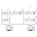
- FIG. 1 is a diagram showing the schematic configuration of the medical information system 1.
- the medical information system 1 shown in FIG. 1 captures an examination target region of a patient as a subject, stores medical images acquired by the imaging, This is a system for interpretation of medical images and creation of an interpretation report by an interpreting doctor, and viewing of the interpretation report and detailed observation of the medical image to be interpreted by a doctor of the medical department of the requesting department.
- the medical information system 1 includes a plurality of imaging devices 2, a plurality of image interpretation WSs (WorkStations) 3 which are image interpretation terminals, a medical examination WS 4, an image server 5, an image DB (DataBase) 5A, a report server 6, A report DB (DataBase) 6A, a management server 7, and a management DB (DataBase) 7A are connected to each other via a wired or wireless network 10 so as to be communicable with each other.
- a wired or wireless network 10 so as to be communicable with each other.
- Each device is a computer installed with an application program for functioning as a component of the medical information system 1.
- Application programs are recorded on recording media such as DVDs (Digital Versatile Discs) and CD-ROMs (Compact Discs Read Only Memory) for distribution, and are installed in computers from the recording media.
- recording media such as DVDs (Digital Versatile Discs) and CD-ROMs (Compact Discs Read Only Memory) for distribution, and are installed in computers from the recording media.
- recording media such as DVDs (Digital Versatile Discs) and CD-ROMs (Compact Discs Read Only Memory) for distribution, and are installed in computers from the recording media.
- it is stored in a storage device of a server computer connected to the network 10 or a network storage in a state accessible from the outside, and is downloaded and installed in a computer upon request.
- the imaging device 2 is a device (modality) that generates a medical image representing the diagnostic target region by imaging the diagnostic target region of the patient. Specifically, they are plain X-ray equipment, CT equipment, MRI equipment, and PET (Positron Emission Tomography) equipment. A medical image generated by the imaging device 2 is transmitted to the image server 5 and stored in the image DB 5A.
- the interpretation WS3 is a computer used by, for example, an interpretation doctor in a radiology department to interpret medical images and create an interpretation report, and includes an information processing apparatus (details will be described later) according to the present embodiment.
- the interpretation WS 3 requests the image server 5 to view medical images, performs various image processing on the medical images received from the image server 5 , displays the medical images, and accepts input of remarks on the medical images.
- the interpretation WS 3 also performs analysis processing for medical images, supports creation of interpretation reports based on the analysis results, requests registration and viewing of interpretation reports to the report server 6, and displays interpretation reports received from the report server 6. . These processes are performed by the interpretation WS3 executing a software program for each process.
- the clinical WS 4 is a computer used by, for example, doctors in clinical departments for detailed observation of images, viewing of interpretation reports, and preparation of electronic medical charts. It consists of a device.
- the medical examination WS 4 requests the image server 5 to view images, displays the images received from the image server 5 , requests the report server 6 to view interpretation reports, and displays the interpretation reports received from the report server 6 . These processes are performed by the clinical WS 4 executing a software program for each process.
- the image server 5 is a general-purpose computer installed with a software program that provides the functions of a database management system (DBMS). Further, the image server 5 has a storage in which an image DB 5A is configured. This storage may be a hard disk device connected to the image server 5 by a data bus, or a disk device connected to NAS (Network Attached Storage) and SAN (Storage Area Network) connected to network 10. may be When the image server 5 receives a request for registering a medical image from the imaging device 2, the image server 5 prepares the medical image into a database format and registers it in the image DB 5A.
- DBMS database management system
- the incidental information includes, for example, an image ID (identification) for identifying individual medical images, a patient ID for identifying a patient, an examination ID for identifying an examination, a unique ID assigned to each medical image ( UID: unique identification), examination date when the medical image was generated, examination time, type of imaging device used in the examination to acquire the medical image, patient information such as patient name, age, gender, examination site (imaging part), imaging information (imaging protocol, imaging sequence, imaging method, imaging conditions, use of contrast agent, etc.), and information such as series number or collection number when multiple medical images are acquired in one examination .
- image ID identification
- UID unique ID assigned to each medical image
- UID unique ID assigned to each medical image
- examination date when the medical image was generated examination time
- type of imaging device used in the examination to acquire the medical image patient information such as patient name, age, gender, examination site (imaging part), imaging information (imaging protocol, imaging sequence, imaging method, imaging conditions, use of contrast agent, etc.), and information such as series number or collection number when multiple
- the image server 5 When the image server 5 receives a viewing request from the interpretation WS 3 and the medical care WS 4 via the network 10, the image server 5 searches for medical images registered in the image DB 5A, and distributes the retrieved medical images to the requesting interpretation WS 3 and the medical care WS 4. Send to WS4.
- the report server 6 incorporates a software program that provides the functions of a database management system to a general-purpose computer.
- the report server 6 receives a registration request for an interpretation report from the interpretation WS 3, the interpretation report is formatted for a database and registered in the report DB 6A.
- the interpretation report contains information such as, for example, the medical image to be interpreted, the image ID for identifying the medical image, the interpretation doctor ID for identifying the interpretation doctor who performed the interpretation, the lesion name, the position information of the lesion, and the properties of the lesion. may contain In this embodiment, an interpretation report and one or more medical images for which the interpretation report is created are associated and registered in the report DB 6A.
- An interpretation report is an example of a medical document of the present disclosure.
- the report server 6 When the report server 6 receives a viewing request for an interpretation report from the interpretation WS 3 and the medical care WS 4 via the network 10, the report server 6 searches for the interpretation report registered in the report DB 6A, and sends the retrieved interpretation report to the requested interpretation report. Send to WS3 and medical care WS4.
- the management server 7 is a general-purpose computer installed with a software program that provides database management system functions.
- the management server 7 includes an information management device (details will be described later) according to this embodiment.
- the management server 7 registers the derived correspondence information in the management DB 7A as will be described later.
- the network 10 is a wired or wireless local area network that connects various devices in the hospital. If the image interpretation WS3 is installed in another hospital or clinic, the network 10 may be configured by connecting the local area networks of each hospital via the Internet or a dedicated line.
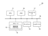
- the information management device 20 includes a CPU (Central Processing Unit) 11, a nonvolatile storage 13, and a memory 16 as a temporary storage area.
- the information management device 20 also includes a display 14 such as a liquid crystal display, an input device 15 such as a keyboard and a pointing device such as a mouse, and a network I/F (InterFace) 17 connected to the network 10 .
- CPU 11 , storage 13 , display 14 , input device 15 , memory 16 and network I/F 17 are connected to bus 18 .
- the CPU 11 is an example of a processor in the present disclosure.
- the storage 13 is realized by HDD (Hard Disk Drive), SSD (Solid State Drive), flash memory, and the like.
- An information management program 12 is stored in the storage 13 as a storage medium.
- the CPU 11 reads out the information management program 12 from the storage 13 , expands it in the memory 16 , and executes the expanded information management program 12 .
- the information processing device 30 includes a CPU 41, a nonvolatile storage 43, and a memory 46 as a temporary storage area.
- the information processing device 30 also includes a display 44 such as a liquid crystal display, an input device 45 such as a keyboard and a pointing device such as a mouse, and a network I/F 47 connected to the network 10 .
- CPU 41 , storage 43 , display 44 , input device 45 , memory 46 and network I/F 47 are connected to bus 48 .
- the CPU 41 is an example of a processor in the present disclosure.
- the storage 43 like the storage 13, is implemented by HDD, SSD, flash memory, and the like.
- An information processing program 42 is stored in the storage 43 as a storage medium.
- the CPU 41 reads the information processing program 42 from the storage 43 , expands it in the memory 46 , and executes the expanded information processing program 42 .
- FIG. 4 is a diagram showing the functional configuration of the information management device according to this embodiment.
- the information management device 20 includes an information acquisition section 21 , a first analysis section 22 , a second analysis section 23 and a corresponding information derivation section 24 .
- the CPU 11 functions as an information acquisition section 21 , a first analysis section 22 , a second analysis section 23 and a corresponding information derivation section 24 .
- the information acquisition unit 21 acquires medical images and interpretation reports associated with the medical images from the image server 5 and the report server 6 via the network I/F 17, respectively.
- FIG. 5 is a diagram showing an example of an interpretation report.
- the interpretation report 50 includes an observation sentence 51 .
- the observation sentence 51 is "A solid nodule with a clear boundary with a long axis of 1 cm is found in the upper lobe of the right lung. Adenocarcinoma of the lung is suspected. A ground-glass nodule with a similar circular shape is found in S8 of the left lung.”
- the interpretation report 50 is associated with the medical image 52 from which the interpretation report 50 is created. Specifically, it is associated with a tomographic image including a region of interest whose findings are described in the interpretation report 50, among a plurality of tomographic images included in the medical image.
- the first analysis unit 22 acquires findings about the region of interest included in the medical image 52 by analyzing the finding sentence 51 included in the interpretation report 50 . Specifically, the first analysis unit 22 analyzes the character string included in the interpretation report using natural language processing technology, thereby extracting findings about the region of interest included in the interpretation report from the interpretation report. . Findings specifically include the position and type of the region of interest, the disease type when the region of interest includes an abnormal site, the name of the disease, the characteristics of the disease, the size of the disease, and the like. In addition, since the finding text 51 also includes findings when the region of interest includes a normal site, the first analysis unit 22 also acquires findings when the region of interest includes a normal site. Note that an example of a finding sentence including a finding about a normal site is "It is normal because the characteristic of A can be seen.” Here, "A" is a finding seen in normal cases.
- the interpretation report describes the properties of the region of interest. For example, if the region of interest is in the lung, the type of absorption value (solid type and ground glass type), presence or absence of spicules, presence or absence of calcification, presence or absence of cavities, presence or absence of pleural indentation, presence or absence of pleural contact, and pleural infiltration Properties are described for a plurality of items such as presence or absence.
- the first analysis unit 22 acquires the properties of the region of interest described in the interpretation report as findings.
- the measurement direction of the size may be predetermined.
- the major axis is described in the interpretation report
- the minor axis is described in the interpretation report.
- the size of the cyst is not important, and only information about whether the cyst is small or large is included in the interpretation report. For this reason, the first analysis unit 22 acquires the size measurement direction of the size and the size information indicating the size (small, minute, large, etc.) included in the interpretation report as findings.
- anatomical segment can be divided into left and right lungs, and five lobes (left upper lobe, left lower lobe, right upper lobe, right middle lobe, and right lower lobe) can be divided into anatomical regions.
- the anatomical regions of regions S1-S8 can be separated for the left and right lungs respectively.
- anatomical segments can be divided into regions S1 to S8 in addition to left and right lobes.
- the anatomic area can be divided into left and right regions.
- the first analysis unit 22 acquires anatomical level information representing the level of the anatomical region of the organ in which the region of interest exists, included in the interpretation report, as findings.
- Natural language processing is a series of technologies that allow computers to process natural language that humans use on a daily basis. With natural language processing, sentences can be segmented into words, syntactically analyzed, semantically analyzed, and the like.
- the first analysis unit 22 obtains findings by dividing the character string included in the interpretation report into words and analyzing the syntax using natural language processing technology. For example, if the finding text included in the interpretation report is "A solid nodule with a clear boundary with a long axis of 1 cm is found in the upper lobe of the right lung.
- Lung adenocarcinoma is suspected.” , “upper lobe of right lung”, “nodule”, “clearly demarcated”, “solid type”, “major axis”, “1 cm” and “lung adenocarcinoma” are extracted as findings.
- the first analysis unit 22 identifies which part of the human body the finding sentence describes from the acquired finding.
- the first analysis unit 22 also identifies the type of disease from the acquired findings. Specifically, the first analysis unit 22 identifies a disease type and a disease name as the type of disease.
- the disease type is identified based on the findings extracted from the interpretation report. For example, if the findings include "nodule”, the first analysis unit 22 identifies the disease type as "nodular”. Moreover, since the disease type of tumor is nodular, the first analysis unit 22 identifies the disease type as "nodular” when "tumor" is included in the findings. Also, when the findings are "pneumonia”, "fatty liver”, etc., the first analysis unit 22 identifies the disease type as "diffuse”. Furthermore, when the findings are "renal enlargement” and “pancreatic duct dilatation", the first analysis unit 22 identifies the disease type as "structural abnormality". In this embodiment, the identified disease type is also one of the findings.
- the first analysis unit 22 identifies the disease name based on the findings corresponding to the disease name among the findings acquired from the interpretation report. For example, if the findings include "lung adenocarcinoma”, the first analysis unit 22 identifies the disease name as "lung adenocarcinoma”. Also, if the finding includes "interstitial pneumonia”, the first analysis unit 22 specifies the disease name as "interstitial pneumonia”. In this embodiment, the identified disease name is also one of the findings.
- a focal finding is a finding related to disease occurring in a part of an organ, and examples include findings such as nodules, tumors, cysts, calcifications, and stones.
- Non-focal findings are those related to disease related to the whole organ, including findings related to organ shape such as enlargement, atrophy and deformity, and organs such as interstitial pneumonia, emphysema, fatty liver, cirrhosis and pleural effusion. Examples include idiosyncratic findings.
- the second analysis unit 23 derives medical image information from the medical image 52. Specifically, by analyzing the medical image, the second analysis unit 23 derives an image including a region of interest such as an abnormal shadow candidate included in the medical image as medical image information. For this reason, the second analysis unit 23 has a trained model (not shown) that has undergone machine learning so as to detect an abnormal shadow candidate in a medical image as a region of interest. In this embodiment, the trained model determines whether or not each pixel (voxel) in the medical image represents an abnormal shadow candidate. As specified in , it consists of a convolutional neural network (CNN (Convolutional Neural Network)) subjected to deep learning using teacher data.
- CNN Convolutional Neural Network
- a trained model is prepared for each organ, for example, and is trained to identify regions of interest, which are candidates for abnormal shadows contained in the organ.
- any trained model such as a support vector machine (SVM) can be used in addition to the convolutional neural network.
- SVM support vector machine
- the second analysis unit 23 detects a region of interest from a tomographic image including the region of interest whose findings are described in the interpretation report 50, among the medical images 52 composed of a plurality of tomographic images. Then, the second analysis unit 23 extracts an image of a local region including the detected region of interest (hereinafter referred to as a local image) from the tomographic image as medical image information.
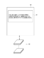
- FIG. 6 is a diagram for explaining extraction of medical image information. As shown in FIG. 6, the second analysis unit 23 identifies the tomographic image 52A associated with the interpretation report 50, and uses the local image 53 including the region of interest in the tomographic image 52A as medical image information, as indicated by the arrow A1. It is extracted from the tomographic image 52A. A plurality of regions of interest may be extracted from one medical image. In this case, the second analysis unit 23 derives a plurality of local images 53 for each of the plurality of regions of interest as medical image information.
- the second analysis unit 23 may derive the feature amount 54 of the local image 53 as medical image information, as indicated by an arrow A2.
- the feature amount may be, for example, a set of vectors whose elements are the pixel position of each pixel in the local image 53 and the pixel value of each pixel position. It may be a set of vectors whose elements are values obtained by performing processing and positions on the local image 53 corresponding to the values.
- FIG. 6 when the plane of the local image 53 is the xy plane, the distribution in the three-dimensional space of vectors whose elements are the pixel positions of the local image 53 and the value F after convolution at each pixel position is shown. It is shown as feature quantity 54 .
- the second analysis unit 23 may use position information 55 representing the position of the local image 53 in the medical image 52 as the medical image information, as indicated by an arrow A3.
- position information coordinate values (x1, y1, z1), (x2, y2, z1), (x3, y3, z1) and (x4, y4, z1).
- the location information also includes a link to the medical image 52 .
- the link becomes a link to the medical image 52 stored in the image server 5 .
- the correspondence information deriving unit 24 derives correspondence information that associates findings with medical image information related to regions of interest.
- the derived correspondence information is arranged in a database format and registered in the management DB 7A.
- the information management device 20 of the management server 7 uses a large number of medical images and interpretation reports to obtain findings about regions of interest included in each of the medical images, and associates the findings with medical image information about the regions of interest. Derive correspondence information. Then, the information management device 20 registers the derived correspondence information in the management DB 7A.
- FIG. 7 is a diagram showing an example of the management DB 7A.
- the management DB 7A registers a plurality of correspondence information derived from a plurality of combinations of medical images and interpretation reports.
- Each piece of correspondence information includes a site, disease type, disease name, multiple disease features, anatomical level information, size level information, medical image information, and finding statement generation information.
- disease features, anatomical level information, size level information, medical image information, and finding statement generation information are associated with each site, disease type, or disease name.
- a plurality of findings are acquired from a plurality of combinations of medical images and interpretation reports. Among the acquired multiple findings, related findings, particularly disease characteristics, are included in one piece of corresponding information.
- a site is a site based on findings acquired by the first analysis unit 22 from an interpretation report.
- the disease type and disease name are the disease type and disease name based on the findings acquired by the first analysis unit 22 from the interpretation report.
- a disease feature is a finding obtained by the first analysis unit 22 as a property from an interpretation report.
- FIG. 7 shows three disease features for one disease type or disease name, the number of disease features depends on the number of findings obtained from the interpretation report.
- the anatomical level information is information representing the level of the anatomical region of the organ in which the region of interest exists, which is included in the interpretation report.
- the size level information is information indicating the measurement direction of the size and the magnitude of the size included in the interpretation report.
- the correspondence information includes local images of medical images related to findings as medical image information.
- rectangles representing local images indicate that the local images of the medical image are registered.
- a plurality of pieces of medical image information may be included in one piece of correspondence information.
- the medical image information may include a feature amount derived from the local image instead of or in addition to the local image.
- the medical image information may include a combination of the link of the medical image from which the corresponding information is acquired and the positional information of the region of interest in the linked medical image instead of the local image.
- the observation text generation information included in the correspondence information is information indicating whether to use a template or natural language processing when generating an observation text with reference to the management DB 7A, as will be described later.
- the management DB 7A stores ⁇ template> representing generation of an observation sentence by a template and ⁇ NLP> (Natural Language Processing) representing generation of an observation sentence by natural language processing. ) is described.
- the finding generation information is a template, for example, " ⁇ feature 2> with ⁇ feature 1> of ⁇ size> at ⁇ location> is allowed", " ⁇ feature 1> at ⁇ location> I agree.”
- a template having blanks corresponding to the findings registered in the correspondence information is stored in the management server 7 .
- the finding generation information is NLP
- the finding text is derived by natural language processing in interpretation WS3, which will be described later.
- the finding statement generation information is to be a template or an NLP is registered in the management DB 7A by the operator depending on the number of findings included in the finding statement. Further, whether the finding sentence generation information is used as a template or NLP may be set according to the site or disease. For example, regarding the lung, there is a tendency to describe a relatively long observation sentence in the interpretation report, so NLP is described in the corresponding information for the lung part.
- postoperative is a disease name for a normal site.
- Disease names other than "postoperative” are disease names of abnormal sites.
- FIG. 8 is a diagram showing the functional configuration of the information processing apparatus according to this embodiment.
- the information processing device 30 includes an image acquisition unit 31 , an image analysis unit 32 , a finding identification unit 33 , a medical information generation unit 34 and a display control unit 35 .
- the CPU 41 By executing the information processing program 42 by the CPU 41 , the CPU 41 functions as an image acquisition unit 31 , an image analysis unit 32 , a finding identification unit 33 , a medical information generation unit 34 and a display control unit 35 .
- the image acquisition unit 31 acquires a medical image to be interpreted for creating an interpretation report from the image server 5 in accordance with an instruction from the input device 45 by the interpreting doctor who is the operator.
- a medical image to be interpreted is referred to as a target medical image G0 in the following description.
- the image analysis unit 32 identifies a target region of interest such as an abnormal shadow candidate included in the target medical image G0 by analyzing the target medical image G0.
- the image analysis unit 32 like the first analysis unit 22 of the information management apparatus 20 according to the present embodiment, determines abnormal shadow candidates in the medical image, and detects the determined abnormal shadow candidates as target regions of interest. It has a trained model (not shown) that has undergone machine learning as follows.
- the finding specifying unit 33 refers to the management DB 7A to specify the finding corresponding to the medical image information for the corresponding region of interest.
- the management DB 7A may be downloaded to the interpretation WS 3 and stored in the storage 43.
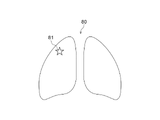
- FIG. FIG. 9 is a diagram for explaining identification of findings.
- the finding identification unit 33 extracts the target region of interest 60 detected from the target medical image G0 by the image analysis unit 32 and the medical image information included in each of the plurality of correspondence information registered in the management DB 7A. and the target region of interest 60 is derived.
- the finding identification unit 33 derives the feature amount of the target region of interest 60 and derives the degree of similarity with the medical image information.
- the finding specifying unit 33 refers to the position information registered in the management DB 7A, By referring to the medical image linked to the information, the region of interest for the medical image registered in the management DB 7A is specified, and the degree of similarity between the region of interest and the target region of interest 60 is derived.
- the finding specifying unit 33 specifies a finding by referring to a predetermined number of high-ranking correspondence information with a high degree of similarity in the management DB 7A. For example, when the predetermined number is 3, the first to third findings are identified by referring to the top three correspondence information having the highest degree of similarity. For example, in the management DB 7A, when “ring-shaped hyperplasia” and “nodule”, “hemangioma”, and “cyst” are registered as findings in the three corresponding information with the highest similarity, The finding identification unit 33 identifies “ring-shaped hyperplasia” and “nodule” as the first finding, “hemangioma” as the second finding, and “cyst” as the third finding.
- the identified findings are related to diseases that occur in a part of the organ such as nodules, tumors, cysts, calcifications, and stones, the findings are identified as localized findings.
- the identified findings relate to organ shape such as enlargement, atrophy and deformity, or to organ-specific diseases such as interstitial pneumonia, emphysema, fatty liver, cirrhosis and pleural effusion, non- A focal finding finding has been identified.
- the identified findings are related to changes from the healthy state of the target region of interest.
- findings such as nodules, tumors, cysts, calcifications, and calculi included in the focal findings described above are associated with changes from a healthy state when part of the organ is the region of interest.
- findings such as interstitial pneumonia, emphysema, fatty liver, liver cirrhosis, and pleural effusion included in non-localized findings are related to changes from a healthy state when the entire organ is the region of interest.
- the medical information generation unit 34 generates medical information including the identified findings.
- a finding sentence including the specified finding is generated as medical information.
- the medical information generation unit 34 refers to finding statement generation information registered in correspondence information including the specified finding in the management DB 7A.
- an observation sentence is generated by a generation method according to the registered observation sentence generation information.
- the finding text generation information is a template
- the medical information generating unit 34 acquires the corresponding template from the management server 7, and generates the finding text by inserting the specified finding into the blanks of the acquired template. .
- the medical information generating unit 34 generates a finding sentence of "A ⁇ nodule> accompanied by ⁇ ring-shaped deepening> of ⁇ 2 cm size> is recognized in ⁇ right lung S5>.”
- the medical information generation unit 34 creates the finding sentence by natural language processing.
- the medical information generation unit 34 has a trained model 34A that has been machine-learned so as to generate an observation statement from the analysis results and observations of the image analysis unit 32 .
- a recurrent neural network can be used as the trained model 34A.
- the trained model 34A uses a large amount of teacher data consisting of combinations of analysis results, observations, and observation sentences to machine a recurrent neural network. It is constructed by learning.
- the finding sentence generation information is NLP
- the medical information generating unit 34 inputs the analysis result and the finding by the image analyzing unit 32 to the learned model 34A, and generates the finding sentence by outputting the finding sentence.
- the generation unit 34 uses the learned model 34A to generate an observation sentence such as "A nodule with a major axis of 1.5 cm and a major axis of 3 mm is found in the left lung S6, and lung metastasis is suspected.”
- the image analysis unit 32, the finding identification unit 33, and the medical information generation unit 34 perform the above-described processing to generate medical information.
- the medical information generated by the medical information generation unit 34 is stored in the storage 43 in association with the target region of interest identified in the target medical image G0.
- the display control unit 35 displays the medical information generated by the medical information generation unit 34.
- the medical information generator 34 derives a plurality of observation sentences as medical information. Therefore, the display control unit 35 displays a plurality of observation sentences in a selectable manner.
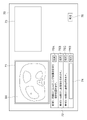
- FIG. 10 is a diagram showing a display screen of medical information.
- the display screen 70 includes an image display area 71 , a candidate display area 72 and a text display area 73 .
- the image display area 71 a tomographic image representing a tomographic plane of the target medical image G0 is displayed in a switchable manner.
- the radiologist selects the adoption button corresponding to the observation sentence candidate. As a result, the selected observation sentence is displayed in the sentence display area 73 .
- the radiologist enters the finding text in the input box 74 and selects the adoption button 75D. . As a result, the observation text input by the radiologist in the input box 74 is displayed in the text display area 73 .
- the interpreting doctor interprets the image while switching the tomographic images displayed in the image display area 71, clicks on the lesion position, displays the finding sentence candidate in the candidate display area 72, and selects the finding sentence by repeating the sentence display area. 73 describes the remarks.
- an interpretation report including the observation text entered in the text display area 73 is created.
- the created interpretation report is stored in the storage 43 together with the target medical image G0 and the detection result of the region of interest. Thereafter, the created interpretation report is transferred to the report server 6 together with the target medical image G0 and the detection results.
- the report server 6 stores the transferred interpretation report together with the target medical image G0 and the detection result.
- the management server 7 uses the new interpretation report and the medical image for which the new interpretation report is created to derive the corresponding information in the same manner as described above, and derives the corresponding information.
- the management DB 7A may be updated using the new correspondence information.
- FIG. 11 is a flowchart showing information management processing performed in this embodiment.
- the information acquisition unit 21 acquires the medical image and the interpretation report associated with the medical image from the image server 5 and the report server 6 (information acquisition; step ST1).
- the first analysis unit 22 acquires findings about the region of interest included in the medical image 52 by analyzing the finding sentence 51 included in the interpretation report 50 (step ST2).
- the second analysis unit 23 derives medical image information from the medical image 52 (step ST3).
- the correspondence information derivation unit 24 derives correspondence information that associates the findings with the medical image information related to the region of interest (step ST4). Then, the correspondence information derivation unit 24 registers the derived correspondence information in the management DB 7A (step ST5). Next, it is determined whether or not registration of correspondence information for all interpretation reports to be registered in the management DB 7A has been completed (step ST6). If step ST6 is negative, the information acquisition unit 21 acquires a new medical image and an interpretation report associated with the medical image from the image server 5 and the report server 6 (acquisition of new information; step ST7). Then, the process returns to step ST2, and the processes after step ST2 are repeated. If step ST6 is affirmative, the process is terminated.
- FIG. 12 is a flowchart showing information processing performed in this embodiment.
- the processing is started when the interpretation doctor issues an instruction to create an interpretation report, and the image acquisition unit 31 acquires the target medical image G0 to be interpreted from the image server 5 (step ST11).
- the image analysis unit 32 analyzes the target medical image G0 to specify a target region of interest such as an abnormal shadow candidate included in the target medical image G0 (step ST12).
- the finding specifying unit 33 refers to the management DB 7A to specify a finding corresponding to the medical image information for the corresponding region of interest (step ST13).
- the medical information generation unit 34 generates medical information including the identified findings (step ST14).
- the generated medical information is saved in the storage 43 .
- the display control unit 35 displays the target medical image G0 on the display 44 (step ST15). Further, the display control unit 35 accepts designation of a lesion in the displayed target medical image G0 by the interpreting doctor (step ST16), and medical information derived for the corresponding region of interest corresponding to the designated lesion, that is, a plurality of finding sentences. is displayed in the candidate display area 72 on the display screen 70 (step ST17).
- step ST18 When one of the plurality of observations displayed in the candidate display area 72 is selected (step ST18: Yes), the display control unit 35 displays the selected observation in the text display area 73 (step ST19). Then, it is determined whether or not the confirm button 76 has been selected (step ST20), and if step ST20 is negative, the process returns to step ST16, and the processes after step ST16 are repeated. If step ST20 is affirmative, an interpretation report is generated (step ST21), and the process ends.
- the created interpretation report is stored in the storage 13 together with the target medical image G0 and the detection results, and the created interpretation report is transferred to the report server 6 together with the target medical image G0 and the detection results.
- a medical document such as an interpretation report associated with a medical image
- the findings are obtained by specifying the findings about the region of interest included in the medical image in the medical document, thereby obtaining the region of interest included in the medical image.
- Interpreter-specific findings can be obtained for
- the management DB 7A is referenced to identify findings corresponding to medical image information about the target region of interest. Furthermore, in the present embodiment, a statement of findings including findings is generated as medical information. Therefore, it is possible to efficiently create an interpretation report including an observation sentence. In addition, it is possible to generate homogeneous observation statements that do not depend on radiologists.
- the correspondence information for each medical image is registered in the management DB 7A, but it is not limited to this.
- a set of correspondence information of a plurality of related medical images may be registered in the management DB 7A.
- correspondence information about two medical images may be grouped together and registered in the management DB 7A as correspondence information about lungs in the second row from the top.
- a broken line indicates a delimiter between pieces of correspondence information of two medical images grouped together.
- the disease type is diffuse for both of the two medical images, and the finding text generation information is NLP for both of the two medical images.
- the disease name is interstitial pneumonia in both of the two medical images
- the disease feature 1 is ground-glass shadow in both of the two medical images.
- disease features 2 and 3 are included only in the corresponding information of medical images in the upper row.
- Examples of multiple related medical images include multiple medical images taken at different times for the same patient and medical images with different modalities for the same patient.
- Examples of a plurality of medical images with different modalities include a combination of a CT image and an MRI image.
- the management DB 7A is derived in this way, in the information processing apparatus according to the present embodiment, for example, for two related medical images, the management DB 7A is referred to for correspondence information including medical image information similar to both of the two medical images. can be referenced to identify findings and generate a finding statement for the two medical images.
- a plurality of observation sentences are generated as medical information, but the present invention is not limited to this. You may generate
- the finding specifying unit 33 may specify the finding by referring to the correspondence information with the highest degree of similarity in the management DB 7A, and the medical information generating unit 34 may generate the finding sentence using the specified finding.
- the medical information generation unit 34 derives an observation sentence as medical information, but it is not limited to this.
- Medical information may be derived that includes a graphical representation of the identified findings.
- FIG. 13 is a diagram showing an example of medical information including a graphical representation of identified findings.
- the specified findings are "right upper lobe of lung" and "pulmonary nodule”.
- the medical information generator 34 may derive medical information in which a mark 81 is added to the position of the right upper lobe of the lung in the lung schema 80 as graphical information. By deriving and displaying medical information including such graphical representations, it is possible to easily recognize the position of an abnormal site.
- the information processing device 30 derives medical information by referring to the management DB 7A generated by the information management device according to this embodiment, but it is not limited to this.
- the correspondence information derived by associating findings and medical image information by manual operation is registered in the management DB 7A, and the management DB 7A in which the correspondence information derived by such manual operation is registered is referred to.
- Information may be derived.
- the medical information is generated when the target medical image G0 is acquired, but it is not limited to this.
- the target medical image G0 may be displayed, and every time an interpreting doctor designates an abnormal site in the displayed image, an observation sentence about the designated abnormal site may be generated as medical information.
- the finding identifying unit 33 identifies findings about the designated abnormal site, and the medical information generating unit 34 generates medical information about the designated abnormal site based on the identified findings. .
- the information management apparatus includes the first analysis unit 22 and obtains findings by analyzing the interpretation report, but the present invention is not limited to this. Findings may be acquired based on input by the operator using the input device 15, or may be acquired by analyzing medical images.
- the processing of the first analysis unit 22 and the second analysis unit 23 in the information management device 20 included in the management server 7 is performed by an external device such as another analysis server connected to the network 10, for example. You may do so.
- the external device acquires an interpretation report from the report server 6 , acquires findings by analyzing the interpretation report, and transmits the findings to the management server 7 .
- the external device also derives medical image information from the medical image associated with the interpretation report, and transmits the derived medical image information to the management server 7 .
- the information management device 20 of the management server 7 derives correspondence information using the findings and medical image information derived by the external device, and registers the derived correspondence information in the management DB 7A.
- the hardware structure of the processing unit (Processing Unit) that executes various processes such as the unit 32, the finding identification unit 33, the medical information generation unit 34, and the display control unit 35 includes the following various processors. can be used.
- the various processors include, in addition to the CPU, which is a general-purpose processor that executes software (programs) and functions as various processing units, circuits such as FPGAs (Field Programmable Gate Arrays), etc.
- Programmable Logic Device PLD which is a processor whose configuration can be changed, ASIC (Application Specific Integrated Circuit) etc. Circuits, etc. are included.
- One processing unit may be configured with one of these various processors, or a combination of two or more processors of the same or different type (for example, a combination of multiple FPGAs or a combination of a CPU and an FPGA). ).
- a plurality of processing units may be configured by one processor.
- a plurality of processing units may be configured by one processor.
- this processor functions as a plurality of processing units.
- SoC System On Chip
- SoC System On Chip
- the various processing units are configured using one or more of the above various processors as a hardware structure.
- an electric circuit in which circuit elements such as semiconductor elements are combined can be used.
Landscapes
- Engineering & Computer Science (AREA)
- Health & Medical Sciences (AREA)
- Medical Informatics (AREA)
- General Health & Medical Sciences (AREA)
- Public Health (AREA)
- Primary Health Care (AREA)
- Epidemiology (AREA)
- Biomedical Technology (AREA)
- Radiology & Medical Imaging (AREA)
- Nuclear Medicine, Radiotherapy & Molecular Imaging (AREA)
- Physics & Mathematics (AREA)
- Life Sciences & Earth Sciences (AREA)
- Pathology (AREA)
- Data Mining & Analysis (AREA)
- Computer Vision & Pattern Recognition (AREA)
- Quality & Reliability (AREA)
- Theoretical Computer Science (AREA)
- General Physics & Mathematics (AREA)
- Databases & Information Systems (AREA)
- Heart & Thoracic Surgery (AREA)
- Surgery (AREA)
- Animal Behavior & Ethology (AREA)
- Veterinary Medicine (AREA)
- Molecular Biology (AREA)
- Biophysics (AREA)
- Business, Economics & Management (AREA)
- General Business, Economics & Management (AREA)
- Medical Treatment And Welfare Office Work (AREA)
- Measuring And Recording Apparatus For Diagnosis (AREA)
Priority Applications (3)
| Application Number | Priority Date | Filing Date | Title |
|---|---|---|---|
| EP22770867.4A EP4310853A4 (en) | 2021-03-19 | 2022-01-24 | Information management device, method, and program, and information processing device, method, and program |
| JP2023506814A JPWO2022196105A1 (https=) | 2021-03-19 | 2022-01-24 | |
| US18/460,712 US20230410305A1 (en) | 2021-03-19 | 2023-09-05 | Information management apparatus, method, and program and information processing apparatus, method, and program |
Applications Claiming Priority (4)
| Application Number | Priority Date | Filing Date | Title |
|---|---|---|---|
| JP2021046613 | 2021-03-19 | ||
| JP2021-046613 | 2021-03-19 | ||
| JP2021-208718 | 2021-12-22 | ||
| JP2021208718 | 2021-12-22 |
Related Child Applications (1)
| Application Number | Title | Priority Date | Filing Date |
|---|---|---|---|
| US18/460,712 Continuation US20230410305A1 (en) | 2021-03-19 | 2023-09-05 | Information management apparatus, method, and program and information processing apparatus, method, and program |
Publications (1)
| Publication Number | Publication Date |
|---|---|
| WO2022196105A1 true WO2022196105A1 (ja) | 2022-09-22 |
Family
ID=83320244
Family Applications (1)
| Application Number | Title | Priority Date | Filing Date |
|---|---|---|---|
| PCT/JP2022/002461 Ceased WO2022196105A1 (ja) | 2021-03-19 | 2022-01-24 | 情報管理装置、方法およびプログラム、並びに情報処理装置、方法およびプログラム |
Country Status (4)
| Country | Link |
|---|---|
| US (1) | US20230410305A1 (https=) |
| EP (1) | EP4310853A4 (https=) |
| JP (1) | JPWO2022196105A1 (https=) |
| WO (1) | WO2022196105A1 (https=) |
Families Citing this family (2)
| Publication number | Priority date | Publication date | Assignee | Title |
|---|---|---|---|---|
| JP7706987B2 (ja) * | 2021-08-17 | 2025-07-14 | 富士フイルム株式会社 | 学習装置、方法およびプログラム、並びに情報処理装置、方法およびプログラム |
| JP7718915B2 (ja) * | 2021-08-30 | 2025-08-05 | 富士フイルム株式会社 | 学習装置、方法およびプログラム、並びに情報処理装置、方法およびプログラム |
Citations (8)
| Publication number | Priority date | Publication date | Assignee | Title |
|---|---|---|---|---|
| JPH07323024A (ja) | 1994-06-01 | 1995-12-12 | Konica Corp | 画像診断支援装置 |
| JP2007305107A (ja) * | 2006-04-10 | 2007-11-22 | Fujifilm Corp | レポート作成支援装置、レポート作成支援方法およびそのプログラム |
| WO2009041586A1 (ja) * | 2007-09-28 | 2009-04-02 | Canon Kabushiki Kaisha | 診断支援装置及びその制御方法 |
| JP2009110485A (ja) * | 2007-11-01 | 2009-05-21 | Konica Minolta Medical & Graphic Inc | 情報処理システム、及びプログラム |
| JP2011118543A (ja) * | 2009-12-01 | 2011-06-16 | Shizuoka Prefecture | 症例画像検索装置、方法およびプログラム |
| JP2017068380A (ja) | 2015-09-28 | 2017-04-06 | キヤノン株式会社 | 情報処理装置及びその方法、情報処理システム、コンピュータプログラム |
| JP2018534029A (ja) * | 2015-10-02 | 2018-11-22 | コーニンクレッカ フィリップス エヌ ヴェKoninklijke Philips N.V. | 所見を関連する心エコー図ループにマッピングするためのシステム |
| JP2019153250A (ja) * | 2018-03-06 | 2019-09-12 | 富士フイルム株式会社 | 医療文書作成支援装置、方法およびプログラム |
Family Cites Families (4)
| Publication number | Priority date | Publication date | Assignee | Title |
|---|---|---|---|---|
| JP3332104B2 (ja) * | 1993-07-19 | 2002-10-07 | 株式会社東芝 | 読影レポート作成支援装置 |
| JP5618787B2 (ja) * | 2010-11-29 | 2014-11-05 | キヤノン株式会社 | レポート作成支援装置及びその作成支援方法、並びにプログラム |
| JP6510196B2 (ja) * | 2014-08-12 | 2019-05-08 | キヤノンメディカルシステムズ株式会社 | 読影レポート作成支援装置 |
| WO2017189758A1 (en) * | 2016-04-26 | 2017-11-02 | Ascend Hit Llc | System and methods for medical image analysis and reporting |
-
2022
- 2022-01-24 JP JP2023506814A patent/JPWO2022196105A1/ja active Pending
- 2022-01-24 EP EP22770867.4A patent/EP4310853A4/en active Pending
- 2022-01-24 WO PCT/JP2022/002461 patent/WO2022196105A1/ja not_active Ceased
-
2023
- 2023-09-05 US US18/460,712 patent/US20230410305A1/en active Pending
Patent Citations (8)
| Publication number | Priority date | Publication date | Assignee | Title |
|---|---|---|---|---|
| JPH07323024A (ja) | 1994-06-01 | 1995-12-12 | Konica Corp | 画像診断支援装置 |
| JP2007305107A (ja) * | 2006-04-10 | 2007-11-22 | Fujifilm Corp | レポート作成支援装置、レポート作成支援方法およびそのプログラム |
| WO2009041586A1 (ja) * | 2007-09-28 | 2009-04-02 | Canon Kabushiki Kaisha | 診断支援装置及びその制御方法 |
| JP2009110485A (ja) * | 2007-11-01 | 2009-05-21 | Konica Minolta Medical & Graphic Inc | 情報処理システム、及びプログラム |
| JP2011118543A (ja) * | 2009-12-01 | 2011-06-16 | Shizuoka Prefecture | 症例画像検索装置、方法およびプログラム |
| JP2017068380A (ja) | 2015-09-28 | 2017-04-06 | キヤノン株式会社 | 情報処理装置及びその方法、情報処理システム、コンピュータプログラム |
| JP2018534029A (ja) * | 2015-10-02 | 2018-11-22 | コーニンクレッカ フィリップス エヌ ヴェKoninklijke Philips N.V. | 所見を関連する心エコー図ループにマッピングするためのシステム |
| JP2019153250A (ja) * | 2018-03-06 | 2019-09-12 | 富士フイルム株式会社 | 医療文書作成支援装置、方法およびプログラム |
Also Published As
| Publication number | Publication date |
|---|---|
| EP4310853A4 (en) | 2024-09-04 |
| JPWO2022196105A1 (https=) | 2022-09-22 |
| EP4310853A1 (en) | 2024-01-24 |
| US20230410305A1 (en) | 2023-12-21 |
Similar Documents
| Publication | Publication Date | Title |
|---|---|---|
| JP7436698B2 (ja) | 医用画像処理装置、方法およびプログラム | |
| US20230005580A1 (en) | Document creation support apparatus, method, and program | |
| JP7684374B2 (ja) | 情報保存装置、方法およびプログラム、並びに解析記録生成装置、方法およびプログラム | |
| US20190279408A1 (en) | Medical image processing apparatus, medical image processing method, and medical image processing program | |
| US20230281810A1 (en) | Image display apparatus, method, and program | |
| US12527528B2 (en) | Image display apparatus, method, and program | |
| EP3954277A1 (en) | Medical document generation device, method, and program | |
| US12211600B2 (en) | Information processing apparatus, information processing method, and information processing program | |
| WO2021107098A1 (ja) | 文書作成支援装置、文書作成支援方法及び文書作成支援プログラム | |
| JP7844358B2 (ja) | 情報処理装置、方法およびプログラム | |
| WO2022196106A1 (ja) | 文書作成装置、方法およびプログラム | |
| WO2021157705A1 (ja) | 文書作成支援装置、方法およびプログラム | |
| WO2021177357A1 (ja) | 情報処理装置、情報処理方法及び情報処理プログラム | |
| WO2021107099A1 (ja) | 文書作成支援装置、文書作成支援方法及びプログラム | |
| WO2022196105A1 (ja) | 情報管理装置、方法およびプログラム、並びに情報処理装置、方法およびプログラム | |
| JP7299314B2 (ja) | 医療文書作成装置、方法およびプログラム、学習装置、方法およびプログラム、並びに学習済みモデル | |
| WO2021193548A1 (ja) | 文書作成支援装置、方法およびプログラム | |
| JP2025178348A (ja) | 医用画像表示装置、方法およびプログラム | |
| WO2023054645A1 (ja) | 情報処理装置、情報処理方法及び情報処理プログラム | |
| WO2023199957A1 (ja) | 情報処理装置、情報処理方法及び情報処理プログラム | |
| JP2023067186A (ja) | 情報処理装置、情報処理方法及び情報処理プログラム | |
| WO2021107142A1 (ja) | 文書作成支援装置、方法およびプログラム | |
| JP7368592B2 (ja) | 文書作成支援装置、方法およびプログラム | |
| JP7376715B2 (ja) | 経過予測装置、経過予測装置の作動方法および経過予測プログラム | |
| WO2023054646A1 (ja) | 情報処理装置、情報処理方法及び情報処理プログラム |
Legal Events
| Date | Code | Title | Description |
|---|---|---|---|
| 121 | Ep: the epo has been informed by wipo that ep was designated in this application |
Ref document number: 22770867 Country of ref document: EP Kind code of ref document: A1 |
|
| WWE | Wipo information: entry into national phase |
Ref document number: 2023506814 Country of ref document: JP |
|
| WWE | Wipo information: entry into national phase |
Ref document number: 2022770867 Country of ref document: EP |
|
| NENP | Non-entry into the national phase |
Ref country code: DE |
|
| ENP | Entry into the national phase |
Ref document number: 2022770867 Country of ref document: EP Effective date: 20231019 |