WO2022181515A1 - Polythérapie avec un inhibiteur du signal pd-1 - Google Patents
Polythérapie avec un inhibiteur du signal pd-1 Download PDFInfo
- Publication number
- WO2022181515A1 WO2022181515A1 PCT/JP2022/006843 JP2022006843W WO2022181515A1 WO 2022181515 A1 WO2022181515 A1 WO 2022181515A1 JP 2022006843 W JP2022006843 W JP 2022006843W WO 2022181515 A1 WO2022181515 A1 WO 2022181515A1
- Authority
- WO
- WIPO (PCT)
- Prior art keywords
- cells
- mice
- cell
- tcr
- signaling
- Prior art date
Links
- 239000003112 inhibitor Substances 0.000 title claims abstract description 72
- 238000002648 combination therapy Methods 0.000 title abstract description 4
- 210000004027 cell Anatomy 0.000 claims abstract description 260
- 108091008874 T cell receptors Proteins 0.000 claims abstract description 120
- 102000016266 T-Cell Antigen Receptors Human genes 0.000 claims abstract description 119
- 101000738771 Homo sapiens Receptor-type tyrosine-protein phosphatase C Proteins 0.000 claims abstract description 52
- 102100037422 Receptor-type tyrosine-protein phosphatase C Human genes 0.000 claims abstract description 52
- 239000000126 substance Substances 0.000 claims abstract description 48
- 239000008194 pharmaceutical composition Substances 0.000 claims abstract description 23
- 230000002401 inhibitory effect Effects 0.000 claims abstract description 19
- 239000003623 enhancer Substances 0.000 claims abstract description 7
- 101710089372 Programmed cell death protein 1 Proteins 0.000 claims description 125
- 206010028980 Neoplasm Diseases 0.000 claims description 82
- 230000011664 signaling Effects 0.000 claims description 68
- 230000002708 enhancing effect Effects 0.000 claims description 50
- 201000011510 cancer Diseases 0.000 claims description 27
- 238000000034 method Methods 0.000 claims description 18
- 208000035473 Communicable disease Diseases 0.000 claims description 15
- 150000001875 compounds Chemical class 0.000 claims description 12
- 239000003814 drug Substances 0.000 claims description 12
- 208000015181 infectious disease Diseases 0.000 claims description 7
- LSQZKIQSQHZVQS-UHFFFAOYSA-N 2-(4-acetylanilino)-3-chloronaphthalene-1,4-dione Chemical compound C1=CC(C(=O)C)=CC=C1NC1=C(Cl)C(=O)C2=CC=CC=C2C1=O LSQZKIQSQHZVQS-UHFFFAOYSA-N 0.000 claims description 6
- 239000002246 antineoplastic agent Substances 0.000 claims description 5
- VZQDDSYKVYARDW-UHFFFAOYSA-N n-(9,10-dioxophenanthren-2-yl)-2,2-dimethylpropanamide Chemical compound C1=CC=C2C3=CC=C(NC(=O)C(C)(C)C)C=C3C(=O)C(=O)C2=C1 VZQDDSYKVYARDW-UHFFFAOYSA-N 0.000 claims description 4
- 229940124597 therapeutic agent Drugs 0.000 claims description 4
- 102100023990 60S ribosomal protein L17 Human genes 0.000 claims 6
- 230000005764 inhibitory process Effects 0.000 abstract description 36
- 238000002560 therapeutic procedure Methods 0.000 abstract description 11
- 241000699670 Mus sp. Species 0.000 description 158
- 102100040678 Programmed cell death protein 1 Human genes 0.000 description 119
- 210000001744 T-lymphocyte Anatomy 0.000 description 79
- 230000014509 gene expression Effects 0.000 description 51
- 210000001266 CD8-positive T-lymphocyte Anatomy 0.000 description 29
- 230000000259 anti-tumor effect Effects 0.000 description 28
- 102100032912 CD44 antigen Human genes 0.000 description 27
- 101000868273 Homo sapiens CD44 antigen Proteins 0.000 description 27
- 101001018097 Homo sapiens L-selectin Proteins 0.000 description 27
- 102100033467 L-selectin Human genes 0.000 description 27
- 108090000623 proteins and genes Proteins 0.000 description 27
- 238000002054 transplantation Methods 0.000 description 25
- 210000001165 lymph node Anatomy 0.000 description 22
- 230000000694 effects Effects 0.000 description 20
- 230000004060 metabolic process Effects 0.000 description 20
- 230000000638 stimulation Effects 0.000 description 20
- 210000000662 T-lymphocyte subset Anatomy 0.000 description 19
- 230000006698 induction Effects 0.000 description 19
- 230000032683 aging Effects 0.000 description 18
- 239000000427 antigen Substances 0.000 description 17
- 108091007433 antigens Proteins 0.000 description 17
- 102000036639 antigens Human genes 0.000 description 17
- 108010058846 Ovalbumin Proteins 0.000 description 16
- 239000000203 mixture Substances 0.000 description 16
- 230000001629 suppression Effects 0.000 description 16
- 229940092253 ovalbumin Drugs 0.000 description 15
- 238000011282 treatment Methods 0.000 description 15
- 241000699666 Mus <mouse, genus> Species 0.000 description 14
- NWIBSHFKIJFRCO-WUDYKRTCSA-N Mytomycin Chemical compound C1N2C(C(C(C)=C(N)C3=O)=O)=C3[C@@H](COC(N)=O)[C@@]2(OC)[C@@H]2[C@H]1N2 NWIBSHFKIJFRCO-WUDYKRTCSA-N 0.000 description 14
- 230000015654 memory Effects 0.000 description 14
- 230000002093 peripheral effect Effects 0.000 description 14
- 230000004913 activation Effects 0.000 description 13
- 238000002347 injection Methods 0.000 description 13
- 239000007924 injection Substances 0.000 description 13
- 102000007624 ZAP-70 Protein-Tyrosine Kinase Human genes 0.000 description 12
- 108010046882 ZAP-70 Protein-Tyrosine Kinase Proteins 0.000 description 12
- 238000009472 formulation Methods 0.000 description 12
- 230000004614 tumor growth Effects 0.000 description 12
- 239000012636 effector Substances 0.000 description 11
- 230000026731 phosphorylation Effects 0.000 description 11
- 238000006366 phosphorylation reaction Methods 0.000 description 11
- 238000010162 Tukey test Methods 0.000 description 10
- 210000003462 vein Anatomy 0.000 description 10
- 238000011740 C57BL/6 mouse Methods 0.000 description 9
- 208000006552 Lewis Lung Carcinoma Diseases 0.000 description 9
- 230000000735 allogeneic effect Effects 0.000 description 9
- 210000004988 splenocyte Anatomy 0.000 description 9
- 230000004083 survival effect Effects 0.000 description 9
- 238000000692 Student's t-test Methods 0.000 description 8
- 230000006044 T cell activation Effects 0.000 description 8
- 238000000540 analysis of variance Methods 0.000 description 8
- 238000004458 analytical method Methods 0.000 description 8
- 230000004069 differentiation Effects 0.000 description 8
- 238000000338 in vitro Methods 0.000 description 8
- 230000007246 mechanism Effects 0.000 description 8
- 210000004881 tumor cell Anatomy 0.000 description 8
- 108010074708 B7-H1 Antigen Proteins 0.000 description 7
- 108700018351 Major Histocompatibility Complex Proteins 0.000 description 7
- 241001465754 Metazoa Species 0.000 description 7
- 102100024216 Programmed cell death 1 ligand 1 Human genes 0.000 description 7
- 239000003795 chemical substances by application Substances 0.000 description 7
- 229960004857 mitomycin Drugs 0.000 description 7
- 230000002829 reductive effect Effects 0.000 description 7
- 230000020382 suppression by virus of host antigen processing and presentation of peptide antigen via MHC class I Effects 0.000 description 7
- LFQSCWFLJHTTHZ-UHFFFAOYSA-N Ethanol Chemical compound CCO LFQSCWFLJHTTHZ-UHFFFAOYSA-N 0.000 description 6
- 230000015572 biosynthetic process Effects 0.000 description 6
- 230000037396 body weight Effects 0.000 description 6
- 238000001325 log-rank test Methods 0.000 description 6
- 239000000243 solution Substances 0.000 description 6
- 239000002904 solvent Substances 0.000 description 6
- -1 Compound salts Chemical class 0.000 description 5
- 241000282414 Homo sapiens Species 0.000 description 5
- 230000006023 anti-tumor response Effects 0.000 description 5
- 238000002659 cell therapy Methods 0.000 description 5
- 230000003247 decreasing effect Effects 0.000 description 5
- 230000030609 dephosphorylation Effects 0.000 description 5
- 238000006209 dephosphorylation reaction Methods 0.000 description 5
- 238000002474 experimental method Methods 0.000 description 5
- 238000002513 implantation Methods 0.000 description 5
- 238000011081 inoculation Methods 0.000 description 5
- 230000002503 metabolic effect Effects 0.000 description 5
- 230000037353 metabolic pathway Effects 0.000 description 5
- 238000010208 microarray analysis Methods 0.000 description 5
- 230000004898 mitochondrial function Effects 0.000 description 5
- 230000035755 proliferation Effects 0.000 description 5
- 230000001105 regulatory effect Effects 0.000 description 5
- 230000000241 respiratory effect Effects 0.000 description 5
- 108091032973 (ribonucleotides)n+m Proteins 0.000 description 4
- 206010009944 Colon cancer Diseases 0.000 description 4
- 241001559542 Hippocampus hippocampus Species 0.000 description 4
- 241000282412 Homo Species 0.000 description 4
- 101000914514 Homo sapiens T-cell-specific surface glycoprotein CD28 Proteins 0.000 description 4
- 101100407308 Mus musculus Pdcd1lg2 gene Proteins 0.000 description 4
- 108700030875 Programmed Cell Death 1 Ligand 2 Proteins 0.000 description 4
- 102100024213 Programmed cell death 1 ligand 2 Human genes 0.000 description 4
- 230000005867 T cell response Effects 0.000 description 4
- 102100027213 T-cell-specific surface glycoprotein CD28 Human genes 0.000 description 4
- 125000003277 amino group Chemical group 0.000 description 4
- 239000006143 cell culture medium Substances 0.000 description 4
- 230000007812 deficiency Effects 0.000 description 4
- 238000011161 development Methods 0.000 description 4
- 230000018109 developmental process Effects 0.000 description 4
- 238000010586 diagram Methods 0.000 description 4
- 208000037265 diseases, disorders, signs and symptoms Diseases 0.000 description 4
- 229940079593 drug Drugs 0.000 description 4
- 230000012010 growth Effects 0.000 description 4
- 238000001727 in vivo Methods 0.000 description 4
- 201000001441 melanoma Diseases 0.000 description 4
- 108020004707 nucleic acids Proteins 0.000 description 4
- 102000039446 nucleic acids Human genes 0.000 description 4
- 150000007523 nucleic acids Chemical class 0.000 description 4
- 238000001543 one-way ANOVA Methods 0.000 description 4
- 230000036284 oxygen consumption Effects 0.000 description 4
- 108090000765 processed proteins & peptides Proteins 0.000 description 4
- 230000009467 reduction Effects 0.000 description 4
- 230000029058 respiratory gaseous exchange Effects 0.000 description 4
- 230000004044 response Effects 0.000 description 4
- 230000019491 signal transduction Effects 0.000 description 4
- 210000000952 spleen Anatomy 0.000 description 4
- WEVYAHXRMPXWCK-UHFFFAOYSA-N Acetonitrile Chemical compound CC#N WEVYAHXRMPXWCK-UHFFFAOYSA-N 0.000 description 3
- 102000007469 Actins Human genes 0.000 description 3
- 108010085238 Actins Proteins 0.000 description 3
- 108091003079 Bovine Serum Albumin Proteins 0.000 description 3
- 102000017420 CD3 protein, epsilon/gamma/delta subunit Human genes 0.000 description 3
- 238000012413 Fluorescence activated cell sorting analysis Methods 0.000 description 3
- 108010002350 Interleukin-2 Proteins 0.000 description 3
- OUYCCCASQSFEME-QMMMGPOBSA-N L-tyrosine Chemical compound OC(=O)[C@@H](N)CC1=CC=C(O)C=C1 OUYCCCASQSFEME-QMMMGPOBSA-N 0.000 description 3
- OKKJLVBELUTLKV-UHFFFAOYSA-N Methanol Chemical compound OC OKKJLVBELUTLKV-UHFFFAOYSA-N 0.000 description 3
- 240000007594 Oryza sativa Species 0.000 description 3
- 235000007164 Oryza sativa Nutrition 0.000 description 3
- 230000005809 anti-tumor immunity Effects 0.000 description 3
- 229910052799 carbon Inorganic materials 0.000 description 3
- 239000000969 carrier Substances 0.000 description 3
- 230000024245 cell differentiation Effects 0.000 description 3
- 208000029742 colonic neoplasm Diseases 0.000 description 3
- 230000002950 deficient Effects 0.000 description 3
- 230000001419 dependent effect Effects 0.000 description 3
- 201000010099 disease Diseases 0.000 description 3
- 239000012091 fetal bovine serum Substances 0.000 description 3
- 238000000684 flow cytometry Methods 0.000 description 3
- 238000009169 immunotherapy Methods 0.000 description 3
- 230000001976 improved effect Effects 0.000 description 3
- 238000001802 infusion Methods 0.000 description 3
- 238000011835 investigation Methods 0.000 description 3
- 239000003446 ligand Substances 0.000 description 3
- 108020004999 messenger RNA Proteins 0.000 description 3
- 210000005259 peripheral blood Anatomy 0.000 description 3
- 239000011886 peripheral blood Substances 0.000 description 3
- 229940002612 prodrug Drugs 0.000 description 3
- 239000000651 prodrug Chemical class 0.000 description 3
- 238000003753 real-time PCR Methods 0.000 description 3
- 208000016691 refractory malignant neoplasm Diseases 0.000 description 3
- 230000002441 reversible effect Effects 0.000 description 3
- 235000009566 rice Nutrition 0.000 description 3
- 238000009097 single-agent therapy Methods 0.000 description 3
- 239000012453 solvate Substances 0.000 description 3
- 229960005322 streptomycin Drugs 0.000 description 3
- 238000012360 testing method Methods 0.000 description 3
- 230000001225 therapeutic effect Effects 0.000 description 3
- 210000001519 tissue Anatomy 0.000 description 3
- OUYCCCASQSFEME-UHFFFAOYSA-N tyrosine Natural products OC(=O)C(N)CC1=CC=C(O)C=C1 OUYCCCASQSFEME-UHFFFAOYSA-N 0.000 description 3
- XLYOFNOQVPJJNP-UHFFFAOYSA-N water Substances O XLYOFNOQVPJJNP-UHFFFAOYSA-N 0.000 description 3
- 238000002689 xenotransplantation Methods 0.000 description 3
- KDCGOANMDULRCW-UHFFFAOYSA-N 7H-purine Chemical compound N1=CNC2=NC=NC2=C1 KDCGOANMDULRCW-UHFFFAOYSA-N 0.000 description 2
- 208000030507 AIDS Diseases 0.000 description 2
- 206010006187 Breast cancer Diseases 0.000 description 2
- 208000026310 Breast neoplasm Diseases 0.000 description 2
- 208000011691 Burkitt lymphomas Diseases 0.000 description 2
- 208000007190 Chlamydia Infections Diseases 0.000 description 2
- 102000004190 Enzymes Human genes 0.000 description 2
- 108090000790 Enzymes Proteins 0.000 description 2
- VZCYOOQTPOCHFL-OWOJBTEDSA-N Fumaric acid Chemical compound OC(=O)\C=C\C(O)=O VZCYOOQTPOCHFL-OWOJBTEDSA-N 0.000 description 2
- 206010017533 Fungal infection Diseases 0.000 description 2
- DHMQDGOQFOQNFH-UHFFFAOYSA-N Glycine Chemical compound NCC(O)=O DHMQDGOQFOQNFH-UHFFFAOYSA-N 0.000 description 2
- VEXZGXHMUGYJMC-UHFFFAOYSA-N Hydrochloric acid Chemical compound Cl VEXZGXHMUGYJMC-UHFFFAOYSA-N 0.000 description 2
- 229940076838 Immune checkpoint inhibitor Drugs 0.000 description 2
- 108091008026 Inhibitory immune checkpoint proteins Proteins 0.000 description 2
- 102000037984 Inhibitory immune checkpoint proteins Human genes 0.000 description 2
- 208000008839 Kidney Neoplasms Diseases 0.000 description 2
- 108010092694 L-Selectin Proteins 0.000 description 2
- 102000016551 L-selectin Human genes 0.000 description 2
- 208000007764 Legionnaires' Disease Diseases 0.000 description 2
- 206010058467 Lung neoplasm malignant Diseases 0.000 description 2
- 208000003445 Mouth Neoplasms Diseases 0.000 description 2
- 206010062207 Mycobacterial infection Diseases 0.000 description 2
- 101150094707 PHGDH gene Proteins 0.000 description 2
- 206010061902 Pancreatic neoplasm Diseases 0.000 description 2
- 102000004160 Phosphoric Monoester Hydrolases Human genes 0.000 description 2
- 108090000608 Phosphoric Monoester Hydrolases Proteins 0.000 description 2
- NBIIXXVUZAFLBC-UHFFFAOYSA-N Phosphoric acid Chemical compound OP(O)(O)=O NBIIXXVUZAFLBC-UHFFFAOYSA-N 0.000 description 2
- 208000000474 Poliomyelitis Diseases 0.000 description 2
- 206010060862 Prostate cancer Diseases 0.000 description 2
- 208000000236 Prostatic Neoplasms Diseases 0.000 description 2
- 102000001253 Protein Kinase Human genes 0.000 description 2
- 239000012980 RPMI-1640 medium Substances 0.000 description 2
- 206010038389 Renal cancer Diseases 0.000 description 2
- MTCFGRXMJLQNBG-UHFFFAOYSA-N Serine Natural products OCC(N)C(O)=O MTCFGRXMJLQNBG-UHFFFAOYSA-N 0.000 description 2
- 201000003176 Severe Acute Respiratory Syndrome Diseases 0.000 description 2
- 208000024313 Testicular Neoplasms Diseases 0.000 description 2
- IQFYYKKMVGJFEH-XLPZGREQSA-N Thymidine Chemical compound O=C1NC(=O)C(C)=CN1[C@@H]1O[C@H](CO)[C@@H](O)C1 IQFYYKKMVGJFEH-XLPZGREQSA-N 0.000 description 2
- PVNJLUVGTFULAE-UHFFFAOYSA-N [NH4+].[Cl-].[K] Chemical compound [NH4+].[Cl-].[K] PVNJLUVGTFULAE-UHFFFAOYSA-N 0.000 description 2
- 239000004480 active ingredient Substances 0.000 description 2
- 125000003368 amide group Chemical group 0.000 description 2
- 239000003963 antioxidant agent Substances 0.000 description 2
- 201000009361 ascariasis Diseases 0.000 description 2
- 230000002238 attenuated effect Effects 0.000 description 2
- 229960000074 biopharmaceutical Drugs 0.000 description 2
- 239000010836 blood and blood product Substances 0.000 description 2
- 229940125691 blood product Drugs 0.000 description 2
- 125000001246 bromo group Chemical group Br* 0.000 description 2
- 239000002775 capsule Substances 0.000 description 2
- 239000001913 cellulose Substances 0.000 description 2
- 229920002678 cellulose Polymers 0.000 description 2
- 239000003153 chemical reaction reagent Substances 0.000 description 2
- 208000028512 chlamydia infectious disease Diseases 0.000 description 2
- 125000001309 chloro group Chemical group Cl* 0.000 description 2
- 239000003086 colorant Substances 0.000 description 2
- 229940000425 combination drug Drugs 0.000 description 2
- 238000012790 confirmation Methods 0.000 description 2
- 230000007423 decrease Effects 0.000 description 2
- 238000005516 engineering process Methods 0.000 description 2
- 238000010201 enrichment analysis Methods 0.000 description 2
- 210000003743 erythrocyte Anatomy 0.000 description 2
- 238000011156 evaluation Methods 0.000 description 2
- 239000000284 extract Substances 0.000 description 2
- 230000004907 flux Effects 0.000 description 2
- 230000006870 function Effects 0.000 description 2
- 125000000524 functional group Chemical group 0.000 description 2
- 239000008187 granular material Substances 0.000 description 2
- 230000009675 homeostatic proliferation Effects 0.000 description 2
- 230000006801 homologous recombination Effects 0.000 description 2
- 238000002744 homologous recombination Methods 0.000 description 2
- 125000002887 hydroxy group Chemical group [H]O* 0.000 description 2
- 230000037451 immune surveillance Effects 0.000 description 2
- 239000012274 immune-checkpoint protein inhibitor Substances 0.000 description 2
- 230000001771 impaired effect Effects 0.000 description 2
- 230000001939 inductive effect Effects 0.000 description 2
- 239000000543 intermediate Substances 0.000 description 2
- 238000007918 intramuscular administration Methods 0.000 description 2
- 238000001990 intravenous administration Methods 0.000 description 2
- 238000002955 isolation Methods 0.000 description 2
- 201000010982 kidney cancer Diseases 0.000 description 2
- 239000007788 liquid Substances 0.000 description 2
- 201000005202 lung cancer Diseases 0.000 description 2
- 208000020816 lung neoplasm Diseases 0.000 description 2
- 210000004698 lymphocyte Anatomy 0.000 description 2
- HQKMJHAJHXVSDF-UHFFFAOYSA-L magnesium stearate Chemical compound [Mg+2].CCCCCCCCCCCCCCCCCC([O-])=O.CCCCCCCCCCCCCCCCCC([O-])=O HQKMJHAJHXVSDF-UHFFFAOYSA-L 0.000 description 2
- 208000015486 malignant pancreatic neoplasm Diseases 0.000 description 2
- 238000004519 manufacturing process Methods 0.000 description 2
- 239000002207 metabolite Substances 0.000 description 2
- 230000002438 mitochondrial effect Effects 0.000 description 2
- 230000006540 mitochondrial respiration Effects 0.000 description 2
- 230000004048 modification Effects 0.000 description 2
- 238000012986 modification Methods 0.000 description 2
- 230000009456 molecular mechanism Effects 0.000 description 2
- 210000001616 monocyte Anatomy 0.000 description 2
- 238000010172 mouse model Methods 0.000 description 2
- 208000027531 mycobacterial infectious disease Diseases 0.000 description 2
- 201000002528 pancreatic cancer Diseases 0.000 description 2
- 208000008443 pancreatic carcinoma Diseases 0.000 description 2
- 230000003071 parasitic effect Effects 0.000 description 2
- 210000004976 peripheral blood cell Anatomy 0.000 description 2
- 239000000546 pharmaceutical excipient Substances 0.000 description 2
- 239000000843 powder Substances 0.000 description 2
- 239000003755 preservative agent Substances 0.000 description 2
- 102000004196 processed proteins & peptides Human genes 0.000 description 2
- 230000002062 proliferating effect Effects 0.000 description 2
- 108060006633 protein kinase Proteins 0.000 description 2
- 102000004169 proteins and genes Human genes 0.000 description 2
- 150000003839 salts Chemical class 0.000 description 2
- 230000033237 signal complex assembly Effects 0.000 description 2
- DAEPDZWVDSPTHF-UHFFFAOYSA-M sodium pyruvate Chemical compound [Na+].CC(=O)C([O-])=O DAEPDZWVDSPTHF-UHFFFAOYSA-M 0.000 description 2
- 238000007619 statistical method Methods 0.000 description 2
- 238000007920 subcutaneous administration Methods 0.000 description 2
- 208000024891 symptom Diseases 0.000 description 2
- 239000003826 tablet Substances 0.000 description 2
- 201000003120 testicular cancer Diseases 0.000 description 2
- 230000009258 tissue cross reactivity Effects 0.000 description 2
- 238000012546 transfer Methods 0.000 description 2
- MNULEGDCPYONBU-WMBHJXFZSA-N (1r,4s,5e,5'r,6'r,7e,10s,11r,12s,14r,15s,16s,18r,19s,20r,21e,25s,26r,27s,29s)-4-ethyl-11,12,15,19-tetrahydroxy-6'-[(2s)-2-hydroxypropyl]-5',10,12,14,16,18,20,26,29-nonamethylspiro[24,28-dioxabicyclo[23.3.1]nonacosa-5,7,21-triene-27,2'-oxane]-13,17,23-trio Polymers O([C@@H]1CC[C@@H](/C=C/C=C/C[C@H](C)[C@@H](O)[C@](C)(O)C(=O)[C@H](C)[C@@H](O)[C@H](C)C(=O)[C@H](C)[C@@H](O)[C@H](C)/C=C/C(=O)O[C@H]([C@H]2C)[C@H]1C)CC)[C@]12CC[C@@H](C)[C@@H](C[C@H](C)O)O1 MNULEGDCPYONBU-WMBHJXFZSA-N 0.000 description 1
- MNULEGDCPYONBU-DJRUDOHVSA-N (1s,4r,5z,5'r,6'r,7e,10s,11r,12s,14r,15s,18r,19r,20s,21e,26r,27s)-4-ethyl-11,12,15,19-tetrahydroxy-6'-(2-hydroxypropyl)-5',10,12,14,16,18,20,26,29-nonamethylspiro[24,28-dioxabicyclo[23.3.1]nonacosa-5,7,21-triene-27,2'-oxane]-13,17,23-trione Polymers O([C@H]1CC[C@H](\C=C/C=C/C[C@H](C)[C@@H](O)[C@](C)(O)C(=O)[C@H](C)[C@@H](O)C(C)C(=O)[C@H](C)[C@H](O)[C@@H](C)/C=C/C(=O)OC([C@H]2C)C1C)CC)[C@]12CC[C@@H](C)[C@@H](CC(C)O)O1 MNULEGDCPYONBU-DJRUDOHVSA-N 0.000 description 1
- MTCFGRXMJLQNBG-REOHCLBHSA-N (2S)-2-Amino-3-hydroxypropansäure Chemical compound OC[C@H](N)C(O)=O MTCFGRXMJLQNBG-REOHCLBHSA-N 0.000 description 1
- MNULEGDCPYONBU-YNZHUHFTSA-N (4Z,18Z,20Z)-22-ethyl-7,11,14,15-tetrahydroxy-6'-(2-hydroxypropyl)-5',6,8,10,12,14,16,28,29-nonamethylspiro[2,26-dioxabicyclo[23.3.1]nonacosa-4,18,20-triene-27,2'-oxane]-3,9,13-trione Polymers CC1C(C2C)OC(=O)\C=C/C(C)C(O)C(C)C(=O)C(C)C(O)C(C)C(=O)C(C)(O)C(O)C(C)C\C=C/C=C\C(CC)CCC2OC21CCC(C)C(CC(C)O)O2 MNULEGDCPYONBU-YNZHUHFTSA-N 0.000 description 1
- MNULEGDCPYONBU-VVXVDZGXSA-N (5e,5'r,7e,10s,11r,12s,14s,15r,16r,18r,19s,20r,21e,26r,29s)-4-ethyl-11,12,15,19-tetrahydroxy-6'-[(2s)-2-hydroxypropyl]-5',10,12,14,16,18,20,26,29-nonamethylspiro[24,28-dioxabicyclo[23.3.1]nonacosa-5,7,21-triene-27,2'-oxane]-13,17,23-trione Polymers C([C@H](C)[C@@H](O)[C@](C)(O)C(=O)[C@@H](C)[C@H](O)[C@@H](C)C(=O)[C@H](C)[C@@H](O)[C@H](C)/C=C/C(=O)OC([C@H]1C)[C@H]2C)\C=C\C=C\C(CC)CCC2OC21CC[C@@H](C)C(C[C@H](C)O)O2 MNULEGDCPYONBU-VVXVDZGXSA-N 0.000 description 1
- MSTNYGQPCMXVAQ-RYUDHWBXSA-N (6S)-5,6,7,8-tetrahydrofolic acid Chemical compound C([C@H]1CNC=2N=C(NC(=O)C=2N1)N)NC1=CC=C(C(=O)N[C@@H](CCC(O)=O)C(O)=O)C=C1 MSTNYGQPCMXVAQ-RYUDHWBXSA-N 0.000 description 1
- 101150072531 10 gene Proteins 0.000 description 1
- YQTCQNIPQMJNTI-UHFFFAOYSA-N 2,2-dimethylpropan-1-one Chemical group CC(C)(C)[C]=O YQTCQNIPQMJNTI-UHFFFAOYSA-N 0.000 description 1
- NHBKXEKEPDILRR-UHFFFAOYSA-N 2,3-bis(butanoylsulfanyl)propyl butanoate Chemical compound CCCC(=O)OCC(SC(=O)CCC)CSC(=O)CCC NHBKXEKEPDILRR-UHFFFAOYSA-N 0.000 description 1
- FWMNVWWHGCHHJJ-SKKKGAJSSA-N 4-amino-1-[(2r)-6-amino-2-[[(2r)-2-[[(2r)-2-[[(2r)-2-amino-3-phenylpropanoyl]amino]-3-phenylpropanoyl]amino]-4-methylpentanoyl]amino]hexanoyl]piperidine-4-carboxylic acid Chemical compound C([C@H](C(=O)N[C@H](CC(C)C)C(=O)N[C@H](CCCCN)C(=O)N1CCC(N)(CC1)C(O)=O)NC(=O)[C@H](N)CC=1C=CC=CC=1)C1=CC=CC=C1 FWMNVWWHGCHHJJ-SKKKGAJSSA-N 0.000 description 1
- MNULEGDCPYONBU-UHFFFAOYSA-N 4-ethyl-11,12,15,19-tetrahydroxy-6'-(2-hydroxypropyl)-5',10,12,14,16,18,20,26,29-nonamethylspiro[24,28-dioxabicyclo[23.3.1]nonacosa-5,7,21-triene-27,2'-oxane]-13,17,23-trione Polymers CC1C(C2C)OC(=O)C=CC(C)C(O)C(C)C(=O)C(C)C(O)C(C)C(=O)C(C)(O)C(O)C(C)CC=CC=CC(CC)CCC2OC21CCC(C)C(CC(C)O)O2 MNULEGDCPYONBU-UHFFFAOYSA-N 0.000 description 1
- 241000589291 Acinetobacter Species 0.000 description 1
- 206010001257 Adenoviral conjunctivitis Diseases 0.000 description 1
- 208000009746 Adult T-Cell Leukemia-Lymphoma Diseases 0.000 description 1
- 208000016683 Adult T-cell leukemia/lymphoma Diseases 0.000 description 1
- GUBGYTABKSRVRQ-XLOQQCSPSA-N Alpha-Lactose Chemical compound O[C@@H]1[C@@H](O)[C@@H](O)[C@@H](CO)O[C@H]1O[C@@H]1[C@@H](CO)O[C@H](O)[C@H](O)[C@H]1O GUBGYTABKSRVRQ-XLOQQCSPSA-N 0.000 description 1
- 206010001986 Amoebic dysentery Diseases 0.000 description 1
- 206010061424 Anal cancer Diseases 0.000 description 1
- 102000006306 Antigen Receptors Human genes 0.000 description 1
- 108010083359 Antigen Receptors Proteins 0.000 description 1
- UIFFUZWRFRDZJC-UHFFFAOYSA-N Antimycin A1 Natural products CC1OC(=O)C(CCCCCC)C(OC(=O)CC(C)C)C(C)OC(=O)C1NC(=O)C1=CC=CC(NC=O)=C1O UIFFUZWRFRDZJC-UHFFFAOYSA-N 0.000 description 1
- NQWZLRAORXLWDN-UHFFFAOYSA-N Antimycin-A Natural products CCCCCCC(=O)OC1C(C)OC(=O)C(NC(=O)c2ccc(NC=O)cc2O)C(C)OC(=O)C1CCCC NQWZLRAORXLWDN-UHFFFAOYSA-N 0.000 description 1
- 208000007860 Anus Neoplasms Diseases 0.000 description 1
- 206010073360 Appendix cancer Diseases 0.000 description 1
- 201000002909 Aspergillosis Diseases 0.000 description 1
- 208000036641 Aspergillus infections Diseases 0.000 description 1
- 208000032791 BCR-ABL1 positive chronic myelogenous leukemia Diseases 0.000 description 1
- 241000193738 Bacillus anthracis Species 0.000 description 1
- 208000035143 Bacterial infection Diseases 0.000 description 1
- 206010004146 Basal cell carcinoma Diseases 0.000 description 1
- DWRXFEITVBNRMK-UHFFFAOYSA-N Beta-D-1-Arabinofuranosylthymine Natural products O=C1NC(=O)C(C)=CN1C1C(O)C(O)C(CO)O1 DWRXFEITVBNRMK-UHFFFAOYSA-N 0.000 description 1
- 206010004593 Bile duct cancer Diseases 0.000 description 1
- 206010005003 Bladder cancer Diseases 0.000 description 1
- 208000013165 Bowen disease Diseases 0.000 description 1
- 208000019337 Bowen disease of the skin Diseases 0.000 description 1
- 208000003174 Brain Neoplasms Diseases 0.000 description 1
- 238000011814 C57BL/6N mouse Methods 0.000 description 1
- 241000222122 Candida albicans Species 0.000 description 1
- 206010007134 Candida infections Diseases 0.000 description 1
- 241000283707 Capra Species 0.000 description 1
- 229920002134 Carboxymethyl cellulose Polymers 0.000 description 1
- 241000700199 Cavia porcellus Species 0.000 description 1
- 108010029240 Cell-Tak Proteins 0.000 description 1
- 206010008342 Cervix carcinoma Diseases 0.000 description 1
- 241000242722 Cestoda Species 0.000 description 1
- 201000006082 Chickenpox Diseases 0.000 description 1
- 201000009182 Chikungunya Diseases 0.000 description 1
- 208000004293 Chikungunya Fever Diseases 0.000 description 1
- 206010008631 Cholera Diseases 0.000 description 1
- 208000010833 Chronic myeloid leukaemia Diseases 0.000 description 1
- 241000588923 Citrobacter Species 0.000 description 1
- 241000193403 Clostridium Species 0.000 description 1
- 229920002261 Corn starch Polymers 0.000 description 1
- 201000007336 Cryptococcosis Diseases 0.000 description 1
- 241000221204 Cryptococcus neoformans Species 0.000 description 1
- 241000223935 Cryptosporidium Species 0.000 description 1
- 206010011831 Cytomegalovirus infection Diseases 0.000 description 1
- 208000001490 Dengue Diseases 0.000 description 1
- 206010012310 Dengue fever Diseases 0.000 description 1
- 201000011001 Ebola Hemorrhagic Fever Diseases 0.000 description 1
- 208000030820 Ebola disease Diseases 0.000 description 1
- 206010014096 Echinococciasis Diseases 0.000 description 1
- 208000009366 Echinococcosis Diseases 0.000 description 1
- 241000196324 Embryophyta Species 0.000 description 1
- 241000588914 Enterobacter Species 0.000 description 1
- 241000194033 Enterococcus Species 0.000 description 1
- 208000007985 Erythema Infectiosum Diseases 0.000 description 1
- 241000588724 Escherichia coli Species 0.000 description 1
- 208000000461 Esophageal Neoplasms Diseases 0.000 description 1
- 201000006353 Filariasis Diseases 0.000 description 1
- 208000007212 Foot-and-Mouth Disease Diseases 0.000 description 1
- 241000710198 Foot-and-mouth disease virus Species 0.000 description 1
- 208000022072 Gallbladder Neoplasms Diseases 0.000 description 1
- 206010051066 Gastrointestinal stromal tumour Diseases 0.000 description 1
- 108010010803 Gelatin Proteins 0.000 description 1
- 206010067807 Gingival cancer Diseases 0.000 description 1
- WQZGKKKJIJFFOK-GASJEMHNSA-N Glucose Natural products OC[C@H]1OC(O)[C@H](O)[C@@H](O)[C@@H]1O WQZGKKKJIJFFOK-GASJEMHNSA-N 0.000 description 1
- 239000004471 Glycine Substances 0.000 description 1
- 102000004457 Granulocyte-Macrophage Colony-Stimulating Factor Human genes 0.000 description 1
- 108010017213 Granulocyte-Macrophage Colony-Stimulating Factor Proteins 0.000 description 1
- 206010061192 Haemorrhagic fever Diseases 0.000 description 1
- 208000006968 Helminthiasis Diseases 0.000 description 1
- 206010019799 Hepatitis viral Diseases 0.000 description 1
- 201000006219 Herpangina Diseases 0.000 description 1
- 208000007514 Herpes zoster Diseases 0.000 description 1
- 201000002563 Histoplasmosis Diseases 0.000 description 1
- 208000017604 Hodgkin disease Diseases 0.000 description 1
- 208000010747 Hodgkins lymphoma Diseases 0.000 description 1
- 101001002657 Homo sapiens Interleukin-2 Proteins 0.000 description 1
- 229920002153 Hydroxypropyl cellulose Polymers 0.000 description 1
- 101150106931 IFNG gene Proteins 0.000 description 1
- 101150055020 Il2rb gene Proteins 0.000 description 1
- 102100034343 Integrase Human genes 0.000 description 1
- 102000004889 Interleukin-6 Human genes 0.000 description 1
- 108090001005 Interleukin-6 Proteins 0.000 description 1
- 206010070494 Japanese spotted fever Diseases 0.000 description 1
- 241000588748 Klebsiella Species 0.000 description 1
- 241000588747 Klebsiella pneumoniae Species 0.000 description 1
- ZDXPYRJPNDTMRX-VKHMYHEASA-N L-glutamine Chemical compound OC(=O)[C@@H](N)CCC(N)=O ZDXPYRJPNDTMRX-VKHMYHEASA-N 0.000 description 1
- 229930182816 L-glutamine Natural products 0.000 description 1
- GUBGYTABKSRVRQ-QKKXKWKRSA-N Lactose Natural products OC[C@H]1O[C@@H](O[C@H]2[C@H](O)[C@@H](O)C(O)O[C@@H]2CO)[C@H](O)[C@@H](O)[C@H]1O GUBGYTABKSRVRQ-QKKXKWKRSA-N 0.000 description 1
- 206010023825 Laryngeal cancer Diseases 0.000 description 1
- 206010023927 Lassa fever Diseases 0.000 description 1
- 208000004554 Leishmaniasis Diseases 0.000 description 1
- 206010024229 Leprosy Diseases 0.000 description 1
- 206010024238 Leptospirosis Diseases 0.000 description 1
- 206010061523 Lip and/or oral cavity cancer Diseases 0.000 description 1
- 241000186781 Listeria Species 0.000 description 1
- 208000016604 Lyme disease Diseases 0.000 description 1
- 206010025323 Lymphomas Diseases 0.000 description 1
- 229940125568 MGD013 Drugs 0.000 description 1
- 208000000932 Marburg Virus Disease Diseases 0.000 description 1
- 201000011013 Marburg hemorrhagic fever Diseases 0.000 description 1
- 208000005450 Maxillary Sinus Neoplasms Diseases 0.000 description 1
- 201000005505 Measles Diseases 0.000 description 1
- 102000018697 Membrane Proteins Human genes 0.000 description 1
- 108010052285 Membrane Proteins Proteins 0.000 description 1
- 206010027260 Meningitis viral Diseases 0.000 description 1
- 206010027406 Mesothelioma Diseases 0.000 description 1
- 208000034578 Multiple myelomas Diseases 0.000 description 1
- 208000005647 Mumps Diseases 0.000 description 1
- 241001529936 Murinae Species 0.000 description 1
- 241000699660 Mus musculus Species 0.000 description 1
- 101100112779 Mus musculus Cd247 gene Proteins 0.000 description 1
- 241000204031 Mycoplasma Species 0.000 description 1
- 208000031888 Mycoses Diseases 0.000 description 1
- 208000033761 Myelogenous Chronic BCR-ABL Positive Leukemia Diseases 0.000 description 1
- 206010028767 Nasal sinus cancer Diseases 0.000 description 1
- 241000588652 Neisseria gonorrhoeae Species 0.000 description 1
- 208000015914 Non-Hodgkin lymphomas Diseases 0.000 description 1
- 206010030155 Oesophageal carcinoma Diseases 0.000 description 1
- 102000015636 Oligopeptides Human genes 0.000 description 1
- 108010038807 Oligopeptides Proteins 0.000 description 1
- 241000150452 Orthohantavirus Species 0.000 description 1
- 241000283973 Oryctolagus cuniculus Species 0.000 description 1
- 208000010191 Osteitis Deformans Diseases 0.000 description 1
- 206010033128 Ovarian cancer Diseases 0.000 description 1
- 206010061535 Ovarian neoplasm Diseases 0.000 description 1
- 208000027868 Paget disease Diseases 0.000 description 1
- 208000003937 Paranasal Sinus Neoplasms Diseases 0.000 description 1
- 201000005702 Pertussis Diseases 0.000 description 1
- 206010035148 Plague Diseases 0.000 description 1
- 206010035226 Plasma cell myeloma Diseases 0.000 description 1
- 208000005384 Pneumocystis Pneumonia Diseases 0.000 description 1
- 206010073755 Pneumocystis jirovecii pneumonia Diseases 0.000 description 1
- 108010029485 Protein Isoforms Proteins 0.000 description 1
- 102000001708 Protein Isoforms Human genes 0.000 description 1
- 102000004022 Protein-Tyrosine Kinases Human genes 0.000 description 1
- 108090000412 Protein-Tyrosine Kinases Proteins 0.000 description 1
- 241000588769 Proteus <enterobacteria> Species 0.000 description 1
- 206010037075 Protozoal infections Diseases 0.000 description 1
- 241000589517 Pseudomonas aeruginosa Species 0.000 description 1
- 206010037151 Psittacosis Diseases 0.000 description 1
- 206010037660 Pyrexia Diseases 0.000 description 1
- 206010037688 Q fever Diseases 0.000 description 1
- 108010092799 RNA-directed DNA polymerase Proteins 0.000 description 1
- 238000011529 RT qPCR Methods 0.000 description 1
- 206010037742 Rabies Diseases 0.000 description 1
- 241000700159 Rattus Species 0.000 description 1
- 208000006265 Renal cell carcinoma Diseases 0.000 description 1
- 241000606651 Rickettsiales Species 0.000 description 1
- 239000006146 Roswell Park Memorial Institute medium Substances 0.000 description 1
- 208000004337 Salivary Gland Neoplasms Diseases 0.000 description 1
- 206010061934 Salivary gland cancer Diseases 0.000 description 1
- 206010039587 Scarlet Fever Diseases 0.000 description 1
- 241000607720 Serratia Species 0.000 description 1
- 208000000453 Skin Neoplasms Diseases 0.000 description 1
- 208000031726 Spotted Fever Group Rickettsiosis Diseases 0.000 description 1
- 206010041925 Staphylococcal infections Diseases 0.000 description 1
- 241000191967 Staphylococcus aureus Species 0.000 description 1
- 241000191963 Staphylococcus epidermidis Species 0.000 description 1
- 229920002472 Starch Polymers 0.000 description 1
- 208000005718 Stomach Neoplasms Diseases 0.000 description 1
- CZMRCDWAGMRECN-UGDNZRGBSA-N Sucrose Chemical compound O[C@H]1[C@H](O)[C@@H](CO)O[C@@]1(CO)O[C@@H]1[C@H](O)[C@@H](O)[C@H](O)[C@@H](CO)O1 CZMRCDWAGMRECN-UGDNZRGBSA-N 0.000 description 1
- 229930006000 Sucrose Natural products 0.000 description 1
- 230000006052 T cell proliferation Effects 0.000 description 1
- 206010057644 Testis cancer Diseases 0.000 description 1
- 206010043376 Tetanus Diseases 0.000 description 1
- 208000024770 Thyroid neoplasm Diseases 0.000 description 1
- 208000002474 Tinea Diseases 0.000 description 1
- 206010062129 Tongue neoplasm Diseases 0.000 description 1
- 201000005485 Toxoplasmosis Diseases 0.000 description 1
- 241000893966 Trichophyton verrucosum Species 0.000 description 1
- 208000034784 Tularaemia Diseases 0.000 description 1
- 208000007097 Urinary Bladder Neoplasms Diseases 0.000 description 1
- 208000006105 Uterine Cervical Neoplasms Diseases 0.000 description 1
- 206010046980 Varicella Diseases 0.000 description 1
- 241000700647 Variola virus Species 0.000 description 1
- 208000036142 Viral infection Diseases 0.000 description 1
- 201000006449 West Nile encephalitis Diseases 0.000 description 1
- 206010057293 West Nile viral infection Diseases 0.000 description 1
- 208000003152 Yellow Fever Diseases 0.000 description 1
- 241000607479 Yersinia pestis Species 0.000 description 1
- 210000001015 abdomen Anatomy 0.000 description 1
- 230000005856 abnormality Effects 0.000 description 1
- 238000009825 accumulation Methods 0.000 description 1
- 125000002777 acetyl group Chemical group [H]C([H])([H])C(*)=O 0.000 description 1
- 239000002253 acid Substances 0.000 description 1
- 150000007513 acids Chemical class 0.000 description 1
- 208000009621 actinic keratosis Diseases 0.000 description 1
- 239000012190 activator Substances 0.000 description 1
- 239000000654 additive Substances 0.000 description 1
- 201000005188 adrenal gland cancer Diseases 0.000 description 1
- 208000024447 adrenal gland neoplasm Diseases 0.000 description 1
- 201000006966 adult T-cell leukemia Diseases 0.000 description 1
- 239000000443 aerosol Substances 0.000 description 1
- 125000000217 alkyl group Chemical group 0.000 description 1
- 230000000961 alloantigen Effects 0.000 description 1
- 229910000147 aluminium phosphate Inorganic materials 0.000 description 1
- 150000001413 amino acids Chemical class 0.000 description 1
- 230000009831 antigen interaction Effects 0.000 description 1
- 230000000890 antigenic effect Effects 0.000 description 1
- UIFFUZWRFRDZJC-SBOOETFBSA-N antimycin A Chemical compound C[C@H]1OC(=O)[C@H](CCCCCC)[C@@H](OC(=O)CC(C)C)[C@H](C)OC(=O)[C@H]1NC(=O)C1=CC=CC(NC=O)=C1O UIFFUZWRFRDZJC-SBOOETFBSA-N 0.000 description 1
- PVEVXUMVNWSNIG-UHFFFAOYSA-N antimycin A3 Natural products CC1OC(=O)C(CCCC)C(OC(=O)CC(C)C)C(C)OC(=O)C1NC(=O)C1=CC=CC(NC=O)=C1O PVEVXUMVNWSNIG-UHFFFAOYSA-N 0.000 description 1
- 201000011165 anus cancer Diseases 0.000 description 1
- 208000021780 appendiceal neoplasm Diseases 0.000 description 1
- 238000003556 assay Methods 0.000 description 1
- 238000003149 assay kit Methods 0.000 description 1
- 229960003852 atezolizumab Drugs 0.000 description 1
- 125000004429 atom Chemical group 0.000 description 1
- 229950002916 avelumab Drugs 0.000 description 1
- 210000003719 b-lymphocyte Anatomy 0.000 description 1
- 208000022362 bacterial infectious disease Diseases 0.000 description 1
- 125000003236 benzoyl group Chemical group [H]C1=C([H])C([H])=C(C([H])=C1[H])C(*)=O 0.000 description 1
- 125000001797 benzyl group Chemical group [H]C1=C([H])C([H])=C(C([H])=C1[H])C([H])([H])* 0.000 description 1
- WQZGKKKJIJFFOK-VFUOTHLCSA-N beta-D-glucose Chemical compound OC[C@H]1O[C@@H](O)[C@H](O)[C@@H](O)[C@@H]1O WQZGKKKJIJFFOK-VFUOTHLCSA-N 0.000 description 1
- IQFYYKKMVGJFEH-UHFFFAOYSA-N beta-L-thymidine Natural products O=C1NC(=O)C(C)=CN1C1OC(CO)C(O)C1 IQFYYKKMVGJFEH-UHFFFAOYSA-N 0.000 description 1
- 208000026900 bile duct neoplasm Diseases 0.000 description 1
- 239000011230 binding agent Substances 0.000 description 1
- 229940121415 bintrafusp alfa Drugs 0.000 description 1
- 230000031018 biological processes and functions Effects 0.000 description 1
- 230000005540 biological transmission Effects 0.000 description 1
- 238000010322 bone marrow transplantation Methods 0.000 description 1
- 239000000872 buffer Substances 0.000 description 1
- 239000007853 buffer solution Substances 0.000 description 1
- 238000010804 cDNA synthesis Methods 0.000 description 1
- 229950007712 camrelizumab Drugs 0.000 description 1
- 230000005907 cancer growth Effects 0.000 description 1
- 238000002619 cancer immunotherapy Methods 0.000 description 1
- 201000003984 candidiasis Diseases 0.000 description 1
- 150000001720 carbohydrates Chemical class 0.000 description 1
- 239000001768 carboxy methyl cellulose Substances 0.000 description 1
- 235000010948 carboxy methyl cellulose Nutrition 0.000 description 1
- 239000008112 carboxymethyl-cellulose Substances 0.000 description 1
- 210000000170 cell membrane Anatomy 0.000 description 1
- 230000007969 cellular immunity Effects 0.000 description 1
- 230000036755 cellular response Effects 0.000 description 1
- 229940121420 cemiplimab Drugs 0.000 description 1
- 201000010881 cervical cancer Diseases 0.000 description 1
- 230000008859 change Effects 0.000 description 1
- 201000004948 cheek mucosa cancer Diseases 0.000 description 1
- 238000006243 chemical reaction Methods 0.000 description 1
- 208000006990 cholangiocarcinoma Diseases 0.000 description 1
- 238000011278 co-treatment Methods 0.000 description 1
- 238000010835 comparative analysis Methods 0.000 description 1
- 238000013329 compounding Methods 0.000 description 1
- 238000011443 conventional therapy Methods 0.000 description 1
- 239000008120 corn starch Substances 0.000 description 1
- 210000004748 cultured cell Anatomy 0.000 description 1
- 208000035250 cutaneous malignant susceptibility to 1 melanoma Diseases 0.000 description 1
- 230000016396 cytokine production Effects 0.000 description 1
- 230000006735 deficit Effects 0.000 description 1
- 210000004443 dendritic cell Anatomy 0.000 description 1
- 208000025729 dengue disease Diseases 0.000 description 1
- 239000000539 dimer Substances 0.000 description 1
- 206010013023 diphtheria Diseases 0.000 description 1
- 239000007884 disintegrant Substances 0.000 description 1
- 208000035475 disorder Diseases 0.000 description 1
- 229940121432 dostarlimab Drugs 0.000 description 1
- 229950009791 durvalumab Drugs 0.000 description 1
- 208000001848 dysentery Diseases 0.000 description 1
- 210000003162 effector t lymphocyte Anatomy 0.000 description 1
- 239000003995 emulsifying agent Substances 0.000 description 1
- 229940121556 envafolimab Drugs 0.000 description 1
- 208000028104 epidemic louse-borne typhus Diseases 0.000 description 1
- 230000006718 epigenetic regulation Effects 0.000 description 1
- 201000004101 esophageal cancer Diseases 0.000 description 1
- 239000003797 essential amino acid Substances 0.000 description 1
- 235000020776 essential amino acid Nutrition 0.000 description 1
- 201000004454 ethmoid sinus cancer Diseases 0.000 description 1
- 125000001495 ethyl group Chemical group [H]C([H])([H])C([H])([H])* 0.000 description 1
- 230000001747 exhibiting effect Effects 0.000 description 1
- 238000001943 fluorescence-activated cell sorting Methods 0.000 description 1
- 125000001153 fluoro group Chemical group F* 0.000 description 1
- 150000002224 folic acids Chemical class 0.000 description 1
- 201000004457 frontal sinus cancer Diseases 0.000 description 1
- 239000001530 fumaric acid Substances 0.000 description 1
- 208000024386 fungal infectious disease Diseases 0.000 description 1
- 201000010175 gallbladder cancer Diseases 0.000 description 1
- 206010017758 gastric cancer Diseases 0.000 description 1
- 201000011243 gastrointestinal stromal tumor Diseases 0.000 description 1
- 229920000159 gelatin Polymers 0.000 description 1
- 239000008273 gelatin Substances 0.000 description 1
- 235000019322 gelatine Nutrition 0.000 description 1
- 235000011852 gelatine desserts Nutrition 0.000 description 1
- 210000004392 genitalia Anatomy 0.000 description 1
- 239000008103 glucose Substances 0.000 description 1
- 125000005843 halogen group Chemical group 0.000 description 1
- 201000010536 head and neck cancer Diseases 0.000 description 1
- 208000014829 head and neck neoplasm Diseases 0.000 description 1
- 210000003958 hematopoietic stem cell Anatomy 0.000 description 1
- 239000001863 hydroxypropyl cellulose Substances 0.000 description 1
- 235000010977 hydroxypropyl cellulose Nutrition 0.000 description 1
- 210000002865 immune cell Anatomy 0.000 description 1
- 230000001900 immune effect Effects 0.000 description 1
- 230000036737 immune function Effects 0.000 description 1
- 230000028993 immune response Effects 0.000 description 1
- 230000003308 immunostimulating effect Effects 0.000 description 1
- 230000001024 immunotherapeutic effect Effects 0.000 description 1
- 230000006872 improvement Effects 0.000 description 1
- 230000002779 inactivation Effects 0.000 description 1
- 201000001371 inclusion conjunctivitis Diseases 0.000 description 1
- 238000011534 incubation Methods 0.000 description 1
- 201000006747 infectious mononucleosis Diseases 0.000 description 1
- 206010022000 influenza Diseases 0.000 description 1
- 150000002484 inorganic compounds Chemical class 0.000 description 1
- 229910010272 inorganic material Inorganic materials 0.000 description 1
- 230000003834 intracellular effect Effects 0.000 description 1
- 238000007912 intraperitoneal administration Methods 0.000 description 1
- 238000010253 intravenous injection Methods 0.000 description 1
- 230000000366 juvenile effect Effects 0.000 description 1
- 239000008101 lactose Substances 0.000 description 1
- 206010023841 laryngeal neoplasm Diseases 0.000 description 1
- 208000032839 leukemia Diseases 0.000 description 1
- 210000000265 leukocyte Anatomy 0.000 description 1
- 208000012987 lip and oral cavity carcinoma Diseases 0.000 description 1
- 239000012669 liquid formulation Substances 0.000 description 1
- 201000007270 liver cancer Diseases 0.000 description 1
- 208000014018 liver neoplasm Diseases 0.000 description 1
- 239000000314 lubricant Substances 0.000 description 1
- 235000019359 magnesium stearate Nutrition 0.000 description 1
- 201000004792 malaria Diseases 0.000 description 1
- 208000027202 mammary Paget disease Diseases 0.000 description 1
- 239000000463 material Substances 0.000 description 1
- 201000004488 maxillary sinus cancer Diseases 0.000 description 1
- 208000019303 maxillary sinus carcinoma Diseases 0.000 description 1
- 230000001404 mediated effect Effects 0.000 description 1
- 210000003071 memory t lymphocyte Anatomy 0.000 description 1
- 230000004066 metabolic change Effects 0.000 description 1
- GXHMMDRXHUIUMN-UHFFFAOYSA-N methanesulfonic acid Chemical compound CS(O)(=O)=O.CS(O)(=O)=O GXHMMDRXHUIUMN-UHFFFAOYSA-N 0.000 description 1
- 208000015688 methicillin-resistant staphylococcus aureus infectious disease Diseases 0.000 description 1
- 125000002496 methyl group Chemical group [H]C([H])([H])* 0.000 description 1
- 238000002493 microarray Methods 0.000 description 1
- 239000011325 microbead Substances 0.000 description 1
- 230000006676 mitochondrial damage Effects 0.000 description 1
- 230000019418 mitochondrial tRNA methylation Effects 0.000 description 1
- 230000019174 mitochondrial tRNA modification Effects 0.000 description 1
- 239000000178 monomer Substances 0.000 description 1
- 208000010805 mumps infectious disease Diseases 0.000 description 1
- 230000035772 mutation Effects 0.000 description 1
- 208000025189 neoplasm of testis Diseases 0.000 description 1
- 229960003301 nivolumab Drugs 0.000 description 1
- 208000002154 non-small cell lung carcinoma Diseases 0.000 description 1
- 229930191479 oligomycin Natural products 0.000 description 1
- MNULEGDCPYONBU-AWJDAWNUSA-N oligomycin A Polymers O([C@H]1CC[C@H](/C=C/C=C/C[C@@H](C)[C@H](O)[C@@](C)(O)C(=O)[C@@H](C)[C@H](O)[C@@H](C)C(=O)[C@@H](C)[C@H](O)[C@@H](C)/C=C/C(=O)O[C@@H]([C@@H]2C)[C@@H]1C)CC)[C@@]12CC[C@H](C)[C@H](C[C@@H](C)O)O1 MNULEGDCPYONBU-AWJDAWNUSA-N 0.000 description 1
- 150000002894 organic compounds Chemical class 0.000 description 1
- 230000008520 organization Effects 0.000 description 1
- 201000000901 ornithosis Diseases 0.000 description 1
- 201000008968 osteosarcoma Diseases 0.000 description 1
- 230000001151 other effect Effects 0.000 description 1
- 230000003647 oxidation Effects 0.000 description 1
- 238000007254 oxidation reaction Methods 0.000 description 1
- 201000007052 paranasal sinus cancer Diseases 0.000 description 1
- 208000014837 parasitic helminthiasis infectious disease Diseases 0.000 description 1
- 244000052769 pathogen Species 0.000 description 1
- 230000001717 pathogenic effect Effects 0.000 description 1
- 230000037361 pathway Effects 0.000 description 1
- 229960002621 pembrolizumab Drugs 0.000 description 1
- 201000001369 pharyngoconjunctival fever Diseases 0.000 description 1
- 239000002953 phosphate buffered saline Substances 0.000 description 1
- 230000035790 physiological processes and functions Effects 0.000 description 1
- 239000002504 physiological saline solution Substances 0.000 description 1
- 201000000317 pneumocystosis Diseases 0.000 description 1
- 229920001184 polypeptide Polymers 0.000 description 1
- 229920001592 potato starch Polymers 0.000 description 1
- 230000002028 premature Effects 0.000 description 1
- 230000002265 prevention Effects 0.000 description 1
- 125000002924 primary amino group Chemical group [H]N([H])* 0.000 description 1
- 230000008569 process Effects 0.000 description 1
- 238000012545 processing Methods 0.000 description 1
- 206010036807 progressive multifocal leukoencephalopathy Diseases 0.000 description 1
- 230000002035 prolonged effect Effects 0.000 description 1
- 125000001436 propyl group Chemical group [H]C([*])([H])C([H])([H])C([H])([H])[H] 0.000 description 1
- 102000005962 receptors Human genes 0.000 description 1
- 108020003175 receptors Proteins 0.000 description 1
- 238000011084 recovery Methods 0.000 description 1
- 230000006697 redox regulation Effects 0.000 description 1
- 230000026016 regulation of mitochondrial translation Effects 0.000 description 1
- 230000008844 regulatory mechanism Effects 0.000 description 1
- 238000011160 research Methods 0.000 description 1
- 230000004043 responsiveness Effects 0.000 description 1
- 201000009410 rhabdomyosarcoma Diseases 0.000 description 1
- 229940080817 rotenone Drugs 0.000 description 1
- JUVIOZPCNVVQFO-UHFFFAOYSA-N rotenone Natural products O1C2=C3CC(C(C)=C)OC3=CC=C2C(=O)C2C1COC1=C2C=C(OC)C(OC)=C1 JUVIOZPCNVVQFO-UHFFFAOYSA-N 0.000 description 1
- 201000005404 rubella Diseases 0.000 description 1
- 239000000523 sample Substances 0.000 description 1
- 229940018073 sasanlimab Drugs 0.000 description 1
- 201000004409 schistosomiasis Diseases 0.000 description 1
- 206010039766 scrub typhus Diseases 0.000 description 1
- 229940121497 sintilimab Drugs 0.000 description 1
- 208000037968 sinus cancer Diseases 0.000 description 1
- 201000000849 skin cancer Diseases 0.000 description 1
- 150000003384 small molecules Chemical class 0.000 description 1
- 229940054269 sodium pyruvate Drugs 0.000 description 1
- 239000002689 soil Substances 0.000 description 1
- 239000007787 solid Substances 0.000 description 1
- 229950007213 spartalizumab Drugs 0.000 description 1
- 210000003718 sphenoid sinus Anatomy 0.000 description 1
- 239000003381 stabilizer Substances 0.000 description 1
- 235000019698 starch Nutrition 0.000 description 1
- 239000008223 sterile water Substances 0.000 description 1
- 201000011549 stomach cancer Diseases 0.000 description 1
- 230000035882 stress Effects 0.000 description 1
- 238000006467 substitution reaction Methods 0.000 description 1
- 239000000829 suppository Substances 0.000 description 1
- 239000004094 surface-active agent Substances 0.000 description 1
- 239000000375 suspending agent Substances 0.000 description 1
- 208000011580 syndromic disease Diseases 0.000 description 1
- 230000002195 synergetic effect Effects 0.000 description 1
- 208000006379 syphilis Diseases 0.000 description 1
- 239000006188 syrup Substances 0.000 description 1
- 235000020357 syrup Nutrition 0.000 description 1
- 230000008685 targeting Effects 0.000 description 1
- 239000005460 tetrahydrofolate Substances 0.000 description 1
- 230000002992 thymic effect Effects 0.000 description 1
- 229940104230 thymidine Drugs 0.000 description 1
- 210000001541 thymus gland Anatomy 0.000 description 1
- 201000002510 thyroid cancer Diseases 0.000 description 1
- 229950007123 tislelizumab Drugs 0.000 description 1
- 201000006134 tongue cancer Diseases 0.000 description 1
- 229940121514 toripalimab Drugs 0.000 description 1
- 206010044325 trachoma Diseases 0.000 description 1
- VZCYOOQTPOCHFL-UHFFFAOYSA-N trans-butenedioic acid Natural products OC(=O)C=CC(O)=O VZCYOOQTPOCHFL-UHFFFAOYSA-N 0.000 description 1
- 230000026683 transduction Effects 0.000 description 1
- 238000010361 transduction Methods 0.000 description 1
- 230000009466 transformation Effects 0.000 description 1
- 230000009261 transgenic effect Effects 0.000 description 1
- 238000011830 transgenic mouse model Methods 0.000 description 1
- 230000007704 transition Effects 0.000 description 1
- 201000008827 tuberculosis Diseases 0.000 description 1
- 208000029729 tumor suppressor gene on chromosome 11 Diseases 0.000 description 1
- 210000003171 tumor-infiltrating lymphocyte Anatomy 0.000 description 1
- 230000002100 tumorsuppressive effect Effects 0.000 description 1
- 206010061393 typhus Diseases 0.000 description 1
- 241001430294 unidentified retrovirus Species 0.000 description 1
- 230000003827 upregulation Effects 0.000 description 1
- 201000005112 urinary bladder cancer Diseases 0.000 description 1
- 229960005486 vaccine Drugs 0.000 description 1
- 235000015112 vegetable and seed oil Nutrition 0.000 description 1
- 239000008158 vegetable oil Substances 0.000 description 1
- 201000001862 viral hepatitis Diseases 0.000 description 1
- 230000009385 viral infection Effects 0.000 description 1
- 201000010044 viral meningitis Diseases 0.000 description 1
- 238000012800 visualization Methods 0.000 description 1
Images


Classifications
-
- A—HUMAN NECESSITIES
- A61—MEDICAL OR VETERINARY SCIENCE; HYGIENE
- A61K—PREPARATIONS FOR MEDICAL, DENTAL OR TOILETRY PURPOSES
- A61K45/00—Medicinal preparations containing active ingredients not provided for in groups A61K31/00 - A61K41/00
- A61K45/06—Mixtures of active ingredients without chemical characterisation, e.g. antiphlogistics and cardiaca
-
- A—HUMAN NECESSITIES
- A61—MEDICAL OR VETERINARY SCIENCE; HYGIENE
- A61K—PREPARATIONS FOR MEDICAL, DENTAL OR TOILETRY PURPOSES
- A61K31/00—Medicinal preparations containing organic active ingredients
- A61K31/13—Amines
- A61K31/135—Amines having aromatic rings, e.g. ketamine, nortriptyline
- A61K31/136—Amines having aromatic rings, e.g. ketamine, nortriptyline having the amino group directly attached to the aromatic ring, e.g. benzeneamine
-
- A—HUMAN NECESSITIES
- A61—MEDICAL OR VETERINARY SCIENCE; HYGIENE
- A61K—PREPARATIONS FOR MEDICAL, DENTAL OR TOILETRY PURPOSES
- A61K31/00—Medicinal preparations containing organic active ingredients
- A61K31/16—Amides, e.g. hydroxamic acids
- A61K31/165—Amides, e.g. hydroxamic acids having aromatic rings, e.g. colchicine, atenolol, progabide
- A61K31/167—Amides, e.g. hydroxamic acids having aromatic rings, e.g. colchicine, atenolol, progabide having the nitrogen of a carboxamide group directly attached to the aromatic ring, e.g. lidocaine, paracetamol
-
- A—HUMAN NECESSITIES
- A61—MEDICAL OR VETERINARY SCIENCE; HYGIENE
- A61K—PREPARATIONS FOR MEDICAL, DENTAL OR TOILETRY PURPOSES
- A61K35/00—Medicinal preparations containing materials or reaction products thereof with undetermined constitution
- A61K35/12—Materials from mammals; Compositions comprising non-specified tissues or cells; Compositions comprising non-embryonic stem cells; Genetically modified cells
-
- A—HUMAN NECESSITIES
- A61—MEDICAL OR VETERINARY SCIENCE; HYGIENE
- A61K—PREPARATIONS FOR MEDICAL, DENTAL OR TOILETRY PURPOSES
- A61K39/00—Medicinal preparations containing antigens or antibodies
- A61K39/395—Antibodies; Immunoglobulins; Immune serum, e.g. antilymphocytic serum
- A61K39/39533—Antibodies; Immunoglobulins; Immune serum, e.g. antilymphocytic serum against materials from animals
- A61K39/3955—Antibodies; Immunoglobulins; Immune serum, e.g. antilymphocytic serum against materials from animals against proteinaceous materials, e.g. enzymes, hormones, lymphokines
-
- A—HUMAN NECESSITIES
- A61—MEDICAL OR VETERINARY SCIENCE; HYGIENE
- A61P—SPECIFIC THERAPEUTIC ACTIVITY OF CHEMICAL COMPOUNDS OR MEDICINAL PREPARATIONS
- A61P35/00—Antineoplastic agents
-
- C—CHEMISTRY; METALLURGY
- C07—ORGANIC CHEMISTRY
- C07K—PEPTIDES
- C07K16/00—Immunoglobulins [IGs], e.g. monoclonal or polyclonal antibodies
- C07K16/18—Immunoglobulins [IGs], e.g. monoclonal or polyclonal antibodies against material from animals or humans
- C07K16/28—Immunoglobulins [IGs], e.g. monoclonal or polyclonal antibodies against material from animals or humans against receptors, cell surface antigens or cell surface determinants
- C07K16/2803—Immunoglobulins [IGs], e.g. monoclonal or polyclonal antibodies against material from animals or humans against receptors, cell surface antigens or cell surface determinants against the immunoglobulin superfamily
- C07K16/2827—Immunoglobulins [IGs], e.g. monoclonal or polyclonal antibodies against material from animals or humans against receptors, cell surface antigens or cell surface determinants against the immunoglobulin superfamily against B7 molecules, e.g. CD80, CD86
-
- A—HUMAN NECESSITIES
- A61—MEDICAL OR VETERINARY SCIENCE; HYGIENE
- A61K—PREPARATIONS FOR MEDICAL, DENTAL OR TOILETRY PURPOSES
- A61K39/00—Medicinal preparations containing antigens or antibodies
- A61K2039/505—Medicinal preparations containing antigens or antibodies comprising antibodies
-
- C—CHEMISTRY; METALLURGY
- C07—ORGANIC CHEMISTRY
- C07K—PEPTIDES
- C07K2317/00—Immunoglobulins specific features
- C07K2317/70—Immunoglobulins specific features characterized by effect upon binding to a cell or to an antigen
- C07K2317/73—Inducing cell death, e.g. apoptosis, necrosis or inhibition of cell proliferation
-
- C—CHEMISTRY; METALLURGY
- C07—ORGANIC CHEMISTRY
- C07K—PEPTIDES
- C07K2317/00—Immunoglobulins specific features
- C07K2317/70—Immunoglobulins specific features characterized by effect upon binding to a cell or to an antigen
- C07K2317/76—Antagonist effect on antigen, e.g. neutralization or inhibition of binding
Definitions
- Non-Patent Document 1- 4 The results of recent clinical trials have revealed that PD-1 signal inhibition therapy is more effective than conventional therapies in many cancer types (1-4) (Non-Patent Document 1- 4).
- the response rate of anti-PD-1 antibody treatment was 20-30%, which was dramatically improved compared to conventional anticancer agents.
- the purpose of the present invention is to provide a new combination therapy with PD-1 signal inhibition therapy.
- T cell activation P. S. Chowdhury, K. Chamoto, T. Honjo, Combination therapy strategies for improving PD-1 blockade efficiency: a new era in cancer immunotherapy. J Intern Med 283, 110-120 (2016).; K. Chamoto et al., Mitochondrial activation chemicals synergize with surface receptor PD-1 blockade for T cell-dependent antitumor activity. A 114, E761-E770 (2017).).
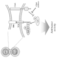
- T cell activation is facilitated by the T cell receptor (TCR) signaling cascade. Phosphorylation and dephosphorylation of TCR signaling molecules affect signal complex formation and TCR signal transduction.
- CD45 is a transmembrane phosphatase that plays a major role in regulating TCR signaling by regulating the level of tyrosine phosphorylation of lymphocyte protein kinase (Lck) and its downstream factors (J. Rossy, D J. Williamson, K. Gauss, How does the kinase Lck phosphorylate the T cell receptor? Spatial organization as a regulatory mechanism. Front Immunol 3, 167 (2012).) (R. J. Brownlie, R. Zamoyska, T cell receptor signaling networks: branched, diversified and bounded. Nat Rev Immunol 13, 257-269 (2013).).
- Lck lymphocyte protein kinase
- CD45 inhibitors support anti-tumor immunity when used in combination with PD-1 signal inhibitors, maintain synergistic effects of suppressing cancer growth, and can be used as monotherapy for PD-1 signal inhibition. It is thought to be effective even against cancers that exhibit unresponsiveness. A similar effect was seen with cell transplantation (xenogeneic cells, allogeneic cells). Enhanced TCR signaling was also confirmed by cell transplantation.
- the gist of the present invention is as follows.
- a pharmaceutical composition comprising a substance capable of enhancing T-cell receptor (TCR) signaling and administered either before, after or simultaneously with administration of a PD-1 signaling inhibitor.
- TCR T-cell receptor
- the CD45 inhibitor is 2-(4-Acetylanilino)-3-chloronaphthoquinone, N-(9,10-Dioxo-9,10-dihydrophenanthren-2-yl)-2,2-dimethylpropionamide and analogues thereof
- the pharmaceutical composition according to (2) which is at least one compound selected from the group consisting of: (4) The pharmaceutical composition according to (2), wherein the cells are xenocells, allocells or a combination thereof.
- a PD-1 signal inhibitory activity enhancer comprising a CD45 inhibitor and/or cells.
- a TCR signal enhancer comprising a CD45 inhibitor and/or cells.
- a method of preventing and/or treating cancer, an infectious disease, or a combination thereof comprising: (11) administration of an effective amount of a substance capable of enhancing T cell receptor (TCR) signaling to the subject either before, after, or concurrently with the administration of the PD-1 signaling inhibitor; A method of enhancing PD-1 signaling inhibitory activity, comprising: (12) A method of enhancing TCR signaling, comprising administering an effective amount of a CD45 inhibitor and/or cells to a subject. (13) A substance capable of enhancing T-cell receptor (TCR) signaling for use in preventing and/or treating cancer, infectious disease, or a combination thereof, which is a PD-1 signal inhibitor The substance administered either before, after, or concurrently with the administration of
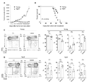
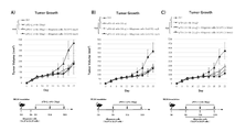
- MC38 cells were inoculated into the skin of young and aged PD-1 KO mice.
- A MC38 tumor size in young (3-4 months old) and aged (15 months old) PD-1 KO mice.
- B Kaplan-Meier plot of survival of MC38 tumor-bearing PD-1 KO mice.
- C and D Analysis of CD8-positive T cell subsets in young (C, 2-3 months old) or aged (D, 15-21 months old) PD-1 KO mice with or without inoculation of MC38 cells. . Peripheral and regional lymph node cells stained on Day 9.
- CD44 and CD62L expression in CD3+CD8+ cells and percentages of CD8+ cell subsets CD44 low /CD62L high (naive; P1), CD44 high /CD62L high (central memory; P2), CD44 high /CD62L low (effects/memory; P3), CD44 low /CD62L low (P4).
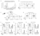
- MC38-OVA cells were injected into young OT-1 mice via the tail vein and the respective subsets were isolated from splenocytes. Five days after intradermal implantation of MC38-OVA cells, P3 or P4 subset cells were adoptively transferred into CD8 KO mice.
- A Tumor volume in MC38-OVA tumor-bearing CD8 KO mice with or without transfer of P3 or P4 cells.
- B FACS analysis of transplanted CD8-positive T cells in day 11 peripheral blood.
- C Scheme of CD8-positive T-cell subset isolation from splenocytes of young PD-1 KO mice.
- a to E Microarray analysis of P1, P2, P3, and P4 cells isolated from young PD-1 KO mice (1 to 3 months old; mix of 9 mice).
- A Hierarchical clustering heatmap of all genes.
- B Scatter plot showing the normalized log intensity of each probe. Dotted lines indicate log 2-fold differences. Genes that have been shown to be involved in the activation and differentiation of CD8-positive cells are listed.
- C Top 10 gene ontology (GO) involving genes upregulated in P4 cells. GO terms involved in 1C metabolism are shown in red.
- D Schematic representation of the 1C metabolic pathway. THF, tetrahydrofolate.
- E Heatmap showing the expression of 1C metabolic-related genes in CD8-positive T-cell subsets.
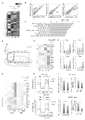
- OCR Oxygen consumption rate
- FIG. 1 Representative diagrams showing the expression of CD44 and CD62L on CD8-positive T cells and the percentage of each CD8-positive T-cell subset in the mice shown 5 days after MC38-OVA transplantation.
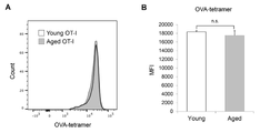
- C Percentage of pZAP70-positive CD8-positive T cells in peripheral lymph nodes from young or aged OT-1 mice with or without MC38-OVA transplantation.
- D Representative plots of CD45RB expression levels and fluorescence intensity in total CD8+ T cells or respective subsets from young (2-3 months old) or aged (14-17 months old) OT-1 mice. (MFI).
- A Schematic diagram of the experimental schedule.
- mice Ten days after the transplantation (day 0), the mice were implanted with MC38 (E to G) or MC38-OVA (H and I) cells in the skin and used for the experiments described below.
- G Percentage of pZAP70-positive cells in CD8-positive T cells from day 6 regional lymph nodes.
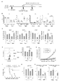
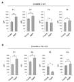
- a and B MC38 cells were inoculated into the skin of young (2 months old) and aged (14 months old) C57BL/6 wild-type mice. The mice then received anti-PD-L1 antibody on days 5, 11, and 17.
- A MC38 tumor size in young and aged wild-type mice.
- B Kaplan-Meier plot of survival of MC38 tumor-bearing wild-type mice. P values were calculated by log-rank test.
- C and D Analysis of CD8-positive T cell subsets in young (C, 2 months old) or aged (D, 15 months old) wild-type mice with or without inoculation of MC38 cells.
- mice (B and C) MC38 tumor size in young (B) and aged (C) wild-type mice.
- (D and E) MC38 tumor-bearing young (D) and aged (E) wild-type mouse survival. Data represent the mean ⁇ s.e.m. of mice (n 5-6). *p ⁇ 0.05; **p ⁇ 0.01; n.s., no significant difference (evaluated by ANOVA followed by Tukey's test or log-rank test). Restoration of age-related mitochondrial damage by xenotransplantation.
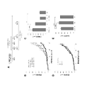
- MC38-OVA cells were implanted into the skin of young (3-4 months old) and aged (17-18 months old) PD-1 KO mice that had been transplanted with PBS (Ctrl) or Daudi cells 10 days earlier. .
- OCR trace (A), basal respiration (B) and reserve respiratory capacity were calculated from OCR values (C).
- MMC-treated splenocytes from C57BL/6 (Ctrl) or Balb/c (Allo) mice were injected into the tail vein of young (1 month old) and aged (14 to 18 months old) C57BL/6 wild-type mice. ported by Subsequent experimental conditions are the same as in FIG. 9A.
- C and D MC38 tumor-bearing young (C) and aged (D) survival of C57BL/6 wild-type mice.
- Experimental method and flow Enhanced PD-1 signaling inhibitory effect by CD45 inhibition. Pre-administration: PD-1 knockout (KO) mice were administered vehicle (control) or 211 and 3 days later were transplanted with MC38 cells (1 ⁇ 10 6 ).
- 211 was administered every 2 days from Day 5, 5 days after MC38 cell transplantation to PD-1 KO mice, for a total of 3 doses.
- the present invention provides a pharmaceutical composition comprising a substance capable of enhancing T cell receptor (TCR) signaling and administered either before, after or simultaneously with administration of a PD-1 signaling inhibitor. do.
- TCR T cell receptor
- T cell receptor is an antigen receptor molecule expressed on the cell membrane of T cells.
- TCR T cell receptor
- intracellular signals are transduced through the phosphorylation of downstream factors such as Lck and ZAP-70, resulting in activation of T cells.
- the enhancement of TCR signals can be confirmed by evaluating the phosphorylation of ZAP-70 in CD8-positive T cells under antigen stimulation. A higher level of ZAP-70 phosphorylation indicates an enhanced TCR signal. Phosphorylation of ZAP-70 can be detected by flow cytometric analysis using an anti-p-ZAP-70 antibody.
- TCR signal By enhancing the TCR signal, it is possible to enhance the anti-tumor immune effect of CD8-positive T cells. Therefore, by enhancing the TCR signal, it is possible to enhance the antitumor effect of PD-1 inhibition therapy, and to treat aging animals and humans that are resistant to PD-1 signal inhibition, as well as resistant cancers. can have an effect.
- CD45 inhibitors and/or cells can be exemplified as substances capable of enhancing TCR signals. These substances may also have the effect of inducing P4 cells.
- P4 cells are a CD44 low CD62L low CD8 positive T cell subset.
- the surface markers CD44 and CD62L L-selectin are used to distinguish between three major CD8+ T cells: naive (also called P1; CD44 low CD62L high ) and central memory (P2; CD44 high CD62L high ). ), effector/memory (P3; CD44 high CD62L low ).
- the remaining CD44 low CD62L low (P4) CD8 positive T cell subset is a very minor population.
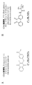
- CD45 inhibitors examples include 2-(4-Acetylanilino)-3-chloronaphthoquinone, N-(9,10-Dioxo-9,10-dihydrophenanthren-2-yl)-2,2-dimethylpropionamide and their analogues. can do.
- analog is a concept including salts of compounds, derivatives of compounds, prodrugs for active metabolites, active metabolites for prodrugs, solvates thereof, and the like.
- Compound salts include salts formed with acids such as hydrochloric acid, mesylic acid (methanesulfonic acid), fumaric acid, and phosphoric acid when the compound has an amino group (including substituted amino groups) or an amide group. can do.
- a derivative of a compound is a compound that has undergone modifications such as introduction or substitution of a functional group, oxidation, reduction, or replacement of an atom, to the extent that the structure or properties of the parent substance are not significantly changed.
- Derivatives of -chloronaphthoquinone include those in which the chloro (-Cl) group is replaced with other halogen groups (e.g., fluoro (-F) group, bromo (-Br) group, etc.), and those in which the acetyl (CH 3 CO-) group is substituted.
- Derivatives of ,10-dihydrophenanthren-2-yl)-2,2-dimethylpropionamide include those in which the dimethylpropionyl group is substituted with other alkyl groups (e.g., methyl, ethyl, propyl, benzyl, etc.). can be exemplified.
- prodrugs include compounds in which the amino group of the active compound is acylated, alkylated, or phosphorylated (for example, the amino group of the active compound is eicosanoylated, alanylated, pentylaminocarbonylated, (5-methyl-2 -oxo-1,3-dioxolen-4-yl)methoxycarbonylated, tetrahydrofuranylated, pyrrolidylmethylated, pivaloyloxymethylated, t-butylated compounds, etc.), where the amide group of the active compound is Alkylated (for example, methylated, ethylated, provylated, etc.) and the like can be exemplified.
- the amino group of the active compound is acylated, alkylated, or phosphorylated
- the amino group of the active compound is eicosanoylated, alanylated, pentylaminocarbonylated, (5
- solvates include solvates with solvents such as water, methanol, ethanol, and acetonitrile.
- the solvent may be a single solvent or a mixture of multiple solvents.
- Cells capable of enhancing TCR signals may be xenocells (heterologous cells) or allocells (allogeneic cells) for humans or animals to which the pharmaceutical composition is administered. or a combination thereof.
- xenogeneic Daudi cells and allogeneic splenocytes were transplanted into mice.
- xenocells are heterogeneous cells, and allocells are homogenous cells with different HLA (human leukocyte antigen) or MHC (major histocompatibility complex).
- Various cells xenocells, allocells
- tissues to be administered to living bodies for the prevention or treatment of diseases are known, and these cells may be used in the present invention.
- Allogeneic e.g., breast cancer cell lines, chronic myelogenous leukemia cell lines, lung cancer cells, non-small cell lung cancer cell lines, melanoma cell lines, monocytes, pancreatic cancer cell lines, prostate cancer cell lines, renal cancer cell lines, etc.
- Cells are used to treat various cancers (Human Vaccines & Immunotherapeutics 10:1, 52-63; January 2014). These cells may be irradiated to prevent proliferation and may be genetically modified to secrete immune stimulators such as GM-CSF.
- Heterologous cells such as murine melanoma B16 and Lewis lung carcinoma (LLC) cells have also been used in clinical trials to administer to melanoma patients (Eur J Dermatol 2016 Apr 1;26(2):138- 43).
- cells used for blood products for transfusion erythrocyte products, platelet products, whole blood products
- hematopoietic stem cells used for bone marrow transplantation can also be used. These cells may be either allo cells or xeno cells.
- PD-1 signal refers to the information transduction mechanism carried by PD-1, and as one of them, PD-1 is its ligand PD-L1, in cooperation with PD-L2, Signal transduction mechanisms that suppress activation of T cells can be exemplified.
- PD-1 programmed cell death-1
- PD-L1 and PD-L2 are antigens of monocytes and dendritic cells. It is expressed in various cells such as presenting cells and cancer cells.
- PD-1, PD-L1 and PD-L2 act as suppressors that suppress T cell activation.
- Certain cancer cells and virus-infected cells express ligands for PD-1 to suppress T-cell activation and evade host immune surveillance.
- PD-1 signal inhibitors include substances that specifically bind to PD-1, PD-L1 or PD-L2, and such substances include proteins, polypeptides, oligopeptides, nucleic acids (natural (including nucleic acids and artificial nucleic acids), low-molecular-weight organic compounds, inorganic compounds, cell extracts, extracts from animals, plants, soil, and the like. Substances may be natural or synthetic.
- Preferred PD-1 signal inhibitors are antibodies, more preferably antibodies such as anti-PD-1 antibodies, anti-PD-L1 antibodies and anti-PD-L2 antibodies.
- Antibodies may be any of polyclonal antibodies, monoclonal antibodies, chimeric antibodies, single-chain antibodies, humanized antibodies, and humanized antibodies as long as they can inhibit PD-1 signaling. Methods for producing those antibodies are known. Antibodies can be derived from any organism, such as human, mouse, rat, rabbit, goat, guinea pig. Also, as used herein, the antibody refers to Fab, F (ab) '2, ScFv, Diabody, VH, VL, Sc (Fv) 2, Bispecific sc (Fv) 2, Minibody, scFv-Fc monomer, scFv- This concept also includes low-molecular-weight compounds such as Fc dimer.
- PD-1 signaling inhibitors that target PD-1 include nivolumab (Bristol-Myers Squibb), pembrolizumab (Merck), cemiplimab (Regeneron/Sanofi), camrelizumab (Jiangsu Hengrui), sintilimab (Innovent Biologics/Lilly), toripalimab (Shanghai Junshi Bioscience), PD-L1-targeting atezolizumab (Genetech/Roche), durvalumab (AstraZeneca), avelumab (Merck/Pfizer) are approved globally and also target PD-1 tislelizumab (BeiGene), dostarlimab (AnaptysBio/Tesaro), HLX-04 (Shanghai Henlius Biotech), AK-105 (Akeso Biopharma), spartalizumab (Novartis), BCD-100 (Biocad), MGA-012 (
- the pharmaceutical composition of the present invention can be used as an anticancer agent, a therapeutic agent for infectious diseases, or a combination thereof.
- target cancers or tumors include leukemia, lymphoma (Hodgkin's disease, non-Hodgkin's lymphoma, etc.), multiple myeloma, brain tumor, breast cancer, and uterine body.
- lymphoma Hodgkin's disease, non-Hodgkin's lymphoma, etc.
- multiple myeloma brain tumor, breast cancer, and uterine body.
- cancer cervical cancer, ovarian cancer, esophageal cancer, stomach cancer, appendix cancer, colon cancer, liver cancer, gallbladder cancer, bile duct cancer, pancreatic cancer, adrenal cancer, gastrointestinal stromal tumor, mesothelioma, head and neck cancer (Laryngeal cancer, etc.), Oral cancer (Floor of the mouth cancer, etc.), Gingival cancer, Tongue cancer, Buccal mucosa cancer, Salivary gland cancer, Paranasal sinus cancer (Maxillary sinus cancer, Frontal sinus cancer, Ethmoid sinus cancer, Sphenoid sinus cancer) ), thyroid cancer, kidney cancer, lung cancer, osteosarcoma, prostate cancer, testicular tumor (testicular cancer), renal cell carcinoma, bladder cancer, rhabdomyosarcoma, skin cancer (basal cell carcinoma, spinous cell cancer) cancer, malignant melanoma, actinic keratosis, Bowen's disease, Paget's disease, etc.), anal cancer,
- target infectious diseases include bacterial infections (streptococci (group A ⁇ -hemolytic streptococci, pneumococci, etc.), Staphylococcus aureus (MSSA, MRSA), Staphylococcus epidermidis, Enterococcus, Listeria, Meningococcus, Neisseria gonorrhoeae, Pathogenic Escherichia coli (0157:H7, etc.), Klebsiella (Klebsiella pneumoniae), Proteus, Pertussis, Pseudomonas aeruginosa, Serratia, Citrobacter, Acinetobacter, Various infectious diseases caused by Enterobacter, Mycoplasma, Clostridium, etc., tuberculosis, cholera, plague, diphtheria, dysentery, scarlet fever, anthrax, syphilis, tetanus, lepros
- bacterial infections streptococci (group A ⁇ -
- the pharmaceutical composition of the present invention contains a substance capable of enhancing TCR signaling, and is administered either before, after, or at the same time as administration of a PD-1 signaling inhibitor.
- the PD-1 signal inhibitor and the substance capable of enhancing the TCR signal may be administered separately at different times or at the same time, or may be administered together at the same time.
- good. comprising a PD-1 signal inhibitor and a substance capable of enhancing TCR signal when the PD-1 signal inhibitor and the substance capable of enhancing TCR signal are administered together at the same time It may be used as a compounding agent.
- composition of the present invention is administered to a subject systemically or locally, orally or parenterally.
- Subjects include humans and non-human animals.
- PD-1 signal inhibitors e.g., anti-PD-1 antibody, anti-PD-L1 antibody, anti-PD-L2 antibody
- a buffer solution such as PBS, physiological saline, sterile water, etc.
- this solution contains additives (for example, colorants, emulsifiers, suspending agents, surfactants, solubilizers, stabilizers, preservatives, antioxidants, buffers, tonicity agents, etc.).
- additives for example, colorants, emulsifiers, suspending agents, surfactants, solubilizers, stabilizers, preservatives, antioxidants, buffers, tonicity agents, etc.
- the route of administration includes intravenous, intramuscular, intraperitoneal, subcutaneous, and intradermal administration.
- PD-1 signal inhibitors e.g., anti-PD-1 antibody, anti-PD-L1 antibody, anti-PD-L2 antibody
- content in the formulation varies depending on the type of formulation, usually 1 to 100% by weight, preferably 50-100% by weight.
- the formulations may be formulated in unit dosage formulations.
- Dosage, number and frequency of administration of PD-1 signal inhibitors are subject to symptoms, age, body weight, administration method, administration Although it varies depending on the form and the like, for example, in terms of the amount of the active ingredient, an adult usually administers 0.1 to 100 mg/kg body weight, preferably 1 to 10 mg/kg body weight, at least once to achieve the desired effect. It should be administered at a frequency that can be confirmed.
- Substances capable of enhancing TCR signaling may be included in formulations containing PD-1 signaling inhibitors, alone or mixed with excipients or carriers, in tablets, capsules, powders, granules. , liquids, syrups, aerosols, suppositories, injections and the like.
- Excipients or carriers are conventionally used in the art and may be pharmaceutically acceptable, and their type and composition may be changed as appropriate. For example, water, vegetable oil and the like are used as the liquid carrier.
- solid carriers saccharides such as lactose, white sugar and glucose, starches such as potato starch and corn starch, and cellulose derivatives such as crystalline cellulose are used.
- a lubricant such as magnesium stearate, a binder such as gelatin and hydroxypropylcellulose, a disintegrant such as carboxymethylcellulose, and the like may be added.
- antioxidants, coloring agents, corrigents, preservatives and the like may be added.
- Substances capable of enhancing TCR signals can be administered by various routes such as oral, nasal, rectal, transdermal, subcutaneous, intravenous and intramuscular.
- the content of a substance that can enhance the TCR signal in the formulation varies depending on the type of formulation, but is usually 1-100% by weight, preferably 50-100% by weight.
- the content of the substance capable of enhancing the TCR signal in the formulation is preferably 1-100% by weight.
- the content of the substance capable of enhancing the TCR signal in the formulation is usually about 10-100% by weight, preferably 50-100% by weight, and the balance is the carrier. is.
- the formulations may be formulated in unit dosage formulations.
- the dose, frequency and frequency of administration of the substance capable of enhancing the TCR signal vary depending on the type of the substance capable of enhancing the TCR signal, subject's symptoms, age, body weight, administration method, administration form, etc.
- the substance capable of enhancing the TCR signal is a CD45 inhibitor
- it is usually 0.005 ⁇ g (or ⁇ l) to 25000 mg (or ml) per adult in terms of the amount of the active ingredient.
- the substance capable of enhancing the TCR signal is a cell, it is usually about 1 x 10 6 cells/person to 1 x 10 11 cells/person, preferably 5 x 10 6 cells/person to an adult. About 2 x 10 10 cells/person should be administered at least once at a frequency that allows confirmation of the desired effect.
- the ratio of the PD-1 signal inhibitor (e.g., anti-PD-1 antibody, anti-PD-L1 antibody, anti-PD-L2 antibody) and the substance capable of enhancing the TCR signal is the substance capable of enhancing the TCR signal is a CD45 inhibitor, 1:0.1 to 1:100 (mass ratio) is suitable, preferably 1:1 to 1:50, and the substance capable of enhancing the TCR signal is the cell , 1 x 10 6 to 1 x 10 10 cells, preferably 5 x 10 6 to 2 x 10 9 cells, are suitable for 240 mg of the PD-1 signal inhibitor.
- the present invention also provides administration of a pharmaceutically effective amount of a substance capable of enhancing TCR signaling to a subject either before, after, or at the same time as administration of a PD-1 signaling inhibitor. Also provided are methods of preventing and/or treating cancer, infectious diseases, or combinations thereof, including: Furthermore, the present invention relates to the use of a substance capable of enhancing TCR signaling for the treatment of cancer, infectious disease or a combination thereof, wherein the agent is administered before, after or simultaneously with a PD-1 signaling inhibitor. A substance capable of enhancing the TCR signal is administered during any of the above uses.
- the present invention relates to the use of a substance capable of enhancing TCR signaling for use in a method of treating cancer, infectious disease or a combination thereof, comprising administering a PD-1 signaling inhibitor Also provided is said use wherein a substance capable of enhancing the TCR signal is administered either before, after or simultaneously.
- the present invention provides a substance capable of enhancing T cell receptor (TCR) signaling for use in preventing and/or treating cancer, infectious diseases, or a combination thereof, which inhibits PD-1 signaling Also provided are said substances administered either before, after or concurrently with the administration of the agent.
- TCR T cell receptor
- the present invention also provides agents that enhance PD-1 signaling inhibitory activity, including substances capable of enhancing TCR signaling.
- the present invention is to administer to a subject an effective amount of a substance capable of enhancing T cell receptor (TCR) signals either before, after or at the same time as administration of a PD-1 signal inhibitor.
- TCR T cell receptor
- methods of enhancing PD-1 signaling inhibitory activity comprising:
- the drug of the present invention can be used as a combination drug or combination drug with a PD-1 signal inhibitor. Combinations and formulations of PD-1 signal inhibitors and substances capable of enhancing TCR signals are described above.
- the agent of the present invention can be used not only as a medicine but also as an experimental reagent.
- the present invention provides a TCR signal enhancer comprising a CD45 inhibitor and/or cells.
- the invention also provides a method of enhancing TCR signaling comprising administering to a subject an effective amount of a CD45 inhibitor and/or cells.
- CD45 inhibitors and cells are described above.
- the agent of the present invention can be used not only as a medicine but also as an experimental reagent.
- CD8-positive cells play a major role in anti-tumor responses and directly kill cancer cells. Strong suppression of CD8-positive cell immunity in the elderly is associated with cancer and other age-related diseases.
- This study investigated the role of T-cell function in age-related resistance to PD-1 inhibitory therapy. We found that formation of CD44 low CD62L low CD8 positive T cells was reduced in regional lymph nodes of aged tumor-bearing mice. First, P4 cells were induced from naive CD8+ T cells by in vitro stimulation and then differentiated into effector/memory cells.
- Immunosenescence is caused by quantitative and qualitative changes in immune cells.
- One of the major quantitative changes in T cells is a reduction in TCR repertoire diversity due to thymic regression, leading to a reduction in naive cells in the periphery (13-15).
- functional deficits such as impaired TCR signaling, reduced ability to differentiate into effector and memory cells, and reduced cytokine production accumulate in peripheral T cells in the elderly (16, 17).
- age-related changes greatly affect the proportion of well-defined CD8-positive T-cell subsets (18-20).
- CD44 and CD62L are used to distinguish between three major CD8+ T cells: naive (also called P1; CD44 low CD62L high ) and central memory (P2; CD44 high CD62L high ). ), effector/memory (P3; CD44 high CD62L low ).
- the CD44 low CD62L low (P4) CD8 positive T cell subset that remains in naive mice is a very minor population.
- Naive T cells differentiate into effector T cells upon antigen stimulation (14, 21). Some effector cells differentiate into memory cells and are capable of rapidly responding to the same antigenic stimulus and effector cell differentiation.
- TCR T cell receptor
- Phosphorylation and dephosphorylation of TCR signaling molecules affect signal complex formation and TCR signal propagation.
- CD45 is a transmembrane phosphatase that plays a major role in regulating TCR signaling by regulating the level of tyrosine phosphorylation of lymphocyte protein kinase (Lck). This Lck acts as an activator of TCRz, z-chain-associated protein (ZAP)-70, and the linker of activated T cells (LAT), which are downstream factors of TCR signaling (24, 25).
- Lck lymphocyte protein kinase
- 1C metabolism In activated and proliferating cells, 1C metabolism is generally enhanced and supports survival and differentiation through purine and thymidine biosynthesis, amino acid production, epigenetic regulation, and redox regulation (29, 29). 30). Recent reports indicate that 1C metabolism is one of the most frequently induced metabolic pathways during early CD4+ or CD8+ T cell activation (31, 32). After T cell activation, increased 1C metabolism promotes serine processing required for the biosynthesis of new nucleic acids derived from the 1C unit, which is responsible for antigen-specific T cell proliferation and differentiation and effector function. is necessary. This 1C metabolic pathway is also involved in energy production through regulation of mitochondrial protein synthesis by mitochondrial tRNA methylation (33, 34). Since mitochondrial function and 1C metabolism are lower in aged CD4+ T cells than in young CD4+ T cells (35), aging of T cells may be involved in the inactivation of 1C metabolism. .
- P4 cells are intermediate between P1 and P3 cells and retain potential for anti-tumor activity.
- the P3 and P4 subsets of CD8-positive T cells were induced by intravenous injection of OVA-expressing MC38 tumor cells (MC38-OVA) into the tail vein of OT-1 mice and isolated from the spleens 5 days later.
- the same number of P3 or P4 cells were adoptively transferred to young CD8 KO mice engrafted with MC38-OVA.
- MC38-OVA proliferation was strongly suppressed in mice transfected with P3 or P4 cells (Fig. 2A).
- Fig. 2B all transplanted P4 cells in peripheral blood 6 days after transplantation differentiated into P3 cells.
- P1 and P2 cells are known to differentiate into P3 cells during activation, but the characteristics of P4 cells are scarce due to their low numbers.
- P1, P2, and P4 cells were isolated from young PD-1 KO mice and treated with anti-CD3/CD28 antibodies in vitro. stimulated by IL-2 (Fig. 2C). Consistent with the in vivo results, isolated P4 cells differentiated predominantly into P3 cells under these conditions (Fig. 2D).
- FIG. 2E and 2F P4 cells were formed from P1 cells, but not from P2 cells, two days after in vitro stimulation.
- Tumor inoculation increased the expression of 1C metabolic-related genes in total CD8+ T cells from young wild-type and PD-1 KO mice (Fig. 3G), which could be explained by the formation of P4 cells. ( Figures 1C and 6C). Consistent with the high efficiency of P4 cell induction in PD-1 KO mice (Fig. 1C and Fig. 6C), CD8-positive T cells in young PD-1 KO mice were more likely to express most 1C metabolism-related genes than in wild-type mice. was expressed at high levels in response to MC38 tumor cell inoculation (Fig. 3G). In contrast, the expression of these genes in total CD8-positive T cells from aged mice showed no appreciable variation with tumor inoculation, regardless of mouse genotype.
- MC38-OVA injection increased the percentage of CD8-positive T cells expressing phosphorylated ZAP-70 in young mice (Fig. 4C). This suggests that the activation of TCR signaling by antigen stimulation is inhibited in aged OT-1 mice. Remarkably, no significant difference was found in the intensity of OVA-specific MHC tetramers (Fig. 7A&B), indicating no difference in TCR expression levels and avidity of OT-I T cells from young and aged mice. matter was shown. Therefore, the inhibition of antigen-stimulated TCR activation seen in aged mice may involve mechanisms other than TCR-MHC/antigen interactions.
- CD45RB an isoform of CD45 that is predominantly expressed on naive and memory T cells in mice.
- the effect of aging on CD45RB expression was greater in P1 and P4 cells than in P2 and P3 cells (Fig. 4D).
- the highest increase in CD45RB expression was observed in P1 cells of aged wild-type and PD-1 KO mice (Fig.
- Heterologous cell therapy enhances P4 cell induction in aged mice and restores the antitumor potential of CD8+ T cells.
- strong in vitro stimulation restores suppression of the P1 to P4 transition in aged mice, suggesting that strong stimulation using heterologous cells (human Burkitt's lymphoma, Daudi cells) is associated with aging.
- Heterologous cells human Burkitt's lymphoma, Daudi cells
- Fig. 5A Ten days after tail vein injection of Daudi cells (day 0: before MC38 tumor cell transplantation), the proportion of P4 cells was strongly increased in the peripheral lymph nodes of young and aged PD-1 KO mice, but the proportion of P1 to P3 cells decreased. The percentage did not increase significantly (Fig.
- Daudi cell injection strongly suppressed MC38 tumor growth in aged mice and prolonged survival (Fig. 5E&F). Age-related unresponsiveness to anti-PD-L1 antibodies in wild-type mice was also restored by Daudi cell injection (FIGS. 9A-E).
- Fig. 5G the percentage of p-ZAP-70-positive cells was increased in PD-1 KO mice injected with Daudi cells (Fig. 5G), indicating that xenostimulation enhances TCR signals to tumor antigens. It was suggested.
- Daudi cell therapy enhanced the proliferation of tumor antigen-reactive CD8+ T cells.
- MC38-OVA cells were transplanted into PD-1 KO mice 10 days after Daudi cell injection, and the appearance of OVA tetramer-positive CD8-positive T cells was monitored.
- the percentage of OVA tetramer-positive CD8-positive T cells was increased by infusion of Daudi cells in regional lymph nodes and tumor tissues of aged PD-1 KO mice (Fig. 5H&I), indicating that this treatment resulted in antigen-specific CD8 It was shown to increase the induction of positive T cells. Given that prestimulation with Daudi cells enhances P4 cell induction and 1C metabolism in CD8+ T cells from aged mice, it is plausible that Daudi cell infusion increased both basal respiration and respiratory reserve. result (Fig. 10A-C).
- T cells are directly (eg, donor peptide presented by donor MHC) or indirectly (eg, donor peptide presented by recipient MHC) activated by allo- or heterologous MHC molecules (54-56).
- donor MHC donor peptide presented by donor MHC
- recipient MHC donor peptide presented by recipient MHC
- mice and Cells All mice were bred at Kyoto University graduate School of Medicine Animal Research Facility or RIKEN Life Science Research Center under an SPF environment free of specific pathogens that would interfere with experiments, and in accordance with appropriate experimental guidelines. and was used in PD-1-/- (PD-1KO) mice were generated in our laboratory by homologous recombination (59). C57BL/6 wild-type mice were obtained from Charles River Laboratories Japan. OT-1 TCR transgenic and CD8 ⁇ / ⁇ (CD8 KO) mice were purchased from The Jackson Laboratory (Bar Harbor, Maine USA) (deposited by M. B. Bevan, Washington Medical Center or T. Mak, University of Toronto). A mouse colon carcinoma (MC38) cell line was kindly provided by J. P.
- OVA-expressing MC38 cells were isolated from MC38 by a pMXs-based OVA-F2A-EGFO retrovirus. generated by transformation. Cells were cultured in the absence of mycobacterial infection in RPMI 1640 (Gibco; 11875-093) containing 10% inactivated fetal bovine serum and 1% penicillin-streptomycin (Nacalai Tesque; 26253-84). did
- mice Mouse Treatment Model MC38 cells (5 ⁇ 10 5 or 2 ⁇ 10 6 ) were implanted intradermally into the right abdomen of mice (day 0). Five days after MC38 cell transplantation (day 5), the mice were treated with an anti-PD-L1 antibody (clone 1-111A.4, produced and stored at Kyoto University graduate School of Medicine Immunology and Genomic Medicine Laboratory (Immunology Letters 84 (2002) ) 57-62) (6 mg/kg) was administered intraperitoneally, and this anti-PD-L1 antibody administration was repeated three times every 6 days (days 5, 11, and 17).
- an anti-PD-L1 antibody clone 1-111A.4, produced and stored at Kyoto University graduate School of Medicine Immunology and Genomic Medicine Laboratory (Immunology Letters 84 (2002) ) 57-62
- this anti-PD-L1 antibody administration was repeated three times every 6 days (days 5, 11, and 17).
- Antibody isotype (Rat IgG2a) was used as a control.Tumor growth was monitored by measuring tumor size using a caliper, and volume was calculated by the formula ⁇ (length ⁇ width ⁇ height)/6.
- the transfer of non-autologous cells was Burkitt's lymphoma Daudi cells or BALB/c treated with mitomycin C (MMC) 10 days before tumor cell transplantation, or C57BL/6N-derived splenocytes (2 ⁇ 10 6 cells/mouse) as a control. was performed by injection through the tail vein of
- Peripheral and regional lymph node cells and tumor-infiltrating lymphocytes were isolated as previously described (39).
- splenocyte analysis spleens were briefly disaggregated and suspended in potassium ammonium chloride solution to lyse red blood cells. Splenocytes were washed and treated with RPMI containing 10% fetal bovine serum, L-glutamine, 55 ⁇ M 2-mercap and ethanol, penicillin-streptomycin, 1 mM sodium pyruvate (Gibco), 1% MEM non-essential amino acids solution (Gibco). Resuspended in 1640 internal solution (T cell culture medium).
- CD8a Ly-2 microbeads
- FACSAria BD Biosciences
- peripheral blood cells collected peripheral blood cells were treated with potassium ammonium chloride solution and washed with T cell culture medium. Purified cells were stimulated with T cell culture medium supplemented with anti-CD3/28 Dynabeads (Gibco) and recombinant human IL-2 (20 U/mL; PeptoTech).
- CD8-positive T cells using MC38-OVA cells
- CD8-positive T cells isolated from lymph nodes of OT-1 mice were combined with MMC-treated MC38-OVA cells in T-cell culture medium. cultured. One day after stimulation, the cells were treated with 0.05 ⁇ M CD45 inhibitor VI (EDM Millipore) for 1 hour.
- CD3 (clone 145-2C11) and CD8 (clone 53-6.7) (Invitrogen); phospho-ZAP70/Syk (clone n3kobu5) (Invitrogen), CD45.1 (clone A20), CD45.2 (clone 104), CD44 ( Clone IM7) and CD62L (clone MEL-14) (TONBO biosciences or BioLegend); CD8 (clone KT15, MBL Life Science); CD45RB (clone 16A, BD Pharmingen).
- H2Kb Negative (SIY) Tetramer-SIRYYGL-APC and H-2Kb OVA Tetramer-SINFEKL-APC (TS-M008-2) were analyzed by MBL Life Science flow cytometric analysis using FACSCant II (BD Biosciences) and FlowJo software (FlowJo LLC). was used.
- CD8 KO and OT-1 Mouse Models To induce P3 and P4 cells, MC38-OVA cells (1-2 ⁇ 10 6 ) were transferred into OT-1 mice by tail vein injection. Five days later, P3 and P4 cells were isolated from those mice. Each isolated CD8-positive T cell was injected via the tail vein of CD8 KO mice that had been intradermally implanted with 2 ⁇ 10 5 MC38-OVA cells 5 days earlier. Peripheral blood was then obtained 6 days later to analyze the differentiation of the transplanted cells.
- Real-time PCR was performed to amplify the indicated mRNAs using the Applied Biosystems 7500 Fast Real-Time PCT system (ABI) and PowerUp SYBR Green Master Mix (Applied Biosystems). The expression level of each gene was normalized by the ⁇ -actin mRNA level.
- the primer sequences are as follows: Shmt1 forward (Fw), 5'-CCAGAGTGCTGTGGCAACTC-3' (SEQ ID NO: 1); Shmt1 reverse (Rv), 5'-GCAAAACACAGGCTGTTCCTG-3' (SEQ ID NO: 2); Shmt2 Fw, 5'-GACAGTTGAGGACACCTGGC-3' (SEQ ID NO: 3); Shmt2 Rv, 5'-CCAGAGAGGAGTGACATCTC-3' (SEQ ID NO: 4); Phgdh Fw, 5'-TGGCCTCGGCAGAATTGGAAG-3' (SEQ ID NO: 5); Phgdh Rv, 5'-TGTCATTCAGCAAGCCTGTGGT-3' (SEQ ID NO: 6); Psat1 Fw, 5'-GATGAACATCCCATTTCGCATTGG-3' (SEQ ID NO: 7); Psat1 Rv, 5'-GCGTTATACAGAGAGGCACGAATG-3' (SEQ ID
- Oxygen Consumption Rate (OCR) Analysis The OCR assay was performed as previously described with some modifications (39). Oxygen consumption rates of CD8+ T cells isolated from regional lymph nodes were analyzed with the XFe96 Extracellular Flux analyzer (Agilent Technologies) using the Seahorse XFe96 Extracellular Flux assay kit and Seahorse XF Cell Mito Stress test kit (Agilent Technologies). rice field. CD8-positive T cells were seeded (3 ⁇ 10 5 /well) on XFe96 plates coated with Cell-Tak (Corning). Reserve respiratory capacity was calculated from OCR graphs as previously described (60). Basal respiration was calculated by subtraction with non-mitochondrial respiration (values before oligomycin addition to values after rotenone/antimycin A addition) (61).
- mice and Cells All mice were bred at the Kyoto University graduate School of Medicine Animal Experimental Facility under an SPF environment free of specific pathogens that would interfere with experiments, and were used under appropriate experimental guidelines.
- C57BL/6 was obtained from Charles River Laboratories Japan.
- PD-1-/- (PD-1KO) mice were generated in our laboratory by homologous recombination (see Nishimura et al.) (11).
- Mouse colon cancer-derived MC38 cells were a gift from J. P. Allison, Memorial Sloan-Kettering Cancer (New York, NY). LLC cells derived from Lewis lung carcinoma were purchased from the American Type Culture Collection (ATCC).
- ATCC American Type Culture Collection
- Mouse therapeutic model CD45 inhibition ( Figure 12): 2-(4-Acetylanilino)-3-chloronaphthoquinone (211) (530197, EDM Millipore) (12) (3 mg/kg) or N-(9,10-Dioxo-9) ,10-dihydrophenanthren-2-yl)-2,2-dimethylpropionamide (PTP) (540215, Sigma-Aldrich) (13) (3 mg/kg) was administered to mice before or after (5 or 7 days after) tumor cell implantation. was administered intraperitoneally. MC38 cells (5 ⁇ 10 5 or 1 ⁇ 10 6 ) or LLC cells (2 ⁇ 10 6 ) were implanted intradermally into the right flank of mice (day 0).
- mice Seven days after LLC cell transplantation (day 7), the mice were intraperitoneally administered anti-PD-L1 antibody (clone 1-111A.4) (2 mg/kg). This anti-PD-L1 antibody administration was repeated three times every 6 days (days 7, 13, and 19). The tumor volume was measured with a caliper and calculated by the formula ⁇ (length ⁇ width ⁇ height)/6.
- PD-1 KO mice (2 months old) were intraperitoneally injected with MC38 mouse colon cancer cells once 3 days before transplantation (pre-administration) or 3 times 5 days after transplantation (post-administration) with CD45 inhibitor 211 at 3 mg/day. kg was dosed (FIGS. 13A & B). MC38 cells were implanted intradermally into the right flank of mice at a rate of 1 ⁇ 10 6 /tumor. As a result, the tumor volume was significantly reduced in the group administered with 211 before or after MC38 transplantation, compared with the control group administered with only the vehicle (FIGS. 14A & B). These results suggest that CD45 inhibition enhances the antitumor effect of PD-1 signal inhibition.
- Balb/c (allogeneic) spleen treated with anti-PD-L1 antibody (clone 1-111A.4) (1.5 mg/kg) intraperitoneally plus mitomycin C after 9, 16 and 23 days
- Cells (5x10⁵/mouse or 2x10⁶/mouse) were injected (A) only once after 6 days, (B) only once after 9 days, or (C) after 9, 16 and 23 days for a total of 3 It was intravenously injected twice, and changes in tumor size over time were measured (Fig. 18). Points in each line graph are means of 5-6 mice, error bars are standard errors. * means p-value less than 0.05, ns means no significant difference (comparison by Tukey test after one-way ANOVA). As a result, it was found that three administrations of allogeneic cells had a higher effect in combination with an immune checkpoint inhibitor than one administration.
- the pharmaceutical composition of the present invention can be used as an anticancer agent, a therapeutic agent for infectious diseases, or a combination thereof.
Abstract
Priority Applications (3)
| Application Number | Priority Date | Filing Date | Title |
|---|---|---|---|
| EP22759551.9A EP4299075A1 (fr) | 2021-02-26 | 2022-02-21 | Polythérapie avec un inhibiteur du signal pd-1 |
| US18/276,583 US20240148676A1 (en) | 2021-02-26 | 2022-02-21 | Combination therapy with pd-1 signal inhibitor |
| JP2023502370A JPWO2022181515A1 (fr) | 2021-02-26 | 2022-02-21 |
Applications Claiming Priority (2)
| Application Number | Priority Date | Filing Date | Title |
|---|---|---|---|
| JP2021031041 | 2021-02-26 | ||
| JP2021-031041 | 2021-02-26 |
Publications (1)
| Publication Number | Publication Date |
|---|---|
| WO2022181515A1 true WO2022181515A1 (fr) | 2022-09-01 |
Family
ID=83047998
Family Applications (1)
| Application Number | Title | Priority Date | Filing Date |
|---|---|---|---|
| PCT/JP2022/006843 WO2022181515A1 (fr) | 2021-02-26 | 2022-02-21 | Polythérapie avec un inhibiteur du signal pd-1 |
Country Status (4)
| Country | Link |
|---|---|
| US (1) | US20240148676A1 (fr) |
| EP (1) | EP4299075A1 (fr) |
| JP (1) | JPWO2022181515A1 (fr) |
| WO (1) | WO2022181515A1 (fr) |
Citations (3)
| Publication number | Priority date | Publication date | Assignee | Title |
|---|---|---|---|---|
| WO2004072286A1 (fr) * | 2003-01-23 | 2004-08-26 | Ono Pharmaceutical Co., Ltd. | Substance specifique a pd-1 humain |
| WO2017004167A1 (fr) * | 2015-07-01 | 2017-01-05 | Augusta University Research Institute, Inc. | Méthodes et compositions pour le traitement du cancer |
| JP2021031041A (ja) | 2019-08-29 | 2021-03-01 | ジー・オー・ピー株式会社 | キャスターおよび運搬台車 |
-
2022
- 2022-02-21 WO PCT/JP2022/006843 patent/WO2022181515A1/fr active Application Filing
- 2022-02-21 US US18/276,583 patent/US20240148676A1/en active Pending
- 2022-02-21 EP EP22759551.9A patent/EP4299075A1/fr active Pending
- 2022-02-21 JP JP2023502370A patent/JPWO2022181515A1/ja active Pending
Patent Citations (3)
| Publication number | Priority date | Publication date | Assignee | Title |
|---|---|---|---|---|
| WO2004072286A1 (fr) * | 2003-01-23 | 2004-08-26 | Ono Pharmaceutical Co., Ltd. | Substance specifique a pd-1 humain |
| WO2017004167A1 (fr) * | 2015-07-01 | 2017-01-05 | Augusta University Research Institute, Inc. | Méthodes et compositions pour le traitement du cancer |
| JP2021031041A (ja) | 2019-08-29 | 2021-03-01 | ジー・オー・ピー株式会社 | キャスターおよび運搬台車 |
Non-Patent Citations (71)
| Title |
|---|
| A. C. MAUE ET AL.: "T-cell immunosenescence: lessons learned from mouse models of aging", TRENDS IMMUNOL, vol. 30, 2009, pages 301 - 305, XP026251275, DOI: 10.1016/j.it.2009.04.007 |
| A. L. SHERGOLDR. MILLARR. J. B. NIBBS: "Understanding and overcoming the resistance of cancer to PD-1/PD-L1 blockade", PHARMACOL RES, vol. 145, 2019, pages 104258 |
| A. PADRON ET AL.: "Age effects of distinct immune checkpoint blockade treatments in a mouse melanoma model", EXP GERONTOL, vol. 105, 2018, pages 146 - 154, XP085365724, DOI: 10.1016/j.exger.2017.12.025 |
| A. RIBAS ET AL.: "Association of Pembrolizumab With Tumor Response and Survival Among Patients With Advanced Melanoma", JAMA, vol. 315, 2016, pages 1600 - 1609 |
| B. Y MA ET AL.: "The lectin Jacalin induces human B-lymphocyte apoptosis through glycosylation-dependent interaction with CD45", IMMUNOLOGY, vol. 127, 2009, pages 477 - 488 |
| B. YOUNGBLOODJ. S. HALER. AHMED: "T-cell memory differentiation: insights from transcriptional signatures and epigenetics", IMMUNOLOGY, vol. 139, 2013, pages 277 - 284, XP071276396, DOI: 10.1111/imm.12074 |
| C. H. JUNE: "Principles of adoptive T cell cancer therapy", J CLIN INVEST, vol. 117, 2007, pages 1204 - 1212, XP055761950, DOI: 10.1172/JCI31446 |
| C. HELISSEYC. VICIERS. CHAMPIAT: "The development of immunotherapy in older adults: New treatments, new toxicities?", J GERIATR ONCOL, vol. 7, 2016, pages 325 - 333, XP029748707, DOI: 10.1016/j.jgo.2016.05.007 |
| C. S. PALMER: "Emerging Role and Characterization of Immunometabolism:Relevance to HIV Pathogenesis, Serious Non-AIDS Events, and a Cure", J IMMUNOL, vol. 196, 2016, pages 4437 - 4444, XP055624585, DOI: 10.4049/jimmunol.1600120 |
| D. R. MINTON ET AL.: "Serine Catabolism by SHMT2 Is Required for Proper Mitochondrial Translation Initiation and Maintenance of Formylmethionyl-tRNAs", MOL CELL, vol. 69, 2018, pages 610 - 621 |
| D. SAUCE ET AL.: "Lymphopenia-driven homeostatic regulation of naive T cells in elderly and thymectomized young adults", J IMMUNOL, vol. 189, 2012, pages 5541 - 5548 |
| E. H. MA ET AL.: "Serine Is an Essential Metabolite for Effector T Cell Expansion", CELL METAB, vol. 25, 2017, pages 345 - 357, XP029914179, DOI: 10.1016/j.cmet.2016.12.011 |
| E. J. WHERRYR. AHMED: "Memory CD8 T-cell differentiation during viral infection", J VIROL, vol. 78, 2004, pages 5535 - 5545 |
| E. S. EGOROV ET AL.: "The Changing Landscape of Naive T Cell Receptor Repertoire With Human Aging", FRONT IMMUNOL, vol. 9, 2018, pages 1618 |
| EUR J DERMATOL, vol. 26, no. 2, 1 April 2016 (2016-04-01), pages 138 - 43 |
| FERNANDES RICARDO A.; SU LEON; NISHIGA YOKO; REN JUNMING; BHUIYAN ALADDIN M.; CHENG NING; KUO CALVIN J.; PICTON LORA K.; OHTSUKI S: "Immune receptor inhibition through enforced phosphatase recruitment", NATURE, vol. 586, no. 7831, 21 October 2020 (2020-10-21), London, pages 779 - 784, XP037282854, ISSN: 0028-0836, DOI: 10.1038/s41586-020-2851-2 * |
| G. PAWELECK. HIROKAWAT. FULOP: "Altered T cell signalling in ageing", MECH AGEING DEV, vol. 122, 2001, pages 1613 - 1637 |
| G. S. DUCKERJ. D. RABINOWITZ: "One-Carbon Metabolism in Health and Disease", CELL METAB, vol. 25, 2017, pages 27 - 42, XP029879738, DOI: 10.1016/j.cmet.2016.08.009 |
| H. NISHIMURAN. MINATOT. NAKANOT. HONJO: "Immunological studies on PD-1 deficient mice: implication of PD-1 as a negative regulator for B cell responses", INT IMMUNOL, vol. 10, 1998, pages 1563 - 1572, XP000971789, DOI: 10.1093/intimm/10.10.1563 |
| H. TSUKAMOTOS. SENJUK. MATSUMURAS. L. SWAINY. NISHIMURA: "IL-6-mediated environmental conditioning of defective Thl differentiation dampens antitumour immune responses in old age", NAT COMMUN, vol. 6, 2015, pages 6702 |
| H. WANG ET AL.: "ZAP-70: an essential kinase in T-cell signaling", COLD SPRING HARB PERSPECT BIOL, vol. 2, 2010, pages a002279, XP055392786, DOI: 10.1101/cshperspect.a002279 |
| HIROAKI IKEDA , NAOZUMI HARADA: "Possibility of cancer treatment vaccine", PHARMACOLOGY, vol. 76, no. 1, 1 January 2016 (2016-01-01), JP , pages 32 - 38, XP009539283, ISSN: 0372-7629, DOI: 10.14843/jpstj.76.32 * |
| IMMUNOLOGY LETTERS, vol. 84, 2002, pages 57 - 62 |
| J. BRAHMER ET AL.: "Nivolumab versus Docetaxel in Advanced Squamous-Cell Non-Small-Cell Lung Cancer", N ENGL J MED, vol. 373, 2015, pages 123 - 135, XP055391742, DOI: 10.1056/NEJMoa1504627 |
| J. C. NOLZG. R. STARBECK-MILLERJ. T. HARTY: "Naive, effector and memory CD8 T-cell trafficking: parallels and distinctions", IMMUNOTHERAPY, vol. 3, 2011, pages 1223 - 1233 |
| J. H. CHO ET AL.: "CD45-mediated control of TCR tuning in naive and memory CD8(+) T cells", NAT COMMUN, vol. 7, 2016, pages 13373 |
| J. NIKOLICH-ZUGICH: "Aging of the T cell compartment in mice and humans: from no naive expectations to foggy memories", J IMMUNOL, vol. 193, 2014, pages 2622 - 2629 |
| J. NIKOLICH-ZUGICH: "The twilight of immunity: emerging concepts in aging of the immune system", NAT IMMUNOL, vol. 19, 2018, pages 10 - 19, XP036582832, DOI: 10.1038/s41590-017-0006-x |
| J. NIKOLICH-ZUGICHG. LIJ. L. UHRLAUBK. R. RENKEMAM. J. SMITHEY: "Age-related changes in CD8 T cell homeostasis and immunity to infection", SEMIN IMMUNOL, vol. 24, 2012, pages 356 - 364 |
| J. ROSSYD. J. WILLIAMSONK. GAUS: "How does the kinase Lck phosphorylate the T cell receptor? Spatial organization as a regulatory mechanism", FRONT IMMUNOL, vol. 3, 2012, pages 167 |
| J. SCENEAY ET AL.: "Interferon Signaling Is Diminished with Age and Is Associated with Immune Checkpoint Blockade Efficacy in Triple-Negative Breast Cancer", CANCER DISCOV, vol. 9, 2019, pages 1208 - 1227, XP055763897, DOI: 10.1158/2159-8290.CD-18-1454 |
| K. CHAMOTO ET AL.: "Mitochondrial activation chemicals synergize with surface receptor PD-1 blockade for T cell-dependent antitumor activity", PROC NATL ACAD SCI U S A, vol. 114, 2017, pages E761 - E770, XP055616829, DOI: 10.1073/pnas.1620433114 |
| K. F. LINDAHLD. B. WILSON: "Histocompatibility antigen-activated cytotoxic T lymphocytes. II. Estimates of the frequency and specificity of precursors", J EXP MED, vol. 145, 1977, pages 508 - 522 |
| K. NAYLOR ET AL.: "The influence of age on T cell generation and TCR diversity", J IMMUNOL, vol. 174, 2005, pages 7446 - 7452 |
| K. TOMIHARAT. J. CURIELB. ZHANG: "Optimization of immunotherapy in elderly cancer patients", CRIT REV ONCOG, vol. 18, 2013, pages 573 - 583 |
| L. J. D'ORSOGNAD. L. ROELENDOXIADIS, IIF. H. CLAAS: "TCR cross-reactivity and allorecognition: new insights into the immunogenetics of allorecognition", IMMUNOGENETICS, vol. 64, 2012, pages 77 - 85 |
| M. AHMED ET AL.: "Clonal expansions and loss of receptor diversity in the naive CD8 T cell repertoire of aged mice", J IMMUNOL, vol. 182, 2009, pages 784 - 792, XP055344575, DOI: 10.4049/jimmunol.182.2.784 |
| M. CASCALHOJ. L. PLATT: "Xenotransplantation and other means of organ replacement", NAT REV IMMUNOL, vol. 1, 2001, pages 154 - 160 |
| M. D. BRANDD. G. NICHOLLS: "Assessing mitochondrial dysfunction in cells", BIOCHEM J, vol. 435, 2011, pages 297 - 312 |
| M. D. PERRON ET AL.: "Allosteric noncompetitive small molecule selective inhibitors of CD45 tyrosine phosphatase suppress T-cell receptor signals and inflammation in vivo", MOL PHARMACOL, vol. 85, 2014, pages 553 - 563 |
| M. HEBEISEN ET AL.: "Molecular insights for optimizing T cell receptor specificity against cancer", FRONT IMMUNOL, vol. 4, 2013, pages 154 |
| M. PERRONH. U. SARAGOVI: "Inhibition of CD45 Phosphatase Activity Induces Cell Cycle Arrest and Apoptosis of CD45(+) Lymphoid Tumors Ex Vivo and In Vivo", MOL PHARMACOL, vol. 93, 2018, pages 575 - 580, XP055883450, DOI: 10.1124/mol.117.110908 |
| M. RECK ET AL.: "Pembrolizumab versus Chemotherapy for PD-L1-Positive Non-Small-Cell Lung Cancer", N ENGL J MED, vol. 375, 2016, pages 1823 - 1833, XP093013864, DOI: 10.1056/NEJMoa1606774 |
| M. SHARPEN. MOUNT: "Genetically modified T cells in cancer therapy: opportunities and challenges", DIS MODEL MECH, vol. 8, 2015, pages 337 - 350 |
| M. T. VENTURA, M. CASCIARO, S. GANGEMI, R. BUQUICCHIO: "Immunosenescence in aging:between immune cells depletion and cytokines up-regulation", CLIN MOL ALLERGY, vol. 15, 2017, pages 21 |
| M. YANGK. H. VOUSDEN: "Serine and one-carbon metabolism in cancer", NAT REV CANCER, vol. 16, 2016, pages 650 - 662 |
| N. DEGAUQUES. BROUARDJ. P. SOULILLOU: "Cross-Reactivity of TCR Repertoire: Current Concepts, Challenges, and Implication for Allotransplantation", FRONT IMMUNOL, vol. 7, 2016, pages 89 |
| N. E. SCHARPING ET AL.: "The Tumor Microenvironment Represses T Cell Mitochondrial Biogenesis to Drive Intratumoral T Cell Metabolic Insufficiency and Dysfunction", IMMUNITY, vol. 45, 2016, pages 701 - 703 |
| N. M. CHAPMANM. R. BOOTHBYH. CHI: "Metabolic coordination of T cell quiescence and activation", NAT REV IMMUNOL, vol. 20, 2020, pages 55 - 70, XP036976385, DOI: 10.1038/s41577-019-0203-y |
| N. MINATOM. HATTORIY. HAMAZAKI: "Physiology and pathology of T-cell aging", INT IMMUNOL, vol. 32, 2020, pages 223 - 231 |
| N. RON-HAREL ET AL.: "Defective respiration and one-carbon metabolism contribute to impaired naive T cell activation in aged mice", PROC NATL ACAD SCI U S A, vol. 115, 2018, pages 13347 - 13352 |
| N. RON-HAREL: "Mitochondrial Biogenesis and Proteome Remodeling Promote One-Carbon Metabolism for T Cell Activation", CELL METAB, vol. 24, 2016, pages 104 - 117, XP029638760, DOI: 10.1016/j.cmet.2016.06.007 |
| N. S. JOSHIS. M. KAECH: "Effector CD8 T cell development: a balancing act between memory cell potential and terminal differentiation", J IMMUNOL, vol. 180, 2008, pages 1309 - 1315 |
| NAKAJIMA YUKA, CHAMOTO KENJI, OURA TAKUMA, HONJO TASUKU: "Critical role of the CD44 low CD62L low CD8 + T cell subset in restoring antitumor immunity in aged mice", PROCEEDINGS OF THE NATIONAL ACADEMY OF SCIENCES, vol. 118, no. 23, 8 June 2021 (2021-06-08), pages 1 - 10, XP055961621, ISSN: 0027-8424, DOI: 10.1073/pnas.2103730118 * |
| P. S. CHOWDHURYK. CHAMOTOA. KUMART. HONJO: "PPAR-Induced Fatty Acid Oxidation in T Cells Increases the Number of Tumor-Reactive CD8(+) T Cells and Facilitates Anti-PD-1 Therapy", CANCER IMMUNOL RES, vol. 6, 2018, pages 1375 - 1387, XP055956806, DOI: 10.1158/2326-6066.CIR-18-0095 |
| P. S. CHOWDHURYK. CHAMOTOT. HONJO: "Combination therapy strategies for improving PD-1 blockade efficacy: a new era in cancer immunotherapy", J INTERN MED, vol. 283, 2018, pages 110 - 120, XP055703710, DOI: 10.1111/joim.12708 |
| Q. QI ET AL.: "Diversity and clonal selection in the human T-cell repertoire", PROC NATL ACAD SCI U S A, vol. 111, 2014, pages 13139 - 13144, XP055568721, DOI: 10.1073/pnas.1409155111 |
| R. A. MILLER: "Effect of aging on T lymphocyte activation", VACCINE, vol. 18, 2000, pages 1654 - 1660, XP004189055, DOI: 10.1016/S0264-410X(99)00502-2 |
| R. J. BROWNLIER. ZAMOYSKA: "T cell receptor signaling networks: branched, diversified and bounded", NAT REV IMMUNOL, vol. 13, 2013, pages 257 - 269, XP093005882, DOI: 10.1038/nri3403 |
| R. J. BROWNLIER. ZAMOYSKA: "T cell receptor signalling networks: branched, diversified and bounded", NAT REV IMMUNOL, vol. 13, 2013, pages 257 - 269, XP093005882, DOI: 10.1038/nri3403 |
| R. J. DEBERARDINISN. S. CHANDEL: "Fundamentals of cancer metabolism", SCI ADV, vol. 2, 2016, pages e 1600200 |
| R. J. MORSCHER ET AL.: "Mitochondrial translation requires folate-dependent tRNA methylation", NATURE, vol. 554, 2018, pages 128 - 132, XP037473713, DOI: 10.1038/nature25460 |
| R. L. WHISLERM. CHENB. LIUY. G. NEWHOUSE: "Age-related impairments in TCR/CD3 activation of ZAP-70 are associated with reduced tyrosine phosphorylations of zeta-chains and p59fyn/p561ck in human T cells", MECH AGEING DEV, vol. 111, 1999, pages 49 - 66 |
| R. W. JENKINSD. A. BARBIEK. T. FLAHERTY: "Mechanisms of resistance to immune checkpoint inhibitors", BR J CANCER, vol. 118, 2018, pages 9 - 16 |
| S. L. TOPALIAN ET AL.: "Safety, activity, and immune correlates of anti-PD-1 antibody in cancer", N ENGL J MED, vol. 366, 2012, pages 2443 - 2454, XP055098235, DOI: 10.1056/NEJMoa1200690 |
| S. M. KRUMMEY ET AL.: "CD45RB Status of CD8(+) T Cell Memory Defines T Cell Receptor Affinity and Persistence", CELL REP, vol. 30, 2020, pages 1282 - 1291 |
| T. FINKELM. SERRANOM. A. BLASCO: "The common biology of cancer and ageing", NATURE, vol. 448, 2007, pages 767 - 774, XP037798393, DOI: 10.1038/nature05985 |
| T. FULOP, JR., A. LARBI, G. DUPUIS, G. PAWELEC: "Ageing, autoimmunity and arthritis:Perturbations of TCR signal transduction pathways with ageing - a biochemical paradigm for the ageing immune system", ARTHRITIS RES THER, vol. 5, 2003, pages 290 - 302 |
| T. FURUKAWAM. ITOHN. X. KRUEGERM. STREULIH. SAITO: "Specific interaction of the CD45 protein-tyrosine phosphatase with tyrosine-phosphorylated CD3 zeta chain", PROC NATL ACAD SCI U S A, vol. 91, 1994, pages 10928 - 10932, XP055171132, DOI: 10.1073/pnas.91.23.10928 |
| W. ZOU, J. D. WOLCHOK, L. CHEN: "PD-L1 (B7-H1) and PD-1 pathway blockade for cancer therapy: Mechanisms, response biomarkers, and combinations", SCI TRANSL MED, vol. 8, 2016, pages 328rv324 |
| YUKA NAKAJIMA: "PD-1 inhibitory treatment resistance mechanism with aging mediated by a decrease in specific CD8 positive T cell subpopulation", ABSTRACTS OF THE 78TH ANNUAL MEETING OF THE JAPANESE CANCER ASSOCIATION; SEPTEMBER 26 TO 28, 2019, 1 January 2019 (2019-01-01), JP, pages 806, XP009539392 * |
Also Published As
| Publication number | Publication date |
|---|---|
| US20240148676A1 (en) | 2024-05-09 |
| EP4299075A1 (fr) | 2024-01-03 |
| JPWO2022181515A1 (fr) | 2022-09-01 |
Similar Documents
| Publication | Publication Date | Title |
|---|---|---|
| ES2439580T3 (es) | Regulación de células T | |
| ES2643887T3 (es) | Combinación de anticuerpos anti-KIR y anticuerpos anti-PD-1 para tratar el cáncer | |
| Ashino et al. | Janus kinase 1/3 signaling pathways are key initiators of TH2 differentiation and lung allergic responses | |
| EP3184117B1 (fr) | Compositions et leur utilisation pour le traitement du cancer | |
| KR20190054094A (ko) | 키메라 포식작용 수용체 분자 | |
| EP3679950A1 (fr) | Amélioration de la réponse immunitaire | |
| US20190192565A1 (en) | Methods to induce conversion of regulatory t cells into effector t cells for cancer immunotherapy | |
| ES2777227T3 (es) | CD33 soluble para tratar síndromes mielodisplásicos (MDS) | |
| US20210301020A1 (en) | Combination of lilrb1/2 pathway inhibitors and pd-1 pathway inhibitors | |
| KR20210117260A (ko) | 입양 세포 요법을 사용한 치료방법 | |
| US20210251994A1 (en) | Increasing immune activity through modulation of postcellular signaling factors | |
| JP2023509359A (ja) | 鉄依存性細胞分解の誘導物質との併用抗癌療法 | |
| Benson et al. | Modeling the clinical phenotype of BTK inhibition in the mature murine immune system | |
| CA3205637A1 (fr) | Traitement du cancer par des cellules nk et un anticorps ciblant cd20 | |
| JP2022512161A (ja) | 免疫療法のための組成物及び方法 | |
| US20200270573A1 (en) | Compositions and methods for improved t cells | |
| Zhao et al. | TLR2 involved in naive CD4+ T cells rescues stress-induced immune suppression by regulating Th1/Th2 and Th17 | |
| CA3180060A1 (fr) | Cellules vivantes modifiees avec des nanocomplexes biologiquement actifs fonctionnalises par des polyphenols | |
| WO2022181515A1 (fr) | Polythérapie avec un inhibiteur du signal pd-1 | |
| JP2023500334A (ja) | がん治療のためのマイクロバイオータ及び代謝物の力の活用 | |
| US20210290672A1 (en) | Regulation of tumor-associated t cells | |
| US20210220337A1 (en) | Methods of modulating antigenicity to enhance recognition by t-cells | |
| CN109071661A (zh) | 通过膜受体连接的抗病毒免疫疗法 | |
| KR20210104713A (ko) | B 세포 악성 종양 치료를 위한 조작된 t 세포 투약 방법 | |
| WO2022265864A9 (fr) | Modulateurs de tim-3 modulant l'immunité anti-tumorale par régulation de l'activation de l'inflammation |
Legal Events
| Date | Code | Title | Description |
|---|---|---|---|
| 121 | Ep: the epo has been informed by wipo that ep was designated in this application |
Ref document number: 22759551 Country of ref document: EP Kind code of ref document: A1 |
|
| ENP | Entry into the national phase |
Ref document number: 2023502370 Country of ref document: JP Kind code of ref document: A |
|
| WWE | Wipo information: entry into national phase |
Ref document number: 2022759551 Country of ref document: EP |
|
| NENP | Non-entry into the national phase |
Ref country code: DE |
|
| ENP | Entry into the national phase |
Ref document number: 2022759551 Country of ref document: EP Effective date: 20230926 |