WO2022014028A1 - 胎盤を構成する細胞への分化能を有する栄養膜幹細胞様細胞、及びその製造方法 - Google Patents
胎盤を構成する細胞への分化能を有する栄養膜幹細胞様細胞、及びその製造方法 Download PDFInfo
- Publication number
- WO2022014028A1 WO2022014028A1 PCT/JP2020/027766 JP2020027766W WO2022014028A1 WO 2022014028 A1 WO2022014028 A1 WO 2022014028A1 JP 2020027766 W JP2020027766 W JP 2020027766W WO 2022014028 A1 WO2022014028 A1 WO 2022014028A1
- Authority
- WO
- WIPO (PCT)
- Prior art keywords
- cells
- gene
- extrinsic
- trophoblast
- inducible promoter
- Prior art date
- Legal status (The legal status is an assumption and is not a legal conclusion. Google has not performed a legal analysis and makes no representation as to the accuracy of the status listed.)
- Ceased
Links
Images



Classifications
-
- C—CHEMISTRY; METALLURGY
- C12—BIOCHEMISTRY; BEER; SPIRITS; WINE; VINEGAR; MICROBIOLOGY; ENZYMOLOGY; MUTATION OR GENETIC ENGINEERING
- C12N—MICROORGANISMS OR ENZYMES; COMPOSITIONS THEREOF; PROPAGATING, PRESERVING, OR MAINTAINING MICROORGANISMS; MUTATION OR GENETIC ENGINEERING; CULTURE MEDIA
- C12N5/00—Undifferentiated human, animal or plant cells, e.g. cell lines; Tissues; Cultivation or maintenance thereof; Culture media therefor
- C12N5/06—Animal cells or tissues; Human cells or tissues
- C12N5/0602—Vertebrate cells
- C12N5/0603—Embryonic cells ; Embryoid bodies
- C12N5/0605—Cells from extra-embryonic tissues, e.g. placenta, amnion, yolk sac, Wharton's jelly
-
- C—CHEMISTRY; METALLURGY
- C12—BIOCHEMISTRY; BEER; SPIRITS; WINE; VINEGAR; MICROBIOLOGY; ENZYMOLOGY; MUTATION OR GENETIC ENGINEERING
- C12N—MICROORGANISMS OR ENZYMES; COMPOSITIONS THEREOF; PROPAGATING, PRESERVING, OR MAINTAINING MICROORGANISMS; MUTATION OR GENETIC ENGINEERING; CULTURE MEDIA
- C12N15/00—Mutation or genetic engineering; DNA or RNA concerning genetic engineering, vectors, e.g. plasmids, or their isolation, preparation or purification; Use of hosts therefor
- C12N15/09—Recombinant DNA-technology
- C12N15/63—Introduction of foreign genetic material using vectors; Vectors; Use of hosts therefor; Regulation of expression
- C12N15/635—Externally inducible repressor mediated regulation of gene expression, e.g. tetR inducible by tetracyline
-
- C—CHEMISTRY; METALLURGY
- C12—BIOCHEMISTRY; BEER; SPIRITS; WINE; VINEGAR; MICROBIOLOGY; ENZYMOLOGY; MUTATION OR GENETIC ENGINEERING
- C12N—MICROORGANISMS OR ENZYMES; COMPOSITIONS THEREOF; PROPAGATING, PRESERVING, OR MAINTAINING MICROORGANISMS; MUTATION OR GENETIC ENGINEERING; CULTURE MEDIA
- C12N15/00—Mutation or genetic engineering; DNA or RNA concerning genetic engineering, vectors, e.g. plasmids, or their isolation, preparation or purification; Use of hosts therefor
- C12N15/09—Recombinant DNA-technology
- C12N15/63—Introduction of foreign genetic material using vectors; Vectors; Use of hosts therefor; Regulation of expression
- C12N15/79—Vectors or expression systems specially adapted for eukaryotic hosts
- C12N15/85—Vectors or expression systems specially adapted for eukaryotic hosts for animal cells
- C12N15/86—Viral vectors
-
- C—CHEMISTRY; METALLURGY
- C12—BIOCHEMISTRY; BEER; SPIRITS; WINE; VINEGAR; MICROBIOLOGY; ENZYMOLOGY; MUTATION OR GENETIC ENGINEERING
- C12N—MICROORGANISMS OR ENZYMES; COMPOSITIONS THEREOF; PROPAGATING, PRESERVING, OR MAINTAINING MICROORGANISMS; MUTATION OR GENETIC ENGINEERING; CULTURE MEDIA
- C12N2500/00—Specific components of cell culture medium
- C12N2500/05—Inorganic components
- C12N2500/10—Metals; Metal chelators
- C12N2500/20—Transition metals
- C12N2500/24—Iron; Fe chelators; Transferrin
- C12N2500/25—Insulin-transferrin; Insulin-transferrin-selenium
-
- C—CHEMISTRY; METALLURGY
- C12—BIOCHEMISTRY; BEER; SPIRITS; WINE; VINEGAR; MICROBIOLOGY; ENZYMOLOGY; MUTATION OR GENETIC ENGINEERING
- C12N—MICROORGANISMS OR ENZYMES; COMPOSITIONS THEREOF; PROPAGATING, PRESERVING, OR MAINTAINING MICROORGANISMS; MUTATION OR GENETIC ENGINEERING; CULTURE MEDIA
- C12N2501/00—Active agents used in cell culture processes, e.g. differentation
- C12N2501/10—Growth factors
- C12N2501/11—Epidermal growth factor [EGF]
-
- C—CHEMISTRY; METALLURGY
- C12—BIOCHEMISTRY; BEER; SPIRITS; WINE; VINEGAR; MICROBIOLOGY; ENZYMOLOGY; MUTATION OR GENETIC ENGINEERING
- C12N—MICROORGANISMS OR ENZYMES; COMPOSITIONS THEREOF; PROPAGATING, PRESERVING, OR MAINTAINING MICROORGANISMS; MUTATION OR GENETIC ENGINEERING; CULTURE MEDIA
- C12N2501/00—Active agents used in cell culture processes, e.g. differentation
- C12N2501/10—Growth factors
- C12N2501/15—Transforming growth factor beta (TGF-β)
-
- C—CHEMISTRY; METALLURGY
- C12—BIOCHEMISTRY; BEER; SPIRITS; WINE; VINEGAR; MICROBIOLOGY; ENZYMOLOGY; MUTATION OR GENETIC ENGINEERING
- C12N—MICROORGANISMS OR ENZYMES; COMPOSITIONS THEREOF; PROPAGATING, PRESERVING, OR MAINTAINING MICROORGANISMS; MUTATION OR GENETIC ENGINEERING; CULTURE MEDIA
- C12N2501/00—Active agents used in cell culture processes, e.g. differentation
- C12N2501/40—Regulators of development
- C12N2501/415—Wnt; Frizzeled
-
- C—CHEMISTRY; METALLURGY
- C12—BIOCHEMISTRY; BEER; SPIRITS; WINE; VINEGAR; MICROBIOLOGY; ENZYMOLOGY; MUTATION OR GENETIC ENGINEERING
- C12N—MICROORGANISMS OR ENZYMES; COMPOSITIONS THEREOF; PROPAGATING, PRESERVING, OR MAINTAINING MICROORGANISMS; MUTATION OR GENETIC ENGINEERING; CULTURE MEDIA
- C12N2501/00—Active agents used in cell culture processes, e.g. differentation
- C12N2501/70—Enzymes
- C12N2501/72—Transferases [EC 2.]
- C12N2501/727—Kinases (EC 2.7.)
-
- C—CHEMISTRY; METALLURGY
- C12—BIOCHEMISTRY; BEER; SPIRITS; WINE; VINEGAR; MICROBIOLOGY; ENZYMOLOGY; MUTATION OR GENETIC ENGINEERING
- C12N—MICROORGANISMS OR ENZYMES; COMPOSITIONS THEREOF; PROPAGATING, PRESERVING, OR MAINTAINING MICROORGANISMS; MUTATION OR GENETIC ENGINEERING; CULTURE MEDIA
- C12N2533/00—Supports or coatings for cell culture, characterised by material
- C12N2533/50—Proteins
- C12N2533/54—Collagen; Gelatin
-
- C—CHEMISTRY; METALLURGY
- C12—BIOCHEMISTRY; BEER; SPIRITS; WINE; VINEGAR; MICROBIOLOGY; ENZYMOLOGY; MUTATION OR GENETIC ENGINEERING
- C12N—MICROORGANISMS OR ENZYMES; COMPOSITIONS THEREOF; PROPAGATING, PRESERVING, OR MAINTAINING MICROORGANISMS; MUTATION OR GENETIC ENGINEERING; CULTURE MEDIA
- C12N2533/00—Supports or coatings for cell culture, characterised by material
- C12N2533/90—Substrates of biological origin, e.g. extracellular matrix, decellularised tissue
-
- C—CHEMISTRY; METALLURGY
- C12—BIOCHEMISTRY; BEER; SPIRITS; WINE; VINEGAR; MICROBIOLOGY; ENZYMOLOGY; MUTATION OR GENETIC ENGINEERING
- C12N—MICROORGANISMS OR ENZYMES; COMPOSITIONS THEREOF; PROPAGATING, PRESERVING, OR MAINTAINING MICROORGANISMS; MUTATION OR GENETIC ENGINEERING; CULTURE MEDIA
- C12N2740/00—Reverse transcribing RNA viruses
- C12N2740/00011—Details
- C12N2740/10011—Retroviridae
- C12N2740/16011—Human Immunodeficiency Virus, HIV
- C12N2740/16041—Use of virus, viral particle or viral elements as a vector
- C12N2740/16043—Use of virus, viral particle or viral elements as a vector viral genome or elements thereof as genetic vector
-
- C—CHEMISTRY; METALLURGY
- C12—BIOCHEMISTRY; BEER; SPIRITS; WINE; VINEGAR; MICROBIOLOGY; ENZYMOLOGY; MUTATION OR GENETIC ENGINEERING
- C12N—MICROORGANISMS OR ENZYMES; COMPOSITIONS THEREOF; PROPAGATING, PRESERVING, OR MAINTAINING MICROORGANISMS; MUTATION OR GENETIC ENGINEERING; CULTURE MEDIA
- C12N2830/00—Vector systems having a special element relevant for transcription
- C12N2830/001—Vector systems having a special element relevant for transcription controllable enhancer/promoter combination
- C12N2830/002—Vector systems having a special element relevant for transcription controllable enhancer/promoter combination inducible enhancer/promoter combination, e.g. hypoxia, iron, transcription factor
- C12N2830/003—Vector systems having a special element relevant for transcription controllable enhancer/promoter combination inducible enhancer/promoter combination, e.g. hypoxia, iron, transcription factor tet inducible
Definitions
- the present invention relates to trophoblast stem cell-like cells having the ability to differentiate into cells constituting the placenta, and a method for producing the same.
- Trophoblast tissue is the main constituent of placental tissue.
- Trophoblast stem cells are considered to be very useful for elucidating the differentiation control mechanism in trophoblast cells. Therefore, it is desired to establish a TS cell line that can be subcultured while maintaining an undifferentiated state.
- the present inventors recover CD49f antibody-positive cells from a cell suspension obtained from the placenta tissue of an early pregnancy of a mammal and induce differentiation into cells having characteristics similar to TS cells (TS-like cells).
- Patent Document 1 Non-Patent Document 1.
- Patent Document 1 and Non-Patent Document 1 can induce differentiation of TS-like cells from cells (cytotrophoblast: CT cells) obtained from placental tissue in the early stage of pregnancy, but in the middle of pregnancy. Subsequent placenta-derived CT cells cannot be used to induce differentiation of TS-like cells. After the second trimester, diseases such as preeclampsia, placental dysgenesis, underweight, and trisomy become apparent. Therefore, if TS-like cells can be induced to differentiate from placenta-derived CT cells after metaphase, it is useful for research on the above-mentioned diseases. In addition, if the placenta after childbirth can be used, it becomes easier to produce TS-like cells.
- cytotrophoblast CT cells
- an object of the present invention is to provide a method for producing TS-like cells from trophoblast cells derived from placenta after the second trimester of pregnancy, and TS-like cells produced by the above-mentioned production method.
- the present invention includes the following aspects.
- Trophoblast stem cell-like cells capable of differentiating into cells constituting the placenta, which are derived from placenta-derived trophoblast cells after the middle of pregnancy, and are the first extrinsic inducible promoters.
- the SALL4 gene is either an extrinsic SALL4 gene operably linked to the first extrinsic inducible promoter, or is operably linked to the first extrinsic inducible promoter.
- the trophoblast stem cell-like cell according to [1] which is an endogenous SALL4 gene.
- step (ii) introduces a polynucleotide containing an extrinsic SALL4 gene operably linked to a first extrinsic inducible promoter into the vegetative membrane cell, or the first step. Introducing a polynucleotide comprising an extrinsic inducible promoter of the above to the upstream of the endogenous SALL4 gene such that the endogenous SALL4 gene operably ligates to the first extrinsic inducible promoter.
- the first inducing factor that induces the transcriptional activity of the first extrinsic inducible promoter comprising inducing the expression of the extrinsic SALL4 gene or the endogenous SALL4 gene, according to [6].
- Method for producing promoter membrane stem cell-like cells [8]
- a polynucleotide containing an extrinsic p53 dominant negative gene functionally linked to a second extrinsic inducible promoter is introduced into the nutrient membrane cells.
- the step (iii) wherein the exogenous p53 dominant negative gene is induced by a second inducing factor that induces the transcriptional activity of the second extrinsic inducible promoter.
- (B) introduces the exogenous MYC gene operably linked to the second extrinsic inducible promoter into the nutrient membrane cells, and the second extrinsic inducible promoter.
- TS-like cells from trophoblast cells derived from placenta after the second trimester of pregnancy, and TS-like cells produced by the above-mentioned production method.
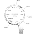
- the structure of the CS-CA-MCS plasmid used for the construction of the Dox-inducible gene transfer wrench vector is shown.
- the outline of the time schedule of the method of inducing TS-like cells from placenta-derived CT cells (middle / late CT cells) after the second trimester is shown.
- An optical micrograph of cells in which the Doxycycline (Dox) -induced SALL4 gene and the Dox-induced p53 dominant negative (p53DN) gene have been introduced into middle- and late-stage CT cells and cultured is shown.
- the cells of Day 12 and Day 17 were cultured in a medium containing Dox, and the cells of Day 40 were cultured in a medium containing no Dox.
- the morphology of TS-like cells was maintained even on Day 40.
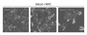
- An optical micrograph of cells in which the Doxycycline (Dox) -inducible SALL4 gene and the Dox-inducible MYC gene are introduced into middle- and late-stage CT cells and cultured is shown.
- the cells of Day6 and Day15 were cultured in the medium containing Dox, and the cells of Day29 were cultured in the medium containing no Dox. Day 29 did not maintain the morphology of TS-like cells.
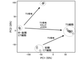
- TS-like cells derived from middle / late CT cells (middle / late TS-like cells), TS-like cells derived from early placenta-derived CT cells (early TS-like cells), TS cells, middle / late CT cells,
- the results of main component analysis on gene expression in early CT cells are shown.
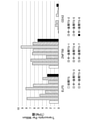
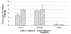
- the results of expression analysis of TS cell markers (positive markers: ELF5, ZNF750; negative markers: CDX2) for early TS-like cells and middle / late TS-like cells are shown.
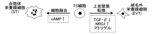
- Induction of differentiation of extravillous cytotrophoblast (EVT) and syncytiotrophoblast (ST) from TS cells is shown.
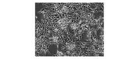
- An optical micrograph in which EVT is induced to differentiate from TS-like cells induced by introducing the SALL4 gene and the p53DN gene into middle- and late-stage CT cells is shown.
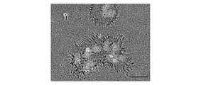
- An optical micrograph in which ST is induced to differentiate from TS-like cells induced by introducing the SALL4 gene and the p53DN gene into middle- and late-stage CT cells is shown.
- the results of CCR7 expression analysis in early CT cells and middle / late CT cells are shown.
- the results of expression analysis of TS cell markers (positive markers: ELF5, ZNF750; negative markers: CDX2) of diseased CT cell-derived TS-like cells and healthy human CT cell-derived TS-like cells are shown.
- polynucleotide and “nucleic acid” are used interchangeably and refer to a nucleotide polymer in which nucleotides are bound by phosphodiester bonds.
- the "polynucleotide” and “nucleic acid” may be DNA, RNA, or may be composed of a combination of DNA and RNA. Further, the “polynucleotide” and “nucleic acid” may be a polymer of a natural nucleotide, and the natural nucleotide and the unnatural nucleotide (an analog of the natural nucleotide, a base portion, a sugar portion and a phosphate portion at least one portion thereof) may be used.
- polynucleotide or “nucleic acid” is described by a generally accepted one-letter code. Unless otherwise specified, the base sequence is described from the 5'side to the 3'side. Nucleotide residues constituting a "polynucleotide” or “nucleic acid” may be simply described by adenine, thymine, cytosine, guanine, uracil, etc., or a one-letter code thereof.
- gene refers to a polynucleotide that contains at least one open reading frame that encodes a particular protein.
- the gene can include both exons and introns.
- polypeptide refers to a polymer of amino acids bound by an amide bond.
- the "polypeptide”, “peptide” or “protein” may be a polymer of natural amino acids, or may be a polymer of natural amino acids and unnatural amino acids (chemical analogs of natural amino acids, modified derivatives, etc.). It may be a polymer of unnatural amino acids. Unless otherwise specified, the amino acid sequence is described from the N-terminal side to the C-terminal side.
- the term "functionally linked" as used with respect to a polynucleotide means that the first base sequence is located close enough to the second base sequence and the first base sequence is the second base sequence or the second base sequence. It means that it can affect the area under the control of.
- functionally ligating a polynucleotide to a promoter means that the polynucleotide is ligated to be expressed under the control of the promoter.
- expression vector refers to a vector containing a target polynucleotide, which comprises a system for making the target polynucleotide expressible in the cell into which the vector has been introduced.
- Endogenous refers to what cells naturally have. Endogenous polynucleotides are polynucleotides that cells naturally contain in their genomes (nuclear genomes, organella genomes). An endogenous polypeptide is a polypeptide produced by transcription and translation of an endogenous polynucleotide.
- exogenous polypeptide refers to factors that are externally given to a cell.
- An extrinsic polynucleotide refers to a polynucleotide introduced into a cell from the outside.
- An extrinsic polypeptide is a polypeptide produced by transcription and translation from an extrinsic polynucleotide, or is an externally donated polypeptide to a cell.
- the extrinsic factors may be extrinsic chemicals, extrinsic physiological conditions (eg, pH, temperature, radiation, osmotic pressure, saline gradient) and the like.
- inducible promoter refers to a promoter in which transcriptional activity is initiated or enhanced in the presence of an inducing factor as compared to the absence of the inducing factor. That is, in the presence of an inducing factor, transcription of a gene functionally linked to an inducible promoter is initiated or enhanced.
- inducing factor refers to a factor that induces transcriptional activity of an inducible promoter.
- the inducing factor is preferably an extrinsic stimulus. Exogenous stimuli include, for example, extrinsic chemicals, extrinsic physiological conditions (eg, pH, temperature, light, radiation, osmotic pressure, saline gradient) and the like.
- the term "dominant negative” refers to a polypeptide that acts dominantly (dominantly) with respect to a normal polypeptide and has an action of inhibiting the action of the normal polypeptide.
- the dominant negative may be an inactive variant of the normal polypeptide.
- An inactive variant that functions as a dominant negative can be a variant that maintains the binding activity of the normal polypeptide to the ligand and does not exhibit the function that is expressed when the normal polypeptide binds to the ligand.
- trophoblast stem cell-like cells capable of differentiating into the cells constituting the placenta, which are derived from placenta-derived trophoblast cells after the middle of pregnancy, are the first extrinsic induction. It provides trophoblast stem cell-like cells containing the SALL4 gene functionally linked to a sex promoter.
- the cell of this embodiment is a TS-like cell having characteristics similar to those of a TS cell. Like TS cells, TS-like cells have the ability to differentiate into the cells that make up the placenta. Examples of cells constituting the placenta include extravillous cytotrophoblast (EVT) and syncytiotrophoblast (ST).
- EVT extravillous cytotrophoblast
- ST syncytiotrophoblast
- Whether or not a cell has the ability to differentiate into EVT can be determined by culturing the cell under the conditions used as a condition for inducing differentiation into EVT and confirming whether or not the cell has differentiated into EVT.
- Examples of the conditions for inducing differentiation into EVT include the conditions described in Examples described later.
- HLA-G is a marker for EVT, and its expression is upregulated by the differentiation of TS cells into EVT. Therefore, if the expression level of HLA-G is increased in the cells after culturing under the above conditions as compared with the cells before culturing under the above conditions, it is determined that the cells have differentiated into EVT. be able to.
- Examples of the HLA-G include those registered under the GenBank accession number NM_001363567.1. Or NM_002127.5.
- Whether or not a cell has the ability to differentiate into ST can be determined by culturing the cell under the conditions used as a condition for inducing differentiation into ST and confirming whether or not the cell has differentiated into ST.
- Examples of the conditions for inducing differentiation into ST include the conditions described in Examples described later.
- CG ⁇ also known as CGB3 (choronic gonadotropin subunit beta 3)
- CGB3 choronic gonadotropin subunit beta 3
- CG ⁇ is a marker for ST, and its expression is upregulated by differentiation from TS cells to ST. Therefore, if the expression level of CG ⁇ is increased in the cells after culturing under the above conditions as compared with the cells before culturing under the above conditions, it can be determined that the cells have differentiated into ST. can.
- Examples of the human CG ⁇ NCBI Gene ID: 1082
- NCBI Gene ID: 1082 include those registered under the GenBank accession number NM_0007373.
- the cells of this embodiment are TS-like cells having characteristics similar to TS cells and express TS cell markers.
- TS cell marker include ELF5 (E74 like ETS transcription factor 5), ZNF750 (zinc finger protein 750) and the like.
- ELF5 E74 like ETS transcription factor 5
- ZNF750 zinc finger protein 750
- human ELF5 NCBI Gene ID: 2001
- human ZNF750 NCBI Gene ID: 79755
- NCBI Gene ID: 79755 include those registered under the GenBank accession number NM_0247022.3.
- the cells of the present embodiment have low expression of CDX2 (cadal type homeobox 2), similar to TS cells.
- CDX2 cadal type homeobox 2
- Examples of the human CDX2 include those registered under the GenBank accession number NM_001265.6.
- the cells of this embodiment are derived from placenta-derived trophoblast cells after the second trimester of pregnancy.
- "After mid-pregnancy” means the gestation period and after childbirth after 1/3 of the gestation period has passed.
- the "placenta after the second trimester” means a placenta after one-third of the pregnancy period, and may be a placenta during the pregnancy period or a placenta after childbirth.
- the placenta is not particularly limited as long as it is a placenta in which the placenta is formed during pregnancy.
- embryos include primates (humans, chimpanzees, rhesus monkeys, marmosets, etc.), rodents (mouses, rats, hamsters, guinea pigs, etc.), meats (dogs, cats, weasels, etc.) and the like.
- Embryonic discs are preferably primates, more preferably humans.
- "after mid-gestation” refers to the gestation period and postpartum after 12 weeks of gestation.
- the human placenta after the second trimester may be a placenta during the gestation period after the 12th week of pregnancy or a placenta after delivery.
- placenta of humans after 12 weeks of gestation for example, those excreted from the body by artificial abortion, miscarriage or childbirth can be used.
- the placenta may be a placenta due to normal pregnancy (hereinafter, also referred to as "normal placenta”) or a placenta due to abnormal pregnancy (hereinafter, also referred to as “disease placenta”).
- the placenta may be the placenta of an individual who has developed a disease of pregnancy hypertension, polyhydramnios, polyhydramnios, fetal growth restriction, low weight birth, chromosomal abnormality (trisomy, etc.), miscarriage, premature birth.
- TS-like cells derived from the placenta of individuals who have developed the disease can be used for the study of these diseases and the development of therapeutic agents and methods for these diseases.
- “Placenta-derived trophoblast cells after the second trimester” means trophoblast cells isolated from the placenta after the second trimester.
- the trophoblast cell is preferably a cytotrophoblast (CT cell).
- CT cells are progenitor cells present in placental tissue.
- Placenta-derived CT cells in the early stages of pregnancy can be differentiated into TS-like cells by adjusting the medium components, but placenta-derived CT cells after the second trimester of pregnancy can be differentiated into TS-like cells even if the medium components are adjusted. I can't let you.
- TS-like cells can be derived from placenta-derived CT cells after metaphase.
- the TS-like cell of this embodiment is a TS-like cell produced by the method described later.
- the method for isolating trophoblast cells from the placenta after the second trimester of pregnancy is not particularly limited. Specific examples include the method described in the examples.
- mechanical and / or enzymatic treatment of placental tissue after metaphase can be appropriately used to prepare cell suspensions.
- a cell suspension may be prepared by finely chopping the placental tissue, washing it with physiological saline or the like, and immersing it in a cell dispersion.
- the cell dispersion for example, a so-called proteolytic enzyme solution, cell adhesion stripping solution, or the like may be used. Specific examples of the cell dispersion include those commercially available under product names such as Accumax and TrypLE.
- the cell dispersion may be used alone or in combination of two or more.
- the cells may be further dispersed using a cell strainer or the like.
- the cell suspension prepared as described above may be treated to remove erythrocytes. Examples of the method for removing erythrocytes include treatment with colloidal silicate particles coated with polyvinylpyrrolidone (commercially available as Percoll®) and the like.
- CD49f antibody-positive cells are collected from the cell suspension.
- the method for recovering CD49f antibody-positive cells is not particularly limited, and known methods can be used. For example, a method using an anti-CD49f antibody and the like can be mentioned. After or before the recovery of the CD49f antibody-positive cells, a treatment for removing the CD45 antibody-positive cells may be performed. The method for removing the CD45 antibody-positive cells is not particularly limited, and a known method can be used. For example, a method using an anti-CD45 antibody and the like can be mentioned.
- the trophoblast cells isolated as described above can be maintained in a medium containing a growth factor and a ROCK inhibitor.
- Mid- and late-stage trophoblast cells are said to have lower expression of CCR7 than placenta-derived trophoblast cells (preferably CT cells) in the early stages of pregnancy (prior to 1/3 of the gestation period). It has characteristics.
- the expression of CCR7 is low in both the middle and late trophoblast cells derived from normal placenta and the middle and late trophoblast cells derived from diseased placenta. More specifically, in the middle / late trophoblast cells, the TPM (transcripts permilion) of CCR7 is preferably 50 or less, 40 or less, 30 or less, or 20 or less.
- the TPM of CCR7 is preferably 100 or more, 150 or more, 160 or more, 170 or more, or 180 or more. ..
- the TPM is a value that can be obtained by using the next-generation sequencer, and is a value indicating how many target transcripts are present when there are 1 million total transcripts in the sample.
- the TPM (TPM t ) of the transcript t can be calculated by the following formula.
- T t is the number of reads per 1,000 bp of the transcript t, and can be calculated by the following formula.
- Yt is the read count mapped to the transcript t
- L t is the length of the transcript t.
- the CCR7 (CC motif chemokine receptor 7) gene encodes a G protein-coupled receptor family.
- the sequence of the CCR7 gene is known and can be obtained from a known database such as GenBank.
- examples of the human CCR7 include those registered under the GenBank accession number NM_001301714.2.
- CCR7 is low in the middle and late trophoblast cells, which is the starting material of the TS-like cells of the present embodiment, but is high in the TS-like cells of the present embodiment.
- the TS-like cells of this embodiment are characterized by containing the SALL4 gene operably linked to a first extrinsic inducible promoter. Therefore, it is distinguishable from TS cells in that it contains the SALL4 gene functionally linked to the first extrinsic inducible promoter.
- the inducible promoter used as the first extrinsic inducible promoter is not particularly limited, and known promoters can be used.
- the inducible promoter is preferably an inducible promoter in which transcriptional activity is induced by an extrinsic inducing factor.
- the first extrinsic inducible promoter includes, for example, a promoter in which transcriptional activity is induced by an extrinsic chemical substance, and extrinsic physiological conditions (eg, pH, temperature, light, radiation, osmotic pressure, saline gradient). Examples thereof include promoters whose transcriptional activity is induced by the above.
- inducible promoter examples include a tetracycline-induced promoter, a lac operon promoter, an IPTG-induced promoter, a steroid-induced promoter, a rapamycin-induced promoter, a temperature-induced promoter, a pH-induced promoter, a salt-induced promoter, and a radiation-induced promoter. Promoters and the like can be mentioned, but the present invention is not limited to these.
- the tetracycline-inducible promoter is an inducible promoter in which transcriptional activity is induced in the presence of tetracycline or an analog thereof.
- the tetracycline-inducible promoter preferably comprises a minimal promoter operably linked to the Tet operator (tetO) sequence.
- tetO Tet operator
- the transcriptional activator binds to the teto sequence and induces the transcriptional activity of the minimal promoter. This induces transcription of the gene functionally linked to the minimal promoter.
- the "tetracycline analog" is a compound that exhibits structural homology with tetracycline and is capable of inducing the transcriptional activity of a tetracycline-inducible promoter.
- the tetracycline analog is not particularly limited, and examples thereof include Doxycycline (Dox), chlorotetracycline, and anhydrotetracycline.
- minimal promoter is meant a promoter having a minimal sequence as a promoter.
- a minimal promoter can be said to be a promoter containing all the necessary elements to properly initiate transcription of a functionally linked gene.
- the minimal promoter may include, for example, a transcription initiation site, an RNA polymerase binding site, and a TATA box.
- the minimal promoter examples include a thymidine kinase promoter, a ⁇ -globulins promoter, a cytomegalovirus (CMV: cytomegalovirus) promoter, an SV40 promoter and the like.
- the inducible promoter is a tetracycline-inducible promoter
- the TS-like cell further comprises a transcriptional activator (eg, reverse tetracycline-regulated transactivator: rtTA) coding sequence operably linked to the appropriate promoter. Is preferable.
- a commercially available expression system containing a tetracycline-inducible promoter may be used.
- a pTetOne vector (Takara) or the like can be used.
- a commercially available expression system containing other inducible promoters can be used.
- SALL4 gene The SALL4 (spart like transcription factor 4) gene encodes SALL4, a transcription factor containing zinc fingers.
- the sequence of the SALL4 gene is known and can be obtained from a known database such as GenBank.
- examples of the human SALL4 NCBI Gene ID: 57167
- examples of the human SALL4 include those registered with GenBank accession number NM_001318031.2 (SEQ ID NO: 1 and 2) or NM_020366.5 (SEQ ID NO: 3 and 4). ..
- the human SALL4 gene is not limited to the above, and may be homologs (orthologs, paralogs) thereof and variants thereof. Sequence information of the SALL4 gene possessed by other mammals can also be obtained from a known database such as GenBank.
- the SALL4 gene functionally linked to the first extrinsic inducible promoter may be an endogenous SALL4 gene or an extrinsic SALL4 gene.
- the first extrinsic inducible promoter is the endogenous SALL4 gene using genome editing technology or the like. It may be inserted upstream.
- genome editing technique known ones can be used, and examples thereof include a CRISPR / Cas system, a zinc finger nuclease (ZFN), a transcription activator-like effector nucleicase (TALEN), and the like.
- the Tet operator (tetO) sequence (tetO) sequence so that the endogenous SALL4 gene promoter is functionally linked upstream of the promoter of the endogenous SALL4 gene.
- tetO Tet operator sequence
- TRE tetracycline response factor
- the extrinsic SALL4 gene expression cassette (also referred to as “exogenous SALL4 gene expression cassette”) may be externally introduced using a known gene transfer technique.
- the exogenous SALL4 expression cassette may be present in the genome (for example, the nuclear genome), or may be present outside the genome as a plasmid or the like.
- the SALL4 gene functionally linked to the first extrinsic inducible promoter is preferably the extrinsic SALL4 gene.
- the extrinsic SALL4 gene functionally linked to the first extrinsic inducible promoter is preferably present on the nuclear genome.
- the organism from which the exogenous SALL4 gene is derived is the same as the organism from which the middle / anaphase trophoblast cells are derived.
- the human middle / late trophoblast cells it is preferably the human SALL4 gene.
- the extrinsic SALL4 gene is not limited to the wild type, and may contain mutations (either deletion, substitution, insertion, or addition) as long as it has the ability to induce TS-like cells.
- the "ability to induce TS-like cells” means the function of inducing the differentiation of middle- and late-stage trophoblast cells into TS-like cells by inducing the expression of the gene.
- extrinsic SALL4 gene examples include the following (A) to (G).
- A) Wild-type SALL4 gene eg, a polynucleotide consisting of the nucleotide sequence set forth in SEQ ID NO: 1 or 3.
- B A polynucleotide consisting of a nucleotide sequence encoding a wild-type SALL4 protein (eg, a protein consisting of the amino acid sequence set forth in SEQ ID NO: 2 or 4).
- a wild-type SALL4 protein (eg, the amino acid sequence set forth in SEQ ID NO: 2 or 4), it encodes a protein consisting of an amino acid sequence in which one or more amino acids are mutated, and has the ability to induce TS-like cells.
- Polynucleotide having. (D) Encodes a protein consisting of an amino acid sequence of a wild-type SALL4 protein (eg, the amino acid sequence set forth in SEQ ID NO: 2 or 4) and an amino acid sequence having 70% or more sequence identity, and into TS-like cells. Polynucleotide with the ability to induce.
- E In the wild-type SALL4 gene (eg, a polynucleotide consisting of the nucleotide sequence set forth in SEQ ID NO: 1 or 3), one or more nucleotides are mutated nucleotide sequences and the ability to induce TS-like cells. Polynucleotide with.
- F It consists of a wild-type SALL4 gene (eg, a polynucleotide consisting of the nucleotide sequence set forth in SEQ ID NO: 1 or 3) and a nucleotide sequence having 70% or more sequence identity, and has the ability to induce TS-like cells. Polynucleotide with.
- a polynucleotide that hybridizes with a wild-type SALL4 gene eg, a polynucleotide consisting of the nucleotide sequence set forth in SEQ ID NO: 1 or 3 under stringent conditions and has the ability to induce TS-like cells.
- the "mutation” may be any of deletion, substitution, addition, and insertion, and may be a combination thereof.
- the above (C) and “plurality” are not particularly limited, but for example, 2 to 60 pieces, 2 to 50 pieces, 2 to 40 pieces, 2 to 30 pieces, 2 to 20 pieces, 2 to 15 pieces, 2 to 10 pieces, 2 to 90 pieces, 2 to 8 pieces, 2 to 7 pieces, 2 to 6 pieces, 2 to 5 pieces, 2 to 4 pieces, 2 to 3 pieces, or 2 pieces can be mentioned.
- the "plurality” is not particularly limited, but for example, 2 to 100, 2 to 90, 2 to 80, 2 to 70, 2 to 60, 2 to 50, 2 ⁇ 40 pieces, 2 ⁇ 30 pieces, 2 ⁇ 20 pieces, 2 ⁇ 15 pieces, 2 ⁇ 10 pieces, 2 ⁇ 9 pieces, 2 ⁇ 8 pieces, 2 ⁇ 7 pieces, 2 ⁇ 6 pieces, 2 ⁇ 5 pieces, 2 ⁇ 4, 2-3, or 2 may be mentioned.
- the sequence identity is not particularly limited as long as it is 70% or more, but is 80% or more, 85% or more, 90% or more, 95% or more, 96% or more, 97% or more. , 98% or more, or 99% or more.
- Sequence identity between amino acid sequences or nucleotide sequences can be obtained by juxtaposing two amino acid sequences or nucleotide sequences with gaps in the parts corresponding to insertions and deletions so that the corresponding amino acids or nucleotides match most. It is determined as the ratio of matching amino acids or nucleotides to the entire amino acid sequence or the entire nucleotide sequence excluding the gap in the aligned. Sequence identity between amino acid sequences or nucleotide sequences can be determined using various homology search software known in the art. For example, the sequence identity value of an amino acid sequence or a nucleotide sequence can be obtained by calculation based on the alignment obtained by the known homology search software BLASTP.
- the “stringent condition” includes, for example, the conditions described in Molecular Cloning-A LABORATORY MANUAL THIRD EDITION (Sambrook et al., Cold Spring Harbor Laboratory Press). For example, 6 ⁇ SSC (composition of 20 ⁇ SSC: 3M sodium chloride, 0.3M citrate solution, pH 7.0), 5 ⁇ Denhardt solution (composition of 100 ⁇ Denhardt solution: 2% by mass, bovine serum albumin, 2% by mass). Incubate at 42-70 ° C.
- hybridization buffer consisting of Ficoll, 2% by mass polyvinylpyrrolidone), 0.5% by weight SDS, 0.1 mg / mL salmon sperm DNA, and 50% formamide.
- the conditions for hybridization are exemplified by performing the above.
- the washing buffer used for washing after incubation include a 1 ⁇ SSC solution containing 0.1% by mass SDS, and more preferably a 0.1 ⁇ SSC solution containing 0.1% by mass SDS.
- the degenerate codons used are those having a high codon usage frequency in the cells of the embryotheria to be used.
- codons frequently used in human cells are used. That is, it is preferably optimized for human codons.
- the SALL4 gene functionally linked to the first extrinsic inducible promoter is preferably the extrinsic SALL4 gene.
- the TS-like cells of the present embodiment are at least selected from the group consisting of the following (a) and (b). It may have one feature.
- the TS-like cells of the present embodiment may have suppressed p53 activity as compared with the middle / late trophoblast cells.
- p53 is a tumor suppressor protein and has a function of controlling the suppression of cell proliferation cycles such as DNA repair, cell proliferation arrest, and apoptosis.
- the sequence of the p53 gene is known and can be obtained from a known database such as GenBank.
- GenBank GenBank
- the human p53 gene is not limited to the above, and may be homologs (orthologs, paralogs) thereof and variants thereof. Sequence information of the p53 gene possessed by other mammals can also be obtained from a known database such as GenBank.
- Suppression of p53 activity can be achieved, for example, by the presence of a p53 activity inhibitor or inhibition of p53 gene expression.
- a p53 activity inhibitor or inhibition of p53 gene expression For example, when the amount of the intracellular activity inhibitor of p53 is larger than that of the middle / late trophoblast cells, the cells are suppressed in the activity of p53 as compared with the middle / late trophoblast cells. , Can be determined. Further, for example, when the intracellular mRNA amount or protein amount of p53 is smaller than that of the middle / late trophoblast cells, the activity of p53 is suppressed in the cells as compared with the middle / late trophoblast cells. It can be determined that it is.
- the activity inhibitor of p53 is not particularly limited, and examples thereof include dominant negative of p53.
- the dominant negative of p53 is not particularly limited as long as it is a polypeptide capable of predominantly acting on p53 and inhibiting the function of p53.
- Examples of the p53 dominant negative include a truncated protein in which a part of p53 is deleted.
- the amino acid sequence to be deleted is not particularly limited as long as the P53 dominant negative binds to the ligand of p53 and inhibits the function of p53.
- the P53 dominant negative may be one in which the N-terminal of p53 is deleted, or one in which the C-terminal is deleted.
- Specific examples of human p53 dominant negative include a polypeptide (SEQ ID NO: 8) containing the amino acid sequence at positions 300 to 393 of the amino acid sequence set forth in SEQ ID NO: 6.
- P53 dominant negative may be introduced into cells by ligating a cell-permeable peptide or the like.
- the p53 dominant negative linked to the cell-permeable peptide may be present intracellularly.
- the exogenous p53 dominant negative gene functionally linked to the second extrinsic inducible promoter may be introduced into the cell to induce the expression of the exogenous p53 dominant negative gene in the cell. good.
- the TS-like cells of this embodiment contain an extrinsic p53 dominant negative gene operably linked to a second extrinsic inducible promoter.
- the p53 dominant negative gene may be present in the genome (for example, a nuclear genome), or may be present outside the genome as a plasmid or the like.
- the second extrinsic inducible promoter is not particularly limited. ..
- Examples of the second extrinsic inducible promoter include those similar to those mentioned above as the first extrinsic inducible promoter.
- the second extrinsic inducible promoter may be the same as or different from the first extrinsic inducible promoter.
- the second extrinsic inducible promoter is preferably the same as the first extrinsic inducible promoter, as transcriptional activity can be induced by the same inducing factors as the first extrinsic inducible promoter.
- extrinsic p53 dominant negative gene functionally linked to the second extrinsic inducible promoter examples include the following (A) to (G).
- a polynucleotide eg, SEQ ID NO: 7 that is a truncated gene of the wild-type p53 gene (eg, SEQ ID NO: 5) and encodes a protein that functions as a p53 dominant negative.
- B A polynucleotide consisting of a nucleotide sequence encoding a shortened protein (eg, SEQ ID NO: 8) of a wild-type p53 protein (eg, SEQ ID NO: 6) that functions as a dominant negative of p53.
- (C) A protein that consists of an amino acid sequence in which one or more amino acids are mutated in a shortened protein (eg, SEQ ID NO: 8) of a wild-type p53 protein (eg, SEQ ID NO: 6) and functions as a dominant negative of p53.
- (D) Consists of an amino acid sequence (eg, SEQ ID NO: 8) of a shortened protein of wild-type p53 protein (eg, SEQ ID NO: 6) and an amino acid sequence having 70% or more sequence identity, as a dominant negative of p53.
- nucleotides consist of a mutated nucleotide sequence and function as a dominant negative of p53.
- a polynucleotide that encodes a protein is a shortened gene (eg, SEQ ID NO: 7) of the wild-type p53 gene (eg, SEQ ID NO: 5) and a nucleotide sequence having 70% or more sequence identity, and functions as a dominant negative of p53.
- G A polynucleotide encoding a protein that hybridizes to a shortened gene (eg, SEQ ID NO: 7) of the wild-type p53 gene (eg, SEQ ID NO: 5) under stringent conditions and functions as a dominant negative of p53. ..
- the "mutation” may be any of deletion, substitution, addition, and insertion, and may be a combination thereof.
- the above (C) and “plurality” are not particularly limited, but for example, 2 to 10 pieces, 2 to 90 pieces, 2 to 8 pieces, 2 to 7 pieces, 2 to 6 pieces, 2 to 5 pieces, 2 to There are four, two to three, or two.
- the “plurality” is not particularly limited, but for example, 2 to 30, 2 to 20, 2 to 15, 2 to 10, 2 to 9, 2 to 8, and 2 -7, 2-6, 2-5, 2-4, 2-3, or 2.
- sequence identity is not particularly limited as long as it is 70% or more, but is 80% or more, 85% or more, 90% or more, 95% or more, 96% or more, 97% or more. , 98% or more, or 99% or more.
- the "stringent condition" in (G) above is the same as described above.
- the degenerate codons used are those having a high codon usage frequency in the cells of the embryotheria to be used.
- codons frequently used in human cells are used. That is, it is preferably optimized for human codons.
- the expression state of the extrinsic p53 dominant negative is not particularly limited.
- the TS-like cell of the present embodiment may be one in which the extrinsic p53 dominant negative gene is not expressed and the extrinsic p53 dominant negative is not detected.
- the TS-like cells of the present embodiment may be those in which the activity of p53 is not suppressed as compared with the middle / late trophoblast cells.
- Transcription inhibition of the p53 gene can be achieved by, for example, small molecule interfering nucleic acids such as siRNA.
- small molecule interfering nucleic acid refers to a small molecule nucleic acid having about 18 to 25 base pairs that suppresses the expression of a target gene by RNA interference. In the low molecular weight interfering nucleic acid, one nucleic acid molecule may form a base pair by a hairpin structure, or two nucleic acid molecules may form a base pair.
- the small molecule interfering nucleic acid may consist only of RNA or may contain RNA and DNA. The design of small interfering nucleic acids that suppress the expression of the p53 gene can be performed based on known techniques.
- the small molecule interfering nucleic acid for the p53 gene may be introduced into cells using a transfection reagent or the like. In this case, a small molecule interfering nucleic acid for the p53 gene may be present in the cell. Alternatively, a gene encoding a small interfering nucleic acid for the p53 gene functionally linked to a second extrinsic inducible promoter is introduced into the cell to induce the expression of the small interfering nucleic acid in the cell. May be good. In this case, the TS-like cells of this embodiment contain a gene encoding a small molecule interfering nucleic acid for the p53 gene operably linked to a second extrinsic inducible promoter.
- the second extrinsic inducible promoter the same as the p53 dominant negative gene can be used.
- the expression state of the gene is not particularly limited.
- the TS-like cell of the present embodiment may be one in which the gene is not expressed and a small molecule interfering nucleic acid for the p53 gene is not detected.
- the TS-like cells of the present embodiment may be those in which the activity of p53 is not suppressed as compared with the middle / late trophoblast cells.
- Transcription inhibition of the p53 gene may be achieved by, for example, an antisense nucleic acid such as antisense RNA.
- an antisense nucleic acid refers to a nucleic acid that hybridizes to the mRNA of the target gene and suppresses the expression of the target gene.
- the antisense nucleic acid may consist only of RNA or may contain RNA and DNA. The design of an antisense nucleic acid that suppresses the expression of the p53 gene can be performed based on known techniques.
- the antisense nucleic acid for the p53 gene may be introduced into cells using a transfection reagent or the like. In this case, the antisense nucleic acid for the p53 gene may be present in the cell. Alternatively, a gene encoding an antisense nucleic acid for the p53 gene functionally linked to a second extrinsic inducible promoter may be introduced into the cell to induce the expression of the antisense nucleic acid in the cell. .. In this case, the TS-like cells of this embodiment contain a gene encoding an antisense nucleic acid against the p53 gene operably linked to a second extrinsic inducible promoter.
- the second extrinsic inducible promoter the same as the p53 dominant negative gene can be used.
- the expression state of the gene is not particularly limited.
- the TS-like cell of the present embodiment may be one in which the gene is not expressed and antisense nucleic acid for the p53 gene is not detected.
- the TS-like cells of the present embodiment may be those in which the activity of p53 is not suppressed as compared with the middle / late trophoblast cells.
- the TS-like cells of the present embodiment may have promoted expression of the MYC gene as compared with the middle / late trophoblast cells.
- the expression is promoted as compared with the middle / late trophoblast cells means that the expression is detected in the middle / late trophoblast cells when the expression is not detected in the middle / late trophoblast cells, and that the expression is detected in the middle / late trophoblast cells. This includes the introduction of an extrinsic gene that is not contained in late trophoblast cells and the expression of the extrinsic gene.
- MYC is a nuclear phosphoprotein encoded by the protooncogene and is involved in cell cycle progression, apoptosis, and cell transformation.
- the sequence of the MYC gene is known and can be obtained from a known database such as GenBank.
- examples of the human MYC NCBI Gene ID: 4609) include those registered with NM_0013548870.1 (SEQ ID NO: 9, 10) or NM_002467.6 (SEQ ID NOs: 11 and 12).
- the human MYC gene is not limited to the above, and may be homologs (orthologs, paralogs) thereof and variants thereof. Sequence information of the MYC gene possessed by other mammals can also be obtained from a known database such as GenBank.
- promotion of MYC gene expression can be achieved, for example, by introducing the MYC gene functionally linked to a second extrinsic inducible promoter and inducing the expression of the MYC gene.
- the cell contains an extrinsic MYC gene operably linked to a second extrinsic inducible promoter.
- the second extrinsic inducible promoter is not particularly limited.
- the second extrinsic inducible promoter the same as the p53 dominant negative gene can be used.
- the exogenous MYC gene is preferably in an expressed state.
- the TS-like cells of this embodiment are exogenous p53 functionally linked to an extrinsic SALL4 gene operably linked to a first extrinsic inducible promoter and a second extrinsic inducible promoter. It preferably contains a dominant negative gene.
- the first extrinsic inducible promoter and the second extrinsic inducible promoter are preferably the same inducible promoter because the same inducing factor can induce transcriptional activity.
- a tetracycline-inducible promoter can be used, but is not limited thereto.
- the TS-like cells of this embodiment can be produced by the method for producing TS-like cells described later. Since the TS-like cells of the present embodiment have the ability to differentiate into cells constituting the placenta, they can be differentiated into EVT, ST, or the like. Therefore, it can be used as a research material for basic research such as early development of placental species and placental function analysis. In addition, the TS-like cells of the present embodiment are derived from trophoblast cells derived from the placenta after the second trimester of pregnancy when a disease related to pregnancy becomes apparent.
- TS-like cells derived from middle- and late-stage trophoblast cells derived from individuals who have developed the disease can be used for elucidation of the pathophysiology of pregnancy-related diseases and development of therapeutic methods. Furthermore, it can be expected to be applied to reproductive medicine and regenerative medicine.
- the present invention provides a method for producing trophoblast stem cell-like cells (TS-like cells) having the ability to differentiate into cells constituting the placenta.
- the method for producing TS-like cells of the present embodiment includes the following steps (i) to (iii). (I) A step of preparing trophoblast cells derived from the placenta after the second trimester of pregnancy. (Ii) A step of inducing the expression of the SALL4 gene in the trophoblast cells.
- Step (i) is a step of preparing trophoblast cells derived from placenta after the second trimester of pregnancy.
- the trophoblast cells (middle / late trophoblast cells) derived from the placenta after the middle of pregnancy are the " ⁇ trophoblast cells>" of the above-mentioned "[trophoblast stem cell like cell (TS-like cells)]". It is the same as that described in the section.
- the metaphase and late trophoblast cells can be isolated from the placenta after the second trimester by a known method. Examples of the method for isolating middle / late trophoblast cells include the methods described in the above section “ ⁇ Trophoblast cells>”.
- Step (ii) is a step of inducing the expression of the SALL4 gene in the middle and late trophoblast cells.
- the method for inducing the expression of the SALL4 gene in the middle / late trophoblast cells is not particularly limited, and a known method can be used. Examples of the method for inducing the expression of the SALL4 gene include methods including the following (ii-1) and (ii-2).
- (Ii-1) For middle and late trophoblast cells, Introducing a polynucleotide containing an extrinsic SALL4 gene operably linked to a first extrinsic inducible promoter, or introducing a polynucleotide containing the first extrinsic inducible promoter into an endogenous SALL4 gene. Introducing the endogenous SALL4 gene upstream of the exogenous SALL4 gene so as to be functionally linked to the first extrinsic inducible promoter. (Ii-2) Inducing the expression of the extrinsic SALL4 gene or the endogenous SALL4 gene by the first inducing factor that induces the transcriptional activity of the first extrinsic inducible promoter.
- (Ii-1) may be an operation of introducing a polynucleotide containing an extrinsic SALL4 gene functionally linked to a first extrinsic inducible promoter into middle / late trophoblast cells.
- the SALL4 gene functionally linked to the first extrinsic inducible promoter is similar to that described in the section "[trophoblast stem cell like cell]" above. be.
- the method for introducing a polynucleotide containing the extrinsic SALL4 gene functionally linked to the first extrinsic inducible promoter into middle / late trophoblast cells is not particularly limited, and a known method can be used. can.
- the extrinsic SALL4 gene functionally linked to the first extrinsic inducible promoter is cloned into an appropriate expression vector that can be expressed in middle and late trophoblast cells that serve as host cells, and the expression vector is used. It may be introduced into middle / late trophoblast cells.
- the expression vector is a polynucleotide containing an extrinsic SALL4 gene operably linked to a first extrinsic inducible promoter, as well as enhancers, poly A addition signals, marker genes, replication initiation sites, replication initiation, if desired. It may contain a gene encoding a protein that binds to a point and controls replication.
- the "marker gene” refers to a gene that enables selection and selection of cells by introducing the marker gene into cells. Examples of the marker gene include, but are not limited to, a drug resistance gene, a fluorescent protein gene, a luminescent enzyme gene, and a color-developing enzyme gene. The marker gene may be used alone or in combination of two or more.
- Examples of the drug resistance gene include, but are not limited to, a neomycin resistance gene, a tetracycline resistance gene, a canamycin resistance gene, a zeosin resistance gene, a hyglomycin resistance gene, and a puromycin resistance gene.
- Examples of the fluorescent protein gene include, but are not limited to, a green fluorescent protein (GFP) gene, a yellow fluorescent protein (YFP) gene, a red fluorescent protein (RFP) gene, and the like.
- Examples of the luminescent enzyme gene include, but are not limited to, a luciferase gene and the like.
- Examples of the color-developing enzyme gene include, but are not limited to, ⁇ -galactosidase gene, ⁇ -glucuronidase gene, alkaline phosphatase gene and the like.
- the type of expression vector is not particularly limited, and a known expression vector can be used.
- Examples of the expression vector include an episomal vector, an artificial chromosome vector, a plasmid vector, a virus vector and the like.
- the episomal vector examples include a vector containing a sequence required for autonomous replication derived from EBV, SV40, etc. as a vector element.
- the vector elements required for autonomous replication are a replication origin and a gene encoding a protein that binds to the replication origin and controls replication.
- the replication origin oriP the replication origin oriP.
- the EBNA-1 gene the replication origin ori and the SV40LT gene can be mentioned.
- examples of the artificial chromosome vector include a YAC (Yeast artificial chromosome) vector, a BAC (Basic artificial chromosome) vector, and a PAC (P1-derivated artificial chromosome) vector.
- YAC yeast artificial chromosome
- BAC Basic artificial chromosome
- PAC P1-derivated artificial chromosome
- the plasmid vector is not particularly limited as long as it is a plasmid vector that can be expressed in pluripotent stem cells to be introduced.
- Examples of the plasmid vector for animal cell expression include pA1-11, pXT1, pRc / CMV, pRc / RSV, pcDNAI / Neo and the like.
- Virus vectors include retrovirus (including lentivirus) vector, adenovirus vector, adeno-associated virus vector, Sendai virus vector, herpesvirus vector, vaccinia virus vector, poxvirus vector, poliovirus vector, sylvis virus vector, and loved. Examples thereof include a virus vector, a paramixovirus vector, an orthomixovirus vector, and the like.
- the method for introducing the expression vector into the middle / late trophoblast cells is not particularly limited, and can be appropriately selected depending on the type of the expression vector.
- Examples of the method for introducing the expression vector into the middle / late trophoblast cells include a lipofection method, a microinjection method, a DEAE dextran method, a gene gun method, an electroporation method, and a calcium phosphate method.
- examples of the method for infecting middle and late trophoblast cells include the polybrene method.
- the cells into which the expression vector has been introduced are introduced by culturing the cells in a medium containing the antibiotic corresponding to the antibiotic resistance gene after the introduction of the expression vector. It can be selected efficiently.
- (Ii-1) functionally transfers a polynucleotide containing a first extrinsic inducible promoter upstream of the endogenous SALL4 gene, and the endogenous SALL4 gene functionally into the first extrinsic inducible promoter. It may be an operation of introducing so as to connect.
- the first extrinsic inducible promoter is similar to the above.
- the method for introducing a polynucleotide containing the first extrinsic inducible promoter upstream of the endogenous SALL4 gene is not particularly limited, and a known site-specific introduction method can be used.
- a method for introducing a polynucleotide in a site-specific manner for example, a genome editing technique using a site-specific nuclease can be mentioned.
- Genome editing techniques include CRISPR / Cas systems, zinc finger nucleases (ZFNs), TALENs and the like. Methods for site-specific insertion of desired polynucleotides using these genome editing techniques are known.
- the protospacer-adjacent motif (PAM) sequence upstream of the endogenous SALL4 gene is searched and the 20 bases upstream of the PAM sequence are adjacent.
- the gRNA can be designed with the degree as the target sequence.
- the gRNA may be designed using known gRNA design software or the like.
- (Ii-2) (Ii-2) is an operation of inducing the expression of the extrinsic SALL4 gene or the endogenous SALL4 gene by the first inducing factor that induces the transcriptional activity of the first extrinsic inducible promoter.
- the first inducing factor is a factor that induces the transcriptional activity of the first extrinsic inducible promoter.
- the first inducing factor can be appropriately selected depending on the type of the first extrinsic inducible promoter. For example, when the first extrinsic inducible promoter is a tetracycline-inducible promoter, tetracycline or an analog thereof can be used as the first inducing factor.
- the transcriptional activity of the first extrinsic inducible promoter is induced. Thereby, it is possible to induce the expression of the extrinsic SALL4 gene or the endogenous SALL4 gene functionally linked to the first extrinsic inducible promoter.
- the period for inducing the expression of the SALL4 gene in the step (ii) is not particularly limited.
- the period for inducing the expression of the SALL4 gene can be, for example, 5 days or more, 8 days or more, 10 days or more, 12 days or more, 15 days or more, 18 days or more, or 20 days or more.
- the upper limit of the period for inducing the expression of the SALL4 gene is not particularly limited.
- the expression of the SALL4 gene may be induced by culturing in a medium containing the first inducing factor until immediately before induction into cells constituting the placenta (EVT, ST, etc.).
- the induction of SALL4 gene expression may be stopped. If the TS-like cells are induced, the TS-like cells can be maintained without inducing the expression of the SALL4 gene while maintaining the ability to differentiate into the cells constituting the placenta. Therefore, for example, the period for inducing the expression of the SALL4 gene may be, for example, 50 days or less, 40 days or less, 30 days or less, or 25 days or less.
- the period for inducing the expression of the SALL4 gene can be controlled by the presence of the first inducing factor in the medium in which the cells are cultured.
- the step (iii) is a step of performing at least one selected from the group consisting of the following (A) and (B).
- A Suppresses the activity of p53 in middle and late trophoblast cells.
- B Induces the expression of the MYC gene in middle and late trophoblast cells.
- (A) is a step of suppressing the activity of p53 of middle and late trophoblast cells.
- the method for suppressing the activity of p53 in the middle and late trophoblast cells is not particularly limited, and a known method can be used. Examples of the method for suppressing the activity of p53 include a method for allowing a substance for inhibiting the activity of p53 to exist in the cells of the middle / late trophoblast cells, a method for suppressing the expression of the p53 gene in the middle / late trophoblast cells, and the like. Be done.
- Examples of the p53 activity inhibitor include p53 dominant negative.
- the p53 dominant negative is the same as that described in the section of "trophoblast stem cell like cell (TS-like cell)".
- the p53 dominant negative may be introduced into cells by ligating a cell-permeable peptide or the like.
- cells may be introduced with an extrinsic p53 dominant negative gene functionally linked to a second extrinsic inducible promoter.
- the extrinsic p53 dominant negative gene functionally linked to the second extrinsic inducible promoter was described in the section "trophoblast stem cell like cell”. Similar to the one.
- (A) may include the following (A-1) and (A-2): .. (A-1) Introducing a polynucleotide containing an extrinsic p53 dominant negative gene functionally linked to a second extrinsic inducible promoter into middle- and late-stage trophoblast cells. (A-2) Inducing the expression of the extrinsic p53 dominant negative gene by a second inducing factor that induces the transcriptional activity of the second extrinsic inducible promoter.
- (A-1) is an operation of introducing a polynucleotide containing an extrinsic p53 dominant negative gene functionally linked to a second extrinsic inducible promoter into middle / late trophoblast cells.
- the method for introducing the polynucleotide into the middle / late trophoblast cells is the same as the method for introducing the polynucleotide containing the extrinsic SALL4 gene functionally linked to the first extrinsic inducible promoter.
- the second extrinsic inducible promoter may be the same as or different from the first extrinsic inducible promoter.
- the second extrinsic inducible promoter is preferably the same as the first extrinsic inducible promoter, as transcriptional activity can be induced by the same inducing factors as the first extrinsic inducible promoter.
- (A-2) is an operation of inducing the expression of the extrinsic p53 dominant negative gene by a second inducing factor that induces the transcriptional activity of the second extrinsic inducible promoter.
- the second inducing factor is a factor that induces the transcriptional activity of the second extrinsic inducible promoter.
- the second inducing factor can be appropriately selected depending on the type of the second extrinsic inducible promoter. For example, if the second extrinsic inducible promoter is a tetracycline-inducible promoter, tetracycline or an analog thereof can be used as the second inducing factor.
- the transcriptional activity of the second extrinsic inducible promoter is induced.
- (Ii-1) and (A-1) may be performed at the same time or separately. Since the introduction operation can be performed once, it is preferable to perform (ii-1) and (A-1) at the same time. In this case, the expression vector used in (ii-1) and the expression vector used in (A-1) can be mixed and introduced into the middle / late trophoblast cells.
- the method of suppressing the activity of p53 in the middle / late trophoblast cells may be a method of suppressing the expression of the p53 gene in the middle / late trophoblast cells.
- a small molecule interfering nucleic acid or antisense nucleic acid for the p53 gene may be used.
- the small molecule interfering nucleic acid and antisense nucleic acid for the p53 gene are the same as those described in the above-mentioned section "Trophoblast stem cell like cell (TS-like cell)".
- the small molecule interfering nucleic acid and antisense nucleic acid for the p53 gene may be introduced into middle and late trophoblast cells using a transfection reagent or the like.
- a polynucleotide containing a gene encoding a small molecule interference or antisense nucleic acid to p53 functionally linked to a second extrinsic inducible promoter may be introduced into mid- and late trophoblast cells. ..
- the method for introducing the polynucleotide into the middle / late trophoblast cells is the same as the method for introducing the polynucleotide containing the extrinsic SALL4 gene functionally linked to the first extrinsic inducible promoter. Can be used.
- the expression of a low molecular weight interfering nucleic acid or an antisense nucleic acid for the p53 gene can be induced.
- the period during which the activity of p53 is suppressed in (A) is not particularly limited.
- the period for suppressing the activity of p53 can be, for example, 5 days or more, 8 days or more, 10 days or more, 12 days or more, 15 days or more, 18 days or more, or 20 days or more.
- the upper limit of the period for suppressing the activity of p52 is not particularly limited.
- the cells are cultured in a medium containing the second inducing factor to express the expression of the p53 dominant negative gene, the small interfering nucleic acid gene, or the antisense nucleic acid gene. You may induce it.
- the suppression of p53 activity may be stopped. If TS-like cells are induced, TS-like cells can be maintained without suppressing the activity of p53 while maintaining the ability to differentiate into the cells constituting the placenta. Therefore, for example, the period for suppressing the activity of p53 may be, for example, 50 days or less, 40 days or less, 30 days or less, or 25 days or less.
- the period during which the activity of p53 is suppressed can be controlled, for example, by the presence of a second inducing factor in the medium in which the cells are cultured.
- the period for suppressing the activity of p53 may be the same as or different from the period for inducing the expression of the SALL4 gene, but it is preferable that the period is the same.
- (B) is a step of inducing the expression of the MYC gene in the middle and late trophoblast cells.
- the method for inducing the expression of the MYC gene in the middle / late trophoblast cells is not particularly limited, and a known method can be used. Examples of the method for inducing the expression of the MYC gene include methods including the following (B-1) and (B-2).
- B-1) Introducing an extrinsic MYC gene functionally linked to a second extrinsic inducible promoter.
- B-2 Inducing the expression of the extrinsic MYC gene by a second inducing factor that induces the transcriptional activity of the second extrinsic inducible promoter.
- (B-1) can be carried out in the same manner as in (A-1) above, except that the gene functionally linked to the second extrinsic inducible promoter is an extrinsic MYC gene.
- the exogenous MYC gene is the same as that described in the section of "trophoblast stem cell like cell (TS-like cell)".
- (B-2) can be carried out in the same manner as in (A-2) above, except that the gene functionally linked to the second extrinsic inducible promoter is the extrinsic MYC gene.
- the expression induction of the exogenous MYC gene is maintained after the start of the expression induction. By continuing to maintain the induction of expression of the exogenous MYC gene, TS-like cells can be maintained while retaining the ability to differentiate into the cells constituting the placenta.
- Examples of the medium for culturing the cells in the step (ii) and the step (iii) include a medium containing a growth factor and a ROCK inhibitor.
- the medium can be prepared, for example, by adding a growth factor and a ROCK inhibitor to the basal medium, which is a medium generally used for culturing animal cells.
- Examples of the basal medium include Doubecco's modified Eagle's Medium (DMEM) medium, DMEM / F12 medium, IMDM medium, Medium 199 medium, Eagle's Minimum Essential Medium (EMEM) medium, ⁇ MEM medium, and ⁇ MEM medium.
- Examples include RPMI1640 medium, Fisher's medium, and a mixed medium thereof.
- Preferred basal medium includes, for example, DMEM / F12.
- the growth factor is not particularly limited, and examples thereof include epidermal growth factor (EGF), insulin, transforming growth factor (TGF), and the like. As these growth factors, various commercially available ones can be used. Among them, EGF is preferable as the growth factor.
- the concentration of the growth factor in the medium is not particularly limited, but can be, for example, 10 to 100 ng / mL, preferably 20 to 80 ng / mL, more preferably 30 to 70 ng / mL, and further 40 to 60 ng / mL. preferable.
- the ROCK (Rho assisted coiled-coil protein kinase) inhibitor is not particularly limited as long as it can suppress the function of Rho-binding kinase.
- the ROCK inhibitor include trans-N- (4-pyridyl) -4- (1-aminoethyl) -cyclohexanecarboxamide and 1- (5-isoquinolinylsulfonyl) homopiperazine (1- (5-Isoquinolinylsulphonyl). ) Homopiperazine), or salts thereof and the like.
- small molecule inhibitors such as Fasudil / HA1077, H-1152, and Wf-536, and derivatives thereof and the like can be mentioned.
- antisense nucleic acid against ROCK, siRNA, dominant negative mutant, expression vector thereof and the like can be mentioned.
- Y27632 ((R)-(+)-trans-N- (4-pyridyl)- 4- (1-aminoethyl) -cyclohexanecarboxamide ⁇ 2HCl ⁇ H 2 O) and the like can be mentioned.
- the ROCK inhibitor may be used alone or in combination of two or more. It is preferable to use Y27632 as the ROCK inhibitor.
- the concentration of the ROCK inhibitor in the medium is not particularly limited, but can be, for example, 0.1 to 50 ⁇ M, preferably 1 to 20 ⁇ M, more preferably 1 to 10 ⁇ M, still more preferably 3 to 8 ⁇ M.
- the medium preferably contains at least one selected from the group consisting of ALK5 inhibitors and GSK3 ⁇ inhibitors, in addition to growth factors and ROCK inhibitors.
- the ALK5 inhibitor is not particularly limited as long as it can suppress the function of ALK5.
- Examples of the ALK5 inhibitor include 4- [4- (1,3-benzodioxole-5-yl) -5- (2-pyridyl) -1H-imidazol-2-yl] benzamide or a salt thereof. Can be mentioned.
- A83-01 (3- (6-Methylpyridin-2-yl) -N-phenyl-4-quinolinin-4-ylpyrazole-1-carbothioamide), 2- (3- (6-methylpyridine-2-yl)).
- Examples include small molecule inhibitors such as -1H-pyrazole-4-yl) -1,5-naphthylidine, Wnt3a / BIO, GW788388, SM16, IN-1130, GW6604, SB-505124, and pyrimidine derivatives.
- antisense nucleic acid against ALK5, siRNA, dominant negative mutant, expression vector thereof and the like can be mentioned.
- Examples of commercially available products of 4- [4- (1,3-benzodioxole-5-yl) -5- (2-pyridyl) -1H-imidazol-2-yl] benzamide or a salt thereof include SB431542 and the like. Be done.
- the ALK5 inhibitor may be used alone or in combination of two or more.
- SB431542 or A83-01 is preferably used, and both SB431542 and A83-01 are preferably used.
- the concentration of the ALK5 inhibitor in the medium is not particularly limited, but can be, for example, 0.1 to 20 ⁇ M, preferably 0.2 to 10 ⁇ M, more preferably 0.5 to 5 ⁇ M, and 0.5 to 3 ⁇ M. Is even more preferable.
- the concentration of SB431542 in the medium can be, for example, 0.1 to 10 ⁇ M, preferably 0.2 to 5 ⁇ M, more preferably 0.5 to 3 ⁇ M, and 0. 7 to 2 ⁇ M is more preferable.
- the concentration of A83-01 in the medium can be, for example, 0.1 to 5 ⁇ M, preferably 0.2 to 3 ⁇ M, more preferably 0.3 to 2 ⁇ M. It is preferable, and 0.3 to 1 ⁇ M is more preferable.
- the GSK3 ⁇ inhibitor is not particularly limited as long as it can suppress the function of GSK3 ⁇ .
- Examples of the GSK3 ⁇ inhibitor include 6-[[2-[[4- (2,4-dichlorophenyl) -5- (4-methyl-1H-imidazol-2-yl) -2-pyrimidinyl] amino] ethyl].
- Amino] Nicotinonitrile and the like can be mentioned.
- Examples thereof include antisense nucleic acids against GSK3 ⁇ , siRNAs, dominant negative variants, and expression vectors thereof. 6-[[2-[[4- (2,4-dichlorophenyl)-. Examples of commercially available products of 5- (4-methyl-1H-imidazol-2-yl) -2-pyrimidinyl] amino] ethyl] amino] nicotinonitrile include CHIR99021 and the like.
- One GSK3 ⁇ inhibitor is used alone. It may be used in combination of two or more kinds. As the GSK3 ⁇ inhibitor, it is preferable to use CHIR99021.
- the concentration of the GSK3 ⁇ inhibitor in the medium is not particularly limited, but can be, for example, 0.1 to 20 ⁇ M, preferably 0.2 to 10 ⁇ M, more preferably 0.5 to 5 ⁇ M, and 0.5 to 3 ⁇ M. Is even more preferable.
- the medium may further contain a histone deacetylase (HDAC) inhibitor in addition to the above components.
- HDAC histone deacetylase
- the HDAC inhibitor is not particularly limited as long as it can suppress the function of HDAC.
- HDAC inhibitors include small molecule inhibitors such as valproic acid (VPA), tricostatin A, sodium butyrate, MC1293, M344 and the like.
- VPA valproic acid
- tricostatin A sodium butyrate
- MC1293, M344 cyclostatin A
- antisense nucleic acid against HDAC, siRNA, dominant negative mutant, expression vector thereof and the like can be mentioned.
- the HDAC inhibitor may be used alone or in combination of two or more. It is preferable to use VPA as the HDAC inhibitor.
- the concentration of the HDAC inhibitor in the medium is not particularly limited, but can be, for example, 0.01 to 10 mM, preferably 0.1 to 5 mM, more preferably 0.5 to 2 mM, and 0.5 to 1 mM. Is even more preferable.
- Other components may be added to the basal medium as needed.
- Other components include, for example, serum (such as fetal bovine serum (FBS)), albumin, transferase, sodium selenate, ITS-X (Invitrogen), knockout serum substitute (KSR); ES cells.
- FBS fetal bovine serum
- albumin albumin
- transferase sodium selenate
- ITS-X Invitrogen
- KSR knockout serum substitute
- Other preferred components include FBS, bovine serum albumin (BSA), ITS-X, L-ascorbic acid, 2-mercaptomethanol, and antibiotics (penicillin, streptomycin, etc.).
- the preferred medium include the Term-1 medium and the Term-2 medium described in Examples.
- the medium to which the first inducing factor or the second inducing factor is added may be used.
- the production method of the present embodiment can be used for the production of TS-like cells of the above-mentioned embodiment.
- CT cells were isolated according to the following procedure. (1) The villous tissue of the human placenta at 20-40 weeks gestation was finely chopped. (2) The finely chopped chorionic villous tissue was washed with physiological saline several times. (3) 50 mL of Accumax and 50 mL of TrypLE were added and incubated at 37 ° C. for 0 minutes. (4) The digested tissue was passed through a 70 ⁇ m cell strainer. (5) 20 mL of PBS-DMEM was added to the obtained fluid. (6) Centrifugation was performed at 1,400 rpm for 5 minutes. (7) 5 mL of PBS-DMEM was added to the cell pellet and maintained at 4 ° C.
- L-ascorbic acid 0.58 g was dissolved in 10 mL of water to prepare 200 mM L-ascorbic acid. 200 mM L-ascorbic acid was filtered and stored at -20 ° C until use. 500 ⁇ g of EGF was dissolved in 5 mL of PBS / 0.2% BSA to prepare 100 ⁇ g / mL EGF. 100 ⁇ g / mL EGF was stored at ⁇ 20 ° C. until use. 1 mg of Doxycycline (Dox) was dissolved in 10 mL of water to prepare 100 ⁇ g / mL Doxy. 100 ⁇ g / mL Dox was filtered and stored at ⁇ 20 ° C. until use.
- Dox Doxycycline
- Basal medium was prepared with the composition shown in Table 3. Basal medium was stored at 4 ° C. and used within 2 weeks after preparation.
- Term-1 medium was prepared with the composition shown in Table 4. Term-1 medium was stored at 4 ° C. and used within 2 weeks after preparation.
- Term-2 medium was prepared with the composition shown in Table 5. Term-2 medium was stored at 4 ° C. and used within 2 weeks after preparation.
- Cloning was performed using an In-Fusion HD Cloning kit (Takara) to prepare a pCS-CA-Tet3G vector.
- the CAG promoter of the CS-CA-MCS plasmid was replaced with the TRE3Gs promoter of the pTetOne vector to prepare a pCS-3G vector.
- the cDNA of the human gene shown in Table 6 was inserted into the multi-cloning site of the pCS-3G vector and used as a Dox-inducible gene transfer lentiviral vector. Lentiviral vectors for Dox-inducible genes of 32 kinds of genes shown in Table 6 were prepared respectively.
- the cDNA of p53 dominant negative (p53DN) was PCR-amplified from T7-p53-pkDNA3 (addgene).
- the p53DN cDNA contained in T7-p53-pcDNA3 encodes the truncated protein (amino acid sequence at positions 300-393) of p53 (AB082923).
- TS-like cells were prepared according to the following procedure. (1) 1 mL of PBS containing 5 ⁇ L of Col IV and 1 ⁇ L of iMatrix 511 was placed in each well and allowed to stand at 37 ° C. for 1 hour to coat each well. (2) PBS / Col IV / iMatrix 511 was removed by suction. (3) Wells were washed with PBS. (4) PBS was removed by suction. (5) Term-1 medium was added to the wells and kept at 37 ° C. for 15 minutes or longer. (6) ⁇ 5x10 5 CT cells (isolated in "1.
- SALL4 + p53DN and SALL4 + MYC were found as a combination of genes capable of establishing TS-like cells when the culture was continued in the presence of Dox.
- SALL4 + p53DN was used, TS-like cells were maintained even when cultured in the absence of Dox from the 21st day of culture (see FIG. 3).
- SALL4 + MYC was used, TS-like cells could not be maintained in the absence of Dox (see FIG. 4: showing the case of SALL4 + MYC).
- TS-like cells derived from the middle / late CT cells by the combination of SALL4 + MYC need to continue to express SALL4 + MYC in order to maintain the TS-like cells.
- TS cells induced by the combination of SALL4 + p53DN show that it is not necessary to continue to express SALL4 + p53DN for its maintenance.
- TS-like cells derived from middle / late CT cells (middle / late TS-like cells), TS-like cells derived from early placenta-derived CT cells (early TS-like cells), The similarity between TS cells, middle / late CT cells, and early CT cells was examined.
- middle / late TS-like cells those prepared by introducing SALL4 + p53DN into the middle / late CT cells were used.
- cells cultured in Dox-free medium after the 21st day of culture were used (see FIG. 2).
- Early TS-like cells were prepared according to the method described in Japanese Patent No. 6400732.
- gene expression analysis was also performed on cells in which middle and late CT cells were treated by the same method as that described in Japanese Patent No. 64008332 (method for inducing early TS-like cells).
- a library for the next-generation sequencer was prepared using the TruSeq RNA Sample Prep Kit.
- Sequence analysis was performed using a next-generation sequencer.
- the gene expression level (TPM value: transcripts per million) was calculated using the next-generation sequencer analysis software Strand NGS.
- ELF5 and ZNF750 are positive markers for TS cells and CDX2 is a negative marker for TS cells.
- FIG. 1 One clone (early TS) was analyzed for early TS-like cells, and eight clones (middle / late TS-1-8) were analyzed for middle / late TS-like cells. Both the clones of the early TS-like cells and the middle and late TS-like cells were ELF5 and ZNF750 positive and CDX2 negative. This result indicates that the middle and late TS-like cells are similar to TS cells.
- middle / late TS-like cells prepared by introducing SALL4 + p53DN into middle / late CT cells were used.
- cells cultured in a Dox-free medium after the 21st day of culture were used (see FIG. 2).
- EVT medium was prepared by adding the following components to DMEM / F12 (048-29785; Fujifilm Wako, Osaka, Japan). 0.5% Penicillin-Streptomycin (Thermo Fisher Scientific) 0.3% BSA (Fujifilm Wako) 1% ITS-X supplement (Fujifilm Wako) 100 ng / ml NRG1 (5218SC; Cell Signaling) 7.5 ⁇ M A83-01 (Fujifilm Wako) 2.5 ⁇ M Y27632 (Fujifilm Wako) 4% KSR (Thermo Fisher Scientific)
- Results A micrograph of the cells after induction of differentiation is shown in FIG. 7B. As a result of microscopic observation, it was confirmed that the middle and late TS-like cells were induced to differentiate into EVT.
- ST medium ST medium was prepared by adding the following components to DMEM / F12 (048-29785; Fujifilm Wako, Osaka, Japan). 0.1 mM 2-mercaptoethanol (Thermo Fisher Scientific) 0.5% Penicillin-Streptomycin (Thermo Fisher Scientific) 0.3% BSA (Fujifilm Wako) 1% ITS-X supplement (Fujifilm Wako) 2.5 ⁇ M Y27632 (Fujifilm Wako) 2 ⁇ M forskolin (Fujifilm Wako) 4% KSR (Thermo Fisher Scientific)
- FIG. 8 shows three clones of early CT cells (early -1, early -2, early -3) and three clones of middle / late CT cells (middle / late -1, middle / late -2, middle / late-). The result of the expression analysis of CCR7 in 3) is shown. It was confirmed that CCR7 was positive in early CT cells and negative in middle and late CT cells.
- CT cells The villous tissue of the human placenta of a preeclampsia patient at 20-40 weeks gestation was finely chopped. Subsequent isolation steps were carried out in the same manner as in (2) to (31) of "1-3. Isolation of CT cells".
- the obtained CT cells are also referred to as “disease CT cells” below.
- CT cells (hereinafter, also referred to as “healthy person CT cells”) were similarly isolated from the human placenta of a healthy person.
- the Basal medium was prepared as shown in Table 3 above, stored at 4 ° C., and used within 2 weeks after preparation.
- the Term-1 medium was prepared as shown in Table 4 above, stored at 4 ° C., and used within 2 weeks after preparation.
- the Term-2 medium was prepared as shown in Table 5 above, stored at 4 ° C., and used within 2 weeks after preparation.
- Dox-Inducible Gene Introductory Wrench Virus Vector A Dox-inducible gene-introduced wrench vector was constructed as described in "2-3. Construction of Dox-inducible gene-introduced lentiviral vector". As the gene to be inserted into the multicloning site of the pCS-3G vector, SALL4 was selected as the gene for reprogramming and p53DN was selected for cell proliferation.
- TS-like cells (TS cells derived from diseased CT cells) from diseased CT cells TS-like cells were prepared in the same manner as in "2-4. Preparation of TS-like cells” except that the diseased CT cells were used. In addition, TS-like cells were similarly produced from healthy human CT cells.
- TS-like cells from trophoblast cells derived from placenta after the second trimester of pregnancy, and TS-like cells produced by the above-mentioned production method.
- the TS-like cells provided by the present invention can be used for early developmental studies of mammals, placental function analysis studies, pathological studies of pregnancy-related diseases, or development of therapeutic methods. It can also be applied to regenerative medicine.
Landscapes
- Health & Medical Sciences (AREA)
- Life Sciences & Earth Sciences (AREA)
- Engineering & Computer Science (AREA)
- Genetics & Genomics (AREA)
- Biomedical Technology (AREA)
- Organic Chemistry (AREA)
- Wood Science & Technology (AREA)
- Chemical & Material Sciences (AREA)
- Biotechnology (AREA)
- Zoology (AREA)
- Bioinformatics & Cheminformatics (AREA)
- General Engineering & Computer Science (AREA)
- Molecular Biology (AREA)
- Microbiology (AREA)
- Biochemistry (AREA)
- General Health & Medical Sciences (AREA)
- Developmental Biology & Embryology (AREA)
- Gynecology & Obstetrics (AREA)
- Reproductive Health (AREA)
- Plant Pathology (AREA)
- Biophysics (AREA)
- Physics & Mathematics (AREA)
- Pregnancy & Childbirth (AREA)
- Cell Biology (AREA)
- Micro-Organisms Or Cultivation Processes Thereof (AREA)
Abstract
胎盤を構成する細胞への分化能を有する栄養膜幹細胞様細胞であって、妊娠中期以降の胎盤由来の栄養膜細胞から誘導されたものであり、第1の外因性の誘導性プロモーターに機能的に連結されたSALL4遺伝子を含む、栄養膜幹細胞様細胞。また、前記胎盤を構成する細胞への分化能を有する栄養膜幹細胞様細胞の製造方法。
Description
本発明は、胎盤を構成する細胞への分化能を有する栄養膜幹細胞様細胞、及びその製造方法に関する。
胎盤組織は、栄養膜組織(トロホブラスト)が主要な構成成分である。栄養膜細胞における分化制御機構の解明には栄養膜幹細胞(TS細胞)が非常に有益であると考えられる。そのため、未分化状態を維持したまま継代培養することができるTS細胞株の樹立が望まれている。
本発明者らは、哺乳動物の妊娠初期の胎盤組織から得た細胞懸濁液から、CD49f抗体陽性細胞を回収し、TS細胞に類似した特徴を有する細胞(TS様細胞)に分化誘導する方法を開発している(特許文献1、非特許文献1)。
Okae H, et al., Derivation of Human Trophoblast Stem Cells. Cell Stem Cell. 2018 Jan; 22: 50-63.
特許文献1及び非特許文献1に記載の方法は、妊娠初期の胎盤組織から得た細胞(細胞性栄養膜細胞;cytotrophoblast:CT細胞)からTS様細胞を分化誘導することができるが、妊娠中期以降の胎盤に由来するCT細胞からTS様細胞を分化誘導することはできない。妊娠中期以降では、妊娠高血圧症、胎盤発育不全、低体重症、トリソミー等の疾患が顕在化する。そのため、妊娠中期以降の胎盤由来のCT細胞からTS様細胞を分化誘導できれば、前記のような疾患の研究に有用である。また、出産後の胎盤を利用することができれば、TS様細胞の作製がより容易となる。
そこで、本発明は、妊娠中期以降の胎盤に由来する栄養膜細胞からTS様細胞を製造する方法、および前記製造方法により製造されるTS様細胞を提供することを目的とする。
本発明は以下の態様を含む。
[1]胎盤を構成する細胞への分化能を有する栄養膜幹細胞様細胞であって、妊娠中期以降の胎盤由来の栄養膜細胞から誘導されたものであり、第1の外因性の誘導性プロモーターに機能的に連結されたSALL4遺伝子を含む、栄養膜幹細胞様細胞。
[2]前記SALL4遺伝子が、前記第1の外因性の誘導性プロモーターに機能的に連結された外因性SALL4遺伝子であるか、又は前記第1の外因性の誘導性プロモーターに機能的に連結された内因性SALL4遺伝子である、[1]に記載の栄養膜幹細胞様細胞。
[3]下記(a)及び(b)からなる群より選択される少なくとも一つの特徴を有する、[1]又は[2]に記載の栄養膜幹細胞様細胞:(a)前記栄養膜細胞と比較してp53の活性が抑制されている;及び(b)前記栄養膜細胞と比較してMYC遺伝子の発現が促進されている。
[4]第2の外因性の誘導性プロモーターに機能的に連結された外因性の遺伝子をさらに含み、前記外因性の遺伝子が、外因性p53ドミナントネガティブ遺伝子、及び外因性MYC遺伝子からなる群より選択される少なくとも1種である、[1]~[3]のいずれか一つに記載の栄養膜幹細胞様細胞。
[5]前記外因性の遺伝子が、前記外因性p53ドミナントネガティブ遺伝子である、[4]に記載の栄養膜幹細胞様細胞。
[6]胎盤を構成する細胞への分化能を有する栄養膜幹細胞様細胞の製造方法であって、(i)妊娠中期以降の胎盤由来の栄養膜細胞を準備する工程と、(ii)前記栄養膜細胞のSALL4遺伝子の発現を誘導する工程と、(iii)下記(A)及び(B)からなる群より選択される少なくとも1つを行う工程と:(A)前記栄養膜細胞のp53の活性を抑制する;及び(B)前記栄養膜細胞のMYC遺伝子の発現を誘導する、を含む、栄養膜幹細胞様細胞の製造方法。
[7]前記(ii)の工程が、前記栄養膜細胞に、第1の外因性の誘導性プロモーターに機能的に連結された外因性SALL4遺伝子を含むポリヌクレオチドを導入すること、又は前記第1の外因性の誘導性プロモーターを含むポリヌクレオチドを、内因性SALL4遺伝子の上流に、前記内因性SALL4遺伝子が前記第1の外因性の誘導性プロモーターに機能的に連結するように導入すること、並びに
前記第1の外因性の誘導性プロモーターの転写活性を誘導する第1の誘導因子により、前記外因性SALL4遺伝子若しくは前記内因性SALL4遺伝子の発現を誘導すること、を含む、[6]に記載の栄養膜幹細胞様細胞の製造方法。
[8]前記(iii)の工程の前記(A)が、前記栄養膜細胞に、第2の外因性の誘導性プロモーターに機能的に連結された外因性p53ドミナントネガティブ遺伝子を含むポリヌクレオチドを導入すること、並びに前記第2の外因性の誘導性プロモーターの転写活性を誘導する第2の誘導因子により、前記外因性p53ドミナントネガティブ遺伝子の発現を誘導すること、を含み、前記(iii)の工程の前記(B)が、前記栄養膜細胞に、前記第2の外因性の誘導性プロモーターに機能的に連結された外因性MYC遺伝子を導入すること、並びに前記第2の外因性の誘導性プロモーターの転写活性を誘導する第2の誘導因子により、前記外因性MYC遺伝子の発現を誘導すること、を含む、[7]に記載の栄養膜幹細胞様細胞の製造方法。
[9]前記(iii)の工程が前記(A)を含む、[6]~[8]のいずれか一つに記載の栄養膜幹細胞様細胞の製造方法。
本発明によれば、妊娠中期以降の胎盤に由来する栄養膜細胞からTS様細胞を製造する方法、および前記製造方法により製造されるTS様細胞が提供される。
[定義]
「ポリヌクレオチド」及び「核酸」という用語は、相互に互換的に使用され、ヌクレオチドがホスホジエステル結合によって結合したヌクレオチドポリマーを指す。「ポリヌクレオチド」及び「核酸」は、DNAであってもよく、RNAであってもよく、DNAとRNAとの組み合わせから構成されてもよい。また、「ポリヌクレオチド」及び「核酸」は、天然ヌクレオチドのポリマーであってもよく、天然ヌクレオチドと非天然ヌクレオチド(天然ヌクレオチドの類似体、塩基部分、糖部分及びリン酸部分のうち少なくとも一つの部分が修飾されているヌクレオチド(例えば、ホスホロチオエート骨格)等)とのポリマーであってもよく、非天然ヌクレオチドのポリマーであってもよい。
「ポリヌクレオチド」又は「核酸」の塩基配列は、特に明示しない限り、一般的に認められている1文字コードで記載される。特に明示しない限り、塩基配列は、5’側から3’側に向かって記載する。
「ポリヌクレオチド」又は「核酸」を構成するヌクレオチド残基は、単に、アデニン、チミン、シトシン、グアニン、又はウラシル等、あるいはそれらの1文字コードで記載される場合がある。
「遺伝子」という用語は、特定のタンパク質をコードする少なくとも1つのオープンリーディングフレームを含むポリヌクレオチドを指す。遺伝子は、エクソン及びイントロンの両方を含み得る。
「ポリペプチド」、「ペプチド」及び「タンパク質」という用語は、相互に互換的に使用され、アミド結合によって結合したアミノ酸のポリマーを指す。「ポリペプチド」、「ペプチド」又は「タンパク質」は、天然アミノ酸のポリマーであってもよく、天然アミノ酸と非天然アミノ酸(天然アミノ酸の化学的類似体、修飾誘導体等)とのポリマーであってもよく、非天然アミノ酸のポリマーであってもよい。特に明示しない限り、アミノ酸配列は、N末端側からC末端側に向かって記載する。
ポリヌクレオチドに関して用いる「機能的に連結」という用語は、第一の塩基配列が第二の塩基配列に十分に近くに配置され、第一の塩基配列が第二の塩基配列又は第二の塩基配列の制御下の領域に影響を及ぼしうることを意味する。例えば、ポリヌクレオチドがプロモーターに機能的に連結するとは、当該ポリヌクレオチドが、当該プロモーターの制御下で発現するように連結されていることを意味する。
「発現可能な状態」という用語は、ポリヌクレオチドが導入された細胞内で、該ポリヌクレオチドが転写され得る状態にあることを指す。
「発現ベクター」という用語は、対象ポリヌクレオチドを含むベクターであって、該ベクターを導入した細胞内で、対象ポリヌクレオチドを発現可能な状態にするシステムを備えたベクターを指す。
「発現可能な状態」という用語は、ポリヌクレオチドが導入された細胞内で、該ポリヌクレオチドが転写され得る状態にあることを指す。
「発現ベクター」という用語は、対象ポリヌクレオチドを含むベクターであって、該ベクターを導入した細胞内で、対象ポリヌクレオチドを発現可能な状態にするシステムを備えたベクターを指す。
「内因性」という用語は、細胞が天然に備えていることを指す。内因性のポリヌクレオチドは、細胞が、天然に、ゲノム(核ゲノム、オルガネラゲノム)中に含んでいるポリヌクレオチドである。内因性のポリペプチドは、内因性のポリヌクレオチドの転写及び翻訳により生成されるポリペプチドである。
「外因性」という用語は、細胞に外部から与えられる因子を指す。外因性のポリヌクレオチドは、外部から細胞に導入されたポリヌクレオチドを指す。外因性のポリペプチドは、外因性のポリヌクレオチドから転写及び翻訳により生成されるポリペプチドであるか、外部から細胞に供与されるポリペプチドである。外因性因子は、外因性の化学物質、外因性の生理条件(例えば、pH、温度、放射線、浸透圧、食塩水勾配)等であってもよい。
「外因性」という用語は、細胞に外部から与えられる因子を指す。外因性のポリヌクレオチドは、外部から細胞に導入されたポリヌクレオチドを指す。外因性のポリペプチドは、外因性のポリヌクレオチドから転写及び翻訳により生成されるポリペプチドであるか、外部から細胞に供与されるポリペプチドである。外因性因子は、外因性の化学物質、外因性の生理条件(例えば、pH、温度、放射線、浸透圧、食塩水勾配)等であってもよい。
「誘導性プロモーター」という用語は、誘導因子が存在しない場合と比較して、誘導因子が存在する場合に転写活性が開始される若しくは増強されるプロモーターを指す。すなわち、誘導因子の存在下では、誘導性プロモーターに機能的に連結された遺伝子の転写が開始又は増強される。
「誘導因子」という用語は、誘導性プロモーターの転写活性を誘導する因子を指す。誘導因子は、外因性の刺激であることが好ましい。外因性の刺激としては、例えば、外因性の化学物質、外因性の生理条件(例えば、pH、温度、光、放射線、浸透圧、食塩水勾配)等が挙げられる。
「誘導因子」という用語は、誘導性プロモーターの転写活性を誘導する因子を指す。誘導因子は、外因性の刺激であることが好ましい。外因性の刺激としては、例えば、外因性の化学物質、外因性の生理条件(例えば、pH、温度、光、放射線、浸透圧、食塩水勾配)等が挙げられる。
「ドミナントネガティブ」という用語は、正常ポリペプチドに対してドミナント(優位)に働いて、正常ポリペプチドの作用を阻害する作用を有するポリペプチドを指す。ドミナントネガティブは、正常ポリペプチドの不活性型変異体であってもよい。ドミナントネガティブとして機能する不活性型変異体は、正常ポリペプチドのリガンドに対する結合活性を維持し、正常ポリペプチドがリガンドと結合したときに発現する機能を発現しない変異体であり得る。
[栄養膜幹細胞様細胞(trophoblast stem cell like cell:TS様細胞)]
一実施形態において、胎盤を構成する細胞への分化能を有する栄養膜幹細胞様細胞であって、妊娠中期以降の胎盤由来の栄養膜細胞から誘導されたものであり、第1の外因性の誘導性プロモーターに機能的に連結されたSALL4遺伝子を 含む、栄養膜幹細胞様細胞を提供する。
一実施形態において、胎盤を構成する細胞への分化能を有する栄養膜幹細胞様細胞であって、妊娠中期以降の胎盤由来の栄養膜細胞から誘導されたものであり、第1の外因性の誘導性プロモーターに機能的に連結されたSALL4遺伝子を 含む、栄養膜幹細胞様細胞を提供する。
本実施形態の細胞は、TS細胞に類似した特徴を有するTS様細胞である。TS様細胞は、TS細胞と同様に、胎盤を構成する細胞への分化能を有する。胎盤を構成する細胞としては、例えば、絨毛外栄養膜細胞(extravillous cytotrophoblast:EVT)及び合胞体栄養膜細胞(syncytiotrophoblast:ST)が挙げられる。
細胞が、EVTへの分化能を有するか否かは、EVTへの分化誘導条件として用いられる条件で細胞を培養し、EVTに分化したかを確認することにより、判定することができる。EVTへの分化誘導条件としては、例えば、後述の実施例に記載の条件が挙げられる。具体的には、以下のようにEVTへの分化誘導を行うことができる。
EVT培地(実施例参照)を含むCollagen IV(Col IV)コーティングプレートに細胞を播種し、マトリゲルを培地体積の2%で添加して細胞を培養する。播種後3日目に、培地を、NRG1を含まないEVT培地と交換し、マトリゲルを培地体積の0.5%で添加して、さらに培養する。播種後6日目に、培地を、NRG1およびKSRを含まないEVT培地と交換し、マトリゲルを培地体積の0.5%で添加して、さらに6~8日培養する。
上記のようなEVTへの分化誘導条件で培養した細胞が、EVTに分化したか否かは、顕微鏡での形態的な観察に加えて、HLA-Gの発現上昇によっても確認することができる。HLA-Gは、EVTのマーカーであり、TS細胞からEVTへの分化により発現が上方制御される。したがって、上記条件での培養前の細胞と比較して、上記条件での培養後の細胞において、HLA-Gの発現量が上昇している場合には、当該細胞はEVTに分化したと判断することができる。
HLA-G(NCBI Gene ID:3135)としては、例えば、GenBankアクセッション番号NM_001363567.1、またはNM_002127.5で登録されているもの等が挙げられる。
細胞が、STへの分化能を有するか否かは、STへの分化誘導条件として用いられる条件で細胞を培養し、STに分化したかを確認することにより、判定することができる。STへの分化誘導条件としては、例えば、後述の実施例に記載の条件が挙げられる。具体的には、以下のようにSTへの分化誘導を行うことができる。
ST培地(実施例参照)を含むCollagen IV(Col IV)コーティングプレートに細胞を播種して細胞を培養する。播種後3日目に、新しいST培地に交換し、さらに3日細胞を培養する。
上記のようなSTへの分化誘導条件で培養した細胞が、STに分化したか否かは、顕微鏡での形態的な観察に加えて、CGβの発現上昇によっても確認することができる。CGβ(別名:CGB3(chorionic gonadotropin subunit beta 3))は、STのマーカーであり、TS細胞からSTへの分化により発現が上方制御される。したがって、上記条件での培養前の細胞と比較して、上記条件での培養後の細胞において、CGβの発現量が上昇している場合には、当該細胞はSTに分化したと判断することができる。
ヒトCGβ(NCBI Gene ID:1082)としては、例えば、GenBankアクセッション番号NM_000737.3で登録されているもの等が挙げられる。
本実施形態の細胞は、TS細胞に類似した特徴を有するTS様細胞であり、TS細胞マーカーを発現する。TS細胞マーカーとしては、例えば、ELF5(E74 like ETS transcription factor 5)、ZNF750(zinc finger protein 750)等が挙げられる。ヒトELF5(NCBI Gene ID:2001)としては、例えば、GenBankアクセッション番号NM_001243080.2で登録されているもの等が挙げられる。ヒトZNF750(NCBI Gene ID:79755)としては、例えば、GenBankアクセッション番号NM_024702.3で登録されているもの等が挙げられる。
また、本実施形態の細胞は、TS細胞と同様に、CDX2(caudal type homeobox 2)の発現が低い。ヒトCDX2(NCBI Gene ID:1045)としては、例えば、GenBankアクセッション番号NM_001265.6で登録されているもの等が挙げられる。
<栄養膜細胞>
本実施形態の細胞は、妊娠中期以降の胎盤由来の栄養膜細胞から誘導されたものである。「妊娠中期以降」とは、妊娠期間の1/3を経過した以降の妊娠期間及び出産後を意味する。「妊娠中期以降の胎盤」といは、妊娠期間の1/3を経過した以降の胎盤を意味し、妊娠期間中の胎盤であってもよく、出産後の胎盤であってもよい。
胎盤は、妊娠時に胎盤が形成される有胎盤類のものであれば特に限定されない。有胎盤類としては、例えば、霊長類(ヒト、チンパンジー、アカゲザル、マーモセット等)、げっ歯類(マウス、ラット、ハムスター、モルモット等)、食肉類(イヌ、ネコ、イタチ等)等が挙げられる。有胎盤類は、好ましくは霊長類であり、より好ましくはヒトである。
ヒトの胎盤を用いる場合、「妊娠中期以降」は、妊娠12週以降の妊娠期間及び出産後を指す。ヒトの妊娠中期以降の胎盤は、妊娠12週以降の妊娠期間中の胎盤であってもよく、出産後の胎盤であってもよい。ヒトの妊娠12週以降の胎盤は、例えば、人工妊娠中絶、流産又は出産によって体外に排出されるものを用いることができる。
胎盤は、正常な妊娠による胎盤(以下、「正常胎盤」ともいう)であってもよく、異常な妊娠による胎盤(以下、「疾患胎盤」ともいう)であってもよい。例えば、胎盤は、妊娠高血圧症、羊水過小、羊水過多、胎児発育不全、低体重出生、染色体異常(トリソミー等)、流産、早産の疾患を発症した個体の胎盤であってもよい。疾患を発症した個体の胎盤から誘導されたTS様細胞は、これらの疾患の研究、並びにこれらの疾患の治療薬及び治療法の開発に利用することができる。
「妊娠中期以降の胎盤由来の栄養膜細胞」(以下、「中・後期栄養膜細胞」というもいう)とは、妊娠中期以降の胎盤から単離された栄養膜細胞を意味する。栄養膜細胞は、好ましくは細胞性栄養膜細胞(cytotrophoblast:CT細胞)である。CT細胞は、胎盤組織中に存在する前駆細胞である。妊娠初期の胎盤由来のCT細胞は、培地成分の調整により、TS様細胞に分化させることができるが、妊娠中期以降の胎盤由来のCT細胞は、培地成分を調整してもTS様細胞に分化させることができない。しかしながら、後述の方法を用いることにより、妊娠中期以降の胎盤由来のCT細胞から、TS様細胞を誘導することができる。本実施形態のTS様細胞は、後述の方法により作製されたTS様細胞である。
妊娠中期以降の胎盤から栄養膜細胞を単離する方法は、特に限定されない。具体例としては、例えば、実施例に記載の方法が挙げられる。例えば、妊娠中期以降の胎盤組織に機械的及び/又は酵素的処理を適宜使用して、細胞懸濁液を調製することができる。例えば、胎盤組織を細かく刻み、適宜生理食塩水等で洗浄し、細胞分散液に漬けること等により、細胞懸濁液を調製してもよい。
前記細胞分散液としては、例えば、タンパク質分解酵素液、又は細胞接着剥離液等と呼ばれるものを用いてもよい。細胞分散液の具体的な例としては、例えば、Accumax、TrypLE等の製品名で市販されているもの等が挙げられる。細胞分散液は1種単独で用いてもよく、2種以上を混合して用いてもよい。細胞分散液で処理後、さらにセルストレイナー等を用いて、細胞を分散させてもよい。上記のように調製された細胞懸濁液に対して、赤血球を除去する処理を行ってもよい。赤血球を除去する方法としては、例えば、ポリビニルピロリドンでコートされたケイ酸コロイド粒子(Percoll(登録商標)として市販されているもの等)での処理等が挙げられる。
次いで、前記細胞懸濁液からCD49f抗体陽性細胞を回収する。CD49f抗体陽性細胞の回収方法は、特に限定されず、公知の方法を用いることができる。例えば、抗CD49f抗体を用いる方法等が挙げられる。
CD49f抗体陽性細胞の回収後又は回収前に、CD45抗体陽性細胞を除去する処理を行ってもよい。CD45抗体陽性細胞を除去する方法は、特に限定されず、公知の方法を用いることができる。例えば、抗CD45抗体を用いる方法等が挙げられる。
CD49f抗体陽性細胞の回収後又は回収前に、CD45抗体陽性細胞を除去する処理を行ってもよい。CD45抗体陽性細胞を除去する方法は、特に限定されず、公知の方法を用いることができる。例えば、抗CD45抗体を用いる方法等が挙げられる。
上記のように単離した栄養膜細胞は、成長因子及びROCK阻害剤を含む培地で維持することができる。
中・後期栄養膜細胞(好ましくはCT細胞)は、妊娠初期(妊娠期間の1/3経過前)の胎盤由来の栄養膜細胞(好ましくはCT細胞)と比較して、CCR7の発現が低いという特徴を有する。正常胎盤由来の中・後期栄養膜細胞であっても、疾患胎盤由来の中・後期栄養膜細胞であっても、CCR7の発現は低い。より具体的には、中・後期栄養膜細胞では、CCR7のTPM(transcripts per million)が、好ましくは、50以下、40以下、30以下、又は20以下である。一方、妊娠初期の胎盤由来の栄養膜細胞(以下、「初期栄養膜細胞」ともいう)では、CCR7のTPMが、好ましくは、100以上、150以上、160以上、170以上、又は180以上である。TPMは、次世代シーケンサーを用いて得ることができる値であり、サンプル中の全転写産物が100万個存在するときに、対象転写産物が何個存在するかを表す値である。転写産物tのTPM(TPMt)は、以下の式により求めることができる。

上記式中、Ttは、転写産物tの1,000bpあたりのリード数であり、下記式により算出することができる。

上記式中、Ytは、転写産物tにマッピングされたリードカウントであり、Ltは、転写産物tの長さである。
CCR7(C-C motif chemokine receptor 7)遺伝子は、Gタンパク質共役受容体ファミリーをコードする。CCR7遺伝子の配列は、公知であり、GenBank等の公知のデータベースから取得することができる。例えば、ヒトCCR7(NCBI Gene ID:1236)としては、GenBankアクセッション番号NM_001301714.2で登録されているもの等が挙げられる。
CCR7の発現は、本実施形態のTS様細胞の出発材料である中・後期栄養膜細胞では低いが、本実施形態のTS様細胞では高くなる。
<第1の外因性誘導性プロモーターに機能的に連結されたSALL4遺伝子>
本実施形態のTS様細胞は、第1の外因性誘導性プロモーターに機能的に連結されたSALL4遺伝子を含むという特徴を有する。したがって、第1の外因性誘導性プロモーターに機能的に連結されたSALL4遺伝子を含むという点で、TS細胞と区別可能である。
本実施形態のTS様細胞は、第1の外因性誘導性プロモーターに機能的に連結されたSALL4遺伝子を含むという特徴を有する。したがって、第1の外因性誘導性プロモーターに機能的に連結されたSALL4遺伝子を含むという点で、TS細胞と区別可能である。
(第1の外因性の誘導性プロモーター)
第1の外因性の誘導性プロモーターとして用いる誘導性プロモーターは、特に限定されず、公知のものを用いることができる。誘導性プロモーターは、好ましくは、外因性の誘導因子により転写活性が誘導される誘導性プロモーターが好ましい。第1の外因性誘導性プロモーターとしては、例えば、外因性の化学物質により転写活性が誘導されるプロモーター、外因性の生理条件(例えば、pH、温度、光、放射線、浸透圧、食塩水勾配)により転写活性が誘導されるプロモーター等が挙げられる。誘導性プロモーターとしては、例えば、テトラサイクリン誘導性プロモーター、lacオペロンプロモーター、IPTG誘導性プロモーター、ステロイド誘導性プロモーター、ラパマイシン誘導性プロモーター、温度誘導性プロモーター、pH誘導性プロモーター、塩誘導性プロモーター、放射線誘導性プロモーター等が挙げられるが、これらに限定されない。
第1の外因性の誘導性プロモーターとして用いる誘導性プロモーターは、特に限定されず、公知のものを用いることができる。誘導性プロモーターは、好ましくは、外因性の誘導因子により転写活性が誘導される誘導性プロモーターが好ましい。第1の外因性誘導性プロモーターとしては、例えば、外因性の化学物質により転写活性が誘導されるプロモーター、外因性の生理条件(例えば、pH、温度、光、放射線、浸透圧、食塩水勾配)により転写活性が誘導されるプロモーター等が挙げられる。誘導性プロモーターとしては、例えば、テトラサイクリン誘導性プロモーター、lacオペロンプロモーター、IPTG誘導性プロモーター、ステロイド誘導性プロモーター、ラパマイシン誘導性プロモーター、温度誘導性プロモーター、pH誘導性プロモーター、塩誘導性プロモーター、放射線誘導性プロモーター等が挙げられるが、これらに限定されない。
テトラサイクリン誘導性プロモーターは、テトラサイクリン又はそのアナログの存在下で転写活性が誘導される誘導性プロモーターである。テトラサイクリン誘導性プロモーターは、Tetオペレーター(tetO)配列に機能的に連結されたミニマルプロモーターを含むことが好ましい。テトラサイクリン又はそのアナログの存在下で、転写アクチベーターはtetO配列に結合し、ミニマルプロモーターの転写活性が誘導される。これにより、ミニマルプロモーターに機能的に連結された遺伝子の転写が誘導される。「テトラサイクリンアナログ」とは、テトラサイクリンと構造的な相同性を示し、且つテトラサイクリン誘導性プロモーターの転写活性を誘導することができる化合物である。テトラサイクリンアナログとしては、特に限定されないが、例えば、ドキシサイクリン(Doxycycline:Dox)、クロロテトラサイクリン、およびアンヒドロテトラサイクリン等が挙げられる。「ミニマルプロモーター」とは、プロモーターとして最小限の配列を有するプロモーターを意味する。ミニマルプロモーターは、機能的に連結された遺伝子の転写を適切に開始するために、必要である全ての要素を含むプロモーターであるといえる。ミニマルプロモーターは、例えば、転写開始部位、RNAポリメラーゼ結合部位、及びTATAボックスを含み得る。ミニマルプロモーターとしては、例えば、チミジンキナーゼプロモーター、β-グロブリンプロモーター、サイトメガロウィルス(CMV:cytomegalovirus)のプロモーター、SV40プロモーター等が挙げられる。誘導性プロモーターがテトラサイクリン誘導性プロモーターである場合、TS様細胞は、適切なプロモーターに機能的に連結された転写アクチベーター(例えば、リバーステトラサイクリン調節性トランス活性化因子:rtTA)コード配列をさらに含むことが好ましい。
テトラサイクリン誘導性プロモーターを含む発現システムは、市販のものを用いてもよい。例えば、pTetOneベクター(Takara)等を利用可能である。また、他の誘導性プロモーターを含む発現システムも市販のものを用いることができる。
(SALLL4遺伝子)
SALL4(spalt like transcription factor 4)遺伝子は、ジンクフィンガーを含む転写因子であるSALL4をコードする。SALL4遺伝子の配列は、公知であり、GenBank等の公知のデータベースから取得することができる。例えば、ヒトSALL4(NCBI Gene ID:57167)としては、GenBankアクセッション番号NM_001318031.2(配列番号1、2)、又はNM_020436.5(配列番号3、4)で登録されているもの等が挙げられる。ヒトSALL4遺伝子は、前記のものに限定されず、それらのホモログ(オーソログ、パラログ)及びそれらの変異体であってもよい。他の哺乳動物が有するSALL4遺伝子の配列情報も、GenBank等の公知のデータベースから取得可能である。
SALL4(spalt like transcription factor 4)遺伝子は、ジンクフィンガーを含む転写因子であるSALL4をコードする。SALL4遺伝子の配列は、公知であり、GenBank等の公知のデータベースから取得することができる。例えば、ヒトSALL4(NCBI Gene ID:57167)としては、GenBankアクセッション番号NM_001318031.2(配列番号1、2)、又はNM_020436.5(配列番号3、4)で登録されているもの等が挙げられる。ヒトSALL4遺伝子は、前記のものに限定されず、それらのホモログ(オーソログ、パラログ)及びそれらの変異体であってもよい。他の哺乳動物が有するSALL4遺伝子の配列情報も、GenBank等の公知のデータベースから取得可能である。
第1の外因性の誘導性プロモーターに機能的に連結されたSALL4遺伝子は、内因性のSALL4遺伝子であってもよく、外因性のSALL4遺伝子であってもよい。
第1の外因性の誘導性プロモーターに機能的に連結されたSALL4遺伝子が内因性である場合、第1の外因性の誘導性プロモーターは、ゲノム編集技術等を用いて、内因性のSALL4遺伝子の上流に挿入されたものであってもよい。ゲノム編集技術は、公知のものを用いることができ、例えば、CRISPR/Casシステム、ジンクフィンガーヌクレアーゼ(ZFN)、転写活性化因子様エフェクターヌクレアーゼ(transcription activator-like effector nuclease:TALEN)等が挙げられる。
第1の外因性の誘導性プロモーターがテトラサイクリン誘導性プロモーターである場合、内因性のSALL4遺伝子のプロモーターの上流に、内因性SALL4遺伝子プロモーターが機能的に連結するように、Tetオペレーター(tetO)配列(例えば、tetO反復配列を有するテトラサイクリン応答因子:TRE)を導入してもよい。
第1の外因性の誘導性プロモーターがテトラサイクリン誘導性プロモーターである場合、内因性のSALL4遺伝子のプロモーターの上流に、内因性SALL4遺伝子プロモーターが機能的に連結するように、Tetオペレーター(tetO)配列(例えば、tetO反復配列を有するテトラサイクリン応答因子:TRE)を導入してもよい。
第1の外因性の誘導性プロモーターに機能的に連結されたSALL4遺伝子が外因性である場合、第1の外因性の誘導性プロモーターに機能的に連結された外因性のSALL4遺伝子(以下、「外因性SALL4遺伝子発現カセット」ともいう)は、公知の遺伝子導入技術を用いて外部から導入されたものであってもよい。外因性SALL4発現カセットは、ゲノム(例えば、核ゲノム)に存在してもよく、プラスミド等としてゲノム外に存在してもよい。
第1の外因性の誘導性プロモーターに機能的に連結されたSALL4遺伝子は、外因性SALL4遺伝子であることが好ましい。第1の外因性の誘導性プロモーターに機能的に連結された外因性SALL4遺伝子は、核ゲノム上に存在することが好ましい。
SALL4遺伝子が外因性である場合、外因性SALL4遺伝子が由来する生物は、中・後期栄養膜細胞が由来する生物と同一であることが好ましい。例えば、ヒトの中・後期栄養膜細胞を用いる場合には、ヒトのSALL4遺伝子であることが好ましい。
外因性SALL4遺伝子は、野生型のものに限定されず、TS様細胞への誘導能を有する限り、変異(欠失、置換、挿入、及び付加のいずれか)を含むものであってもよい。「TS様細胞への誘導能」とは、当該遺伝子の発現が誘導されることにより、中・後期栄養膜細胞をTS様細胞に分化誘導する機能を意味する。
外因性SALL4遺伝子としては、例えば、以下の(A)~(G)が挙げられる。
(A)野生型のSALL4遺伝子(例、配列番号1又は3に記載のヌクレオチド配列からなるポリヌクレオチド)。
(B)野生型のSALL4タンパク質(例、配列番号2又は4に記載のアミノ酸配列からなるタンパク質)をコードするヌクレオチド配列からなるポリヌクレオチド。
(C)野生型のSALL4タンパク質(例、配列番号2又は4に記載のアミノ酸配列)において、1又は複数個のアミノ酸が変異したアミノ酸配列からなるタンパク質をコードし、且つTS様細胞への誘導能を有するポリヌクレオチド。
(D)野生型のSALL4タンパク質のアミノ酸配列(例、配列番号2又は4に記載のアミノ酸配列)と、70%以上の配列同一性を有するアミノ酸配列からなるタンパク質をコードし、かつTS様細胞への誘導能を有するポリヌクレオチド。
(E)野生型のSALL4遺伝子(例、配列番号1又は3に記載のヌクレオチド配列からなるポリヌクレオチド)において、1又は複数個のヌクレオチドが変異したヌクレオチド配列からなり、且つTS様細胞への誘導能を有するポリヌクレオチド。
(F)野生型のSALL4遺伝子(例、配列番号1又は3に記載のヌクレオチド配列からなるポリヌクレオチド)と、70%以上の配列同一性を有するヌクレオチド配列からなり、且つTS様細胞への誘導能を有するポリヌクレオチド。
(G)野生型のSALL4遺伝子(例、配列番号1又は3に記載のヌクレオチド配列からなるポリヌクレオチド)とストリンジェントな条件でハイブリダイズし、且つTS様細胞への誘導能を有するポリヌクレオチド。
上記(C)及び(E)において、「変異」は、欠失、置換、付加、及び挿入のいずれであってもよく、これらの組合せであってもよい。
上記(C)、「複数個」は、特に限定されないが、例えば、2~60個、2~50個、2~40個、2~30個、2~20個、2~15個、2~10個、2~90個、2~8個、2~7個、2~6個、2~5個、2~4個、2~3個、又は2個が挙げられる。
上記(E)において、「複数個」は、特に限定されないが、例えば、2~100個、2~90個、2~80個、2~70個、2~60個、2~50個、2~40個、2~30個、2~20個、2~15個、2~10個、2~9個、2~8個、2~7個、2~6個、2~5個、2~4個、2~3個、又は2個が挙げられる。
上記(D)及び(F)において、配列同一性は、70%以上である限り、特に限定されないが、80%以上、85%以上、90%以上、95%以上、96%以上、97%以上、98%以上、又は99%以上が挙げられる。アミノ酸配列同士又はヌクレオチド配列同士の配列同一性は、2つのアミノ酸配列又はヌクレオチド配列を、対応するアミノ酸又はヌクレオチドが最も多く一致するように、挿入及び欠失に当たる部分にギャップを入れながら並置し、得られたアラインメント中のギャップを除くアミノ酸配列全体又はヌクレオチド配列全体に対する一致したアミノ酸又はヌクレオチドの割合として求められる。アミノ酸配列同士又はヌクレオチド配列同士の配列同一性は、当該技術分野で公知の各種相同性検索ソフトウェアを用いて求めることができる。例えば、アミノ酸配列又はヌクレオチド配列の配列同一性の値は、公知の相同性検索ソフトウェアBLASTPにより得られたアライメントを元にした計算によって得ることができる。
上記(G)において、「ストリンジェントな条件」とは、例えば、Molecular Cloning-A LABORATORY MANUAL THIRD EDITION (Sambrookら、Cold Spring Harbor Laboratory Press)に記載の条件が挙げられる。例えば、6×SSC(20×SSCの組成:3M塩化ナトリウム、0.3Mクエン酸溶液、pH7.0)、5×デンハルト溶液(100×デンハルト溶液の組成:2質量%ウシ血清アルブミン、2質量%フィコール、2質量%ポリビニルピロリドン)、0.5質量%のSDS、0.1mg/mLサケ精子DNA、及び50%フォルムアミドからなるハイブリダイゼーションバッファー中で、42~70℃で数時間から一晩インキュベーションを行うことによりハイブリダイズさせる条件が例示される。なお、インキュベーション後の洗浄の際に用いる洗浄バッファーとしては、好ましくは0.1質量%SDS含有1×SSC溶液、より好ましくは0.1質量%SDS含有0.1×SSC溶液が挙げられる。
上記(B)~(E)において、縮重コドンは、使用する有胎盤類の細胞において、コドン使用頻度が高いものが用いられていることが好ましい。例えば、ヒトの中・後期栄養膜細胞に用いる場合には、ヒト細胞において使用頻度の高いコドンが用いられていることが好ましい。すなわち、ヒトコドンに最適化されていることが好ましい。
第1の外因性の誘導性プロモーターに機能的に連結されたSALL4遺伝子は、外因性SALL4遺伝子であることが好ましい。
<任意の構成>
本実施形態のTS様細胞は、第1の外因性の誘導性プロモーターに機能的に連結されたSALL4遺伝子を含むことに加えて、下記(a)及び(b)からなる群より選択される少なくとも一つの特徴を有していてもよい。
(a)前記栄養膜細胞と比較してp53の活性が抑制されている。
(b)前記栄養膜細胞と比較してMYC遺伝子の発現が促進されている
本実施形態のTS様細胞は、第1の外因性の誘導性プロモーターに機能的に連結されたSALL4遺伝子を含むことに加えて、下記(a)及び(b)からなる群より選択される少なくとも一つの特徴を有していてもよい。
(a)前記栄養膜細胞と比較してp53の活性が抑制されている。
(b)前記栄養膜細胞と比較してMYC遺伝子の発現が促進されている
(特徴(a):p53の活性抑制)
本実施形態のTS様細胞は、中・後期栄養膜細胞と比較して、p53の活性が抑制されていてもよい。p53は、がん抑制タンパク質であり、DNA修復、細胞増殖停止、アポトーシス等の細胞増殖サイクルの抑制を制御する機能を有する。p53遺伝子の配列は、公知であり、GenBank等の公知のデータベースから取得することができる。例えば、ヒトp53(NCBI Gene ID:7157)としては、AB082923.1(配列番号5、6)で登録されているもの等が挙げられる。ヒトp53遺伝子は、前記のものに限定されず、それらのホモログ(オーソログ、パラログ)及びそれらの変異体であってもよい。他の哺乳動物が有するp53遺伝子の配列情報も、GenBank等の公知のデータベースから取得可能である。
本実施形態のTS様細胞は、中・後期栄養膜細胞と比較して、p53の活性が抑制されていてもよい。p53は、がん抑制タンパク質であり、DNA修復、細胞増殖停止、アポトーシス等の細胞増殖サイクルの抑制を制御する機能を有する。p53遺伝子の配列は、公知であり、GenBank等の公知のデータベースから取得することができる。例えば、ヒトp53(NCBI Gene ID:7157)としては、AB082923.1(配列番号5、6)で登録されているもの等が挙げられる。ヒトp53遺伝子は、前記のものに限定されず、それらのホモログ(オーソログ、パラログ)及びそれらの変異体であってもよい。他の哺乳動物が有するp53遺伝子の配列情報も、GenBank等の公知のデータベースから取得可能である。
p53の活性抑制は、例えば、p53の活性阻害物質の存在、又はp53遺伝子の発現阻害により達成することができる。例えば、中・後期栄養膜細胞と比較して、細胞内のp53の活性阻害物質の量が多い場合、当該細胞は、中・後期栄養膜細胞と比較して、p53の活性が抑制されている、と判定することができる。また、例えば、中・後期栄養膜細胞と比較して、細胞内のp53のmRNA量若しくはタンパク質量が少ない場合、当該細胞は、中・後期栄養膜細胞と比較して、p53の活性が抑制されている、と判定することができる。
p53の活性阻害物質としては、特に限定されないが、例えば、p53のドミナントネガティブが挙げられる。p53のドミナントネガティブは、p53に対して優位に作用し、p53の機能を阻害可能なポリペプチドであれば、特に限定されない。p53ドミナントネガティブとしては、例えば、p53の一部が欠失した短縮型タンパク質(truncated protein)が挙げられる。P53ドミナントネガティブは、p53のリガンドに結合してp53の機能を阻害するものであれば、欠失するアミノ酸配列は特に限定されない。例えば、P53ドミナントネガティブは、p53のN末端が欠失したものでもよく、C末端が欠失したものでもよい。ヒトp53ドミナントネガティブの具体例としては、例えば、配列番号6に記載のアミノ酸配列の300~393位のアミノ酸配列を含むポリペプチド(配列番号8)が挙げられる。
p53ドミナントネガティブは、細胞透過性ペプチド等を連結して、細胞に導入してもよい。この場合、細胞透過性ペプチドに連結されたp53ドミナントネガティブが細胞内に存在していてもよい。あるいは、第2の外因性の誘導性プロモーターに機能的に連結された外因性のp53ドミナントネガティブ遺伝子を細胞に導入して、細胞内で前記外因性のp53ドミナントネガティブ遺伝子の発現を誘導してもよい。この場合、本実施形態のTS様細胞は、第2の外因性の誘導性プロモーターに機能的に連結された外因性のp53ドミナントネガティブ遺伝子を含む。前記p53ドミナントネガティブ遺伝子は、ゲノム(例えば、核ゲノム)に存在してもよく、プラスミド等としてゲノム外に存在してもよい。
本実施形態のTS様細胞が、第2の外因性の誘導性プロモーターに機能的に連結された外因性のp53ドミナントネガティブ遺伝子を含む場合、第2の外因性の誘導性プロモーターは、特に限定されない。第2の外因性の誘導性プロモーターとしては、前記第1の外因性の誘導性プロモーターとして挙げたものと同様のものが挙げられる。第2の外因性の誘導性プロモーターは、前記第1の外因性の誘導性プロモーターと同じであってもよく、異なっていてもよい。第1の外因性の誘導性プロモーターと同じ誘導因子で転写活性を誘導できることから、第2の外因性の誘導性プロモーターは、第1の外因性の誘導性プロモーターと同じことが好ましい。
第2の外因性の誘導性プロモーターに機能的に連結される外因性のp53ドミナントネガティブ遺伝子としては、例えば、以下の(A)~(G)が挙げられる。
(A)野生型のp53遺伝子(例、配列番号5)の短縮型遺伝子(truncated gene)であって、p53ドミナントネガティブとして機能するタンパク質をコードするポリヌクレオチド(例、配列番号7)。
(B)野生型のp53タンパク質(例、配列番号6)の短縮型タンパク質(例、配列番号8)であって、p53のドミナントネガティブとして機能するタンパク質をコードするヌクレオチド配列からなるポリヌクレオチド。
(C)野生型のp53タンパク質(例、配列番号6)の短縮型タンパク質(例、配列番号8)において、1又は複数個のアミノ酸が変異したアミノ酸配列からなり、p53のドミナントネガティブとして機能するタンパク質をコードするポリヌクレオチド。
(D)野生型のp53タンパク質(例、配列番号6)の短縮型タンパク質のアミノ酸配列(例、配列番号8)と、70%以上の配列同一性を有するアミノ酸配列からなり、p53のドミナントネガティブとして機能するタンパク質をコードするポリヌクレオチド。
(E)野生型のp53遺伝子(例、配列番号5)の短縮型遺伝子(例、配列番号7)において、1又は複数個のヌクレオチドが変異したヌクレオチド配列からなり、且つp53のドミナントネガティブとして機能するタンパク質をコードするポリヌクレオチド。
(F)野生型のp53遺伝子(例、配列番号5)の短縮型遺伝子(例、配列番号7)と、70%以上の配列同一性を有するヌクレオチド配列からなり、且つp53のドミナントネガティブとして機能するタンパク質をコードするポリヌクレオチド。
(G)野生型のp53遺伝子(例、配列番号5)の短縮型遺伝子(例、配列番号7)とストリンジェントな条件でハイブリダイズし、且つp53のドミナントネガティブとして機能するタンパク質をコードするポリヌクレオチド。
上記(C)及び(E)において、「変異」は、欠失、置換、付加、及び挿入のいずれであってもよく、これらの組合せであってもよい。
上記(C)、「複数個」は、特に限定されないが、例えば、2~10個、2~90個、2~8個、2~7個、2~6個、2~5個、2~4個、2~3個、又は2個が挙げられる。
上記(E)において、「複数個」は、特に限定されないが、例えば、2~30個、2~20個、2~15個、2~10個、2~9個、2~8個、2~7個、2~6個、2~5個、2~4個、2~3個、又は2個が挙げられる。
上記(D)及び(F)において、配列同一性は、70%以上である限り、特に限定されないが、80%以上、85%以上、90%以上、95%以上、96%以上、97%以上、98%以上、又は99%以上が挙げられる。
上記(G)における「ストリンジェントな条件」は、前記と同様である。
上記(B)~(E)において、縮重コドンは、使用する有胎盤類の細胞において、コドン使用頻度が高いものが用いられていることが好ましい。例えば、ヒトの中・後期栄養膜細胞に用いる場合には、ヒト細胞において使用頻度の高いコドンが用いられていることが好ましい。すなわち、ヒトコドンに最適化されていることが好ましい。
本実施形態のTS様細胞が、第2の外因性の誘導性プロモーターに機能的に連結された外因性p53ドミナントネガティブ遺伝子を含む場合、前記外因性p53ドミナントネガティブの発現状態は特に限定されない。本実施形態のTS様細胞は、前記外因性p53ドミナントネガティブ遺伝子が発現しておらず、外因性p53ドミナントネガティブが検出されないものであってもよい。この場合、本実施形態のTS様細胞は、中・後期栄養膜細胞と比較してp53の活性が抑制されていないものであってもよい。
p53遺伝子の転写阻害は、例えば、siRNA等の低分子干渉核酸により達成され得る。「低分子干渉核酸」とは、RNA干渉により標的遺伝子の発現を抑制する18~25塩基対程度の低分子核酸を指す。低分子干渉核酸は、1本の核酸分子がヘアピン構造により塩基対を形成していてもよく、2本の核酸分子により塩基対を形成していてもよい。低分子干渉核酸は、RNAのみからなるものであってもよく、RNAとDNAとを含むものであってもよい。p53遺伝子の発現を抑制する低分子干渉核酸の設計は、公知技術に基づいて行うことができる。
p53遺伝子に対する低分子干渉核酸は、トランスフェクション試薬等を用いて、細胞に導入してもよい。この場合、p53遺伝子に対する低分子干渉核酸が細胞内に存在していてもよい。あるいは、第2の外因性の誘導性プロモーターに機能的に連結されたp53遺伝子に対する低分子干渉核酸をコードする遺伝子を細胞に導入して、細胞内で前記低分子干渉核酸の発現を誘導してもよい。この場合、本実施形態のTS様細胞は、第2の外因性の誘導性プロモーターに機能的に連結されたp53遺伝子に対する低分子干渉核酸をコードする遺伝子を含む。第2の外因性の誘導性プロモーターは、前記p53ドミナントネガティブ遺伝子と同様のものを用いることができる。本実施形態のTS様細胞が、第2の外因性の誘導性プロモーターに機能的に連結されたp53遺伝子に対する低分子干渉核酸をコードする遺伝子を含む場合、前記遺伝子の発現状態は特に限定されない。本実施形態のTS様細胞は、前記遺伝子が発現しておらず、p53遺伝子に対する低分子干渉核酸が検出されないものであってもよい。この場合、本実施形態のTS様細胞は、中・後期栄養膜細胞と比較してp53の活性が抑制されていないものであってもよい。
p53遺伝子の転写阻害は、例えば、アンチセンスRNA等のアンチセンス核酸によって達成されてもよい。「アンチセンス核酸」とは、標的遺伝子のmRNAにハイブリダイズし、標的遺伝子の発現を抑制する核酸を指す。アンチセンス核酸は、RNAのみからなるものであってもよく、RNAとDNAとを含むものであってもよい。p53遺伝子の発現を抑制するアンチセンス核酸の設計は、公知技術に基づいて行うことができる。
p53遺伝子に対するアンチセンス核酸は、トランスフェクション試薬等を用いて、細胞に導入してもよい。この場合、p53遺伝子に対するアンチセンス核酸が細胞内に存在していてもよい。あるいは、第2の外因性の誘導性プロモーターに機能的に連結されたp53遺伝子に対するアンチセンス核酸をコードする遺伝子を細胞に導入して、細胞内で前記アンチセンス核酸の発現を誘導してもよい。この場合、本実施形態のTS様細胞は、第2の外因性の誘導性プロモーターに機能的に連結されたp53遺伝子に対するアンチセンス核酸をコードする遺伝子を含む。第2の外因性の誘導性プロモーターは、前記p53ドミナントネガティブ遺伝子と同様のものを用いることができる。本実施形態のTS様細胞が、第2の外因性の誘導性プロモーターに機能的に連結されたp53遺伝子に対するアンチセンス核酸をコードする遺伝子を含む場合、前記遺伝子の発現状態は特に限定されない。本実施形態のTS様細胞は、前記遺伝子が発現しておらず、p53遺伝子に対するアンチセンス核酸が検出されないものであってもよい。この場合、本実施形態のTS様細胞は、中・後期栄養膜細胞と比較してp53の活性が抑制されていないものであってもよい。
(特徴(b):MYC遺伝子の発現促進)
本実施形態のTS様細胞は、中・後期栄養膜細胞と比較して、MYC遺伝子の発現が促進されていてもよい。ここで、「中・後期栄養膜細胞と比較して、発現が促進される」とは、中・後期栄養膜細胞において発現が検出されない場合に当該細胞で発現が検出されること、及び中・後期栄養膜細胞が含まない外因性の遺伝子が導入されて当該外因性の遺伝子が発現すること、を包含する。
本実施形態のTS様細胞は、中・後期栄養膜細胞と比較して、MYC遺伝子の発現が促進されていてもよい。ここで、「中・後期栄養膜細胞と比較して、発現が促進される」とは、中・後期栄養膜細胞において発現が検出されない場合に当該細胞で発現が検出されること、及び中・後期栄養膜細胞が含まない外因性の遺伝子が導入されて当該外因性の遺伝子が発現すること、を包含する。
MYCは、がん原遺伝子によってコードされる核リンタンパク質であり、細胞周期の進行、アポトーシス、細胞の形質転換に関与する。MYC遺伝子の配列は、公知であり、GenBank等の公知のデータベースから取得することができる。例えば、ヒトMYC(NCBI Gene ID:4609)としては、NM_001354870.1(配列番号9、10)又はNM_002467.6(配列番号11、12)で登録されているもの等が挙げられる。ヒトMYC遺伝子は、前記のものに限定されず、それらのホモログ(オーソログ、パラログ)及びそれらの変異体であってもよい。他の哺乳動物が有するMYC遺伝子の配列情報も、GenBank等の公知のデータベースから取得可能である。
MYC遺伝子の発現促進は、例えば、第2の外因性の誘導性プロモーターに機能的に連結されたMYC遺伝子を導入し、MYC遺伝子の発現を誘導することにより達成することができる。この場合、細胞は、第2の外因性の誘導性プロモーターに機能的に連結された外因性のMYC遺伝子を含む。
本実施形態のTS様細胞が、第2の外因性の誘導性プロモーターに機能的に連結された外因性のMYC遺伝子を含む場合、第2の外因性の誘導性プロモーターは、特に限定されない。第2の外因性の誘導性プロモーターは、前記p53ドミナントネガティブ遺伝子と同様のものを用いることができる。本実施形態のTS様細胞が、第2の外因性の誘導性プロモーターに機能的に連結された外因性のMYC遺伝子を含む場合、外因性のMYC遺伝子は発現状態であることが好ましい。
本実施形態のTS様細胞は、第1の外因性の誘導性プロモーターに機能的に連結された外因性SALL4遺伝子と、第2の外因性の誘導性プロモーターに機能的に連結された外因性p53ドミナントネガティブ遺伝子と、を含むことが好ましい。前記第1の外因性の誘導性プロモーターと、前記第2の外因性の誘導性プロモーターは、同じ誘導因子で転写活性を誘導できることから、同じ誘導性プロモーターであることが好ましい。第1の外因性の誘導性プロモーター及び第2の外因性の誘導性プロモーターとしては、例えば、テトラサイクリン誘導性プロモーターを用いることができるが、これに限定されない。
本実施形態のTS様細胞は、後述のTS様細胞の製造方法により製造することができる。本実施形態のTS様細胞は、胎盤を構成する細胞への分化能を有するため、EVT又はST等に分化させることができる。そのため、有胎盤類の初期発生、胎盤機能解析等の基礎研究の研究材料として利用することができる。また、本実施形態のTS様細胞は、妊娠に関連する疾患が顕在化する妊娠中期以降の胎盤に由来する栄養膜細胞から誘導される。疾患を発症した個体に由来する中・後期栄養膜細胞から誘導されたTS様細胞は、妊娠関連疾患の病態の解明及び治療方法の開発に利用することができる。さらに、生殖医療や再生医療への応用も期待できる。
[栄養膜幹細胞様細胞(TS様細胞)の製造方法]
一実施形態において、本発明は、胎盤を構成する細胞への分化能を有する栄養膜幹細胞様細胞(TS様細胞)の製造方法を提供する。本実施形態のTS様細胞の製造方法は、下記(i)~(iii)の工程を含む。
(i)妊娠中期以降の胎盤由来の栄養膜細胞を準備する工程。
(ii)前記栄養膜細胞のSALL4遺伝子の発現を誘導する工程。
(iii)下記(A)及び(B)からなる群より選択される少なくとも1つを行う工程:
(A)前記栄養膜細胞のp53の活性を抑制する;及び
(B)前記栄養膜細胞のMYC遺伝子の発現を誘導する。
一実施形態において、本発明は、胎盤を構成する細胞への分化能を有する栄養膜幹細胞様細胞(TS様細胞)の製造方法を提供する。本実施形態のTS様細胞の製造方法は、下記(i)~(iii)の工程を含む。
(i)妊娠中期以降の胎盤由来の栄養膜細胞を準備する工程。
(ii)前記栄養膜細胞のSALL4遺伝子の発現を誘導する工程。
(iii)下記(A)及び(B)からなる群より選択される少なくとも1つを行う工程:
(A)前記栄養膜細胞のp53の活性を抑制する;及び
(B)前記栄養膜細胞のMYC遺伝子の発現を誘導する。
<工程(i)>
工程(i)は、妊娠中期以降の胎盤由来の栄養膜細胞を準備する工程である。
妊娠中期以降の胎盤由来の栄養膜細胞(中・後期栄養膜細胞)は、前記「[栄養膜幹細胞様細胞(trophoblast stem cell like cell:TS様細胞)]」の「<栄養膜細胞>」の項で説明したものと同様である。中・後期栄養膜細胞は、公知の方法で、妊娠中期以降の胎盤から単離することができる。中・後期栄養膜細胞の単離方法としては、前記「<栄養膜細胞>」の項で説明した方法が挙げられる。
工程(i)は、妊娠中期以降の胎盤由来の栄養膜細胞を準備する工程である。
妊娠中期以降の胎盤由来の栄養膜細胞(中・後期栄養膜細胞)は、前記「[栄養膜幹細胞様細胞(trophoblast stem cell like cell:TS様細胞)]」の「<栄養膜細胞>」の項で説明したものと同様である。中・後期栄養膜細胞は、公知の方法で、妊娠中期以降の胎盤から単離することができる。中・後期栄養膜細胞の単離方法としては、前記「<栄養膜細胞>」の項で説明した方法が挙げられる。
<工程(ii)>
工程(ii)は、中・後期栄養膜細胞のSALL4遺伝子の発現を誘導する工程である。
中・後期栄養膜細胞のSALL4遺伝子の発現を誘導する方法は、特に限定されず、公知の方法を用いることができる。
SALL4遺伝子の発現を誘導する方法としては、例えば、下記(ii-1)及び(ii-2)を含む方法が挙げられる。
(ii-1)中・後期栄養膜細胞に、
第1の外因性の誘導性プロモーターに機能的に連結された外因性SALL4遺伝子を含むポリヌクレオチドを導入すること、又は
前記第1の外因性の誘導性プロモーターを含むポリヌクレオチドを、内因性SALL4遺伝子の上流に、前記内因性SALL4遺伝子が前記第1の外因性の誘導性プロモーターに機能的に連結するように導入すること。
(ii-2)前記第1の外因性の誘導性プロモーターの転写活性を誘導する第1の誘導因子により、前記外因性SALL4遺伝子若しくは前記内因性SALL4遺伝子の発現を誘導すること。
(ii-1)
(ii-1)は、中・後期栄養膜細胞に、第1の外因性の誘導性プロモーターに機能的に連結された外因性SALL4遺伝子を含むポリヌクレオチドを導入する操作であってもよい。第1の外因性の誘導性プロモーターに機能的に連結されたSALL4遺伝子は、前記「[栄養膜幹細胞様細胞(trophoblast stem cell like cell:TS様細胞)]」の項で説明したものと同様である。
(ii-1)は、中・後期栄養膜細胞に、第1の外因性の誘導性プロモーターに機能的に連結された外因性SALL4遺伝子を含むポリヌクレオチドを導入する操作であってもよい。第1の外因性の誘導性プロモーターに機能的に連結されたSALL4遺伝子は、前記「[栄養膜幹細胞様細胞(trophoblast stem cell like cell:TS様細胞)]」の項で説明したものと同様である。
中・後期栄養膜細胞に、第1の外因性の誘導性プロモーターに機能的に連結された外因性SALL4遺伝子を含むポリヌクレオチドを導入する方法は、特に限定されず、公知の方法を用いることができる。例えば、第1の外因性の誘導性プロモーターに機能的に連結された外因性SALL4遺伝子を、ホスト細胞となる中・後期栄養膜細胞で発現可能な適切な発現ベクターにクローニングし、前記発現ベクターを中・後期栄養膜細胞に導入してもよい。
発現ベクターは、第1の外因性の誘導性プロモーターに機能的に連結された外因性SALL4遺伝子を含むポリヌクレオチドに加えて、所望によりエンハンサー、ポリA付加シグナル、マーカー遺伝子、複製開始点、複製開始点に結合して複製を制御するタンパク質をコードする遺伝子等を含んでいてもよい。「マーカー遺伝子」とは、該マーカー遺伝子を細胞に導入することにより、細胞の選別及び選択を可能とするような遺伝子をいう。マーカー遺伝子としては、例えば、薬剤耐性遺伝子、蛍光タンパク質遺伝子、発光酵素遺伝子、発色酵素遺伝子等が挙げられるが、これらに限定されない。マーカー遺伝子は、1種単独で使用してもよいし、2種以上を併用してもよい。薬剤耐性遺伝子としては、例えば、ネオマイシン耐性遺伝子、テトラサイクリン耐性遺伝子、カナマイシン耐性遺伝子、ゼオシン耐性遺伝子、ハイグロマイシン耐性遺伝子、ピューロマイシン耐性遺伝子等が挙げられるが、これらに限定されない。蛍光タンパク質遺伝子としては、例えば、緑色蛍光タンパク質(GFP)遺伝子、黄色蛍光タンパク質(YFP)遺伝子、赤色蛍光タンパク質(RFP)遺伝子等が挙げられるが、これらに限定されない。発光酵素遺伝子としては、例えば、ルシフェラーゼ遺伝子等が挙げられるが、これに限定されない。発色酵素遺伝子としては、例えば、βガラクトシターゼ遺伝子、βグルクロニダーゼ遺伝子、アルカリホスファターゼ遺伝子等が挙げられるが、これらに限定されない。
発現ベクターの種類は、特に限定されず、公知の発現ベクターを用いることができる。発現ベクターとしては、例えば、エピソーマルベクター、人工染色体ベクター、プラスミドベクター、ウイルスベクター等が挙げられる。
エピソーマルベクターとしては、例えば、EBV、SV40等に由来する自律複製に必要な配列をベクター要素として含むベクターが挙げられる。自律複製に必要なベクター要素としては、具体的には、複製開始点と、複製開始点に結合して複製を制御するタンパク質をコードする遺伝子であり、例えば、EBVにあっては複製開始点oriPとEBNA-1遺伝子、SV40にあっては複製開始点oriとSV40LT遺伝子が挙げられる。
また、上記人工染色体ベクターとしては、YAC(Yeast artificial chromosome)ベクター、BAC(Bacterial artificial chromosome)ベクター、PAC(P1-derived artificial chromosome)ベクター等が挙げられる。
また、プラスミドベクターとしては、導入対象となる多能性幹細胞内で発現可能なプラスミドベクターであれば、限り特に限定されない。動物細胞発現用プラスミドベクターとしては、例えば、pA1-11、pXT1、pRc/CMV、pRc/RSV、pcDNAI/Neo等が挙げられる。
ウイルスベクターとしては、レトロウイルス(レンチウイルスを含む)ベクター、アデノウイルスベクター、アデノ随伴ウイルスベクター、センダイウイルスベクター、ヘルペスウイルスベクター、ワクシニアウイルスベクター、ポックスウイルスベクター、ポリオウイルスベクター、シルビスウイルスベクター、ラブドウイルスベクター、パラミクソウイルスベクター、オルソミクソウイルスベクター等が挙げられる。
発現ベクターを中・後期栄養膜細胞に導入する方法は、特に限定されず、発現ベクターの種類に応じて適宜選択することができる。発現ベクターの中・後期栄養膜細胞への導入方法としては、例えば、リポフェクション法、マイクロインジェクション法、DEAEデキストラン法、遺伝子銃法、エレクトロポレーション法、リン酸カルシウム法等が挙げられる。発現ベクターがウイルスベクターである場合には、ウイルスベクターを中・後期栄養膜細胞へ感染させる方法としては、例えば、ポリブレン法が挙げられる。
発現ベクターが、選択マーカーとして抗生物質耐性遺伝子を含む場合、発現ベクターを導入後に、当該抗生物質耐性遺伝子に対応する抗生物質を含む培地で細胞を培養することにより、発現ベクターが導入された細胞を効率よく選択することができる。
(ii-1)は、第1の外因性の誘導性プロモーターを含むポリヌクレオチドを、内因性SALL4遺伝子の上流に、前記内因性SALL4遺伝子が前記第1の外因性の誘導性プロモーターに機能的に連結するように導入する操作であってもよい。第1の外因性の誘導性プロモーターは、上記と同様である。
第1の外因性の誘導性プロモーターを含むポリヌクレオチドを、内因性SALL4遺伝子の上流に導入する方法は、特に限定されず、公知の部位特異的導入方法を用いることができる。部位特異的にポリヌクレオチドを導入する方法としては、例えば、部位特異的ヌクレアーゼを用いるゲノム編集技術が挙げられる。ゲノム編集技術としては、CRISPR/Casシステム、ジンクフィンガーヌクレアーゼ(ZFN)、TALEN等が挙げられる。これらのゲノム編集技術を用いて部位特異的に所望のポリヌクレオチドを挿入する方法は公知である。例えば、CRISPR/Casシステムを用いる場合、内因性SALL4遺伝子の上流のプロトスペーサー隣接モチーフ(PAM)配列(S. pyogenesのCRISPR/Cas9システムではNGG)を検索し、前記PAM配列の上流隣接する20塩基程度を標的配列としてgRNAを設計することができる。gRNAの設計は、公知のgRNA設計ソフト等を用いて行ってもよい。
(ii-2)
(ii-2)は、前記第1の外因性の誘導性プロモーターの転写活性を誘導する第1の誘導因子により、前記外因性SALL4遺伝子若しくは前記内因性SALL4遺伝子の発現を誘導する操作である。
(ii-2)は、前記第1の外因性の誘導性プロモーターの転写活性を誘導する第1の誘導因子により、前記外因性SALL4遺伝子若しくは前記内因性SALL4遺伝子の発現を誘導する操作である。
第1の誘導因子は、第1の外因性の誘導性プロモーターの転写活性を誘導する因子である。第1の誘導因子は、第1の外因性の誘導性プロモーターの種類に応じて、適宜選択可能である。例えば、第1の外因性の誘導性プロモーターがテトラサイクリン誘導性プロモーターである場合、第1の誘導因子としてはテトラサイクリン又はそのアナログを用いることができる。(ii-1)後の細胞を、第1の誘導因子を含む培地で培養することにより、第1の外因性の誘導性プロモーターの転写活性が誘導される。これにより、第1の外因性の誘導性プロモーターに機能的に連結する外因性SALL4遺伝子若しくは前記内因性SALL4遺伝子の発現を誘導することができる。
工程(ii)においてSALL4遺伝子の発現誘導を行う期間は、特に限定されない。SALL4遺伝子の発現を誘導する期間は、例えば、5日以上、8日以上、10日以上、12日以上、15日以上、18日以上、又は20日以上とすることができる。前記の下限値以上の期間SALL4遺伝子の発現を誘導することにより、胎盤を構成する細胞への分化能を有する均質なTS様細胞を得ることができる。SALL4遺伝子の発現を誘導する期間の上限は、特に限定されない。例えば、胎盤を構成する細胞(EVT、ST等)に誘導する直前まで、第1の誘導因子を含む培地で培養し、SALL4遺伝子の発現を誘導してもよい。あるいは、TS様細胞が誘導された後は、SALL4遺伝子の発現誘導をやめてもよい。TS様細胞が誘導されれば、SALL4遺伝子の発現を誘導しなくても、胎盤を構成する細胞への分化能を保持したままTS様細胞を維持することができる。そのため、例えば、SALL4遺伝子の発現誘導を行う期間は、例えば、50日以下、40日以下、30日以下、又は25日以下であってもよい。
SALL4遺伝子の発現誘導を行う期間は、細胞を培養する培地中の第1の誘導因子の存在により制御することができる。
SALL4遺伝子の発現誘導を行う期間は、細胞を培養する培地中の第1の誘導因子の存在により制御することができる。
<工程(iii)>
工程(iii)は、下記(A)及び(B)からなる群より選択される少なくとも1つを行う工程である。
(A)中・後期栄養膜細胞のp53の活性を抑制する。
(B)中・後期栄養膜細胞のMYC遺伝子の発現を誘導する。
工程(iii)は、下記(A)及び(B)からなる群より選択される少なくとも1つを行う工程である。
(A)中・後期栄養膜細胞のp53の活性を抑制する。
(B)中・後期栄養膜細胞のMYC遺伝子の発現を誘導する。
(A)
(A)は、中・後期栄養膜細胞のp53の活性を抑制する工程である。中・後期栄養膜細胞のp53の活性を抑制する方法は、特に限定されず、公知の方法を用いることができる。p53の活性を抑制する方法としては、例えば、中・後期栄養膜細胞の細胞内にp53の活性阻害物質を存在させる方法、中・後期栄養膜細胞のp53遺伝子の発現を抑制する方法等が挙げられる。
(A)は、中・後期栄養膜細胞のp53の活性を抑制する工程である。中・後期栄養膜細胞のp53の活性を抑制する方法は、特に限定されず、公知の方法を用いることができる。p53の活性を抑制する方法としては、例えば、中・後期栄養膜細胞の細胞内にp53の活性阻害物質を存在させる方法、中・後期栄養膜細胞のp53遺伝子の発現を抑制する方法等が挙げられる。
p53の活性阻害物質としては、例えば、p53のドミナントネガティブが挙げられる。p53ドミナントネガティブは、前記「栄養膜幹細胞様細胞(trophoblast stem cell like cell:TS様細胞)]」の項で説明したものと同様である。p53ドミナントネガティブは、細胞透過性ペプチド等を連結して、細胞に導入してもよい。あるいは、第2の外因性の誘導性プロモーターに機能的に連結された外因性のp53ドミナントネガティブ遺伝子を細胞に導入してもよい。第2の外因性の誘導性プロモーターに機能的に連結された外因性のp53ドミナントネガティブ遺伝子は、前記「栄養膜幹細胞様細胞(trophoblast stem cell like cell:TS様細胞)]」の項で説明したものと同様である。
第2の外因性の誘導性プロモーターに機能的に連結された外因性のp53ドミナントネガティブ遺伝子を用いる場合、(A)は、下記(A-1)及び(A-2)を含んでいてもよい。
(A-1)中・後期栄養膜細胞に、第2の外因性の誘導性プロモーターに機能的に連結された外因性p53ドミナントネガティブ遺伝子を含むポリヌクレオチドを導入すること。
(A-2)前記第2の外因性の誘導性プロモーターの転写活性を誘導する第2の誘導因子により、前記外因性p53ドミナントネガティブ遺伝子の発現を誘導すること。
(A-1)中・後期栄養膜細胞に、第2の外因性の誘導性プロモーターに機能的に連結された外因性p53ドミナントネガティブ遺伝子を含むポリヌクレオチドを導入すること。
(A-2)前記第2の外因性の誘導性プロモーターの転写活性を誘導する第2の誘導因子により、前記外因性p53ドミナントネガティブ遺伝子の発現を誘導すること。
(A-1)は、中・後期栄養膜細胞に、第2の外因性の誘導性プロモーターに機能的に連結された外因性p53ドミナントネガティブ遺伝子を含むポリヌクレオチドを導入する操作である。中・後期栄養膜細胞に前記ポリヌクレオチドを導入する方法は、前記第1の外因性の誘導性プロモーターに機能的に連結された外因性SALL4遺伝子を含むポリヌクレオチドを導入する方法と同様の方法を用いることができる。第2の外因性の誘導性プロモーターは、前記第1の外因性の誘導性プロモーターと同じであってもよいし、異なっていてもよい。第1の外因性の誘導性プロモーターと同じ誘導因子で転写活性を誘導できることから、第2の外因性の誘導プロモーターは、第1の外因性の誘導性プロモーターと同じであることが好ましい。
(A-2)は、第2の外因性の誘導性プロモーターの転写活性を誘導する第2の誘導因子により、前記外因性p53ドミナントネガティブ遺伝子の発現を誘導する操作である。
第2の誘導因子は、第2の外因性の誘導性プロモーターの転写活性を誘導する因子である。第2の誘導因子は、第2の外因性の誘導性プロモーターの種類に応じて、適宜選択可能である。例えば、第2の外因性の誘導性プロモーターがテトラサイクリン誘導性プロモーターである場合、第2の誘導因子としてはテトラサイクリン又はそのアナログを用いることができる。(A-1)後の細胞を、第2の誘導因子を含む培地で培養することにより、第2の外因性の誘導性プロモーターの転写活性が誘導される。これにより、第2の外因性の誘導性プロモーターに機能的に連結する外因性p53ドミナントネガティブ遺伝子の発現を誘導することができる。第2の外因性の誘導性プロモーターが第1の外因性の誘導性プロモーターと同じ場合、第2の誘導因子は第1の誘導因子と同じものを用いることができる。
(ii-1)及び(A-1)は同時に行ってもよく、別々に行ってもよい。導入操作を1度で行えることから、(ii-1)及び(A-1)は同時に行うことが好ましい。この場合、(ii-1)で用いる発現ベクターと(A-1)で用いる発現ベクターとを混合し、中・後期栄養膜細胞への導入操作を行うことができる。
(ii-2)及び(A-2)は、同時に行うことが好ましい。(ii-2)及び(A-2)を同時に行うことにより、SALL4遺伝子及びp53ドミナントネガティブ遺伝子を同時に発現誘導することができる。SALL4及びp53ドミナントネガティブを同時に作用させることにより、中・後期栄養膜細胞をTS様細胞に効率よく誘導することができる。
中・後期栄養膜細胞のp53の活性を抑制する方法は、中・後期栄養膜細胞のp53遺伝子の発現を抑制する方法であってもよい。この場合、p53遺伝子に対する低分子干渉核酸又はアンチセンス核酸を用いてもよい。p53遺伝子に対する低分子干渉核酸及びアンチセンス核酸は、前記「栄養膜幹細胞様細胞(trophoblast stem cell like cell:TS様細胞)]」の項で説明したものと同様である。p53遺伝子に対する低分子干渉核酸及びアンチセンス核酸は、トランスフェクション試薬等を用いて、中・後期栄養膜細胞に導入してもよい。
あるいは、第2の外因性の誘導プロモーターに機能的に連結されたp53に遺伝子対する低分子干渉若しくはアンチセンス核酸をコードする遺伝子を含むポリヌクレオチドを、中・後期栄養膜細胞に導入してもよい。中・後期栄養膜細胞に前記ポリヌクレオチドを導入する方法は、前記第1の外因性の誘導性プロモーターに機能的に連結された外因性SALL4遺伝子を含むポリヌクレオチドを導入する方法と同様の方法を用いることができる。
前記ポリヌクレオチドを導入後の細胞を、第2の誘導因子を含む培地で培養することにより、p53遺伝子に対する低分子干渉核酸又はアンチセンス核酸の発現を誘導することができる。
前記ポリヌクレオチドを導入後の細胞を、第2の誘導因子を含む培地で培養することにより、p53遺伝子に対する低分子干渉核酸又はアンチセンス核酸の発現を誘導することができる。
(A)においてp53の活性を抑制する期間は、特に限定されない。p53の活性を抑制する期間は、例えば、5日以上、8日以上、10日以上、12日以上、15日以上、18日以上、又は20日以上とすることができる。前記の下限値以上の期間p53の活性を抑制することにより、胎盤を構成する細胞への分化能を有する均質なTS様細胞を得ることができる。p52の活性を抑制する期間の上限は、特に限定されない。例えば、胎盤を構成する細胞(EVT、ST等)に誘導する直前まで、第2の誘導因子を含む培地で培養し、p53ドミナントネガティブ遺伝子、低分子干渉核酸遺伝子、又はアンチセンス核酸遺伝子の発現を誘導してもよい。あるいは、TS様細胞が誘導された後は、p53の活性抑制をやめてもよい。TS様細胞が誘導されれば、p53の活性を抑制しなくても、胎盤を構成する細胞への分化能を維持したままTS様細胞を維持することができる。そのため、例えば、p53の活性を抑制する期間は、例えば、50日以下、40日以下、30日以下、又は25日以下であってもよい。
p53の活性を抑制する期間は、例えば、細胞を培養する培地中の第2の誘導因子の存在により制御することができる。p53の活性を抑制する期間は、SALL4遺伝子の発現を誘導する期間と同じであってもよく、異なっていてもよいが、同じであることが好ましい。
(B)
(B)は、中・後期栄養膜細胞のMYC遺伝子の発現を誘導する工程である。中・後期栄養膜細胞のMYC遺伝子の発現を誘導する方法は、特に限定されず、公知の方法を用いることができる。MYC遺伝子の発現を誘導する方法としては、例えば、下記(B-1)及び(B-2)を含む方法が挙げられる。
(B-1)第2の外因性の誘導性プロモーターに機能的に連結された外因性MYC遺伝子を導入すること。
(B-2)前記第2の外因性の誘導性プロモーターの転写活性を誘導する第2の誘導因子により、前記外因性MYC遺伝子の発現を誘導すること。
(B)は、中・後期栄養膜細胞のMYC遺伝子の発現を誘導する工程である。中・後期栄養膜細胞のMYC遺伝子の発現を誘導する方法は、特に限定されず、公知の方法を用いることができる。MYC遺伝子の発現を誘導する方法としては、例えば、下記(B-1)及び(B-2)を含む方法が挙げられる。
(B-1)第2の外因性の誘導性プロモーターに機能的に連結された外因性MYC遺伝子を導入すること。
(B-2)前記第2の外因性の誘導性プロモーターの転写活性を誘導する第2の誘導因子により、前記外因性MYC遺伝子の発現を誘導すること。
(B-1)は、第2の外因性の誘導性プロモーターに機能的に連結されている遺伝子が外因性MYC遺伝子であること以外は、前記(A-1)と同様に行うことができる。外因性MYC遺伝子は、前記「栄養膜幹細胞様細胞(trophoblast stem cell like cell:TS様細胞)」の項で説明したものと同様である。
(B-2)は、第2の外因性の誘導性プロモーターに機能的に連結されている遺伝子が外因性MYC遺伝子であること以外は、前記(A-2)と同様に行うことができる。ただし、外因性MYC遺伝子の発現誘導は、発現誘導の開始後は維持し続けることが好ましい。外因性MYC遺伝子の発現誘導を維持し続けることにより、胎盤を構成する細胞への分化能を保持したままTS様細胞を維持することができる。
<培地>
工程(ii)及び工程(iii)で細胞を培養する培地としては、例えば、成長因子及びROCK阻害剤を含有する培地が挙げられる。前記培地は、例えば、動物細胞の培養に一般的に用いられる培地を基礎培地に、成長因子およびROCK阻害剤を添加することにより調製することができる。基礎培地としては、例えば、Doulbecco’s modified Eagle’s Medium(DMEM)培地、DMEM/F12培地、IMDM培地、Medium199培地、Eagle’sMinimum Essential Medium(EMEM)培地、αMEM培地、Ham’s F12培地、RPMI1640培地、Fischer’s培地、及びこれらの混合培地等が挙げられる。好ましい基礎培地としては、例えば、DMEM/F12が挙げられる。
工程(ii)及び工程(iii)で細胞を培養する培地としては、例えば、成長因子及びROCK阻害剤を含有する培地が挙げられる。前記培地は、例えば、動物細胞の培養に一般的に用いられる培地を基礎培地に、成長因子およびROCK阻害剤を添加することにより調製することができる。基礎培地としては、例えば、Doulbecco’s modified Eagle’s Medium(DMEM)培地、DMEM/F12培地、IMDM培地、Medium199培地、Eagle’sMinimum Essential Medium(EMEM)培地、αMEM培地、Ham’s F12培地、RPMI1640培地、Fischer’s培地、及びこれらの混合培地等が挙げられる。好ましい基礎培地としては、例えば、DMEM/F12が挙げられる。
成長因子としては、特に限定されないが、例えば、上皮成長因子(Epidermal Growth Factor:EGF)、インスリン、トランスフォーミング成長因子(Transforming growth factor:TGF)等が挙げられる。これらの成長因子は、各種市販されているものを用いることができる。中でも、成長因子は、EGFが好ましい。
培地中の成長因子の濃度は特に限定されないが、例えば、10~100ng/mLとすることができ、20~80ng/mLが好ましく、30~70ng/mLがより好ましく、40~60ng/mLがさらに好ましい。
培地中の成長因子の濃度は特に限定されないが、例えば、10~100ng/mLとすることができ、20~80ng/mLが好ましく、30~70ng/mLがより好ましく、40~60ng/mLがさらに好ましい。
ROCK(Rho associated coiled-coil containing protein kinase:Rho結合キナーゼ)阻害剤は、Rho結合キナーゼの機能を抑制できるものでれば特に限定されない。ROCK阻害剤としては、例えば、trans-N-(4-ピリジル)-4-(1-アミノエチル)-シクロヘキサンカルボキサミド、1-(5-イソキノリニルスルホニル)ホモピペラジン(1-(5-Isoquinolinylsulfonyl)homopiperazine)、またはこれらの塩等が挙げられる。また、例えば、Fasudil/HA1077、H-1152、およびWf-536等の低分子阻害剤、並びにそれらの誘導体等が挙げられる。また、ROCKに対するアンチセンス核酸、siRNA、ドミナントネガティブ変異体、およびそれらの発現ベクター等が挙げられる。
trans-N-(4-ピリジル)-4-(1-アミノエチル)-シクロヘキサンカルボキサミド又はその塩の市販品としては、Y27632((R)-(+)-trans-N-(4-pyridyl)-4-(1-aminoethyl)-cyclohexanecarboxamide・2HCl・H2O)等が挙げられる。ROCK阻害剤は、1種を単独で用いてもよく、2種以上を組み合わせて用いてもよい。
ROCK阻害剤は、Y27632を用いることが好ましい。
培地中のROCK阻害剤の濃度は、特に限定されないが、例えば、0.1~50μMとすることができ、1~20μMが好ましく、1~10μMがより好ましく、3~8μMがさらに好ましい。
培地は、成長因子およびROCK阻害剤に加えて、ALK5阻害剤、およびGSK3β阻害剤からなる群より選択される少なくとも1種を含有することが好ましい。
ALK5阻害剤としては、ALK5の機能を抑制できるものであれば、特に限定されない。ALK5阻害剤としては、例えば、4-[4-(1,3-ベンゾジオキソール-5-イル)-5-(2-ピリジル)-1H-イミダゾール-2-イル]ベンズアミド又はその塩等が挙げられる。また、A83-01(3-(6-Methylpyridin-2-yl)-N-phenyl-4-quinolin-4-ylpyrazole-1-carbothioamide)、2-(3-(6-メチルピリジン-2-イル)-1H-ピラゾール-4-イル)-1,5-ナフチリジン、Wnt3a/BIO、GW788388、SM16、IN-1130、GW6604、SB-505124、およびピリミジン誘導体等の低分子阻害剤が挙げられる。また、ALK5に対するアンチセンス核酸、siRNA、ドミナントネガティブ変異体、およびそれらの発現ベクター等が挙げられる。4-[4-(1,3-ベンゾジオキソール-5-イル)-5-(2-ピリジル)-1H-イミダゾール-2-イル]ベンズアミド又はその塩の市販品としては、SB431542等が挙げられる。ALK5阻害剤は、1種を単独で用いてもよく、2種以上を組み合わせて用いてもよい。
ALK5阻害剤は、SB431542またはA83-01を用いることが好ましく、SB431542およびA83-01の両方を用いることが好ましい。
培地中のALK5阻害剤の濃度は、特に限定されないが、例えば、0.1~20μMとすることができ、0.2~10μMが好ましく、0.5~5μMがより好ましく、0.5~3μMがさらに好ましい。ALK5阻害剤としてSB431542を用いる場合、SB431542の培地中の濃度としては、例えば、0.1~10μMとすることができ、0.2~5μMが好ましく、0.5~3μMがより好ましく、0.7~2μMがさらに好ましい。ALK5阻害剤としてA83-01を用いる場合、A83-01の培地中の濃度としては、例えば、0.1~5μMとすることができ、0.2~3μMが好ましく、0.3~2μMがより好ましく、0.3~1μMがさらに好ましい。
GSK3β阻害剤としては、GSK3βの機能を抑制できるものであれば、特に限定されない。GSK3β阻害剤としては、例えば、6-[[2-[[4-(2,4-ジクロロフェニル)-5-(4-メチル-1H-イミダゾール-2-イル)-2-ピリミジニル]アミノ]エチル]アミノ]ニコチノニトリル等が挙げられる。また、ケンパウロン(Kenpaullone)、1-アザケンパウロン(Azakenpaullone)、CHIR98014、AR-A014418、CT99021、CT20026、SB216763、AR-A014418、リチウム、SB415286、TDZD-8、BIO、BIO-アセトキシム、(5-メチル-1H-ピラゾール-3-イル)-(2-フェニルキナゾリン-4-イル)アミン、ピリドカルバゾール-シクロペンタジエニルルテニウム(cyclopenadienylruthenium)複合体、TDZD-8 4-ベンジル-2-メチル-1,2,4-チアジアゾリジン-3,5-ジオン、2-チオ(3-ヨードベンジル)-5-(1-ピリジル)-[1,3,4]-オキサジアゾール、OTDZT、アルファ-4-ジブロモアセトフェノン、AR-AO 144-18、3-(1-(3-ヒドロキシプロピル)-1H-ピロロ[2,3-b]ピリジン-3-イル]-4-ピラジン-2-イル-ピロール-2,5-ジオン;TWSl 19ピロロピリミジン化合物、L803 H-KEAPPAPPQSpP-NH2またはそのミリストイル化形態;2-クロロ-1-(4,5-ジブロモ-チオフェン-2-イル)-エタノン、SB216763、およびSB415286等の低分子阻害剤が挙げられる。また、GSK3βに対するアンチセンス核酸、siRNA、ドミナントネガティブ変異体、およびそれらの発現ベクター等が挙げられる。6-[[2-[[4-(2,4-ジクロロフェニル)-5-(4-メチル-1H-イミダゾール-2-イル)-2-ピリミジニル]アミノ]エチル]アミノ]ニコチノニトリルの市販品としては、CHIR99021等が挙げられる。GSK3β阻害剤は、1種を単独で用いてもよく、2種以上を組み合わせて用いてもよい。
GSK3β阻害剤は、CHIR99021を用いることが好ましい。
培地中のGSK3β阻害剤の濃度は、特に限定されないが、例えば、0.1~20μMとすることができ、0.2~10μMが好ましく、0.5~5μMがより好ましく、0.5~3μMがさらに好ましい。
GSK3β阻害剤は、CHIR99021を用いることが好ましい。
培地中のGSK3β阻害剤の濃度は、特に限定されないが、例えば、0.1~20μMとすることができ、0.2~10μMが好ましく、0.5~5μMがより好ましく、0.5~3μMがさらに好ましい。
培地は、上記の成分に加えて、さらにヒストンデアセチラーゼ(Histone Deacetylase:HDAC)阻害剤を含有していてもよい。
HDAC阻害剤としては、HDACの機能を抑制できるものであれば、特に限定されない。HDAC阻害剤としては、例えば、バルプロ酸(VPA)、トリコスタチンA、酪酸ナトリウム、MC1293、M344等の低分子阻害剤が挙げられる。また、HDACに対するアンチセンス核酸、siRNA、ドミナントネガティブ変異体、およびそれらの発現ベクター等が挙げられる。HDAC阻害剤は、1種を単独で用いてもよく、2種以上を組み合わせて用いてもよい。
HDAC阻害剤は、VPAを用いることが好ましい。
培地中のHDAC阻害剤の濃度は、特に限定されないが、例えば、0.01~10mMとすることができ、0.1~5mMが好ましく、0.5~2mMがより好ましく、0.5~1mMがさらに好ましい。
前記基礎培地には、上記成分に加えて、必要に応じて、他の成分を添加してもよい。他の成分としては、例えば、血清(牛胎児血清(FBS)など)、あるいはアルブミン、トランスフェリン、亜セレン酸ナトリウム、ITS-X(Invitrogen)、ノックアウト血清代替物(Knockout Serum Replacement(KSR);ES細胞培養時のFBSの血清代替物)、N2サプリメント(Invitrogen)、B27サプリメント(Invitrogen)、脂肪酸、インスリン、コラーゲン前駆体、微量元素、2-メルカプトエタノール、3’-チオールグリセロールなどの1つ以上の血清代替物が挙げられる。また、例えば、脂質、アミノ酸、L-グルタミン、Glutamax、非必須アミノ酸、ビタミン、増殖因子、抗生物質、抗酸化剤、ピルビン酸、緩衝剤、および無機塩類から選択される1つ以上の物質が挙げられる。好ましい他の成分としては、FBS、ウシ血清アルブミン(BSA)、ITS-X、L-アスコルビン酸、2-メルカプトメタノール、および抗生物質(ペニシリン、ストレプトマイシン等)が挙げられる。
好ましい培地の具体例としては、実施例に記載のTerm-1培地又はTerm-2培地が挙げられる。第1の外因性の誘導性プロモーター又は第2の外因性の誘導性プロモーターの転写活性を誘導する場合、前記培地に第1の誘導因子又は第2の誘導因子を添加したものを用いればよい。
本実施形態の製造方法は、前記実施形態のTS様細胞の製造に用いることができる。
以下、実施例により本発明を説明するが、本発明は以下の実施例に限定されるものではない。
1.妊娠中期以降の胎盤からのCT細胞の単離
1-1.試薬類及び器具類
試験に用いた試薬類及び器具類を表1に示す。
1-1.試薬類及び器具類
試験に用いた試薬類及び器具類を表1に示す。
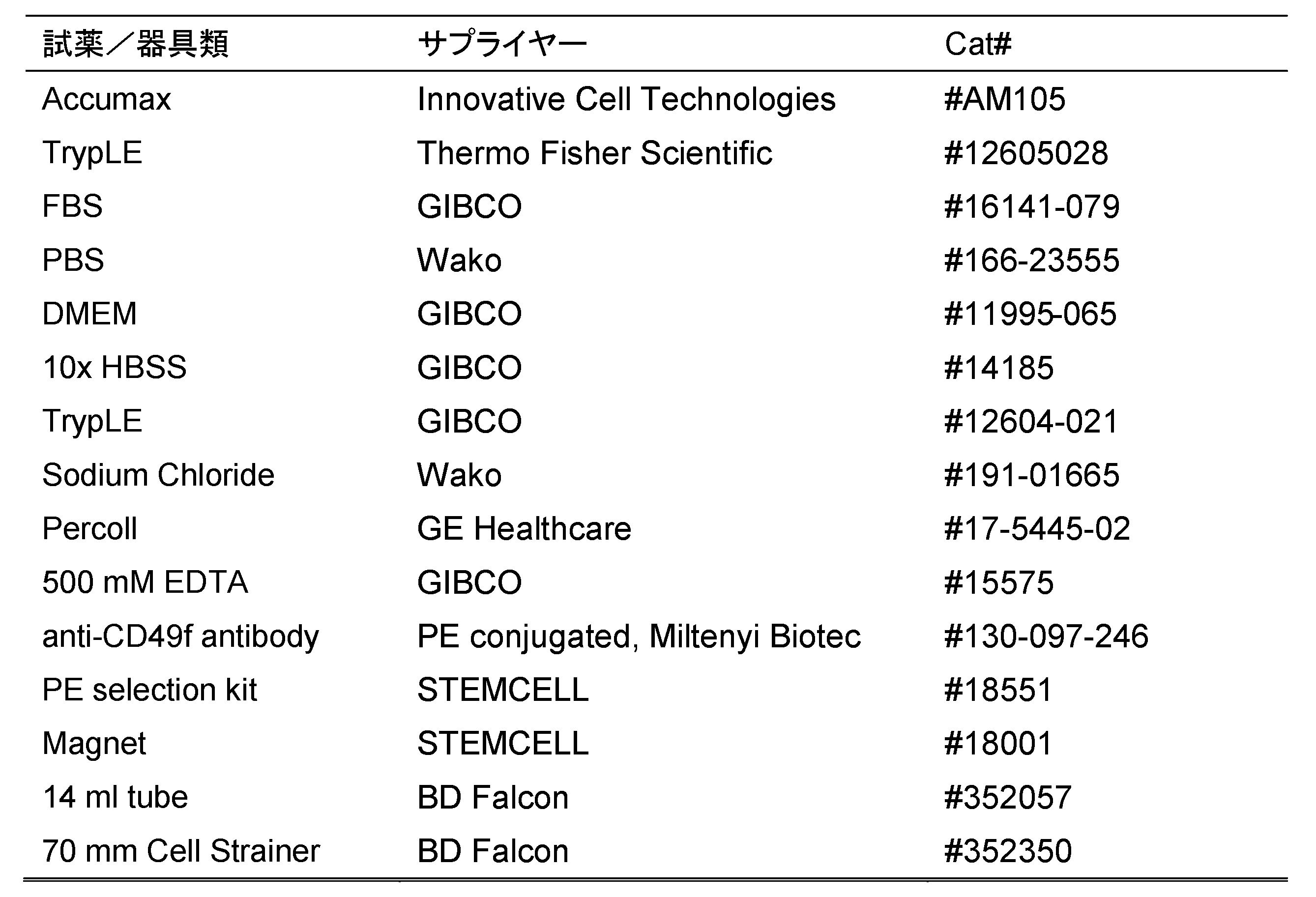
1-2.培地の調製
試薬及び培地を以下のように調整した。
9gの塩化ナトリウムを、1000mLの水に溶解して、生理食塩水(0.9%塩化ナトリウム)を調製した。生理食塩水はオートクレーブし、使用するまで室温で保存した。
196mLのPBS、400μLの500mM EDTA、及び4mLのFBSを混合し、PBS-EDTAを調製した。PBS-EDTAは、使用するまで4℃で保存した。
160mLのPBS、36mLのDMEM、及び4mLのFBSを混合し、PBS-DMEMを調製した。PBS-DMEMは、使用するまで4℃で保存した。
35mLのPercoll、5mLの10xHBSS、及び10mLの水を混合し、70%Percollを調製した。70%Percollは、使用するまで4℃で保存した。
7.5mLのPercoll、5mLの10xHBSS、及び37.5mLの水を混合し、15%Percollを調製した。15%Percollは、使用するまで4℃で保存した。
1-3.CT細胞の単離
以下の手順に従って、CT細胞を単離した。
(1)妊娠20~40週のヒト胎盤の絨毛組織を細かく刻んだ。
(2)細かく刻んだ絨毛組織を生理食塩水で数回洗浄した。
(3)50mLのAccumax及び50mLのTrypLEを添加し、37℃で0分間インキュベートした。
(4)消化された組織を、70μmのセルストレイナーに通した。
(5)得られた流体に、20mLのPBS-DMEMを添加した。
(6)1,400rpm、5分間遠心分離した。
(7)細胞ペレットに5mLのPBS-DMEMを添加し、4℃に維持した。
(8)(4)~(7)を繰り返した。
(9)細胞懸濁液(総量10mL)を70μmのセルストレイナーに通した。
(10)1,400rpmで5分間遠心分離した。
(11)細胞ペレットに1mLのPBS-DMEMを添加した。
(12)Percoll濃度勾配(15/70%)上に細胞懸濁液を積層した。
(13)1,200gで20分間遠心分離した。
(14)15%から70%の間のPercoll界面の細胞を回収した。
(15)細胞に20mLのPBS-EMEMを添加した。
(16)1,400rpmで5分間遠心分離した。
(17)細胞ペレットに10mLのPBSを添加した。
(18)1,400rpmで5分間遠心分離した。
(19)細胞ペレットに1mLのPBS-EDTAを添加して細胞を懸濁し、細胞懸濁液を14mLチューブに移した。
(20)30μLの抗CD49f抗体を添加した。
(21)光から保護しながら、室温で10分間、細胞懸濁液をインキュベートした。
(22)30μLのPE-selection cocktailを添加した。
(23)細胞懸濁液を室温で10分間インキュベートした。
(24)20μLのMagnetic particleを添加した。
(25)細胞懸濁液を室温で10分間インキュベートした。
(26)PBS-EDTAを添加して、液量を10mLとした。
(27)磁石内に14mLチューブを置き、室温で7分間静置した。
(28)磁石を反転させて、チューブ内の培地を流し出した。
(29)10mLのPBS-EDTAを14mLチューブに添加した。
(30)(27)~(29)を3回繰り返した。
(31)1,400rpmで5分間遠心分離し、CT細胞を回収した。
2.TS様細胞の作製
2-1.試薬類
試験に用いた試薬類を表2に示す。
2-1.試薬類
試験に用いた試薬類を表2に示す。
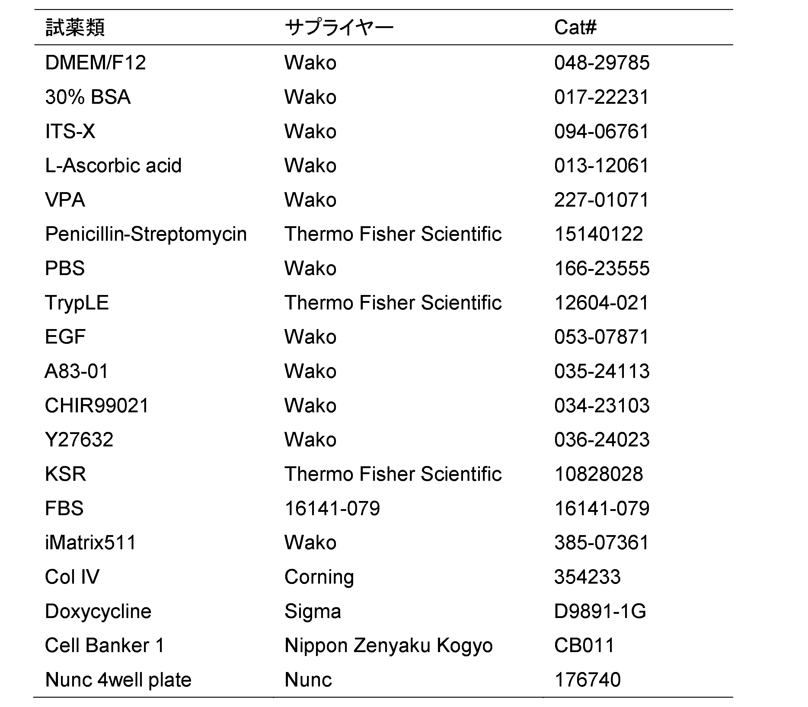
2-2.培地の調製
培地に用いる各試薬を以下のように調整した。
10mgのA83-01を、2.37mLのジメチルスルホキシド(DMSO)に溶解して、10mM A83-01を調製した。10mM A83-01は、使用するまで-20℃で保存した。
5mgのCHIR99021を、2.69mLのDMSOに溶解して、4mM CHIR99021を調製した。4mM CHIR99021は、使用するまで-20℃で保存した。
5mgのY27632を、1.48mLの滅菌水に溶解して、10mM Y27632を調製した。5mgのY27632は、使用するまで-20℃で保存した。
0.58gのL-アスコルビン酸(L-Ascorbic acid)を、10mLの水に溶解して、200mM L-アスコルビン酸を調製した。200mM L-アスコルビン酸は、フィルター濾過後、使用するまで-20℃で保存した。
500μgのEGFを、5mLのPBS/0.2%BSAに溶解して、100μg/mL EGFを調製した。100μg/mL EGFは、使用するまで-20℃で保存した。
1mgのDoxycycline(Dox)を、10mLの水に溶解して、100μg/mL Doxを調製した。100μg/mL Doxは、フィルター濾過後、使用するまで-20℃で保存した。
表3に示す組成で、Basal培地を調製した。Basal培地は、4℃で保存し、調製後2週間以内に使用した。

表4に示す組成で、Term-1培地を調製した。Term-1培地は、4℃で保存し、調製後2週間以内に使用した。
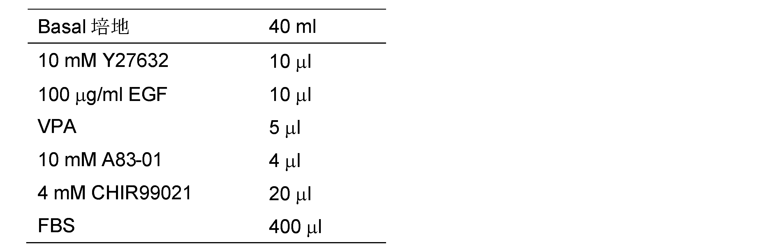
表5に示す組成で、Term-2培地を調製した。Term-2培地は、4℃で保存し、調製後2週間以内に使用した。
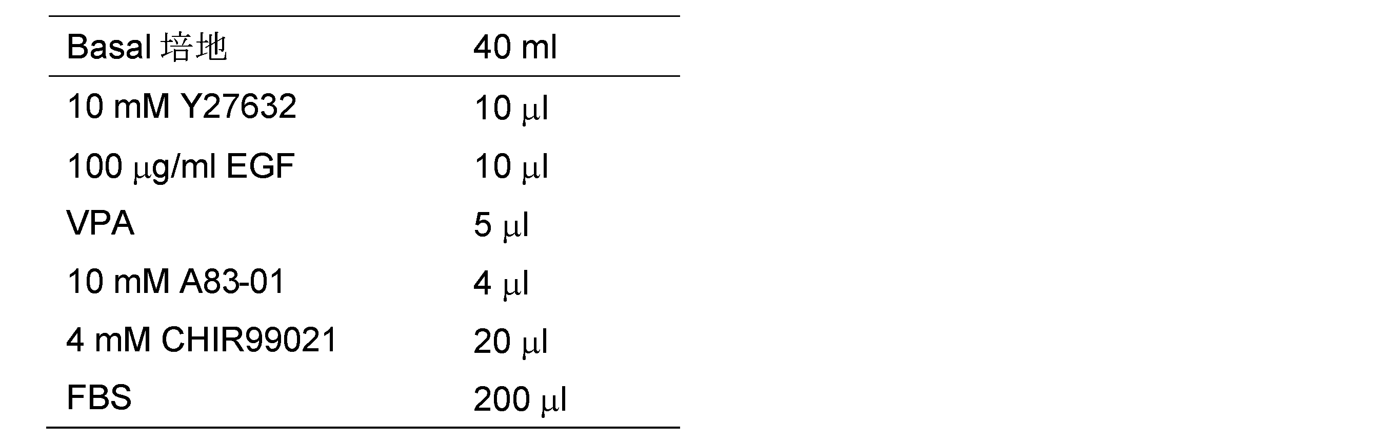
2-3.Dox誘導性遺伝子導入用レンチウイルスベクターの構築
Takahashiら(Takahashi et al., Proc Natl Acad Sci U S A. 2019 Dec 2;116(52):26606-26613.)に記載の方法に従って、Dox誘導性遺伝子導入用レンチベクターを構築した。具体的な方法を以下に示す。
(1)pTetOneベクター(Takara)のTet-On 3GトランスアクチベーターをPCR増幅し、CS-CA-MCSプラスミド(図1;理研バイオリソースセンター(茨城、日本)から譲渡された)のマルチクローニングサイトに、In-Fusion HD Cloning kit(Takara)を用いてクローニングし、pCS-CA-Tet3Gベクターを作製した。
(2)CS-CA-MCSプラスミドのCAGプロモーターをpTetOneベクターのTRE3Gsプロモーターで置き換えて、pCS-3Gベクターを作製した。
(3)表6に示すヒト遺伝子のcDNAを、pCS-3Gベクターのマルチクローニングサイトに挿入し、Dox誘導性遺伝子導入用レンチウイルスベクターとして用いた。表6に示す32種の遺伝子のDox誘導性遺伝子用レンチウイルスベクターをそれぞれ作製した。

p53ドミナントネガティブ(p53DN)のcDNAは、T7-p53-pcDNA3(addgene)からPCR増幅したものを用いた。T7-p53-pcDNA3が含むp53DN cDNAは、p53(AB082923)のtruncatedタンパク質(300~393位のアミノ酸配列)をコードする。
2-4.TS様細胞の作製
操作のタイムスケジュールの概略を図2に示した。TS様細胞は、以下の手順に従って作製した。
(1)5μLのCol IV及び1μLのiMatrix511を含む1mLのPBSを各ウェルに入れ、37℃で1時間静置し、各ウェルをコートした。
(2)PBS/Col IV/iMatrix511を吸引除去した。
(3)PBSでウェルを洗浄した。
(4)PBSを吸引除去した。
(5)Term-1培地をウェルに添加し、37℃で15分以上保持した。
(6)Term-1培地に~5x10⁵個のCT細胞(「1.妊娠中期以降の胎盤からのCT細胞の単離」で単離したもの)を懸濁し(Day0)、37℃で培養した。
(7)培養2日目(Day2)に、Term-1培地を吸引除去し、2mLの予め37℃に温めたTerm-2培地を添加した。
(8)レンチウイルス粒子(5μLのpCS-CA-Tet3Gベクター、3~5μLのDox誘導性遺伝子導入用レンチウイルスベクター)を添加した。
(9)培養3日目(Day3)に、2μLの100μg/mL Doxを添加した。
(10)3日毎に培地を交換し、Doxを含むTerm-2培地で培養を継続した。
(11)培養21日目(Day21)に、Doxを含まないTerm-2培地に交換し、培養を継続した。
3.TS様細胞作製に必要な遺伝子の検討
妊娠中期以降の胎盤由来のCT細胞(中・後期CT細胞)に導入することにより、TS様細胞を樹立可能な遺伝子を検討した。表6に記載の遺伝子を検討した結果を表7に示す。表7中、「Yes」は、TS様細胞が樹立できたことを示し、「No」はTS様細胞を樹立できなかったことを示す。
妊娠中期以降の胎盤由来のCT細胞(中・後期CT細胞)に導入することにより、TS様細胞を樹立可能な遺伝子を検討した。表6に記載の遺伝子を検討した結果を表7に示す。表7中、「Yes」は、TS様細胞が樹立できたことを示し、「No」はTS様細胞を樹立できなかったことを示す。
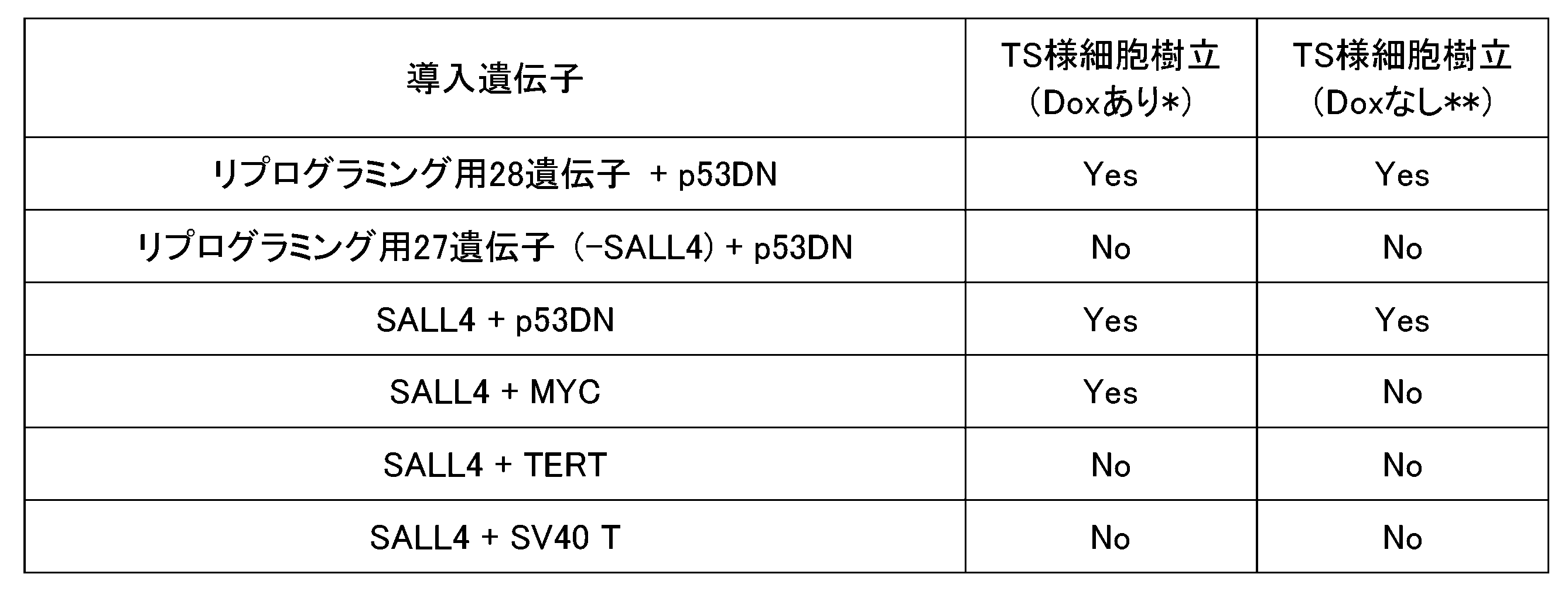
表7の結果から、Dox存在下で培養を続けた場合、SALL4+p53DN及びSALL4+MYCが、TS様細胞を樹立可能な遺伝子の組み合わせとして見出された。これらの中でも、SALL4+p53DNを用いた場合、培養21日目からDox非存在下で培養しても、TS様細胞が維持された(図3参照)。一方、SALL4+MYCを用いた場合、Dox非存在下ではTS様細胞を維持できなかった(図4参照:SALL4+MYCの場合を示す)。この結果から、SALL4+MYCの組合せで中・後期CT細胞から誘導されたTS様細胞は、その維持に、SALL4+MYCが発現し続ける必要があることを示す。一方、SALL4+p53DNの組合せで誘導されたTS細胞は、その維持に、SALL4+p53DNが発現し続ける必要がないことを示す。
4.TS様細胞の遺伝子発現解析
4-1.試薬類及び機器類
試験に用いた試薬類及び機器類を以下に示す。
RNeasy mini kit(Qiagen,Cat#:74104)
TruSeq RNA Sample Prep Kit(Illumina,Cat#:RS-122-2001)
次世代シークエンサー(IlluminaのHiSeq2500を使用)
次世代シークエンサー解析ソフト Strand NGS(Digital Biology)
4-1.試薬類及び機器類
試験に用いた試薬類及び機器類を以下に示す。
RNeasy mini kit(Qiagen,Cat#:74104)
TruSeq RNA Sample Prep Kit(Illumina,Cat#:RS-122-2001)
次世代シークエンサー(IlluminaのHiSeq2500を使用)
次世代シークエンサー解析ソフト Strand NGS(Digital Biology)
4-2.遺伝子発現解析
遺伝子発現解析を行い、中・後期CT細胞から誘導されたTS様細胞(中・後期TS様細胞)、初期胎盤由来のCT細胞から誘導されたTS様細胞(初期TS様細胞)、TS細胞、中・後期CT細胞、及び初期CT細胞の類似性を検証した。
中・後期TS様細胞は、中・後期CT細胞にSALL4+p53DNを導入して作製したものを用いた。解析には、培養21日目以降のDox非含有培地で培養されている細胞を用いた(図2参照)。
初期TS様細胞は、特許第6400832号に記載の方法に従って作製した。
また、中・後期CT細胞を、特許第6400832号に記載の方法(初期TS様細胞の誘導方法)と同様の方法で処理した細胞についても遺伝子発現解析を行った。
以下の手法に従い、遺伝子発現解析を行った。
(1)RNeasy mini kitを用いて、細胞から全RNAを抽出した。
(2)TruSeq RNA Sample Prep Kitを用いて、次世代シークエンサー用ライブラリーを作製した。
(3)次世代シークエンサーを用い、シークエンス解析を行った。
(4)次世代シークエンサー解析ソフト Strand NGSを用い、遺伝子の発現量(TPM値:transcripts per million)を計算した。
(1)RNeasy mini kitを用いて、細胞から全RNAを抽出した。
(2)TruSeq RNA Sample Prep Kitを用いて、次世代シークエンサー用ライブラリーを作製した。
(3)次世代シークエンサーを用い、シークエンス解析を行った。
(4)次世代シークエンサー解析ソフト Strand NGSを用い、遺伝子の発現量(TPM値:transcripts per million)を計算した。
4-3.主成分分析
次世代シークエンサーで解析された遺伝子発現量に基づき、Rを用いて主成分分析を行った。その結果を図5に示す。
図5に示すように、中・後期TS様細胞は、TS細胞及び初期TS様細胞と類似の発現プロファイルを示すことが確認された。一方、中・後期CT細胞を、初期CT細胞と同様の方法で誘導した場合には、TS細胞とは異なる発現プロファイルを示した。
次世代シークエンサーで解析された遺伝子発現量に基づき、Rを用いて主成分分析を行った。その結果を図5に示す。
図5に示すように、中・後期TS様細胞は、TS細胞及び初期TS様細胞と類似の発現プロファイルを示すことが確認された。一方、中・後期CT細胞を、初期CT細胞と同様の方法で誘導した場合には、TS細胞とは異なる発現プロファイルを示した。
4-4.TS細胞マーカーの発現解析
初期TS様細胞及び中・後期TS様細胞について、ELF5、ZNF750、及びCDX2の発現を確認した。ELF5及びZNF750はTS細胞の陽性マーカーであり、CDX2はTS細胞の陰性マーカーである。
その結果を図6に示す。初期TS様細胞では1クローン(初期TS)を解析し、中・後期TS様細胞では8クローン(中・後期TS-1~8)を解析した。初期TS様細胞、及び中・後期TS様細胞のいずれのクローンも、ELF5及びZNF750陽性であり、CDX2陰性であった。この結果は、中・後期TS様細胞が、TS細胞に類似していることを示す。
初期TS様細胞及び中・後期TS様細胞について、ELF5、ZNF750、及びCDX2の発現を確認した。ELF5及びZNF750はTS細胞の陽性マーカーであり、CDX2はTS細胞の陰性マーカーである。
その結果を図6に示す。初期TS様細胞では1クローン(初期TS)を解析し、中・後期TS様細胞では8クローン(中・後期TS-1~8)を解析した。初期TS様細胞、及び中・後期TS様細胞のいずれのクローンも、ELF5及びZNF750陽性であり、CDX2陰性であった。この結果は、中・後期TS様細胞が、TS細胞に類似していることを示す。
5.EVTまたはSTへの分化誘導
中・後期TS様細胞が、胎盤を構成する細胞への分化能を有していることを確認した。TS細胞は、TGF-βの濃度が低く、NRG1及びマトリゲルの濃度が高い環境下で、上皮間葉転換し、絨毛外栄養膜細胞(extravillous cytotrophoblast:EVT)に分化誘導される(図7A参照)。また、TS細胞は、cAMPの濃度が高い環境下で、細胞誘導し、合胞体栄養膜細胞(syncytiotrophoblast:ST)に分化誘導される(図7A参照)。前記のような環境下で培養することにより、中・後期TS様細胞もTS細胞と同様に、EVT及びSTに分化誘導されるかを確認した。
中・後期TS様細胞が、胎盤を構成する細胞への分化能を有していることを確認した。TS細胞は、TGF-βの濃度が低く、NRG1及びマトリゲルの濃度が高い環境下で、上皮間葉転換し、絨毛外栄養膜細胞(extravillous cytotrophoblast:EVT)に分化誘導される(図7A参照)。また、TS細胞は、cAMPの濃度が高い環境下で、細胞誘導し、合胞体栄養膜細胞(syncytiotrophoblast:ST)に分化誘導される(図7A参照)。前記のような環境下で培養することにより、中・後期TS様細胞もTS細胞と同様に、EVT及びSTに分化誘導されるかを確認した。
分化誘導のための中・後期TS様細胞として、中・後期CT細胞にSALL4+p53DNを導入して作製した中・後期TS様細胞を用いた。また、培養21日目以降のDox非含有培地で培養されている細胞を用いた(図2参照)。
5-1.EVTへの分化誘導
5-1-1.EVT培地の調製
DMEM/F12(048-29785; Fujifilm Wako, Osaka, Japan)に、下記成分を添加して、EVT培地を調製した。
0.5% Penicillin-Streptomycin(Thermo Fisher Scientific)
0.3% BSA(Fujifilm Wako)
1% ITS-X supplement(Fujifilm Wako)
100 ng/ml NRG1(5218SC; Cell Signaling)
7.5 μM A83-01(Fujifilm Wako)
2.5 μM Y27632(Fujifilm Wako)
4% KSR(Thermo Fisher Scientific)
5-1-2.EVTへの分化誘導
以下の手順に従い、中・後期TS様細胞からEVTへの分化誘導を行った。
(1)中・後期TS様細胞を、EVT培地を含む1μg/mLのCol IVコーティングプレート(Corning)上に、8,000細胞/cm2の密度で播種した。
(2)マトリゲル(Corning)を培地体積の2%で添加した。
(3)播種後3日目に、培地を、NRG1(Cell Signaling)を含まないEVT培地と交換し、マトリゲル(Corning)を培地体積の0.5%で添加した。
(4)播種後6日目に、培地を、NRG1(Cell Signaling)およびKSR(Thermo Fisher Scientific)を含まないEVT培地と交換し、マトリゲル(Corning)を培地体積の0.5%で添加した。
(5)前記培地交換後6~8日目に、細胞の顕微鏡観察を行った。
5-1-3.結果
分化誘導後の細胞の顕微鏡写真を図7Bに示す。顕微鏡観察の結果、中・後期TS様細胞は、EVTに分化誘導されていることが確認された。
分化誘導後の細胞の顕微鏡写真を図7Bに示す。顕微鏡観察の結果、中・後期TS様細胞は、EVTに分化誘導されていることが確認された。
5-2.STへの分化誘導
5-2-1.ST培地の調製
DMEM/F12(048-29785; Fujifilm Wako, Osaka, Japan)に、下記成分を添加して、ST培地を調製した。
0.1 mM 2-mercaptoethanol(Thermo Fisher Scientific)
0.5% Penicillin-Streptomycin(Thermo Fisher Scientific)
0.3% BSA(Fujifilm Wako)
1% ITS-X supplement(Fujifilm Wako)
2.5μM Y27632(Fujifilm Wako)
2μM forskolin(Fujifilm Wako)
4% KSR(Thermo Fisher Scientific)
5-2-2.STへの分化誘導
以下の手順に従い、中・後期TS様細胞からSTへの分化誘導を行った。
(1)中・後期TS様細胞を、ST培地を含む2.5μg/mLのCol IVコーティングプレート(Corning)上に、10,000細胞/cm2の密度で播種した。
(2)播種後3日目に、ST培地を交換した。
(3)播種後6日目に、細胞の顕微鏡観察を行った。
以下の手順に従い、中・後期TS様細胞からSTへの分化誘導を行った。
(1)中・後期TS様細胞を、ST培地を含む2.5μg/mLのCol IVコーティングプレート(Corning)上に、10,000細胞/cm2の密度で播種した。
(2)播種後3日目に、ST培地を交換した。
(3)播種後6日目に、細胞の顕微鏡観察を行った。
5-2-3.結果
分化誘導後の細胞の顕微鏡写真を図7Cに示す。顕微鏡観察の結果、中・後期TS様細胞は、STに分化誘導されていることが確認された。
分化誘導後の細胞の顕微鏡写真を図7Cに示す。顕微鏡観察の結果、中・後期TS様細胞は、STに分化誘導されていることが確認された。
6.初期CT細胞及び中・後期CT細胞を区別するマーカー
初期CT細胞及び中・後期CT細胞について、上記「4-2.遺伝子発現解析」と同様の手法で遺伝子発現解析を行い、初期CT細胞及び中・後期CT細胞を区別するマーカーを検討した。
初期CT細胞及び中・後期CT細胞について、上記「4-2.遺伝子発現解析」と同様の手法で遺伝子発現解析を行い、初期CT細胞及び中・後期CT細胞を区別するマーカーを検討した。
その結果、初期CT細胞で発現し、中・後期CT細胞で発現しない遺伝子として、CCR7が見出された。図8に、初期CT細胞の3クローン(初期-1、初期-2、初期-3)、及び中・後期CT細胞の3クローン(中・後期-1、中・後期-2、中・後期-3)におけるCCR7の発現解析結果を示す。CCR7は、初期CT細胞では陽性であり、中・後期CT細胞では陰性であることが確認された。
7.妊娠高血圧症患者の胎盤からのCT細胞の単離
7-1.試薬類及び器具類
試験に用いた試薬類及び器具類は、上記表1の通りである。
7-1.試薬類及び器具類
試験に用いた試薬類及び器具類は、上記表1の通りである。
7-2.培地の調製
試薬及び培地は、「1-2.培地の調製」に記載のとおりに調製した。
試薬及び培地は、「1-2.培地の調製」に記載のとおりに調製した。
7-3.CT細胞の単離
妊娠20~40週の妊娠高血圧症患者のヒト胎盤の絨毛組織を細かく刻んだ。その後の単離工程は、「1-3.CT細胞の単離」の(2)~(31)までと同様に行った。得られたCT細胞を以下「疾患CT細胞」とも記載する。また、健常人のヒト胎盤からも同様にCT細胞(以下、「健常人CT細胞」ともいう)を単離した。
妊娠20~40週の妊娠高血圧症患者のヒト胎盤の絨毛組織を細かく刻んだ。その後の単離工程は、「1-3.CT細胞の単離」の(2)~(31)までと同様に行った。得られたCT細胞を以下「疾患CT細胞」とも記載する。また、健常人のヒト胎盤からも同様にCT細胞(以下、「健常人CT細胞」ともいう)を単離した。
8.疾患CT細胞からのTS様細胞の作製
8-1.試薬類
試験に用いた試薬類は、上記表2のとおりである。
8-1.試薬類
試験に用いた試薬類は、上記表2のとおりである。
8-2.培地の調製
培地に用いる各試薬は、「2-2.培地の調製」に記載のとおりに調製した。Basal培地の調製は、上記表3のとおりに行い、4℃で保存し、調製後2週間以内に使用した。Term-1培地の調製は、上記表4のとおりに行い、4℃で保存し、調製後2週間以内に使用した。Term-2培地の調製は、上記表5のとおりに行い、4℃で保存し、調製後2週間以内に使用した。
培地に用いる各試薬は、「2-2.培地の調製」に記載のとおりに調製した。Basal培地の調製は、上記表3のとおりに行い、4℃で保存し、調製後2週間以内に使用した。Term-1培地の調製は、上記表4のとおりに行い、4℃で保存し、調製後2週間以内に使用した。Term-2培地の調製は、上記表5のとおりに行い、4℃で保存し、調製後2週間以内に使用した。
8-3.Dox誘導性遺伝子導入用レンチウイルスベクターの構築
「2-3.Dox誘導性遺伝子導入用レンチウイルスベクターの構築」に記載のとおりに、Dox誘導性遺伝子導入用レンチベクターを構築した。pCS-3Gベクターのマルチクローニングサイトに挿入する遺伝子としては、リプログラミング用遺伝子としてSALL4、細胞増殖用としてp53DNを選択した。
「2-3.Dox誘導性遺伝子導入用レンチウイルスベクターの構築」に記載のとおりに、Dox誘導性遺伝子導入用レンチベクターを構築した。pCS-3Gベクターのマルチクローニングサイトに挿入する遺伝子としては、リプログラミング用遺伝子としてSALL4、細胞増殖用としてp53DNを選択した。
8-4.疾患CT細胞からのTS様細胞(疾患CT細胞由来TS細胞)の作製
疾患CT細胞を用いたこと以外は、「2-4.TS様細胞の作製」と同様に、TS様細胞を作製した。また、健常人CT細胞からも同様にTS様細胞を作製した。
疾患CT細胞を用いたこと以外は、「2-4.TS様細胞の作製」と同様に、TS様細胞を作製した。また、健常人CT細胞からも同様にTS様細胞を作製した。
9.疾患CT細胞由来TS様細胞の遺伝子発現解析
9-1.試薬類及び機器類
試験に用いた試薬類及び機器類は、「4-1.試薬類及び機器類」に記載の通りである。
9-1.試薬類及び機器類
試験に用いた試薬類及び機器類は、「4-1.試薬類及び機器類」に記載の通りである。
9-2.遺伝子発現解析
「4-2.遺伝子発現解析」に記載の手順に従い、遺伝子発現解析を行った。
「4-2.遺伝子発現解析」に記載の手順に従い、遺伝子発現解析を行った。
9-3.TS細胞マーカーの発現解析
疾患CT細胞由来TS様細胞について、ELF5、ZNF750、及びCDX2の発現を確認した。また、健常人CT細胞由来TS様細胞についても同様に、ELF5、ZNF750、及びCDX2の発現を確認した。
その結果を図9に示す。疾患CT細胞由来TS様細胞3クローン、健常人CT細胞由来TS様細胞4クローンをそれぞれ解析した。いずれのクローンも、ELF5及びZNF750陽性であり、CDX2陰性であった。この結果は、疾患CT細胞由来TS様細胞が、TS細胞に類似していることを示す。
疾患CT細胞由来TS様細胞について、ELF5、ZNF750、及びCDX2の発現を確認した。また、健常人CT細胞由来TS様細胞についても同様に、ELF5、ZNF750、及びCDX2の発現を確認した。
その結果を図9に示す。疾患CT細胞由来TS様細胞3クローン、健常人CT細胞由来TS様細胞4クローンをそれぞれ解析した。いずれのクローンも、ELF5及びZNF750陽性であり、CDX2陰性であった。この結果は、疾患CT細胞由来TS様細胞が、TS細胞に類似していることを示す。
本発明によれば、妊娠中期以降の胎盤に由来する栄養膜細胞からTS様細胞を製造する方法、および前記製造方法により製造されるTS様細胞が提供される。本発明により提供されるTS様細胞は、哺乳動物の初期発生研究、胎盤機能解析研究、及び妊娠に関連する疾患の病態研究若しくは治療法開発に利用することができる。また、再生医療への応用も可能である。
Claims (9)
- 胎盤を構成する細胞への分化能を有する栄養膜幹細胞様細胞であって、
妊娠中期以降の胎盤由来の栄養膜細胞から誘導されたものであり、
第1の外因性の誘導性プロモーターに機能的に連結されたSALL4遺伝子を含む、
栄養膜幹細胞様細胞。 - 前記SALL4遺伝子が、
前記第1の外因性の誘導性プロモーターに機能的に連結された外因性SALL4遺伝子であるか、又は
前記第1の外因性の誘導性プロモーターに機能的に連結された内因性SALL4遺伝子である、
請求項1に記載の栄養膜幹細胞様細胞。 - 下記(a)及び(b)からなる群より選択される少なくとも一つの特徴を有する、請求項1又は2に記載の栄養膜幹細胞様細胞:
(a)前記栄養膜細胞と比較してp53の活性が抑制されている;及び
(b)前記栄養膜細胞と比較してMYC遺伝子の発現が促進されている。 - 第2の外因性の誘導性プロモーターに機能的に連結された外因性の遺伝子をさらに含み、前記外因性の遺伝子が、外因性p53ドミナントネガティブ遺伝子、及び外因性MYC遺伝子からなる群より選択される少なくとも1種である、請求項1~3のいずれか一項に記載の栄養膜幹細胞様細胞。
- 前記外因性の遺伝子が、前記外因性p53ドミナントネガティブ遺伝子である、請求項4に記載の栄養膜幹細胞様細胞。
- 胎盤を構成する細胞への分化能を有する栄養膜幹細胞様細胞の製造方法であって、
(i)妊娠中期以降の胎盤由来の栄養膜細胞を準備する工程と、
(ii)前記栄養膜細胞のSALL4遺伝子の発現を誘導する工程と、
(iii)下記(A)及び(B)からなる群より選択される少なくとも1つを行う工程と:
(A)前記栄養膜細胞のp53の活性を抑制する;及び
(B)前記栄養膜細胞のMYC遺伝子の発現を誘導する、
を含む、栄養膜幹細胞様細胞の製造方法。 - 前記(ii)の工程が、
前記栄養膜細胞に、
第1の外因性の誘導性プロモーターに機能的に連結された外因性SALL4遺伝子を含むポリヌクレオチドを導入すること、又は
前記第1の外因性の誘導性プロモーターを含むポリヌクレオチドを、内因性SALL4遺伝子の上流に、前記内因性SALL4遺伝子が前記第1の外因性の誘導性プロモーターに機能的に連結するように導入すること、並びに
前記第1の外因性の誘導性プロモーターの転写活性を誘導する第1の誘導因子により、前記外因性SALL4遺伝子若しくは前記内因性SALL4遺伝子の発現を誘導すること、
を含む、請求項6に記載の栄養膜幹細胞様細胞の製造方法。 - 前記(iii)の工程の前記(A)が、
前記栄養膜細胞に、
第2の外因性の誘導性プロモーターに機能的に連結された外因性p53ドミナントネガティブ遺伝子を含むポリヌクレオチドを導入すること、並びに
前記第2の外因性の誘導性プロモーターの転写活性を誘導する第2の誘導因子により、前記外因性p53ドミナントネガティブ遺伝子の発現を誘導すること、
を含み、
前記(iii)の工程の前記(B)が、
前記栄養膜細胞に、
前記第2の外因性の誘導性プロモーターに機能的に連結された外因性MYC遺伝子を導入すること、並びに
前記第2の外因性の誘導性プロモーターの転写活性を誘導する第2の誘導因子により、前記外因性MYC遺伝子の発現を誘導すること、
を含む、
請求項7に記載の栄養膜幹細胞様細胞の製造方法。 - 前記(iii)の工程が前記(A)を含む、請求項6~8のいずれか一項に記載の栄養膜幹細胞様細胞の製造方法。
Priority Applications (4)
| Application Number | Priority Date | Filing Date | Title |
|---|---|---|---|
| JP2022536084A JP7670356B2 (ja) | 2020-07-17 | 2020-07-17 | 胎盤を構成する細胞への分化能を有する栄養膜幹細胞様細胞、及びその製造方法 |
| EP20945461.0A EP4183873A4 (en) | 2020-07-17 | 2020-07-17 | TROPHOBLAST STEM CELL-LIKE CELLS FOR DIFFERENTIATION INTO PLACENTA-CONSTITUTING CELLS AND METHODS FOR THE PRODUCTION THEREOF |
| PCT/JP2020/027766 WO2022014028A1 (ja) | 2020-07-17 | 2020-07-17 | 胎盤を構成する細胞への分化能を有する栄養膜幹細胞様細胞、及びその製造方法 |
| US18/005,511 US20230313130A1 (en) | 2020-07-17 | 2020-07-17 | Trophoblast stem cell-like cells capable of differentiating into placenta-constituting cells, and method for producing same |
Applications Claiming Priority (1)
| Application Number | Priority Date | Filing Date | Title |
|---|---|---|---|
| PCT/JP2020/027766 WO2022014028A1 (ja) | 2020-07-17 | 2020-07-17 | 胎盤を構成する細胞への分化能を有する栄養膜幹細胞様細胞、及びその製造方法 |
Publications (1)
| Publication Number | Publication Date |
|---|---|
| WO2022014028A1 true WO2022014028A1 (ja) | 2022-01-20 |
Family
ID=79554547
Family Applications (1)
| Application Number | Title | Priority Date | Filing Date |
|---|---|---|---|
| PCT/JP2020/027766 Ceased WO2022014028A1 (ja) | 2020-07-17 | 2020-07-17 | 胎盤を構成する細胞への分化能を有する栄養膜幹細胞様細胞、及びその製造方法 |
Country Status (4)
| Country | Link |
|---|---|
| US (1) | US20230313130A1 (ja) |
| EP (1) | EP4183873A4 (ja) |
| JP (1) | JP7670356B2 (ja) |
| WO (1) | WO2022014028A1 (ja) |
Cited By (2)
| Publication number | Priority date | Publication date | Assignee | Title |
|---|---|---|---|---|
| WO2024060269A1 (zh) * | 2022-09-25 | 2024-03-28 | 干细胞转化研究中心有限公司 | 哺乳动物源性细胞模型及其在抗衰老药物、天然产物、保健食品、添加剂的筛选和/或制备中的应用 |
| WO2025023316A1 (ja) | 2023-07-26 | 2025-01-30 | 国立大学法人東北大学 | 着床率向上剤及び着床率向上剤の製造方法 |
Citations (1)
| Publication number | Priority date | Publication date | Assignee | Title |
|---|---|---|---|---|
| JPS64832B2 (ja) | 1983-10-12 | 1989-01-09 | Mitsubishi Electric Corp |
Family Cites Families (3)
| Publication number | Priority date | Publication date | Assignee | Title |
|---|---|---|---|---|
| EP2599859A1 (en) * | 2011-11-30 | 2013-06-05 | IMBA-Institut für Molekulare Biotechnologie GmbH | Haploid cells |
| WO2016005985A2 (en) * | 2014-07-09 | 2016-01-14 | Yissum Research Development Company Of The Hebrew University Of Jerusalem Ltd. | Method for reprogramming cells |
| EP3447130A4 (en) * | 2016-04-22 | 2019-11-13 | Kyoto University | METHOD FOR PRODUCING DOPAMINE-PROPERING NEURAL PRESERVATOR CELLS |
-
2020
- 2020-07-17 US US18/005,511 patent/US20230313130A1/en active Pending
- 2020-07-17 WO PCT/JP2020/027766 patent/WO2022014028A1/ja not_active Ceased
- 2020-07-17 EP EP20945461.0A patent/EP4183873A4/en active Pending
- 2020-07-17 JP JP2022536084A patent/JP7670356B2/ja active Active
Patent Citations (1)
| Publication number | Priority date | Publication date | Assignee | Title |
|---|---|---|---|---|
| JPS64832B2 (ja) | 1983-10-12 | 1989-01-09 | Mitsubishi Electric Corp |
Non-Patent Citations (10)
| Title |
|---|
| "GenBank", Database accession no. NM_001318031.2 |
| "NCBI Gene", Database accession no. 1236 |
| "NCBI", Database accession no. 3135 |
| HIROAKI OKAE, SOTA TAKAHASHI, TAKAHIRO ARIMA: "3AW-18-6 Why are trophoblast tumors rare? A study using human trophoblast stem cells", PROGRAM / ABSTRACTS OF THE 42ND ANNUAL MEETING OF THE MOLECULAR BIOLOGY SOCIETY OF JAPAN; DECEMBER 3-6, 2019Y OF JAPAN, JP, 19 November 2019 (2019-11-19) - 6 December 2019 (2019-12-06), JP, pages 3AW-18-6, XP009534367 * |
| KIO KOBAYASHI, HIROAKI OKAE, SOTA TAKAHASHI, HITOSHI HIURA, TAKAHIRO ARIMA: "P-0326 Transdifferentiation of human embryonic stem cells to trophoblast stem cells", PROGRAM / ABSTRACTS OF THE 42ND ANNUAL MEETING OF THE MOLECULAR BIOLOGY SOCIETY OF JAPAN; DECEMBER 3-6, 2019Y OF JAPAN, JP, 19 November 2019 (2019-11-19) - 6 December 2019 (2019-12-06), JP, pages P-0326, XP009534368 * |
| OKAE H ET AL.: "Derivation of Human Trophoblast Stem Cells", CELL STEM CELL, vol. 22, January 2018 (2018-01-01), pages 50 - 63 |
| OKAE HIROAKI: "Creation of undifferentiated placental stem cell model of human disease and elucidation of pathophysiology", THE UEHARA MEMORIAL FOUNDATION RESEARCH REPORT COLLECTION, 4 February 2020 (2020-02-04), XP055898068, Retrieved from the Internet <URL:https://www.ueharazaidan.or.jp/houkokushu/Vol.33/pdf/report/137_report.pdf> * |
| OKAE HIROAKI; TOH HIDEHIRO; SATO TETSUYA; HIURA HITOSHI; TAKAHASHI SOTA; SHIRANE KENJIRO; KABAYAMA YUKA; SUYAMA MIKITA; SASAKI HIR: "Derivation of Human Trophoblast Stem Cells", CELL STEM CELL, ELSEVIER, CELL PRESS, AMSTERDAM, NL, vol. 22, no. 1, 14 December 2017 (2017-12-14), AMSTERDAM, NL , pages 50, XP085331006, ISSN: 1934-5909, DOI: 10.1016/j.stem.2017.11.004 * |
| See also references of EP4183873A4 |
| TAKAHASHI ET AL., PROC NATL ACAD SCI USA., vol. 116, no. 52, 2 December 2019 (2019-12-02), pages 26606 - 26613 |
Cited By (2)
| Publication number | Priority date | Publication date | Assignee | Title |
|---|---|---|---|---|
| WO2024060269A1 (zh) * | 2022-09-25 | 2024-03-28 | 干细胞转化研究中心有限公司 | 哺乳动物源性细胞模型及其在抗衰老药物、天然产物、保健食品、添加剂的筛选和/或制备中的应用 |
| WO2025023316A1 (ja) | 2023-07-26 | 2025-01-30 | 国立大学法人東北大学 | 着床率向上剤及び着床率向上剤の製造方法 |
Also Published As
| Publication number | Publication date |
|---|---|
| EP4183873A4 (en) | 2024-04-24 |
| US20230313130A1 (en) | 2023-10-05 |
| JPWO2022014028A1 (ja) | 2022-01-20 |
| JP7670356B2 (ja) | 2025-04-30 |
| EP4183873A1 (en) | 2023-05-24 |
Similar Documents
| Publication | Publication Date | Title |
|---|---|---|
| Wang et al. | The p53 family coordinates Wnt and nodal inputs in mesendodermal differentiation of embryonic stem cells | |
| Huang et al. | STAT3 phosphorylation at tyrosine 705 and serine 727 differentially regulates mouse ESC fates | |
| JP4332348B2 (ja) | 細胞の運命を変化させる方法 | |
| Mu et al. | EZH1 in germ cells safeguards the function of PRC2 during spermatogenesis | |
| JP2021530999A (ja) | 免疫改変多能性幹細胞由来のキメラ抗原受容体t細胞 | |
| Le et al. | Enhanced telomere rejuvenation in pluripotent cells reprogrammed via nuclear transfer relative to induced pluripotent stem cells | |
| US20190264223A1 (en) | Novel method | |
| EA011953B1 (ru) | Pdx1-экспрессирующая энтодерма | |
| US20240091274A1 (en) | TRANSPLANTED CELL PROTECTION VIA Fc SEQUESTRATION | |
| JP2004511209A (ja) | 細胞の脱分化及び組織の再生のための組成物及び方法 | |
| CN109996873A (zh) | 移植用细胞群及其制造方法 | |
| CN113226475B (zh) | 改造tcr及其制造方法 | |
| US7838503B2 (en) | Methods for extending the replicative lifespan of cells | |
| US10443043B2 (en) | Methods for making induced pluripotent stem cells | |
| JP7670356B2 (ja) | 胎盤を構成する細胞への分化能を有する栄養膜幹細胞様細胞、及びその製造方法 | |
| Ito et al. | Critical roles of transcriptional coactivator MED1 in the formation and function of mouse adipose tissues | |
| JP2025065482A (ja) | Myclの一過性発現による増殖可能な膵島前駆細胞様細胞の誘導とインスリン陽性細胞への分化誘導 | |
| JP2023544324A (ja) | 人工多能性幹細胞のための改善されたリプログラミング、維持及び保存 | |
| WO2020250438A1 (ja) | 胎盤を構成する細胞への分化能を有する細胞、およびその製造方法 | |
| CN118946660A (zh) | 生成人耳蜗毛细胞的方法 | |
| JPWO2007094301A1 (ja) | 硝子体細胞株 | |
| Kitamura et al. | Identification of human HAPRIN potentially involved in the acrosome reaction | |
| Schätzlein et al. | Telomeres and telomerase in liver generation and cirrhosis | |
| Varga et al. | The GINS complex is required for the survival of rapidly proliferating retinal and tectal progenitor cells during zebrafish development | |
| JP4995654B2 (ja) | クロマチンタンパク質又はその変異体による幹細胞未分化制御方法。 |
Legal Events
| Date | Code | Title | Description |
|---|---|---|---|
| 121 | Ep: the epo has been informed by wipo that ep was designated in this application |
Ref document number: 20945461 Country of ref document: EP Kind code of ref document: A1 |
|
| ENP | Entry into the national phase |
Ref document number: 2022536084 Country of ref document: JP Kind code of ref document: A |
|
| NENP | Non-entry into the national phase |
Ref country code: DE |
|
| ENP | Entry into the national phase |
Ref document number: 2020945461 Country of ref document: EP Effective date: 20230217 |