WO2021187602A1 - Procédé de purification de cardiomyocytes - Google Patents
Procédé de purification de cardiomyocytes Download PDFInfo
- Publication number
- WO2021187602A1 WO2021187602A1 PCT/JP2021/011232 JP2021011232W WO2021187602A1 WO 2021187602 A1 WO2021187602 A1 WO 2021187602A1 JP 2021011232 W JP2021011232 W JP 2021011232W WO 2021187602 A1 WO2021187602 A1 WO 2021187602A1
- Authority
- WO
- WIPO (PCT)
- Prior art keywords
- cells
- cell
- cardiomyocytes
- cell population
- myocardial
- Prior art date
Links
Images


Classifications
-
- A—HUMAN NECESSITIES
- A61—MEDICAL OR VETERINARY SCIENCE; HYGIENE
- A61P—SPECIFIC THERAPEUTIC ACTIVITY OF CHEMICAL COMPOUNDS OR MEDICINAL PREPARATIONS
- A61P9/00—Drugs for disorders of the cardiovascular system
- A61P9/10—Drugs for disorders of the cardiovascular system for treating ischaemic or atherosclerotic diseases, e.g. antianginal drugs, coronary vasodilators, drugs for myocardial infarction, retinopathy, cerebrovascula insufficiency, renal arteriosclerosis
-
- A—HUMAN NECESSITIES
- A61—MEDICAL OR VETERINARY SCIENCE; HYGIENE
- A61K—PREPARATIONS FOR MEDICAL, DENTAL OR TOILETRY PURPOSES
- A61K35/00—Medicinal preparations containing materials or reaction products thereof with undetermined constitution
- A61K35/12—Materials from mammals; Compositions comprising non-specified tissues or cells; Compositions comprising non-embryonic stem cells; Genetically modified cells
- A61K35/34—Muscles; Smooth muscle cells; Heart; Cardiac stem cells; Myoblasts; Myocytes; Cardiomyocytes
-
- A—HUMAN NECESSITIES
- A61—MEDICAL OR VETERINARY SCIENCE; HYGIENE
- A61L—METHODS OR APPARATUS FOR STERILISING MATERIALS OR OBJECTS IN GENERAL; DISINFECTION, STERILISATION OR DEODORISATION OF AIR; CHEMICAL ASPECTS OF BANDAGES, DRESSINGS, ABSORBENT PADS OR SURGICAL ARTICLES; MATERIALS FOR BANDAGES, DRESSINGS, ABSORBENT PADS OR SURGICAL ARTICLES
- A61L27/00—Materials for grafts or prostheses or for coating grafts or prostheses
- A61L27/36—Materials for grafts or prostheses or for coating grafts or prostheses containing ingredients of undetermined constitution or reaction products thereof, e.g. transplant tissue, natural bone, extracellular matrix
- A61L27/38—Materials for grafts or prostheses or for coating grafts or prostheses containing ingredients of undetermined constitution or reaction products thereof, e.g. transplant tissue, natural bone, extracellular matrix containing added animal cells
- A61L27/3804—Materials for grafts or prostheses or for coating grafts or prostheses containing ingredients of undetermined constitution or reaction products thereof, e.g. transplant tissue, natural bone, extracellular matrix containing added animal cells characterised by specific cells or progenitors thereof, e.g. fibroblasts, connective tissue cells, kidney cells
- A61L27/3834—Cells able to produce different cell types, e.g. hematopoietic stem cells, mesenchymal stem cells, marrow stromal cells, embryonic stem cells
-
- A—HUMAN NECESSITIES
- A61—MEDICAL OR VETERINARY SCIENCE; HYGIENE
- A61L—METHODS OR APPARATUS FOR STERILISING MATERIALS OR OBJECTS IN GENERAL; DISINFECTION, STERILISATION OR DEODORISATION OF AIR; CHEMICAL ASPECTS OF BANDAGES, DRESSINGS, ABSORBENT PADS OR SURGICAL ARTICLES; MATERIALS FOR BANDAGES, DRESSINGS, ABSORBENT PADS OR SURGICAL ARTICLES
- A61L27/00—Materials for grafts or prostheses or for coating grafts or prostheses
- A61L27/36—Materials for grafts or prostheses or for coating grafts or prostheses containing ingredients of undetermined constitution or reaction products thereof, e.g. transplant tissue, natural bone, extracellular matrix
- A61L27/38—Materials for grafts or prostheses or for coating grafts or prostheses containing ingredients of undetermined constitution or reaction products thereof, e.g. transplant tissue, natural bone, extracellular matrix containing added animal cells
- A61L27/3895—Materials for grafts or prostheses or for coating grafts or prostheses containing ingredients of undetermined constitution or reaction products thereof, e.g. transplant tissue, natural bone, extracellular matrix containing added animal cells using specific culture conditions, e.g. stimulating differentiation of stem cells, pulsatile flow conditions
-
- C—CHEMISTRY; METALLURGY
- C12—BIOCHEMISTRY; BEER; SPIRITS; WINE; VINEGAR; MICROBIOLOGY; ENZYMOLOGY; MUTATION OR GENETIC ENGINEERING
- C12N—MICROORGANISMS OR ENZYMES; COMPOSITIONS THEREOF; PROPAGATING, PRESERVING, OR MAINTAINING MICROORGANISMS; MUTATION OR GENETIC ENGINEERING; CULTURE MEDIA
- C12N5/00—Undifferentiated human, animal or plant cells, e.g. cell lines; Tissues; Cultivation or maintenance thereof; Culture media therefor
- C12N5/06—Animal cells or tissues; Human cells or tissues
- C12N5/0602—Vertebrate cells
- C12N5/0652—Cells of skeletal and connective tissues; Mesenchyme
- C12N5/0657—Cardiomyocytes; Heart cells
-
- C—CHEMISTRY; METALLURGY
- C12—BIOCHEMISTRY; BEER; SPIRITS; WINE; VINEGAR; MICROBIOLOGY; ENZYMOLOGY; MUTATION OR GENETIC ENGINEERING
- C12N—MICROORGANISMS OR ENZYMES; COMPOSITIONS THEREOF; PROPAGATING, PRESERVING, OR MAINTAINING MICROORGANISMS; MUTATION OR GENETIC ENGINEERING; CULTURE MEDIA
- C12N2500/00—Specific components of cell culture medium
- C12N2500/02—Atmosphere, e.g. low oxygen conditions
-
- C—CHEMISTRY; METALLURGY
- C12—BIOCHEMISTRY; BEER; SPIRITS; WINE; VINEGAR; MICROBIOLOGY; ENZYMOLOGY; MUTATION OR GENETIC ENGINEERING
- C12N—MICROORGANISMS OR ENZYMES; COMPOSITIONS THEREOF; PROPAGATING, PRESERVING, OR MAINTAINING MICROORGANISMS; MUTATION OR GENETIC ENGINEERING; CULTURE MEDIA
- C12N2500/00—Specific components of cell culture medium
- C12N2500/05—Inorganic components
- C12N2500/10—Metals; Metal chelators
- C12N2500/20—Transition metals
- C12N2500/24—Iron; Fe chelators; Transferrin
-
- C—CHEMISTRY; METALLURGY
- C12—BIOCHEMISTRY; BEER; SPIRITS; WINE; VINEGAR; MICROBIOLOGY; ENZYMOLOGY; MUTATION OR GENETIC ENGINEERING
- C12N—MICROORGANISMS OR ENZYMES; COMPOSITIONS THEREOF; PROPAGATING, PRESERVING, OR MAINTAINING MICROORGANISMS; MUTATION OR GENETIC ENGINEERING; CULTURE MEDIA
- C12N2500/00—Specific components of cell culture medium
- C12N2500/30—Organic components
- C12N2500/44—Thiols, e.g. mercaptoethanol
-
- C—CHEMISTRY; METALLURGY
- C12—BIOCHEMISTRY; BEER; SPIRITS; WINE; VINEGAR; MICROBIOLOGY; ENZYMOLOGY; MUTATION OR GENETIC ENGINEERING
- C12N—MICROORGANISMS OR ENZYMES; COMPOSITIONS THEREOF; PROPAGATING, PRESERVING, OR MAINTAINING MICROORGANISMS; MUTATION OR GENETIC ENGINEERING; CULTURE MEDIA
- C12N2501/00—Active agents used in cell culture processes, e.g. differentation
- C12N2501/065—Modulators of histone acetylation
-
- C—CHEMISTRY; METALLURGY
- C12—BIOCHEMISTRY; BEER; SPIRITS; WINE; VINEGAR; MICROBIOLOGY; ENZYMOLOGY; MUTATION OR GENETIC ENGINEERING
- C12N—MICROORGANISMS OR ENZYMES; COMPOSITIONS THEREOF; PROPAGATING, PRESERVING, OR MAINTAINING MICROORGANISMS; MUTATION OR GENETIC ENGINEERING; CULTURE MEDIA
- C12N2501/00—Active agents used in cell culture processes, e.g. differentation
- C12N2501/10—Growth factors
- C12N2501/15—Transforming growth factor beta (TGF-β)
-
- C—CHEMISTRY; METALLURGY
- C12—BIOCHEMISTRY; BEER; SPIRITS; WINE; VINEGAR; MICROBIOLOGY; ENZYMOLOGY; MUTATION OR GENETIC ENGINEERING
- C12N—MICROORGANISMS OR ENZYMES; COMPOSITIONS THEREOF; PROPAGATING, PRESERVING, OR MAINTAINING MICROORGANISMS; MUTATION OR GENETIC ENGINEERING; CULTURE MEDIA
- C12N2501/00—Active agents used in cell culture processes, e.g. differentation
- C12N2501/10—Growth factors
- C12N2501/155—Bone morphogenic proteins [BMP]; Osteogenins; Osteogenic factor; Bone inducing factor
-
- C—CHEMISTRY; METALLURGY
- C12—BIOCHEMISTRY; BEER; SPIRITS; WINE; VINEGAR; MICROBIOLOGY; ENZYMOLOGY; MUTATION OR GENETIC ENGINEERING
- C12N—MICROORGANISMS OR ENZYMES; COMPOSITIONS THEREOF; PROPAGATING, PRESERVING, OR MAINTAINING MICROORGANISMS; MUTATION OR GENETIC ENGINEERING; CULTURE MEDIA
- C12N2501/00—Active agents used in cell culture processes, e.g. differentation
- C12N2501/10—Growth factors
- C12N2501/16—Activin; Inhibin; Mullerian inhibiting substance
-
- C—CHEMISTRY; METALLURGY
- C12—BIOCHEMISTRY; BEER; SPIRITS; WINE; VINEGAR; MICROBIOLOGY; ENZYMOLOGY; MUTATION OR GENETIC ENGINEERING
- C12N—MICROORGANISMS OR ENZYMES; COMPOSITIONS THEREOF; PROPAGATING, PRESERVING, OR MAINTAINING MICROORGANISMS; MUTATION OR GENETIC ENGINEERING; CULTURE MEDIA
- C12N2501/00—Active agents used in cell culture processes, e.g. differentation
- C12N2501/10—Growth factors
- C12N2501/165—Vascular endothelial growth factor [VEGF]
-
- C—CHEMISTRY; METALLURGY
- C12—BIOCHEMISTRY; BEER; SPIRITS; WINE; VINEGAR; MICROBIOLOGY; ENZYMOLOGY; MUTATION OR GENETIC ENGINEERING
- C12N—MICROORGANISMS OR ENZYMES; COMPOSITIONS THEREOF; PROPAGATING, PRESERVING, OR MAINTAINING MICROORGANISMS; MUTATION OR GENETIC ENGINEERING; CULTURE MEDIA
- C12N2501/00—Active agents used in cell culture processes, e.g. differentation
- C12N2501/40—Regulators of development
- C12N2501/415—Wnt; Frizzeled
-
- C—CHEMISTRY; METALLURGY
- C12—BIOCHEMISTRY; BEER; SPIRITS; WINE; VINEGAR; MICROBIOLOGY; ENZYMOLOGY; MUTATION OR GENETIC ENGINEERING
- C12N—MICROORGANISMS OR ENZYMES; COMPOSITIONS THEREOF; PROPAGATING, PRESERVING, OR MAINTAINING MICROORGANISMS; MUTATION OR GENETIC ENGINEERING; CULTURE MEDIA
- C12N2501/00—Active agents used in cell culture processes, e.g. differentation
- C12N2501/70—Enzymes
- C12N2501/72—Transferases (EC 2.)
- C12N2501/727—Kinases (EC 2.7.)
-
- C—CHEMISTRY; METALLURGY
- C12—BIOCHEMISTRY; BEER; SPIRITS; WINE; VINEGAR; MICROBIOLOGY; ENZYMOLOGY; MUTATION OR GENETIC ENGINEERING
- C12N—MICROORGANISMS OR ENZYMES; COMPOSITIONS THEREOF; PROPAGATING, PRESERVING, OR MAINTAINING MICROORGANISMS; MUTATION OR GENETIC ENGINEERING; CULTURE MEDIA
- C12N2501/00—Active agents used in cell culture processes, e.g. differentation
- C12N2501/70—Enzymes
- C12N2501/73—Hydrolases (EC 3.)
-
- C—CHEMISTRY; METALLURGY
- C12—BIOCHEMISTRY; BEER; SPIRITS; WINE; VINEGAR; MICROBIOLOGY; ENZYMOLOGY; MUTATION OR GENETIC ENGINEERING
- C12N—MICROORGANISMS OR ENZYMES; COMPOSITIONS THEREOF; PROPAGATING, PRESERVING, OR MAINTAINING MICROORGANISMS; MUTATION OR GENETIC ENGINEERING; CULTURE MEDIA
- C12N2506/00—Differentiation of animal cells from one lineage to another; Differentiation of pluripotent cells
- C12N2506/45—Differentiation of animal cells from one lineage to another; Differentiation of pluripotent cells from artificially induced pluripotent stem cells
-
- C—CHEMISTRY; METALLURGY
- C12—BIOCHEMISTRY; BEER; SPIRITS; WINE; VINEGAR; MICROBIOLOGY; ENZYMOLOGY; MUTATION OR GENETIC ENGINEERING
- C12N—MICROORGANISMS OR ENZYMES; COMPOSITIONS THEREOF; PROPAGATING, PRESERVING, OR MAINTAINING MICROORGANISMS; MUTATION OR GENETIC ENGINEERING; CULTURE MEDIA
- C12N2509/00—Methods for the dissociation of cells, e.g. specific use of enzymes
-
- C—CHEMISTRY; METALLURGY
- C12—BIOCHEMISTRY; BEER; SPIRITS; WINE; VINEGAR; MICROBIOLOGY; ENZYMOLOGY; MUTATION OR GENETIC ENGINEERING
- C12N—MICROORGANISMS OR ENZYMES; COMPOSITIONS THEREOF; PROPAGATING, PRESERVING, OR MAINTAINING MICROORGANISMS; MUTATION OR GENETIC ENGINEERING; CULTURE MEDIA
- C12N2510/00—Genetically modified cells
-
- C—CHEMISTRY; METALLURGY
- C12—BIOCHEMISTRY; BEER; SPIRITS; WINE; VINEGAR; MICROBIOLOGY; ENZYMOLOGY; MUTATION OR GENETIC ENGINEERING
- C12N—MICROORGANISMS OR ENZYMES; COMPOSITIONS THEREOF; PROPAGATING, PRESERVING, OR MAINTAINING MICROORGANISMS; MUTATION OR GENETIC ENGINEERING; CULTURE MEDIA
- C12N2533/00—Supports or coatings for cell culture, characterised by material
- C12N2533/50—Proteins
- C12N2533/52—Fibronectin; Laminin
-
- C—CHEMISTRY; METALLURGY
- C12—BIOCHEMISTRY; BEER; SPIRITS; WINE; VINEGAR; MICROBIOLOGY; ENZYMOLOGY; MUTATION OR GENETIC ENGINEERING
- C12N—MICROORGANISMS OR ENZYMES; COMPOSITIONS THEREOF; PROPAGATING, PRESERVING, OR MAINTAINING MICROORGANISMS; MUTATION OR GENETIC ENGINEERING; CULTURE MEDIA
- C12N2533/00—Supports or coatings for cell culture, characterised by material
- C12N2533/90—Substrates of biological origin, e.g. extracellular matrix, decellularised tissue
Definitions
- the present invention relates to a method for producing and purifying cardiomyocytes, and more particularly to a method for producing and purifying cardiomyocytes using a histone deacetylase inhibitor.
- One of the methods for stably providing uniform myocardial cells is a method for inducing differentiation of myocardial cells from stem cells or myocardial progenitor cells, and in order to establish an efficient method for inducing differentiation into myocardial cells, Various efforts have been made.
- a differentiation-inducing method a method of promoting differentiation induction from pluripotent stem cells to myocardial cells by culturing pluripotent stem cells in a medium containing an EGFR inhibitor (Patent Document 1), artificial pluripotent stem cells
- Patent Document 2 A method of maturing myocardial cells by contacting the myocardial cells with a Neuregulin 1 antagonist or an ErbB antagonist after inducing differentiation into myocardial cells (Patent Document 2), with undifferentiated progenitor cells such as myoblasts.
- a method for promoting differentiation induction of undifferentiated progenitor cells by contacting them with a deacetylating enzyme inhibitor (Patent Document 3), adult progenitor cells such as myocardial progenitor cells, and histone deacetylating enzyme (HDAC).
- a method of promoting differentiation induction of progenitor cells by contacting with an inhibitor (Patent Document 4) has been reported.
- a method that does not undergo a differentiation induction process from stem cells has also been reported.
- myocardial progenitor cells or myocardial cells are produced from somatic cells such as fibroblasts by direct reprogramming. The method is disclosed.
- the present inventors did not promote the induction of differentiation from undifferentiated cells to myocardial cells, but instead included myocardial cells in a cell population that already contained myocardial cells.
- myocardial cells By adding a compound that suppresses the proliferation of cells other than myocardial cells or a compound that reduces the number of cells other than myocardial cells, the proportion of myocardial cells in the cell population can be increased, that is, myocardial cells can be purified. I got the idea that it might be.
- the present invention provides the following.
- [1] A method for producing a cell population containing cardiomyocytes. (1) A step of contacting a cell population containing myocardial cells and cells other than myocardial cells obtained by culturing pluripotent stem cells in a medium for myocardial cell differentiation with a histone deacetylase inhibitor, and (2) Step of culturing the cell population, Including methods.
- [2] The method according to [1], wherein the contact between the cell population in the step (1) and the histone deacetylase inhibitor is performed on or after the 7th day from the start of differentiation induction of pluripotent stem cells.
- a cell transplant therapy agent comprising the cell population according to [8].
- a method for purifying cardiomyocytes (1) A step of contacting a cell population containing myocardial cells and cells other than myocardial cells obtained by culturing pluripotent stem cells in a medium for myocardial cell differentiation with a histone deacetylase inhibitor, and (2) Step of culturing the cell population, Including methods.
- a cell population containing cardiomyocytes with high purity is provided.
- Such a cell population can be suitably used for cell transplantation therapy for heart disease.
- Also provided are a method of purifying cardiomyocytes with high purity from a cell population containing cardiomyocytes, and a method of reducing cells other than cardiomyocytes (that is, non-cardiomyocytes) from a cell population containing cardiomyocytes.
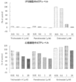
- the intracellular ATP levels of undifferentiated iPS cells (myocardial reporter iPS cell line) and purified cardiomyocytes derived from the same iPS cells after treatment with histone deacetylase inhibitor in the compound screening of Test Example 1 are shown.
- the intracellular ATP levels of iPS cells and cardiomyocytes derived from the same iPS cells in Test Example 2 after treatment with a histone deacetylase inhibitor are shown.
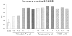
- the sarcomere ⁇ -actinin positive cell rate by flow cytometer analysis of CiRA clinical iPS cell-derived cardiomyocytes after treatment with histone deacetylase inhibitor in Test Example 3 is shown.
- the sarcomere ⁇ -actinin positive cell rate by flow cytometer analysis of iPS cell-derived cardiomyocytes after treatment with histone deacetylase inhibitor in Test Example 4 is shown.
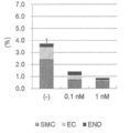
- the non-cardiomyocyte rate after compound treatment in Test Example 5 is shown (END: endoderm lineage cells, SMC: smooth muscle-like cells, EC: endothelial-like cells).
- the present invention provides a method for producing a cell population containing cardiomyocytes (hereinafter, also referred to as “the production method of the present invention”).
- the production method of the present invention comprises (1) contacting a cell population containing cardiomyocytes and cells other than cardiomyocytes with a histone deacetylase (HDAC) inhibitor, and (2) culturing the cell population. , including.
- HDAC histone deacetylase
- myocardial cell means a cell in which at least one of sarcomere ⁇ -actinin, myocardial troponin T and troponin I type 1 (TNNI1) is positive, preferably. , Sarcomere ⁇ -actinin positive cells. Also, typically, it is a myocardial cell having a self-beating ability.
- Cells other than cardiomyocytes are cells that do not correspond to cardiomyocytes, and specific examples thereof include smooth muscle cells, endothelial cells, stem cells (eg, pluripotent stem cells), and myocardial progenitor cells.
- Protein detection can be performed using antibody-based immunological assays such as ELISA, immunostaining, and flow cytometry.
- ELISA antibody-based immunological assays
- the reporter protein is expressed together with the protein, and the target protein is detected by detecting the reporter protein.
- Gene detection can be performed using, for example, a nucleic acid amplification method such as RT-PCR, biochip (eg, microarray), RNAseq, and / or a nucleic acid detection method.
- the expression of a protein or gene can be determined by a general method. For example, when flow cytometry is used, the expression of the protein is relatively expressed as compared with the expression level in the negative control group. When the amount is high, it can be determined that the protein is detectable and expressed.
- negative means that the expression level of a protein or gene is less than the lower limit of detection by all or any of the above-mentioned known methods.
- the lower limit of detection of protein or gene expression may differ depending on each method, but can be determined by a general method.
- the term "cell population” means a population consisting of two or more cells of the same type or different types. "Cell population” also means a mass of cells of the same or different types.
- a cell population containing cardiomyocytes and cells other than cardiomyocytes used in the step (1) can be produced by culturing pluripotent stem cells in a medium for cardiomyocyte differentiation. Therefore, the production method of the present invention may include a step (0) of inducing differentiation of cardiomyocytes from pluripotent stem cells before the step (1).
- pluripotent stem cells used in the present invention include induced pluripotent stem cells (iPS cells), embryonic stem cells (embryonic stem cells: ES cells), and embryos derived from cloned embryos obtained by nuclear transplantation.
- Specified stem cells nuclear transfer Embryonic stem cells: ntES cells
- pluripotent germLine stem cells mGS cells
- EG cells embryonic stem cells
- multi-line embryonic stem cells
- iPS cells more preferably human iPS cells
- the pluripotent stem cell is an ES cell or an arbitrary cell derived from a human embryo, the cell is a cell produced by destroying an embryo, even if the cell is produced by destroying the embryo.
- the pluripotent stem cells are preferably derived from mammals (eg, mice, rats, hamsters, guinea pigs, dogs, monkeys, orangutans, chimpanzees, humans), and more preferably humans. Therefore, the most preferable pluripotent stem cell used in the present invention is a human iPS cell.
- iPS cell Induced pluripotent stem cell
- iPS cell refers to a cell obtained by introducing a specific factor (nuclear reprogramming factor) into a mammalian somatic cell or an undifferentiated stem cell and reprogramming it.
- a specific factor nuclear reprogramming factor
- iPS cells there are various types of “artificial pluripotent stem cells (iPS cells)", and by introducing four factors, Oct3 / 4, Sox2, Klf4, and c-Myc, into mouse fibroblasts by Yamanaka et al.
- iPS cells derived from human cells established by introducing the same four factors into human fibroblasts (Takahashi K, Yamanaka S., Cell, (2006) 126: 663-676) Takahashi K, Yamanaka S., et al. Cell, (2007) 131: 861-872.), After introducing the above four factors, Nanog expression was selected as an index and established Nanog-iPS cells (Okita, K., Ichisaka, T., and Yamanaka, S. (2007).
- C-Myc-free iPS cells (Nakagawa M, Yamanaka S., et al. Nature Biotechnology, (2008) 26, 101 --106), iPS cells established by introducing 6 factors by virus-free method (Okita K et al. Nat. Methods 2011 May; 8 (5): 409-12, Okita K et al . Stem Cells. 31 (3): 458-66.) Can also be used.
- induced pluripotent stem cells established by introducing the four factors of OCT3 / 4, SOX2, NANOG, and LIN28 prepared by Thomson et al. (Yu J., Thomson JA.
- any of the induced pluripotent stem cells known in the art can be used.
- As the induced pluripotent stem cell line various iPS cell lines established by NIH, RIKEN, Kyoto University, etc. can be used.
- RIKEN's HiPS-RIKEN-1A strain, HiPS-RIKEN-2A strain, HiPS-RIKEN-12A strain, NiPS-B2 strain, Kyoto University's 253G1 strain, 201B7 strain, 409B2 strain examples thereof include 454E2 strain, 606A1 strain, 610B1 strain, 648A1 strain, iPS cell stock for regenerative medicine and the like.
- the term "somatic cell” means any animal cell (preferably a mammalian cell including human) except germline cells such as eggs, egg mother cells, and ES cells or totipotent cells.
- the somatic cells include, but are not limited to, fetal (pup) somatic cells, neonatal (pup) somatic cells, and mature healthy or diseased somatic cells, and primary cultured cells. , Passed cells, and established cells are all included.
- the somatic cells include, for example, (1) tissue stem cells (somatic stem cells) such as nerve stem cells, hematopoietic stem cells, mesenchymal stem cells, and dental pulp stem cells, (2) tissue precursor cells, (3) lymphocytes, and epithelium.
- Endothelial cells muscle cells, fibroblasts (skin cells, etc.), hair cells, hepatocytes, gastric mucosal cells, intestinal cells, splenocytes, pancreatic cells (pancreatic exocrine cells, etc.), brain cells, lung cells, renal cells And differentiated cells such as fat cells are exemplified.
- ES cells are stem cells that are pluripotent and have the ability to proliferate by self-renewal, established from the inner cell mass of early embryos (for example, blastocysts) of mammals such as humans and mice.
- ES cells were discovered in mice in 1981 (MJ Evans and MH Kaufman (1981), Nature 292: 154-156), and ES cell lines were subsequently established in primates such as humans and monkeys (JA Thomson et). al. (1998), Science 282: 1145-1147; JA Thomson et al. (1995), Proc. Natl. Acad. Sci. USA, 92: 7844-7848; JA Thomson et al. (1996), Biol.
- ES cells can be established by removing the inner cell mass from the blastocyst of the fertilized egg of the target animal and culturing the inner cell mass on a fibroblast feeder.
- a fibroblast feeder for methods of establishing and maintaining ES cells in humans and monkeys, for example, USP5,843,780; Thomson JA, et al. (1995), Proc Natl. Acad. Sci. U S A. 92: 7844-7848; Thomson JA. , Et al. (1998), Science. 282: 1145-1147; Suemori H. et al. (2006), Biochem. Biophys.
- ES cells can be established using only single blastomeres of embryos in the cleavage stage before the blastocyst stage (Chung Y. et al.
- ES cells various mouse ES cell lines established by inGenious targeting laboratory, RIKEN (RIKEN), etc. can be used for mouse ES cells, and Wisconsin University for human ES cells.
- RIKEN inGenious targeting laboratory
- RIKEN RIKEN
- Wisconsin University for human ES cells.
- NIH, RIKEN, Kyoto University, National Center for Child Health and Development, Cellartis, etc. are available.
- CHB-1 to CHB-12 strains sold by ESI Bio RUES1 strains, RUES2 strains, HUES1 to HUES28 strains, etc.
- H1 strains sold by WiCell Research H9 strains, etc.
- KhES-1, KhES-2, KhES-3, KhES-4, KhES-5, SSES1, SSES2, SSES3 and the like to be sold can be used.
- nt ES cells are ES cells derived from cloned embryos produced by nuclear transplantation technology and have almost the same characteristics as ES cells derived from fertilized eggs (Wakayama T. et al. (Wakayama T. et al.) 2001), Science, 292: 740-743; S. Wakayama et al. (2005), Biol. Reprod., 72: 932-936; Byrne J. et al. (2007), Nature, 450: 497-502) ..
- ES cells established from the inner cell mass of blastocysts derived from cloned embryos obtained by replacing the nuclei of unfertilized eggs with the nuclei of somatic cells are nt ES (nuclear transfer ES) cells.
- nt ES nuclear transfer ES
- nuclear transplantation technology Cibelli JB et al. (1998), Nature Biotechnol., 16: 642-646)
- ES cell production technology above is used (Wakayama). Seika et al. (2008), Experimental Medicine, Vol. 26, No. 5 (Special Edition), pp. 47-52).
- somatic cell nuclei can be injected into enucleated unfertilized eggs of mammals and cultured for several hours to initialize them.
- MGS cells are testis-derived pluripotent stem cells, which are the origin cells for spermatogenesis. Similar to ES cells, these cells can induce differentiation into cells of various lineages, and have the property of producing chimeric mice when transplanted into mouse blastocysts (Kanatsu-Shinohara M. et al. (Kanatsu-Shinohara M. et al.). 2003) Biol. Reprod., 69: 612-616; Shinohara K. et al. (2004), Cell, 119: 1001-1012). It is self-renewable in a culture medium containing glial cell line-developed neurotrophic factor (GDNF), and reproduces by repeating passage under the same culture conditions as ES cells. Stem cells can be obtained (Masanori Takebayashi et al. (2008), Experimental Medicine, Vol. 26, No. 5 (Special Edition), pp. 41-46, Glial Cell Line (Tokyo, Japan)).
- GDNF glial cell line
- EG cells are cells with pluripotency similar to ES cells, which are established from embryonic primordial germ cells. It can be established by culturing primordial germ cells in the presence of substances such as LIF, bFGF, and stem cell factor (Matsui Y. et al. (1992), Cell, 70: 841-847; JL Resnick). et al. (1992), Nature, 359: 550-551).
- Muse cells are non-neoplastic pluripotent stem cells that are endogenous to the living body, and can be produced, for example, by the method described in WO 2011/007900. Specifically, Muse cells are pluripotent cells obtained by long-term trypsinization of fibroblasts or bone marrow stromal cells, preferably 8 or 16 hours of trypsinization, followed by suspension culture. SSEA-3 and CD105 are positive.
- the step (0) is not particularly limited as long as the pluripotent stem cells can be induced to differentiate into cardiomyocytes.
- the pluripotent stem cells are differentiated into cardiomyocytes by culturing them in a cardiomyocyte differentiation medium. Can be induced.
- the step (0) is (0-1) a step of inducing differentiation of pluripotent stem cells into mesoderm cells, and (0-2) the steps of mesoderm cells into cardiomyocytes. And may include a step of inducing differentiation.
- the "cardiomyocyte differentiation medium” includes a factor that promotes the induction of differentiation into cardiomyocytes such as cytokines (hereinafter, may be referred to as "cardiomyocyte differentiation inducing factor”) and a basal medium. Means medium.
- the above-mentioned cardiomyocyte differentiation-inducing factor also includes factors necessary for inducing differentiation of pluripotent stem cells into intermediate cells (for example, mesoderm cells) in the process of inducing cardiomyocyte differentiation.
- basal medium used in the present invention examples include StemFit (eg, StemFit AK03N, StemFit AK02N) (Ajinomoto), StemPro-34 (Thermo Fisher Scientific), PECM (Primate ES Cell Medium), PECM (Primate ES Cell Medium), and Eagle's Medium. : Glasgow Minimum Essential Medium, IMDM (Iskov's Modified Dulbecco's Medium), 199 Medium, Eagle's Minimum Essential Medium (Eagle's Minimum Esentical Dulbecco's moderate Eagle's Medium (DMEM), Ham's F12 medium, RPMI 1640 medium, Fisher's medium, and a mixed medium thereof are included.
- StemFit eg, StemFit AK03N, StemFit AK02N
- Thermo Fisher Scientific Thermo Fisher Scientific
- PECM Primary ES Cell Medium
- PECM Primary ES Cell Medium
- Eagle's Medium Glasgow Minimum Essential Medium
- IMDM Iskov's Modified
- the basal medium includes ROCK inhibitors (eg, Y-27632, Fasudil / HA1077, SR3677, GSK269962, H-1152, Wf-536, etc.), serum (eg, bovine fetal serum (FBS), human serum, horse serum, etc.).
- ROCK inhibitors eg, Y-27632, Fasudil / HA1077, SR3677, GSK269962, H-1152, Wf-536, etc.
- serum eg, bovine fetal serum (FBS), human serum, horse serum, etc.
- serum substitutes insulin, various vitamins (eg vitamin Cs (eg ascorbic acid)), L-glutamine, various amino acids such as non-essential amino acids, 2-mercaptoethanol, thioglycerol (eg ⁇ -monothio) Gglycerol (MTG)), various cytokines, stem cell factors (SCF (Stem cell factor)), activin, etc.), various hormones, various growth factors (leukemia inhibitor (LIF), basic fibroblast growth factor (bFGF), TGF - ⁇ etc.), various extracellular matrices, various cell adhesion molecules, antibiotics such as penicillin / streptomycin and puromycin, pH indicators such as phenol red and the like can be appropriately added.
- Serum alternatives include albumin, transferrin, fatty acids, insulin, collagen precursors, trace elements, Knockout Serum Replacement (KSR), ITS-supplements and mixtures thereof.
- vitamin Cs mean L-ascorbic acid and its derivatives
- L-ascorbic acid derivatives mean those that become vitamin C by an enzymatic reaction in the living body.
- vitamin C phosphate eg, ascorbic acid-2 phosphate
- ascorbic acid glucoside e.g. ascorbic acid-2 phosphate
- ascorbic acid glucoside e.g. ascorbic acid-2 phosphate
- vitamin C ester eg., ascorbic acid phosphate
- ascorbic acid glucoside eg, ascorbic acid glucoside, ascorbic ethyl, vitamin C ester, ascobyl tetrahexyldecanoate, ascobyl stearate and -2 phosphorus ascorbic acid Acid-6 palmitic acid
- Vitamin C phosphate eg, Ascorbic acid 2-phospate
- examples thereof include phosphate-L-ascorbic acid salts such as Na phosphate-L-ascorbic acid or Mg phosphate-L-ascorbic acid. Be done.
- the culture of induced pluripotent stem cells or embryoid bodies may be adhesive culture or suspension culture.
- adhesive culture it may be carried out using a culture vessel coated with an extracellular matrix component, or it may be co-cultured with a feeder cell.
- the feeder cell is not particularly limited, and examples thereof include fibroblasts (mouse fetal fibroblast (MEF), mouse fibroblast (STO), etc.). It is preferable that the feeder cells are inactivated by a method known per se, for example, irradiation with radiation (gamma rays or the like) or treatment with an anticancer agent (mitomycin C or the like).
- matrigel Niwa A, et al.
- fibrous proteins such as gelatin, collagen, and elastin, and glucosaminoglycans such as hyaluronic acid and chondroitin sulfate.
- cell-adhesive proteins such as proteoglycan, fibronectin, vitronectin, and laminin.
- the conditions of the culture temperature are not particularly limited, but are preferably about 37 ° C. to 42 ° C. and 37 ° C. to 39 ° C., for example.
- the cells may be cultured under hypoxic conditions, and in the present invention, the hypoxic conditions are exemplified by oxygen concentrations of 15%, 10%, 9%, 8%, 7%, 6%, 5% or less.
- the hypoxic conditions are exemplified by oxygen concentrations of 15%, 10%, 9%, 8%, 7%, 6%, 5% or less.
- Suspension culture refers to culturing cells in a non-adherent state in a culture vessel, and is not particularly limited, but is artificially treated for the purpose of improving adhesion to cells (for example, coating with extracellular matrix or the like).
- Untreated culture vessel or treatment to artificially suppress adhesion for example, coating treatment with polyhydroxyethyl methacrylate (poly-HEMA) or nonionic surfactant (Pluronic F-127, etc.)
- This can be done using the culture vessel that has been prepared.
- stirring blades such as a single-use bioreactor (Biot Co., Ltd.), a single-use bioreactor (Thermo Fisher), a single-use bioreactor (Sartorius Stedium), and a single-use bioreactor (GE Healthcare Life Science).
- Suspension culture may be performed using an incubator.
- the type of incubator to be used and the stirring speed can be appropriately selected by those skilled in the art depending on the type of cells to be cultured. Examples of stirring speeds include, but are not limited to, 0-100 rpm, 20-80 rpm, or 45-65 rpm.
- the step (0) may include a step of forming an embryoid body from pluripotent stem cells.
- a step it is preferable to dissociate the colonized pluripotent stem cells into single cells and then form embryoid bodies.
- the step of dissociating pluripotent stem cells the cells that adhere to each other to form a population are dissociated (separated) into individual cells.
- Methods for dissociating pluripotent stem cells include, for example, a method of mechanically dissociating, a dissociation solution having protease activity and collagenase activity (for example, Accutase TM and Accumax TM), or dissociation having only collagenase activity.
- a dissociation method using a solution can be mentioned.
- a method of dissociating pluripotent stem cells using a dissociation solution having protease activity and collagenase activity is used.
- the medium used in the above steps preferably contains thioglycerol, L-glutamine and / or ascorbic acid.
- Examples of the myocardial cell differentiation-inducing factor used in the above step (0-1) include Wnt signal activators, activin A, BMP4, and bFGF, which may be used alone or in combination of two or more. .. In one aspect of the invention, a combination of activin A, BMP4 and bFGF is used.
- the medium used in the above step (0-1) preferably contains thioglycerol, L-glutamine and / or ascorbic acid.
- Wnt signal activator means a substance that activates the Wnt signaling pathway.
- Wnt signal activator examples include Wnt protein, GSK3 ⁇ inhibitor (eg, BIO, CHIR99021, etc.) and the like. These may be used alone or in combination of two or more.
- a Wnt signal activator When a Wnt signal activator is used, its concentration in the medium is not particularly limited.
- BIO or CHIR99021 is used as the Wnt signal activator, it is preferably used at a final concentration of 100 nM to 100 ⁇ M, preferably 1 ⁇ M to 10 ⁇ M in the medium.
- its concentration in the medium is preferably 1 ng / mL to 100 ng / mL, 1 ng / mL, 2 ng / mL, 3 ng / mL, 4 ng / mL, 5 ng / mL, 6 ng / mL, 7 ng / mL, 8 ng / mL, 9 ng / mL, 10 ng / mL, 11 ng / mL, 12 ng / mL, 13 ng / mL, 14 ng / mL, 15 ng / mL, 16 ng / mL, 17 ng / mL, 18 ng / mL, 19 ng / mL Examples thereof are mL, 20 ng / mL, 30 ng / mL, 40 ng / mL, 50 ng / mL, 60 ng / mL
- its concentration in the medium is preferably 1 ng / mL to 1 ⁇ g / mL, 1 ng / mL, 2 ng / mL, 3 ng / mL, 4 ng / mL, 5 ng / mL, 6 ng / mL, 7 ng.
- its concentration in the medium is preferably 1 ng / mL to 100 ng / mL, 1 ng / mL, 2 ng / mL, 3 ng / mL, 4 ng / mL, 5 ng / mL, 6 ng / mL, 7 ng.
- the period of the above step (0-1) is not particularly limited as long as mesoderm cells can be obtained, but is preferably 12 hours or more (example: 1 day, 2 days or more), and 6 days or less (example: 5). Days, 4 days, 3 days or less).
- the mesoderm marker gene include T, MIXL1, NODAL and the like.
- Examples of the myocardial cell differentiation-inducing factor used in the above step (0-2) include a Wnt inhibitor and VEGF, which may be used alone or in combination of two or more.
- the medium used in the above step (0-2) preferably contains thioglycerol, L-glutamine and / or ascorbic acid.
- Wnt inhibitor means a substance that inhibits the signal transduction that follows from the binding of Wnt to the receptor to the accumulation of ⁇ -catenin, and inhibits the binding to the Frizzled family of receptors. It may be a substance or a substance that promotes the decomposition of ⁇ -catenin. Examples of Wnt inhibitors include DKK1 protein (for example, in the case of humans, NCBI accession number: NM_122242), sclerostin (for example, in the case of humans, NCBI accession number: NM_025237), IWR-1 (Merck Millipore).
- DKK1 protein for example, in the case of humans, NCBI accession number: NM_122242
- sclerostin for example, in the case of humans, NCBI accession number: NM_025237
- IWR-1 Merck Millipore
- IWP-2 Sigma-Aldrich
- IWP-3 Sigma-Aldrich
- IWP-4 Sigma-Aldrich
- PNU-74654 Sigma-Aldrich
- XAV939 Sigma-Aldrich
- IWP-3 or IWP-4 is preferable. Only one type of Wnt inhibitor may be used, or a plurality of types may be used in combination.
- a Wnt inhibitor When a Wnt inhibitor is used, its concentration in the medium is preferably 1 nM to 50 ⁇ M, for example, 1 nM, 10 nM, 50 nM, 100 nM, 500 nM, 750 nM, 1 ⁇ M, 2 ⁇ M, 3 ⁇ M, 4 ⁇ M, 5 ⁇ M, 6 ⁇ M, 7 ⁇ M. , 8 ⁇ M, 9 ⁇ M, 10 ⁇ M, 15 ⁇ M, 20 ⁇ M, 25 ⁇ M, 30 ⁇ M, 40 ⁇ M, 50 ⁇ M, but is not limited thereto. More preferably, it is 1 ⁇ M.
- VEGF vascular endothelial growth factor
- its concentration in the medium is preferably 1 to 100 ng / mL, 1 ng / mL, 2 ng / mL, 3 ng / mL, 4 ng / mL, 5 ng / mL, 6 ng / mL, 7 ng / mL.
- ng / mL 8 ng / mL, 9 ng / mL, 10 ng / mL, 11 ng / mL, 12 ng / mL, 13 ng / mL, 14 ng / mL, 15 ng / mL, 16 ng / mL, 17 ng / mL, 18 ng / mL, 19 ng / mL, 20 ng Examples include / mL, 30 ng / mL, 40 ng / mL, 50 ng / mL, 60 ng / mL, 70 ng / mL, 80 ng / mL, 90 ng / mL and 100 ng / mL.
- a BMP inhibitor and / or a TGF ⁇ inhibitor may be further added to the basal medium as a myocardial cell differentiation inducing factor.
- BMP inhibitor includes protein inhibitors such as Chordin, Noggin, and Follistatin, and Dorsomorphin (6- [4- (2-piperidin-1-yl-ethoxy) phenyl] -3-pyridin-. 4-yl-pyrazolo [1,5-a] pyrimidine) and its derivatives (P. B. Yu et al. (2007), Circulation, 116: II # 60; PB Yu et al. (2008), Nat. Chem . Biol., 4: 33-41; J.
- its concentration in the medium is preferably 1 nM to 50 ⁇ M, for example, 1 nM, 10 nM, 50 nM, 100 nM, 500 nM, 600 nM, 700 nM, 800 nM, 900 nM, 1 ⁇ M, 2 ⁇ M, 3 ⁇ M, 4 ⁇ M. 5, 5 ⁇ M, 6 ⁇ M, 7 ⁇ M, 8 ⁇ M, 9 ⁇ M, 10 ⁇ M, 15 ⁇ M, 20 ⁇ M, 25 ⁇ M, 30 ⁇ M, 40 ⁇ M, 50 ⁇ M, but not limited to these.
- the TGF ⁇ inhibitor means a substance that inhibits the signal transduction of TGF ⁇ from binding to a receptor to SMAD, even if it is a substance that inhibits binding to the ALK family of receptors. , A substance that inhibits the phosphorylation of SMAD by the ALK family.
- TGF ⁇ inhibitors include Lefty-1 (NCBI Accession No. includes mouse: NM_010094 and human: NM_020997), SB431542, SB202190 (above, RKLindemann et al., Mol. Cancer, 2003, 2).
- SB431542 is preferable.
- a TGF ⁇ inhibitor When a TGF ⁇ inhibitor is used, its concentration in the medium is preferably 1 nM to 50 ⁇ M, for example, 1 nM, 10 nM, 50 nM, 100 nM, 500 nM, 750 nM, 1 ⁇ M, 2 ⁇ M, 3 ⁇ M, 4 ⁇ M, 5 ⁇ M, 5.2 ⁇ M. It is, but is not limited to, 5.4 ⁇ M, 5.6 ⁇ M, 5.8 ⁇ M, 6 ⁇ M, 7 ⁇ M, 8 ⁇ M, 9 ⁇ M, 10 ⁇ M, 15 ⁇ M, 20 ⁇ M, 25 ⁇ M, 30 ⁇ M, 40 ⁇ M, and 50 ⁇ M.
- the period of the above step (0-2) is not particularly limited as long as cardiomyocytes can be obtained, but is 1 day or more (example: 1 day, 2 days, 3 days, 4 days, 5 days, 6 days, 7 days or More than that).
- cardiomyocytes since there is no effect on the establishment of cardiomyocytes by culturing for a long period of time, there is no particular upper limit, but it is typically 40 days or less.
- the step (0) further includes a step of culturing the cardiomyocytes obtained in the step (0-2) in the presence or absence of the step (0-3) VEGF and / or bFGF. You may be.
- the medium used in this step preferably contains thioglycerol, L-glutamine and / or ascorbic acid.
- the medium used in this step is a myocardial maturation compound (eg, N- (1,1-dioxo-2,3-dihydro-1H-1-benzothiophen-5-yl) -2- (4- ⁇ 5).
- VEGF When VEGF is used in step (0-3), its concentration in the medium is preferably 1 to 100 ng / mL, 1 ng / mL, 2 ng / mL, 3 ng / mL, 4 ng / mL, 5 ng / mL, 6 ng.
- / ML 7 ng / mL, 8 ng / mL, 9 ng / mL, 10 ng / mL, 11 ng / mL, 12 ng / mL, 13 ng / mL, 14 ng / mL, 15 ng / mL, 16 ng / mL, 17 ng / mL, 18 ng / mL , 19 ng / mL, 20 ng / mL, 30 ng / mL, 40 ng / mL, 50 ng / mL, 60 ng / mL, 70 ng / mL, 80 ng / mL, 90 ng / mL and 100 ng / mL.
- bFGF When bFGF is used in step (0-3), its concentration in the medium is preferably 1 to 100 ng / mL, 1 ng / mL, 2 ng / mL, 3 ng / mL, 4 ng / mL, 5 ng / mL, 6 ng.
- ng / ML 7 ng / mL, 8 ng / mL, 9 ng / mL, 10 ng / mL, 11 ng / mL, 12 ng / mL, 13 ng / mL, 14 ng / mL, 15 ng / mL, 16 ng / mL, 17 ng / mL, 18 ng / mL , 19 ng / mL, 20 ng / mL, 30 ng / mL, 40 ng / mL, 50 ng / mL, 60 ng / mL, 70 ng / mL, 80 ng / mL, 90 ng / mL and 100 ng / mL. More preferably, it is 5 ng / mL.
- the period of the above step (0-3) is not particularly limited, but is 1 day or more (eg: 1 day, 2 days, 3 days, 4 days, 5 days, 6 days, 7 days, 8 days, 9 days, 10 days, 11 days, 12 days, 13 days, 14 days, 15 days, 16 days, 17 days, 18 days, 19 days, 20 days, 21 days, 22 days, 23 days, 24 days, 25 days, 26 days , 27 days, 28 days, or more).
- no upper limit is set, but it is typically 60 days or less.
- the embryoid body may be dissociated by the same method as described above before performing the step (0-2) or the step (0-3).
- END2 cells which are mouse-derived supporting cells, and pluripotent stem cells (Mummery, C., et al., Circulation. 107 (21), 2733-40 (2003)), embryonic bodies. , BMP4, FGF2, methods for inducing myocardial cells by culturing with insulin and serum (Paul, WB., Et al., PLoSone. 6 (4), e18293 (2011).) Be done.
- HDAC inhibition is applied to a cell population containing (1') pluripotent stem cells obtained by culturing pluripotent stem cells in a medium for myocardial cell differentiation and containing myocardial cells and cells other than myocardial cells.
- a method for purifying myocardial cells (hereinafter, also referred to as “purification method of the present invention”), which comprises a step of contacting an agent and (2') a step of culturing the cell population.
- a method for reducing cells other than cardiomyocytes from a cell population containing cardiomyocytes and cells other than cardiomyocytes which comprises the above steps (1') and (2') (hereinafter, “this”. Also referred to as “the method for reducing non-cardiomyocytes of the invention”).
- purifying cardiomyocytes means that the reduction rate of the number of cells other than cardiomyocytes (“the number of cells” means the number of living cells; the same applies hereinafter) of cardiomyocytes by HDAC inhibitor is the same. Percentage of cardiomyocytes in the cell population (in the cell population) when the rate of decrease is exceeded or the growth rate of cells other than cardiomyocytes is inhibited and the growth rate of cardiomyocytes exceeds the growth rate of cells other than cardiomyocytes. It means that the number of cardiomyocytes / the total number of cells in the cell population) increases.
- an increase in the proportion of myocardial cells in the purification of myocardial cells is due to an increase in the proportion of myocardial cells due to promotion of induction of differentiation from myocardial progenitor cells to myocardial cells or inhibition of induction of differentiation from myocardial progenitor cells to cells other than myocardial cells.
- the reduction rate of the pluripotent stem cell number by the HDAC inhibitor treatment is higher than the reduction rate of the cardiomyocyte number, and further, the cardiomyocyte in the cell population by the HDAC inhibitor treatment.
- the reduction in cell number by HDAC inhibitors is the result of induction of cell apoptosis by HDAC inhibitors.
- the HDAC inhibitor reduces the proportion of undifferentiated cells such as pluripotent stem cells contained in the cell population, or removes the undifferentiated cells from the cell population, thereby causing myocardium in the cell population. It is presumed that the proportion of cells increased.
- a step of contacting a cell population containing myocardial cells contaminated with undifferentiated cells with a histone deacetylase inhibitor and (II) a step of culturing the cell population.
- a method for removing or reducing the undifferentiated cells from the cell population (hereinafter, also referred to as “method for removing undifferentiated cells of the present invention”) is provided.
- undifferentiated cell means a cell that retains pluripotency, that is, pluripotency, multipotency, oligopotency, or unipotency.
- the undifferentiated cells include the above-mentioned pluripotent stem cells, mesenchymal cells having differentiation potential, myocardial progenitor cells and the like.
- the period of contact between the cell population containing cardiomyocytes and the HDAC inhibitor is not particularly limited, but is, for example, 1 hour or more (eg, 2 hours, 3). Time, 5 hours, 12 hours, 1 day, 2 days, 3 days or more). In addition, since long-term culture does not affect the establishment of cardiomyocytes or myocardial progenitor cells, there is no particular upper limit, but typically 60 days or less (eg, 50 days, 40 days, 30 days). , 20 days, 14 days, 13 days, 12 days, 11 days or less).
- the contact between the cell population containing myocardial cells and the HDAC inhibitor may be carried out by adding the HDAC inhibitor to the medium containing the cell population, or to the medium to which the HDAC inhibitor has been added in advance. This may be done by seeding a cell population.
- the timing of contacting the HDAC inhibitor is not particularly limited as long as the cell population contains cardiomyocytes, but for example, it is preferable to contact the HDAC inhibitor in the above steps (0-2) or (0-3), and it is pluripotent. Based on the start date of differentiation induction of sex stem cells, it is preferable to perform the procedure on or after the 7th day from the start of differentiation induction.
- HDACs inhibited by the HDAC inhibitors used in the present invention include class I HDACs (eg, HDAC1, HDAC2, HDAC3, HDAC8) and class II HDACs (eg, HDAC4, HDAC5, HDAC6, HDAC7, HDAC9, HDAC10).
- Class III HDACs eg SirT1, SirT2, SirT3, SirT4, SirT5, SirT6, SirT7
- Class IV HDACs eg HDAC11
- Yes more preferably an inhibitor of class I HDACs, especially HDAC1 or HDAC2.
- an inhibitor of class I against HDAC has other activity, for example, an inhibitory activity against class I HDAC, if it has a class I inhibitory activity against HDAC. It may also have a class I HDAC-specific inhibitory activity. The same is true for inhibitors of other classes of HDAC.
- Examples of the inhibitor against HDAC1 used in the present invention include Trichostatin A, CI994 (Tacedinaline), Quisinostat (JNJ-26481585), CUDC-907, PCI-24781 (Abexinostat), RG2833 (RGFP109), RiFP109. , Resminostat, Pracinostat (SB939), Rocylinostat (ACY-1215), Mocetinostat (MGCD0103), CAY10603, Entinostat (MS-275), 4SC-202, HPOB, PCI-34051, Tubastat, etc.
- Examples of the inhibitor against HDAC2 include Trichostatin A, Quisinostat (JNJ-26481585), CUDC-907, CUDC-101, PCI-24781 (Abequinostat), Romidepsin (FK228, Depsipeptide, Depsipeptide), and Depsipeptide. SB939), Mocetinostat (MGCD0103), 4SC-202, HPOB and the like.
- Examples of the inhibitor against HDAC3 include Trichostatin A, RGFP966, CUDC-907, Quisinostat (JNJ-26481585), RG2833 (RGFP109), PCI-24781 (Abexinostat), CUDC-101, Pracetinostat (Pracinostat) ACY-1215), 4SC-202, Mocetinostat (MGCD0103), HPOB, Entinostat (MS-275), Droxinostat and the like.
- Examples of the inhibitor against HDAC4 include Trichostatin A, Tasquinimod, Quisinostat (JNJ-26481585), LMK-235, CUDC-101, Pracinostat (SB939), TMP269, CUDC-907, Rocil, etc. ..
- Examples of the inhibitor against HDAC5 include Quisinostat (JNJ-26481585), LMK-235, CUDC-101, Pracinostat (SB939), TMP269, CUDC-907, and Rocilinostat (ACY-1215).
- Examples of the inhibitor against HDAC6 include Trichostatin A, CAY10603, Tubacin, Rocilinostat (ACY-1215), Nexturastat A, Tubasatin A HCl, Tubasatin A, HPOB, CUDC-101, PCI-281.
- Examples thereof include Resminostat, Quisinostat (JNJ-26481585), Pracinostat (SB939), Droxinostat, PCI-34051 and the like.
- Examples of the inhibitor against HDAC7 include TMP269, Quisinostat (JNJ-26481585), Pracinostat (SB939), CUDC-101, CUDC-907, and Rocilinostat (ACY-1215).
- Examples of the inhibitor against HDAC8 include PCI-34051, Quisinostat (JNJ-26481585), CUDC-101, Rocilinostat (ACY-1215), Pracinostat (SB939), CUDC-907, PCI-24781 (Abexinostat). , Droxinostat, HPOB and the like.
- Examples of the inhibitor against HDAC9 include TMP269, Quisinostat (JNJ-26481585), CUDC-101, Pracinostat (SB939), and CUDC-907.
- Examples of the inhibitor against HDAC10 include Trichostatin A, Quisinostat (JNJ-26481585), CUDC-907, PCI-24781 (Abexinostat), CUDC-101, Pracinostat (SB939), HPOB, PCI-45051 and the like.
- Examples of the inhibitor against HDAC11 include Quisinostat (JNJ-26481585), CUDC-907, Pracinostat (SB939), Mocetinostat (MGCD0103) and the like.
- Inhibitors for class III HDAC include Sirtinol, SirReal2, Nicotinamide (Vitamin B3), Selisistat (EX 527), Thiomyristoyl, Salermide, OSS_128167, AK7,3-TYP, Ten. Inauhzin and the like can be mentioned.
- HDAC inhibitor a non-selective HDAC inhibitor may be used, and examples of the inhibitor include Vorinostat (SAHA), Panobinostat (LBH589), Belinostat (PXD101) and the like. Of these, FK228, Entinostat, Trichostatin A or Panobinostat are preferable. Only one type of HDAC inhibitor may be used, or a plurality of types may be used in combination.
- SAHA Vorinostat
- LH589 Panobinostat
- PXD101 Belinostat
- FK228, Entinostat, Trichostatin A or Panobinostat are preferable. Only one type of HDAC inhibitor may be used, or a plurality of types may be used in combination.
- the concentration of the HDAC inhibitor in the medium can be appropriately selected by those skilled in the art, but for example, 0.1 nM to 10 ⁇ M is preferable, and specifically, 0.5 nM, 1 nM, 2 nM, 3 nM, 5 nM, 10 nM. , 20 nM, 30 nM, 40 nM, 50 nM, 0.1 ⁇ M, 0.2 ⁇ M, 0.3 ⁇ M, 0.4 ⁇ M, 0.5 ⁇ M, 1.0 ⁇ M, 1.5 ⁇ M, 2 ⁇ M, 3 ⁇ M, 4 ⁇ M, 5 ⁇ M, 6 ⁇ M, 7 ⁇ M, 8 ⁇ M , 9 ⁇ M, 10 ⁇ M and the like.
- the concentration can be appropriately changed depending on the type of the compound. For example, when Trichostatin A is used, 0.1 ⁇ M to 10 ⁇ M is preferable, and when Panobinostat is used, 0.1 to 3 ⁇ M is preferable, and FK228 is preferable. When using, 1 nM to 1 ⁇ M is preferable, and when Entinostat is used, 0.1 ⁇ M to 10 ⁇ M is preferable, but the concentration is not limited to these.
- the method for culturing the cell population in the steps (2), (2') and (II) is the same as in (0-3) above.
- the culture period is the same, and the culture may be continued at least while the cell population is in contact with the HDAC inhibitor.
- the present invention also comprises a cell population containing cardiomyocytes obtained by the production method, purification method, non-cardiomyocyte reduction method or undifferentiated cell removal method of the present invention (hereinafter, "cells of the present invention”). Also called “group”).
- the cell population of the present invention contains cardiomyocytes with high purity. High purity means that the ratio of cardiomyocytes in the cell population (number of cardiomyocytes in the cell population / total number of cells in the cell population) is specifically 80% or more (eg, 85%, 90%, 91). %, 92%, 93%, 94%, 95%, 96%, 97%, 98%, 99% or more).
- the ratio of the cardiomyocytes is before mixing the other cells or the cell population when the cell population is used in combination with other cells or cell populations such as mesenchymal stem cells.
- the cell population of the present invention comprises a higher proportion of cardiomyocytes than the cell population obtained by conventional methods of inducing cardiomyocytes from pluripotent stem cells.
- Such a cell population may be further purified by cell sorting or the like, and the cell population thus purified is also included in the "cell population of the present invention".
- the present invention also provides a cell transplantation therapy agent (hereinafter, also referred to as “the cell transplantation therapy agent of the present invention”), which comprises the cell population of the present invention.
- the cell transplantation therapy agent of the present invention may be used for autologous transplantation or allogeneic transplantation. It may also be used in combination with other drugs, such as immunosuppressants.
- the cell population of the present invention contains myocardial cells with high purity, the cell population of the present invention is suitable for use as a raw material for a cell transplantation therapeutic agent, and the cell population of the present invention or the present invention Cell transplant therapeutic agents are useful in the treatment or prevention of heart disease.
- an effective amount of the cell population or cell transplant therapy agent of the present invention is administered or transplanted to a mammal (eg, human, mouse, rat, monkey, cow, horse, pig, dog, etc.) to be treated or prevented.
- a mammal eg, human, mouse, rat, monkey, cow, horse, pig, dog, etc.
- Methods for treating or preventing heart disease are also included in the present invention.
- Heart diseases to be treated or prevented include diseases or disorders such as heart failure, ischemic heart disease, myocardial infarction, cardiomyopathy, cardiomyopathy, hypertrophic cardiomyopathy, dilated phase hypertrophic cardiomyopathy, and dilated cardiomyopathy. Examples include, but are not limited to, defects.
- the cell population of the present invention is used as a cell transplant therapy agent, it is derived from iPS cells established from somatic cells having the same or substantially the same HLA genotype of the transplanted individual from the viewpoint that rejection does not occur. It is desirable to use a cell population that includes the cells to be used.
- substantially the same means that the transplanted cells have the same HLA genotype to the extent that the immune response can be suppressed by the immunosuppressant, and for example, HLA-A and HLA-B.
- HLA-DR 3 loci or HLA-C added 4 loci matching HLA type somatic cells. It is also possible to implant in a capsule such as polyethylene glycol or silicon, a porous container, or the like to avoid rejection.
- the cell population of the present invention is produced as a parenteral preparation such as an injection, a suspension, or an infusion by mixing with a pharmaceutically acceptable carrier according to conventional means.
- Pharmaceutically acceptable carriers that can be contained in the parenteral preparation include, for example, isotonic solutions containing physiological saline, glucose and other adjuvants (eg, D-sorbitol, D-mannitol, sodium chloride, etc.) and the like.
- An aqueous solution for injection can be mentioned.
- the cell transplantation therapeutic agent of the present invention is, for example, a buffer (for example, phosphate buffer, sodium acetate buffer), a soothing agent (for example, benzalkonium chloride, procaine hydrochloride, etc.), and a stabilizer (for example, human). It may be blended with serum albumin, polyethylene glycol, etc.), preservatives, antioxidants, etc.
- a cell population containing cardiomyocytes is suspended in the aqueous solution so that the number of cells is about 1 ⁇ 10 6 to about 1 ⁇ 10 8 cells / mL. Just let me do it. It can also be administered with scaffolding materials that promote engraftment.
- the scaffolding material is exemplified by, but is not limited to, a biological component such as collagen and a synthetic polymer such as polylactic acid which substitutes for the component.
- the treatment of heart disease may be performed by sheeting the obtained cardiomyocytes and attaching them to the patient's heart.
- the myocardial sheet is administered, it is achieved by arranging it so as to cover the desired portion.
- the arrangement so as to cover the desired portion can be performed by using a technique well known in the art.
- the tissue may be arranged so as to surround the tissue.
- the administration can also be placed several times in the same portion to obtain the desired effect. When several placements are performed, it is desirable to allow sufficient time for the desired cells to engraft in the tissue and perform angiogenesis.
- the mechanism of treatment for such heart disease may be the effect produced by engraftment of the myocardial sheet, or an indirect effect not dependent on cell engraftment (eg, the recipient by secreting an attractant).
- the effect of mobilizing the derived cells to the damaged site) may be used.
- a cell scaffold material such as collagen, fibronectin, or laminin may be contained in addition to cardiomyocytes.
- any cell type (s) can be included.
- the number of cardiomyocytes used for the treatment of heart disease is not particularly limited as long as the administered myocardial sheet is effective in the treatment of heart disease, and the size of the affected area and the size of the body are not particularly limited. It can be adjusted by increasing or decreasing as appropriate.
- the cell population of the present invention can also be used for drug screening for the treatment of heart disease and cardiotoxicity assessment of the drug.
- the effect and toxicity of the test drug can be evaluated by administering the test drug to the cell population of the present invention and examining the response of cardiomyocytes.
- Test example 1 Screening of compounds that have a purifying effect on cardiomyocytes Based on the difference in proliferative properties between cardiomyocytes and non-cardiomyocytes, it is assumed that there are differences in susceptibility to compounds targeting cell proliferation and cell cycle, and iPS cell lines for research are used. It was used to screen for compounds that have a purifying effect on cardiomyocytes.
- iPS cell line was prepared. The maintenance culture of the reporter iPS cell line was performed by a conventional method (Okita K, et al. Stem Cells. 2012 Nov 29. doi: 10.1002 / stem.1293).
- the human iPS cells are episomal vectors (loading genes; OCT3 / 4, KLF4, SOX2, L-MYC, LIN28, mouse p53DD) using PBMC (LP_167, Sample ID: 2013018) purchased from CTL. (Reference; Okita K, et al. Stem Cells. 2012 Nov 29. doi: 10.1002 / stem. 1293).
- Induction of differentiation into cardiomyocytes was performed on a 6-well plate according to the method described in the paper (Miki et al, Cell Stem Cell. 2015 Jun 4; 16. doi: 10.1016 / j.stem. 2015.04.005.). Briefly, differentiation into cardiomyocytes is induced by treating the reporter iPS cell line with TripLE select (Life Technologies) diluted in 1/2 with 0.5 mM EDTA / PBS for 4 to 5 minutes, and then using the cell scraper (IWAKI). ), And the cells were dissociated into single cells by pipetting.
- the medium was removed by centrifugation at 1,000 rpm for 5 minutes, and the obtained cells were seeded in 2 ⁇ 10 6 cells per 1 well of a 6-well plate, and 1% L-glutamine and transferrin 150 ⁇ g / mL were added to the StemPro34 medium.
- the 6-well plate was tilted to allow the embryoid bodies to settle, 80-90% of the medium was removed, and then 1.5 mL of IMDM was added to each well.
- the well plate is tilted again to allow the embryoid body to settle, and after removing 80 to 90% of the medium, 1% L-glutamine, transferase 150 ⁇ g / mL, ascorbic acid 50 ⁇ g / mL (Sigma), in StemPro34 medium, Monothioglycerol 4 ⁇ 10 -4 M, 10 ng / mL VEGF, 1 ⁇ M IWP-3, 0.6 ⁇ M Dorsomorphin and 5.4 ⁇ M SB431542 were cultured in a medium supplemented with 37 ° C.
- the 6-well plate was tilted and allowed to settle to settle the embryoid body, and after removing 80 to 90% of the medium, 1% L-glutamine, transferrin 150 ⁇ g / mL, ascorbic acid 50 ⁇ g / StemPro34 medium supplemented with mL (Sigma), monothioglycerol 4 ⁇ 10 -4 M and 5 ng / mL VEGF was added.
- the cells were cultured for 10 days at 37 ° C. under 5% oxygen conditions. During this period, the medium was replaced with a medium under the same conditions once every 2 to 3 days. After the 10th day, the cells were cultured under 37 ° C. and 21% oxygen conditions.
- EBs were single-celled using the Papain Dissociation System (Worthington), centrifuged (70 g, 6 minutes), suspended in 2% FBS / PBS, and GFP-positive cells were separated by a flow cytometer. After centrifugation, the supernatant was removed, and myocardial differentiation medium based on StemPro34 medium (Ascorbic acid 50 ⁇ g / mL, L-glutamine 2 mM, transferrin 150 mg / mL, monothioglycerol 4 ⁇ 10 -4 M, VEGF 5 ng in Stepro34). (Mixed / ml) was resuspended in 100 mL.
- StemPro34 medium Ascorbic acid 50 ⁇ g / mL, L-glutamine 2 mM, transferrin 150 mg / mL, monothioglycerol 4 ⁇ 10 -4 M, VEGF 5 ng in Stepro34.
- the resuspended cells were seeded on a pre-coated CellCarrier-384 plate Ultra Microplate at 2.0 ⁇ 10 3 cells / well.
- the cell population obtained by the culture is hereinafter referred to as cardiomyocyte.
- the iPS cell line was treated with 0.5 ⁇ TrypLE select (Life Technologies, diluted 1/2 with 0.5 mM EDTA / PBS) for 4 to 5 minutes, and then the cells were detached with a cell scraper (IWAKI) and pipetting was performed. It dissociated into a single cell.
- the medium was removed by centrifugation (1,000 rpm, 5 minutes), resuspended in StemFit AK02 medium supplemented with Y-27632 10 ⁇ M, and coated with iMatrix-511 (Nippi) CellCarrier-384 Ultra Microplate (PerkinElmer / 6057300). ) was seeded on 2.0 ⁇ 10 3 cells / well.
- the evaluation compound was added 2 days after seeding, and the cells were cultured for 2 days. Forty-eight hours after compound treatment, viable cell numbers were assessed by measuring intracellular ATP levels using CellTiter-Glo (PerkinElmer).
- Test example 2 Verification of Purifying Effect in Clinical iPS Cell Lines Next, it was determined by ATP assay whether the HDAC inhibitor found in the screening of Test Example 1 also had a purifying effect on cardiomyocytes derived from clinical iPS cell lines. confirmed.
- ⁇ IPS cell line> A clinical iPS cell line prepared with CiRA was used. The maintenance culture of the iPS cell line was performed according to the conventional method (Okita K, et al. Stem Cells. 2012 Nov 29. doi: 10.1002 / stem.1293).
- a solution containing 10 ⁇ g / mL of DNase was added to 2 mL of TrypLE select per dish, and the mixture was allowed to stand at 37 ° C. under normal oxygen conditions for 15 minutes. Then, a medium containing 10 ⁇ g / mL of DNase was added to 2 mL of IMDM per dish, and the cells were made into single cells by pipetting and then centrifuged (100 g, 4 ° C., 5 minutes). .. After centrifugation, the supernatant was removed, suspended in I3 medium (myocardial differentiation medium similar to Test Example 1), and seeded on a fibronectin-coated 96-well plate at a seeding density of 2x10 4 cells / well.
- I3 medium myocardial differentiation medium similar to Test Example 1
- cardiomyocyte The cell population obtained by the culture is hereinafter referred to as cardiomyocyte.
- some cells were fixed, stained with anti-sarcomere ⁇ -actinin antibody, and the expression of sarcomere ⁇ -actinin was analyzed with a flow cytometer.
- the sarcomere ⁇ -actinin positive rate was 99.4%.
- iPS cells cultured in a 10 cm dish were washed with 5 mL of PBS, 5 mL of Accutase was added, and the cells were allowed to stand at 37 ° C. under normal oxygen conditions for 7 minutes.
- the cells were detached by pipetting to form single cells, which were then subjected to centrifugation (1,000 rpm, 5 minutes). After centrifugation, the supernatant was removed, suspended in undifferentiated maintenance medium supplemented with Y-27632 10 ⁇ M, and then seeded on a 96-well plate coated with iMatrix-511 (Nippi) at a seeding density of 5x10 3 cells / well. ..
- iMatrix-511 Nippi
- Test example 3 Verification of the effect of HDAC inhibitors on germ layer bodies in which cardiomyocytes and non-cardiomyocytes coexist 1 Based on the above results, the present inventors considered that the use of HDAC inhibitors could purify cardiomyocytes in embryoid bodies in which cardiomyocytes and non-cardiomyocytes such as iPS cells coexist. Therefore, the effect of the HDAC inhibitor on the embryoid body was verified by the following procedure.
- ⁇ IPS cell line> A clinical iPS cell line prepared with CiRA was used. The maintenance culture of the iPS cell line was performed according to the conventional method (Okita K, et al. Stem Cells. 2012 Nov 29. doi: 10.1002 / stem.1293).
- the cells were cultured at 37 ° C. under normal oxygen conditions for 3 days.
- the 6-well plate was allowed to stand to submerge the embryoid body, and the embryoid body was collected in a 1.5 mL tube by a pipetman with a wide-diameter tip. Centrifugated at 500 g for 1 minute. The supernatant was removed, a solution containing 10 ⁇ g / mL of DNase and 100 ⁇ g / mL of Liberase was added to 500 ⁇ L of IMDM per tube, and the mixture was allowed to stand at 37 ° C. under normal oxygen conditions for 1 hour.
- the tube was centrifuged at 500 g for 1 minute and the supernatant was removed so as not to suck the embryoid body. Then, a solution added so as to have DNase of 10 ⁇ g / mL was added to 500 ⁇ L of TrypLE select per tube, and the mixture was allowed to stand at 37 ° C. under normal oxygen conditions for 15 minutes. Then, after making a single cell by pipetting, a medium containing 10 ⁇ g / mL of DNase was added to 500 ⁇ L of IMDM per tube, mixed by inversion, and subjected to centrifugation (700 g, 5 minutes).
- Test example 4 Verification of the effect of HDAC inhibitors on germ layer bodies in which cardiomyocytes and non-cardiomyocytes coexist 2 Next, the same verification was performed with the same cell line as in Test Example 3, and it was confirmed that the cardiomyocyte rate was improved even with Entinostat and that the cardiomyocyte rate was improved even with a lower concentration of FK228.
- the cells were cultured at 37 ° C. under normal oxygen conditions for 4 days.
- the 6-well plate was allowed to stand to submerge the embryoid body, and the embryoid body was collected in a 1.5 mL tube by a pipetman with a wide-diameter tip. Centrifugated at 500 g for 1 minute. The supernatant was removed, a solution containing 10 ⁇ g / mL of DNase and 100 ⁇ g / mL of Liberase was added to 500 ⁇ L of IMDM per tube, and the mixture was allowed to stand at 37 ° C. under normal oxygen conditions for 1 hour.
- the tube was centrifuged at 500 g for 1 minute and the supernatant was removed so as not to suck the embryoid body. Then, a solution containing 10 ⁇ g / mL of DNase was added to 500 uL of TrypLE select per tube, and the mixture was allowed to stand at 37 ° C. under normal oxygen conditions for 15 minutes. Then, after making a single cell by pipetting, a medium containing 10 ⁇ g / mL of DNase was added to 500 ⁇ L of IMDM per tube, mixed by inversion, and subjected to centrifugation (700 g, 5 minutes).
- Test example 5 Verification of the effect of HDAC inhibitors on germ layer bodies in which cardiomyocytes and non-cardiomyocytes coexist 3 Next, verification was performed with the same cell line as in Test Example 3, and it was confirmed that the non-cardiomyocyte rate was reduced by FK-228 treatment.
- the 6-well plate was allowed to stand to submerge the embryoid body, and the embryoid body was collected in a 1.5 mL tube by a pipetman with a wide-diameter tip. Centrifugated at 500 g for 1 minute. After removing the supernatant, add 3 mL of a solution of DNase 10 ⁇ g / mL and Liberase 100 ⁇ g / mL to 1 mL of IMDM (Iscover's Modified Dulbecco's Media) per tube, and add 3 mL of the solution to each tube at 37 ° C., normal oxygen. It was allowed to stand for 1 hour under the conditions.
- the tube was centrifuged at 500 g for 1 minute and the supernatant was removed so as not to suck the embryoid body.
- 500 ⁇ L of a solution prepared by adding 10 ⁇ g / mL of DNase to TrypLE select (Thermo) was added to each tube, and the mixture was allowed to stand at 37 ° C. under normal oxygen conditions for 10 minutes. After standing, a single cell was formed by pipetting, and 500 ⁇ L of a medium prepared by adding 10 ⁇ g / mL of DNase to IMDM was added to each tube and mixed by inversion to prepare a single cell suspension.
- the above single cell suspension was centrifuged at 400 g for 3 minutes, the supernatant was removed, the suspension was suspended in Bambanker, and the suspension was cryopreserved at -80 ° C.
- the cryopreserved cells were thawed in a warm bath at 37 ° C., centrifuged at 400 g for 5 minutes, and the supernatant was removed.
- PBS containing 1% BSA was added, and the mixture was centrifuged at 300 g for 1 minute to remove the supernatant.
- the myocardial cells induced to differentiate from the iPS cell line into the myocardium differ in the expression of these three surface markers, resulting in CD326-positive cells (endometrial lineage cells), CD326-negative CD31-positive cells (vascular endothelial-like cells), and CD326-negative CD31.
- CD326-positive cells endometrial lineage cells
- CD326-negative CD31-positive cells vascular endothelial-like cells
- CD326-negative CD31 vascular endothelial-like cells
- Negative CD49a-positive cells smooth muscle-like cells
- CD326-negative CD31-negative CD49a-negative cells were isolated into four main cell populations.
- HDAC inhibition can purify cardiomyocytes in the embryoid body.
- the present invention provides a cell population containing cardiomyocytes with high purity. Such a cell population is useful because it can be suitably used for cell transplantation therapy for heart disease and screening of therapeutic agents for heart disease.
Landscapes
- Health & Medical Sciences (AREA)
- Life Sciences & Earth Sciences (AREA)
- Engineering & Computer Science (AREA)
- Biomedical Technology (AREA)
- Chemical & Material Sciences (AREA)
- Zoology (AREA)
- Cell Biology (AREA)
- General Health & Medical Sciences (AREA)
- Bioinformatics & Cheminformatics (AREA)
- Organic Chemistry (AREA)
- Biotechnology (AREA)
- Veterinary Medicine (AREA)
- Medicinal Chemistry (AREA)
- Animal Behavior & Ethology (AREA)
- Public Health (AREA)
- Wood Science & Technology (AREA)
- Genetics & Genomics (AREA)
- Cardiology (AREA)
- Epidemiology (AREA)
- Chemical Kinetics & Catalysis (AREA)
- Developmental Biology & Embryology (AREA)
- Oral & Maxillofacial Surgery (AREA)
- Botany (AREA)
- Dermatology (AREA)
- Transplantation (AREA)
- Urology & Nephrology (AREA)
- Rheumatology (AREA)
- Microbiology (AREA)
- Biochemistry (AREA)
- General Engineering & Computer Science (AREA)
- Pharmacology & Pharmacy (AREA)
- Vascular Medicine (AREA)
- Virology (AREA)
- Immunology (AREA)
- Nuclear Medicine, Radiotherapy & Molecular Imaging (AREA)
- General Chemical & Material Sciences (AREA)
- Heart & Thoracic Surgery (AREA)
- Hematology (AREA)
- Micro-Organisms Or Cultivation Processes Thereof (AREA)
Abstract
La présente invention concerne un procédé de production d'une population de cellules contenant des cardiomyocytes, le procédé comprenant les étapes consistant à : (1) mettre en contact un inhibiteur d'histone désacétylase avec une population de cellules qui contient des cardiomyocytes et des cellules autres que les cardiomyocytes, et sont obtenues par culture de cellules souches pluripotentes dans des milieux pour la différenciation de cardiomyocytes ; et (2) cultiver la population cellulaire.
Priority Applications (4)
| Application Number | Priority Date | Filing Date | Title |
|---|---|---|---|
| CN202180022365.2A CN115427552A (zh) | 2020-03-19 | 2021-03-18 | 心肌细胞的精制方法 |
| US17/912,214 US20230212519A1 (en) | 2020-03-19 | 2021-03-18 | Method for purifying cardiomyocytes |
| JP2022508445A JPWO2021187602A1 (fr) | 2020-03-19 | 2021-03-18 | |
| EP21772548.0A EP4123016A1 (fr) | 2020-03-19 | 2021-03-18 | Procédé de purification de cardiomyocytes |
Applications Claiming Priority (4)
| Application Number | Priority Date | Filing Date | Title |
|---|---|---|---|
| JP2020-050269 | 2020-03-19 | ||
| JP2020050269 | 2020-03-19 | ||
| JP2020-145097 | 2020-08-28 | ||
| JP2020145097 | 2020-08-28 |
Publications (1)
| Publication Number | Publication Date |
|---|---|
| WO2021187602A1 true WO2021187602A1 (fr) | 2021-09-23 |
Family
ID=77768144
Family Applications (1)
| Application Number | Title | Priority Date | Filing Date |
|---|---|---|---|
| PCT/JP2021/011232 WO2021187602A1 (fr) | 2020-03-19 | 2021-03-18 | Procédé de purification de cardiomyocytes |
Country Status (5)
| Country | Link |
|---|---|
| US (1) | US20230212519A1 (fr) |
| EP (1) | EP4123016A1 (fr) |
| JP (1) | JPWO2021187602A1 (fr) |
| CN (1) | CN115427552A (fr) |
| WO (1) | WO2021187602A1 (fr) |
Citations (13)
| Publication number | Priority date | Publication date | Assignee | Title |
|---|---|---|---|---|
| US5843780A (en) | 1995-01-20 | 1998-12-01 | Wisconsin Alumni Research Foundation | Primate embryonic stem cells |
| WO2007069666A1 (fr) | 2005-12-13 | 2007-06-21 | Kyoto University | Facteur de reprogrammation nucleaire |
| US20080236610A1 (en) | 2004-04-10 | 2008-10-02 | Holger Bartels | Hair Rollers |
| WO2008118220A2 (fr) | 2006-11-28 | 2008-10-02 | Veritainer Corporation | Unité de détection de rayonnement pour monter un capteur de rayonnement sur une grue de contenant |
| WO2008124133A1 (fr) | 2007-04-07 | 2008-10-16 | Whitehead Institute For Biomedical Research | Reprogrammation de cellules somatiques |
| WO2008151058A2 (fr) | 2007-05-30 | 2008-12-11 | The General Hospital Corporation | Procédés de génération de cellules pluripotentes à partir de cellules somatiques |
| JP2008307007A (ja) | 2007-06-15 | 2008-12-25 | Bayer Schering Pharma Ag | 出生後のヒト組織由来未分化幹細胞から誘導したヒト多能性幹細胞 |
| US20090047263A1 (en) | 2005-12-13 | 2009-02-19 | Kyoto University | Nuclear reprogramming factor and induced pluripotent stem cells |
| WO2009073618A2 (fr) * | 2007-11-30 | 2009-06-11 | New York Medical College | Compositions comprenant des inhibiteurs hdac et procédés permettant de les utiliser pour réparer la fonction des cellules souches et empêcher l'insuffisance cardiaque |
| WO2009146408A1 (fr) | 2008-05-30 | 2009-12-03 | Summa Health Systems Llc | Procédés d'utilisation d'inhibiteurs du récepteur tgf-b ou des inhibiteurs a-83-01 et sb-431542 de kinase de type activine (alk) 5 pour traiter une maladie des yeux et la guérison de blessure |
| WO2011007900A1 (fr) | 2009-07-15 | 2011-01-20 | Dezawa Mari | Cellule souche pluripotente isolée à partir de tissue organique |
| JP2017104091A (ja) * | 2015-11-27 | 2017-06-15 | タカラバイオ株式会社 | 間葉系細胞の製造方法 |
| JP2017108705A (ja) * | 2015-12-18 | 2017-06-22 | 国立大学法人京都大学 | 心筋細胞の製造方法 |
Family Cites Families (3)
| Publication number | Priority date | Publication date | Assignee | Title |
|---|---|---|---|---|
| KR101564044B1 (ko) * | 2007-10-31 | 2015-10-28 | 고쿠리츠 다이가쿠 호진 교토 다이가쿠 | 핵초기화 방법 |
| BR112016030723B8 (pt) * | 2014-07-16 | 2024-02-27 | Heartseed Inc | Meios de cultura de células, método para indução da morte celular de células-tronco indiferenciadas e métodos para seleção de cardiomiócitos |
| JP6265515B2 (ja) * | 2014-09-16 | 2018-01-24 | 国立大学法人大阪大学 | 多能性幹細胞由来心筋細胞集団の製造方法 |
-
2021
- 2021-03-18 CN CN202180022365.2A patent/CN115427552A/zh active Pending
- 2021-03-18 WO PCT/JP2021/011232 patent/WO2021187602A1/fr unknown
- 2021-03-18 JP JP2022508445A patent/JPWO2021187602A1/ja active Pending
- 2021-03-18 EP EP21772548.0A patent/EP4123016A1/fr active Pending
- 2021-03-18 US US17/912,214 patent/US20230212519A1/en active Pending
Patent Citations (17)
| Publication number | Priority date | Publication date | Assignee | Title |
|---|---|---|---|---|
| US5843780A (en) | 1995-01-20 | 1998-12-01 | Wisconsin Alumni Research Foundation | Primate embryonic stem cells |
| US20080236610A1 (en) | 2004-04-10 | 2008-10-02 | Holger Bartels | Hair Rollers |
| WO2007069666A1 (fr) | 2005-12-13 | 2007-06-21 | Kyoto University | Facteur de reprogrammation nucleaire |
| JP2008283972A (ja) | 2005-12-13 | 2008-11-27 | Kyoto Univ | 誘導多能性幹細胞の製造方法 |
| US20090047263A1 (en) | 2005-12-13 | 2009-02-19 | Kyoto University | Nuclear reprogramming factor and induced pluripotent stem cells |
| WO2008118220A2 (fr) | 2006-11-28 | 2008-10-02 | Veritainer Corporation | Unité de détection de rayonnement pour monter un capteur de rayonnement sur une grue de contenant |
| WO2008124133A1 (fr) | 2007-04-07 | 2008-10-16 | Whitehead Institute For Biomedical Research | Reprogrammation de cellules somatiques |
| WO2008151058A2 (fr) | 2007-05-30 | 2008-12-11 | The General Hospital Corporation | Procédés de génération de cellules pluripotentes à partir de cellules somatiques |
| WO2009006930A1 (fr) | 2007-06-15 | 2009-01-15 | Izumi Bio, Inc. | Cellules souches pluripotentes humaines induites à partir des cellules souches indifférenciées issues d'un tissu postnatal humain |
| WO2009006997A1 (fr) | 2007-06-15 | 2009-01-15 | Izumi Bio, Inc. | Cellules souches pluripotentes humaines et leur utilisation à des fins médicales |
| WO2009007852A2 (fr) | 2007-06-15 | 2009-01-15 | Izumi Bio, Inc | Cellules multipotentes/pluripotentes et procédés s'y rapportant |
| JP2008307007A (ja) | 2007-06-15 | 2008-12-25 | Bayer Schering Pharma Ag | 出生後のヒト組織由来未分化幹細胞から誘導したヒト多能性幹細胞 |
| WO2009073618A2 (fr) * | 2007-11-30 | 2009-06-11 | New York Medical College | Compositions comprenant des inhibiteurs hdac et procédés permettant de les utiliser pour réparer la fonction des cellules souches et empêcher l'insuffisance cardiaque |
| WO2009146408A1 (fr) | 2008-05-30 | 2009-12-03 | Summa Health Systems Llc | Procédés d'utilisation d'inhibiteurs du récepteur tgf-b ou des inhibiteurs a-83-01 et sb-431542 de kinase de type activine (alk) 5 pour traiter une maladie des yeux et la guérison de blessure |
| WO2011007900A1 (fr) | 2009-07-15 | 2011-01-20 | Dezawa Mari | Cellule souche pluripotente isolée à partir de tissue organique |
| JP2017104091A (ja) * | 2015-11-27 | 2017-06-15 | タカラバイオ株式会社 | 間葉系細胞の製造方法 |
| JP2017108705A (ja) * | 2015-12-18 | 2017-06-22 | 国立大学法人京都大学 | 心筋細胞の製造方法 |
Non-Patent Citations (39)
| Title |
|---|
| CELL, vol. 70, 1992, pages 841 - 847 |
| CIBELLI J.B. ET AL., NATURE BIOTECHNOL., vol. 16, 1998, pages 642 - 646 |
| HUANGFU D.MELTON, DA. ET AL., NATURE BIOTECHNOLOGY, vol. 26, no. 7, 2008, pages 795 - 797 |
| J. HAO ET AL., PLOS ONE, vol. 3, no. 8, 2008, pages e2904 |
| J.A. THOMSON ET AL., BIOL. REPROD., vol. 55, 1996, pages 254 - 259 |
| J.A. THOMSON ET AL., PROC. NATL. ACAD. SCI. USA, vol. 92, 1995, pages 7844 - 7848 |
| J.A. THOMSON ET AL., SCIENCE, vol. 282, 1998, pages 1145 - 1147 |
| J.A. THOMSONV.S. MARSHALL, CURR. TOP. DEV. BIOL., vol. 38, 1998, pages 133 - 165 |
| J.L. RESNICK ET AL., NATURE, vol. 359, 1992, pages 550 - 551 |
| KANATSU-SHINOHARA M. ET AL., BIOL. REPROD, vol. 69, 2003, pages 612 - 616 |
| KAWASAKI H. ET AL., PROC. NATL. ACAD. SCI. USA, vol. 99, 2002, pages 1580 - 1585 |
| KIM JB.SCHOLER HR ET AL., NATURE, vol. 292, 1981, pages 154 - 156 |
| KLIMANSKAYA I. ET AL., NATURE, vol. 444, 2006, pages 481 - 485 |
| LIAN X ET AL., PROC NATL ACAD SCI USA., vol. 109, no. 27, 3 July 2012 (2012-07-03), pages E1848 - 57 |
| LINDEMANN ET AL., MOL. CANCER, vol. 2, 2003, pages 20 |
| MASANORI TAKEHASHI ET AL., EXPERIMENTAL MEDICINE, vol. 26, no. 5, 2008, pages 41 - 46 |
| MIKI ET AL., CELL STEM CELL, vol. 16, 4 June 2015 (2015-06-04) |
| MINAMI I ET AL., CELL REP., vol. 2, no. 5, 29 November 2012 (2012-11-29), pages 1448 - 60 |
| MUMMERY, C ET AL., CIRCULATION, vol. 107, no. 21, 2003, pages 2733 - 40 |
| NIWA A ET AL., PLOS ONE, vol. 6, no. 7, 2011, pages e22261 |
| OKITA K ET AL., NAT. METHODS, vol. 8, no. 5, May 2011 (2011-05-01), pages 409 - 12 |
| OKITA K ET AL., STEM CELLS, 29 November 2012 (2012-11-29) |
| OKITA K ET AL., STEM CELLS., vol. 31, no. 3, pages 458 - 66 |
| OKITA, K.ICHISAKA, T.YAMANAKA, S, NATURE, vol. 450, 2007, pages 497 - 502 |
| P. B. YU ET AL., CIRCULATION, vol. 116, 2007, pages 11 |
| P. B. YU ET AL., NAT. CHEM. BIOL., vol. 4, 2008, pages 33 - 41 |
| PAUL, W B. ET AL., PLOSONE, vol. 6, no. 4, 2011, pages e18293 |
| S. WAKAYAMA ET AL., BIOL. REPROD., vol. 72, 2005, pages 932 - 936 |
| SCIENCE, vol. 292, 2001, pages 740 - 743 |
| SHI Y.DING S. ET AL., CELL STEM CELL, vol. 2, 2008, pages 113 - 117 |
| SHINOHARA K. ET AL., CELL, vol. 119, 2004, pages 1001 - 1012 |
| SUEMORI H. ET AL., BIOCHEM. BIOPHYS. RES. COMMUN., vol. 345, 2006, pages 926 - 932 |
| SUEMORI H. ET AL., DEV. DYN., vol. 222, 2001, pages 273 - 279 |
| TAKAHASHI KYAMANAKA S. ET AL., CELL, vol. 131, 2007, pages 861 - 872 |
| TAKAHASHI KYAMANAKA S., CELL, vol. 126, 2006, pages 663 - 676 |
| THOMSON JA ET AL., PROC NATL. ACAD. SCI. U S A., vol. 92, 1995, pages 7844 - 7848 |
| UENO M. ET AL., PROC. NATL. ACAD. SCI. USA, vol. 103, 2006, pages 9554 - 9559 |
| YU J.THOMSON JA ET AL., SCIENCE, vol. 318, 2007, pages 1917 - 1920 |
| ZHANG X. ET AL., STEM CELLS, vol. 24, 2006, pages 2669 - 2676 |
Also Published As
| Publication number | Publication date |
|---|---|
| EP4123016A1 (fr) | 2023-01-25 |
| CN115427552A (zh) | 2022-12-02 |
| US20230212519A1 (en) | 2023-07-06 |
| JPWO2021187602A1 (fr) | 2021-09-23 |
Similar Documents
| Publication | Publication Date | Title |
|---|---|---|
| US10828335B2 (en) | Production of midbrain dopaminergic neurons and methods for the use thereof | |
| KR102487142B1 (ko) | 만능 세포를 분화시키는 방법 | |
| US20190153385A1 (en) | Method of producing progenitor cells from differentiated cells | |
| US20180171290A1 (en) | Method of producing progenitor cells from differentiated cells | |
| KR20170072940A (ko) | 신경 조직의 제조 방법 | |
| EP3260536B1 (fr) | Nouveau procédé d'induction de chondrocyte | |
| JPWO2016143803A1 (ja) | 肺胞上皮細胞の分化誘導法 | |
| US9422522B2 (en) | Method of producing adipocytes from fibroblast cells | |
| JP2017108705A (ja) | 心筋細胞の製造方法 | |
| JPWO2018199142A1 (ja) | 神経堤細胞および交感神経細胞の製造方法 | |
| WO2020203538A1 (fr) | Population de cellules comprenant des cellules souches pluripotentes et son procédé de production | |
| US20220010271A1 (en) | Method for producing brain organoids | |
| WO2021187601A1 (fr) | Procédé de purification de cardiomyocytes | |
| WO2021187602A1 (fr) | Procédé de purification de cardiomyocytes | |
| JP7344486B2 (ja) | 心筋細胞成熟促進剤 | |
| RU2813532C1 (ru) | Способ очистки кардиомиоцитов | |
| US10174285B2 (en) | Methods of obtaining cell populations enriched with desired cells | |
| US20240084265A1 (en) | Methods to convert somatic human cell to a totipotency-like state with engineered sox17 | |
| JP2020115771A (ja) | 多能性幹細胞から軟骨組織を製造する方法 |
Legal Events
| Date | Code | Title | Description |
|---|---|---|---|
| 121 | Ep: the epo has been informed by wipo that ep was designated in this application |
Ref document number: 21772548 Country of ref document: EP Kind code of ref document: A1 |
|
| ENP | Entry into the national phase |
Ref document number: 2022508445 Country of ref document: JP Kind code of ref document: A |
|
| NENP | Non-entry into the national phase |
Ref country code: DE |
|
| ENP | Entry into the national phase |
Ref document number: 2021772548 Country of ref document: EP Effective date: 20221019 |