WO2021145395A1 - 抗線維化剤、及び抗線維化作用を有する細胞外小胞の製造方法 - Google Patents
抗線維化剤、及び抗線維化作用を有する細胞外小胞の製造方法 Download PDFInfo
- Publication number
- WO2021145395A1 WO2021145395A1 PCT/JP2021/001131 JP2021001131W WO2021145395A1 WO 2021145395 A1 WO2021145395 A1 WO 2021145395A1 JP 2021001131 W JP2021001131 W JP 2021001131W WO 2021145395 A1 WO2021145395 A1 WO 2021145395A1
- Authority
- WO
- WIPO (PCT)
- Prior art keywords
- extracellular vesicles
- derived
- affinity
- solution
- extracellular
- Prior art date
- Legal status (The legal status is an assumption and is not a legal conclusion. Google has not performed a legal analysis and makes no representation as to the accuracy of the status listed.)
- Ceased
Links
Images




Classifications
-
- A—HUMAN NECESSITIES
- A61—MEDICAL OR VETERINARY SCIENCE; HYGIENE
- A61K—PREPARATIONS FOR MEDICAL, DENTAL OR TOILETRY PURPOSES
- A61K35/00—Medicinal preparations containing materials or reaction products thereof with undetermined constitution
- A61K35/12—Materials from mammals; Compositions comprising non-specified tissues or cells; Compositions comprising non-embryonic stem cells; Genetically modified cells
-
- A—HUMAN NECESSITIES
- A61—MEDICAL OR VETERINARY SCIENCE; HYGIENE
- A61P—SPECIFIC THERAPEUTIC ACTIVITY OF CHEMICAL COMPOUNDS OR MEDICINAL PREPARATIONS
- A61P43/00—Drugs for specific purposes, not provided for in groups A61P1/00-A61P41/00
-
- A—HUMAN NECESSITIES
- A61—MEDICAL OR VETERINARY SCIENCE; HYGIENE
- A61K—PREPARATIONS FOR MEDICAL, DENTAL OR TOILETRY PURPOSES
- A61K35/00—Medicinal preparations containing materials or reaction products thereof with undetermined constitution
- A61K35/12—Materials from mammals; Compositions comprising non-specified tissues or cells; Compositions comprising non-embryonic stem cells; Genetically modified cells
- A61K35/28—Bone marrow; Haematopoietic stem cells; Mesenchymal stem cells of any origin, e.g. adipose-derived stem cells
-
- C—CHEMISTRY; METALLURGY
- C12—BIOCHEMISTRY; BEER; SPIRITS; WINE; VINEGAR; MICROBIOLOGY; ENZYMOLOGY; MUTATION OR GENETIC ENGINEERING
- C12N—MICROORGANISMS OR ENZYMES; COMPOSITIONS THEREOF; PROPAGATING, PRESERVING, OR MAINTAINING MICROORGANISMS; MUTATION OR GENETIC ENGINEERING; CULTURE MEDIA
- C12N5/00—Undifferentiated human, animal or plant cells, e.g. cell lines; Tissues; Cultivation or maintenance thereof; Culture media therefor
- C12N5/06—Animal cells or tissues; Human cells or tissues
- C12N5/0602—Vertebrate cells
- C12N5/0652—Cells of skeletal and connective tissues; Mesenchyme
- C12N5/0662—Stem cells
- C12N5/0663—Bone marrow mesenchymal stem cells (BM-MSC)
-
- C—CHEMISTRY; METALLURGY
- C12—BIOCHEMISTRY; BEER; SPIRITS; WINE; VINEGAR; MICROBIOLOGY; ENZYMOLOGY; MUTATION OR GENETIC ENGINEERING
- C12N—MICROORGANISMS OR ENZYMES; COMPOSITIONS THEREOF; PROPAGATING, PRESERVING, OR MAINTAINING MICROORGANISMS; MUTATION OR GENETIC ENGINEERING; CULTURE MEDIA
- C12N5/00—Undifferentiated human, animal or plant cells, e.g. cell lines; Tissues; Cultivation or maintenance thereof; Culture media therefor
- C12N5/06—Animal cells or tissues; Human cells or tissues
- C12N5/0602—Vertebrate cells
- C12N5/0652—Cells of skeletal and connective tissues; Mesenchyme
- C12N5/0662—Stem cells
- C12N5/0667—Adipose-derived stem cells [ADSC]; Adipose stromal stem cells
-
- C—CHEMISTRY; METALLURGY
- C12—BIOCHEMISTRY; BEER; SPIRITS; WINE; VINEGAR; MICROBIOLOGY; ENZYMOLOGY; MUTATION OR GENETIC ENGINEERING
- C12N—MICROORGANISMS OR ENZYMES; COMPOSITIONS THEREOF; PROPAGATING, PRESERVING, OR MAINTAINING MICROORGANISMS; MUTATION OR GENETIC ENGINEERING; CULTURE MEDIA
- C12N5/00—Undifferentiated human, animal or plant cells, e.g. cell lines; Tissues; Cultivation or maintenance thereof; Culture media therefor
- C12N5/06—Animal cells or tissues; Human cells or tissues
- C12N5/0602—Vertebrate cells
- C12N5/0652—Cells of skeletal and connective tissues; Mesenchyme
- C12N5/0662—Stem cells
- C12N5/0668—Mesenchymal stem cells from other natural sources
Definitions
- the present invention relates to an antifibrotic agent using extracellular vesicles derived from mesenchymal stem cells.
- Extracellular vesicles which are cell-derived small membrane vesicles composed of lipid bilayers, are responsible for cell-cell communication via the transport of nucleic acids such as mRNA and microRNA contained therein, or proteins.
- Non-Patent Document 1 Non-Patent Document 1
- mesenchymal stem cells are somatic stem cells derived from mesoderm tissue. MSC can be separated from fat, bone marrow, umbilical cord matrix, etc., all of which are adhesive, CD105, CD73, CD90 positive, CD45, CD34, CD14, CD11b, CD79a, CD19, HLA-Class II ( DR) Negative, it is thought to have in common the ability to differentiate into bone, fat and cartilage.
- Non-Patent Document 2 Extracellular vesicles obtained from extracellular vesicles derived from mesenchymal stem cells by the ultracentrifugal method have an antifibrotic effect (antifibrotic effect) (Non-Patent Document 2). ).
- an object of the present invention is to provide a therapeutic preparation composed of a population of extracellular vesicles having a higher therapeutic effect, particularly an antifibrotic agent.
- the present inventors have diligently studied extracellular vesicles selectively collected by various methods for obtaining extracellular vesicles, and as a result, a substance having an affinity for phosphatidylserine (PS).
- PS affinity method phosphatidylserine
- a population of extracellular vesicles with the characteristic of PS positive obtained by the method using (PS affinity method) and / and extracellular vesicles derived from mesenchymal stem cells stimulated by fibroblast growth factor.
- the population has found to be highly effective as an anti-fibrotic agent, and has completed the present invention.
- the basic aspect of the present invention is (1) An antifibrotic agent containing extracellular vesicles derived from mesenchymal stem cells as an active ingredient, which can be obtained by a method using a substance having an affinity for phosphatidylserine. Antifibrotic agents, and / and extracellular vesicles derived from mesenchymal stem cells stimulated by fibroblastic proliferative factors; (2) The mesenchymal stem cells are derived from iPS cells or one or more tissues selected from the group consisting of umbilical cord, umbilical cord blood, bone marrow, adipose, muscle, nerve, skin, amniotic membrane and placenta.
- the present invention is also a method for producing extracellular vesicles as such an antifibrotic agent. (9) While obtaining extracellular vesicles from extracellular vesicles derived from mesenchymal stem cells by a method using a substance having an affinity for phosphatidylserine, or / and stimulating with transforming growth factor ⁇ .
- a method for producing extracellular vesicles having an antifibrotic effect which comprises obtaining extracellular vesicles from mesenchymal stem cells; (10) The extracellular vesicle according to (9) above, which comprises obtaining extracellular vesicles from extracellular vesicles derived from mesenchymal stem cells by a method using a substance having an affinity for phosphatidylserine.
- Production method (11) The method for producing extracellular vesicles according to (10) above, wherein the substance having an affinity for phosphatidylserine is a Tim protein; (12) The method for producing extracellular vesicles according to (11) above, wherein the Tim protein is selected from Tim4 protein, Tim3 protein and Tim1 protein; (13) The method for producing extracellular vesicles according to (9) to (12) above, which comprises obtaining extracellular vesicles from mesenchymal stem cells stimulated by transforming growth factor ⁇ ; Is.
- the present invention provides an antifibrotic agent containing extracellular vesicles having an antifibrotic effect as an active ingredient from mesenchymal stem cells. Since the antifibrotic agent provided by the present invention has an action of regulating the production of factors involved in tissue fibrosis, for example, collagen III, Collagen V, and ⁇ -SMA collagen, which are fibrosis-related genes, it is a living tissue or organ. By suppressing the progress of fibrosis in collagen, it is possible to regenerate the functions of living tissues and organs that have become dysfunctional or dysfunctional.
- FIG. 1 is a diagram showing the results of analyzing the particle size distribution of MSC-derived extracellular vesicles obtained by the PS affinity method and the ultracentrifugation method by the NTA method (nanoparticle tracking analysis method).
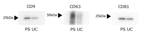
- FIG. 2 is a diagram showing the results of detecting CD9, CD63, and CD81 of MSC-derived extracellular vesicles obtained by the PS affinity method and the ultracentrifugation method by Western blotting.
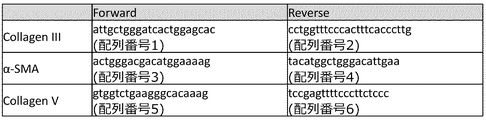
- FIG. 3 is a diagram showing primers used for quantitative PCR.
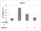
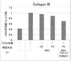
- FIG. 4 is a diagram showing the results of evaluating the antifibrotic effect of bone marrow MSC-derived extracellular vesicles using the mRNA expression level of Collagen III as an index.
- FIG. 1 is a diagram showing the results of analyzing the particle size distribution of MSC-derived extracellular vesicles obtained by the PS affinity method and the ultracentrifugation method by the NTA method (nanoparticle tracking analysis method).
- FIG. 2 is a
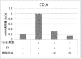
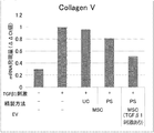
- FIG. 5 is a diagram showing the results of evaluating the antifibrotic effect of bone marrow MSC-derived extracellular vesicles using the mRNA expression level of Collagen V as an index.
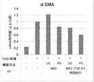
- FIG. 6 is a diagram showing the results of evaluating the antifibrotic effect of bone marrow MSC-derived extracellular vesicles using the mRNA expression level of ⁇ SMA as an index.
- FIG. 7 is a diagram showing the results of evaluating the antifibrotic effect of bone marrow MSC-derived extracellular vesicles using the mRNA expression level of Collagen III as an index.
- FIG. 8 is a diagram showing the results of evaluating the antifibrotic effect of bone marrow MSC-derived extracellular vesicles using the mRNA expression level of Collagen V as an index.
- FIG. 9 is a diagram showing the results of evaluating the antifibrotic effect of bone marrow MSC-derived extracellular vesicles using the expression level of ⁇ -SMA mRNA as an index.
- FIG. 10 is a diagram showing the results of evaluating the antifibrotic action of adipose MSC-derived extracellular vesicles and umbilical cord MSC-derived extracellular vesicles using the mRNA expression level of Collagen III as an index.
- FIG. 11 is a diagram showing the results of evaluating the antifibrotic action of adipose MSC-derived extracellular vesicles and umbilical cord MSC-derived extracellular vesicles using the mRNA expression level of Collagen V as an index.
- FIG. 12 is a diagram showing the results of evaluating the antifibrotic effect of adipose MSC-derived extracellular vesicles and umbilical cord MSC-derived extracellular vesicles using the mRNA expression level of ⁇ SMA as an index.
- the basis of the present invention is extracellular vesicles and / and transforming growth obtained by a method utilizing a substance having an affinity for phosphatidylserine from extracellular vesicles derived from mesenchymal stem cells. It is an antifibrotic agent containing extracellular vesicles derived from mesenchymal stem cells stimulated by factor ⁇ as an active ingredient.
- Extracellular vesicles are small membrane vesicles derived from cells and composed of a lipid bilayer membrane.
- the extracellular vesicles usually have a diameter of 20 nm to 1000 nm, preferably 50 nm to 800 nm, more preferably 50 nm to 500 nm, and particularly preferably 50 nm to 200 nm.
- Examples of the extracellular vesicles include Nature Reviews Immunology 9,581-593 (March 2009), "Obesity Study” Vol. 13 No. 2 2007 Topics As described in Naoto Aoki and others, those classified in various ways according to their origin and the size of small membrane vesicles can be mentioned.
- exosomes include exosomes, microvesicles, ectosomes, membrane particles, exosome-like vesicles, apoptotic bodies, adiposomes and the like, with exosomes and vesicles being preferred, and exosomes being more preferred.
- the exosome is a cell-derived small membrane vesicle composed of a lipid bilayer membrane, and examples thereof include those having a diameter of 50 nm to 200 nm, preferably 50 nm to 150 nm, and 50 nm to 100 nm. The one is more preferable. Exosomes are thought to be derived from late endosomes.
- the microvesicles are cell-derived small membrane vesicles composed of a lipid bilayer membrane, and examples thereof include those having a diameter of 100 nm to 1000 nm, preferably 100 nm to 800 nm, and 100 nm to 500 nm. Is more preferable.
- the microvesicles are thought to be derived from the cell membrane.
- MSC is a stem cell capable of differentiating into cells belonging to mesoderm-derived tissues (mesoderm system) such as osteoblasts, adipocytes, muscle cells, and chondrocytes.
- the MSC can be separated from fat, bone marrow, umbilical cord matrix, etc., and the MSC used in the present invention (hereinafter, may be abbreviated as “MSC according to the present invention”) is derived from, for example, mesoderm. It can be obtained by a method of separating from a tissue or a method of inducing from stem cells such as iPS cells and ES cells.
- the MSC according to the present invention includes those derived from one or more tissues selected from the group consisting of umbilical cord, umbilical cord blood, bone marrow, fat, muscle, nerve, skin, amniotic membrane and placenta, and those derived from iPS cells. It is preferably used.
- the MSC according to the present invention may be one that has undergone pretreatment such as recovery, concentration, purification, isolation, dilution with a buffer solution, filtration sterilization, and the like. These pretreatments may be appropriately performed according to a conventional method. Further, the MSC according to the present invention is preferably stimulated with transforming growth factor ⁇ , and more preferably stimulated with TGF ⁇ 1 or TGF ⁇ 3.
- extracellular vesicle which is an active ingredient in the antifibrotic agent provided by the present invention is a cell derived from the MSC according to the present invention.
- Extracellular vesicles isolated and obtained from extracellular vesicles using a substance having an affinity for phosphatidylserine hereinafter, may be abbreviated as "PS-positive extracellular vesicles”
- Extracellular vesicles derived from MSC according to the present invention stimulated by growth factor ⁇ hereinafter, may be abbreviated as “stimulated MSC-derived extracellular vesicles”).
- the PS-positive extracellular vesicle is the PS-positive (PS-containing) extracellular vesicle in which phosphatidylserine is considered to be exposed on the membrane surface of the extracellular vesicle.
- PS-affinitive substance As the substance having an affinity for phosphatidylserine (hereinafter, may be abbreviated as "PS-affinitive substance"), it is possible to specifically bind to phosphatidylserine constituting the membrane of extracellular vesicles.
- Tim1 T-cell immunoglobulin / mucin domain-containing molecule 1, T-cell immunoglobulin-mucin-domain 1) protein
- Tim2 T-cell immunoglobulin / Mucin domain-containing molecule 2, T-cell immunoglobulin-mucin-domain 2) protein
- Tim3 T-cell immunoglobulin / mucin domain-containing molecule 3, T-cell immunoglobulin-mucin-domain 3) protein
- Tim4 T-cell immunoglobulin / Tim proteins such as mucin domain-containing molecule 4, T-cell immunoglobulin-mucin-domain 4) protein
- Tim protein is preferable, and Tim4 protein and Tim3 protein are preferable.
- Tim1 protein are more preferred, Tim4 protein and Tim1 protein are even more preferred, and Tim4 protein is particularly preferred.
- PS-positive extracellular vesicles have an affinity for phosphatidylserine from the cell culture supernatant of MSC according to the present invention containing EV (hereinafter, may be abbreviated as "cell culture supernatant containing EV").
- EV obtained after isolation using a substance having sex or obtaining EV from the cell culture supernatant of MSC according to the present invention containing EV by a conventional method in this field such as ultracentrifugation. It is obtained by isolating from phosphatidylserine using a substance having an affinity for phosphatidylserine.
- the cell culture supernatant of the MSC according to the present invention containing EV can be obtained, for example, by proliferating the MSC according to the present invention by cell culture and further culturing the proliferated cells in an EV production medium.
- the cell culture of MSC and the culture in the EV production medium according to the present invention may be carried out according to a conventional method performed in this field, and the medium and culture conditions used are not particularly limited.
- PS affinity method The outline of the method for obtaining extracellular vesicles (hereinafter, may be abbreviated as "PS affinity method") by a method using a substance having an affinity for phosphatidylserine is described below. Further, as a PS affinity method, for example, a specific example is described in Patent Document 1.
- the cell culture supernatant containing EV in the presence of calcium ions, the cell culture supernatant containing EV is brought into contact with the PS-affinitive substance, and the extracellular vesicles in the cell culture supernatant are combined with the PS-affinitive substance. This is done by forming a body (hereinafter, sometimes abbreviated as "complex according to the present invention"), separating the PS-affinitive substance from the complex, and obtaining PS-positive extracellular vesicles. ..
- the preferred method of the PS affinity method includes the following steps. (1) In the presence of calcium ions, the cell culture supernatant containing EV and the PS-affinitive substance are brought into contact with each other, and a complex of PS-positive extracellular vesicles and the PS-affinitive substance in the cell culture supernatant is brought into contact with each other. Forming (complex according to the present invention) (hereinafter, may be abbreviated as “complex forming step"), (2) Separation of the complex according to the present invention obtained in the complex formation step from the cell culture supernatant containing the EV (hereinafter, may be abbreviated as “complex separation step”). (3) Separation of PS-positive extracellular vesicles from the complex according to the present invention to obtain PS-positive extracellular vesicles (hereinafter, may be abbreviated as "acquisition step”).
- the cell culture supernatant containing EV and the PS-affinity substance in the PS-affinity method are the same as those described above, and the preferred ones and specific examples are also the same.
- the PS-affinity substance used in the complex formation step is preferably one immobilized (bonded) to an insoluble carrier.
- the complex according to the present invention can be separated from the cell culture supernatant containing the EV by a known B / F separation method in the complex separation step.
- the insoluble carrier for immobilizing the PS-affinity substance examples include an insoluble carrier used in immunological measurement methods. Specifically, for example, polystyrene, polyacrylic acid, polymethacrylic acid, polymethylmethacrylate, polyacrylamide, polyglycidyl methacrylate, polypropylene, polyolefin, polyimide, polyurethane, polyester, polyvinyl chloride, polyethylene, polychlorocarbonate, silicone.
- Organic substances such as resins, silicone rubbers, agaroses, dextrans, ethylene-maleic anhydride copolymers; inorganic substances such as glass, silicon oxide, silica, porous glass, suriglass, alumina, silica gel, metal oxides; iron, cobalt, Magnetic materials such as nickel, magnetite, and chromate; and those prepared using an alloy of these magnetic materials as a material can be mentioned.
- examples of the usage form of these carriers include particles (beads), microplates, tubes, disc-shaped pieces and the like.
- the form of the insoluble carrier is preferably particles (beads), and the size of the particles is not particularly limited, but examples thereof include those having a size of 10 nm to 100 ⁇ m, and those having a size of 100 nm to 10 ⁇ m are preferable.
- Examples of the method for binding the PS-affinitive substance to the insoluble carrier include a method known per se for binding a protein to the carrier.
- a method of binding by affinity binding for example, a method of binding by chemical bonding (for example, the method described in Japanese Patent Application Laid-Open No. 3269554 and WO2012 / 039395), and a method of binding by physical adsorption (for example, JP-A-5-41946).
- the method described in (1) and the like are preferable, and a method of binding by physical adsorption and a method of binding by affinity bonding are preferable, and a method of binding by physical adsorption is more preferable because it is simple.
- the PS-affinity substance and the insoluble carrier are bound under the condition that the PS-affinity substance and the insoluble carrier are bound according to a method known per se. Should be brought into contact with.
- the amount of the PS-affinity substance to be bound to the insoluble carrier may be, for example, 0.1 ⁇ g to 50 ⁇ g, preferably 0.1 ⁇ g to 30 ⁇ g, based on 1 mg of the insoluble carrier, for example, when the insoluble carrier is particles (beads). , 0.1 ⁇ g to 20 ⁇ g is more preferable.
- Physical adsorption of the PS-affinitive substance and the insoluble carrier may be performed, for example, by bringing the solution containing the PS-affinitive substance into contact with the insoluble carrier.
- the solution for dissolving the PS-affinitive substance may be a solution that dissolves the PS-affinitive substance in a stable state, for example, purified water, for example, pH 6.0 to 9.8.
- a buffer solution having a buffering action of 7.0 to 9.6 for example, a good buffer solution such as MOPS, a carbonate buffer solution, PBS, TBS, TBS-T, HBS, etc.
- the buffer concentration in these buffers may be appropriately selected from the range of usually 5 to 100 mM, preferably 10 to 100 mM.
- the concentration when NaCl is contained is, for example, 100 to 200 mM, preferably 140 to 160 mM.
- the solution containing the PS-affinitive substance for example, saccharides, salts such as NaCl, and surfactants such as Tween TM 20 as long as the amount does not interfere with the binding between the PS-affinitive substance and the insoluble carrier.
- Preservatives, proteins and the like may be contained.
- the method of binding the PS-affinity substance and the insoluble carrier by physical adsorption include the following methods.
- 1 mg of the bead (particle) carrier is brought into contact with a solution containing the PS-affinitive substance containing 0.1 ⁇ g to 50 ⁇ g, preferably 0.1 ⁇ g to 30 ⁇ g, more preferably 0.1 ⁇ g to 20 ⁇ g, and the temperature is 2 ° C.
- the reaction is carried out at ⁇ 37 ° C., preferably 4 ° C. to 11 ° C. for 0.5 to 48 hours, preferably 0.5 to 24 hours.
- the insoluble carrier on which the PS-affinity substance immobilized as described above may be subjected to a blocking treatment usually performed in this field.
- the complex formation step is performed in the presence of calcium ions.
- Calcium ions are present when the PS-affinitive substance is brought into contact with the PS-positive extracellular vesicles in the cell culture supernatant containing the EV.
- the calcium ion concentration at the time of contacting the PS-affinitive substance with the PS-positive extracellular vesicles in the cell culture supernatant containing the EV is usually 0.5 mM to 100 mM, preferably 1.0 mM to 10 mM, more preferably. Is 2.0 mM to 5.0 mM.
- the complex according to the present invention is formed between the PS-affinitive substance and the PS-positive extracellular vesicle in the cell culture supernatant containing the EV and the complex separation step is carried out, that is, the complex according to the present invention.
- Calcium ions having the above-mentioned concentration are required in the solution containing the complex according to the present invention until the step of separating the bodies.
- the origin of calcium ions is not particularly limited, and examples thereof include calcium chloride, calcium hydroxide, calcium hydrogencarbonate, calcium iodide, calcium bromide, calcium acetate, and calcium chloride, calcium hydrogencarbonate, and calcium iodide are preferable. Calcium chloride and calcium hydrogen carbonate are more preferable.
- the cell culture supernatant containing the PS-affinitive substance and the EV is used as a method for allowing calcium ions to be present when the PS-affinitive substance and the PS-positive extracellular vesicle in the cell culture supernatant containing the EV are brought into contact with each other.
- the cell culture supernatant containing the PS-affinitive substance and the EV is used in the cell culture supernatant containing EV and / and the solution containing the PS-affinitive substance.
- the calcium ion concentration at the time of contact with the PS-positive extracellular vesicles in the solution is within the above range.
- the calcium ion as described above may be contained.
- a solution containing an amount of calcium ions in which the calcium ion concentration at the time of contacting the PS-affinitive substance with the PS-positive extracellular vesicles in the cell culture supernatant containing the EV is within the above range (hereinafter, , "Calcium ion-containing solution according to the present invention"), the cell culture supernatant containing EV, and the solution containing PS-affinitive substance may be mixed.
- the solution for dissolving calcium ions may be any solution that does not interfere with the binding between PS-positive extracellular vesicles and PS-affinitive substances, for example, water, pH 7.0 to pH 8.
- a buffer solution having a buffering action at .0 is mentioned, and a buffer solution having a buffering action at pH 7.2 to pH 7.6 (for example, TBS, HBS, etc.) is preferable.
- the phosphate buffer is not preferable because it binds to calcium and precipitates.
- the buffer concentration in these buffer solutions is usually appropriately selected from the range of 5 mM to 50 mM, preferably 10 mM to 30 mM. When NaCl is contained, the concentration is usually selected from the range of 100 mM to 200 mM, preferably 140 mM to 160 mM.
- the calcium ion-containing solution according to the present invention for example, saccharides, salts such as NaCl, surfactants, preservatives, BSA, as long as they do not interfere with the binding between PS-positive extracellular vesicles and PS-affinitive substances. And the like may be contained.
- the surfactant include Tween TM 20 and the like, and the concentration of the surfactant in the calcium ion-containing solution according to the present invention is usually 0.00001% to 0.2%, preferably 0.0005%. It is ⁇ 0.1%.
- the amount of the cell culture supernatant containing the EV to be contacted with 1 ⁇ g of the PS-affinitive substance is usually 0.1 ml to 100 ml, and is 0. .1 ml to 10 ml is preferable, and 0.1 ml to 1.0 ml is more preferable.
- the temperature at which the cell culture supernatant containing EV is brought into contact with the PS-affinitive substance is usually 2 to 37 ° C, preferably 4 to 37 ° C, and more preferably 4 to 30 ° C.
- the contact time between the cell culture supernatant containing EV and the PS-affinitive substance is usually 0.5 to 24 hours, preferably 0.5 to 8 hours, and more preferably 0.5 to 4 hours.
- the amount of the carrier is usually 0.1 mg to 20 mg per 1 mL of the solution for forming the complex according to the present invention. It is preferably 0.3 mg to 10 mg, more preferably 0.5 mg to 6.0 mg.
- the complex formation step may be performed by, for example, the following method. That is, usually 0.1 mg to 20 mg, preferably 0.1 mg to 20 mg, per 1 mL of the solution after mixing the cell culture supernatant containing the EV, the insoluble carrier on which the PS-affinitive substance is immobilized, and the calcium ion-containing solution according to the present invention.
- Calcium ion according to the present invention in an amount such that the calcium ion concentration in the solution after mixing with the ion-containing solution is usually 0.5 mM to 100 mM, preferably 1.0 mM to 10 mM, more preferably 2.0 mM to 5.0 mM.
- Cell culture supernatant containing the EV usually 0.1 ml to 100 ml, preferably 0.1 ml to 10 ml, more preferably 0.1 ml to 1.0 ml per 1 mg of the insoluble carrier on which the PS-affinitive substance is immobilized and the containing solution.
- the solution is usually 4.0 to 37 ° C., preferably 4.0 to 25 ° C., more preferably 4.0 ° C.
- a complex of a PS-affinitive substance bound to a carrier and PS-positive extracellular vesicles in a cell culture supernatant containing the EV after contact for 0.5 to 4.0 hours is formed.
- the complex separation step may be any method as long as the complex according to the present invention and the cell culture supernatant containing the EV can be separated to obtain the complex according to the present invention. It is good, but for example, the following methods can be mentioned.
- a container containing the complex according to the present invention obtained by the complex forming step is installed on a magnet stand, if necessary. A method of assembling the composite according to the present invention on a tube wall using magnetic force and removing the supernatant to separate them.
- the container containing the complex according to the present invention obtained by the complex forming step is centrifuged to carry out the present invention. A method of separating the complexes by assembling them as a precipitate and then removing the supernatant.
- the complex separation step includes the following methods.
- the magnetic carrier is used as the insoluble carrier
- the container subjected to the complex forming step is installed on a magnet stand if necessary, and the complex according to the present invention obtained on the tube wall is assembled by using magnetic force, and the supernatant is prepared. Exclude the sample.
- the obtained complex according to the present invention may be washed with a calcium ion-containing washing solution (hereinafter, may be abbreviated as "washing operation").
- washing operation impurities in the biological sample such as cell-derived components adhering to the surface of the insoluble carrier on which the PS-affinity substance is immobilized can be removed.
- a cleaning method usually used in this field can be used except that a calcium ion-containing cleaning solution is used.
- the calcium ion-containing washing solution used in the washing operation contains calcium ions usually 0.5 to 100 mM, preferably 1 to 10 mM, more preferably 2 mM to 5 mM, and is immobilized on PS-positive extracellular vesicles and an insoluble carrier. Any solution that does not affect the binding to the PS-affinitive substance obtained may be used, for example, it usually contains 0.5 mM to 100 mM, preferably 1 mM to 10 mM, and more preferably 2 mM to 5 mM of calcium ions.
- buffer solutions that do not precipitate calcium having a buffering action for example, TBS, TBS-T, HBS
- TBS Trigger Base
- TBS-T Trigger Base
- buffer solutions that do not precipitate calcium having a buffering action for example, TBS, TBS-T, HBS
- the buffer concentration in these buffer solutions is usually selected from the range of 5 mM to 50 mM, preferably 10 mM to 30 mM, and the concentration when NaCl is contained is usually 100 mM to 200 mM, preferably 140 mM to 160 mM. It is appropriately selected from the range.
- the insoluble carrier for example, saccharides, salts such as NaCl, surfactants, preservatives, etc., as long as they do not interfere with the binding of PS-positive extracellular vesicles and PS-affinitive substances immobilized on the insoluble carrier.
- the insoluble carrier may contain a protein or the like.
- the surfactant include Tween TM 20 (Fujifilm Wako Pure Chemical Industries, Ltd.), and the concentration of the surfactant in the washing solution is usually 0.00001% to 0.2%, preferably usually 0.00001% to 0.2%. It is 0.0005% to 0.1%.
- the calcium ion-containing washing solution according to the present invention is added to the container containing the complex according to the present invention obtained by the complex separation step, and the mixture is stirred. After that, the container is placed on a magnet stand, the composite according to the present invention is assembled on the tube wall using magnetic force, and the solution in the container is discarded. These cleaning operations may be repeated several times as needed.
- the acquisition step may be any method as long as it can acquire PS-positive extracellular vesicles from the complex according to the present invention, and a method of reducing the calcium ion concentration is preferable.
- Examples of the method for reducing the concentration of calcium ions include a method using a calcium ion chelating agent. That is, after the complex separation step, if necessary, after a washing operation, it was brought in from a solution containing calcium ions (calcium ions bound to the complex according to the present invention and the complex according to the present invention) in the reaction system.
- PS-positive extracellular vesicles can be separated from the complex according to the present invention by allowing a calcium ion chelating agent to act on calcium ions to chelate calcium ions and reduce the effective concentration of calcium ions in the reaction system. Just do it.
- the calcium ion chelating agent used in this method may be any compound capable of chelating calcium ions, for example, EDTA (ethylenediaminetetraacetic acid), NTA (nitrilotriacetic acid), DTPA (diethylenetriaminetetraacetic acid), GLDA.
- EDTA ethylenediaminetetraacetic acid
- NTA nitrilotriacetic acid
- DTPA diethylenetriaminetetraacetic acid
- GLDA GLDA
- the calcium ion chelating agent is usually used as a solution.
- the solution for dissolving the calcium ion chelating agent may be any solution that dissolves the calcium ion chelating agent, and examples thereof include purified water and a buffer solution.
- a buffer solution having a buffering action usually at pH 7.0 to pH 8.0, preferably pH 7.2 to pH 7.6 (for example, PBS, TBS, HBS, etc.) is preferable.
- the buffer concentration in these buffer solutions is usually appropriately selected from the range of 5 Mm to 50 Mm, preferably 10 Mm to 30 Mm, and the concentration when NaCl is contained is usually 100 Mm to 200 Mm, preferably 140 Mm to 160 Mm. It is appropriately selected from the range.
- the solution containing a calcium ion chelating agent may contain, for example, sugars, salts such as NaCl, preservatives, proteins and the like.
- the concentration of the calcium ion chelating agent in the calcium ion chelating agent-containing solution is usually 0.5 mM to 500 mM, preferably 0.5 mM to 100 mM, and more preferably 0.5 mM to 50 mM.
- the pH of the calcium ion chelating agent-containing solution is usually pH 6.0 to pH 9.0, preferably pH 7.0 to pH 8.0, and more preferably pH 7.2 to pH 7.6.
- the calcium ion chelating agent-containing solution is brought into contact with the complex according to the present invention (for example, in pellet form) to bring the present invention into contact with the complex according to the present invention. This is performed by reacting the calcium ion bound to the complex according to the above with the calcium ion chelating agent in the calcium ion chelating agent-containing solution.
- the contact between the calcium ion chelating agent-containing solution and the complex according to the present invention is, for example, a method of suspending the complex according to the present invention in a calcium ion chelating agent-containing solution (insolubility of an insoluble carrier on which a PS-affinitive substance is immobilized).
- a method of immersing the complex according to the present invention in a solution containing a calcium ion chelating agent when the insoluble carrier of the insoluble carrier on which a PS-affinitive substance is immobilized is a disk-shaped piece, a tube, etc.) ) Etc.
- the concentration of calcium ions in the solution after contact with the complex according to the present invention is less than the effective concentration, and the complex according to the present invention is used. Any amount may be used as long as the extracellular vesicles are separated.
- the temperature and time for the calcium ion chelating agent to act (contact) the complex according to the present invention are usually 4.0 ° C. to 37 ° C., preferably 10 ° C. to 30 ° C., more preferably 20 ° C. to 30 ° C. , Usually 1 to 10 minutes, preferably 5 to 15 minutes.
- the acquisition step will be described below by taking as an example a method using a carrier (Tim carrier) in which Tim protein is bound to an insoluble carrier. That is, after the complex separation step, if necessary, after a further washing operation, the obtained complex according to the present invention is usually 0.5 mM to 500 mM, preferably 0.5 mM to 100 mM, more preferably 0.5 mM.
- a solution containing ⁇ 50 mM calcium ion chelating agent is usually added at 10 ⁇ L to 500 ⁇ L, preferably 20 ⁇ L to 200 ⁇ L ⁇ L, more preferably 50 ⁇ L to 100 ⁇ L ⁇ L per 1 mg of Tim carrier, and usually 4.0 ° C. to 37 ° C., preferably 10 ° C.
- the reaction is carried out at 30 ° C., more preferably 20 ° C. to 30 ° C. for usually 1 to 30 minutes, preferably 5 to 15 minutes to separate PS-positive extracellular vesicles from the complex according to the present invention.
- the calcium ion chelating agent-containing solution brought into contact with the complex according to the present invention was separated (freed) from the insoluble carrier on which the PS-affinitive substance was immobilized and the complex according to the present invention. It will be contained in extracellular vesicles. Therefore, if the carrier on which the PS-affinity substance is immobilized is removed from the solution and only the solution is recovered, a solution containing PS-positive extracellular vesicles can be obtained.
- the stimulated MSC-derived extracellular vesicle contains the EV stimulated by transforming growth factor ⁇ in the cell culture supernatant of the MSC according to the present invention (that is, the EV stimulated by transforming growth factor ⁇ ). Obtained by isolation from cell culture supernatant).
- the method for obtaining the stimulated MSC-derived extracellular vesicles may be any conventional method used when isolating EV from a sample, for example, an affinity method (for example, PS affinity method) or a fractional centrifugation method (for example, a fractional centrifugation method).
- an affinity method for example, PS affinity method
- a fractional centrifugation method for example, a fractional centrifugation method
- pellet down method for example, scroll cushion method, ultracentrifugation method such as density gradient centrifugation method
- immunoprecipitation method for example, chromatography method (for example, ion exchange chromatography method, gel permeation chromatography method), density gradient method (for example, for example).
- Sucrose density gradient method Sucrose density gradient method
- electrophoresis eg organella electrophoresis
- magnetic separation eg magnetically activated cell sorting (MACS)
- ultracentrifugation enrichment eg nanomembrane ultracentrifugation
- Percor gradient isolation method method using microfluidic device, PEG precipitation method, etc.
- affinity method that can obtain highly purified extracellular membrane vesicles, or theoretically unbiased recovery is possible.
- the fractional centrifugation method is preferable, the affinity method or the ultracentrifugation method is more preferable, and the affinity method is particularly preferable.
- the affinity methods the PS affinity method is preferable.
- the affinity method and the fractional centrifugation method may be carried out according to, for example, the methods described in JP-A-2016-088689. As these isolation methods, only one kind may be used, or two or more kinds may be combined. In addition, isolation by one isolation method may be repeated twice or more.
- the cell culture supernatant of the MSC according to the present invention containing EV stimulated by the transforming growth factor ⁇ is, for example, the MSC according to the present invention stimulated by the transforming growth factor ⁇ is grown by cell culture. , Obtained by further culturing the grown cells in an EV-producing medium.
- the transforming growth factor is not particularly limited, and TGF ⁇ 1 and TGF ⁇ 3 are preferable.
- the stimulation of the MSC according to the present invention by transforming growth factor ⁇ may be performed by culturing the MSC according to the present invention in the coexistence of transforming growth factor ⁇ .
- the cell culture of MSC and the culture in the EV production medium according to the present invention may be carried out according to a conventional method performed in this field, and the medium and culture conditions used are not particularly limited.
- the extracellular vesicle according to the present invention is preferably a PS-positive extracellular vesicle, is derived from the MSC according to the present invention stimulated by transforming growth factor ⁇ , and has an affinity for phosphatidylserine.
- the one obtained by a method using a substance is more preferable, and it is derived from the MSC according to the present invention stimulated by transforming growth factor ⁇ , and the PS affinity method using Tim protein.
- the one obtained by TGF ⁇ 1 or TGF ⁇ 3 is more preferable, and the one derived from the MSC according to the present invention stimulated with TGF ⁇ 1 or TGF ⁇ 3, and the one obtained by the PS affinity method using Tim4 protein, Tim3 protein or Tim1 protein is particularly preferable.
- TGF ⁇ 1 or TGF ⁇ 3 stimulated by the MSC according to the present invention and most preferably obtained by the PS affinity method using Tim4 protein.
- the extracellular vesicles according to the present invention thus prepared had factors involved in tissue fibrosis, for example, collagen III, Collagen V, ⁇ -SMA collagen production-regulating action, which are fibrosis-related genes. Therefore, the antifibrosis agent provided by the present invention has an action of regulating the production of fibrosis-related genes by extracellular vesicles according to the present invention as an active ingredient, for example, Collagen III, Collagen V, ⁇ -SMA collagen. Based on this, by suppressing the progress of fibrosis of living tissues or organs, it is possible to regenerate the functions of living tissues and organs that have become dysfunctional or dysfunctional.
- the antifibrotic agents provided by the present invention include diseases in which fibrosis can occur, such as pulmonary fibrosis, idiopathic pulmonary fibrosis, retroperitoneal fibrosis, myeloid fibrosis, rheumatoid arthritis, and liver fibrosis.
- the antifibrinolytic agent provided by the present invention basically contains the extracellular vesicles according to the present invention obtained by isolation from MSC-derived extracellular vesicles as an active ingredient, and the administration agent thereof.
- the form is formulated as a solution containing the extracellular vesicles according to the present invention as it is, or as a liquid agent, a suspending agent, a lipo agent, etc. together with a pharmaceutically acceptable carrier and an additive, if necessary.
- it is formulated as a powder by freeze-drying as a solid preparation such as tablets together with a pharmaceutically acceptable additive.
- Pharmaceutically acceptable carriers and additives used in the formulation include, for example, tonicity agents, thickeners, sugars, sugar alcohols, preservatives (preservatives), bactericides or antibacterial agents, pH. Regulators, stabilizers, chelating agents, oily bases, gel bases, surfactants, suspending agents, binders, excipients, lubricants, disintegrants, foaming agents, fluidizing agents, dispersants , Emulsifiers, buffers, solubilizers, antioxidants, sweeteners, acidity agents, colorants, flavoring agents, fragrances, cooling agents and the like, but are not limited thereto.
- Examples of typical carriers, additives, etc. include the following.
- Examples of the carrier include an aqueous carrier such as water and hydrous ethanol.
- Examples of the tonicity agent include inorganic salts such as sodium chloride, potassium chloride, calcium chloride, and magnesium chloride.
- Examples of the polyhydric alcohol include glycerin, propylene glycol, polyethylene glycol and the like.
- Examples of the thickener include carboxyvinyl polymer, hydroxyethyl cellulose, hydroxypropyl methyl cellulose, methyl cellulose, alginic acid, polyvinyl alcohol (completely or partially saponified product), polyvinylpyrrolidone, macrogol and the like.
- sugars include cyclodextrin, glucose, fructose, lactose and the like.
- sugar alcohols include sugar alcohols such as xylitol, sorbitol, and mannitol.
- preservatives, bactericides or antibacterial agents include dibutylhydroxytoluene, benzalkonium chloride, benzethonium chloride, chlorhexidine gluconate, sodium dehydroacetate, methyl paraoxybenzoate, ethyl paraoxybenzoate, propyl paraoxybenzoate, paraoxybenzoate. Acid butyl sugar can be mentioned.
- Examples of the pH adjuster include hydrochloric acid, boric acid, aminoethylsulfonic acid, citric acid, acetic acid, sodium hydroxide, potassium hydroxide, calcium hydroxide, magnesium hydroxide, sodium hydrogencarbonate, sodium carbonate, borosand, and bird.
- Examples thereof include ethanolamine, monoethanolamine, diisopropanolamine, sulfuric acid, magnesium sulfate, phosphoric acid, polyphosphoric acid, propionic acid, oxalic acid, gluconic acid, fumaric acid, lactic acid, tartrate acid, malic acid, and succinic acid.
- the stabilizer examples include dibutylhydroxytoluene, tromethamole, sodium formaldehyde sulfoxylate (longalit), tocopherol, sodium pyrosulfite, monoethanolamine, aluminum monostearate, glycerin monostearate, sodium hydrogen sulfite, sodium sulfite and the like.
- the base examples include vegetable oils such as olive oil, corn oil, soybean oil, sesame oil, and cottonseed oil; oily bases such as medium-chain fatty acid triglyceride; aqueous bases such as macrogol 400; carboxyvinyl polymers, gums, etc. Gel base can be mentioned.
- surfactant examples include polysorbate 80, hardened castor oil, glycerin fatty acid ester, sorbitan sesquioleate, and the like
- suspending agent examples include sala shimitsuro, various surfactants, arabic rubber, and arabic rubber. Powder, xanthan gum, soy lecithin and the like can be mentioned.
- examples of the binder include hydroxyethyl cellulose, hydroxypropyl cellulose, hydroxypropyl methyl cellulose, sodium carboxymethyl cellulose, polyvinylpyrrolidone, polyvinyl alcohol and the like
- examples of the excipient include sucrose, lactose, starch and corn starch. , Crystalline cellulose, light anhydrous silicic acid and the like.
- examples of the lubricant include sucrose fatty acid ester, magnesium stearate, talc and the like
- examples of the disintegrant include low-substituted hydroxypropyl. Examples thereof include cellulose, crospovidone, and croscarmellose sodium
- examples of the fluidizing agent include sodium aluminometasilicate and light anhydrous silicic acid.
- the antifibrinolytic agent provided by the present invention is preferably formulated as a solution, suspension or lipoagent, and can be basically isolated from extracellular vesicles derived from MSC. It is obtained by mixing the above-mentioned solution containing extracellular vesicles according to the present invention with the above-mentioned carriers and additives as necessary with, for example, physiological saline, 5% glucose solution, lipo emulsion or the like. The lyophilized powder can also be used to dissolve or suspend the product before use.
- the anti-fibrotic agent provided by the present invention is a liquid agent, a suspension agent or a lipo agent
- its pH is particularly limited as long as it is within a pharmaceutically, pharmacologically or physiologically acceptable range.
- a range of pH 2.5 to 9.0, preferably 3.0 to 8.5, and more preferably 3.5 to 8.0 can be mentioned, and the pH is adjusted appropriately with a pH adjuster. can do.
- the administration route of the antifibrotic agent provided by the present invention includes oral administration, subcutaneous administration, intramuscular administration, intravenous administration, intraarterial administration, intrathecal administration, and intraperitoneal administration, depending on the dosage form.
- Example 1 Acquisition of extracellular vesicles by PS affinity method (1)
- Cell culture Poietics TM human mesenchymal stem cells (LONZA), which are bone marrow-derived mesenchymal stem cells, are mixed with 15% FBS (Selborne Biological Technology Pty.) MEM ⁇ ( L-glutamine, phenol red-containing, Fujifilm Wako Junyaku Co., Ltd. was used as a growth medium for culturing. Then, bone marrow-derived mesenchymal stem cells were seeded in a dish 100mm cell culture cells number 3 ⁇ 10 5 (Corning International, Inc.), in 5% CO 2, 37 °C conditions for cell culture incubator set at the cultured 72 hours cultures were grown to a cell number 3 ⁇ 10 6.
- extracellular vesicle solution PS affinity method, bone marrow-derived MSC
- Comparative example 1 Acquisition of extracellular vesicles by ultracentrifugation
- the culture supernatant prepared according to the same method as in Examples 1 (1)-(2) was prepared using an ultracentrifugator (Beckman: Optima TM L-100XP) at 110, Centrifugation was performed at 000 ⁇ g for 70 minutes. 1 mL of PBS was added to the obtained pellet, and the mixture was centrifuged again at 110,000 ⁇ g for 70 minutes. The finally obtained pellet was lysed by PBS to which EV-Save TM extracellular vesicle blocking reagent (Fujifilm Wako Pure Chemical Industries, Ltd.) was added.
- the obtained solution may be referred to as "extracellular vesicle solution (ultracentrifugation method, bone marrow-derived MSC)".
- Experimental example 1 Measurement of the number of extracellular vesicle particles by nanotracking analysis
- the number of particles per unit volume of the "extracellular vesicle solution (PS affinity method, bone marrow-derived MSC)" obtained in Example 1 was determined by NanoSign (Malvern Panasonic). ) was used for measurement according to the procedure described in the NanoSight manual by the nanoparticle tracking analysis method (Nano Tracking Analysis method), and the average particle size and the average number of particles per unit volume [particles / mL] were calculated.
- the graph of the obtained particle size distribution is shown in FIG. 1 together with the results of Experimental Example 2.
- the vertical axis represents the number of particles and the horizontal axis represents the particle size.
- the average particle size of the extracellular vesicles obtained by the PS affinity method was 138.1 ⁇ 0.2 nm, and the average number of particles per unit volume was 1.76 ⁇ 10 10 [particles / mL].
- extracellular vesicle solution PS affinity methods, bone marrow-derived MSC
- extracellular vesicle solution PS affinity method, bone marrow-derived MSC
- SDS-PAGE Sample Buffer 4-fold concentrate without reducing agent
- Electrophoresis was performed on a -20% acrylamide gel (Fujifilm Wako Pure Chemical Industries, Ltd.).
- the membrane on which the anti-CD9 antibody and the anti-CD81 antibody were reacted was reacted with a secondary antibody solution obtained by diluting an HRP-labeled anti-rat IgG antibody (Jackson Immuno Research) with a PBS-T solution 10000 times to obtain an anti-CD63 antibody.
- the reacted membranes were each reacted with a secondary antibody solution diluted 10000 times with an HRP-labeled anti-mouse IgG antibody (DAKO) PBS-T solution.
- the extracellular vesicles [extracellular vesicles in "extracellular vesicle solution (PS affinity method, bone marrow-derived MSC)" obtained by the PS affinity method, Experimental Example 3] were obtained by the ultracentrifugation method. It was found that the expression levels of CD9 and CD63 were higher than those of the obtained extracellular vesicles [extracellular vesicles in "extracellular vesicle solution (ultracentrifugation, bone marrow-derived MSC)", Experimental Example 4].
- Example 2-4 Evaluation of the antifibrotic effect of extracellular vesicles obtained by the PS affinity method
- the antifibrotic effect of extracellular vesicles in the "extracellular vesicle solution (PS affinity method, bone marrow-derived MSC)" prepared in Example 1. was evaluated.
- TIG3 cells distributed from JCRB
- DMEM Flujifilm Wako Pure Chemical Industries, Ltd.
- FBS Biosera
- Example 2 Twenty-four hours after cell seeding, the "extracellular vesicle solution (PS affinity method, bone marrow-derived MSC)" obtained in Example 1 was subjected to a final concentration of 1 ⁇ 10 9 parts / mL with reference to the measurement results of Experimental Example 1. The amount was added to each well and cultured for 16 hours. Then, an amount having a final concentration of 5 ng / mL TGF ⁇ 1 (R & D systems) was added to each well for stimulation, and the cells were further cultured for 24 hours. Using the PureLink TM RNA Mini kit (Thermo Fisher Scientific), each RNA was extracted according to the procedure described in the instructions attached to the kit.
- cDNA was synthesized using Revertra Ace TM qPCR RT Master Mix with gDNA Remover (Toyobo Co., Ltd.) according to the procedure described in the instruction manual attached to the kit.
- KOD SYBR qPCR Mix (Toyo Spinning Co., Ltd.) used the fibrosis-related genes Collagen III (Example 2), Collagen V (Example 3), ⁇ -SMA (Example 4), And the mRNA expression level of GAPDH, which is an internal standard, was measured by quantitative PCR, respectively.
- the primers used for quantitative PCR are shown in FIG. 3 (SEQ ID NO: 1-6), respectively.
- FIG. 4 shows the result of collagen III mRNA expression level
- FIG. 5 shows the result of collagen V mRNA expression level
- FIG. 6 shows the result of ⁇ -SMA mRNA expression level.
- the horizontal axis "TGF ⁇ 1 stimulation (-) EV (-) purification method (-)” shows the results in TIG3 without stimulation, and "TGF ⁇ 1 stimulation (+) EV (-) purification method (-)".
- TIG3 control to which TGF ⁇ 1 stimulation was applied
- TGF ⁇ 1 stimulation (+) EV (+) purification method (PS) was described in "Extracellular vesicle solution (PS)” to TIG3 to which TGF ⁇ 1 stimulation was applied.
- affinity method, bone marrow-derived MSC The results when “affinity method, bone marrow-derived MSC)" are shown (Example 2 (Fig. 4), Example 3 (Fig. 5), Example 4 (Fig. 6)), "TGF ⁇ 1 stimulation (+) EV ( +) Purification method (UC) "is the result when" extracellular vesicle solution (ultracentrifugation, bone marrow-derived MSC) "was used for TIG3 stimulated with TGF ⁇ 1 (Comparative Example 2 (FIG.
- Comparative Example 2-4 Evaluation of anti-fibrotic effect of extracellular vesicles obtained by ultracentrifugal method Instead of "extracellular vesicle solution (PS affinity method, bone marrow-derived MSC)", “extracellular vesicle solution obtained in Comparative Example 1” (Ultracentrifugal method, bone marrow-derived MSC) ”, the fibrosis-related genes Collagen III (Comparative Example 2), Collagen V (Comparative Example 3), ⁇ by the same method as in Example 2-4.
- -The mRNA expression levels of SMA (Comparative Example 4) and GAPDH, which is an internal standard, were measured by quantitative PCR, respectively, and the antifibrotic effect of extracellular vesicles was evaluated. The results of quantitative PCR are shown in FIG. 4-6 together with the results of Example 2-4.
- Examples 5-7 Evaluation of the anti-fibrotic effect of extracellular vesicles obtained by the PS affinity method
- the "extracellular vesicle solution (PS affinity method, bone marrow-derived MSC)" obtained in Example 1 was used as a “final concentration 1 x 10 9 parts /”. Instead of using “amount of mL”, “extracellular vesicle solution (PS affinity method, bone marrow-derived MSC)” obtained in Example 1 was used except for "amount of final concentration 6 ⁇ 10 8 parts / mL”.
- the fibrosis-related genes Collagen III (Example 5), Collagen V (Example 6), ⁇ -SMA (Example 7) and the internal standard GAPDH.
- FIG. 7 shows the mRNA expression level of Collagen III
- FIG. 8 shows the mRNA expression level of Collagen V
- FIG. 9 shows the mRNA expression level of ⁇ -SMA.
- the horizontal axis in the figure represents the same meaning as in FIG. 4-6.
- Comparative Example 5-7 Evaluation of the anti-fibrotic effect of extracellular vesicles obtained by the ultracentrifugation method
- the "extracellular vesicle solution (ultracentrifugation method, bone marrow-derived MSC)” obtained in Comparative Example 1 was used as "final concentration 1 x 10 9 parts / Instead of using “amount of mL”, "extracellular vesicle solution (ultracentrifugation, bone marrow-derived MSC)" obtained in Comparative Example 1 was used except for "amount of final concentration 6 ⁇ 10 8 parts / mL".
- Examples 8-10 Evaluation of antifibrotic effect of TGF ⁇ 1-stimulated mesenchymal stem cell-derived extracellular vesicles obtained by PS affinity method
- Example 1 (1) After proliferating bone marrow-derived mesenchymal stem cells by the same method as in cell culture, TGF ⁇ 1 (R & D systems) was added at a final concentration of 20 ng / mL for stimulation, and further culturing was carried out for 24 hours. Next, extracellular vesicles were isolated by the same method as in Example 1 (2) Production of extracellular vesicles and Example 1 (3) Acquisition of extracellular vesicles by the PS affinity method, and EV-Save TM extracellular.
- extracellular vesicle solution PS affinity method, bone marrow-derived MSC, TGF ⁇ 1 stimulation
- the average particle size and the average number of particles per unit volume [particles / mL] of the obtained "extracellular vesicle solution (PS affinity method, bone marrow-derived MSC, TGF ⁇ 1 stimulation)" were calculated by the same method as in Experimental Example 1. ..
- Example 2 except that "extracellular vesicle solution (PS affinity method, bone marrow-derived MSC, TGF ⁇ 1 stimulation)" was used instead of “extracellular vesicle solution (PS affinity method, bone marrow-derived MSC)".
- extracellular vesicle solution PS affinity method, bone marrow-derived MSC
- the mRNA expression levels of the fibrosis-related genes Collagen III (Example 8), Collagen V (Example 9), ⁇ -SMA (Example 10) and the internal standard GAPDH were determined. Each was measured by quantitative PCR, and the antifibrotic effect of extracellular vesicles obtained by the PS affinity method was evaluated. The results of quantitative PCR are shown in FIGS. 7-9 together with the results of Examples 5-7 and 11 and Comparative Example 5-7, respectively.
- Example 11 Evaluation of antifibrotic effect of TGF ⁇ 1-stimulated mesenchymal stem cell-derived extracellular vesicles obtained by ultracentrifugal method
- TGF ⁇ 1 R & D systems
- TGF ⁇ 1 R & D systems
- Extracellular vesicle production was used in a supercentrifugator (Beckman: Optima TM L-100XP) at 110,000 ⁇ g for 70 minutes. Centrifuged.
- extracellular vesicle solution (ultracentrifugation method, bone marrow-derived MSC, TGF ⁇ 1 stimulation)
- the average particle size and the average number of particles per unit volume [particles / mL] of the obtained "extracellular vesicle solution (PS affinity method, bone marrow-derived MSC, TGF ⁇ 1 stimulation)" were calculated by the same method as in Experimental Example 1. .. In addition, Example 4 except that "extracellular vesicle solution (ultracentrifugal method, bone marrow-derived MSC, TGF ⁇ 1 stimulation)" was used instead of "extracellular vesicle solution (PS affinity method, bone marrow-derived MSC)".
- Example 11 the mRNA expression levels of ⁇ -SMA (Example 11), which is a fibrosis-related gene, and GAPDH, which is an internal standard, were measured by quantitative PCR, respectively, and extracellular vesicles obtained by the PS affinity method were measured. The antifibrotic effect was evaluated.
- the results of quantitative PCR are shown in FIG. 7-9 together with the results of Example 5-10 and Comparative Example 5-7, respectively.
- the increase in mRNA expression level of fibrosis-related genes due to TGF ⁇ is suppressed by using extracellular vesicles obtained by the PS affinity method (extracellular vesicles obtained by the PS affinity method). It has an anti-fibrotic effect).
- the extracellular vesicles obtained by the PS finity method (Example 5-7) have a significantly higher antifibrotic effect than the extracellular vesicles obtained by the ultracentrifugation method (Comparative Example 5-7). It turned out. From FIG.
- extracellular vesicles obtained from mesenchymal stem cells stimulated with TGF ⁇ are extracellular vesicles obtained from unstimulated mesenchymal stem cells (Example 5). It was found to have a higher antifibrotic effect than -7, Comparative Example 5-7). Further, the extracellular vesicles obtained from the mesenchymal stem cells stimulated with TGF ⁇ by the PS affinity method (Example 10) are extracellular vesicles obtained from the mesenchymal stem cells stimulated with TGF ⁇ by the ultracentrifugation method (Example 10). It was found that it has a particularly remarkable antifibrotic effect as compared with extracellular vesicles obtained by other methods such as 11).
- Example 12-13 Acquisition of extracellular vesicles by PS affinity method Instead of "Poietics TM human mesenchymal stem cells (LONZA), which are bone marrow-derived mesenchymal stem cells", “fat-derived mesenchymal stem cells””Human Adipose-Derived Stem Cells (LONZA)” (Example 12) or “Human Mesenchymal Stem Cells from Umbilical Cord Matrix (PromoCell), except for Example 13" (Example 12).
- Poietics TM human mesenchymal stem cells LONZA
- Human Mesenchymal Stem Cells from Umbilical Cord Matrix PromoCell
- extracellular vesicles were obtained by the same method as in Example 1, and "extracellular vesicle solution (PS affinity method, adipose-derived MSC)" (Example 12) and “extracellular vesicle solution (PS affinity method, PS affinity method)” were obtained.
- Umbilical band-derived MSCs) (Example 13) were obtained respectively.
- Comparative example 8 Acquisition of extracellular vesicles by ultracentrifugation Instead of "Poietics TM human mesenchymal stem cells (LONZA), which are bone marrow-derived mesenchymal stem cells", “Human Adipose-Delivered Stem Cells, which are adipose-derived mesenchymal stem cells” (LONZA) ”, the culture supernatant prepared according to the same method as in Examples 1 (1)-(2) was prepared using an ultracentrifugator (Beckman: Optima TM L-100XP) 110. Centrifugation was performed at 000 ⁇ g for 70 minutes.
- Examples 14-19 Evaluation of the antifibrotic effect of extracellular vesicles obtained by the PS affinity method Evaluation of the antifibrotic effect of extracellular vesicles in the "extracellular vesicle solution (PS affinity method)" prepared in Examples 12 and 13. did. Except that the "extracellular vesicle solution (PS affinity method, fat-derived MSC)" obtained in Example 12 was used instead of the "extracellular vesicle solution (PS affinity method, bone marrow-derived MSC)”. MRNA expression of the fibrosis-related genes Collagen III (Example 14), Collagen V (Example 16), ⁇ -SMA (Example 18), and the internal standard GAPDH by the same method as in Example 5-7. Each amount was measured by quantitative PCR.
- Example 13 the "extracellular vesicle solution (PS affinity method, MSC derived from umbilical cord)" obtained in Example 13 was used instead of the "extracellular vesicle solution (PS affinity method, MSC derived from bone marrow)".
- the mRNA expression levels of Collagen III (Example 15), Collagen V (Example 17), ⁇ -SMA (Example 19), and the internal standard GAPDH were measured, respectively. It was measured by quantitative PCR.
- the primers used for quantitative PCR are shown in FIG. 3 (SEQ ID NO: 1-6), respectively.
- FIG. 10-12 shows the results of collagen III mRNA expression level
- FIG. 11 shows the result of collagen V mRNA expression level
- FIG. 12 shows the result of ⁇ -SMA mRNA expression level.
- TGF ⁇ (-) EV (-) derived (-) on the horizontal axis is the result in TIG3 without stimulation
- TGF ⁇ stimulation (+) EV (-) derived (-) is TGF ⁇ 1 stimulation.
- TIG3 control added with TGF ⁇ 1 were shown, and "TGF ⁇ stimulation (+) EV (PS) -derived (fat)” was added to TIG3 with TGF ⁇ 1 stimulation "extracellular vesicle solution (PS affinity method, fat-derived MSC). ) ”Is shown in [Example 14 (FIG. 10), Example 16 (FIG. 11), Example 18 (FIG. 12)], and“ TGF ⁇ stimulation (+) EV (UC) -derived (fat) Is the result when "extracellular vesicle solution (ultracentrifugal method, fat-derived MSC)” was used for TIG3 to which TGF ⁇ 1 stimulation was applied [Comparative Example 9 (FIG. 10), Comparative Example 10 (FIG.
- Example 11 Comparison.
- Example 11 “TGF ⁇ -stimulated (+) EV (PS) -derived (umbilical band)” is "extracellular vesicle solution (PS affinity method, umbilical band-derived MSC)" in TIG3 to which TGF ⁇ 1 stimulation was applied.
- Example 15 FIG. 10
- Example 17 FIG. 11
- Example 19 FIG. 12
- the numerical value obtained from qPCR performed using the primers for each gene was normalized to GAPDH and shown as the ⁇ ct value relative to the expression of the target mRNA in the control, and the vertical axis of each figure was used. ..
- Comparative Example 9-11 Evaluation of anti-fibrotic effect of extracellular vesicles obtained by ultracentrifugation Instead of "extracellular vesicle solution (PS affinity method, bone marrow-derived MSC)", “extracellular vesicle solution (ultracentrifugal method, fat-derived)”
- MSC extracellular vesicle solution
- the fibrosis-related genes Collagen III (Comparative Example 9), Collagen V (Comparative Example 10), and ⁇ -SMA (Comparative Example 11) were used.
- the mRNA expression level of the internal standard GAPDH were measured by quantitative PCR, respectively.
- the primers used for quantitative PCR are shown in FIG. 3 (SEQ ID NO: 1-6), respectively.
- the results of quantitative PCR are shown in FIGS. 10-12 together with the results of Examples 14-19, respectively.
- an antifibrotic agent containing extracellular vesicles having an antifibrotic effect obtained from extracellular vesicles derived from mesenchymal stem cells as an active ingredient is provided. Since the antifibrotic agent provided by the present invention has a function of regulating the production of factors involved in fibrosis of living tissues or organs, for example, fibrosis-related genes, it is possible to suppress the progress of fibrosis of living tissues or organs. , The industrial utility is great in that an effective therapeutic agent for the disease in which fibrosis may occur is provided.
Landscapes
- Health & Medical Sciences (AREA)
- Life Sciences & Earth Sciences (AREA)
- Engineering & Computer Science (AREA)
- Biomedical Technology (AREA)
- Chemical & Material Sciences (AREA)
- Developmental Biology & Embryology (AREA)
- Biotechnology (AREA)
- Organic Chemistry (AREA)
- Zoology (AREA)
- Bioinformatics & Cheminformatics (AREA)
- General Health & Medical Sciences (AREA)
- Cell Biology (AREA)
- Wood Science & Technology (AREA)
- Genetics & Genomics (AREA)
- Public Health (AREA)
- Animal Behavior & Ethology (AREA)
- Pharmacology & Pharmacy (AREA)
- Veterinary Medicine (AREA)
- Medicinal Chemistry (AREA)
- Immunology (AREA)
- General Engineering & Computer Science (AREA)
- Rheumatology (AREA)
- Microbiology (AREA)
- Biochemistry (AREA)
- General Chemical & Material Sciences (AREA)
- Epidemiology (AREA)
- Nuclear Medicine, Radiotherapy & Molecular Imaging (AREA)
- Chemical Kinetics & Catalysis (AREA)
- Virology (AREA)
- Hematology (AREA)
- Micro-Organisms Or Cultivation Processes Thereof (AREA)
- Medicines Containing Material From Animals Or Micro-Organisms (AREA)
- Peptides Or Proteins (AREA)
Priority Applications (4)
| Application Number | Priority Date | Filing Date | Title |
|---|---|---|---|
| CN202180010028.1A CN115361956A (zh) | 2020-01-15 | 2021-01-14 | 抗纤维化剂及具有抗纤维化作用的细胞外囊泡的制造方法 |
| EP21741345.9A EP4091617A4 (en) | 2020-01-15 | 2021-01-14 | Antifibrotic agent, and method for producing extracellular vesicles having antifibrotic action |
| JP2021571238A JP7425092B2 (ja) | 2020-01-15 | 2021-01-14 | 抗線維化剤、及び抗線維化作用を有する細胞外小胞の製造方法 |
| US17/865,875 US20220362290A1 (en) | 2020-01-15 | 2022-07-15 | Anti-fibrosis agent and method of producing extracellular vesicle having anti-fibrosis action |
Applications Claiming Priority (2)
| Application Number | Priority Date | Filing Date | Title |
|---|---|---|---|
| JP2020-004765 | 2020-01-15 | ||
| JP2020004765 | 2020-01-15 |
Related Child Applications (1)
| Application Number | Title | Priority Date | Filing Date |
|---|---|---|---|
| US17/865,875 Continuation US20220362290A1 (en) | 2020-01-15 | 2022-07-15 | Anti-fibrosis agent and method of producing extracellular vesicle having anti-fibrosis action |
Publications (1)
| Publication Number | Publication Date |
|---|---|
| WO2021145395A1 true WO2021145395A1 (ja) | 2021-07-22 |
Family
ID=76864638
Family Applications (1)
| Application Number | Title | Priority Date | Filing Date |
|---|---|---|---|
| PCT/JP2021/001131 Ceased WO2021145395A1 (ja) | 2020-01-15 | 2021-01-14 | 抗線維化剤、及び抗線維化作用を有する細胞外小胞の製造方法 |
Country Status (5)
| Country | Link |
|---|---|
| US (1) | US20220362290A1 (cg-RX-API-DMAC7.html) |
| EP (1) | EP4091617A4 (cg-RX-API-DMAC7.html) |
| JP (1) | JP7425092B2 (cg-RX-API-DMAC7.html) |
| CN (1) | CN115361956A (cg-RX-API-DMAC7.html) |
| WO (1) | WO2021145395A1 (cg-RX-API-DMAC7.html) |
Cited By (3)
| Publication number | Priority date | Publication date | Assignee | Title |
|---|---|---|---|---|
| CN114306278A (zh) * | 2021-12-30 | 2022-04-12 | 上海纳米技术及应用国家工程研究中心有限公司 | 一种EV装载CPP-siRNA纳米颗粒的制备方法及其产品和应用 |
| JPWO2022080461A1 (cg-RX-API-DMAC7.html) * | 2020-10-16 | 2022-04-21 | ||
| CN119157906A (zh) * | 2024-09-30 | 2024-12-20 | 广州飞来爱生命科技有限公司 | 一种脐带间充质干细胞制剂及其在治疗肝脏疾病的用途 |
Families Citing this family (1)
| Publication number | Priority date | Publication date | Assignee | Title |
|---|---|---|---|---|
| CN117899007A (zh) * | 2024-01-22 | 2024-04-19 | 华北理工大学 | 一种负载ADSCs来源外泌体的水凝胶及其制备方法和应用 |
Citations (5)
| Publication number | Priority date | Publication date | Assignee | Title |
|---|---|---|---|---|
| JPH0541946B2 (cg-RX-API-DMAC7.html) | 1983-08-05 | 1993-06-25 | Wako Pure Chem Ind Ltd | |
| JP3269554B2 (ja) | 1990-11-05 | 2002-03-25 | ミネソタ・マイニング・アンド・マニュファクチュアリング・カンパニー | アズラクトン官能性の高分子担体に共有結合で固定化した生理活性物質とその製法 |
| WO2012039395A1 (ja) | 2010-09-24 | 2012-03-29 | 和光純薬工業株式会社 | small RNAの取得方法 |
| JP2016088689A (ja) | 2014-11-05 | 2016-05-23 | セイコーエプソン株式会社 | 媒体保持具、媒体装填装置、記録装置 |
| WO2016088689A1 (ja) | 2014-12-05 | 2016-06-09 | 和光純薬工業株式会社 | Timタンパク質結合担体、当該担体を用いた細胞外膜小胞及びウイルスの取得方法、除去方法、検出方法並びに当該担体を含むキット |
Family Cites Families (5)
| Publication number | Priority date | Publication date | Assignee | Title |
|---|---|---|---|---|
| US8658374B2 (en) * | 2002-02-28 | 2014-02-25 | Microsens Biphage Limited | Binding of aggregated forms of proteins |
| WO2010107068A1 (ja) * | 2009-03-17 | 2010-09-23 | 和光純薬工業株式会社 | β-グルカンの測定方法及びそれに用いられるβ-グルカン結合性蛋白質 |
| KR101844519B1 (ko) * | 2016-08-05 | 2018-04-02 | 한국생명공학연구원 | 섬유증 질환 모델의 제조방법 및 섬유증 질환 모델의 용도 |
| CN109891236A (zh) * | 2016-10-13 | 2019-06-14 | 合同会社明乐康中央研究所 | 回收胞外囊泡的方法 |
| CA3099042A1 (en) * | 2018-05-09 | 2019-11-14 | Children's Medical Center Corporation | Mesenchymal stromal cell exosome -treated monocytes and uses thereof |
-
2021
- 2021-01-14 EP EP21741345.9A patent/EP4091617A4/en active Pending
- 2021-01-14 WO PCT/JP2021/001131 patent/WO2021145395A1/ja not_active Ceased
- 2021-01-14 CN CN202180010028.1A patent/CN115361956A/zh active Pending
- 2021-01-14 JP JP2021571238A patent/JP7425092B2/ja active Active
-
2022
- 2022-07-15 US US17/865,875 patent/US20220362290A1/en active Pending
Patent Citations (5)
| Publication number | Priority date | Publication date | Assignee | Title |
|---|---|---|---|---|
| JPH0541946B2 (cg-RX-API-DMAC7.html) | 1983-08-05 | 1993-06-25 | Wako Pure Chem Ind Ltd | |
| JP3269554B2 (ja) | 1990-11-05 | 2002-03-25 | ミネソタ・マイニング・アンド・マニュファクチュアリング・カンパニー | アズラクトン官能性の高分子担体に共有結合で固定化した生理活性物質とその製法 |
| WO2012039395A1 (ja) | 2010-09-24 | 2012-03-29 | 和光純薬工業株式会社 | small RNAの取得方法 |
| JP2016088689A (ja) | 2014-11-05 | 2016-05-23 | セイコーエプソン株式会社 | 媒体保持具、媒体装填装置、記録装置 |
| WO2016088689A1 (ja) | 2014-12-05 | 2016-06-09 | 和光純薬工業株式会社 | Timタンパク質結合担体、当該担体を用いた細胞外膜小胞及びウイルスの取得方法、除去方法、検出方法並びに当該担体を含むキット |
Non-Patent Citations (10)
| Title |
|---|
| DONALD G PHINNEY; MARK F PITTENGER: "Concise Review: MSC-Derived Exosomes for Cell - Free Therapy", STEM CELLS, vol. 35, no. 4, April 2017 (2017-04-01), pages 851 - 858, XP055591076, DOI: http:// dx. doi.org/10.1002/stem.2575 * |
| FERGUSON ET AL.: "The microRNA regulatory landscape of MSC-derived exosomes: a systems view", SCI REP, vol. 8, no. 1, 2018, pages 1419 |
| FERGUSON SCOTT W., WANG JINLI, LEE CHRISTINE J., LIU MAIXIAN, NEELAMEGHAM SRIRAM, CANTY JOHN M., NGUYEN JULIANE: "The microRNA regulatory landscape of MSC-derived exosomes: a systems view", SCINETIFIC REPORTS, vol. 8, no. 1419, 23 January 2018 (2018-01-23), pages 1 - 12, XP055842509, DOI: 10.1038/s41598-018-19581-x * |
| MATHIEU MMARTIN-JAULAR LLAVIEU GTHERY C: "Specificities of secretion and uptake of exosomes and other extracellular vesicles for cell-to-cell communication", NAT CELL BIOL, vol. 21, no. 1, 2019, pages 9 - 17, XP036664265, DOI: 10.1038/s41556-018-0250-9 |
| NAKAI WATARU, YOSHIDA TAKESHI, DIEZ DIEGO, MIYATAKE YUJI, NISHIBU TAKAHIRO, IMAWAKA NAOKO, NARUSE KEN, SADAMURA YOSHIFUSA, HANAYAM: "A novel affinity-based method for the isolation of highly purified extracellular vesicles", SCIENTIFIC REPORTS, vol. 6, no. 33935, 23 September 2016 (2016-09-23), pages 1 - 11, XP055842512, DOI: 10.1038/srep33935 * |
| NATURE REVIEWS IMMUNOLOGY, vol. 9, August 2009 (2009-08-01), pages 581 - 593 |
| OHARA MASATSUGU, OHNISHI SHUNSUKE, HOSONO HIDETAKA, YAMAMOTO KOJI, YUYAMA KOHEI, NAKAMURA HIDEKI, FU QINGJIE, MAEHARA OSAMU, SUDA : "Extracellular Vesicles from Amnion-Derived Mesenchymal Stem Cells Ameliorate Hepatic Inflammation and Fibrosis in Rats", STEM CELLS INTERNATIONAL, vol. 2018, 24 December 2018 (2018-12-24), pages 1 - 15, XP055842501, DOI: https:// doi.org/10.1155/2018/3212643 * |
| TOLOMEO, A. M. ET AL.: "Annexin a5(An5)-bound extracellular vesicles (EVs) from mesenchymal stromal cells (MSCs) show enhanced and specific antiinflammatory effects", J. EXTRACELLULAR VESICLES, vol. 7, no. 10, 3 May 2018 (2018-05-03), pages 1 - 2, XP055842521, ISSN: 2001-3078, DOI: 10.1080/20013078.2018.1461450 * |
| TOPICS NAOTO AOKI, OBESITY RESEARCH, vol. 13, no. 2, 2007 |
| YAMANE, MASAYUKI; TAKAMASA, ISHITOME: "Purification of highly active MSC-derived exosomes realized by PS affinity method proposal of a completely new strategy in regenerative therapy using exosomes", WAKO PURE CHEMICAL NEWS, vol. 88, no. 4, 15 October 2020 (2020-10-15), JP , pages 10 - 11, XP009537673, ISSN: 1347-4804 * |
Cited By (4)
| Publication number | Priority date | Publication date | Assignee | Title |
|---|---|---|---|---|
| JPWO2022080461A1 (cg-RX-API-DMAC7.html) * | 2020-10-16 | 2022-04-21 | ||
| WO2022080461A1 (ja) * | 2020-10-16 | 2022-04-21 | 富士フイルム和光純薬株式会社 | 細胞外小胞の産生用培地、培地キット、添加剤及び細胞外小胞の産生方法 |
| CN114306278A (zh) * | 2021-12-30 | 2022-04-12 | 上海纳米技术及应用国家工程研究中心有限公司 | 一种EV装载CPP-siRNA纳米颗粒的制备方法及其产品和应用 |
| CN119157906A (zh) * | 2024-09-30 | 2024-12-20 | 广州飞来爱生命科技有限公司 | 一种脐带间充质干细胞制剂及其在治疗肝脏疾病的用途 |
Also Published As
| Publication number | Publication date |
|---|---|
| US20220362290A1 (en) | 2022-11-17 |
| JP7425092B2 (ja) | 2024-01-30 |
| EP4091617A1 (en) | 2022-11-23 |
| EP4091617A4 (en) | 2023-08-09 |
| CN115361956A (zh) | 2022-11-18 |
| JPWO2021145395A1 (cg-RX-API-DMAC7.html) | 2021-07-22 |
Similar Documents
| Publication | Publication Date | Title |
|---|---|---|
| US20220362290A1 (en) | Anti-fibrosis agent and method of producing extracellular vesicle having anti-fibrosis action | |
| US20260091064A1 (en) | Method for isolation and purification of microvesicles from cell culture supernatants and biological fluids | |
| Blaber et al. | Analysis of in vitro secretion profiles from adipose-derived cell populations | |
| US9198937B2 (en) | Adipose-derived regenerative cells for treating liver injury | |
| JP2022538004A (ja) | 疾患治療用エキソソーム | |
| CN113973498B (zh) | 源自间充质干细胞的外泌体生产方法及利用其制备的培养液、药学组合物、化妆品组合物 | |
| GB2572005A (en) | Macrophage-based therapy | |
| AU2009240888A1 (en) | Method for collecting functional cells in vivo with high efficiency | |
| Zhang et al. | Current status and future prospects of mesenchymal stem cell-derived exosomes therapy for premature ovarian insufficiency | |
| JP2023538109A (ja) | 骨髄由来間葉系幹細胞からの微小胞を使用する処置の組成物及び方法 | |
| Öztürk et al. | Immune regulation is more effective in the U937 inflammation model with mesenchymal stem cell extracellular vesicles stimulated by pro-inflammatory cytokines | |
| US12533381B2 (en) | Angiogenesis agent and method of producing extracellular vesicle having angiogenesis action | |
| JP7478227B2 (ja) | 細胞老化抑制剤、生体組織修復促進剤、遺伝子発現調節剤及び製造方法 | |
| US20230126684A1 (en) | Anti-inflammatory agent and method of producing extracellular vesicle having anti-inflammatory action | |
| Angulski et al. | Mesenchymal stem/stromal cells as biological factories | |
| Munshi et al. | Comprehensive Proteomic Analysis and Characterization of Human Bone Marrow Mesenchymal Stem/Stromal Derived Extracellular Vesicles | |
| CN121136915A (zh) | 富含蛋白酶体人胎盘源促血管干细胞外囊泡分离及其应用 |
Legal Events
| Date | Code | Title | Description |
|---|---|---|---|
| 121 | Ep: the epo has been informed by wipo that ep was designated in this application |
Ref document number: 21741345 Country of ref document: EP Kind code of ref document: A1 |
|
| ENP | Entry into the national phase |
Ref document number: 2021571238 Country of ref document: JP Kind code of ref document: A |
|
| NENP | Non-entry into the national phase |
Ref country code: DE |
|
| ENP | Entry into the national phase |
Ref document number: 2021741345 Country of ref document: EP Effective date: 20220816 |