WO2021145375A1 - Device and method for measuring blood lipid level - Google Patents
Device and method for measuring blood lipid level Download PDFInfo
- Publication number
- WO2021145375A1 WO2021145375A1 PCT/JP2021/001038 JP2021001038W WO2021145375A1 WO 2021145375 A1 WO2021145375 A1 WO 2021145375A1 JP 2021001038 W JP2021001038 W JP 2021001038W WO 2021145375 A1 WO2021145375 A1 WO 2021145375A1
- Authority
- WO
- WIPO (PCT)
- Prior art keywords
- wavelength
- blood vessel
- light
- attenuation coefficient
- blood
- Prior art date
Links
Images


Classifications
-
- A—HUMAN NECESSITIES
- A61—MEDICAL OR VETERINARY SCIENCE; HYGIENE
- A61B—DIAGNOSIS; SURGERY; IDENTIFICATION
- A61B5/00—Measuring for diagnostic purposes; Identification of persons
- A61B5/145—Measuring characteristics of blood in vivo, e.g. gas concentration, pH value; Measuring characteristics of body fluids or tissues, e.g. interstitial fluid, cerebral tissue
- A61B5/1455—Measuring characteristics of blood in vivo, e.g. gas concentration, pH value; Measuring characteristics of body fluids or tissues, e.g. interstitial fluid, cerebral tissue using optical sensors, e.g. spectral photometrical oximeters
-
- G—PHYSICS
- G01—MEASURING; TESTING
- G01N—INVESTIGATING OR ANALYSING MATERIALS BY DETERMINING THEIR CHEMICAL OR PHYSICAL PROPERTIES
- G01N21/00—Investigating or analysing materials by the use of optical means, i.e. using sub-millimetre waves, infrared, visible or ultraviolet light
- G01N21/17—Systems in which incident light is modified in accordance with the properties of the material investigated
- G01N21/47—Scattering, i.e. diffuse reflection
- G01N21/49—Scattering, i.e. diffuse reflection within a body or fluid
Landscapes
- Physics & Mathematics (AREA)
- Health & Medical Sciences (AREA)
- Life Sciences & Earth Sciences (AREA)
- Pathology (AREA)
- General Health & Medical Sciences (AREA)
- Medical Informatics (AREA)
- Spectroscopy & Molecular Physics (AREA)
- Engineering & Computer Science (AREA)
- Biomedical Technology (AREA)
- Heart & Thoracic Surgery (AREA)
- Optics & Photonics (AREA)
- Molecular Biology (AREA)
- Surgery (AREA)
- Animal Behavior & Ethology (AREA)
- Biophysics (AREA)
- Public Health (AREA)
- Veterinary Medicine (AREA)
- Chemical & Material Sciences (AREA)
- Analytical Chemistry (AREA)
- Biochemistry (AREA)
- General Physics & Mathematics (AREA)
- Immunology (AREA)
- Measurement Of The Respiration, Hearing Ability, Form, And Blood Characteristics Of Living Organisms (AREA)
Abstract
[Problem] To provide a device and a method by which blood lipid level can be stably measured. [Solution] A device for measuring blood lipid level that comprises: an irradiation unit that irradiates a specific position of a target with light of a first wavelength and with light of a second wavelength, said second wavelength leading to higher absorption by hemoglobin than the first wavelength; a light intensity detection unit that is placed either at a position apart a preset distance from the light irradiation position of the irradiation unit or at a continuous position and detects the intensity of light emitted from the target at multiple positions; and a control unit that calculates the blood vessel depth and the blood vessel size on the basis of the intensity of the light of the second wavelength, calculates the variation in a first effective attenuation coefficient on the basis of the intensity of the light of the first wavelength, and then calculates the lipid level on the basis of the variation in the first effective attenuation coefficient, the blood vessel depth and the blood vessel size.
Description
本発明は、血中脂質濃度計測装置及びその方法に関する。
The present invention relates to a blood lipid concentration measuring device and a method thereof.
動脈疾患の一因として食後高脂血症が近年注目されており、その診断には採血を必要としない手法が望まれている。血中脂質濃度の変化は、光学的には血液の散乱係数の変化に相当するため、光学的手法による経皮無侵襲計測が期待できる(例えば、特許文献1参照)。
Postprandial hyperlipidemia has been attracting attention in recent years as a cause of arterial disease, and a method that does not require blood sampling for its diagnosis is desired. Since the change in blood lipid concentration optically corresponds to the change in the scattering coefficient of blood, percutaneous non-invasive measurement by an optical method can be expected (see, for example, Patent Document 1).
特許文献1の手法によれば、非侵襲脂質計測により、採血を無くすことができる。これにより医療機関のみならず家庭でも血中脂質を計測できるようになる。即時的なデータ取得を可能とすることで、時間的に連続した血中脂質を計測することが可能となる。
According to the method of Patent Document 1, blood sampling can be eliminated by non-invasive lipid measurement. This makes it possible to measure blood lipids not only at medical institutions but also at home. By enabling immediate data acquisition, it becomes possible to measure blood lipids continuously over time.
従来の非侵襲脂質計測手法は、測定対象が一様散乱媒質であることを前提としていた。しかしながら、生体は血管や一般組織を含み、一様散乱媒質とはいえず、計測位置や個人の違いによって計測値が変動する場合がある。計測の安定のためには、生体内の血管(静脈)のサイズや深さを考慮する必要がある。
The conventional non-invasive lipid measurement method was based on the premise that the measurement target was a uniform scattering medium. However, the living body includes blood vessels and general tissues, and cannot be said to be a uniform scattering medium, and the measured values may fluctuate depending on the measurement position and individual differences. For stable measurement, it is necessary to consider the size and depth of blood vessels (veins) in the living body.
本発明は、このような従来の問題を解決するためになされたもので、非侵襲的な血中成分の計測を安定化することが可能な血中脂質濃度計測装置及び方法を提供することである。
The present invention has been made to solve such a conventional problem, and by providing a blood lipid concentration measuring device and a method capable of stabilizing the measurement of non-invasive blood components. be.
本発明の血中脂質濃度計測装置は、第1波長の光と、第1波長よりヘモグロビンによる吸収が大きい第2波長の光と、を被験体の所定位置に光を照射する照射部と、照射部による光の照射位置から所定間隔をあけて、あるいは、連続的な位置での、被検体から放出される、複数の位置の光強度を検出する光強度検出部と、第2波長の光強度から血管深さと血管太さを算出し、第1波長の光強度から第1実効減衰係数の変化量を算出し、第1実効減衰係数の変化量と、血管深さと、血管太さと、から、脂質濃度を算出する、制御部と、を有する。
The blood lipid concentration measuring device of the present invention irradiates an irradiation unit that irradiates a predetermined position of a subject with light having a first wavelength and light having a second wavelength that is absorbed by hemoglobin more than the first wavelength. A light intensity detection unit that detects the light intensity at a plurality of positions emitted from the subject at a predetermined interval from the light irradiation position by the unit or at a continuous position, and a light intensity of the second wavelength. The blood vessel depth and blood vessel thickness are calculated from, the amount of change in the first effective attenuation coefficient is calculated from the light intensity of the first wavelength, and the amount of change in the first effective attenuation coefficient is calculated from the blood vessel depth and blood vessel thickness. It has a control unit for calculating the lipid concentration.
本発明の血中脂質濃度計測方法は、第1波長の光と、第1波長よりヘモグロビンによる吸収が大きい第2波長の光と、を被験体の所定位置に光を照射し、光の照射位置から所定間隔をあけて、あるいは、連続的な位置での、被検体から放出される、複数の位置の光強度を検出し、第2波長の光強度から血管深さと血管太さを算出し、第1波長の光強度から第1実効減衰係数の変化量を算出し、第1実効減衰係数の変化量と、血管深さと、血管太さと、から、脂質濃度を算出する。
In the blood lipid concentration measuring method of the present invention, light of the first wavelength and light of the second wavelength, which is absorbed by hemoglobin more than the first wavelength, are irradiated to a predetermined position of the subject, and the irradiation position of the light is applied. The light intensity at multiple positions emitted from the subject is detected at predetermined intervals or at continuous positions, and the blood vessel depth and blood vessel thickness are calculated from the light intensity of the second wavelength. The amount of change in the first effective attenuation coefficient is calculated from the light intensity of the first wavelength, and the lipid concentration is calculated from the amount of change in the first effective attenuation coefficient, the blood vessel depth, and the blood vessel thickness.
本発明の血中脂質濃度計測装置及び方法によれば、非侵襲的な血中成分の計測を安定化することが可能となる。
According to the blood lipid concentration measuring device and method of the present invention, it is possible to stabilize the measurement of non-invasive blood components.
以下、実施形態である血中脂質濃度計測装置及びその方法について、図を参照して詳細に説明をする。
Hereinafter, the blood lipid concentration measuring device and the method thereof, which are the embodiments, will be described in detail with reference to the drawings.
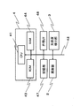
図1は、実施形態の血中脂質濃度計測装置の構成を示す図である。図2は、実施形態の血中脂質濃度計測装置の制御系の構成を示すブロック図である。
FIG. 1 is a diagram showing the configuration of the blood lipid concentration measuring device of the embodiment. FIG. 2 is a block diagram showing a configuration of a control system of the blood lipid concentration measuring device of the embodiment.
図1に示すように、実施形態の血中脂質濃度計測装置1は、照射部2と、光強度検出部3と、制御部4と、を有する。
As shown in FIG. 1, the blood lipid concentration measuring device 1 of the embodiment includes an irradiation unit 2, a light intensity detection unit 3, and a control unit 4.
照射部2は、生体の所定の部位の生体外から生体内に向けて、所定の照射位置21に光を照射するための光源22を有する。光源22は、制御部4によって、照射する光の波長を調整することができる。光源22は、制御部4によって、照射強度を調整できる。
The irradiation unit 2 has a light source 22 for irradiating a predetermined irradiation position 21 with light from outside the living body to the inside of the living body at a predetermined part of the living body. The light source 22 can adjust the wavelength of the emitted light by the control unit 4. The irradiation intensity of the light source 22 can be adjusted by the control unit 4.
実施形態の照射部2は、後述する制御部4による実効減衰係数μeffの算出方法に応じて、光の連続的な照射や光のパルス状の照射等の光を照射する時間の長さを任意に調整することができる。
In the irradiation unit 2 of the embodiment, the length of time for irradiating light such as continuous irradiation of light or pulsed irradiation of light is arbitrary according to the calculation method of the effective attenuation coefficient μeff by the control unit 4 described later. Can be adjusted to.
照射部2は、波長が固定された光源22を用いてもよい。照射部2は、波長が異なる複数の光源あるいは複数の波長の光を混合したものであってもよい。照射部2は、例えば、蛍光灯、LED、レーザー、白熱灯、HID、ハロゲンランプ等である。照射部2の照度は、制御部4により制御される。実施形態では、光源22はLED(Light Emitting Diode)である。
The irradiation unit 2 may use a light source 22 having a fixed wavelength. The irradiation unit 2 may be a mixture of a plurality of light sources having different wavelengths or light having a plurality of wavelengths. The irradiation unit 2 is, for example, a fluorescent lamp, an LED, a laser, an incandescent lamp, a HID, a halogen lamp, or the like. The illuminance of the irradiation unit 2 is controlled by the control unit 4. In the embodiment, the light source 22 is an LED (Light Emitting Diode).
実施形態の光源22は、相異なる第1波長の光及び第2波長の光を照射する。第1波長の光は、ヘモグロビンによる吸収が小さい波長であり、血中の脂質濃度を計測するための光である。第2波長の光は、ヘモグロビンによる吸収が大きい波長であり、静脈の深さと直径を測定するための光である。
The light source 22 of the embodiment irradiates different first wavelength light and second wavelength light. The light of the first wavelength is a wavelength at which absorption by hemoglobin is small, and is light for measuring the lipid concentration in blood. The light of the second wavelength is a wavelength that is largely absorbed by hemoglobin, and is light for measuring the depth and diameter of veins.
第1波長の光は、ヘモグロビンによる吸収が少ない波長が望ましく、800nm±50nm(すなわち、750nm以上850nm以下)の波長域の光であることが好ましい。
The first wavelength light is preferably a wavelength that is less absorbed by hemoglobin, and is preferably light in the wavelength range of 800 nm ± 50 nm (that is, 750 nm or more and 850 nm or less).
第2波長の光は、ヘモグロビンによる吸収が強くなる波長が望ましく、500nm以上600nm以下の光であることが好ましい。
The light of the second wavelength is preferably a wavelength at which absorption by hemoglobin is strong, and is preferably light of 500 nm or more and 600 nm or less.
光強度検出部3は、生体から生体外に放出される光を受光して、その光強度を検出する。複数の光強度検出部3(31、32)を用いる場合には、光強度検出部3(31、32)は、照射位置21から各々異なる距離に設置される。図1に示すように、実施形態では、照射位置21から所定の間隔で同一面上でかつ直線状に、第1光強度検出部31及び第2光強度検出部32が順に並ぶ。光強度検出部3は、フォトダイオードやCCDやCMOSでよい。
The light intensity detection unit 3 receives the light emitted from the living body to the outside of the living body and detects the light intensity. When a plurality of light intensity detecting units 3 (31, 32) are used, the light intensity detecting units 3 (31, 32) are installed at different distances from the irradiation position 21. As shown in FIG. 1, in the embodiment, the first light intensity detecting unit 31 and the second light intensity detecting unit 32 are arranged in order on the same surface and linearly from the irradiation position 21 at predetermined intervals. The light intensity detection unit 3 may be a photodiode, a CCD, or a CMOS.
光強度検出部3で受光した光は光電流に変換され、この光電流は、CPU41で処理される。
The light received by the light intensity detection unit 3 is converted into a photocurrent, and this photocurrent is processed by the CPU 41.
図1に示すように、実施形態では、照射位置21から第1光強度検出部31による第1検出位置331までの距離を第1距離ρ1とし、照射位置21から第2光強度検出部32による第2検出位置332までの距離を第2距離ρ2とする。
As shown in FIG. 1, in the embodiment, the distance from the irradiation position 21 to the first detection position 331 by the first light intensity detection unit 31 is set to the first distance ρ 1, and the distance from the irradiation position 21 to the second light intensity detection unit 32 is set. Let the distance to the second detection position 332 according to be the second distance ρ 2 .
第1光強度検出部31による第1検出位置331と照射部2の照射位置21との第1距離ρ1は、3~8mmとするのが好ましい。入出射距離が3~8mmと短い場合には、静脈血の特性が支配的となる計測結果が得られる。第2光強度検出部32による第2検出位置332と照射部2の照射位置21との第2距離ρ2は、10~15mmとするのが好ましい。入出射距離が10~15mmと長い場合には、静脈外の周囲組織が支配的となる計測結果が得られる。
The first distance ρ 1 between the first detection position 331 by the first light intensity detection unit 31 and the irradiation position 21 of the irradiation unit 2 is preferably 3 to 8 mm. When the entry / exit distance is as short as 3 to 8 mm, measurement results in which the characteristics of venous blood dominate can be obtained. The second distance ρ 2 between the second detection position 332 by the second light intensity detection unit 32 and the irradiation position 21 of the irradiation unit 2 is preferably 10 to 15 mm. When the entry / exit distance is as long as 10 to 15 mm, measurement results can be obtained in which the surrounding tissue outside the vein is dominant.
図3に示すように、光を生体に照射する照射位置21と、生体中の血液(図中のE)から放出される光強度を検出する検出位置33との間に所定の距離ρを設ける。所定の距離ρを設けることにより、照射した光(図中のA)が生体表面及び表面近傍の散乱体により反射して直接的に生体から放出される光(図中のB)の影響を抑制する。照射した光が、血液が存在する深さに達したのち、血液中の脂質(図中のD)によって光が反射する。
As shown in FIG. 3, a predetermined distance ρ is provided between the irradiation position 21 for irradiating the living body with light and the detection position 33 for detecting the light intensity emitted from the blood (E in the figure) in the living body. .. By providing a predetermined distance ρ, the effect of the irradiated light (A in the figure) reflected by the scattering body on the surface of the living body and the scattering body near the surface and directly emitted from the living body (B in the figure) is suppressed. do. After the irradiated light reaches the depth at which blood exists, the light is reflected by the lipid in the blood (D in the figure).
なお、複数の検出位置331、332等を設ける場合の配列は、照射位置21から各々異なる距離に配置されるのであれば直線状に限定されるものではなく、円状、波状、ジグザグ状など、適宜選択することができる。また、照射位置21から検出位置331、332等までの第1距離ρ1や第2距離ρ2、検出位置331、332同士の間隔は、一定の間隔に限定されるものではなく、連続的でもよい。
The arrangement when a plurality of detection positions 331, 332, etc. are provided is not limited to a linear shape as long as they are arranged at different distances from the irradiation position 21, but may be circular, wavy, zigzag, or the like. It can be selected as appropriate. Further, the intervals between the first distance ρ 1 and the second distance ρ 2 from the irradiation position 21 to the detection positions 331, 332, etc., and the detection positions 331 and 332 are not limited to a fixed interval, and may be continuous. good.
次に、血管検知装置1の制御系の構成について説明する。図2は実施形態の血管検知装置1のブロック図である。システムバス42を介して、CPU(Central Processing Unit)41、ROM(Read Only Memory)43、RAM(Random Access Memory)44、記憶部47、外部I/F(Interface)48、照射部2、及び、光強度検出部3が接続される。CPU41とROM43とRAM44とで制御部(コントローラー)4を構成する。
Next, the configuration of the control system of the blood vessel detection device 1 will be described. FIG. 2 is a block diagram of the blood vessel detection device 1 of the embodiment. CPU (Central Processing Unit) 41, ROM (Read Only Memory) 43, RAM (Random Access Memory) 44, storage unit 47, external I / F (Interface) 48, irradiation unit 2, and The light intensity detection unit 3 is connected. The control unit (controller) 4 is composed of the CPU 41, the ROM 43, and the RAM 44.
ROM43は、CPU41により実行されるプログラムや閾値を予め記憶する。
The ROM 43 stores in advance the programs and thresholds executed by the CPU 41.
RAM44は、CPU41が実行するプログラムを展開するエリアと、プログラムによるデータ処理の作業領域となるワークエリアなどの様々なメモリエリア等を有する。RAM44は、後述する図4に示す静脈深さ・直径推定マップの校正データ(第1校正データ)を記憶する記憶エリアを有する。RAM44は、後述する図5に示す血中脂質濃度校正曲線群の校正データ(第2校正データ)を記憶する記憶エリアを有する。
The RAM 44 has various memory areas such as an area for developing a program executed by the CPU 41 and a work area for data processing by the program. The RAM 44 has a storage area for storing calibration data (first calibration data) of the vein depth / diameter estimation map shown in FIG. 4, which will be described later. The RAM 44 has a storage area for storing calibration data (second calibration data) of the blood lipid concentration calibration curve group shown in FIG. 5, which will be described later.
記憶部47は、検知・算出された光強度等やμeff等のデータを記憶する。記憶部47は、HDD(Hard Disk Drive)や、フラッシュメモリや、SSD(Solid State Drive)等の、不揮発性に記憶する内部メモリーでよい。記憶部47は、後述する第1校正データ及び第2校正データを記憶してもよい。
The storage unit 47 stores data such as the detected / calculated light intensity and μ eff. The storage unit 47 may be an internal memory such as an HDD (Hard Disk Drive), a flash memory, or an SSD (Solid State Drive) that stores non-volatilely. The storage unit 47 may store the first calibration data and the second calibration data, which will be described later.
外部I/F48は、例えばクライアント端末(PC)などの外部装置と通信するためのインターフェースである。外部I/F48は、外部装置とデータ通信を行うインターフェースであれば良く、たとえば、外部装置にローカルに接続する機器(USBメモリ等)であっても良いし、ネットワークを介して通信するためのネットワークインターフェイスであっても良い。
The external I / F48 is an interface for communicating with an external device such as a client terminal (PC). The external I / F48 may be an interface for data communication with an external device, for example, a device (USB memory or the like) locally connected to the external device, or a network for communication via a network. It may be an interface.
制御部4は、光検出強度部3(31、32)により検出された光強度に基づき、第2波長の光強度から血管深さと直径情報(血管太さ)を算出し、第1波長の光強度から実効減衰係数の変化量を算出し、実効減衰係数の変化量と血管深さと直径情報とから、血中の脂質濃度を算出する。
The control unit 4 calculates the blood vessel depth and diameter information (blood vessel thickness) from the light intensity of the second wavelength based on the light intensity detected by the light detection intensity unit 3 (31, 32), and the light of the first wavelength. The amount of change in the effective attenuation coefficient is calculated from the intensity, and the blood lipid concentration is calculated from the amount of change in the effective attenuation coefficient, the blood vessel depth, and the diameter information.
以下、制御部4による脂質濃度の算出処理について説明する。
Hereinafter, the lipid concentration calculation process by the control unit 4 will be described.
実施形態は、静脈を計測対象とし、後方散乱光の空間分解計測に基づく無侵襲の血中脂質濃度計測装置及び方法である。実施形態は、2波長の計測により、静脈の深さと直径の同時推定も加えた血中脂質濃度計測装置及び方法である。なお、実施形態では静脈の直径を推定するが、これに限られず、静脈の半径等、静脈の太さを推定してもよい。また、計測対象の血管は、静脈に限られず、例えば、浅い位置にある動脈でもよい。
The embodiment is a non-invasive blood lipid concentration measuring device and method based on spatial decomposition measurement of backscattered light, targeting veins. An embodiment is a blood lipid concentration measuring device and method in which the depth and diameter of a vein are simultaneously estimated by measuring two wavelengths. In the embodiment, the diameter of the vein is estimated, but the present invention is not limited to this, and the thickness of the vein such as the radius of the vein may be estimated. Further, the blood vessel to be measured is not limited to a vein, and may be, for example, an artery at a shallow position.
実施形態では、制御部4は、ヘモグロビンの吸収が小さい波長(第1波長)で得られた光強度から実効減衰係数μeffを算出する。次に、制御部4は、へモグロビンの吸収が大きい波長(第2波長)による測定で得られた静脈の深さと直径の推定値で実効減衰係数μeffを補正し、補正した実効減衰係数μeffから脂質濃度を算出する。
In the embodiment, the control unit 4 calculates the effective attenuation coefficient μ eff from the light intensity obtained at a wavelength (first wavelength) where the absorption of hemoglobin is small. Next, the control unit 4 corrects the effective attenuation coefficient μ eff with the estimated values of the depth and diameter of the vein obtained by the measurement at the wavelength (second wavelength) where the absorption of hemoglobin is large, and the corrected effective attenuation coefficient μ. Calculate the lipid concentration from eff.
まず、制御部4は、ヘモグロビンの吸収が小さい波長(第1波長)で得られた光強度から実効減衰係数μeffを算出する。一様媒質では,後方散乱光強度R(ρ)と入出射間距離ρの関係は以下の式で近似できる。
First, the control unit 4 calculates the effective attenuation coefficient μ eff from the light intensity obtained at a wavelength (first wavelength) in which hemoglobin absorption is small. In a uniform medium, the relationship between the backscattered light intensity R (ρ) and the entrance / exit distance ρ can be approximated by the following equation.
(数1)
 (Number 1)
ここでS0は光源強度、μaは吸収係数、μeffは実効減衰係数を表す。μeffは散乱係数と吸収係数に依存する係数である。通常、赤血球濃度は比較的安定であるため、吸収係数は大きく変化しない。その場合は、μeffは散乱係数のみに大きく依存する。したがって、ρに対する光強度からμeffを算出することで脂質濃度を算出できる。また、実施形態においては脂質濃度ではなく濁度などで表記してもよい。
Here, S 0 represents the light source intensity, μ a represents the absorption coefficient, and μ eff represents the effective attenuation coefficient. μ eff is a coefficient that depends on the scattering coefficient and the absorption coefficient. Normally, the red blood cell concentration is relatively stable, so the absorption coefficient does not change significantly. In that case, μ eff depends largely only on the scattering coefficient. Therefore, the lipid concentration can be calculated by calculating μ eff from the light intensity with respect to ρ. Further, in the embodiment, it may be expressed by turbidity or the like instead of the lipid concentration.
なお、ここでの入出射間距離ρは、後述する第1距離ρ1である。また、第1波長で得られた光強度から算出された実効減衰係数μeffをμeff_1(第1実効減衰係数)とする。
The distance ρ between entry and exit here is the first distance ρ 1 described later. Further, the effective attenuation coefficient μ eff calculated from the light intensity obtained at the first wavelength is defined as μ eff_1 (first effective attenuation coefficient).
次に、制御部4は、へモグロビンの吸収が大きい波長(第2波長)による測定で得られた実効減衰係数μeffから、静脈の深さと直径の推定値を算出する。
Next, the control unit 4 calculates an estimated value of the depth and diameter of the vein from the effective attenuation coefficient μ eff obtained by the measurement at the wavelength (second wavelength) where the hemoglobin is absorbed greatly.
脂質濃度算出は静脈の深さと直径に大きく依存する。一方、血中ヘモグロビンの吸収が大きい波長(第2波長)では静脈内の吸収が支配的であるため、静脈内に入った光はほとんど減衰されてしまい、散乱すなわち脂質濃度が変化しても計測されるμeffの変化は小さい。そのため、第2波長では脂質濃度に依存せずに、静脈の深さと直径を推定できる。
Lipid concentration calculation largely depends on the depth and diameter of the vein. On the other hand, at wavelengths where the absorption of hemoglobin in the blood is large (second wavelength), the absorption in the vein is dominant, so the light entering the vein is almost attenuated, and even if the scattering, that is, the lipid concentration changes, it is measured. The change in μ eff that is made is small. Therefore, at the second wavelength, the depth and diameter of the vein can be estimated independently of the lipid concentration.
入出射間距離が短い場合の実効減衰係数μeffは、静脈血の特性が支配的であり、長い場合は静脈外の周囲組織が支配的である。そこで、制御部4は、距離が短い、例えば3~8mm(第1距離ρ1)にある第1光強度検出部31が検出した光強度からμeff_n(第2実効減衰係数)を算出し、距離が長い、例えば10~15 mm(第2距離ρ2)にある第2光強度検出部32が検出した光強度からμeff_f(第3実効減衰係数)を算出する。
The effective attenuation coefficient μ eff when the distance between entry and exit is short is dominated by the characteristics of venous blood, and when the distance is long, the surrounding tissue outside the vein is dominant. Therefore, the control unit 4 calculates μ eff_n (second effective attenuation coefficient) from the light intensity detected by the first light intensity detection unit 31 at a short distance, for example, 3 to 8 mm (first distance ρ 1). Μ eff_f (third effective attenuation coefficient) is calculated from the light intensity detected by the second light intensity detection unit 32 at a long distance, for example, 10 to 15 mm (second distance ρ 2).
なお、第1距離ρ1は3~8mmに限られるものではなく、4~7mm程度に範囲を狭めたり、浅めの静脈を想定した場合には2~5mm程度と近づけたり、深い静脈を対象とした場合は6~9mm程度と遠ざけることでもよく、測定対象の一個人の決まった場所での測定に応じて好適な距離が定められる。第2距離ρ2も同様である。
The first distance ρ 1 is not limited to 3 to 8 mm, but the range is narrowed to about 4 to 7 mm, and when a shallow vein is assumed, it is approached to about 2 to 5 mm, and deep veins are targeted. In that case, the distance may be as far as 6 to 9 mm, and a suitable distance is determined according to the measurement at a fixed place of one individual to be measured. The same applies to the second distance ρ 2.
図4は、静脈深さ・直径推定マップである。図4は、ヘモグロビンの吸収が大きい波長600nm(第2波長)における、μeff_n/μeff_f(第2実効減衰係数と第3実効減衰係数との比)に対するμeff_n(第2実効減衰係数)を静脈の深さ(血管深さ)と直径(血管太さ)を変えて予め作成・取得した第1校正データである。
FIG. 4 is a vein depth / diameter estimation map. 4, in the absorption of hemoglobin is large wavelength 600 nm (the second wavelength), μ eff_n / μ eff_f μ eff_n for (second effective attenuation coefficient and the ratio of the third effective attenuation coefficient) (second effective attenuation coefficient) This is the first calibration data created / acquired in advance by changing the depth (blood vessel depth) and the diameter (blood vessel thickness) of the vein.
図4には、校正線が3本(図中のA、B、C)記載されている。第2実効減衰係数μeff_nは、静脈血の実効減衰係数μeffが支配的であり、静脈の直径に大きく依存する。したがって、図4において、静脈の直径が1,2,3 mmと増えると、縦軸方向上に校正線がAからB、BからCへとシフトする。
In FIG. 4, three calibration lines (A, B, C in the figure) are shown. The second effective attenuation coefficient μ eff_n is dominated by the effective attenuation coefficient μ eff of venous blood and largely depends on the diameter of the vein. Therefore, in FIG. 4, when the diameter of the vein increases to 1,2,3 mm, the calibration line shifts from A to B and from B to C in the vertical axis direction.
一方、第2実効減衰係数と第3実効減衰係数との比μeff_n/μeff_fは周囲媒質に対する静脈血の比となり、静脈の深さに大きく依存する。静脈が深いと周囲媒質に埋もれ比が小さくなり、浅い場合はその逆となる。これは周囲媒質に対して静脈血のμeffは大きい前提であり、第2波長では問題なく成立する。よって、図4において横軸のμeff_n/μeff_fが小さい場合は静脈が深く、横軸のμeff_n/μeff_fが大きい場合は静脈が浅い校正線となる。
On the other hand, the ratio μ eff_n / μ eff_f between the second effective attenuation coefficient and the third effective attenuation coefficient is the ratio of venous blood to the surrounding medium and largely depends on the depth of the vein. If the vein is deep, it will be buried in the surrounding medium and the ratio will be small, and if it is shallow, the opposite will be true. This is a premise that the μ eff of venous blood is large with respect to the surrounding medium, and it holds without any problem at the second wavelength. Thus, veins if μ eff_n / μ eff_f the horizontal axis is small deeply when μ eff_n / μ eff_f the horizontal axis is large becomes veins shallow calibration line in FIG.
横軸が静脈の太さのみに依存し、縦軸が静脈の深さのみに依存する独立の関係であれば、図4に示すマップを作成する必要性は低いが、実際の静脈の場合には、計測される実効減衰係数は太さと深さの相互影響を受け、独立に扱うことは好ましくない。図4のマップの歪がそれぞれの相互影響を表すことになる。
If the horizontal axis depends only on the thickness of the vein and the vertical axis depends only on the depth of the vein, it is not necessary to create the map shown in FIG. 4, but in the case of an actual vein. The measured effective damping coefficient is affected by the mutual influence of thickness and depth, and it is not preferable to treat them independently. The distortion of the map in FIG. 4 represents their mutual influence.
図4の推定マップは、想定される範囲で静脈の直径、深さを変えて数値シミュレーションにより、μeff_nとμeff_fを求めて図4にプロットすることであらかじめ作成可能である。また、シミュレーションによらず、実験的にも、ファントムモデルまたは超音波診断装置で静脈の直径、深さを予め計測することにより、実際の人体に対しても、原理的にあらかじめ作成可能である。
The estimation map of FIG. 4 can be created in advance by obtaining μ eff_n and μ eff_f by numerical simulation while changing the diameter and depth of veins within the assumed range and plotting them in FIG. In principle, it can be created in advance for an actual human body by measuring the diameter and depth of veins in advance with a phantom model or an ultrasonic diagnostic apparatus, not by simulation but experimentally.
制御部4は、このような第2実効減衰係数と第3実効減衰係数との比に対する第2実効減衰係数を、血管深さと血管太さを変えてあらかじめ作成した静脈の直径、深さの校正曲線の第1校正データを格納し、当該第1校正データに基づいて、μeff_n/μeff_fとμeff_nとから、静脈の深さと直径を算出する。
The control unit 4 calibrates the diameter and depth of the vein prepared in advance by changing the blood vessel depth and the blood vessel thickness with respect to the second effective attenuation coefficient with respect to the ratio of the second effective attenuation coefficient and the third effective attenuation coefficient. The first calibration data of the curve is stored, and the depth and diameter of the vein are calculated from μ eff_n / μ eff_f and μ eff_n based on the first calibration data.
なお、図4の推定マップから静脈の直径、深さをmm単位程度で推定可能であるが、図4のマップは粗いため、線形補間(非線形補間も可能)や粗さを埋めるよう直径、深さを細かく変化させてシミュレーションすることで、小数点以下1から2桁のmm単位程度の確度で直径と深さを求めることでもよい。図4の推定マップ(第1校正データ)は、静脈の直径ごとに作成され、RAM44に記憶される。
The diameter and depth of veins can be estimated in mm units from the estimation map in FIG. 4, but since the map in FIG. 4 is rough, linear interpolation (nonlinear interpolation is also possible) and diameter and depth to fill the roughness are possible. The diameter and depth may be obtained with an accuracy of about 1 to 2 digits after the decimal point in mm units by simulating by finely changing the interpolation value. The estimation map (first calibration data) of FIG. 4 is created for each vein diameter and stored in the RAM 44.
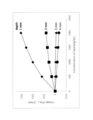
次に、制御部4は、図4の静脈深さ・直径推定マップで得られた静脈の深さと直径の推定値で、実効減衰係数μeffを補正し、補正したμeffから脂質濃度の変化量を算出する。図5に、波長810 nm(第1波長)における脂質濃度の校正曲線の一例を示す。図5の縦軸は、μeff_1(第1実効減衰係数)の変化量を示し、横軸は、脂質濃度を示す。図5は、縦軸、横軸それぞれ脂質がない状態を初期値としてそれからの変化量としてグラフ化したものである。なお、絶対値としての校正線を求めることも可能である。これにより、先に求めた静脈の深さと直径に対応した校正曲線から静脈血脂質濃度の推定が可能となる。
Next, the control unit 4 corrects the effective attenuation coefficient μ eff with the estimated values of the vein depth and diameter obtained from the vein depth / diameter estimation map of FIG. 4, and changes the lipid concentration from the corrected μ eff. Calculate the amount. FIG. 5 shows an example of a calibration curve of lipid concentration at a wavelength of 810 nm (first wavelength). The vertical axis of FIG. 5 shows the amount of change in μ eff_1 (first effective attenuation coefficient), and the horizontal axis shows the lipid concentration. FIG. 5 is a graph showing the state in which there is no lipid on each of the vertical and horizontal axes as the initial value and the amount of change thereafter. It is also possible to obtain the calibration line as an absolute value. This makes it possible to estimate the venous blood lipid concentration from the calibration curve corresponding to the previously obtained vein depth and diameter.
図5は、血中脂質濃度校正曲線群である。図5は、第1実効減衰係数の変化量に対する脂質濃度を、血管深さと血管太さを変えて予め作成・取得した第2校正データである。図5の校正線を導出する手法について説明する。第1距離のヘモグロビンの吸収が小さい第1波長のμeff_1(第1実効減衰係数)を横軸の脂質濃度に相当する血中の散乱係数を変化させて求めた図5のような校正グラフを、静脈の直径を変えたシミュレーションにより、静脈の直径毎の図5のようなグラフを準備する。なお、図5は、静脈の直径を2mmと固定して、深さ(図中のdepth)を変えた校正線をまとめて表示したものである。静脈の直径1mm、3mm等、それぞれの直径毎に図5のような校正線群のグラフを作成する(不図示)。実際には、図5の校正線群のグラフ(第2校正データ)は、静脈の直径ごとに作成され、RAM44に記憶される。
FIG. 5 is a group of blood lipid concentration calibration curves. FIG. 5 is the second calibration data prepared and acquired in advance for the lipid concentration with respect to the amount of change in the first effective attenuation coefficient by changing the blood vessel depth and the blood vessel thickness. The method of deriving the calibration line of FIG. 5 will be described. A calibration graph as shown in FIG. 5 obtained by changing the blood scattering coefficient corresponding to the lipid concentration on the horizontal axis for μ eff_1 (first effective attenuation coefficient) of the first wavelength in which the absorption of hemoglobin at the first distance is small. , Prepare a graph as shown in FIG. 5 for each vein diameter by a simulation in which the vein diameters are changed. Note that FIG. 5 shows the calibration lines in which the diameter of the vein is fixed at 2 mm and the depth (depth in the figure) is changed. A graph of the calibration line group as shown in FIG. 5 is created for each diameter of veins such as 1 mm and 3 mm in diameter (not shown). Actually, the graph of the calibration line group of FIG. 5 (second calibration data) is created for each vein diameter and stored in the RAM 44.
そして、図4の静脈深さ・直径推定マップで先に推定した静脈の直径に応じた図5の校正群を選択する。次に、血中脂質濃度校正曲線群の中から、図4の静脈深さ・直径推定マップで先に推定した静脈の深さ(図中のdepth)の校正線を選択し、その時の第1波長の計測値から求めたμeff_1(第1実効減衰係数)により脂質濃度に換算することができる。
Then, the calibration group of FIG. 5 according to the vein diameter previously estimated by the vein depth / diameter estimation map of FIG. 4 is selected. Next, from the blood lipid concentration calibration curve group, select the calibration line of the vein depth (depth in the figure) previously estimated by the vein depth / diameter estimation map of FIG. 4, and select the first calibration line at that time. It can be converted to lipid concentration by μ eff_1 (first effective attenuation coefficient) obtained from the measured value of wavelength.
制御部4は、このような第1実効減衰係数の変化量に対する脂質濃度を、血管深さと血管太さを変えてあらかじめ作成した血中脂質濃度の第2校正データを格納し、当該第2校正データに基づいて、静脈の深さと直径と第1波長の計測値から求めたμeff_1(第1実効減衰係数)とから、脂質濃度を算出する。
The control unit 4 stores the second calibration data of the blood lipid concentration prepared in advance by changing the blood vessel depth and the blood vessel thickness for the lipid concentration with respect to the amount of change in the first effective attenuation coefficient, and the second calibration is performed. Based on the data, the lipid concentration is calculated from the depth and diameter of the vein and μ eff_1 (first effective attenuation coefficient) obtained from the measured values of the first wavelength.
なお、図5の血中脂質濃度校正曲線群は粗いので、図4の校正マップの説明と同様に、線形補間や細かくシミュレーションするなどして、補間してもよい。
Since the blood lipid concentration calibration curve group in FIG. 5 is rough, it may be interpolated by linear interpolation or detailed simulation as in the explanation of the calibration map in FIG.
以上のような構成を備える実施形態の血中脂質濃度計測装置1において、予め設定されているプログラムに基づいて、血中脂質濃度計測装置1は、血中脂質濃度計測処理を実行する。図6は、実施形態の血中脂質濃度計測処理のフローチャートである。
In the blood lipid concentration measuring device 1 of the embodiment having the above configuration, the blood lipid concentration measuring device 1 executes the blood lipid concentration measuring process based on a preset program. FIG. 6 is a flowchart of the blood lipid concentration measurement process of the embodiment.
照射部が、照射位置21に連続光を照射する(ステップ101)。照射部2は、ヘモグロビンによる吸収が小さい第1波長と、ヘモグロビンによる吸収が大きい第2波長の光と、を照射する。
The irradiation unit irradiates the irradiation position 21 with continuous light (step 101). The irradiation unit 2 irradiates light having a first wavelength that is less absorbed by hemoglobin and light having a second wavelength that is more absorbed by hemoglobin.
第1光強度検出部31が第1検出位置331における光強度を検出するとともに、第2光強度検出部32が第2検出位置332の光強度を検出する(ステップ102)。照射位置21と第1検出位置331との第1距離ρ1は、静脈血の特性が支配的となる距離であり、照射位置21と第2検出位置332との第2距離ρ2は、第1距離ρ1よりも長く、静脈外の周囲組織が支配的となる計測結果が得られる距離である。
The first light intensity detection unit 31 detects the light intensity at the first detection position 331, and the second light intensity detection unit 32 detects the light intensity at the second detection position 332 (step 102). The first distance ρ 1 between the irradiation position 21 and the first detection position 331 is the distance at which the characteristics of venous blood dominate, and the second distance ρ 2 between the irradiation position 21 and the second detection position 332 is the first. It is a distance longer than 1 distance ρ 1 and a measurement result can be obtained in which the surrounding tissue outside the vein is dominant.
制御部4は、第1距離(例えば、3~8mm)における第1波長(例えば、810nm)の強度からμeff_1を算出する。制御部4は、第1距離ρ1(例えば、3~8mm)における第2波長(例えば、600nm)の強度からμeff_nを算出する。制御部4は、第2距離ρ2(例えば、10~15mm)における第2波長(例えば、600nm)の強度からμeff_fを算出する(ステップ103)。なお、μeffの算出法は上述した。
The control unit 4 calculates μ eff_1 from the intensity of the first wavelength (for example, 810 nm) at the first distance (for example, 3 to 8 mm). The control unit 4 calculates μ eff_n from the intensity of the second wavelength (for example, 600 nm) at the first distance ρ 1 (for example, 3 to 8 mm). The control unit 4 calculates μ eff_f from the intensity of the second wavelength (for example, 600 nm) at the second distance ρ 2 (for example, 10 to 15 mm) (step 103). The calculation method of μ eff is described above.
制御部4は、図4に示す、予め作成し記憶した静脈の深さ・直径の校正データに基づき、算出された第2波長におけるμeff_n/μeff_fとμeff_nとから、静脈の深さと直径を推定する(ステップ104)。
The control unit 4 has the vein depth and diameter from μ eff_n / μ eff_f and μ eff_n at the second wavelength calculated based on the vein depth / diameter calibration data created and stored in advance as shown in FIG. Is estimated (step 104).
制御部4は、図5に示す、予め作成し記憶した血中脂質濃度の校正データに基づき、算出された静脈の深さと直径と、第1波長におけるμeff_1の変化量と、から血中脂質濃度を算出する(ステップ105)。
The control unit 4 uses the calculated vein depth and diameter and the amount of change in μ eff_1 at the first wavelength based on the blood lipid concentration calibration data prepared and stored in advance as shown in FIG. Calculate the concentration (step 105).
以上説明したように、本実施形態の血中脂質濃度計測装置及びその方法によれば、血中脂質濃度を安定的に計測することが可能となる。
As described above, according to the blood lipid concentration measuring device and its method of the present embodiment, it is possible to stably measure the blood lipid concentration.
以上、実施形態を説明したが、この実施形態は、例として提示したものであり、発明の範囲を限定することは意図していない。この新規な実施形態は、その他の様々な形態で実施されることが可能であり、発明の要旨を逸脱しない範囲で、種々の省略、置き換え、変更を行うことができる。この実施形態やその変形は、発明の範囲や要旨に含まれるとともに、特許請求の範囲に記載された発明とその均等の範囲に含まれる。
Although the embodiment has been described above, this embodiment is presented as an example and is not intended to limit the scope of the invention. This novel embodiment can be implemented in various other embodiments, and various omissions, replacements, and changes can be made without departing from the gist of the invention. This embodiment and its modifications are included in the scope and gist of the invention, and are also included in the scope of the invention described in the claims and the equivalent scope thereof.
1 血中脂質濃度計測装置
2 照射部
3 光強度検出部
4 制御部 1 Blood lipidconcentration measuring device 2 Irradiation unit 3 Light intensity detection unit 4 Control unit
2 照射部
3 光強度検出部
4 制御部 1 Blood lipid
Claims (7)
- 第1波長の光と、前記第1波長よりヘモグロビンによる吸収が大きい第2波長の光と、を被験体の所定位置に光を照射する照射部と、
前記照射部による光の照射位置から所定間隔をあけて、あるいは、連続的な位置での、前記被検体から放出される、複数の位置の光強度を検出する光強度検出部と、
前記第2波長の光強度から血管深さと血管太さを算出し、
前記第1波長の光強度から第1実効減衰係数の変化量を算出し、
前記第1実効減衰係数の変化量と、前記血管深さと、前記血管太さと、から、脂質濃度を算出する、制御部と、
を有する血中脂質濃度計測装置。 An irradiation unit that irradiates the light of the first wavelength and the light of the second wavelength, which is absorbed by hemoglobin more than the first wavelength, at a predetermined position of the subject.
A light intensity detection unit that detects light intensities at a plurality of positions emitted from the subject at predetermined intervals or at continuous positions from the light irradiation position by the irradiation unit.
The blood vessel depth and blood vessel thickness are calculated from the light intensity of the second wavelength.
The amount of change in the first effective attenuation coefficient is calculated from the light intensity of the first wavelength.
A control unit that calculates the lipid concentration from the amount of change in the first effective attenuation coefficient, the blood vessel depth, and the blood vessel thickness.
Blood lipid concentration measuring device having. - 前記第1波長は、750nm以上850nm以下であり、前記第2波長は、500nm以上600nm以下であることを特徴とする、請求項1に記載の血中脂質濃度計測装置。 The blood lipid concentration measuring apparatus according to claim 1, wherein the first wavelength is 750 nm or more and 850 nm or less, and the second wavelength is 500 nm or more and 600 nm or less.
- 前記光強度検出部は、
前記照射部から第1距離と、当該第1距離より長い第2距離と、における光強度を検出し、
前記制御部は、
前記第2波長における前記第1距離の第2実効減衰係数と、前記第2波長における前記第2距離の第3実効減衰係数と、を算出し、
前記第2実効減衰係数と前記第3実効減衰係数との比と、前記第2実効減衰係数とに基づいて、前記血管深さと前記血管太さを算出する、
請求項1または2に記載の血中脂質濃度計測装置。 The light intensity detection unit
The light intensity at the first distance from the irradiation unit and the second distance longer than the first distance is detected.
The control unit
The second effective attenuation coefficient of the first distance at the second wavelength and the third effective attenuation coefficient of the second distance at the second wavelength were calculated.
The blood vessel depth and the blood vessel thickness are calculated based on the ratio of the second effective attenuation coefficient and the third effective attenuation coefficient and the second effective attenuation coefficient.
The blood lipid concentration measuring device according to claim 1 or 2. - 前記制御部は、
前記第2実効減衰係数と前記第3実効減衰係数との比に対する前記第2実効減衰係数を、前記血管深さと前記血管太さを変えて予め作成した第1校正データを記憶し、当該第1校正データに基づいて、前記第2実効減衰係数と前記第3実効減衰係数との比と、前記第2実効減衰係数とから、前記血管深さと前記血管太さを算出する、請求項3に記載の血中脂質濃度計測装置。 The control unit
The first calibration data prepared in advance by changing the blood vessel depth and the blood vessel thickness for the second effective damping coefficient with respect to the ratio of the second effective damping coefficient and the third effective damping coefficient is stored, and the first calibration data is stored. The third aspect of claim 3, wherein the blood vessel depth and the blood vessel thickness are calculated from the ratio of the second effective attenuation coefficient and the third effective attenuation coefficient and the second effective attenuation coefficient based on the calibration data. Blood lipid concentration measuring device. - 前記制御部は、
前記第1実効減衰係数の変化量に対する前記脂質濃度を、前記血管深さと前記血管太さを変えて予め作成した第2校正データを記憶し、当該第2校正データに基づいて、前記第1実効減衰係数の変化量と前記血管深さと前記血管太さとから、脂質濃度を算出する、請求項4に記載の血中脂質濃度計測装置。 The control unit
The lipid concentration with respect to the amount of change in the first effective attenuation coefficient is stored in the second calibration data prepared in advance by changing the blood vessel depth and the blood vessel thickness, and the first effective is based on the second calibration data. The blood lipid concentration measuring apparatus according to claim 4, wherein the lipid concentration is calculated from the amount of change in the attenuation coefficient, the blood vessel depth, and the blood vessel thickness. - 前記第1距離は、3mm以上8mm以下であり、前記第2距離は、10mm以上15mm以下である請求項3から5のいずれかに記載の血中脂質濃度計測装置。 The blood lipid concentration measuring device according to any one of claims 3 to 5, wherein the first distance is 3 mm or more and 8 mm or less, and the second distance is 10 mm or more and 15 mm or less.
- 第1波長の光と、前記第1波長よりヘモグロビンによる吸収が大きい第2波長の光と、を被験体の所定位置に光を照射し、
前記光の照射位置から所定間隔をあけて、あるいは、連続的な位置での、前記被検体から放出される、複数の位置の光強度を検出し、
前記第2波長の光強度から血管深さと血管太さを算出し、
前記第1波長の光強度から第1実効減衰係数の変化量を算出し、
前記第1実効減衰係数の変化量と、前記血管深さと、前記血管太さと、から、脂質濃度を算出する、血中脂質濃度計測方法。 The light of the first wavelength and the light of the second wavelength, which is absorbed by hemoglobin more than the first wavelength, are irradiated to a predetermined position of the subject.
By detecting the light intensities at a plurality of positions emitted from the subject at predetermined intervals from the light irradiation position or at continuous positions, the light intensity is detected.
The blood vessel depth and blood vessel thickness are calculated from the light intensity of the second wavelength.
The amount of change in the first effective attenuation coefficient is calculated from the light intensity of the first wavelength.
A method for measuring a blood lipid concentration, which calculates a lipid concentration from the amount of change in the first effective attenuation coefficient, the blood vessel depth, and the blood vessel thickness.
Priority Applications (1)
| Application Number | Priority Date | Filing Date | Title |
|---|---|---|---|
| JP2021571228A JPWO2021145375A1 (en) | 2020-01-17 | 2021-01-14 |
Applications Claiming Priority (2)
| Application Number | Priority Date | Filing Date | Title |
|---|---|---|---|
| JP2020005791 | 2020-01-17 | ||
| JP2020-005791 | 2020-01-17 |
Publications (1)
| Publication Number | Publication Date |
|---|---|
| WO2021145375A1 true WO2021145375A1 (en) | 2021-07-22 |
Family
ID=76864401
Family Applications (1)
| Application Number | Title | Priority Date | Filing Date |
|---|---|---|---|
| PCT/JP2021/001038 WO2021145375A1 (en) | 2020-01-17 | 2021-01-14 | Device and method for measuring blood lipid level |
Country Status (2)
| Country | Link |
|---|---|
| JP (1) | JPWO2021145375A1 (en) |
| WO (1) | WO2021145375A1 (en) |
Citations (7)
| Publication number | Priority date | Publication date | Assignee | Title |
|---|---|---|---|---|
| JP2002107291A (en) * | 2000-10-03 | 2002-04-10 | Sysmex Corp | Non-invasive biological measuring device and method |
| JP2014124454A (en) * | 2012-12-27 | 2014-07-07 | Seiko Epson Corp | Method and apparatus for measuring blood component, and medical equipment |
| JP2015521282A (en) * | 2012-04-30 | 2015-07-27 | メイヨ フォンデーシヨン フォー メディカル エジュケーション アンド リサーチ | Spectroscopic system and method for improving focus location of temporal and spatial variation measurements |
| JP2017205264A (en) * | 2016-05-18 | 2017-11-24 | メディカルフォトニクス株式会社 | Blood lipid concentration measuring device and operation method thereof |
| WO2019123559A1 (en) * | 2017-12-20 | 2019-06-27 | メディカルフォトニクス株式会社 | Lipid measurement device and method therefor |
| US20190257759A1 (en) * | 2018-02-21 | 2019-08-22 | Olive Healthcare Inc. | Signal processing device of analyzing bio-signal and bio-signal analyzing apparatus using the same |
| JP2019201760A (en) * | 2018-05-22 | 2019-11-28 | メディカルフォトニクス株式会社 | Blood vessel detection device and method thereof |
-
2021
- 2021-01-14 WO PCT/JP2021/001038 patent/WO2021145375A1/en active Application Filing
- 2021-01-14 JP JP2021571228A patent/JPWO2021145375A1/ja active Pending
Patent Citations (7)
| Publication number | Priority date | Publication date | Assignee | Title |
|---|---|---|---|---|
| JP2002107291A (en) * | 2000-10-03 | 2002-04-10 | Sysmex Corp | Non-invasive biological measuring device and method |
| JP2015521282A (en) * | 2012-04-30 | 2015-07-27 | メイヨ フォンデーシヨン フォー メディカル エジュケーション アンド リサーチ | Spectroscopic system and method for improving focus location of temporal and spatial variation measurements |
| JP2014124454A (en) * | 2012-12-27 | 2014-07-07 | Seiko Epson Corp | Method and apparatus for measuring blood component, and medical equipment |
| JP2017205264A (en) * | 2016-05-18 | 2017-11-24 | メディカルフォトニクス株式会社 | Blood lipid concentration measuring device and operation method thereof |
| WO2019123559A1 (en) * | 2017-12-20 | 2019-06-27 | メディカルフォトニクス株式会社 | Lipid measurement device and method therefor |
| US20190257759A1 (en) * | 2018-02-21 | 2019-08-22 | Olive Healthcare Inc. | Signal processing device of analyzing bio-signal and bio-signal analyzing apparatus using the same |
| JP2019201760A (en) * | 2018-05-22 | 2019-11-28 | メディカルフォトニクス株式会社 | Blood vessel detection device and method thereof |
Also Published As
| Publication number | Publication date |
|---|---|
| JPWO2021145375A1 (en) | 2021-07-22 |
Similar Documents
| Publication | Publication Date | Title |
|---|---|---|
| JP4156373B2 (en) | An assembly device for the measurement of the optical properties of multilayer structures. | |
| US5920390A (en) | Fiberoptic interferometer and associated method for analyzing tissue | |
| JP5271700B2 (en) | System and method for correcting light reflectance measurements | |
| JP5463545B2 (en) | Concentration determination apparatus, concentration determination method and program | |
| JP5186044B2 (en) | Blood glucose level estimation device | |
| JP4872536B2 (en) | Biological component concentration measurement method | |
| JP2013052227A (en) | Apparatus and method for acquiring information on subject | |
| KR20190038513A (en) | Frequency domian based multi-wavelength bio-signal analysing apparatus and method thereof | |
| JP2017518792A (en) | Device and method for noninvasively determining a hematocrit value of a subject | |
| WO2019225612A1 (en) | Blood vessel detection device and method therefor | |
| JP6894088B2 (en) | Scatterer concentration measuring device and its method | |
| WO2021145375A1 (en) | Device and method for measuring blood lipid level | |
| CN105686800B (en) | Subject information acquisition device and its control method | |
| JP2008155011A (en) | Density measuring apparatus and its method | |
| TWI773713B (en) | Lipid measurement device and method therefor | |
| JP6894087B2 (en) | Lipid measuring device and its method | |
| JP6991634B1 (en) | Lipid concentration measuring device, program, and method | |
| Vogt et al. | Measurement and thermal dependence of biological tissue optical properties | |
| WO2020246455A1 (en) | Non-invasive measuring device, method, and program | |
| US20230058347A1 (en) | Blood vessel detection device and method therefor | |
| WO2020080409A1 (en) | Particle concentration measurement device, particle concentration measurement program, and particle concentration measurement method | |
| Hernandez-Quintanar et al. | Characterization of intralipid-10% in the range of 400–700 nm using light emitting diodes | |
| RU2545814C1 (en) | Method of determining physical-biological parameters of skin and concentration of haemoglobin derivatives in blood | |
| JP2023004122A (en) | High-sensitivity particle concentration measurement device | |
| TWI615131B (en) | Image based oxygen saturation measuring device and method thereof |
Legal Events
| Date | Code | Title | Description |
|---|---|---|---|
| 121 | Ep: the epo has been informed by wipo that ep was designated in this application |
Ref document number: 21740925 Country of ref document: EP Kind code of ref document: A1 |
|
| ENP | Entry into the national phase |
Ref document number: 2021571228 Country of ref document: JP Kind code of ref document: A |
|
| NENP | Non-entry into the national phase |
Ref country code: DE |
|
| 122 | Ep: pct application non-entry in european phase |
Ref document number: 21740925 Country of ref document: EP Kind code of ref document: A1 |