WO2021079522A1 - 腫瘍細胞群の特性を決定する方法、キット及びプログラム - Google Patents
腫瘍細胞群の特性を決定する方法、キット及びプログラム Download PDFInfo
- Publication number
- WO2021079522A1 WO2021079522A1 PCT/JP2019/042047 JP2019042047W WO2021079522A1 WO 2021079522 A1 WO2021079522 A1 WO 2021079522A1 JP 2019042047 W JP2019042047 W JP 2019042047W WO 2021079522 A1 WO2021079522 A1 WO 2021079522A1
- Authority
- WO
- WIPO (PCT)
- Prior art keywords
- drug
- cells
- cell group
- breast cancer
- nucleic acid
- Prior art date
- Legal status (The legal status is an assumption and is not a legal conclusion. Google has not performed a legal analysis and makes no representation as to the accuracy of the status listed.)
- Ceased
Links
Images











Classifications
-
- C—CHEMISTRY; METALLURGY
- C12—BIOCHEMISTRY; BEER; SPIRITS; WINE; VINEGAR; MICROBIOLOGY; ENZYMOLOGY; MUTATION OR GENETIC ENGINEERING
- C12Q—MEASURING OR TESTING PROCESSES INVOLVING ENZYMES, NUCLEIC ACIDS OR MICROORGANISMS; COMPOSITIONS OR TEST PAPERS THEREFOR; PROCESSES OF PREPARING SUCH COMPOSITIONS; CONDITION-RESPONSIVE CONTROL IN MICROBIOLOGICAL OR ENZYMOLOGICAL PROCESSES
- C12Q1/00—Measuring or testing processes involving enzymes, nucleic acids or microorganisms; Compositions therefor; Processes of preparing such compositions
- C12Q1/68—Measuring or testing processes involving enzymes, nucleic acids or microorganisms; Compositions therefor; Processes of preparing such compositions involving nucleic acids
- C12Q1/6897—Measuring or testing processes involving enzymes, nucleic acids or microorganisms; Compositions therefor; Processes of preparing such compositions involving nucleic acids involving reporter genes operably linked to promoters
-
- C—CHEMISTRY; METALLURGY
- C12—BIOCHEMISTRY; BEER; SPIRITS; WINE; VINEGAR; MICROBIOLOGY; ENZYMOLOGY; MUTATION OR GENETIC ENGINEERING
- C12M—APPARATUS FOR ENZYMOLOGY OR MICROBIOLOGY; APPARATUS FOR CULTURING MICROORGANISMS FOR PRODUCING BIOMASS, FOR GROWING CELLS OR FOR OBTAINING FERMENTATION OR METABOLIC PRODUCTS, i.e. BIOREACTORS OR FERMENTERS
- C12M1/00—Apparatus for enzymology or microbiology
- C12M1/34—Measuring or testing with condition measuring or sensing means, e.g. colony counters
-
- C—CHEMISTRY; METALLURGY
- C12—BIOCHEMISTRY; BEER; SPIRITS; WINE; VINEGAR; MICROBIOLOGY; ENZYMOLOGY; MUTATION OR GENETIC ENGINEERING
- C12Q—MEASURING OR TESTING PROCESSES INVOLVING ENZYMES, NUCLEIC ACIDS OR MICROORGANISMS; COMPOSITIONS OR TEST PAPERS THEREFOR; PROCESSES OF PREPARING SUCH COMPOSITIONS; CONDITION-RESPONSIVE CONTROL IN MICROBIOLOGICAL OR ENZYMOLOGICAL PROCESSES
- C12Q1/00—Measuring or testing processes involving enzymes, nucleic acids or microorganisms; Compositions therefor; Processes of preparing such compositions
- C12Q1/68—Measuring or testing processes involving enzymes, nucleic acids or microorganisms; Compositions therefor; Processes of preparing such compositions involving nucleic acids
- C12Q1/6876—Nucleic acid products used in the analysis of nucleic acids, e.g. primers or probes
- C12Q1/6883—Nucleic acid products used in the analysis of nucleic acids, e.g. primers or probes for diseases caused by alterations of genetic material
- C12Q1/6886—Nucleic acid products used in the analysis of nucleic acids, e.g. primers or probes for diseases caused by alterations of genetic material for cancer
Definitions
- Embodiments of the present invention relate to methods, kits and programs for determining the characteristics of tumor cell groups.
- drug susceptibility of breast cancer can be predicted by detecting the amount of transcript (mRNA) of a marker gene in breast cancer cells collected from a patient by the PCR method or the microarray method.
- mRNA transcript
- An embodiment of the present invention aims to provide a method, a kit and a program capable of accurately determining the characteristics of a tumor cell group.
- the method according to the embodiment is a method for determining the characteristics of a tumor cell group.
- a contact step of contacting a lipid particle containing a lipid membrane and a first nucleic acid contained in the lipid membrane with a tumor cell group (the first nucleic acid is a marker gene promoter sequence for predicting characteristics and a promoter sequence of a marker gene.
- the culture step of culturing the tumor cell group and the presence or absence of a signal from the reporter protein expressed from the reporter gene in each tumor cell contained in the tumor cell group.
- a detection step for detecting the amount and a counting step for measuring the number of characteristic tumor cells from the results of the detection step are included.
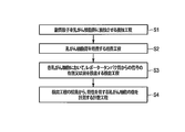
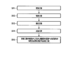
- FIG. 1 is a flowchart showing an example of the method of the first embodiment.
- FIG. 2 is a cross-sectional view showing an example of the lipid particles of the first embodiment.
- FIG. 3 is a schematic diagram showing an example of the first nucleic acid of the first embodiment.
- FIG. 4 is a flowchart showing an example of the method of the first embodiment.
- FIG. 5 is a schematic diagram and a graph showing a mode in which a signal is obtained in the method of the first embodiment.
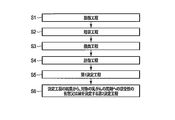
- FIG. 6 is a flowchart showing an example of the method of the second embodiment.
- FIG. 7 is a flowchart showing an example of the method of the second embodiment.
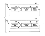
- FIG. 8 is a schematic diagram showing an example of the first nucleic acid used in the third embodiment.
- FIG. 9 is a schematic diagram showing an example of the first nucleic acid used in the third embodiment.
- FIG. 10 is a flowchart showing an example of the method of the third embodiment.
- FIG. 11 is a flowchart showing an example of the method of the third embodiment.
- FIG. 12 is a flowchart showing an example of the method of the fourth embodiment.
- FIG. 13 is a flowchart showing an example of the method of the fourth embodiment.
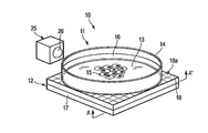
- FIG. 14 is a perspective view showing an example of a state when the cell culture detection device of the fifth embodiment is used.
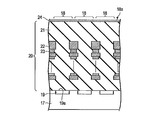
- FIG. 15 is an enlarged view of a cross section when the optical sensor shown in FIG. 14 is cut along AA'.
- FIG. 16 is a block diagram showing an example of the cell characterization system of the fifth embodiment.
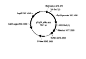
- FIG. 17 is a schematic diagram showing the vector used in Example 1.
- FIG. 18 is a schematic diagram showing the vector used in Example 1.
- FIG. 19 is a graph showing the experimental results of Example 3.
- FIG. 20 is a graph showing the experimental results of Example 3.
- FIG. 21 is a photomicrograph (JPEG format) showing the experimental results of Example 4.
- FIG. 22 is a micrograph (bitmap format) showing the experimental results of Example 4.
- the method according to the embodiment is a method of predicting the characteristics of tumor cells.
- a contact step of contacting a lipid particle containing a lipid membrane and a first nucleic acid contained in the lipid membrane with a tumor cell group (the first nucleic acid is a marker gene promoter sequence for predicting characteristics and a promoter sequence of a marker gene.
- the culture step of culturing the tumor cell group and the presence or absence of a signal from the reporter protein expressed from the reporter gene in each tumor cell contained in the tumor cell group.
- a detection step for detecting the amount and a counting step for measuring the number of characteristic tumor cells from the results of the detection step are included.
- a method according to the first embodiment is a method of determining the susceptibility of a breast cancer cell group to a drug.
- Breast cancer refers to, for example, a malignant tumor (neoplasm) formed in the mammary gland of an animal.
- Breast cancer includes those commonly referred to as “breast cancer” or “breast tumor” and the like, as well as ductal carcinoma and lobular carcinoma.
- Breast cancer includes those of any stage, for example, a state in which the cancer remains in the mammary gland, a state in which the cancer has spread to surrounding tissues, and a state in which the cancer has spread to lymph nodes. , And the condition where the cancer has spread to more distant organs.
- the breast cancer cell group is preferably a breast cancer cell group collected from a subject suffering from breast cancer, and includes a plurality of breast cancer cells.
- the breast cancer cell group is included in a sample obtained by, for example, aspiration with a needle (puncture aspiration, needle biopsy, mammothome biopsy, etc.) or surgical excision of a breast cancer lesion, or collection of mammary gland secretion (milk). It is a thing.
- the target is, for example, a mammal, preferably a human.
- the breast cancer cell group may be appropriately treated after collection. Appropriate treatments include, for example, shredding, dispersion, removal of other cell groups or isolation of breast cancer cell groups.
- the breast cancer cell group may be a cultured cell obtained by culturing a collected or pretreated breast cancer cell group in a manner that does not lose the characteristics of breast cancer.
- the breast cancer cell group may be an established breast cancer cell group or the like.
- the "breast cancer cell group” is also simply referred to as a "cell group”.
- “breast cancer cells” are also simply referred to as "cells”.
- the sensitivity of a cell group to a drug means whether or not the cell group receives the therapeutic effect of the drug or the degree of receiving the therapeutic effect.
- the susceptibility of a cell group to a specific drug means that the cell group receives a therapeutic effect by the drug, for example, the drug reduces or eliminates the cells contained in the cell group.
- the fact that the cell group does not have sensitivity to a specific drug means that the cell group has resistance (resistance) to the drug and has no or little therapeutic effect.
- the drug can be selected from, for example, drugs having antitumor activity commonly used in the treatment of breast cancer.
- the drug is selected from any of anticancer agents, hormonal therapeutic agents, molecular targeted agents, immunostimulators and the like.
- Anticancer agents include, for example, anthracyclines, microtubule agents, alkylating agents, metabolic antagonists or platinum complexes, and hormone therapeutic agents include, for example, aromatase inhibitors, LH-RH agonist preparations, anti-cancer agents. Includes estrogen drugs or progesterone drugs.
- the drug is not limited to the above.
- one drug whose susceptibility in the breast cancer cell group is investigated is selected from any of the above.
- a plurality of agents may be selected as described in the third embodiment. The following is an example of investigating one drug.
- the method for determining the susceptibility of breast cancer cells to a drug according to the embodiment includes, for example, the following steps shown in FIG.
- the lipid particle 1 includes, for example, a lipid membrane 2 and a first nucleic acid 3 encapsulated in a lumen 2a of the lipid membrane 2.
- the first nucleic acid 3 is condensed by, for example, the nucleic acid condensing peptide 4 and encapsulated in the lipid membrane 2. As will be described in detail later, it is used as a carrier for introducing the first nucleic acid 3 into breast cancer cells.
- the lipid membrane 2 is a substantially spherical hollow body formed by arranging a plurality of lipid molecules 2b in a non-covalent bond.
- the lipid membrane 2 may be a lipid monolayer or a lipid bilayer membrane. Further, the lipid membrane 2 may be composed of a single layer membrane or a multi-layered membrane.
- the lipid that is the material of the lipid membrane 2 includes, for example, a lipid that is the main component of the biological membrane (hereinafter, referred to as “base lipid”).
- the base lipid is a phospholipid or sphingolipid, such as diacylphosphatidylcholine, diacylphosphatidylethanolamine, ceramide, sphingomyelin, dihydrosphingomyelin, kefarin or cerebroside, or a combination thereof.
- 1,2-diore oil-sn-glycero-3-phosphoethanolamine DOPE
- 1,2-Stearoyl-sn-glycero-3-phosphoethanolamine DSPE
- 1,2-Dipalmitoyl-sn-glycero-3-phosphatidylcholine DPPC
- 1-Palmityl-2-oleoyl-sn-glycero-3-phosphatidylcholine POPC
- 1,2-di-O-octadecyl-3-trimethylammonium propane DOTMA
- 1,2-Giore Oil-3-Dimethylammonium Propane DODAP
- 1,2-Dimyristoylation-3-dimethylammonium propane 14: 0 DAP
- 1,2-Dipalmitoyl-3-dimethylammonium propane 16: 0 DAP
- 1,2-Distearoyl-3-dimethylammonium propane 18: 0 DAP
- the lipid membrane 2 preferably further contains the first lipid compound, the second lipid compound, or both described below in addition to the base lipid.
- the first lipid compound can be represented by the formula Q-CHR 2.
- Q is a nitrogen-containing aliphatic group containing two or more tertiary nitrogens and containing no oxygen.
- the first lipid compound for example, it is preferable to use a lipid having a structure represented by the following formula because the nucleic acid introduction efficiency is more excellent.
- the second lipid compound can be represented by the formula P- [X-W-Y-W'-Z] 2.
- P is an alkyleneoxy containing one or more ether bonds in the main chain.
- X is a divalent linking group each independently containing a tertiary amine structure.
- W is C 1 to C 6 alkylene, respectively.
- Y is a divalent linking group independently selected from the group consisting of a single bond, an ether bond, a carboxylic acid ester bond, a thiocarboxylic acid ester bond, a thioester bond, an amide bond, a carbamate bond and a urea bond.
- W' is independently a single bond or C 1 to C 6 alkylene, respectively.
- Z is independently a fat-soluble vitamin residues, sterol residue, or C 12 ⁇ C 22 aliphatic hydrocarbon group).
- the oxygen constituting the ether bond contained in P forms a hydrogen bond with the included nucleic acid, so that the amount of nucleic acid included increases.
- a second lipid compound having the following structure because the amount of nucleic acid included is more excellent.
- the total lipid molecule 2b contained in the lipid membrane 2 may be the base lipid, but the first lipid compound and / or the second lipid compound is about 100% of the total lipid molecule 2b. It is preferably contained in an amount of 20% to about 70% (molar ratio).
- the lipid membrane 2 is, for example, a polyamide oligomer derived from a PEG-modified lipid, particularly polyethylene glycol (PEG) dimyristylglycerol (DMG-PEG), omega-amino (oligoethylene glycol) alkanoic acid monomer (eg, US Patent No. 1). 6,320,017), preferably further containing monosialoganglioside.
- PEG polyethylene glycol
- DMG-PEG dimyristylglycerol
- omega-amino oligoethylene glycol alkanoic acid monomer
- Such lipids are preferably contained in an amount of about 1% to about 5% (molar ratio) with respect to the entire lipid molecule 2b.
- the lipid film 2 leaks inclusions such as a relatively highly biocompatible lipid for adjusting the biocompatibility of the lipid particle 1, a lipid having a functional group that binds a ligand to the lipid film 2, and cholesterol. It may contain a lipid such as a lipid for suppressing the above.
- the first nucleic acid 3 is easily introduced into breast cancer cells.
- the lipid particle 1 contains a compound of formula (1-01), a compound of formula (2-01), DOTAP, DOPE, cholesterol, and DMG-PEG, the efficiency of introduction into breast cancer cells Is particularly excellent, and is preferable.
- these components are contained in a ratio of 37: 15: 10.5: 10.5: 30: 2 (molar ratio).
- the first nucleic acid 3 contains, for example, the first reporter expression unit U1 as shown in FIG.
- the first reporter expression unit U1 contains, for example, a promoter sequence 5, a reporter gene 6, and a transcription termination sequence 7.
- Promoter sequence 5 is a promoter sequence of a marker gene for determining the susceptibility of breast cancer cells to a drug.
- the marker gene is selected according to the type of drug investigated by this method.
- the marker gene is, for example, a gene whose expression level differs between breast cancer cells that are sensitive to a drug and breast cancer cells that are not sensitive to the drug.
- the marker gene may be a gene whose expression level changes depending on the presence of the selected drug in breast cancer cells.
- the change in the expression level includes both an increase in the expression level and a decrease in the expression level.
- the marker gene a gene known in the present field as a drug susceptibility prediction marker, a therapeutic effect prediction marker, a drug selection marker, or the like can be used.
- the marker genes are TOP2A, RARA, CDC6, THRA, GSDM1, PSMD3, CSF3, MED24, SNORD124, NR1D1, TRNASTOP-UCA, MSL-1, CASC3, RAPGEFL1, WIPF2, LOC100131821.
- GJD3, LOC390791, LOC728207, IGFBP4, TNS4, CCR7, SMARCE1 and the like can be used.
- the marker genes include STC2, SLC39A6, CA12, ESR1, PDZK1, NPY1R, CD2, MAPT, QDPR, AZGP1, ABAT, ADCY1, CD3D, NAT1, MRPS30, DNAJC12, SCUBE2, KCNE4. , DHA, ATP5J2, VDAC2, DARS, UCP2, UBE2Z, AK2, WIPF2, APPBP2, TRIM2 and the like can be used.
- the marker gene is not limited to the above.
- genes whose expression levels differ between breast cancer cells that are sensitive to drugs and breast cancer cells that are not sensitive to drugs, or genes whose expression levels change when a drug is added to breast cancer cells and use these as markers. It may be used as a gene.
- the full length of the promoter sequence of the marker gene may be used, or a part thereof or a modified sequence may be used as long as it does not affect the promoter activity.
- the promoter sequence 5 is preferably shorter from the viewpoint of stability in the lipid particle 1 and the amount of inclusion.
- STC2 staniotic rutin 2
- STC2 staniotic rutin 2
- topoisomerase 2 alpha TOP2A
- SEQ ID NO: 2 SEQ ID NO: 2
- Reporter gene 6 is operably linked downstream of promoter sequence 5. Functionally linked means that the reporter gene 6 expresses the reporter protein by the promoter activity of the promoter sequence 5.
- reporter gene 6 a gene generally used in the reporter assay can be used.
- Reporter gene 6 is a gene encoding a reporter protein that produces a detectable signal, such as a gene encoding a luminescent enzyme protein such as a firefly luciferase gene, a sea urchin luciferase gene or a NanoLuc® luciferase gene, or green. It is preferably a gene encoding a fluorescent protein such as a fluorescent protein gene, a blue fluorescent protein gene, or a red fluorescent protein gene.
- the reporter gene 6 is a drug resistance gene such as an ampicillin resistance gene or a canamycin resistance gene; a gene encoding an active oxygen-producing enzyme such as a xanthin oxidase gene or a nitrogen monoxide synthase gene; a ⁇ -galactosidase gene or chloramphenicol.
- a gene encoding a color-developing enzyme protein such as an acetyltransferase gene; or a gene encoding a heavy metal-binding protein may be used.
- the transcription termination sequence 7 may be, for example, a sequence containing the transcription termination poly (A) sequence of the reporter gene 6, and may be, for example, a poly (A) addition signal sequence.
- the poly (A) addition signal sequence includes, for example, the poly (A) addition signal sequence of siamine virus (SV) 40, the poly (A) addition signal sequence of the bovine growth hormone gene, and the function of transcription termination of these sequences is maintained. As long as it is possible, a modified sequence or an artificially synthesized poly (A) addition signal sequence can be used.
- the first nucleic acid 3 is, for example, a circular double-stranded DNA.
- the first nucleic acid 3 may be a vector. When it is cyclic, the first nucleic acid 3 can be stably present in the lipid particle 1, and decomposition or breakage of the first nucleic acid 3 is prevented.
- the first nucleic acid 3 may be a linear nucleic acid. When the first nucleic acid is linear, the size of the first nucleic acid 3 can be made smaller than that when the first nucleic acid 3 is cyclic, so that more can be included in the lipid membrane 2.
- the first nucleic acid 3 may contain an additional sequence in addition to the above sequence.
- the first nucleic acid 3 when it is a vector, it may contain a replication initiation sequence for replicating the first nucleic acid 3 itself, a sequence for expressing a gene encoding a replication initiation protein involved in replication initiation, and the like. ..
- Nucleic acid condensed peptide 4 is, for example, a cationic peptide.
- the preferred nucleic acid condensed peptide 4 is, for example, a peptide containing 45% or more of all cationic amino acids.
- a more preferred nucleic acid condensed peptide 4 has RRRRRRR (first amino acid sequence) at one end and the sequence RQRQR (second amino acid sequence) at the other end. Then, between the two amino acid sequences, 0 or 1 or more intermediate sequences consisting of RRRRRRR or RQRQR are included. Further, among the first amino acid sequence, the second amino acid sequence and the intermediate sequence, two or more neutral amino acids are contained between two adjacent sequences.
- the neutral amino acid is, for example, G or Y.
- the nucleic acid condensed peptide 4 preferably has the following amino acid sequence.
- RQRQRYYRQRQRGGRRRRRRR (SEQ ID NO: 3) RQRQRGGRRRRRRR (SEQ ID NO: 4).
- the nucleic acid condensed peptide 4 has RRRRRRR (third amino acid sequence) at one end and RRRRRRR (fourth amino acid sequence) at the other end. Then, between the two amino acid sequences, 0 or 1 or more intermediate sequences consisting of RRRRRRR or RQRQR are included. Further, among the third amino acid sequence, the fourth amino acid sequence and the intermediate sequence, two or more neutral amino acids are contained between two adjacent sequences.
- nucleic acid condensed peptide 4 preferably has the following amino acid sequence.
- nucleic acid condensed peptide 4 having the following amino acid sequence can be used in combination with any of the above nucleic acid condensed peptides 4.
- GNQSSNFGPMKGNGNFGGRSSGPYGGGGQYFAKPRNNQGGY M9 (SEQ ID NO: 6)
- the nucleic acid condensed peptide 4 When the nucleic acid condensed peptide 4 is used, the first nucleic acid 3 is condensed, more first nucleic acid 3 can be encapsulated in the lipid membrane 2, and the size of the lipid particle 1 can be reduced. As a result, the efficiency of introducing the first nucleic acid 3 into breast cancer cells is increased. It is preferable to use the nucleic acid condensed peptide 4, but the nucleic acid condensed peptide 4 may not be used depending on the type of nucleic acid used.
- the lipid particle 1 may contain an additional component in addition to the first nucleic acid 3.
- Compounds that regulate the expression of nucleic acids in cells such as retinoic acid, cyclic adenosine monophosphate (cAMP) or ascorbic acid; peptides, polypeptides, cytokines, growth factors, apoptosis factors, differentiation inducers, other cells It is also possible to include a surface receptor and its ligand and the like.
- Lipid particles 1 can be produced as follows.
- the first nucleic acid 3 is first condensed by stirring and mixing the first nucleic acid 3 with the nucleic acid condensing peptide 4.
- an aqueous buffer solution containing a component to be included in the lipid membrane such as the first nucleic acid 3 is added to the solution containing the lipid which is the material of the lipid membrane 2, and the mixture is stirred and suspended.
- it can be produced by using a known method used for encapsulating small molecules in lipid particles or the like, for example, a Bangham method, an organic solvent extraction method, a surfactant removal method, a freeze-thaw method, or the like.
- the amount of the first nucleic acid 3 contained can be confirmed by using, for example, a commercially available DNA and RNA quantification kit.
- the average particle size of the lipid particles 1 is, for example, about 50 nm to about 300 nm, preferably about 50 nm to about 200 nm.
- the particle size can be reduced by ultrasonic treatment. It is also possible to adjust the size of the lipid particles 1 by allowing the polycarbonate film or ceramic film to permeate.
- the average particle size of the lipid particles 1 can be measured by, for example, a zetasizer using a dynamic light scattering method.
- Lipid particles 1 may be prepared as a composition, for example, by being included in a suitable solvent.
- the solvent is, for example, water, a saline solution such as physiological saline, an aqueous glycine solution, a buffer solution, or the like.
- the lipid particles 1 may be provided in a dry state.
- the lipid particles 1 described above are brought into contact with the breast cancer cell group (contact step S1).
- the contact step S1 is performed by adding lipid particles 1 to a group of breast cancer cells suspended in a suitable solvent or seeded on a medium.
- the contact step S1 may be performed by a method of adding the breast cancer cell group to a container or the like in which the lipid particles 1 are attached or fixed to the inner surface.
- the breast cancer cell group is cultured (culture step S2).
- Culturing can be performed under conditions in which the breast cancer cell group maintains the properties of breast cancer and is favorable for the survival of the breast cancer cell group.
- Culturing is carried out, for example, on a dish or in a tube using a suitable medium such as a solid medium or a liquid medium.
- the culture conditions are preferably 30 to 37 ° C. and 2 to 5% in a CO 2 atmosphere.
- the culturing time is preferably 2 to 72 hours.
- the lipid particles 1 are taken up by breast cancer cells included in the breast cancer cell group by endocytosis or the like during the culture step S2, and the first nucleic acid 3 is released into the cells.
- the lipid particle 1 according to the embodiment it is possible to efficiently and easily introduce the first nucleic acid 3 into breast cancer cells.
- the promoter sequence 5 of the first nucleic acid 3 is activated according to the strength of the promoter of the marker gene existing in the breast cancer cell, and the reporter gene 6 located downstream thereof is expressed. Therefore, the first nucleic acid 3 reflects the strength of the promoter of the marker gene in breast cancer cells in the amount of reporter protein, that is, the amount of signal generated from it.
- the strength of the promoter of the marker gene means the expression frequency of the marker gene
- the strong promoter strength means that the transcription amount and the expression level of the gene are high and the expression of the marker gene is induced at a high rate. (Early genes, etc.) are included.
- the weak promoter strength includes the transcription amount and expression level of the gene being low or absent, and the rate at which the expression of the marker gene is induced is slow (late gene, etc.).
- One breast cancer cell group may contain a plurality of types of breast cancer cells having different or different degrees of drug sensitivity.
- a signal from a reporter protein can be obtained in a breast cancer cell group.
- no signal can be obtained from the reporter protein in breast cancer cells in which the drug susceptibility predictive marker gene is not expressed.
- more signals can be obtained in breast cancer cells with high marker gene promoter intensity.
- the culturing time is preferably 2 to 72 hours as described above, but it is preferable to cultivate until a desired signal amount (expression level of reporter protein) is obtained.
- a desired signal amount expression level of reporter protein
- the culture time is selected according to the type of marker gene used. For example, in the case of a gene having high promoter strength (early gene, etc.), the culture time may be 2 to 4 hours. On the contrary, in the case of a gene having a low promoter strength (late gene or the like), it is preferable to carry out the culture for 24 to 72 hours.
- cells other than breast cancer cells contained in the collected sample for example, blood cells, fibroblasts, adipocytes, etc. may be killed and removed.
- breast cancer cells may proliferate by culturing.
- Breast cancer cells increased in culture also come into contact with lipid particles 1 to introduce the first nucleic acid 3, or when the breast cancer cells into which the first nucleic acid 3 is introduced proliferate, the first nucleic acid 3 also replicates during division. It can be contained in each cell that has been divided.
- the signal from the reporter protein is individually detected in each breast cancer cell included in the breast cancer cell group.
- the reporter gene 6 is a gene encoding a luciferase protein
- the substrate of the luciferase protein is added to the breast cancer cell group before detection, and the substrate is metabolized by the reporter protein (luciferase protein).
- Fluorescence from an object can be a signal.
- the reporter gene is a gene encoding a fluorescent protein
- the fluorescence from the reporter protein itself can be a signal.
- the signal is detected in each breast cancer cell and the presence or absence or amount of the signal is individually determined for each breast cancer cell.
- Such signal detection is preferably performed using a detection device such as an optical sensor, a microscope, a camera or a cell sorter that can detect the presence or absence of a signal for each cell.
- a detection device such as an optical sensor, a microscope, a camera or a cell sorter that can detect the presence or absence of a signal for each cell.
- the detection step S3 may be performed after the culture step S2, but it is preferable to perform the detection step S3 over time while culturing. Thereby, even when the appropriate culture time according to the type of the marker gene is unknown, the signal when the signal amount is maximized can be used. As a result, it is possible to accurately measure the number of cells for which a signal was obtained in the subsequent counting step.
- the time-lapse may be continuous or intermittent.
- the number of drug-sensitive breast cancer cells is measured from the result of the detection step S3.
- the cell from which the signal was obtained (hereinafter, also referred to as “signal-generating cell”) is considered to have high promoter activity and is sensitive to the drug. It may be determined that the cells have (hereinafter, also referred to as “drug-sensitive cells”), and the number thereof may be measured.
- cells for which a signal obtained at or above a threshold value may be regarded as a signal-generating cell and measured as a drug-sensitive cell.
- the threshold value can be determined, for example, according to the amount of signal obtained by introducing the first nucleic acid 3 into breast cancer cells whose presence or absence of drug sensitivity is known.
- non-signal generating cell cells in which no signal is obtained or cells in which the obtained signal amount is below the threshold value. ) Can be measured as drug-sensitive cells.
- the cell count may be performed visually from the obtained microscope image or video, or may be performed using image processing software or the like. Alternatively, it may be performed automatically by using an optical sensor, a cell sorter, or the like having a plurality of sensor elements described later.
- the total number of breast cancer cells contained in the breast cancer cell group may be measured.
- the total number of breast cancer cells may be used to calculate the abundance of drug-sensitive cells, that is, the ratio of the number of drug-sensitive cells to the total number of breast cancer cells in the breast cancer cell population.
- the reporter gene is a drug resistance gene
- the total number of cells contained in the breast cancer cell group is first measured, then the breast cancer cell group is treated with a drug corresponding to the drug resistance gene, and the breast cancer cell group remains alive.
- Breast cancer cells may be detected as a signal and the number thereof may be measured. Detection and cell number measurement can be performed using a detection device such as an optical sensor, a microscope, a camera or a cell sorter.
- the detection step S3 and the counting step S4 it is not always necessary to detect and count the number of signal-generating cells in the entire breast cancer cell group.
- the total number of cells contained therein and the number of signal-generating cells may be counted for a part of the breast cancer cell group, and the value may be used as a representative value as a result of the entire breast cancer cell group.
- the detection step S3 and the counting step S4 may be performed using an image obtained by photographing a part of the breast cancer cell group, a field of view of a microscope including a part of the breast cancer cell group, or the like.
- the number of signal-generating cells per unit number of breast cancer cell groups may be calculated and used from the results obtained from a part of the cells.
- the detection step S3 and the counting step S4 may be performed on a plurality of parts of the breast cancer cell group, and the average value of the results may be used as the result in the breast cancer cell group.
- the number of drug-sensitive cells or the abundance rate thereof can be obtained by the above steps S1 to S4. This information can be used, for example, to determine the drug susceptibility of breast cancer cells taken from a subject or the breast cancer present in the subject.
- the method according to the further embodiment may further include a determination step S5 for determining the presence or absence or degree of sensitivity of the breast cancer cell group to the drug from the result of the counting step S4.
- the "breast cancer cell group” may be the breast cancer cell group used in step S1 in a state of being taken out of the body of the subject.
- the larger the number of drug-sensitive cells in the breast cancer cell group the higher the sensitivity of the breast cancer cell group to the drug.
- drug-sensitive cells are few or absent in a breast cancer cell population, it can be determined that the breast cancer cell population is less sensitive to the drug.
- the abundance rate of drug-sensitive cells When the abundance rate of drug-sensitive cells is calculated, it can be determined that the higher the abundance rate, the higher the sensitivity of the breast cancer cell group to the drug.
- a threshold may be set for the abundance rate, and when the abundance rate is higher than the threshold value, the breast cancer cell group may be determined to be sensitive to the drug.
- the threshold value is determined and is not limited depending on the type of drug and the type of marker gene, but for example, when the abundance of drug-sensitive cells is 10% or more, the breast cancer cell group receives the drug. Can be determined to be sensitive.
- the presence or absence or degree of drug sensitivity of the breast cancer cell group at the time of carrying out this method or at the time of collecting the breast cancer cell group may be determined, or the presence or absence or degree of future drug sensitivity of the breast cancer cell group may be determined.
- “Future” includes, for example, after a breast cancer present in the body of the subject from which the breast cancer cell population was obtained, or after a breast cancer cell population maintained in a state suitable for survival after the method.
- determination includes determination of drug susceptibility of a breast cancer cell group at the time of implementation of the method or at the time of breast cancer cell group collection, and determination of future drug susceptibility (ie, prediction).
- the signal intensity may be further measured in the detection step S3, and the intensity information may be used to determine the degree of drug sensitivity of the breast cancer cell group. For example, when the number of drug-sensitive cells is the same but the obtained signal intensities are different, it can be determined that the cell group having a stronger signal intensity is more drug-sensitive.
- the first nucleic acid is efficiently introduced into the breast cancer cell group using the lipid particle 1, and the number (presence rate) of drug-sensitive cells in the living breast cancer cell group is used as an index. Therefore, the susceptibility of breast cancer cell groups to drugs can be determined more accurately.
- Cell group A Contains 4 cells 8a having a high expression level of the marker gene and 5 cells 8c not expressing the marker gene.
- Cell group B Contains 4 cells 8b and 5 cells 8c having a low expression level of the marker gene.
- Cell group C When using one cell 8a and nine cells 8c having a high expression level of a marker gene, before culturing (0 hours of culturing), as shown in FIG. 5 (b), cells.
- the expression level of the marker gene as a whole group may be high in the cell group A and low in the cell groups B and C.
- the number of cells expressing the marker gene in the cell group B is the same as that in the cell group A. However, it may be judged as negative (false negative). Further, when the cell group B and the cell group C are compared, the expression level of the marker gene is the same as that of the cell group B even though the number of cells expressing the marker gene is small in the cell group C, and the drug sensitivity is high. The determination result may be determined to be the same as that of cell group B (false positive).
- the promoter activity of live breast cancer cells is detected using the first nucleic acid, and further the culture step S2 is performed, so that the reporter protein continues to be expressed during the culture period. Since the expression of the marker gene can be detected as an enhanced signal after culturing, the cells 8b in the cell group B can also be detected as signal-generating cells 9 in the same manner as the cells 8a, as shown in FIG. 5 (c). As a result, as shown in FIG. 5D, the number of signal-generating cells can be determined that the cell group B is sensitive or highly sensitive to the drug as in the cell group A, while the signal is generated. It can be determined that the cell group C in which the number of cells obtained is low or low is drug-insensitive.
- a large number of cells that weakly express a marker gene, such as cell group B, is more sensitive to drugs as a whole breast cancer cell group than a small number of cells that express a large amount of a marker gene, such as cell group C. Is often high. For example, it is conceivable that a cell 8b having a low expression level of a marker gene may become a cell 8a having a high expression level in the future. Further, for example, it is conceivable that the cells 8a in the cell group C have outliers and abnormal values.
- a breast cancer cell group containing a large number of cells expressing at least a marker gene such as cell group B is detected as positive instead of false negative, and the marker gene is detected as in cell group C.
- Breast cancer cell groups with a small number of expressed cells can be detected as negative instead of false positive. Therefore, it is possible to accurately determine drug susceptibility.
- the number of cells contained in the breast cancer cell group and the expression level of the marker gene are not limited to those shown in FIG.
- the determination result of the drug sensitivity of the breast cancer cell group (cultured cells) obtained by the method of the first embodiment is determined in the body of the target. It can be used to assist in determining the susceptibility of breast cancer to drugs present in.
- Second Embodiment there is provided a method for determining, for example, diagnosing the susceptibility of breast cancer present in the body of a subject to a drug. As shown in FIG. 6, this method is based on the results of the determination step S5 (here, referred to as “first determination step S5”) in addition to the above steps S1 to S5, and the breast cancer existing in the body of the subject. Includes a second determination step S6 to determine the presence or absence or degree of susceptibility to the drug.
- the breast cancer cell group used in this method was collected from the subject.
- the result in this breast cancer cell group can be regarded as a representative value of breast cancer in the target body.
- the same drug is applied to the breast cancer in the target body in the second determination step S6. Can be determined to be sensitive or high.
- the breast cancer in the target body is similarly subjected to the drug in the second determination step S6. It can be determined to be insensitive or low.
- the method of the second embodiment also prevents false negatives and false positives, and can accurately determine the susceptibility of the subject's body to the drug for breast cancer.
- the drug to be administered to the subject may be selected according to the results of the first determination step S5 and / or the second determination step S6.
- This method includes, for example, as shown in FIG. 7, a first determination step S5 or a second determination step S6 followed by a third determination step S7 that selects a drug to be administered to the subject.
- the third determination step S7 is based on, for example, the presence or absence of susceptibility to a specific drug of the breast cancer cell group or the target breast cancer determined in the first determination step S5 or the second determination step S6, or the degree of susceptibility. This includes deciding whether or not to administer a drug to the subject, deciding whether or not to administer another drug to the subject, deciding the administration plan of those drugs, and the like.
- the method of the embodiment it is possible to select an appropriate drug more accurately. According to this method, false negatives and false positives can be prevented, so that it is possible to prevent selection of an inappropriate drug for a subject.
- the second determination step S6 may be performed after the counting step S4 without performing the first determination step S5. That is, the drug susceptibility of breast cancer in the target body may be determined from the number and abundance of drug-sensitive cells obtained in the counting step S4.
- a method using a plurality of types of promoter sequences is provided.
- the expression level of a plurality of types of promoter sequences can be investigated at the same time.
- the susceptibility to a plurality of drugs for one breast cancer cell group is determined by selecting the promoter sequences of a plurality of susceptibility predicting marker genes corresponding to a plurality of drugs different from each other as a plurality of types of promoter sequences. can do.
- more accurately determining the susceptibility to the one drug by selecting the promoter sequences of the plurality of susceptibility predictive marker genes corresponding to the one drug as the plurality of promoter sequences. Is also possible.
- the first nucleic acid used in the third embodiment is referred to as each promoter sequence of the first to nth marker genes for determining the susceptibility of breast cancer cells to a drug (hereinafter, referred to as "the first to nth promoter sequence").
- the first to nth reporter genes operably linked downstream of the first to nth promoter sequences, respectively.
- the first to nth promoter sequences are different from each other, the first to nth reporter genes are different from each other, and n is a natural number of 2 or more.
- the first nucleic acid 30 has two reporter gene expression units.
- the first reporter expression unit U1 contains a first promoter sequence 31, a first reporter gene 32, and a first transcription termination sequence 33.
- the second reporter expression unit U2 contains a second promoter sequence 34, a second reporter gene 35, and a second transcription termination sequence 36.
- the first promoter sequence 31 and the second promoter sequence 34 each contain promoter sequences of different marker genes.
- the first promoter sequence 31 and the second promoter sequence 34 may be promoter sequences of marker genes (corresponding to (i) above) for determining the susceptibility of two drugs different from each other, or may be the same. It may be a promoter sequence of two different marker genes (corresponding to (ii) above) for determining the susceptibility of a drug.
- the drug and marker genes can be selected from those listed in the first embodiment.
- the first reporter gene 32 and the second reporter gene 35 are different types of reporter genes, and the signals of the reporter proteins expressed from them are different from each other.
- These reporter genes can be selected from those listed in the first embodiment. For example, it is preferable to use fluorescent protein genes having different fluorescence wavelengths, luciferase protein genes having different fluorescence wavelengths emitted from metabolites, and the like.
- the first transcription termination sequence 33 and the second transcription termination sequence 36 are selected from any of the above transcription termination sequences, and may be the same sequence or different sequences from each other.
- the type of reporter expression unit contained in the first nucleic acid 30 is not limited to two as shown in FIG. 8, and the first nucleic acid containing three or more may be used. Also, the orientation of each reporter expression unit (whether from the 5'side to the 3'side or from the 3'side to the 5'side) does not have to be the same for all units, and one of them has a different orientation. It may be arranged.
- the first nucleic acid may include a plurality of subnucleic acids, each containing a different reporter gene expression unit.
- a first sub-nucleic acid 37a containing a first reporter expression unit U1 and a second sub-nucleic acid 37b containing a second reporter expression unit U2 may be used.
- the first sub-nucleic acid 37a and the second sub-nucleic acid 37b may be encapsulated in the same lipid membrane 2 and prepared as one kind of lipid particle 1.
- each sub-nucleic acid may be condensed with a nucleic acid condensing peptide at once and encapsulated in the lipid membrane 2, or may be condensed separately and encapsulated in the lipid membrane 2.
- the first sub-nucleic acid 37a and the second sub-nucleic acid 37b may be encapsulated in separate lipid membranes 2 and separately prepared as two types of lipid particles 1.
- the types of the plurality of promoter sequences used in this example are not limited to two as shown in FIG. 9, and three or more may be used.
- the form of the nucleic acid does not have to be cyclic as shown in FIGS. 8 and 9, and may be a linear nucleic acid.
- the method according to the third embodiment includes the following steps, for example, as shown in FIG.
- (S11) A contact step of bringing a lipid particle containing a lipid membrane and a first nucleic acid contained in the lipid membrane into contact with a breast cancer cell group.
- (S12) Culture step of culturing a breast cancer cell group,
- (S13) In each breast cancer cell included in the breast cancer cell group, the presence or absence or amount of the first to n signals from the first to n reporter proteins expressed from the first to n reporter genes is detected.
- a counting step of individually measuring the number of characteristic breast cancer cells for each of the first to nth signals from the results of the detection step and the detection step (S14).
- the contact step S11 can be performed in the same manner as the contact step S1 except that a nucleic acid containing a plurality of promoter sequences is used as the first nucleic acid.
- a nucleic acid containing a plurality of promoter sequences is used as the first nucleic acid.
- a first nucleic acid such as the first subnucleic acid 37a and the second subnucleic acid 37b encapsulated in separate lipid membranes 2
- the lipid particles containing each of them may be brought into contact with the breast cancer cell group simultaneously or sequentially. ..
- a subnucleic acid containing the promoter sequence of the marker gene having a weak promoter strength is introduced earlier, and the promoter of the marker gene having a strong promoter strength is introduced.
- the subnucleic acid containing the sequence may be introduced later.
- the culture step S12 can be performed in the same manner as the culture step S2.
- the first to nth signals are individually detected from each breast cancer cell.
- the first to nth signals are detected by the same method as in the detection step S3.
- the signal is an optical signal
- a microscope, an optical sensor, a camera, or the like capable of detecting a plurality of lights having different wavelengths can be used.
- the number of drug-sensitive cells may be measured for each of the first to nth signals by the same method as in the counting step S4.
- the number of cells can be accurately measured regardless of the difference in the optimum culture time and detection time according to the difference in the strength of a plurality of promoter sequences.
- the method according to the third embodiment may further include a determination step (S15) for determining the presence or absence or degree of susceptibility of the breast cancer cell group to the drug from the result of the counting step.
- the determination step S15 (I) When a plurality of promoter sequences correspond to a plurality of types of drugs, the above determination is made from the number of each drug-sensitive cell obtained by individual detection of the first to nth signals or the abundance thereof. Similar to step S5, the presence or absence or degree of susceptibility to a plurality of types of drugs is individually determined. Thereby, the susceptibility of a plurality of drugs can be determined at the same time.
- the number or abundance of each drug-sensitive cell obtained by individual detection of the first to nth signals is used.
- the susceptibility to the drug may be determined comprehensively.
- the susceptibility may be determined by the same method as in the determination step S5 from the average value of the number of drug-sensitive cells in each signal. Thereby, the susceptibility to one drug can be determined more accurately.
- the drug susceptibility of a breast cancer cell group can be accurately determined using a plurality of promoter sequences.
- the method of the third embodiment is the same as that of the second embodiment (for example, the examples shown in FIGS. 6 and 7), and the second determination step of determining the susceptibility of the breast cancer existing in the target body to the drug.
- a third determination step of selecting a drug may be further included.
- the method according to the fourth embodiment is a method for determining the characteristics of tumor cells. That is, in the fourth embodiment, another tumor cell group may be used instead of the breast cancer cell group of the method according to the first embodiment, and instead of the sensitivity of the method according to the first embodiment to a drug. , Other properties of tumor cells may be determined.
- the method according to the fourth embodiment includes, for example, the following steps.
- (S21) Contact step of contacting a lipid particle containing a lipid membrane and a first nucleic acid contained in the lipid membrane with a tumor cell group (the first nucleic acid is a promoter sequence of a marker gene for determining the characteristics of tumor cells. And a reporter gene operably linked downstream of the promoter sequence), (S22) Culture step of culturing a tumor cell group, (S23) In each tumor cell included in the tumor cell group, from the results of the detection step of detecting the presence or absence or amount of the signal from the reporter protein expressed from the reporter gene, and (S24) the detection step, the tumor cells having characteristics A counting process that measures numbers.
- the first nucleic acid is a promoter sequence of a marker gene for determining the characteristics of tumor cells. And a reporter gene operably linked downstream of the promoter sequence
- (S22) Culture step of culturing a tumor cell group, (S23) In each tumor cell included in the tumor cell group, from the results of the detection step of detecting the presence or
- tumors are any malignant tumors that form in the body of an animal, such as lung cancer, esophageal cancer, gastric cancer, colon cancer, bile duct cancer, pancreatic cancer, liver cancer, bladder cancer, and ovary. It includes, but is not limited to, cancer, prostate cancer, brain tumor, sarcoma, uterine body cancer, uterine sarcoma, hematopoietic malignancies, and the like.
- tumor cell prognosis metastatic or infiltrative.
- the characteristics of tumor cells also include whether healthy cells will become tumor cells in the future.
- the promoter sequence of a marker gene for determining a desired characteristic in a desired tumor is used.
- They can be promoter sequences such as marker genes for a desired tumor known in the art, such as prognostic marker genes, metastatic marker genes, infiltrative marker genes or tumor cell marker genes.
- reporter gene 6 and the transcription termination sequence 7 for example, any of those listed in the first embodiment can be used.
- lipid membrane 2 As the material of the lipid membrane 2, a material containing the base lipid, the first lipid compound, the second lipid compound and / or other lipid described in the first embodiment can be used.
- the steps S21 to S25 of the method according to the fourth embodiment can be performed in the same manner as the above steps S1 to S5, respectively.
- a first nucleic acid comprising a promoter sequence of a marker gene that uses the desired tumor cell group instead of the breast cancer cell group of the method of the first embodiment and determines other properties of the tumor cell instead of susceptibility to the drug. Is used to determine the desired properties in the determination step S25.
- the method according to the fourth embodiment may further include a determination step (S25) for determining the characteristics of the tumor cell group from the result of the counting step. Determination of each characteristic of tumor cells in the determination step S25 can be performed according to the type of marker gene and according to the knowledge of those skilled in the art.
- the method of the fourth embodiment may further include a step of determining the characteristics of the tumor existing in the body of the subject by the same method as that of the second embodiment. Further, the present method may be carried out using a plurality of promoter sequences in the same manner as in the third embodiment.
- the cell culture detection device 10 includes, for example, a sample accommodating portion 11 and an optical sensor 12.
- the sample accommodating portion 11 includes, for example, a bottom portion 13 and a wall portion 14 erected from the peripheral edge of the bottom portion 13, and has the shape of a liquid-tight container having an open upper portion.
- the bottom portion 13 and the wall portion 14 are made of a light-transmitting material.
- the light transmissive material can be, for example, a material that transmits visible light, ultraviolet light, infrared light, fluorescence, or light such as chemical, biological or chemical bioluminescence. Examples of such materials include, for example, glass, silicon dioxide, polystyrene or polydimethylsiloxane (PDMS).
- sample accommodating portion 11 a commercially available glass, silicon dioxide, polystyrene, or polydimethylsiloxane (PDMS) petri dish, dish, or multi-well plate may be used as the sample accommodating portion 11.
- PDMS polydimethylsiloxane
- the cell group 15 and the medium 16 and the like can be accommodated on the bottom 13 of the sample accommodating portion 11 to culture the cell group 15.
- the optical sensor 12 includes a plurality of sensor elements 18 arranged in a matrix on the substrate 17.
- a plurality of sensor elements 18 are arranged in a matrix to form a sensor surface 18a capable of two-dimensionally detecting an optical signal as a whole.
- FIG. 15 shows an enlarged view of the three sensor elements 18 in a cross section cut along AA'of FIG.
- the sensor element 18 includes a sensing portion 19 embedded on the surface of the substrate 17 on the sample accommodating portion 11 side.
- the sensing unit 19 is, for example, a light receiving element that detects light by converting an optical signal into an electrical signal, and any known light receiving element can be used.
- the sensing unit 19 is, for example, a photodiode.
- the sensing surface 19a that receives the optical signal of the sensing unit 19 is exposed on the surface of the substrate 17.
- a wiring layer 20 may be provided on the sensing surface 19a of the sensing unit 19.
- one layer or a plurality of layers of wiring 22 for transmitting the optical information detected by the sensing unit 19 are arranged.
- the plurality of wires 22 are electrically connected to each other by, for example, vias 23 of the conductor material.
- the wiring 22 and the via 23 are fixed at a desired position by being provided in the light transmitting layer 21.
- the light transmitting layer 21 is made of a light transmitting material such as SiO 2.
- the wiring 22 and the via 23 are located at positions where the light from the sample accommodating portion 11 does not interfere with reaching the sensing portion 19 via the light transmitting layer 21, for example, adjacent 2 when the optical sensor 12 is viewed in a plan view. It is provided between the two sensing units 19.
- the wiring 22 and the via 23 have a role as a light-shielding member, and prevent light to be detected by another adjacent sensor element 18 from entering the sensing unit 19 to improve the detection sensitivity.
- a protective layer 24 may be further provided on the surface of the light transmitting layer 21 opposite to the substrate 17.
- the protective layer 24 is a member for protecting the surface of the light transmitting layer 21, and is made of a light transmitting material such as a silicon nitride film.
- the solid lines drawn as the boundary lines of the adjacent sensor elements 18 in FIG. 14 are drawn for convenience in order to express the arrangement of the sensor elements 18, and the sensor elements 18 are not necessarily divided into these solid lines. It is not necessary to provide a member to be used.
- the optical sensor 12 can be formed by a semiconductor process, for example, as follows. First, the sensing unit 19 is formed by driving impurities into a place on the substrate 17 where the sensing unit 19 is formed. Subsequently, the light transmitting layer 21 is deposited on the light transmitting layer 21. When the light transmitting layer 21 is deposited, the wiring 22 can be formed while being connected to a desired position on the way by a via 23.
- the protective layer 24 can be formed by depositing on the surface of the light transmitting layer 21 by, for example, a CVD method.
- each sensor element 18 on the surface parallel to the sensor surface 18a can be, for example, 500 nm ⁇ 500 nm to 10 ⁇ m ⁇ 10 ⁇ m.
- the number of sensor elements 18 included in the optical sensor 12 can be about 100 to 100 million.
- the number and size of the sensor elements 18 can be determined, for example, by the size of the cells contained in the cell group 15 housed in the sample storage part 11, the desired resolution of the acquired image, and the like, and are limited to the above. It's not something.
- the number and size of the sensor elements 18 are preferably adjusted so that, for example, optical information from one cell can be detected by one sensor element 18.
- the thickness of the optical sensor 12 can be several ⁇ m, for example, 1 ⁇ m to 4 ⁇ m, but is not limited to this range.
- optical sensor 12 a commercially available optical sensor having a plurality of sensor elements arranged in a matrix may be used, and for example, any known CMOS sensor can be used.
- the sample accommodating portion 11 is arranged on the sensor surface 18a of the optical sensor 12, and the sensor surface 18a and the bottom portion 13 face each other.
- a gap may be provided between the sensor surface 18a and the bottom portion 13, and the distance between the sensor surface 18a and the bottom portion 13 is preferably, for example, 0 to 100 ⁇ m.
- the medium 16 and the cell group 15 in contact with the lipid particles 1 are stored in the sample storage unit 11.
- the cell culture detection device 10 is placed in an incubator or the like set to desired culture conditions, and the cell group 15 is cultured (culture step).
- the detection step and the counting step are performed by irradiating the cell group 15 with light by the light irradiation device 25, for example, as shown in FIG.
- the light source 26 provided in the light irradiation device 25 is, for example, visible light such as red light, green light, blue light, white light, ultraviolet light and infrared light, and a combination of two or more selected from these. It is a light source that emits light such as.
- the light source 26 is, for example, an organic EL, an LED, a laser, or the like.
- the light irradiation device 25 may be fixed to the cell culture detection device 10, or may not be fixed and may be prepared as a separate body.
- the type of light emitted from the light source 26 is selected according to the type of reporter protein.
- the reporter protein is a fluorescent protein
- the light is the excitation light of fluorescence that results from itself.
- the reporter protein is a luciferase protein
- it is the fluorescence excitation light generated from the metabolites of the luciferase protein.
- the light may be light for bright-field observation.
- the light is applied to the cell group 15 through the wall portion 14, and hits the cell group 15 and is scattered.
- the light scattered downward is detected by the optical sensor 12. According to such irradiation, light that becomes noise is less likely to be generated as compared with the case where light is irradiated from the upper part of the sample accommodating portion 11, and high-sensitivity detection is possible.
- the optical sensor 12 can be obtained by combining the optical information from the cell group 15 housed in the sample storage unit 11 with the two-dimensional position information by the plurality of sensor elements 18.
- the desired number of cells can be measured from the position and number of the sensor elements 18 from which the signal is obtained.
- an image or a video may be created from the detection results obtained from all the sensor elements 18.
- the optical signal when the optical signal is fluorescent, detection is performed under bright-field conditions to measure the total number of cells contained in the cell group 15, and then excitation light is irradiated under dark-field conditions to detect the optical signal. Then count the number of signaling cells.
- the reporter gene 6 when the reporter gene 6 is a luciferase protein gene, it further includes a step of adding an enzyme substrate to the sample container 11 prior to the detection step.
- the reporter gene 6 is a drug resistance gene, first, detection is performed under bright visual field conditions to measure the total number of cells contained in the cell group 15, and the corresponding drug is added to the sample container 11 to make the light. Detection is performed under visual field conditions and the number of remaining cells is measured.
- the detection step and the counting step may be performed after culturing, but according to the cell culture detection device 10, it is possible to easily perform detection over time while culturing cells in the sample container 11.
- the cell culture detection device 10 By using the cell culture detection device 10 described above, it is possible to easily perform detection over time during the culture process, and it is possible to detect signals while maintaining cells in an environment suitable for cell survival. , It is possible to determine the characteristics of tumor cells more accurately.
- the optical sensor 12 may include a plurality of types of sensor elements 18.
- the plurality of types of sensor elements 18 include, for example, a first sensor element such as the sensor element 18 shown in FIG. 15, a second sensor element further provided with a filter for detecting light of a desired wavelength on the surface or inside, and /. Alternatively, it is a third sensor element or the like provided with a filter for detecting light having a wavelength different from that of the second sensor element.
- An optical sensor in which one basic block including a group of sensor elements including one type of each of these different types of sensor elements 18 is arranged in a matrix in a two-dimensional region may be used.
- the sample accommodating portion 11 does not have to include the bottom portion 13.
- the cell group 15 can be housed directly on the sensor surface 18a of the optical sensor 12, and the cell group 15 can be cultured.
- Such a cell culture detection device 10 may include a wall portion 14 erected from above the sensor surface 18a.
- an optical sensor 12 including the above basic block further including a chemical sensor element for detecting a chemical signal such as a substance or pH may be used. By using such an optical sensor 12, for example, it is possible to measure the number of signal-generating cells for which an electrical signal or a chemical signal from a reporter protein has been obtained.
- kits According to a further embodiment, a kit for determining the characteristics of tumor cells is provided.
- the kit includes, for example, a cell culture detection device 10 and lipid particles 1.
- the cell culture detection device 10 may be any of the above.
- the lipid particle 1 is any of the above, and is provided as a reagent for determining the characteristics of tumor cells.
- the lipid particles 1 are provided, for example, in a container with a suitable solvent.
- the reagent may further contain a substance that improves storage stability in addition to the lipid particles 1.
- a substance that improves storage stability are not limited, but for example, sugar proteins such as albumin, lipoprotein, apolipoprotein, and globulin: pH adjusters, buffers, tension regulators, etc .; free radicals. Fat-affinity free radical quenchers such as ⁇ -tocopherol that suppress damage caused by; lipid protection such as water-soluble chelators such as ferrioxamine to suppress peroxidation damage of lipids and improve storage stability. Agents, etc.
- the reagent may be sterilized by a general method.
- the reagent may also be provided as a liquid or as a dry powder. Powdered reagents can be used, for example, by dissolving them in a suitable liquid.
- the concentration of the lipid particles 1 contained in the reagent is not limited, but is preferably 0.01 to 30% by mass, preferably 0.05 to 10% by mass. The concentration is appropriately selected according to the purpose.
- the kit may further include reagents for detecting signals from reporter proteins.
- the reagent includes a substrate of the luciferase protein, a drug corresponding to a drug resistance gene, and the like.
- the cell characterization system 100 includes, for example, a cell culture detection device 10, a light irradiation device 25, and a processing device 101.
- the processing device 101 includes an input unit 103, a storage unit 40, a processor 50, a display unit 60, and an output unit 70.
- the input unit 103, the storage unit 40, the processor 50, the display unit 60, and the output unit 70 are electrically connected via the bus 102.
- the cell culture detection device 10 is as described above.
- the cell culture detection device 10 can be detachably attached to the cell characterization system 100.
- the optical sensor 12 is electrically connected to the input unit 103.
- the cell culture detection device 10 includes a plurality of pads (not shown) connected to each sensor element 18 of the optical sensor 12, and each pad and the input unit 103 are electrically connected.
- the light irradiation device 25 is as described above.
- the light source 26 provided in the light irradiation device 25 is arranged so as to irradiate the sample accommodating portion 11 with light.
- the input unit 103 is electrically connected to the optical sensor 12 and receives measurement data from the optical sensor 12.
- the storage unit 40 is used to measure the total number of cells and the number of signal-generating cells from the bright-field measurement data 41 and fluorescence measurement data 42, and the bright-field measurement data 41 and fluorescence measurement data 42 obtained from the optical sensor 12, respectively.
- Number calculation formula 43, total particle number data 44, signal generation cell number data 45, abundance calculation formula 46 for calculating the abundance rate of signal generation cells from each number data, signal generation cell abundance 47, abundance rate From 47, the characteristic determination calculation formula 48 for determining the cell characteristics, the characteristic determination result 49 and / or the program P and the like are stored.
- Program P is for realizing a function of measuring the number of tumor cells having desired characteristics from the bright field measurement data 41 and fluorescence measurement data 42 obtained by the optical sensor 12 and evaluating the characteristics of the tumor cells. Contains the program.
- the program P may further include a program for controlling the on / off of the light from the light source 26 and the like.
- the processor 50 includes a data management unit 51, a number calculation unit 52, an abundance rate calculation unit 53, an evaluation unit 54, a light irradiation management unit 55, and the like.
- the data management unit 51 stores the measurement data received via the input unit 103 in the storage unit 40.
- the number calculation unit 52 measures the number of all particles from the bright field measurement data 41 using the number calculation formula 43 based on the program P, and measures the number of signal-generating cells from the fluorescence measurement data 42.
- the abundance calculation unit 53 calculates the abundance rate of signal-generating cells from the total number of particles data 44 and the number data 45 of signal-generating cells using, for example, the abundance calculation formula 46 based on the program P.
- the evaluation unit 53 determines the cell characteristics from the abundance 47 using, for example, the characteristic determination calculation formula 48 based on the program P.
- the light irradiation management unit 55 controls the on / off of the light source 26 of the light irradiation device 25 via the output unit 70 based on the program P.
- the processor 50 is, for example, a CPU.
- the display unit 60 may include a display, a printer, or the like.
- the output unit 70 is electrically connected to the light irradiation device 25, and outputs a command from the light irradiation management unit 55 to the light irradiation device 25.
- the cell characteristic determination system 100 may include a button, a keyboard, a touch panel, and the like for starting or stopping each operation of the cell characteristic determination system 100 and inputting parameters. These are, for example, electrically connected to the input unit 103.
- the cell characterization system 100 may include a culture section (not shown).
- the culture section includes a chamber.
- the chamber is equipped with various environmental sensors for measuring environmental conditions such as temperature, light conditions, carbon dioxide concentration and oxygen concentration, and equipment such as heaters, lighting and gas cylinders for adjusting these environmental conditions in the chamber.
- the processor 50 may include a condition changing unit that controls each of the above facilities via an output unit so that the environmental conditions in the chamber are appropriate for the culture process based on the measurement result of the environmental sensor.
- the culture step can be performed in the chamber, and the optical sensor 12 and the input unit 103 can be electrically connected.
- the light source 26 may be provided in the chamber and arranged so as to irradiate the sample accommodating portion 11 with light from a desired direction.
- the medium 16 and the cell group 15 are housed in the sample storage part 11, and the culturing step is performed.
- the cell culture detection device 10 after the culture step is attached to the cell characterization system 100.
- the cell culture detection device 10 containing the cell group 15 and the medium 16 may be attached to the culture unit to perform the culture step.
- the light irradiation management unit 55 commands the switch-on of the light source 26 via the output unit 70, and the light source 26 irradiates the sample accommodating unit 11 with light. Then, detection is performed by the optical sensor 12, and bright field measurement data 41 and fluorescence measurement data 42 are obtained. Each data is stored in the storage unit 40 by the data management unit 51 via the input unit 103. At this time, each measurement data associated with the position information of all the sensor elements 18 is stored in the storage unit 40.
- the bright field measurement data 41 and the number calculation formula 43 are taken out from the storage unit 40, and the number of total lipid particles is calculated based on the number calculation formula 43.
- the total number of particles data 44 is stored in the storage unit 40.
- the fluorescence measurement data 42 and the number calculation formula 43 are taken out from the storage unit 40, and the number of signal-generating cells is calculated based on the number calculation formula 43.
- the signal-generating cell number data 45 is stored in the storage unit 40.
- the abundance calculation unit 53 of the processor 50 the total number of particles data 44, the number data 45 of the signal generating cells and the abundance calculation formula 46 are taken out from the storage unit 40, and the abundance rate 47 of the signal generating cells is calculated.
- the abundance rate 47 is stored in the storage unit 40.
- the existence rate 47 may be displayed on the display unit 60.
- the abundance rate 47 and the characteristic determination calculation formula 48 are taken out from the storage unit, and the cell characteristics are determined.
- the result is stored in the storage unit 40 as the characteristic determination result 49, and is also displayed on the display unit 60.
- the characteristic determination calculation formula differs depending on the cell type, the type of the marker gene, and the cell characteristic to be determined, and a plurality of calculation formulas corresponding to any combination of these may be stored in the storage unit 40.
- a characterization formula may be selected based on the cell type, marker gene type, and / or cell characteristics to be determined, which are input by the operator from the input unit, and the cell characteristics may be determined based on the characterization calculation formula. ..
- Each of the above procedures may be automatically performed by the program P, or may be performed according to the input of the operator of the present device or the like.
- the cell culture detection device 10 can be removed from the cell characterization system 100.
- This system does not necessarily have to include the cell culture detection device 10, and may be configured to include a built-in sample container and / or an optical sensor.
- the cell characterization system of the embodiment it is possible to accurately determine the characteristics of tumor cells.
- Example 1 Preparation of reporter vector A basic vector containing an ampicillin resistance gene sequence, a ColE1 origin sequence, an SV40 origin sequence, an SV40 poly A sequence, a NanoLuc gene, and a BGH poly A sequence was prepared. The SacI and XhoI sites upstream of the NanoLuc gene of the basal vector were treated with restriction enzymes to linearize them, and 0.8% agarose gel electrophoresis was performed to purify them.
- a primer set for amplifying the promoter sequence of the STC2 gene (SEQ ID NOs: 7 and 8 shown in Table 3) and a primer set for amplifying the promoter sequence of the TOP2A gene (Table 3) using Human Genetic DNA (Novagen) as a template. It was amplified by the PCR method (conditions in Table 4) using the shown SEQ ID NOs: 9 and 10). Amplification gave the STC2 promoter sequence (SEQ ID NO: 1 shown in Table 1) and the TOP2A promoter sequence (SEQ ID NO: 2 shown in Table 2), respectively. These amplification products were purified by 0.8% agarose gel electrophoresis.
- Example 2 Preparation of lipid particles containing a reporter vector Cationic peptides were added to a DNA solution containing an STC2 reporter vector and a DNA solution containing a TOP2A reporter vector to form a DNA-peptide condensate.
- Example 3 Introduction of reporter vector into cell line-Introduction into human mammary tumor-derived cell line Tamoxyphen-sensitive human mammary tumor-derived cell line (MCF-7) and doxorubicin-sensitive human mammary tumor-derived cell line (MDA-MB-231)
- MCF-7 MEM
- MDA-MB-231 DMEM
- FBS bovine fetal serum
- the medium was removed, washed with PBS, the cells were peeled off by 0.25% Trypsin-EDTA treatment, and then the cells were suspended in a medium supplemented with 10% FBS to inactivate Trypsin.
- 2.0 ⁇ 10 5 cells / mL and comprising as to suspend the cells in medium supplemented with 10% FBS, in a 96-well culture dishes (manufactured by Thermo Fisher Scientific) 200 ⁇ L of cell suspension was added so as to be 4.0 ⁇ 10 4 cells / well.
- HMEC Human normal mammary epithelial cells
- Mummy Life Lifeline Cell Technology
- the medium was removed, the cells were washed with PBS, the cells were peeled off by 0.25% Trypsin-EDTA treatment, and then the cells were suspended in Mammaly Life to inactivate Trypsin.
- NanoLuc luminescence assay Twenty-four hours after mixing the promoter vector-encapsulating lipid particles and breast cancer cells, the culture plate was removed from the incubator, the medium was removed, the cells were washed with PBS, and 100 ⁇ L / well of Glo Lysis Buffer (Promega) was added. Then, it was frozen at ⁇ 80 ° C. for 30 minutes. After thawing at room temperature, the cytolytic solution was collected in a 1.5 mL centrifuge tube. Centrifugation was performed at 15000 rpm for 10 minutes to precipitate cell residues, and 25 ⁇ L of the supernatant was dispensed into a 96-well plate (Black, Nunc).
- FIG. 19 shows the measurement result of NanoLuc emission intensity (RLU) of each breast cancer cell into which STC2 promoter vector-encapsulating lipid particles were introduced.
- RLU NanoLuc emission intensity
- Emission intensity of MCF-7 tamoxifen sensitive breast cancer cell lines are about 27 ⁇ 10 5 RLU, compared with MDA-MB-231 (approximately 0.5 ⁇ 10 5 RLU) and HMEC (about 1 ⁇ 10 5 RLU) And it was obviously expensive. From this result, it was clarified that the STC2 promoter vector-encapsulating particles can determine the susceptibility of breast cancer cells to tamoxifen.
- FIG. 20 shows the measurement results of NanoLuc emission intensity of each breast cancer cell into which TOP2A promoter vector-encapsulating lipid particles were introduced.
- Emission intensity of MDA-MB-231 is doxorubicin-sensitive breast cancer cell line is approximately 7.0 ⁇ 10 5 RLU, MCF- 7 ( about 3.5 ⁇ 10 5 RLU) and HMEC (about 0.6 ⁇ 10 5 It was clearly higher than RLU). From this result, it was clarified that the TOP2A promoter vector-encapsulating particles can determine the susceptibility of breast cancer cells to doxorubicin.
- Example 4 Introduction of reporter vector into patient breast cancer sample cells ⁇ Preparation of sample cells and introduction of nucleic acid by DNA-encapsulating lipid particles A part of the tumor surgically removed from a human suffering from breast cancer was used as a breast cancer sample (the sample is). , Part of a surgical specimen removed from a consenting patient based on an experimental plan for clinical research approved by the Ethics Committee and used in the experiment under appropriate control).
- the samples were breast cancer sample A sensitive to tamoxifen, breast cancer sample B sensitive to doxorubicin, and sample C not sensitive to either tamoxifen or doxorubicin. After collection, it was stored in a sample bottle containing MACS Tissue Storage Solution (Miltenyi Biotec) and transported at 4 ° C.
- MACS Tissue Discovery Kits (Miltenyi Biotec) was used for cell dispersion of the sample.
- To 4.8 ml of RPMI containing 1% BSA add 100 ⁇ L of enzyme H attached to Kit (1/2 amount of Kit protocol) and 20 ⁇ L of enzyme R (1/5 amount of Kit protocol), and enterle MACSC.
- the sample was placed in Tube (Miltenyi Biotec) together with the sample, and the sample was shredded with scissors. This was set in the gentle MACS-Oct Dissociator with heathers (Miltenyi Biotec) and the cells were dispersed using the program set for breast cancer specimens (Table 5).
- the sample cell dispersion after the treatment was passed through a cell strainer (a nylon filter having a pore size of 70 ⁇ m, trade name: BD Falcon (registered trademark)), collected in a centrifuge tube, and centrifuged (300 g, 5 minutes, 4 ° C.). After removing the supernatant, hemolysis was performed to remove the red blood cells. The hemolysis treatment was carried out by suspending the cells in 5 ml of hemolysis buffer (BD Pharma Lyse Lysing Buffer diluted 1/10 with sterile water) and treating at 37 ° C. for 2 minutes. The cells were then centrifuged again (300 g, 5 minutes, 4 ° C.) to collect the cells.
- a cell strainer a nylon filter having a pore size of 70 ⁇ m, trade name: BD Falcon (registered trademark)
- hemolysis buffer diluted 1/10 with sterile water
- the precipitate was suspended in Mammaly Life (Lifeline Cell Technology), and the number of cells was counted.
- 10 ⁇ L of extracellular fluid and 0.4 w / v% Trypan Blue Solution (manufactured by Wako Pure Chemical Industries, Ltd., distributor code: 207-17081) are mixed at a ratio of 1: 1 and the automatic cell measurement device Countess (Countess) Measured with a registered trademark, Life Technologies, Inc.), the cells were seeded in Nano Culture dish so that the number of viable cells having a cell diameter of 5 to 60 ⁇ m was 5000 cells / well (15 ⁇ L / well).
- NanoCulture dish For the NanoCulture dish, a NanoCulture Dish MS pattern with high adhesion and 35 mm (NCD-HS35-5, ORGANO GENIX) coating was used.
- NCD-HS35-5 For coding, place a 3 mm ⁇ , 2 mm thick gasket on the NanoCulture dish, dilute iMatrix-511 (manufactured by Nippi, product code: 892011) 1/60 with PBS, and Adhesamine (manufactured by Wako Pure Chemical Industries, Ltd., distributor code). : 010-23201) was added in 15 ⁇ L of a 1/10 diluted solution, and the mixture was allowed to stand at 37 ° C. for 1 hour.
- NanoLuc luminescence assay Twenty-four hours after the addition of the reporter vector-encapsulating lipid particles, the NanoCulture dish was removed from the incubator, a luminescent substrate (Nano-Glo Luciferase Assay Substate, Promega) was added to the medium so as to be diluted 1/1000, and a high-sensitivity cooled CCD camera (Promega) was added. The cells were observed using a light emitting microscope (LuminoView LV200, manufactured by Olympus) equipped with ImageM EX (manufactured by Hamamatsu Photonics), and bright-field and dark-field photographs were taken. Image processing was performed using MetaMorph (Molecular Device).
- FIGS. 21 and 22 in the photographs of NanoLuc luminescent cells using STC2 promoter vector-encapsulating lipid particles, there are clearly many luminescent cells in tamoxyphen-sensitive sample A, for example, FIG. 21. In the field of view shown in FIG. 22, about 60 cells were observed with the naked eye. On the other hand, 2 luminescent cells were observed in Specimen B and 3 luminescent cells were observed in Specimen C, which were clearly small. 21 and 22 are the same photographs in JPEG format and bitmap format, respectively.
- Example 5 Comparison with real-time PCR method-Determination of drug susceptibility using promoter vector encapsulated lipid particles STC2 promoter vector by the same method as in Example 4 using breast cancer samples D and E different from the samples A, B and C in Example 4. Encapsulating lipid particles were introduced into cells and photographs were taken in bright and dark fields. The number of cells in which luminescence was observed was measured by performing image processing on photographs. Image processing was performed using MetaMorph (Molecular Device). Next, the abundance of luminescent cells was calculated as the number of STC2-positive cells / the number of CMV-positive cells.
- StepOnePlus device Appiled Biosys2 expression was detected by ST.
- the PCR amplification conditions were 50 ° C. for 2 minutes, 95 ° C. for 10 minutes, then 95 ° C. for 15 seconds and 60 ° C. for 1 minute as one cycle, and measurements were taken up to 22 cycles, and the presence or absence of amplification was measured by turbidity.
- Table 6 shows the abundance of luminescent cells in the method using promoter vector-encapsulating lipid particles and the presence or absence of STC2 amplification products in the real-time PCR method.
- the expression of the STC2 gene could be detected by the method using the promoter vector-encapsulating lipid particles in both the sample D and the sample E, but the STC2 amplification product of the sample E was detected by the PCR method, but the expression was not detected in the sample D.
- the method using the promoter vector-encapsulating lipid particles according to the embodiment can obtain more accurate information on the drug susceptibility of breast cancer cells than the PCR method.
- lipid particle 1 ... lipid particle, 2 ... lipid membrane, 3 ... first nucleic acid, 5 ... promoter sequence, 6 ... Reporter gene, 9 ... Signal-generating cells, 10 ... Cell culture detection device, 11 ... sample container, 12 ... optical sensor, 13 ... bottom, 15 ... cell group, 17 ... Substrate, 18 ... Sensor element, 19 ... Sensing unit, 30 ... 1st nucleic acid, 31 ... 1st promoter sequence, 32 ... 1st reporter gene, 33 ... 1st transcription termination sequence, 34 ... 2nd promoter sequence, 35 ... 2nd reporter gene 36 ... 2nd transcription termination sequence
Landscapes
- Chemical & Material Sciences (AREA)
- Health & Medical Sciences (AREA)
- Life Sciences & Earth Sciences (AREA)
- Organic Chemistry (AREA)
- Engineering & Computer Science (AREA)
- Zoology (AREA)
- Wood Science & Technology (AREA)
- Proteomics, Peptides & Aminoacids (AREA)
- Genetics & Genomics (AREA)
- Bioinformatics & Cheminformatics (AREA)
- Analytical Chemistry (AREA)
- Biotechnology (AREA)
- Immunology (AREA)
- General Engineering & Computer Science (AREA)
- Biochemistry (AREA)
- Microbiology (AREA)
- General Health & Medical Sciences (AREA)
- Physics & Mathematics (AREA)
- Molecular Biology (AREA)
- Biophysics (AREA)
- Pathology (AREA)
- Hospice & Palliative Care (AREA)
- Oncology (AREA)
- Sustainable Development (AREA)
- Biomedical Technology (AREA)
- Medicinal Chemistry (AREA)
- Measuring Or Testing Involving Enzymes Or Micro-Organisms (AREA)
- Apparatus Associated With Microorganisms And Enzymes (AREA)
Priority Applications (3)
| Application Number | Priority Date | Filing Date | Title |
|---|---|---|---|
| PCT/JP2019/042047 WO2021079522A1 (ja) | 2019-10-25 | 2019-10-25 | 腫瘍細胞群の特性を決定する方法、キット及びプログラム |
| JP2021553884A JP7273987B2 (ja) | 2019-10-25 | 2019-10-25 | 腫瘍細胞群の特性を決定する方法、キット及びプログラム |
| US17/470,722 US20210404021A1 (en) | 2019-10-25 | 2021-09-09 | Method, kit, and program for determining characteristic of tumor cell group |
Applications Claiming Priority (1)
| Application Number | Priority Date | Filing Date | Title |
|---|---|---|---|
| PCT/JP2019/042047 WO2021079522A1 (ja) | 2019-10-25 | 2019-10-25 | 腫瘍細胞群の特性を決定する方法、キット及びプログラム |
Related Child Applications (1)
| Application Number | Title | Priority Date | Filing Date |
|---|---|---|---|
| US17/470,722 Continuation US20210404021A1 (en) | 2019-10-25 | 2021-09-09 | Method, kit, and program for determining characteristic of tumor cell group |
Publications (1)
| Publication Number | Publication Date |
|---|---|
| WO2021079522A1 true WO2021079522A1 (ja) | 2021-04-29 |
Family
ID=75620616
Family Applications (1)
| Application Number | Title | Priority Date | Filing Date |
|---|---|---|---|
| PCT/JP2019/042047 Ceased WO2021079522A1 (ja) | 2019-10-25 | 2019-10-25 | 腫瘍細胞群の特性を決定する方法、キット及びプログラム |
Country Status (3)
| Country | Link |
|---|---|
| US (1) | US20210404021A1 (https=) |
| JP (1) | JP7273987B2 (https=) |
| WO (1) | WO2021079522A1 (https=) |
Cited By (2)
| Publication number | Priority date | Publication date | Assignee | Title |
|---|---|---|---|---|
| US12571057B2 (en) | 2020-12-16 | 2026-03-10 | Kabushiki Kaisha Toshiba | Nucleic acid construct, kit, detection method, and therapeutic effect prediction method |
| US12584139B2 (en) | 2020-12-04 | 2026-03-24 | Kabushiki Kaisha Toshiba | Nucleic acid construct set, kit, detection method and method for predicting drug effect |
Citations (9)
| Publication number | Priority date | Publication date | Assignee | Title |
|---|---|---|---|---|
| JPH05505457A (ja) * | 1989-12-08 | 1993-08-12 | ケンブリッジ・イメージング・リミテッド | 光伝達システムにおけるおよびそれに関する改良ならびにそこから生じる改良された細胞検査技術 |
| JP2005525779A (ja) * | 2001-04-02 | 2005-09-02 | バイエル アクチェンゲゼルシャフト | 核酸構築物のトランスフェクションによって、身体サンプルから細胞を特異的に検出、単離および特徴付けするための方法 |
| JP2013042721A (ja) * | 2011-08-25 | 2013-03-04 | Toshiba Corp | プラスミド型ベクター、遺伝子プロモーター活性の検出方法、及びアッセイキット |
| JP2015029461A (ja) * | 2013-08-02 | 2015-02-16 | 克昌 藤田 | 撮像装置 |
| JP2015512507A (ja) * | 2012-03-14 | 2015-04-27 | ソーク インスティテュート フォー バイオロジカル スタディーズ | アデノウイルス腫瘍診断 |
| JP2017127227A (ja) * | 2016-01-19 | 2017-07-27 | 東芝メディカルシステムズ株式会社 | 被検細胞の細胞特性を評価するためのレポーターベクター、アッセイキット、方法および装置 |
| JP2017166910A (ja) * | 2016-03-15 | 2017-09-21 | 株式会社東芝 | 光学センサ、分析装置および分析方法 |
| US20180305771A1 (en) * | 2017-04-19 | 2018-10-25 | UNIVERSITé LAVAL | Transcriptional system and uses thereof for single cell detection |
| WO2019176079A1 (ja) * | 2018-03-16 | 2019-09-19 | 株式会社 東芝 | 生分解性化合物、脂質粒子、脂質粒子を含む組成物、およびキット |
-
2019
- 2019-10-25 WO PCT/JP2019/042047 patent/WO2021079522A1/ja not_active Ceased
- 2019-10-25 JP JP2021553884A patent/JP7273987B2/ja active Active
-
2021
- 2021-09-09 US US17/470,722 patent/US20210404021A1/en active Pending
Patent Citations (9)
| Publication number | Priority date | Publication date | Assignee | Title |
|---|---|---|---|---|
| JPH05505457A (ja) * | 1989-12-08 | 1993-08-12 | ケンブリッジ・イメージング・リミテッド | 光伝達システムにおけるおよびそれに関する改良ならびにそこから生じる改良された細胞検査技術 |
| JP2005525779A (ja) * | 2001-04-02 | 2005-09-02 | バイエル アクチェンゲゼルシャフト | 核酸構築物のトランスフェクションによって、身体サンプルから細胞を特異的に検出、単離および特徴付けするための方法 |
| JP2013042721A (ja) * | 2011-08-25 | 2013-03-04 | Toshiba Corp | プラスミド型ベクター、遺伝子プロモーター活性の検出方法、及びアッセイキット |
| JP2015512507A (ja) * | 2012-03-14 | 2015-04-27 | ソーク インスティテュート フォー バイオロジカル スタディーズ | アデノウイルス腫瘍診断 |
| JP2015029461A (ja) * | 2013-08-02 | 2015-02-16 | 克昌 藤田 | 撮像装置 |
| JP2017127227A (ja) * | 2016-01-19 | 2017-07-27 | 東芝メディカルシステムズ株式会社 | 被検細胞の細胞特性を評価するためのレポーターベクター、アッセイキット、方法および装置 |
| JP2017166910A (ja) * | 2016-03-15 | 2017-09-21 | 株式会社東芝 | 光学センサ、分析装置および分析方法 |
| US20180305771A1 (en) * | 2017-04-19 | 2018-10-25 | UNIVERSITé LAVAL | Transcriptional system and uses thereof for single cell detection |
| WO2019176079A1 (ja) * | 2018-03-16 | 2019-09-19 | 株式会社 東芝 | 生分解性化合物、脂質粒子、脂質粒子を含む組成物、およびキット |
Cited By (2)
| Publication number | Priority date | Publication date | Assignee | Title |
|---|---|---|---|---|
| US12584139B2 (en) | 2020-12-04 | 2026-03-24 | Kabushiki Kaisha Toshiba | Nucleic acid construct set, kit, detection method and method for predicting drug effect |
| US12571057B2 (en) | 2020-12-16 | 2026-03-10 | Kabushiki Kaisha Toshiba | Nucleic acid construct, kit, detection method, and therapeutic effect prediction method |
Also Published As
| Publication number | Publication date |
|---|---|
| US20210404021A1 (en) | 2021-12-30 |
| JPWO2021079522A1 (https=) | 2021-04-29 |
| JP7273987B2 (ja) | 2023-05-15 |
Similar Documents
| Publication | Publication Date | Title |
|---|---|---|
| US11866767B2 (en) | Simultaneous spatio-temporal measurement of gene expression and cellular activity | |
| Zhao et al. | RhoE functions as a tumor suppressor in esophageal squamous cell carcinoma and modulates the PTEN/PI3K/Akt signaling pathway | |
| CN104971341B (zh) | 肿瘤干细胞分子标记 | |
| Tamkovich et al. | Blood circulating exosomes contain distinguishable fractions of free and cell-surface-associated vesicles | |
| US20100099108A1 (en) | Method for Detecting, Isolating, and Characterizing Cells from Body Samples by Transfection with Nucleic Acid Constructs | |
| Brancato et al. | 3D tumor microtissues as an in vitro testing platform for microenvironmentally-triggered drug delivery systems | |
| JP7273987B2 (ja) | 腫瘍細胞群の特性を決定する方法、キット及びプログラム | |
| Li et al. | RelB promotes the migration and invasion of prostate cancer DU145 cells via exosomal ICAM1 in vitro | |
| Kim et al. | Anchored protease-activatable polymersomes for molecular diagnostics of metastatic cancer cells | |
| CN105838820B (zh) | 转录因子foxd1的新用途 | |
| CN112662774A (zh) | 一种肝癌循环肿瘤细胞标志物及其应用 | |
| Wang et al. | NAT2 knockdown inhibits the development of colorectal cancer and its clinical significance. | |
| CN114839377A (zh) | 用于评估胆管癌进展的标志物及其应用 | |
| AU2018357091B2 (en) | Fluorescent dye accumulation assay | |
| US20200399681A1 (en) | Assay for detection of androgen receptor variants | |
| JP7657583B2 (ja) | 核酸構築物、キット、検出方法及び治療効果予測方法 | |
| JP7585015B2 (ja) | 核酸構築物のセット、キット、検出方法及び薬剤効果予測方法 | |
| CN111289749A (zh) | C型1类尼曼-匹克蛋白检测物在制备筛查肝细胞癌产品中的应用 | |
| Wang et al. | SMARCA1-NPFF axis inhibits colorectal cancer metastasis by blocking epithelial-mesenchymal transition and macrophage-dependent immune reprogramming | |
| Hsu et al. | TGFBR3 supports anoikis through suppressing ATF4 signaling | |
| Reuter et al. | Overexpression of MicroRNA-1 in Prostate Cancer Cells Modulates the Blood Vessel System of an In Vivo Hen's Egg Test–Chorioallantoic Membrane Model | |
| Liu et al. | Effects of miR-222 on cisplatin resistance of renal cancer cell strains and related mechanisms | |
| CN103525941A (zh) | Cthrc1基因在制备检测/治疗宫颈癌药物中的应用 | |
| CN114921549B (zh) | 乳腺癌生物标志物znf662基因及其检测方法与应用 | |
| Zhang et al. | Overexpression of Calcium Activated Chloride Channel A4 Suppressed the Proliferation, Invasion and Gemcitabine Resistance of Non-Small Cell Lung Cancer Cells |
Legal Events
| Date | Code | Title | Description |
|---|---|---|---|
| 121 | Ep: the epo has been informed by wipo that ep was designated in this application |
Ref document number: 19950024 Country of ref document: EP Kind code of ref document: A1 |
|
| ENP | Entry into the national phase |
Ref document number: 2021553884 Country of ref document: JP Kind code of ref document: A |
|
| NENP | Non-entry into the national phase |
Ref country code: DE |
|
| 122 | Ep: pct application non-entry in european phase |
Ref document number: 19950024 Country of ref document: EP Kind code of ref document: A1 |