WO2021066039A1 - 医療情報処理プログラム、および医療情報処理装置 - Google Patents
医療情報処理プログラム、および医療情報処理装置 Download PDFInfo
- Publication number
- WO2021066039A1 WO2021066039A1 PCT/JP2020/037237 JP2020037237W WO2021066039A1 WO 2021066039 A1 WO2021066039 A1 WO 2021066039A1 JP 2020037237 W JP2020037237 W JP 2020037237W WO 2021066039 A1 WO2021066039 A1 WO 2021066039A1
- Authority
- WO
- WIPO (PCT)
- Prior art keywords
- medical
- data
- case data
- medical information
- information processing
- Prior art date
- Legal status (The legal status is an assumption and is not a legal conclusion. Google has not performed a legal analysis and makes no representation as to the accuracy of the status listed.)
- Ceased
Links
Images




Classifications
-
- G—PHYSICS
- G16—INFORMATION AND COMMUNICATION TECHNOLOGY [ICT] SPECIALLY ADAPTED FOR SPECIFIC APPLICATION FIELDS
- G16H—HEALTHCARE INFORMATICS, i.e. INFORMATION AND COMMUNICATION TECHNOLOGY [ICT] SPECIALLY ADAPTED FOR THE HANDLING OR PROCESSING OF MEDICAL OR HEALTHCARE DATA
- G16H50/00—ICT specially adapted for medical diagnosis, medical simulation or medical data mining; ICT specially adapted for detecting, monitoring or modelling epidemics or pandemics
- G16H50/70—ICT specially adapted for medical diagnosis, medical simulation or medical data mining; ICT specially adapted for detecting, monitoring or modelling epidemics or pandemics for mining of medical data, e.g. analysing previous cases of other patients
Definitions
- the present disclosure relates to a medical information processing program and a medical information processing device for outputting medical information useful for diagnosis based on case data similar to the data of a person to be diagnosed among a plurality of case data.
- the ophthalmic information processing apparatus disclosed in Patent Document 1 facilitates follow-up observation of an eye to be inspected by matching layer thickness information based on a plurality of OCT data obtained on different days.
- case data similar to the data of the person to be diagnosed from a plurality of case data including a plurality of medical images taken at different timings, and output medical information based on the extracted case data.
- the transition of the disease state of the patient is grasped from the plurality of medical images included in the extracted case data, the transition of the disease of the diagnosis target person is appropriately predicted by the output medical information. ..
- the multiple medical images included in the case data and the medical images of the person to be diagnosed are both taken at various timings. Therefore, if the medical information is not output in consideration of the timing at which each medical image is taken, the diagnosis by the doctor may not be appropriately assisted.
- the present disclosure is to provide a medical information processing program and a medical information processing device capable of presenting more useful medical information to a user.
- the medical information processing program provided by the typical embodiment in the present disclosure is medical information processing that outputs medical information useful for diagnosis based on case data similar to the data of a person to be diagnosed among a plurality of case data. It is a medical information processing program executed in the device, and each of the plurality of case data includes data of a plurality of medical images taken at different timings and information on the shooting timing of each medical image.
- the medical information processing program is executed by the control unit of the medical information processing apparatus, the data of at least one medical image relating to the diagnosis target person and the imaging timing of the medical image are determined. From the target data acquisition step for acquiring target data including information and the plurality of case data, the case data including a medical image similar to at least one medical image included in the target data is extracted as similar case data.
- the medical information output step of outputting medical information based on the similar case data in a state where the time axes are matched, is executed by the medical information processing apparatus.
- the medical information processing apparatus is medical information processing that outputs medical information useful for diagnosis based on case data similar to the data of a person to be diagnosed among a plurality of case data. It is an apparatus, and each of the plurality of case data is stored in a database in a state including data of a plurality of medical images taken at different timings and information on the timing of taking each medical image.
- the control unit of the medical information processing apparatus includes a target data acquisition step of acquiring at least one medical image data relating to the diagnosis target person and target data including information on the imaging timing of the medical image, and the plurality of case data.
- a similar case data extraction step for extracting the case data including a medical image similar to at least one medical image included in the target data as similar case data, the similar case data and the target data, and a plurality of the similar case data.
- Medical information based on the similar case data in a state where the time axis matching step of matching the time axis of the acquisition timing of the medical image and the time axis are matched between at least one of the similar case data of the above.
- the medical information processing program illustrated in this disclosure is executed in a medical information processing device.
- the medical information processing device outputs medical information useful for diagnosis based on case data similar to the data of the person to be diagnosed among a plurality of case data.
- Each of the plurality of case data is stored in the database in a state including the data of the plurality of medical images taken at different timings and the information of the shooting timing of each medical image.
- the control unit of the medical information processing apparatus executes a target data acquisition step, a similar case data extraction step, a time axis matching step, and a medical information output step.
- the control unit acquires target data including at least one medical image data regarding the diagnosis target person and information on the timing of taking the medical image.
- the control unit extracts case data (similar case data) including a medical image similar to at least one medical image included in the target data from the plurality of case data.
- case data similar case data
- the control unit matches the time axis of the acquisition timing of the medical image between the similar case data, the target data, and at least one of the plurality of similar case data.
- the control unit outputs medical information based on the extracted case data in a state where the time axes are matched.
- the time axis of the acquisition timing of the medical image is set between a plurality of data (between similar case data and target data, and at least one of a plurality of similar case data). Medical information is output in the matched state. Therefore, the user (for example, a doctor or the like) who confirms the output medical information can appropriately predict the transition of the disease of the diagnosis target person after grasping the timing when each medical image is taken. Therefore, more useful medical information is presented to the user.
- the method for extracting similar case data can be set as appropriate.
- the control unit extracts a value indicating the feature amount of each medical image (for example, SIFT (Scale Invariant Feature Transfer Transfer)), and selects a medical image having a small difference in the feature amount between the target data and the medical image.
- the including case data may be extracted as similar case data.
- the control unit may acquire the similarity of the medical image by inputting the medical image into the mathematical model trained by the machine learning algorithm, and extract similar case data based on the acquired result. Further, when the image quality of the medical image included in the case data is less than the threshold value, the control unit may exclude it from the extraction target or warn the user that the image quality is low.
- the control unit may match the time axis of the medical image imaging timing between the plurality of data according to the instruction input from the user.
- the user can output appropriate medical information to the medical information processing apparatus in a state where the time axis between the plurality of data matches a desired state.
- the specific method for matching the time axis according to the instruction from the user can be appropriately selected. For example, a time axis indicating the acquisition timing of each medical image of a plurality of data may be displayed on the display unit.
- the user may input an instruction for adjusting the time axis to the medical information processing apparatus while confirming the time axis indicating the imaging timing.
- the control unit may match the time axis according to the input adjustment instruction. In this case, the control unit may display the medical image on the display unit together with the time axis.
- the user may also enter an instruction to specify a criterion for matching the time axis.
- control unit may input an instruction to specify a medical image whose imaging timing matches on the time axis as a reference image among the medical images of the plurality of data.
- control unit may match the time axes so that the imaging timings of the medical images designated as the reference images match.
- the control unit may match the time axis by matching the imaging timings of the medical images having the highest degree of similarity among the medical images included in each of the plurality of data.
- the disease states in the two most similar medical images are likely to be similar. Therefore, by matching the time axes so that the acquisition timings of the two medical images having the highest degree of similarity match, the user can more appropriately grasp the transition of the disease from the medical information.
- the case data may include information (treatment information) regarding the treatment (eg, medication, surgery, treatment, etc.) performed on the patient.
- the medical information processing apparatus can output more appropriate medical information based on the treatment information included in the case data.
- treatment data includes information indicating whether or not treatment has been performed on the patient, information indicating the content of treatment performed on the patient, and treatment in which treatment has been performed or started on the patient. It may contain at least one of the timing information.
- the control unit is one of a plurality of case data groups classified in the plurality of case data according to at least one of the presence or absence of treatment indicated by the treatment information and the content of the performed treatment.
- case data including a medical image similar to at least one medical image included in the target data hereinafter, referred to as “similar case data”.
- medical information is output after appropriately extracting case data according to at least one of the presence or absence of treatment and the content of treatment. Therefore, the user can appropriately predict the transition of the disease of the diagnosis target person according to the content of the treatment and the like.
- control unit may extract similar case data from one case data group among a plurality of case data groups. Further, the control unit may extract at least one similar case data from each of the plurality of case data. For example, by extracting similar case data from each of the case data group of treated patients and the case data group of untreated patients, the transition of the disease according to the presence or absence of treatment can be appropriately performed. is expected. In addition, by extracting similar case data from each of a plurality of treatment data groups having different treatment contents, the transition of the disease according to the treatment contents can be appropriately predicted.
- the treatment information included in the case data may include information indicating the treatment timing when the treatment is executed or started.
- the control unit may match the time axis by matching the treatment timing between the plurality of data. In this case, the transition of the diagnosed subject due to the treatment of the disease is appropriately predicted based on the treatment timing.
- the timing of treatment for the diagnosis target person may be the timing of the treatment actually performed for the diagnosis target person, or the timing of the treatment assuming that the diagnosis target person will be treated in the future.
- the control unit may specify the timing (change timing) at which the change in the feature amount of the plurality of medical images becomes equal to or greater than the threshold value for each of the plurality of data.
- the control unit may match the time axis by matching the change timings of the plurality of data.
- the transition of the disease is appropriately predicted based on the change timing.
- the control unit identifies the change timing at which the change in the progress of the disease (progress of deterioration) exceeds the threshold value based on the feature amounts of a plurality of medical images, and matches the change timing of the plurality of data. You may let me. In this case, the user can easily compare a plurality of data based on the timing when the progress of the disease suddenly deteriorates.
- the control unit determines the degree of similarity between the medical image included in the case data before or after the treatment / change timing and the medical image of the target data. Similar case data may be extracted by comparing. In this case, similar case data is extracted more appropriately after considering the treatment / change timing.
- the control unit captures a plurality of images at an imaging interval in which the difference from the imaging interval of the plurality of medical images in the target data is equal to or less than the threshold.
- case data in which a plurality of medical images are similar to each other may be extracted as similar case data.
- the plurality of medical images of the extracted similar case data include medical images taken at intervals close to the shooting intervals of the plurality of medical images of the target data. Therefore, the user can more appropriately predict the transition of the disease based on the case data in which the imaging intervals of the plurality of medical images are close and the time axes are the same.
- the threshold value of the difference between the imaging intervals of the medical image of the target data and the medical image of the case data may be set in advance or may be set according to an instruction input by the user.
- the control unit can extract similar case data by various methods. For example, the control unit sets one or a plurality of medical images to be determined for similarity with the case data among a plurality of medical images included in the target data according to an instruction input from the user. You may. The control unit may extract similar case data by comparing the similarity between the medical image to be determined and the medical image of the case data.
- the control unit displays the medical image included in each of the plurality of data and the time axis indicating the shooting timing of the medical image as medical information on the display unit in a state where the time axes are matched. It may be displayed.
- the user can appropriately grasp the imaging timing of the plurality of medical images displayed on the display unit in a state where the time axes of the plurality of data are matched. Therefore, the user can more easily predict the transition of the disease.
- the control unit may display the medical image included in each of the plurality of similar case data on the display unit together with the time axis. Further, the control unit may display the medical image included in the similar case data and the medical image included in the target data on the display unit together with the time axis. In this case, the user can appropriately compare the medical image of the target data and the medical image of the similar case data after grasping the imaging timing on the time axis.
- the control unit is based on the medical image included in the similar case data, the medical image taken after the same timing as the shooting timing of the medical image of the target data on the matched time axis, and the medical image of the target data. Therefore, a predicted image of the person to be diagnosed may be generated.
- the control unit may display the predicted image generated in a state where the time axes are matched on the display unit as medical information. In this case, an appropriate predicted image is generated and displayed based on the medical images of the similar case data and the target data, taking into consideration the imaging timing. Therefore, the user can more appropriately predict the transition of the disease.
- the control unit may further execute the progress acquisition step and the graph generation step.
- the control unit acquires information on the progress of the disease shown in each of the plurality of medical images.
- the graph generation step the control unit displays a progress transition graph showing the transition of the progress in each of the similar case data and the target data, or a plurality of similar case data, in a state where the time axis between the plurality of data is matched. Generate.
- the control unit may display the progress transition graph generated in a state where the time axes are matched on the display unit as medical information. In this case, the user can easily grasp the transition of the disease progression in the plurality of data. Further, since the progress transition graph is generated in a state where the time axes of the plurality of data match, the user can appropriately compare the progress transition graphs of the plurality of data.
- At least one of the plurality of case data may include heterogeneous data acquired by a device of a type different from the medical imaging device that captured the medical image included in the target data regarding the diagnosis target person.
- the control unit may output the heterogeneous data as medical information when the extracted similar case data includes the heterogeneous data.
- the heterogeneous data is, for example, an image taken by a different type of imaging device from the medical imaging apparatus that captured the medical image included in the target data (that is, the medical image referred to when extracting similar case data). It may be.
- the heterogeneous data is the test result acquired by a test device that tests the patient (for example, in the field of ophthalmology, at least one of the test results such as visual acuity, axial length, intraocular pressure, and visual field of the eye to be examined). It may be. Further, the heterogeneous data may be displayed together with the medical image included in the similar case data, or the heterogeneous data may be output independently (for example, display).
- the user can make a diagnosis after confirming the heterogeneous data included in the similar case data, so that the transition of the disease of the person to be diagnosed can be predicted more appropriately.
- the user can perform the diagnosis of the diagnosis target person more appropriately by checking the heterogeneous data included in the similar case data. it can.
- medical information processing including a server 10, a plurality of medical information processing devices 20 used at each base, and a medical image capturing device 30 for supplying data including medical images to the medical information processing device 20.
- the system 1 is illustrated.
- the configurations of the medical information processing system and the medical image processing apparatus are not limited to the configurations exemplified in this embodiment.
- the storage device of the server 10 is used as a database for storing a plurality of case data.
- the server 10 can be omitted.
- the server 10 or the medical imaging device 30 may function as a medical information processing device.
- a plurality of devices for example, two or more such as a server, a terminal device (personal computer or mobile terminal, etc.), and a medical imaging device
- a server may function as a medical information processing device.
- a terminal device personal computer or mobile terminal, etc.
- a medical imaging device may cooperate to function as a medical information processing device.
- the medical information processing system 1 of the present embodiment includes a server 10 and a plurality of medical information processing devices 20 used at each base (for example, a hospital, a medical examination facility, etc.).
- FIG. 1 illustrates a medical information processing device 20A used at the base A and a medical information processing device 20B used at the base B.
- the server 10 provides various data and the like to the connected device (medical information processing device 20 in this embodiment).
- a server (so-called cloud server) of a manufacturer that provides a cloud service is used as the server 10.
- the server 10 includes a control unit 11 that performs various processing controls and a communication I / F 14.
- the control unit 11 includes a CPU 12 which is a controller that controls control, and a storage device 13 that can store programs, data, and the like.
- the storage device 13 of the server 10 is used as a database for storing case data described later.
- the communication I / F 14 connects the server 10 to an external device (for example, the medical information processing device 20) via the network 5 (for example, the Internet).
- the medical information processing device 20 is used by users at each base (for example, doctors and laboratory technicians). Although the medical information processing device 20 of the present embodiment is a personal computer, a mobile terminal such as a smartphone or a tablet terminal may be used as the medical information processing device.
- the medical information processing device 20 includes a control unit 21 that performs various control processes and a communication I / F 24.
- the control unit 21 includes a CPU 22 which is a controller that controls control, and a storage device 23 that can store programs, data, and the like.
- the storage device 23 stores a medical information processing program for executing various processes described later.
- the communication I / F 24 connects the medical information processing device 20 to an external device (for example, a server 10) via the network 5.
- the medical information processing apparatus 20 receives (acquires) case data from the server 10. Further, the medical information processing apparatus 20 transmits (outputs) case data to the server 10.
- the medical information processing device 20 is connected to the operation unit 25 and the display unit 26.
- the operation unit 25 is operated by the user in order for the user to input various instructions to the medical information processing apparatus 20.
- the operation unit 25 for example, at least one of a keyboard, a mouse, a touch panel, and the like can be used.
- a microphone or the like for inputting various instructions may be used together with the operation unit 25 or instead of the operation unit 25.
- the display unit 26 is a device (for example, a monitor or a projector) capable of displaying various images.
- At least one of the plurality of medical information processing devices 20 can exchange data (for example, medical image data, etc.) with one or a plurality of medical image capturing devices 30 that capture a medical image of a patient. ..
- the method by which the medical information processing apparatus 20 exchanges data and the like with the medical imaging apparatus 30 can be appropriately selected.
- the medical information processing apparatus 20 exchanges data and control signals with the medical imaging apparatus 30 by at least one of wired communication, wireless communication, a detachable storage medium (for example, a USB memory), and the like. May be good.
- the medical imaging device 30 used in the present embodiment includes an OCT device capable of acquiring a tomographic image and a frontal image of the tissue of the eye to be inspected (the fundus in the present embodiment).
- an ophthalmologic imaging device other than the OCT device for example, at least one of a fundus camera, a scanning laser ophthalmoscope (SLO), a corneal shape measuring device, and the like
- SLO scanning laser ophthalmoscope
- a corneal shape measuring device and the like
- a medical imaging device that photographs the tissue of a patient other than the eye to be inspected may be used.
- the medical imaging device 30 includes a control unit 31 that performs various control processes and an imaging unit 35.
- the control unit 31 includes a CPU 32, which is a controller that controls control, and a storage device 33 that can store programs, data, and the like.
- the imaging unit 35 includes various configurations necessary for the medical imaging apparatus 30 to capture a medical image of a patient.
- the imaging unit 35 includes an OCT light source, a scanning unit for scanning OCT light, an optical system for irradiating the eye to be inspected with OCT light, and an eye to be inspected.
- a light receiving element or the like that receives light reflected by the tissue is included.
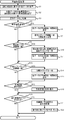
- the medical information processing illustrated in FIG. 2 is executed by the CPU 22 of the medical information processing device 20 according to the medical information processing program stored in the storage device 23 of the medical information processing device 20.
- the medical information processing may be executed by the control unit of another device (for example, the CPU 12 of the server 10 or the CPU 32 of the medical imaging device 30).
- the control units of a plurality of devices for example, the CPU 22 of the medical information processing device 20 and the CPU 12 of the server 10) may cooperate to execute medical information processing.
- the CPU 22 acquires the target data 40 regarding the diagnosis target person (S1). As shown in FIGS. 3 and 4, in the target data 40 of the present embodiment, at least one medical image 41 taken by the medical imaging apparatus 30 and each medical image 41 were taken for the diagnosis target person. Information on the shooting timing (in the present embodiment, information on the shooting date and time) is included.
- the CPU 22 may acquire the target data 40 from the medical imaging apparatus 30 via communication, a detachable storage medium, or the like. Further, the CPU 22 uses the target data of the past target data of the diagnosis target person already stored in the storage device 23, the medical image 41 newly captured by the medical image capturing device 30, and the imaging timing information. It may be acquired as 40. Although the details will be described later, the medical information processing apparatus 20 extracts case data similar to the target data 40 as similar case data from a plurality of case data stored in the database.
- the storage device 13 of the server 10 is used as a database for storing a plurality of case data 50.
- the database that stores the case data 50 may be another storage device (for example, the storage device 23 of the medical information processing device 20).
- the case data 50 includes a plurality of medical images 51 captured at different timings by the medical imaging apparatus 30, and information on the imaging timing of each medical image 51.
- the plurality of case data 50 are stored in the database.
- the case data 50 of the present embodiment includes information regarding the treatment performed on the patient (hereinafter, referred to as “treatment information”).
- the treatment information includes the presence or absence of treatment for the patient, the timing when the treatment is executed or started (hereinafter referred to as "treatment timing"), and the content of the treatment performed (for example, the type of surgery performed). , At least one of the type of drug administered, the method of administration, the type of treatment performed, etc.).
- Each of the plurality of case data 50 stored in the database is classified into one of the plurality of case data groups according to the presence or absence of treatment indicated by the treatment information and the content of the performed treatment. That is, in the present embodiment, each of the plurality of case data 50 is classified into either a case data group of a patient who has not been treated and a case data group of a patient who has been treated. Furthermore, the case data group of the treated patients is further subdivided according to the content of the treatment performed. Although the details will be described later, the medical information processing apparatus 20 can also extract case data 50 similar to the target data 40 according to the classification of the case data group.
- At least one of the plurality of case data stored in the database includes data related to the patient (hereinafter,) acquired by a device of a type different from the medical image capturing device 30 that captures the medical image 41 of the target data 40.
- “Different data” may be included.
- the heterogeneous data includes at least an imaging device (for example, a fundus camera, a scanning laser ophthalmoscope (SLO), a corneal shape measuring device, etc.) different from the medical imaging apparatus 30 (OCT apparatus in the present embodiment) described above. The data of the image taken by either) may be included.
- the heterogeneous data includes test result data (for example, at least one test result data such as visual acuity, axial length, intraocular pressure, visual field, etc.) acquired by a test device that tests a patient. It may be.
- the CPU 22 uses one or a plurality of medical images 41 as a reference for extracting the case data 50 from the one or a plurality of medical images 41 included in the target data 40 acquired in S1 as a reference image.
- Set (S2) the medical information processing apparatus 20 of the present embodiment has a medical image 51 similar to at least one medical image 41 included in the target data 40 from among a plurality of case data 50 stored in the database. Case data 50 including the above is extracted as similar case data.
- the medical image 41 for determining the degree of similarity with the medical image 51 in the case data 50 is set as a reference image.
- the specific method for setting the reference image in S2 can be appropriately selected.
- the CPU 22 sets one medical image 41 in the target data 40 as the reference image.
- the CPU 22 uses one or a plurality of medical images 41 selected by the instruction as the reference image based on the reference image selection instruction input from the user. It may be set.
- the CPU 22 may automatically set one or a plurality of medical images 41 as reference images in the order of newest imaging timing.
- the CPU 22 extracts one or more case data 50 similar to the target data 40 from the plurality of case data 50 stored in the database as similar case data (S3). Specifically, the CPU 22 extracts the case data 50 including the medical image 51 similar to the reference image set in the target data 40 as the similar case data.
- the CPU 22 acquires a value indicating the feature amount of each of the reference image and the plurality of medical images 51 in the case data 50.
- SIFT Scale Invariant Feature Transfer Transfer
- feature quantities other than SIFT may be acquired.
- the CPU 22 extracts the case data 50 including the medical image 51 having a small difference in the feature amount from the reference image as similar case data.
- the CPU 50 may extract case data 50 including a medical image 51 in which the difference in feature amounts is equal to or less than a threshold value. Further, the CPU 50 may extract one or more case data 50 in ascending order of the difference between the feature amounts of the reference image and the medical image 51.
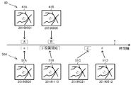
- FIG. 3 an example of a method for extracting similar case data when a plurality of reference images 41A and 41B are set in the target data 40 will be described.
- the CPU 22 specifies the shooting intervals D1 of the plurality of reference images 41A and 41B based on the shooting timing information included in the target data 40.
- the CPU 22 identifies the case data 50 including the plurality of medical images 51 taken at intervals of the plurality of reference images 41A and 41B with an interval D1 which is equal to or less than the threshold value among the plurality of case data 50.
- Case data 50 including a plurality of medical images 41 is extracted as similar case data.
- the threshold value for comparing the shooting intervals may be set in advance or may be set according to an instruction from the user.
- the difference between the imaging interval D2 of the two medical images 51A and 51B included in the case data 50A and the imaging interval D1 of the two reference images 41A and 41B is equal to or less than the threshold value. Further, the similarity between the two medical images 51A and 51B and the two reference images 41A and 41B is high (that is, the similarity between the medical image 51A and the reference image 41A and the similarity between the medical image 51B and the reference image 41B are high. , Both are expensive). Therefore, the CPU 22 extracts the case data 50A as similar case data.
- the two medical images 51X and 51Y included in the case data 50B have a high degree of similarity with the two reference images 41A and 41B, the two medical images 51X and 51Y have an imaging interval D3 and two.
- the difference between the shooting intervals D1 of the reference images 41A and 41B is larger than the threshold value. Therefore, the CPU 22 does not extract the case data 50B as similar case data.
- the extracted similar case data includes medical images 51A and 51B taken at intervals close to the imaging intervals of the plurality of reference images 41A and 41B of the target data 40. ..
- the CPU 22 extracts the case data 50 including the medical image 51 having a high degree of similarity to the one reference image as the similar case data.
- the CPU 22 can extract similar case data from one or a plurality of case data groups among a plurality of case data groups classified by treatment information. For example, when an instruction to extract similar case data from a case data group of a patient who has been treated with a specific content is input, the CPU 22 is one from the case data group corresponding to the content of the instructed treatment. Or extract multiple similar case data. The CPU 22 can also extract one or more similar case data from each of the plurality of case data groups. For example, the CPU 22 can also extract each of the similar case data of the treated patient and the similar case data of the untreated patient. The CPU 22 can also extract a plurality of similar case data having different contents of the performed treatment.
- the CPU 22 uses the medical image 51 before or after the treatment timing indicated by the treatment information among the plurality of medical images 51 included in each case data 50, and the reference image of the target data 40. It is also possible to extract similar case data by comparing the similarities of. For example, when the reference image of the target data 40 is the medical image 41 before the treatment, the CPU 22 compares the similarity between the medical image 51 before the treatment timing and the reference image and extracts similar case data. As a result, case data 50 in which the state of the disease before treatment is close to the state of the disease of the diagnosed subject is appropriately extracted.
- the CPU 22 acquires the similarity of the medical images 41 and 51 by inputting the medical images 41 and 51 into the mathematical model trained by the machine learning algorithm, and extracts similar case data based on the acquired results. You may. Further, when the image quality of the medical image 51 included in the case data 50 is less than the threshold value, the CPU 22 may exclude the case data 50 from the extraction target, or may exclude the extracted similar case data image. The user may be warned that the quality is low.
- the CPU 22 performs a process of matching the time axis of the acquisition timing of the medical image with respect to the plurality of data (S5, S6, S7, S9, S10, S13, S14).
- the plurality of data to be matched on the time axis may be the similar case data 50 and the target data 40, or may be the extracted plurality of similar case data 50.
- the user selects one of a method of manually matching the time axis, a method of automatically matching the time axis according to the similarity of medical images, and a method of automatically matching the time axis according to the treatment timing.
- the instruction to be performed can be input by operating the operation unit 25.
- the CPU 22 uses a plurality of data (similar case data 50 and target data 40, or a plurality of data) for which the time axis is to be matched.
- the medical image of the similar case data 50) of the above is displayed on the display unit 26 together with the time axis (S6).
- the medical images 41A and 41B included in the target data 40 and the medical images 51A, 51B, 51C and 51D included in the extracted similar case data 50A are displayed on the display unit 26 together with the time axis T. Has been done. On the time axis T, the timing of taking each medical image is shown.
- the imaging timing of the medical image 41A is indicated by “A” on the time axis T
- the imaging timing of the medical image 51D is indicated by “d” on the time axis T.
- the treatment timing at which the treatment is executed or started is also shown based on the treatment information included in the similar case data 50A. In the example shown in FIG. 4, the timing of starting the medication coincides with the timing of photographing the medical image 51B.
- the CPU 22 matches the time axis of the acquisition timing of the medical image between the plurality of data (in the example shown in FIG. 4, between the target data 40 and the similar case data 50A) in response to an instruction input from the user. Further, the CPU 22 changes the imaging timing of each medical image shown on the time axis T according to the result of matching the time axes (S7).
- the CPU 22 may change the display positions of a plurality of medical images and the like according to the matched time axis together with the imaging timing on the time axis T. For example, the user may input an instruction to match the time axis by inputting an instruction to slide the shooting timing indicated on the time axis T in a direction along the time axis by the operation unit 25.
- the user can use two medical images whose imaging timings match among the medical images included in each of the plurality of data (in the example shown in FIG. 4, the medical image 41A of the target data 40 and the medical image of the similar case data 50A). You may enter an instruction to specify image 51A).
- the CPU 22 matches the time axes of the plurality of data so that the imaging timings of the two designated medical images match.
- the CPU 22 When an instruction to match the time axis according to the similarity of the medical images is input (S9: YES), the CPU 22 has a plurality of data (similar case data 50 and target data 40, or a plurality of similar case data 50). By comparing the similarity of the medical images included in each of the data among the data and matching the imaging timings of the medical images having the highest similarity, the time axes of the plurality of data are matched (S10).
- the CPU 22 causes the display unit 26 to display both the medical image included in each of the plurality of data and the time axis T indicating the shooting timing of the medical image as medical information in the state where the time axes are matched in S10 (S11). ).
- S10 When comparing medical images of a plurality of data, the disease states in the two medical images having the highest similarity are likely to be similar. Therefore, by performing the processing of S10, the user can more appropriately grasp the transition of the disease from the medical information.
- the CPU 22 When an instruction to match the time axis according to the treatment timing is input (S13: YES), the CPU 22 performs treatment between a plurality of data (similar case data 50 and target data 40, or a plurality of similar case data 50). By matching the timings, the time axes of a plurality of data are matched (S14). The CPU 22 causes the display unit 26 to display both the medical image included in each of the plurality of data and the time axis T indicating the shooting timing of the medical image as medical information in the state where the time axes are matched in S14 (S15). ). By performing the process of S14, the user can compare a plurality of data with reference to the treatment timing, so that the transition of the disease due to the treatment can be appropriately predicted.
- the time is based on the timing of the treatment assuming that the diagnosis target person is to be treated in the future.
- the axes may be aligned.
- the user may input the timing of future treatment, or the timing of treatment may be the time when the latest medical image 41 is taken.
- the CPU 22 determines whether or not an instruction for displaying the predicted image of the diagnosis target person has been input by the user (S17).
- the predicted image is an image that is predicted to show the future state of the disease of the person to be diagnosed.
- the CPU 22 generates a predicted image based on the medical image 41 included in the target data 40 and the medical image 51 included in the similar case data. , Displayed on the display unit 26 (S18).
- the CPU 22 extracts the medical image 41 used for generating the predicted image as the base image 42 from the medical image 41 included in the target data 40 (see FIGS. 3 and 4) for the diagnosis target person.
- the base image 42 used to generate the predicted image is preferably a new image as much as possible. Therefore, when the target data 40 includes a plurality of medical images 41, the CPU 22 extracts the medical image 41 having the latest imaging timing as the base image 42.
- the CPU 22 captures the medical image 51 captured after the same timing as the imaging timing of the base image 42 on the matched time axis among the plurality of medical images 51 included in the similar case data 50 (see FIG. 4).
- the reference image 52 is extracted in a state where the time axes of the imaging timings of the target data 40 and the similar case data 50 are matched. Therefore, the reference image 52 for generating the predicted image 60 is extracted more appropriately.
- the base image 42 includes the diseased portion 43
- the reference image 52 also includes the diseased portion 53.
- the disease progresses as a result of the patient not being treated, and the diseased portion 53 larger than the diseased portion 43 of the base image 42 is reflected.
- the CPU 22 generates a predicted image 60 based on the base image 42 and the reference image 52.
- the CPU 22 generates a diseased portion removal image 45 in which the information of the diseased portion 43 is removed from the basic image 42.
- the CPU 22 generates a diseased part image 55 in which information other than the diseased part 53 is removed from the reference image 52.
- the CPU 22 generates a predicted image 60 based on the diseased part removal image 45 and the diseased part image 55 (for example, by performing composition by image processing or the like).
- a predicted image predicted to show the future disease state of the diagnosed subject is appropriately generated.
- the predicted image 60 may be generated by utilizing a mathematical model trained by a machine learning algorithm.
- the mathematical model may be pre-trained with data from a plurality of medical images so that the predicted image 60 is output by inputting the base image 42 and the reference image 52.
- At least one of the mathematical models that inputs and outputs the predicted image 60 may be used.
- Whether or not the CPU 22 has been input with an instruction to display a progress transition graph showing the transition of the disease progression in each of the plurality of data (similar case data 50 and target data 40, or a plurality of similar case data 50). Is determined (S20).
- the CPU 22 acquires information on the progress of the disease shown in each of the plurality of medical images (S21).
- the CPU 22 generates a progress transition graph showing the transition of the progress in each data in a state where the time axes of the shooting timings among the plurality of data are matched, and displays the graph on the display unit 26 (S22).
- the CPU 22 acquires information on the degree of progression of the disease shown in each of the medical images included in each data.
- the method for acquiring information on the degree of disease progression can be appropriately selected.
- the CPU 22 may acquire information on the degree of progression by inputting a medical image into a mathematical model trained by a machine learning algorithm and outputting the degree of progression (for example, the probability of a disease) to the mathematical model.
- the CPU 22 inputs a medical image into a mathematical model that outputs the analysis result of the disease, and information indicating the distribution of the degree of influence that the mathematical model has influenced when outputting the analysis result (sometimes referred to as an "attention map").
- Information on the degree of disease progression may be obtained based on (there is).
- the CPU 22 may perform image processing on each medical image and acquire information on the degree of progression based on at least one of the size and color of the diseased portion.
- the above-mentioned attention map may be displayed on the display unit 26 together with the original medical image.
- the CPU 22 obtains a progress transition graph showing the transition of the disease progression from the medical image. Create based on the information in.
- the CPU 22 generates a progress transition graph in a state where the time axes of the shooting timings of the plurality of data are matched.
- the time axis of the imaging timing is matched so that the treatment timing in the data of the patient who performed the treatment is matched. Therefore, in the example shown in FIG. 6, the user can easily compare the transition of the degree of progression of the disease according to the presence or absence of treatment and the content of treatment based on the treatment timing.
- the imaging timing of the latest medical image 41 is adjusted to the treatment timing of other data as a provisional treatment timing. Therefore, the user can easily predict the transition of the disease when the treatment is started immediately by the progress transition graph. Further, in the example shown in FIG. 6, among the similar case data of the patients who were not treated, the imaging timing of the medical image 51 having the highest degree of similarity to the latest medical image 41 in the target data 40 is the target data. It is adjusted to the shooting timing of the latest medical image 41 in 40.
- the CPU 22 displays the heterogeneous data together with the medical image included in the similar case data or separately from the medical image. It is displayed on the unit 26. Therefore, the user can more appropriately predict the transition of the disease of the diagnosis target person by confirming the heterogeneous data included in the similar case data. Further, even when the heterogeneous data regarding the diagnosis target person has not been acquired yet, the user can confirm the heterogeneous data of the patients having similar cases, so that the diagnosis of the diagnosis target person can be performed more appropriately. be able to.
- the CPU 22 of the medical information processing apparatus 20 may specify the timing (change timing) at which the change in the feature amount of the plurality of medical images included in each data becomes equal to or greater than the threshold value for each of the plurality of data.
- the CPU 22 may match the time axis by matching the change timings in the data.
- the change timing may be, for example, the timing when the change in the progress of the disease (progress of deterioration) becomes equal to or higher than the threshold value, or the change timing may be the timing when the change in the progress of healing of the disease becomes equal to or higher than the threshold value.
- the CPU 22 obtains similar case data by comparing the medical image before or after the change timing with the medical image 41 of the target data 40 among the plurality of medical images 51 included in the case data 50. It may be extracted.
- the process of acquiring the target data 40 in S1 of FIG. 2 is an example of the “target data acquisition step”.
- the process of extracting similar case data in S3 of FIG. 2 is an example of the “similar case data extraction step”.
- the process of matching the time axis of the shooting timing in S7, S10, and S14 of FIG. 2 is an example of the “time axis matching step”.
- the process of outputting medical information in S7, S11, S15, S18, and S22 of FIG. 2 is an example of the “medical information output step”.
- the process of generating the predicted image in S18 of FIG. 2 is an example of the “predicted image generation step”.
- the process of acquiring the progress information in S21 of FIG. 2 is an example of the “progress acquisition step”.
- the process of generating the progress transition graph in S22 of FIG. 2 is an example of the “graph generation step”.
Landscapes
- Engineering & Computer Science (AREA)
- Medical Informatics (AREA)
- Data Mining & Analysis (AREA)
- Health & Medical Sciences (AREA)
- Public Health (AREA)
- Pathology (AREA)
- Databases & Information Systems (AREA)
- Biomedical Technology (AREA)
- Epidemiology (AREA)
- General Health & Medical Sciences (AREA)
- Primary Health Care (AREA)
- Medical Treatment And Welfare Office Work (AREA)
- Measuring And Recording Apparatus For Diagnosis (AREA)
Priority Applications (1)
| Application Number | Priority Date | Filing Date | Title |
|---|---|---|---|
| JP2021551394A JP7709047B2 (ja) | 2019-09-30 | 2020-09-30 | 医療情報処理プログラム、および医療情報処理装置 |
Applications Claiming Priority (2)
| Application Number | Priority Date | Filing Date | Title |
|---|---|---|---|
| JP2019-180319 | 2019-09-30 | ||
| JP2019180319 | 2019-09-30 |
Publications (1)
| Publication Number | Publication Date |
|---|---|
| WO2021066039A1 true WO2021066039A1 (ja) | 2021-04-08 |
Family
ID=75338085
Family Applications (1)
| Application Number | Title | Priority Date | Filing Date |
|---|---|---|---|
| PCT/JP2020/037237 Ceased WO2021066039A1 (ja) | 2019-09-30 | 2020-09-30 | 医療情報処理プログラム、および医療情報処理装置 |
Country Status (2)
| Country | Link |
|---|---|
| JP (1) | JP7709047B2 (https=) |
| WO (1) | WO2021066039A1 (https=) |
Cited By (2)
| Publication number | Priority date | Publication date | Assignee | Title |
|---|---|---|---|---|
| WO2024162100A1 (ja) | 2023-02-03 | 2024-08-08 | 株式会社ニデック | 医療情報処理プログラムおよび医療情報処理装置 |
| WO2025141847A1 (ja) * | 2023-12-28 | 2025-07-03 | 三菱電機株式会社 | 情報支援サーバ、及び情報支援方法 |
Citations (9)
| Publication number | Priority date | Publication date | Assignee | Title |
|---|---|---|---|---|
| JP2006149654A (ja) * | 2004-11-29 | 2006-06-15 | Hitachi Omron Terminal Solutions Corp | 眼底の病変についての診断の支援 |
| JP2007287018A (ja) * | 2006-04-19 | 2007-11-01 | Fujifilm Corp | 診断支援システム |
| JP2007287027A (ja) * | 2006-04-19 | 2007-11-01 | Fujifilm Corp | 医療用の計画立案支援システム |
| JP2009045121A (ja) * | 2007-08-14 | 2009-03-05 | Fujifilm Corp | 医用画像処理システム、医用画像処理方法、及びプログラム |
| JP2011182960A (ja) * | 2010-03-09 | 2011-09-22 | Fuji Xerox Co Ltd | プログラムおよび情報処理装置 |
| JP2013052245A (ja) * | 2012-10-31 | 2013-03-21 | Canon Inc | 情報処理装置および情報処理方法 |
| JP2013198817A (ja) * | 2013-07-10 | 2013-10-03 | Canon Inc | 情報処理装置、情報処理方法、プログラムおよび記憶媒体 |
| JP2014233611A (ja) * | 2013-06-05 | 2014-12-15 | 株式会社東芝 | 治療計画策定支援装置及び治療計画策定支援システム |
| JP2017033093A (ja) * | 2015-07-29 | 2017-02-09 | 富士フイルム株式会社 | 診療支援装置とその作動方法および作動プログラム、並びに診療支援システム |
-
2020
- 2020-09-30 WO PCT/JP2020/037237 patent/WO2021066039A1/ja not_active Ceased
- 2020-09-30 JP JP2021551394A patent/JP7709047B2/ja active Active
Patent Citations (9)
| Publication number | Priority date | Publication date | Assignee | Title |
|---|---|---|---|---|
| JP2006149654A (ja) * | 2004-11-29 | 2006-06-15 | Hitachi Omron Terminal Solutions Corp | 眼底の病変についての診断の支援 |
| JP2007287018A (ja) * | 2006-04-19 | 2007-11-01 | Fujifilm Corp | 診断支援システム |
| JP2007287027A (ja) * | 2006-04-19 | 2007-11-01 | Fujifilm Corp | 医療用の計画立案支援システム |
| JP2009045121A (ja) * | 2007-08-14 | 2009-03-05 | Fujifilm Corp | 医用画像処理システム、医用画像処理方法、及びプログラム |
| JP2011182960A (ja) * | 2010-03-09 | 2011-09-22 | Fuji Xerox Co Ltd | プログラムおよび情報処理装置 |
| JP2013052245A (ja) * | 2012-10-31 | 2013-03-21 | Canon Inc | 情報処理装置および情報処理方法 |
| JP2014233611A (ja) * | 2013-06-05 | 2014-12-15 | 株式会社東芝 | 治療計画策定支援装置及び治療計画策定支援システム |
| JP2013198817A (ja) * | 2013-07-10 | 2013-10-03 | Canon Inc | 情報処理装置、情報処理方法、プログラムおよび記憶媒体 |
| JP2017033093A (ja) * | 2015-07-29 | 2017-02-09 | 富士フイルム株式会社 | 診療支援装置とその作動方法および作動プログラム、並びに診療支援システム |
Cited By (3)
| Publication number | Priority date | Publication date | Assignee | Title |
|---|---|---|---|---|
| WO2024162100A1 (ja) | 2023-02-03 | 2024-08-08 | 株式会社ニデック | 医療情報処理プログラムおよび医療情報処理装置 |
| EP4661016A1 (en) | 2023-02-03 | 2025-12-10 | Nidek Co., Ltd. | Medical information processing program and medical information processing device |
| WO2025141847A1 (ja) * | 2023-12-28 | 2025-07-03 | 三菱電機株式会社 | 情報支援サーバ、及び情報支援方法 |
Also Published As
| Publication number | Publication date |
|---|---|
| JP7709047B2 (ja) | 2025-07-16 |
| JPWO2021066039A1 (https=) | 2021-04-08 |
Similar Documents
| Publication | Publication Date | Title |
|---|---|---|
| US12096981B2 (en) | Ophthalmologic image processing device and non-transitory computer-readable storage medium storing computer-readable instructions | |
| JP7196908B2 (ja) | 眼科画像処理装置および眼科画像処理プログラム | |
| CN110582223A (zh) | 用于医疗状况诊断、治疗和预后确定的系统及方法 | |
| JP7406901B2 (ja) | 情報処理装置及び情報処理方法 | |
| CN112533526B (zh) | 眼科图像处理装置、oct装置及眼科图像处理程序 | |
| JP2019526334A (ja) | 眼科手術中に医師を支援する予測装置 | |
| JP2019208831A (ja) | 歯科分析システムおよび歯科分析x線システム | |
| JP7521575B2 (ja) | 眼科画像処理装置、oct装置、および眼科画像処理プログラム | |
| CN111344222A (zh) | 执行眼睛检查测试的方法 | |
| JP7709047B2 (ja) | 医療情報処理プログラム、および医療情報処理装置 | |
| JP2018147387A (ja) | 眼科診療情報処理システム及び眼科診療情報処理方法 | |
| JP7435885B2 (ja) | 眼科画像処理装置、および眼科画像処理プログラム | |
| JP2019208832A (ja) | 歯科分析システムおよび歯科分析x線システム | |
| JP7328489B2 (ja) | 眼科画像処理装置、および眼科撮影装置 | |
| JP2019208851A (ja) | 眼底画像処理装置および眼底画像処理プログラム | |
| US20220122730A1 (en) | Using artificial intelligence and biometric data for serial screening exams for medical conditions | |
| WO2020116351A1 (ja) | 診断支援装置、および診断支援プログラム | |
| JP7302184B2 (ja) | 眼科画像処理装置、および眼科画像処理プログラム | |
| JP7563384B2 (ja) | 医療画像処理装置および医療画像処理プログラム | |
| JP7612990B2 (ja) | 眼科画像処理装置および眼科画像処理プログラム | |
| JP7439990B2 (ja) | 医療画像処理装置、医療画像処理プログラム、および医療画像処理方法 | |
| GB2576139A (en) | Ocular assessment | |
| JP7468163B2 (ja) | 眼科画像処理プログラムおよび眼科画像処理装置 | |
| JP2018147386A (ja) | 眼科診療情報処理システム及び眼科診療情報処理方法 | |
| JP2022087936A (ja) | 眼科装置及びその作動方法、並びに、プログラム |
Legal Events
| Date | Code | Title | Description |
|---|---|---|---|
| 121 | Ep: the epo has been informed by wipo that ep was designated in this application |
Ref document number: 20871321 Country of ref document: EP Kind code of ref document: A1 |
|
| ENP | Entry into the national phase |
Ref document number: 2021551394 Country of ref document: JP Kind code of ref document: A |
|
| NENP | Non-entry into the national phase |
Ref country code: DE |
|
| 122 | Ep: pct application non-entry in european phase |
Ref document number: 20871321 Country of ref document: EP Kind code of ref document: A1 |