WO2021060494A1 - 治療デバイスおよび治療方法 - Google Patents
治療デバイスおよび治療方法 Download PDFInfo
- Publication number
- WO2021060494A1 WO2021060494A1 PCT/JP2020/036356 JP2020036356W WO2021060494A1 WO 2021060494 A1 WO2021060494 A1 WO 2021060494A1 JP 2020036356 W JP2020036356 W JP 2020036356W WO 2021060494 A1 WO2021060494 A1 WO 2021060494A1
- Authority
- WO
- WIPO (PCT)
- Prior art keywords
- balloon
- blood vessel
- anchor member
- vessel wall
- expanded
- Prior art date
- Legal status (The legal status is an assumption and is not a legal conclusion. Google has not performed a legal analysis and makes no representation as to the accuracy of the status listed.)
- Ceased
Links
Images




Classifications
-
- A—HUMAN NECESSITIES
- A61—MEDICAL OR VETERINARY SCIENCE; HYGIENE
- A61M—DEVICES FOR INTRODUCING MEDIA INTO, OR ONTO, THE BODY; DEVICES FOR TRANSDUCING BODY MEDIA OR FOR TAKING MEDIA FROM THE BODY; DEVICES FOR PRODUCING OR ENDING SLEEP OR STUPOR
- A61M25/00—Catheters; Hollow probes
- A61M25/01—Introducing, guiding, advancing, emplacing or holding catheters
- A61M25/02—Holding devices, e.g. on the body
- A61M25/04—Holding devices, e.g. on the body in the body, e.g. expansible
-
- A—HUMAN NECESSITIES
- A61—MEDICAL OR VETERINARY SCIENCE; HYGIENE
- A61B—DIAGNOSIS; SURGERY; IDENTIFICATION
- A61B17/00—Surgical instruments, devices or methods
- A61B17/22—Implements for squeezing-off ulcers or the like on inner organs of the body; Implements for scraping-out cavities of body organs, e.g. bones; for invasive removal or destruction of calculus using mechanical vibrations; for removing obstructions in blood vessels, not otherwise provided for
-
- A—HUMAN NECESSITIES
- A61—MEDICAL OR VETERINARY SCIENCE; HYGIENE
- A61M—DEVICES FOR INTRODUCING MEDIA INTO, OR ONTO, THE BODY; DEVICES FOR TRANSDUCING BODY MEDIA OR FOR TAKING MEDIA FROM THE BODY; DEVICES FOR PRODUCING OR ENDING SLEEP OR STUPOR
- A61M25/00—Catheters; Hollow probes
- A61M25/10—Balloon catheters
- A61M25/104—Balloon catheters used for angioplasty
-
- A—HUMAN NECESSITIES
- A61—MEDICAL OR VETERINARY SCIENCE; HYGIENE
- A61B—DIAGNOSIS; SURGERY; IDENTIFICATION
- A61B17/00—Surgical instruments, devices or methods
- A61B17/22—Implements for squeezing-off ulcers or the like on inner organs of the body; Implements for scraping-out cavities of body organs, e.g. bones; for invasive removal or destruction of calculus using mechanical vibrations; for removing obstructions in blood vessels, not otherwise provided for
- A61B17/221—Gripping devices in the form of loops or baskets for gripping calculi or similar types of obstructions
-
- A—HUMAN NECESSITIES
- A61—MEDICAL OR VETERINARY SCIENCE; HYGIENE
- A61B—DIAGNOSIS; SURGERY; IDENTIFICATION
- A61B17/00—Surgical instruments, devices or methods
- A61B17/22—Implements for squeezing-off ulcers or the like on inner organs of the body; Implements for scraping-out cavities of body organs, e.g. bones; for invasive removal or destruction of calculus using mechanical vibrations; for removing obstructions in blood vessels, not otherwise provided for
- A61B2017/22051—Implements for squeezing-off ulcers or the like on inner organs of the body; Implements for scraping-out cavities of body organs, e.g. bones; for invasive removal or destruction of calculus using mechanical vibrations; for removing obstructions in blood vessels, not otherwise provided for with an inflatable part, e.g. balloon, for positioning, blocking, or immobilisation
-
- A—HUMAN NECESSITIES
- A61—MEDICAL OR VETERINARY SCIENCE; HYGIENE
- A61B—DIAGNOSIS; SURGERY; IDENTIFICATION
- A61B17/00—Surgical instruments, devices or methods
- A61B17/22—Implements for squeezing-off ulcers or the like on inner organs of the body; Implements for scraping-out cavities of body organs, e.g. bones; for invasive removal or destruction of calculus using mechanical vibrations; for removing obstructions in blood vessels, not otherwise provided for
- A61B2017/22051—Implements for squeezing-off ulcers or the like on inner organs of the body; Implements for scraping-out cavities of body organs, e.g. bones; for invasive removal or destruction of calculus using mechanical vibrations; for removing obstructions in blood vessels, not otherwise provided for with an inflatable part, e.g. balloon, for positioning, blocking, or immobilisation
- A61B2017/22062—Implements for squeezing-off ulcers or the like on inner organs of the body; Implements for scraping-out cavities of body organs, e.g. bones; for invasive removal or destruction of calculus using mechanical vibrations; for removing obstructions in blood vessels, not otherwise provided for with an inflatable part, e.g. balloon, for positioning, blocking, or immobilisation to be filled with liquid
-
- A—HUMAN NECESSITIES
- A61—MEDICAL OR VETERINARY SCIENCE; HYGIENE
- A61B—DIAGNOSIS; SURGERY; IDENTIFICATION
- A61B17/00—Surgical instruments, devices or methods
- A61B17/22—Implements for squeezing-off ulcers or the like on inner organs of the body; Implements for scraping-out cavities of body organs, e.g. bones; for invasive removal or destruction of calculus using mechanical vibrations; for removing obstructions in blood vessels, not otherwise provided for
- A61B2017/22051—Implements for squeezing-off ulcers or the like on inner organs of the body; Implements for scraping-out cavities of body organs, e.g. bones; for invasive removal or destruction of calculus using mechanical vibrations; for removing obstructions in blood vessels, not otherwise provided for with an inflatable part, e.g. balloon, for positioning, blocking, or immobilisation
- A61B2017/22065—Functions of balloons
- A61B2017/22069—Immobilising; Stabilising
-
- A—HUMAN NECESSITIES
- A61—MEDICAL OR VETERINARY SCIENCE; HYGIENE
- A61M—DEVICES FOR INTRODUCING MEDIA INTO, OR ONTO, THE BODY; DEVICES FOR TRANSDUCING BODY MEDIA OR FOR TAKING MEDIA FROM THE BODY; DEVICES FOR PRODUCING OR ENDING SLEEP OR STUPOR
- A61M25/00—Catheters; Hollow probes
- A61M25/10—Balloon catheters
- A61M2025/1043—Balloon catheters with special features or adapted for special applications
- A61M2025/105—Balloon catheters with special features or adapted for special applications having a balloon suitable for drug delivery, e.g. by using holes for delivery, drug coating or membranes
-
- A—HUMAN NECESSITIES
- A61—MEDICAL OR VETERINARY SCIENCE; HYGIENE
- A61M—DEVICES FOR INTRODUCING MEDIA INTO, OR ONTO, THE BODY; DEVICES FOR TRANSDUCING BODY MEDIA OR FOR TAKING MEDIA FROM THE BODY; DEVICES FOR PRODUCING OR ENDING SLEEP OR STUPOR
- A61M25/00—Catheters; Hollow probes
- A61M25/10—Balloon catheters
- A61M2025/1043—Balloon catheters with special features or adapted for special applications
- A61M2025/1052—Balloon catheters with special features or adapted for special applications for temporarily occluding a vessel for isolating a sector
-
- A—HUMAN NECESSITIES
- A61—MEDICAL OR VETERINARY SCIENCE; HYGIENE
- A61M—DEVICES FOR INTRODUCING MEDIA INTO, OR ONTO, THE BODY; DEVICES FOR TRANSDUCING BODY MEDIA OR FOR TAKING MEDIA FROM THE BODY; DEVICES FOR PRODUCING OR ENDING SLEEP OR STUPOR
- A61M2205/00—General characteristics of the apparatus
- A61M2205/02—General characteristics of the apparatus characterised by a particular materials
- A61M2205/0266—Shape memory materials
Definitions
- the present invention relates to a therapeutic device and a therapeutic method used transvascularly.
- Coronary Artery Bypass Graft was developed by Bailey-Hirose, Garrett, 196 from 19 to 19 Was done.
- CABG Coronary Artery Bypass Graft
- Palmaz-Shatz registered trademark
- the drug-eluting stent was developed with the aim of improving the remaining remote restenosis.
- Johnson & Johnson used Cypher® stents using sirolimus (macrolide immunosuppressant), and Boston Scientific (registered trademark) used paclitaxel (cancer control agent) in the United States.
- paclitaxel cancer control agent
- a stent The surface of these stents is coated with a non-absorbable polymer, which is impregnated with the drug. The impregnated drug is slowly released from the polymer layer and diffuses into the indwelling blood vessels to suppress smooth muscle proliferation, which is the cause of restenosis.
- PCI percutaneous coronary intervention
- the present invention has been made to solve the above-mentioned problems, and provides a therapeutic device and a therapeutic method thereof capable of improving the permeability of a physiologically active substance to the blood vessel wall while maintaining blood flow.
- the purpose is to do.
- a therapeutic device that achieves the above object is a therapeutic device for treating a lesion in a blood vessel, which is arranged and expanded on a shaft portion having at least one lumen and a distal side of the shaft portion.
- the expanded outer diameter of the balloon has a possible balloon and at least one anchor member that can be inserted into the lumen and has a diameter that can be expanded with a distal portion protruding from the lumen.
- the diameter is smaller than the outer diameter of the anchor member, and when the anchor member expands in diameter and comes into contact with the blood vessel wall, the balloon is held away from the blood vessel wall.
- the treatment method according to the present invention that achieves the above object is a treatment method in which a physiologically active substance acts on a blood vessel wall in a blood vessel, and an expandable dilator is expanded in the blood vessel and from the blood vessel wall.
- the treatment device configured as described above can maintain the expanded balloon in the substantially central portion of the blood vessel without contacting the blood vessel wall by bringing the enlarged anchor member into contact with the blood vessel wall.
- This allows the therapeutic device to guide the physiologically active substance released from the upstream side of the balloon to the vicinity of the blood vessel wall by the balloon while maintaining blood flow. Therefore, the physiologically active substance released into the blood vessel can flow in the bloodstream in the vicinity of the blood vessel wall, which has a high shear stress and is advantageous for taking up the substance. Therefore, the therapeutic device can effectively induce the physiologically active substance in the vicinity of the blood vessel wall to improve the permeability of the physiologically active substance to the blood vessel wall.
- At least one of the anchor members may be formed of a shape memory alloy. As a result, the diameter of the anchor member can be increased by restoring the memorized shape.
- At least one of the anchor members may be an anchor balloon that can be expanded by flowing a fluid. As a result, the diameter of the anchor member can be increased by the inflow of fluid.
- the anchor member may be arranged on the distal side and / or the proximal side of the balloon. Thereby, the anchor member can maintain the expanded balloon at a substantially central portion in the blood vessel without contacting the blood vessel wall without hindering the expansion of the balloon.
- At least one of the anchor members may be arranged radially outside the balloon.
- Anchor members arranged radially outward of the balloon effectively prevent the expanded balloon from coming into contact with the vessel wall and can maintain the balloon approximately central within the vessel with high accuracy.
- At least one of the lumens may be arranged so as to pass through the center of the balloon and open on the distal side of the balloon.
- the anchor member passing through the lumen that opens on the distal side of the balloon can be expanded in diameter on the distal side of the balloon.
- At least one of the lumens may be arranged so as to extend along the axis of the balloon and open on the proximal side of the balloon.
- the anchor member passing through the lumen that opens on the proximal side of the balloon can be expanded in diameter on the proximal side of the balloon.
- the outer diameter of the expanded balloon may be less than 4 mm. As a result, even if the balloon expands inside the coronary artery having an inner diameter of about 4 mm, it does not easily come into contact with the blood vessel wall of the coronary artery, and is therefore suitable for treatment of the coronary artery.
- the treatment method configured as described above can maintain blood flow because the dilator does not block the blood vessels.
- the physiologically active substance released from the upstream side of the dilator can be guided to the vicinity of the blood vessel wall by the dilator, the physiologically active substance is placed in the bloodstream and has a high shear stress to take up the substance. It can be flushed near the advantageous vessel wall. Therefore, this therapeutic method can effectively induce the physiologically active substance in the vicinity of the blood vessel wall while maintaining the blood flow, and improve the permeability of the physiologically active substance to the blood vessel wall.
- the vascular wall on the upstream side and / or downstream side of the lesion part of the treated blood vessel which has been treated to expand the lesion part where the stenosis or occlusion has occurred, has an outer diameter larger than that of the dilated body. It may have an arrangement step in which at least one anchor member capable of increasing the diameter is arranged by increasing the diameter. Thereby, in this treatment method, the anchor member can be arranged on the blood vessel wall at a position that does not interfere with the expansion of the dilator. Further, since the outer diameter of the anchor member used is larger than the outer diameter of the dilated body, the dilated body can be maintained at a substantially central portion in the blood vessel without contacting the blood vessel wall.
- the expansion step may be performed after the placement step.
- the extension body can be expanded while the extension body is held in an appropriate position by the placement step. Therefore, when the dilated body is expanded, the dilated body can be maintained at a substantially central portion in the blood vessel with high accuracy without contacting the blood vessel wall.
- the axis of the expansion body may be aligned with the central axis of the blood vessel by expanding the anchor member in the arrangement step.
- the expanded balloon can be maintained at a substantially central portion in the blood vessel with high accuracy without contacting the blood vessel wall.
- the dilator may be held so as not to come into contact with the blood vessel wall in the guidance step.
- the physiologically active substance is moved to the blood vessel wall side. Can be effectively induced.
- the physiologically active substance may be induced toward the blood vessel wall side in the induction step without blocking the blood flow by the dilator.
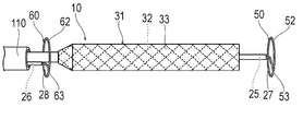
- FIG. 1 is the plan view of the distal part
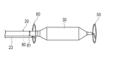
- FIG. 2 is the plan view of the distal part
- FIG. BB shows the sectional view along line BB of (A).
- FIG. BB shows the area of a balloon and the area surrounded by an anchor member in the cross section orthogonal to the axis of a balloon. It is a figure for demonstrating the treatment method using a treatment device.
- the state held by the two anchor members (D) is the state in which the therapeutic device is held by the first anchor member and the second anchor member, and (E) is the state in which the therapeutic device guides the physiologically active substance toward the blood vessel side. Indicates the state of doing. It is a top view which shows the 1st modification of a treatment device. It is a top view which shows the 2nd modification of a treatment device. It is a figure which shows the 3rd modification of the treatment device, (A) is a plan view, (B) is a front view seen from the distal side. It is a figure which shows the 4th modification of a treatment device, (A) is a plan view, (B) is a front view seen from the distal side.
- distal side the side of the device to be inserted into the blood vessel
- proximal side the side of the hand side to be operated
- the therapeutic device 10 is a device for treatment performed following percutaneous coronary intervention (PCI) performed for treatment of acute myocardial infarction (AMI).
- PCI percutaneous coronary intervention
- AMI acute myocardial infarction
- Physiologically active substances can act on damaged blood vessels, myocardium, etc. through the gaps formed between them.
- the treatment device 10 includes an expansion catheter 11 having an expandable balloon 30, and a first anchor member 50 and a second anchor member 60 that can be inserted into the expansion catheter 11. There is.
- the expansion catheter 11 includes a long shaft portion 20, a balloon 30 provided at the distal portion of the shaft portion 20, and a hub 40 fixed to the base end of the shaft portion 20.
- the shaft portion 20 includes a first tube body 21, a second tube body 22 arranged inside the first tube body 21, and a third tube body arranged outside the first tube body 21. It is equipped with 23.
- the second tubular body 22 is arranged coaxially with the first tubular body 21 inside the first tubular body 21.
- the second tubular body 22 extends to the distal side of the first tubular body 21.
- the third tube body 23 is fixed to the outer surface of the first tube body 21 substantially parallel to the first tube body 21. The distal end of the third tube 23 is located proximal to the distal end of the first tube 21.
- An expansion lumen 24 through which an expansion fluid for expanding the balloon 30 flows is formed between the first tube body 21 and the second tube body 22.
- a first wire lumen 25 into which a guide wire and a first anchor member 50 can be inserted is formed inside the second tubular body 22.
- a first distal opening 27 through which the first wire lumen 25 opens is formed.
- a second wire lumen 26 into which the second anchor member 60 can be inserted is formed inside the third tubular body 23 .
- a second distal opening 28 through which the second wire lumen 26 opens is formed at the distal end of the third tubular body 23.
- the hub 40 is fixed to the proximal portion of the first tube body 21, the second tube body 22, and the third tube body 23.
- the hub 40 has a first opening 41 that communicates with the expansion lumen 24 between the first tubular body 21 and the second tubular body 22, and a first wire lumen 25 that communicates with the first wire lumen 25 of the second tubular body 22. It includes two openings 42 and a third opening 43 that communicates with the second wire lumen 26 of the third tubular body 23.
- the first opening 41 functions as a port for allowing the expansion fluid to flow in and out of the expansion lumen 24. When the expansion fluid flows in from the first opening 41, the expansion fluid flows into the inside of the balloon 30 via the expansion lumen 24. As a result, the balloon 30 can be expanded.
- the second opening 42 functions as a port for inserting or removing the guide wire or the first anchor member 50 into the first wire lumen 25.
- the third opening 43 functions as a port for inserting or removing the second anchor member 60 into the second wire lumen 26.
- the proximal end of the second tubular body 22 may be arranged distal to the hub 40 instead of the hub 40.
- first tube body 21, the second tube body 22, and the third tube body 23 have a structure having appropriate flexibility and appropriate rigidity.
- the constituent materials of the first tubular body 21, the second tubular body 22, and the third tubular body 23 are, for example, polyolefins (for example, polyethylene, polypropylene, polybutene, ethylene-propylene copolymer, ethylene-vinyl acetate copolymer weight).
- the first anchor member 50 is a member for holding the balloon 30 at a desired position on the distal side of the balloon 30.
- the first anchor member 50 is inserted into the first wire lumen 25 through the second opening 42 of the hub and is located distal to the balloon 30 as the first distal wire lumen 25. It can protrude from the opening 27.
- the first anchor member 50 is a single wire rod that is elastically deformable.
- the first anchor member 50 has a first proximal linear portion 51 that is substantially linear and a first ring portion that extends in a ring shape so as to draw a substantially perfect circle in a natural state in which no external force acts.
- the first proximal linear portion 51 is located on the proximal side of the first anchor member 50, and the first ring portion 52 is located on the distal side of the first anchor member 50.
- the line X1 passing through the axis of the first proximal linear portion 51 is substantially orthogonal to the plane on which the first ring portion 52 is located.
- the line X1 passing through the axis of the first proximal linear portion 51 passes through the ring center R1 of the first ring portion 52.
- the first connecting portion 53 extends from the first proximal linear portion 51 to the first ring portion 52 along the surface on which the first ring portion 52 is located.
- the shape of the first connecting portion 53 is not particularly limited.
- the first anchor member 50 can be elastically deformed in a substantially linear shape as a whole and inserted into the first wire lumen 25 from the first opening 41. When the first anchor member 50 projects distally from the first distal opening 27, the first ring portion 52 and the first connecting portion 53 are restored to their original shapes by their own elastic force. can do.
- the second anchor member 60 is a member for holding the balloon 30 at a desired position on the proximal side of the balloon 30.
- the second anchor member 60 is inserted into the second wire lumen 26 through the third opening 43 of the hub and is located proximal to the balloon 30 in the second distal portion of the second wire lumen 26. It can protrude from the opening 28.
- the second anchor member 60 is a single wire rod that is elastically deformable.
- the second anchor member 60 includes a second proximal linear portion 61 which is substantially linear in a natural state where no external force acts, and a second ring portion 62 which extends so as to draw a substantially perfect circle.
- the second connecting portion 63 arranged between the second proximal linear portion 61 and the second ring portion 62.
- the second proximal linear portion 61 is located on the proximal side of the second anchor member 60, and the second ring portion 62 is located on the distal side of the second anchor member 60.
- the line X2 passing through the axis of the second proximal linear portion 61 is substantially orthogonal to the plane on which the second ring portion 62 is located.
- the line X2 passing through the axis of the second proximal linear portion 61 passes through a position deviated by a distance L1 from the ring center R2 of the second ring portion 62.
- the second connecting portion 63 extends from the second proximal linear portion 61 to the second ring portion 62 along the surface on which the second ring portion 62 is located.
- the shape of the second connecting portion 63 is not particularly limited.
- the second anchor member 60 can be inserted into the second wire lumen 26 from the second opening 42 by deforming the entire second anchor member 60 in a substantially linear shape. When the second anchor member 60 projects distally from the second distal opening 28, the second ring portion 62 and the second connecting portion 63 are restored to their original shapes by their own elastic force.
- the ring center R2 of the second ring portion 62 deviates from the line X2 passing through the axis of the second proximal linear portion 61 by a distance L1, but the distance L1 is the axis of the first wire lumen 25. It substantially coincides with the separation distance L2 (see FIG. 2) between the center and the axial center of the second wire lumen 26 through which the second proximal linear portion 61 passes. Therefore, the ring center R2 of the second ring portion 62 can substantially coincide with the axis of the first wire lumen 25.
- the second anchor member 60 protrudes from the second wire lumen 26 to expand the second ring portion 62, and then the ring center R2 of the second ring portion 62 comes from the axial center of the first wire lumen 25. It is desirable to limit the rotation within the second wire lumen 26 so that it does not shift.
- the shape of the second distal opening 28 of the second wire lumen 26 is the curved shape of the wire between the second proximal linear portion 61 and the second connecting portion 63. It may have a shape that matches. Thereby, by arranging the wire rod between the second proximal linear portion 61 and the second connecting portion 63 in the second distal opening 28 in an appropriate orientation, the second wire lumen 26 On the other hand, the rotation of the second anchor member 60 can be restricted.
- the constituent materials of the first anchor member 50 and the second anchor member 60 are preferably, for example, a shape memory alloy to which a shape memory effect and superelasticity are imparted by heat treatment.
- a shape memory alloy Ni—Ti type, Cu—Al—Ni type, Cu—Zn—Al type and the like can be preferably used.
- the constituent materials of the first anchor member 50 and the second anchor member 60 are not particularly limited as long as the shape can be elastically restored, and for example, other metals (including alloys) such as stainless steel and the like. It may be resin or the like.
- the first anchor member 50 and the second anchor member 60 may be formed of a plurality of materials.
- the first anchor member 50 and the second anchor member 60 may be coated with a resin material on the outside of the metal material described above.
- the balloon 30 is a member that can be expanded by allowing a fluid to flow into the balloon 30.
- the distal side of the balloon 30 is fixed to the distal portion of the second tubular body 22, and the proximal side is fixed to the distal portion of the first tubular body 21. Therefore, the inside of the balloon 30 communicates with the expansion lumen 24.
- the balloon 30 needs to have a certain degree of flexibility and to have a predetermined outer diameter when expanded. Therefore, the constituent material of the balloon 30 is preferably a non-compliant material that does not over-expand even when the pressure is increased to a predetermined value or more, or a semi-compliant material that slightly over-expands.
- the non-compliant material include polyethylene terephthalate and the like.
- the semi-compliant material include nylon 6, nylon 66, nylon 12, and the like.
- the balloon 30 is likely to have a desired outer diameter because the balloon 30 is made of a material that does not overexpand or is made of a material that slightly overexpands.
- the balloon 30 may have a structure in which the outer diameter can be arbitrarily adjusted by increasing the pressure.
- the constituent material of such a balloon 30 is, for example, a highly elastic material.
- the highly elastic material include various rubbers such as silicone rubber and latex rubber, polyurethane, polyamide, polyester, polystyrene-based thermoplastic elastomer and the like.
- the outer diameter of the expanded balloon 30 is set to be less than the inner diameter of the blood vessel to be treated.
- the inner diameter of the coronary artery is usually about 4 mm, so that the outer diameter of the dilated balloon 30 is preferably less than 4 mm.
- the outer diameter of the expanded balloon 30 is smaller than that of the expanded first anchor member 50 and the second anchor member 60 so that the balloon 30 does not come into contact with the inner wall of the blood vessel. Therefore, in the cross section orthogonal to the axis of the balloon 30, as shown in FIG. 5, the area C occupied by the expanded balloon 30 (the area of the range surrounded by the outer surface of the balloon 30) is the first anchor member. It is smaller than the area D of the range surrounded by the expanded first ring portion 52 and the first ring portion 52 of 50.
- AMI acute myocardial infarction
- the operator expands the lesion where the coronary artery is narrowed or occluded by percutaneous coronary intervention (PCI) with a known balloon 30, and places a known stent 100 in the lesion.
- PCI percutaneous coronary intervention
- the state in which the lesion is patented is maintained by the stent 100.
- the vascular endothelial cells in the lesion are damaged and substantially peeled off, and the substance permeability to the vascular wall in the lesion is remarkably improved.
- exposure to ischemia causes the gap junction between endothelial cells to break, the endothelial cells to lose their barrier function, or to peel off. Material permeability is improved.
- the operator causes a known catheter 110 to reach the upstream side of the lesion in which the stent 100 is placed along a guide wire (not shown).
- the white arrows indicate the direction of blood flow.
- the catheter 110 may be the one used for PCI.
- the operator prepares the treatment device according to the present embodiment and inserts it into the catheter 110 from the proximal side of the catheter 110.
- the first anchor member 50 is arranged so as not to protrude from the first distal opening 27.
- the second anchor member 60 is arranged so as not to protrude from the second distal opening 28.
- the surgeon places the non-expanded therapeutic device 10 of the balloon 30 projecting from the catheter 110 and inside the stent 100, as shown in FIG. 6 (B).
- the operator causes the second anchor member 60 to protrude from the distal opening of the second wire lumen 26.
- the second ring portion 62 and the second connecting portion 63 return to their original shapes by their own restoring force.
- the second ring portion 62 contacts the inner wall of the blood vessel at approximately 360 degrees. Therefore, the second ring portion 62 is fixed to the blood vessel as an anchor member.
- the axis of the second wire lumen 26 through which the second anchor member 60 passes is deviated from the axis of the first wire lumen 25 which substantially coincides with the axis of the balloon 30.
- the ring center R2 of the second ring portion 62 is deviated by a distance L1 with respect to the line X2 passing through the axial center of the second proximal linear portion 61.
- the distance L1 substantially coincides with the separation distance L2 (see FIG. 2) between the axis of the first wire lumen 25 and the axis of the second wire lumen 26. Therefore, the ring center R2 of the second ring portion 62 substantially coincides with the axis of the first wire lumen 25, that is, the axis of the balloon 30.
- the second anchor member 60 protrudes from the second wire lumen 26 to expand the second ring portion 62, and then the ring center R2 of the second ring portion 62 comes from the axial center of the first wire lumen 25. Rotation within the second wire lumen 26 is restricted so that it does not shift.
- the operator causes the first anchor member 50 to protrude from the first distal opening 27 of the first wire lumen 25.
- the first ring portion 52 and the first connecting portion 53 return to their original shapes by their own restoring force.
- the first ring portion 52 contacts the inner wall of the blood vessel at approximately 360 degrees. Therefore, the first ring portion 52 is fixed to the blood vessel as an anchor member.
- the axis of the first wire lumen 25 through which the first anchor member 50 passes substantially coincides with the axis of the balloon 30.
- the ring center of the first ring portion 52 substantially coincides with the axis of the first wire lumen 25, that is, the axis of the balloon 30.
- the operator can align the axis of the balloon 30 with the central axis of the blood vessel between the first anchor member 50 and the second anchor member 60 fixed to the blood vessel by expanding the diameter. it can.
- the balloon 30 is not in contact with the inner wall of the blood vessel.
- the operator may increase the diameter of the first anchor member 50 before the second anchor member 60.
- the operator may increase the diameter of only one of the first anchor member 50 or the second anchor member 60.
- the position of the distal end of the long treatment device 10 becomes unstable due to blood flow. Therefore, it is preferable to use at least the first anchor member 50.
- the operator supplies the expansion fluid from the third opening 43 of the hub 40 to the inside of the balloon 30 via the expansion lumen 24.
- the balloon 30 expands as shown in FIG. 6 (E).
- the outer diameter of the expanded balloon 30 is smaller than the outer diameter of the first ring portion 52 of the first anchor member 50, and smaller than the outer diameter of the second ring portion 62 of the second anchor member 60.
- the outer diameter of the expanded balloon 30 is smaller than the inner diameter of the blood vessel reopened by PCI and the inner diameter of the stent 100. Therefore, the outer surface of the balloon 30 does not come into contact with the stent 100 and the blood vessel in which the stent 100 is placed. Therefore, blood flow is not blocked by the balloon 30.
- the operator releases the physiologically active substance into the blood vessel via the catheter 110 into which the therapeutic device 10 is inserted.
- physiologically active substance injected into the blood vessel sirolimus, everolimus, zotarolimus, biolimus, etc., which have a cell growth inhibitory effect and are also immune reaction inhibitors, can be preferably used.
- an anti-inflammatory agent such as dexamethasone can be preferably used.
- microRNA miRNA
- nucleic acid peptide, protein, or a mixture of two or more thereof can be preferably used as the physiologically active substance.
- microparticles and nanoparticles encapsulating these physiologically active substances may be used.
- physiologically active substances are taken up by the blood vessel wall by rolling (rolling) on vascular endothelial cells while interacting with specific adhesion factors.
- physiologically active substances have a medium molecular weight such as peptides, nucleic acids, and proteins, they are formed on the blood vessel wall in the vicinity of the surface of vascular endothelial cells while causing electrical or hydrophilic / hydrophobic interactions. It is captured.
- the physiologically active substance is not limited to the above-mentioned example, and may be a material having a swelling action such as collagen, hyaluronic acid or alginate.
- the blood flow is the fastest in the central part of the blood vessel and the slowest in the vicinity of the blood vessel wall, as explained by the Hagen-Poiseuille flow. Therefore, when the physiologically active substance is released into the blood vessel, the physiologically active substance easily flows in the fast blood flow in the central part of the blood vessel. Therefore, it becomes difficult for the physiologically active substance released into the blood vessel to reach the vicinity of the blood vessel wall, which has the highest shear stress and is advantageous for the uptake of the substance, and the amount of uptake into the site to be treated (inside the blood vessel wall) is large. It can be inadequate.
- the balloon 30 expands at the central portion of the blood vessel without contacting the blood vessel wall. Therefore, the therapeutic device 10 can guide the physiologically active substance to the vicinity of the blood vessel wall by the balloon 30 while maintaining the reopened blood flow in which the stent 100 is placed. As a result, the physiologically active substance released into the blood vessel can flow in the bloodstream in the vicinity of the blood vessel wall, which has a high shear stress and is advantageous for taking up the substance. Therefore, the physiologically active substance is effectively taken into the vascular tissue through the gap formed between the inner surface of the peripheral blood vessel including the lesion portion which has been damaged and the substance permeability has been improved and the vascular endothelial cells.
- the bioactive substance is a substance of cells or genes that promotes repair and / or regeneration of the myocardium
- the physiologically active substance is efficient to the myocardium through blood vessels with improved substance permeability (substance uptake). Acts on. Therefore, the therapeutic device can effectively promote the repair and / or regeneration of the ischemic-damaged myocardium. Therefore, the outer diameter when the balloon 30, which is an expandable structure, is expanded is smaller than the inner diameter of the blood vessel reopened by PCI, and the blood flow in the central part of the blood vessel is reduced to reduce the blood flow. It is preferable that the diameter is large enough to guide the blood vessel to the wall side.
- Physiologically active substances are formed even downstream of the site of stenosis or occlusion between the inner surface of surrounding blood vessels including lesions whose substance permeability has been improved by exposure to ischemia and between vascular endothelial cells. Therefore, it can be effectively taken into the vascular tissue and act effectively.
- the area C occupied by the expanded balloon 30 (the area of the range surrounded by the outer surface of the balloon 30) is the first expandable first. It is smaller than the area D of the range surrounded by the expanded first ring portion 52 and the first ring portion 52 of the anchor member 50.
- the ratio of the area C to the area D is not particularly limited, but is, for example, 10 to 80%, preferably 25% or more and less than 50%, and more preferably 30 to 45%.
- the smaller the ratio of the area C to the area D the higher the effect of maintaining the blood flow, but the lower the effect of guiding the blood flow near the blood vessel wall.
- the larger the ratio of the area C to the area D the higher the effect of guiding the blood flow to the vicinity of the blood vessel wall, but the lower the effect of maintaining the blood flow.
- the axial length of the balloon 30 is not particularly limited, but as shown in FIG. 6 (E), when used in combination with treatment with the stent 100, the stent 100 is expanded and placed in a blood vessel.
- the length is preferably at least the axial length, more preferably at least twice, and even more preferably at least three times. If the axial length of the balloon 30 is greater than or equal to the axial length of the stent 100, the balloon 30 can direct blood flow closer to the vessel wall throughout the stent 100. If the axial length of the balloon 30 is at least twice the axial length of the stent 100, then in addition to the stent 100, a range upstream of the stent 100 and a range downstream of the stent 100. Can penetrate.
- the balloon 30 to reliably guide blood flow closer to the vessel wall throughout the stent 100. If the axial length of the balloon 30 is three times or more the axial length of the stent 100, then in addition to the stent 100, a range of substantially the same length as the stent 100 on the upstream side of the stent 100 and a range of substantially the same length. It can penetrate a range of substantially the same length as the stent 100 on the downstream side of the stent 100. This allows the balloon 30 to more reliably guide blood flow closer to the vessel wall throughout the stent 100.
- the first anchor member 50 and / or the second anchor member 60 may be inserted into the dilation catheter 11 after the dilation catheter 11 has reached the target position of the coronary artery.
- the operator contracts the balloon 30 as shown in FIG. 6 (D).
- the operator accommodates the distal portion of the first anchor member 50, which has an enlarged diameter in the blood vessel, in the first wire lumen 25.
- the operator accommodates the distal portion of the second anchor member 60, which has an enlarged diameter in the blood vessel, in the second wire lumen 26.
- the operator may accommodate the second anchor member 60 before the first anchor member 50.
- the operator accommodates the treatment device 10 protruding into the blood vessel from the catheter 110 in the catheter 110.
- the operator removes the catheter 110 and the treatment device 10 from the blood vessel. This completes the procedure.
- the treatment device 10 is a treatment device 10 for treating a lesion in a blood vessel, and is located on a shaft portion 20 having at least one lumen and a distal side of the shaft portion 20.
- Out of the expanded balloon 30 having an arranged and expandable balloon 30 and at least one anchor member that is insertable into the lumen and is expandable with a distal portion protruding from the lumen.
- the diameter is smaller than the outer diameter of the expanded anchor member, and when the anchor member expands in diameter and comes into contact with the blood vessel wall in the blood vessel, the balloon 30 is held away from the blood vessel wall.
- the treatment device 10 configured as described above can maintain the expanded balloon 30 at a substantially central portion in the blood vessel without contacting the blood vessel wall by bringing the enlarged anchor member into contact with the blood vessel wall.
- the therapeutic device 10 can guide the physiologically active substance released from the upstream side of the balloon 30 to the vicinity of the blood vessel wall by the balloon 30 while maintaining the blood flow. Therefore, the physiologically active substance released into the blood vessel can flow in the bloodstream in the vicinity of the blood vessel wall, which has a high shear stress and is advantageous for taking up the substance. Therefore, the therapeutic device 10 can effectively induce the physiologically active substance in the vicinity of the blood vessel wall while maintaining the blood flow, and improve the permeability of the physiologically active substance to the blood vessel wall.
- the present treatment device 10 can expand the balloon 30 in a blood vessel in which a site where stenosis or occlusion has occurred is expanded and patrolled.
- the present therapeutic device 10 can effectively take up a physiologically active substance into vascular tissue from vascular endothelial cells that have been injured by being expanded and whose substance permeability has been improved.
- the anchor member may be formed of a shape memory alloy. As a result, the diameter of the anchor member can be increased by restoring the memorized shape.
- first anchor member 50 is arranged on the distal side of the balloon 30, and the second anchor member 60 is arranged on the proximal side of the balloon 30.
- first anchor member 50 and the second anchor member 60 can maintain the expanded balloon 30 at a substantially central portion in the blood vessel without interfering with the expansion of the balloon 30 and without contacting the blood vessel wall.
- both the first anchor member 50 and the second anchor member 60 are provided, the expanded balloon 30 can be maintained with high accuracy at a substantially central portion in the blood vessel without contacting the blood vessel wall.
- first wire lumen 25 is arranged so as to pass through the central portion of the balloon 30 and open on the distal side of the balloon 30.
- the diameter of the first anchor member 50 passing through the first wire lumen 25 that opens on the distal side of the balloon 30 can be expanded on the distal side of the balloon 30.
- the second wire lumen 26 extends along the axis of the balloon 30 and is arranged so as to open on the proximal side of the balloon 30.
- the diameter of the second anchor member 60 passing through the second wire lumen 26 that opens on the proximal side of the balloon 30 can be expanded on the proximal side of the balloon 30.
- the outer diameter of the expanded balloon 30 may be less than 4 mm.
- the balloon 30 is suitable for the treatment of the coronary artery because it is difficult to contact the blood vessel wall of the coronary artery even if it expands inside the coronary artery having an inner diameter of about 4 mm.
- the present invention also includes a therapeutic method in which a physiologically active substance acts on the blood vessel wall in the blood vessel.
- the treatment method involves a dilation step in which the dilatable dilator is dilated within the blood vessel and placed away from the vessel wall, and a physiologically active substance is released from the upstream side of the blood vessel from the dilator. It has an induction step that guides the physiologically active substance toward the blood vessel wall side by the dilator.
- the treatment method configured as described above can maintain blood flow because the dilator does not block the blood vessels.
- the physiologically active substance released from the upstream side of the dilator can be guided to the vicinity of the blood vessel wall by the dilator, the physiologically active substance is placed in the bloodstream and has a high shear stress to take up the substance. It can be flushed near the advantageous vessel wall. Therefore, this therapeutic method can effectively induce the physiologically active substance in the vicinity of the blood vessel wall while maintaining the blood flow, and improve the permeability of the physiologically active substance to the blood vessel wall.
- an anchor member may not be used.
- the expansion body is not limited to the balloon 30.
- the expansion body 31 is a covered stent in which the gap between the mesh-like members 32 formed of a shape memory alloy or the like and capable of expanding the diameter in a tubular shape is covered with a film body 33, for example, as in the first modification shown in FIG. It may have a similar shape.
- the covered stent-like dilator 31 can be dilated by its own restoring force by releasing it from the catheter 110 or removing the sheath (not shown in FIG. 7) in which the dilator 31 is stored in advance.
- the physiologically active substance released from the catheter 110 is guided to the vicinity of the blood vessel by the membrane body 33 supported by the reticulated member 32.
- the film body 33 preferably has no permeability, but may have a certain degree of permeability.
- this treatment method has a larger outer diameter than the dilated dilator on the blood vessel wall upstream and / or downstream of the lesion of the treated blood vessel that has been treated to dilate the lesion where stenosis or occlusion has occurred. It has an arrangement step in which at least one anchor member capable of increasing the diameter is arranged by increasing the diameter. Thereby, in this treatment method, the anchor member can be arranged on the blood vessel wall at a position that does not interfere with the expansion of the dilator. Further, since the outer diameter of the anchor member used is larger than the outer diameter of the dilated body, the dilated body can be maintained at a substantially central portion in the blood vessel without contacting the blood vessel wall. When the anchor member is placed both distally and proximally, the anchor member can maintain the dilated extension with high accuracy in the approximately central part of the vessel without contacting the vessel wall. ..
- the expansion step is carried out after the placement step.
- the extension body can be expanded while the extension body is held in an appropriate position by the placement step. Therefore, when the dilated body is expanded, the dilated body can be maintained at a substantially central portion in the blood vessel with high accuracy without contacting the blood vessel wall.
- the axis of the dilated body is aligned with the central axis of the blood vessel by expanding the anchor member in the placement step.
- the expanded balloon 30 can be maintained at a substantially central portion in the blood vessel with high accuracy without contacting the blood vessel wall.
- the dilator is held so as not to come into contact with the blood vessel wall.
- the physiologically active substance is moved to the blood vessel wall side. Can be effectively induced.
- the physiologically active substance is induced toward the blood vessel wall side without blocking the blood flow by the dilator.
- the blood vessel is not blocked by the dilator, it is possible to suppress the ischemic state on the downstream side, and the safety can be improved.
- the present invention is not limited to the above-described embodiment, and various modifications can be made by those skilled in the art within the technical idea of the present invention.
- a plurality of treatments may be continuously performed while shifting the position of the treatment device 10 in the axial direction.
- the treatment device 10 may have a third anchor member 70 arranged on the radial outer side of the balloon 30.
- the third anchor member 70 arranged radially outside the balloon 30 can be accommodated in the lumen of the fourth tube 29 arranged on the surface of the balloon 30 from the outer peripheral surface of the first tube.
- the fourth tube 29 has an opening on the surface of the balloon 30.
- the proximal end of the fourth tube 29 is located on the outer surface of the first tube 21, but may be located at the hub 40.
- the third anchor member 70 is expandable by projecting from the opening of the fourth tubular body 29 on the surface of the balloon 30.
- the ring center of the third ring portion 71 of the expanded third anchor member 70 is preferably located on the axis of the balloon 30.
- the third anchor member 70 preferably comes into contact with the blood vessel wall at a position where it does not come into contact with the stent 100. That is, the third anchor member 70 is in contact with the blood vessel wall on the upstream side or the downstream side of the stent 100.
- the treatment device 10 provided with the third anchor member 70 arranged radially outside the balloon 30 is effective when the balloon 30 is axially long. When the balloon 30 is long in the axial direction, it easily bends and easily contacts the blood vessel wall. However, in the treatment device 10 in which the third anchor member 70 is provided on the radial outer side of the balloon 30, even if the balloon 30 is long, the third anchor member 70 can prevent the balloon 30 from coming into contact with the blood vessel wall. ..
- the treatment device 10 has a third anchor member 70 when the axial length of the balloon 30 is at least four times the axial length of the expanded stent 100 placed in the blood vessel. Is preferable.
- the form of the first anchor member 50 is not particularly limited as long as the diameter can be expanded on the distal side of the balloon 30.
- the first anchor member 50 may have an anchor balloon 54 that can be expanded by the inflow of fluid.
- the anchor balloon 54 is fixed to the distal portion of the balloon 30, and it is preferable that a fluid is supplied from a lumen different from the expansion lumen that expands the balloon 30 to expand the balloon 30.
- the anchor balloon 54 communicates with the lumen of the second tubular body 22 and is expandable by supplying fluid from the lumen of the second tubular body 22.
- the anchor balloon 54 has a non-circular outer peripheral surface when viewed from the distal side, and has a plurality of projecting portions 55 projecting radially outward from the outer peripheral surface of the balloon 30.
- the number of the protrusions 55 is not particularly limited, but is preferably 3 or more, and is 4 in the example of FIG. Since the anchor balloon 54 has the protrusion 55, the first anchor member 50 can come into contact with the blood vessel wall without blocking the blood flow.
- the first anchor member 50 has an anchor balloon 54 and a long anchor shaft 56 having a lumen communicating with the inside of the anchor balloon 54. You may.
- the anchor balloon 54 can be expanded by being supplied with fluid from the anchor shaft 56. By contracting, the anchor balloon 54 can be accommodated in the first wire lumen 25 of the second tubular body together with the anchor shaft 56.
- the first anchor member 50 supports a net-like member 58 formed of a shape memory alloy or the like and capable of expanding the diameter in a tubular shape, and the member 58, as in the fifth modification shown in FIG. 11 (A). It may have a long support shaft 57 and the like. By contracting, the member 58 can be accommodated in the first wire lumen 25 of the second tubular body 22 together with the support shaft 57.
- the first anchor member 50 is a spiral body 57A formed from a shape memory alloy or the like and spirally wound, and a long length supporting the spiral body 57A, as in the sixth modification shown in FIG. 11 (B). It may have a support shaft 57 and the like.
- the spiral body 57A can be accommodated in the first wire lumen 25 of the second tubular body 22 together with the support shaft 57 by extending and contracting in a straight line.
- the first anchor member 50 includes a mesh structure 59 woven in a bag shape surrounding an internal space with a wire rod made of a shape memory alloy or the like, and a mesh, as in the seventh modification shown in FIG. 11C. It may have a long support shaft 57 that supports the structure 59.
- the mesh structure 59 can be accommodated in the first wire lumen 25 of the second tubular body together with the support shaft 57 by extending and contracting in a straight line.
- the form of the second anchor member 60 is not particularly limited as long as the diameter can be increased on the proximal side of the balloon 30.
- the second anchor member 60 includes a ring-shaped anchor balloon 64 and an anchor shaft 65 having a lumen communicating with the inside of the anchor balloon 64. May have.
- the anchor balloon 64 can be expanded by being supplied with fluid from the anchor shaft 65. By contracting, the anchor balloon 64 can be accommodated in the second wire lumen 26 of the third tubular body 23 together with the anchor shaft 65.
- the second anchor member 60 has a structure in which the second anchor member 60 is independently inserted into the catheter 110 instead of being housed in the third tubular body 23 as in the ninth modification shown in FIG. 12 (B). There may be.
- the second anchor member 60 may have a long support shaft 66, a plurality of branch shafts 67 branching from the distal portion of the support shaft 66, and an annular portion 68 supported by the branch shaft 67. ..
- the branch shaft 67 is formed of a shape memory alloy or the like.
- the annular portion 68 is a discontinuous ring body in which a part of 360 degrees is cut off.
- the annular portion 68 is formed of a material that is easily deformed.
- the second anchor member 60 can be accommodated in the catheter 110 together with the support shaft 66 by deforming the annular portion 68 and deforming the plurality of branch shafts 67 so as to approach each other.
- the therapeutic device 10 may be formed with a lumen 80 and at least one opening 81 for releasing a physiologically active substance into a blood vessel, as in the tenth modification shown in FIG. It is preferable that a large number of openings 81 are provided in order to allow the physiologically active substance to flow more evenly and over the entire circumference of the blood vessel wall. Further, it is generally considered that the larger the diameter of the openings 81 or the larger the number of openings 81, the lower the injection resistance of the solution containing the physiologically active substance, which is advantageous for the injection of a highly viscous solution. ..
- the blood vessel treated by the treatment device 10 may be a blood vessel other than the coronary artery.
- the stent 100 does not have to be placed in the blood vessel in the procedure performed before the treatment using the treatment device 10.
- the procedure performed before the treatment using the treatment device 10 the procedure of expanding the lesion portion by the balloon 30 is performed, and the stent 100 does not have to be placed.
- the treatment using the treatment device 10 is preferably performed continuously in the same operation immediately after the procedure for expanding the lesion, but may be performed in another operation after a certain period of time. ..
- the damage inflicted on the endothelial cells by the procedure of expanding the lesion remains, and it may be performed by another operation, for example, within half a year.
- the lesion and its downstream area are temporarily suspended. Due to exposure to normal ischemia, the barrier function of endothelial cells is lacking, and gaps are formed between endothelial cells, resulting in increased material permeability into vascular tissue. Therefore, from the viewpoint of preventing sudden death due to a fatal VP rupture, it may be performed as a single operation.
- the treatment device 10 may be inserted from the downstream side of the blood vessel.
- the physiologically active substance can be released from the first wire lumen 25.
- Treatment device 20 Shaft part 24 Extended lumen (lumen) 25 1st wire lumen (lumen) 26 Second wire lumen (lumen) 30 Balloon 31 Expansion body 50 First anchor member (anchor member) 60 Second anchor member (anchor member) 64 Balloon for anchor 70 Third anchor member (anchor member) 100 stent 110 catheter
Landscapes
- Health & Medical Sciences (AREA)
- Life Sciences & Earth Sciences (AREA)
- Heart & Thoracic Surgery (AREA)
- Veterinary Medicine (AREA)
- Public Health (AREA)
- General Health & Medical Sciences (AREA)
- Engineering & Computer Science (AREA)
- Animal Behavior & Ethology (AREA)
- Biomedical Technology (AREA)
- Vascular Medicine (AREA)
- Hematology (AREA)
- Anesthesiology (AREA)
- Pulmonology (AREA)
- Biophysics (AREA)
- Surgery (AREA)
- Child & Adolescent Psychology (AREA)
- Orthopedic Medicine & Surgery (AREA)
- Nuclear Medicine, Radiotherapy & Molecular Imaging (AREA)
- Medical Informatics (AREA)
- Molecular Biology (AREA)
- Media Introduction/Drainage Providing Device (AREA)
Priority Applications (2)
| Application Number | Priority Date | Filing Date | Title |
|---|---|---|---|
| JP2021549061A JPWO2021060494A1 (https=) | 2019-09-27 | 2020-09-25 | |
| US17/685,591 US20220184354A1 (en) | 2019-09-27 | 2022-03-03 | Treatment device and treatment method |
Applications Claiming Priority (2)
| Application Number | Priority Date | Filing Date | Title |
|---|---|---|---|
| JP2019176920 | 2019-09-27 | ||
| JP2019-176920 | 2019-09-27 |
Related Child Applications (1)
| Application Number | Title | Priority Date | Filing Date |
|---|---|---|---|
| US17/685,591 Continuation US20220184354A1 (en) | 2019-09-27 | 2022-03-03 | Treatment device and treatment method |
Publications (1)
| Publication Number | Publication Date |
|---|---|
| WO2021060494A1 true WO2021060494A1 (ja) | 2021-04-01 |
Family
ID=75166321
Family Applications (1)
| Application Number | Title | Priority Date | Filing Date |
|---|---|---|---|
| PCT/JP2020/036356 Ceased WO2021060494A1 (ja) | 2019-09-27 | 2020-09-25 | 治療デバイスおよび治療方法 |
Country Status (3)
| Country | Link |
|---|---|
| US (1) | US20220184354A1 (https=) |
| JP (1) | JPWO2021060494A1 (https=) |
| WO (1) | WO2021060494A1 (https=) |
Families Citing this family (2)
| Publication number | Priority date | Publication date | Assignee | Title |
|---|---|---|---|---|
| CN115154852B (zh) * | 2022-08-12 | 2023-07-14 | 深圳市赛禾医疗技术有限公司 | 锚定球囊导管 |
| CN119971268B (zh) * | 2025-04-11 | 2025-07-08 | 鼎科医疗技术(苏州)有限公司 | 一种光纤球囊导管 |
Citations (4)
| Publication number | Priority date | Publication date | Assignee | Title |
|---|---|---|---|---|
| JP2001513374A (ja) * | 1997-08-08 | 2001-09-04 | サンスコープ インターナショナル インコーポレイテッド | バルーンカテーテルおよびその使用方法 |
| US20110218494A1 (en) * | 2008-11-12 | 2011-09-08 | Gerrans Lawrence J | Multi-Balloon Catheter for Extravasated Drug Delivery |
| JP2017516523A (ja) * | 2014-05-22 | 2017-06-22 | カーディオノミック,インク. | 電気的神経調節のためのカテーテル及びカテーテルシステム |
| US20170326344A1 (en) * | 2016-02-09 | 2017-11-16 | Nadar A. Dakak | Angioplasty, self-expanding, stent catheter with low pressure anchor and/or marker balloon assembly and method |
-
2020
- 2020-09-25 WO PCT/JP2020/036356 patent/WO2021060494A1/ja not_active Ceased
- 2020-09-25 JP JP2021549061A patent/JPWO2021060494A1/ja active Pending
-
2022
- 2022-03-03 US US17/685,591 patent/US20220184354A1/en active Pending
Patent Citations (4)
| Publication number | Priority date | Publication date | Assignee | Title |
|---|---|---|---|---|
| JP2001513374A (ja) * | 1997-08-08 | 2001-09-04 | サンスコープ インターナショナル インコーポレイテッド | バルーンカテーテルおよびその使用方法 |
| US20110218494A1 (en) * | 2008-11-12 | 2011-09-08 | Gerrans Lawrence J | Multi-Balloon Catheter for Extravasated Drug Delivery |
| JP2017516523A (ja) * | 2014-05-22 | 2017-06-22 | カーディオノミック,インク. | 電気的神経調節のためのカテーテル及びカテーテルシステム |
| US20170326344A1 (en) * | 2016-02-09 | 2017-11-16 | Nadar A. Dakak | Angioplasty, self-expanding, stent catheter with low pressure anchor and/or marker balloon assembly and method |
Also Published As
| Publication number | Publication date |
|---|---|
| US20220184354A1 (en) | 2022-06-16 |
| JPWO2021060494A1 (https=) | 2021-04-01 |
Similar Documents
| Publication | Publication Date | Title |
|---|---|---|
| US11090467B2 (en) | Apparatus and methods for scaffolding | |
| JP4643573B2 (ja) | 分岐管のための突出した分岐部を備えたステント | |
| JP4953571B2 (ja) | 動脈瘤を処置するための方法およびデバイス | |
| US8690823B2 (en) | Drug coated balloon catheter | |
| JP4976301B2 (ja) | 脈管分枝部治療用段付きバルーンカテーテル | |
| US9056157B2 (en) | Hybrid biodegradable/non-biodegradable stent, delivery system and method of treating a vascular condition | |
| US9956384B2 (en) | Articulating balloon catheter and method for using the same | |
| US9339630B2 (en) | Retractable drug delivery system and method | |
| EP2066388B1 (en) | Systems for local bioactive material delivery | |
| US20080221552A1 (en) | Agent delivery perfusion catheter | |
| JP2009034530A (ja) | 疾患抑制剤を備えた拡張式支持装置とその使用方法 | |
| WO2020180638A1 (en) | Apparatus and methods for restoring tissue | |
| JP2018068724A (ja) | バルーンカテーテル | |
| US20120283624A1 (en) | Drug Eluting Device and Method of Use Thereof | |
| US20220184354A1 (en) | Treatment device and treatment method | |
| US20210030573A1 (en) | Apparatus and methods for restoring tissue | |
| US20100217190A1 (en) | Catheter system for preventing and treating restenosis in coronary and peripheral arteries, in a single vessel or bifurcation | |
| US11752311B2 (en) | Apparatus and methods for restoring tissue | |
| JP2021137460A (ja) | カテーテル | |
| US20230302263A1 (en) | Removable dual function loading tool & protector for drug coated medical devices | |
| WO2026070804A1 (ja) | 医療用デバイスおよび拡張方法 | |
| CN117643678A (zh) | 一种中空式球囊以及相关的系统和方法 | |
| WO2014118905A1 (ja) | 生体管腔用治療システム及びステント |
Legal Events
| Date | Code | Title | Description |
|---|---|---|---|
| 121 | Ep: the epo has been informed by wipo that ep was designated in this application |
Ref document number: 20870126 Country of ref document: EP Kind code of ref document: A1 |
|
| ENP | Entry into the national phase |
Ref document number: 2021549061 Country of ref document: JP Kind code of ref document: A |
|
| NENP | Non-entry into the national phase |
Ref country code: DE |
|
| 122 | Ep: pct application non-entry in european phase |
Ref document number: 20870126 Country of ref document: EP Kind code of ref document: A1 |