WO2021013065A1 - 人源化抗VEGF Fab抗体片段及其用途 - Google Patents
人源化抗VEGF Fab抗体片段及其用途 Download PDFInfo
- Publication number
- WO2021013065A1 WO2021013065A1 PCT/CN2020/102560 CN2020102560W WO2021013065A1 WO 2021013065 A1 WO2021013065 A1 WO 2021013065A1 CN 2020102560 W CN2020102560 W CN 2020102560W WO 2021013065 A1 WO2021013065 A1 WO 2021013065A1
- Authority
- WO
- WIPO (PCT)
- Prior art keywords
- seq
- antibody
- vegf
- amino acid
- acid sequence
- Prior art date
Links
- 102000008394 Immunoglobulin Fragments Human genes 0.000 title claims abstract description 47
- 108010021625 Immunoglobulin Fragments Proteins 0.000 title claims abstract description 47
- 230000027455 binding Effects 0.000 claims abstract description 81
- 208000005590 Choroidal Neovascularization Diseases 0.000 claims abstract description 33
- 206010060823 Choroidal neovascularisation Diseases 0.000 claims abstract description 31
- 150000007523 nucleic acids Chemical group 0.000 claims abstract description 25
- 239000008194 pharmaceutical composition Substances 0.000 claims abstract description 23
- 206010064930 age-related macular degeneration Diseases 0.000 claims abstract description 22
- 208000002780 macular degeneration Diseases 0.000 claims abstract description 22
- 206010012688 Diabetic retinal oedema Diseases 0.000 claims abstract description 20
- 201000011190 diabetic macular edema Diseases 0.000 claims abstract description 19
- 108010054477 Immunoglobulin Fab Fragments Proteins 0.000 claims abstract description 10
- 102000001706 Immunoglobulin Fab Fragments Human genes 0.000 claims abstract description 10
- 201000010183 Papilledema Diseases 0.000 claims abstract description 9
- 206010073286 Pathologic myopia Diseases 0.000 claims abstract description 9
- 206010038886 Retinal oedema Diseases 0.000 claims abstract description 9
- 208000001309 degenerative myopia Diseases 0.000 claims abstract description 9
- 230000004340 degenerative myopia Effects 0.000 claims abstract description 9
- 201000011195 retinal edema Diseases 0.000 claims abstract description 9
- 101000808011 Homo sapiens Vascular endothelial growth factor A Proteins 0.000 claims description 82
- 102000058223 human VEGFA Human genes 0.000 claims description 78
- 125000003275 alpha amino acid group Chemical group 0.000 claims description 65
- 238000000034 method Methods 0.000 claims description 33
- 239000002773 nucleotide Substances 0.000 claims description 31
- 125000003729 nucleotide group Chemical group 0.000 claims description 31
- 239000013604 expression vector Substances 0.000 claims description 24
- 229940049595 antibody-drug conjugate Drugs 0.000 claims description 22
- 239000003814 drug Substances 0.000 claims description 22
- 108020004707 nucleic acids Proteins 0.000 claims description 22
- 102000039446 nucleic acids Human genes 0.000 claims description 22
- 208000037265 diseases, disorders, signs and symptoms Diseases 0.000 claims description 19
- 239000000611 antibody drug conjugate Substances 0.000 claims description 18
- 230000033115 angiogenesis Effects 0.000 claims description 17
- 108010076504 Protein Sorting Signals Proteins 0.000 claims description 15
- 238000011282 treatment Methods 0.000 claims description 13
- 208000035475 disorder Diseases 0.000 claims description 11
- 229940079593 drug Drugs 0.000 claims description 11
- 208000030533 eye disease Diseases 0.000 claims description 10
- 238000004519 manufacturing process Methods 0.000 claims description 10
- 229940124597 therapeutic agent Drugs 0.000 claims description 10
- 201000010099 disease Diseases 0.000 claims description 8
- 239000003795 chemical substances by application Substances 0.000 claims description 5
- 238000012258 culturing Methods 0.000 claims description 3
- 239000003937 drug carrier Substances 0.000 claims description 3
- 239000001963 growth medium Substances 0.000 claims description 3
- 210000004204 blood vessel Anatomy 0.000 claims description 2
- 206010012601 diabetes mellitus Diseases 0.000 claims description 2
- 238000012377 drug delivery Methods 0.000 claims description 2
- 230000001568 sexual effect Effects 0.000 claims description 2
- 230000002792 vascular Effects 0.000 claims description 2
- 208000001344 Macular Edema Diseases 0.000 claims 1
- 206010025415 Macular oedema Diseases 0.000 claims 1
- 201000010230 macular retinal edema Diseases 0.000 claims 1
- 102000005789 Vascular Endothelial Growth Factors Human genes 0.000 abstract description 27
- 108010019530 Vascular Endothelial Growth Factors Proteins 0.000 abstract description 27
- 239000013598 vector Substances 0.000 abstract description 21
- 230000000694 effects Effects 0.000 abstract description 19
- 101000851007 Homo sapiens Vascular endothelial growth factor receptor 2 Proteins 0.000 abstract description 13
- 206010028980 Neoplasm Diseases 0.000 abstract description 8
- 108091028043 Nucleic acid sequence Proteins 0.000 abstract description 7
- 230000035755 proliferation Effects 0.000 abstract description 7
- 230000024203 complement activation Effects 0.000 abstract description 5
- 206010018001 Gastrointestinal perforation Diseases 0.000 abstract description 3
- 208000032843 Hemorrhage Diseases 0.000 abstract description 3
- 206010020772 Hypertension Diseases 0.000 abstract description 3
- 206010061218 Inflammation Diseases 0.000 abstract description 3
- 230000001363 autoimmune Effects 0.000 abstract description 3
- 238000010523 cascade reaction Methods 0.000 abstract description 3
- 230000004054 inflammatory process Effects 0.000 abstract description 3
- 208000022873 Ocular disease Diseases 0.000 abstract description 2
- 238000009169 immunotherapy Methods 0.000 abstract description 2
- 230000028709 inflammatory response Effects 0.000 abstract description 2
- 230000000977 initiatory effect Effects 0.000 abstract 1
- 231100000331 toxic Toxicity 0.000 abstract 1
- 230000002588 toxic effect Effects 0.000 abstract 1
- 210000004027 cell Anatomy 0.000 description 59
- 239000000427 antigen Substances 0.000 description 48
- 102000036639 antigens Human genes 0.000 description 48
- 108091007433 antigens Proteins 0.000 description 48
- 108090000623 proteins and genes Proteins 0.000 description 40
- 239000012634 fragment Substances 0.000 description 35
- 241000283973 Oryctolagus cuniculus Species 0.000 description 34
- 102000004169 proteins and genes Human genes 0.000 description 30
- 108010047041 Complementarity Determining Regions Proteins 0.000 description 29
- 235000018102 proteins Nutrition 0.000 description 27
- 229940076783 lucentis Drugs 0.000 description 25
- 230000035772 mutation Effects 0.000 description 16
- 239000000523 sample Substances 0.000 description 15
- 102100033177 Vascular endothelial growth factor receptor 2 Human genes 0.000 description 12
- 239000012636 effector Substances 0.000 description 12
- 230000006870 function Effects 0.000 description 12
- 230000003472 neutralizing effect Effects 0.000 description 12
- 239000013642 negative control Substances 0.000 description 11
- YBJHBAHKTGYVGT-ZKWXMUAHSA-N (+)-Biotin Chemical compound N1C(=O)N[C@@H]2[C@H](CCCCC(=O)O)SC[C@@H]21 YBJHBAHKTGYVGT-ZKWXMUAHSA-N 0.000 description 10
- 108010081667 aflibercept Proteins 0.000 description 10
- 238000002965 ELISA Methods 0.000 description 9
- 238000000746 purification Methods 0.000 description 9
- 102000005962 receptors Human genes 0.000 description 9
- 108020003175 receptors Proteins 0.000 description 9
- 238000012360 testing method Methods 0.000 description 9
- 229960002685 biotin Drugs 0.000 description 8
- 239000011616 biotin Substances 0.000 description 8
- 238000011161 development Methods 0.000 description 8
- 238000006386 neutralization reaction Methods 0.000 description 8
- 102100026120 IgG receptor FcRn large subunit p51 Human genes 0.000 description 7
- 238000004458 analytical method Methods 0.000 description 7
- 230000003053 immunization Effects 0.000 description 7
- 239000000243 solution Substances 0.000 description 7
- 108700036276 KH902 fusion Proteins 0.000 description 6
- 230000015572 biosynthetic process Effects 0.000 description 6
- 230000004540 complement-dependent cytotoxicity Effects 0.000 description 6
- 229950005748 conbercept Drugs 0.000 description 6
- 239000003623 enhancer Substances 0.000 description 6
- 229940051306 eylea Drugs 0.000 description 6
- 238000002649 immunization Methods 0.000 description 6
- 239000013612 plasmid Substances 0.000 description 6
- 229920001184 polypeptide Polymers 0.000 description 6
- 108090000765 processed proteins & peptides Proteins 0.000 description 6
- 102000004196 processed proteins & peptides Human genes 0.000 description 6
- 238000012216 screening Methods 0.000 description 6
- 239000000126 substance Substances 0.000 description 6
- 101710177940 IgG receptor FcRn large subunit p51 Proteins 0.000 description 5
- 238000003556 assay Methods 0.000 description 5
- 229960000397 bevacizumab Drugs 0.000 description 5
- 235000020958 biotin Nutrition 0.000 description 5
- 210000002889 endothelial cell Anatomy 0.000 description 5
- 230000005764 inhibitory process Effects 0.000 description 5
- 238000002360 preparation method Methods 0.000 description 5
- 241000894007 species Species 0.000 description 5
- 238000003786 synthesis reaction Methods 0.000 description 5
- NFGXHKASABOEEW-UHFFFAOYSA-N 1-methylethyl 11-methoxy-3,7,11-trimethyl-2,4-dodecadienoate Chemical compound COC(C)(C)CCCC(C)CC=CC(C)=CC(=O)OC(C)C NFGXHKASABOEEW-UHFFFAOYSA-N 0.000 description 4
- 101000851018 Homo sapiens Vascular endothelial growth factor receptor 1 Proteins 0.000 description 4
- 102100033178 Vascular endothelial growth factor receptor 1 Human genes 0.000 description 4
- 238000002835 absorbance Methods 0.000 description 4
- 229960002833 aflibercept Drugs 0.000 description 4
- 230000001580 bacterial effect Effects 0.000 description 4
- 239000003153 chemical reaction reagent Substances 0.000 description 4
- 230000000875 corresponding effect Effects 0.000 description 4
- 230000001419 dependent effect Effects 0.000 description 4
- 238000010494 dissociation reaction Methods 0.000 description 4
- 230000005593 dissociations Effects 0.000 description 4
- 230000002401 inhibitory effect Effects 0.000 description 4
- 230000003993 interaction Effects 0.000 description 4
- 238000010899 nucleation Methods 0.000 description 4
- 238000011160 research Methods 0.000 description 4
- 210000001525 retina Anatomy 0.000 description 4
- 238000012163 sequencing technique Methods 0.000 description 4
- VSIVTUIKYVGDCX-UHFFFAOYSA-M sodium;4-[2-(2-methoxy-4-nitrophenyl)-3-(4-nitrophenyl)tetrazol-2-ium-5-yl]benzene-1,3-disulfonate Chemical compound [Na+].COC1=CC([N+]([O-])=O)=CC=C1[N+]1=NC(C=2C(=CC(=CC=2)S([O-])(=O)=O)S([O-])(=O)=O)=NN1C1=CC=C([N+]([O-])=O)C=C1 VSIVTUIKYVGDCX-UHFFFAOYSA-M 0.000 description 4
- 239000000758 substrate Substances 0.000 description 4
- 238000002054 transplantation Methods 0.000 description 4
- 241000701022 Cytomegalovirus Species 0.000 description 3
- PEDCQBHIVMGVHV-UHFFFAOYSA-N Glycerine Chemical compound OCC(O)CO PEDCQBHIVMGVHV-UHFFFAOYSA-N 0.000 description 3
- 241000829100 Macaca mulatta polyomavirus 1 Species 0.000 description 3
- 108010022394 Threonine synthase Proteins 0.000 description 3
- 102100039037 Vascular endothelial growth factor A Human genes 0.000 description 3
- 208000000208 Wet Macular Degeneration Diseases 0.000 description 3
- 238000001042 affinity chromatography Methods 0.000 description 3
- 235000001014 amino acid Nutrition 0.000 description 3
- 229940024606 amino acid Drugs 0.000 description 3
- 125000000539 amino acid group Chemical group 0.000 description 3
- 150000001413 amino acids Chemical class 0.000 description 3
- 230000003321 amplification Effects 0.000 description 3
- 229940125644 antibody drug Drugs 0.000 description 3
- 230000008901 benefit Effects 0.000 description 3
- 230000004071 biological effect Effects 0.000 description 3
- 230000000903 blocking effect Effects 0.000 description 3
- 229950000025 brolucizumab Drugs 0.000 description 3
- 238000004587 chromatography analysis Methods 0.000 description 3
- 239000012228 culture supernatant Substances 0.000 description 3
- 231100000433 cytotoxic Toxicity 0.000 description 3
- 230000001472 cytotoxic effect Effects 0.000 description 3
- 230000003013 cytotoxicity Effects 0.000 description 3
- 231100000135 cytotoxicity Toxicity 0.000 description 3
- 102000004419 dihydrofolate reductase Human genes 0.000 description 3
- 238000009396 hybridization Methods 0.000 description 3
- 238000000338 in vitro Methods 0.000 description 3
- 230000001965 increasing effect Effects 0.000 description 3
- 238000011534 incubation Methods 0.000 description 3
- 239000003446 ligand Substances 0.000 description 3
- 230000005012 migration Effects 0.000 description 3
- 238000013508 migration Methods 0.000 description 3
- 210000000822 natural killer cell Anatomy 0.000 description 3
- 238000003199 nucleic acid amplification method Methods 0.000 description 3
- 238000007500 overflow downdraw method Methods 0.000 description 3
- 210000003819 peripheral blood mononuclear cell Anatomy 0.000 description 3
- 238000003259 recombinant expression Methods 0.000 description 3
- 230000001105 regulatory effect Effects 0.000 description 3
- 230000010076 replication Effects 0.000 description 3
- 210000002966 serum Anatomy 0.000 description 3
- 230000009870 specific binding Effects 0.000 description 3
- 210000001519 tissue Anatomy 0.000 description 3
- 210000003556 vascular endothelial cell Anatomy 0.000 description 3
- IJGRMHOSHXDMSA-UHFFFAOYSA-N Atomic nitrogen Chemical compound N#N IJGRMHOSHXDMSA-UHFFFAOYSA-N 0.000 description 2
- 241000894006 Bacteria Species 0.000 description 2
- 241000283707 Capra Species 0.000 description 2
- 108090000695 Cytokines Proteins 0.000 description 2
- 102000004127 Cytokines Human genes 0.000 description 2
- 102000006402 Endocrine-Gland-Derived Vascular Endothelial Growth Factor Human genes 0.000 description 2
- 108010044063 Endocrine-Gland-Derived Vascular Endothelial Growth Factor Proteins 0.000 description 2
- 108010041308 Endothelial Growth Factors Proteins 0.000 description 2
- 108010087819 Fc receptors Proteins 0.000 description 2
- 102000009109 Fc receptors Human genes 0.000 description 2
- 101000851030 Homo sapiens Vascular endothelial growth factor receptor 3 Proteins 0.000 description 2
- 108010073807 IgG Receptors Proteins 0.000 description 2
- 102000009490 IgG Receptors Human genes 0.000 description 2
- 102000018071 Immunoglobulin Fc Fragments Human genes 0.000 description 2
- 108010091135 Immunoglobulin Fc Fragments Proteins 0.000 description 2
- ZDXPYRJPNDTMRX-VKHMYHEASA-N L-glutamine Chemical compound OC(=O)[C@@H](N)CCC(N)=O ZDXPYRJPNDTMRX-VKHMYHEASA-N 0.000 description 2
- FBOZXECLQNJBKD-ZDUSSCGKSA-N L-methotrexate Chemical compound C=1N=C2N=C(N)N=C(N)C2=NC=1CN(C)C1=CC=C(C(=O)N[C@@H](CCC(O)=O)C(O)=O)C=C1 FBOZXECLQNJBKD-ZDUSSCGKSA-N 0.000 description 2
- 102100035194 Placenta growth factor Human genes 0.000 description 2
- 108010003723 Single-Domain Antibodies Proteins 0.000 description 2
- FAPWRFPIFSIZLT-UHFFFAOYSA-M Sodium chloride Chemical compound [Na+].[Cl-] FAPWRFPIFSIZLT-UHFFFAOYSA-M 0.000 description 2
- 108010090804 Streptavidin Proteins 0.000 description 2
- 108091008605 VEGF receptors Proteins 0.000 description 2
- 108010073929 Vascular Endothelial Growth Factor A Proteins 0.000 description 2
- 102100033179 Vascular endothelial growth factor receptor 3 Human genes 0.000 description 2
- 239000002253 acid Substances 0.000 description 2
- 230000006909 anti-apoptosis Effects 0.000 description 2
- 229940120638 avastin Drugs 0.000 description 2
- 210000003719 b-lymphocyte Anatomy 0.000 description 2
- 230000008827 biological function Effects 0.000 description 2
- 208000034158 bleeding Diseases 0.000 description 2
- 230000000740 bleeding effect Effects 0.000 description 2
- 210000004369 blood Anatomy 0.000 description 2
- 239000008280 blood Substances 0.000 description 2
- 230000017531 blood circulation Effects 0.000 description 2
- 238000004422 calculation algorithm Methods 0.000 description 2
- 230000010261 cell growth Effects 0.000 description 2
- 230000004663 cell proliferation Effects 0.000 description 2
- 210000004978 chinese hamster ovary cell Anatomy 0.000 description 2
- 230000000295 complement effect Effects 0.000 description 2
- 150000001875 compounds Chemical class 0.000 description 2
- 238000007796 conventional method Methods 0.000 description 2
- 210000001151 cytotoxic T lymphocyte Anatomy 0.000 description 2
- 229940127089 cytotoxic agent Drugs 0.000 description 2
- 231100000599 cytotoxic agent Toxicity 0.000 description 2
- 230000006240 deamidation Effects 0.000 description 2
- 238000013461 design Methods 0.000 description 2
- 238000001514 detection method Methods 0.000 description 2
- 206010014801 endophthalmitis Diseases 0.000 description 2
- 210000001508 eye Anatomy 0.000 description 2
- 108020001507 fusion proteins Proteins 0.000 description 2
- 102000037865 fusion proteins Human genes 0.000 description 2
- 230000036541 health Effects 0.000 description 2
- 238000004128 high performance liquid chromatography Methods 0.000 description 2
- 210000002865 immune cell Anatomy 0.000 description 2
- 230000001900 immune effect Effects 0.000 description 2
- 210000000265 leukocyte Anatomy 0.000 description 2
- 210000004698 lymphocyte Anatomy 0.000 description 2
- 210000002540 macrophage Anatomy 0.000 description 2
- 210000004962 mammalian cell Anatomy 0.000 description 2
- 239000003550 marker Substances 0.000 description 2
- 239000002609 medium Substances 0.000 description 2
- 229960000485 methotrexate Drugs 0.000 description 2
- 238000002156 mixing Methods 0.000 description 2
- 239000000203 mixture Substances 0.000 description 2
- 238000010369 molecular cloning Methods 0.000 description 2
- 210000001616 monocyte Anatomy 0.000 description 2
- 108010068617 neonatal Fc receptor Proteins 0.000 description 2
- 210000000440 neutrophil Anatomy 0.000 description 2
- 238000002823 phage display Methods 0.000 description 2
- 230000035790 physiological processes and functions Effects 0.000 description 2
- 230000008569 process Effects 0.000 description 2
- 239000000047 product Substances 0.000 description 2
- 230000001737 promoting effect Effects 0.000 description 2
- 238000011002 quantification Methods 0.000 description 2
- 208000004644 retinal vein occlusion Diseases 0.000 description 2
- 238000010839 reverse transcription Methods 0.000 description 2
- 230000019491 signal transduction Effects 0.000 description 2
- 210000000952 spleen Anatomy 0.000 description 2
- 230000008685 targeting Effects 0.000 description 2
- 230000001225 therapeutic effect Effects 0.000 description 2
- 238000001890 transfection Methods 0.000 description 2
- 238000000108 ultra-filtration Methods 0.000 description 2
- 210000003606 umbilical vein Anatomy 0.000 description 2
- 230000008728 vascular permeability Effects 0.000 description 2
- 230000003612 virological effect Effects 0.000 description 2
- FWMNVWWHGCHHJJ-SKKKGAJSSA-N 4-amino-1-[(2r)-6-amino-2-[[(2r)-2-[[(2r)-2-[[(2r)-2-amino-3-phenylpropanoyl]amino]-3-phenylpropanoyl]amino]-4-methylpentanoyl]amino]hexanoyl]piperidine-4-carboxylic acid Chemical compound C([C@H](C(=O)N[C@H](CC(C)C)C(=O)N[C@H](CCCCN)C(=O)N1CCC(N)(CC1)C(O)=O)NC(=O)[C@H](N)CC=1C=CC=CC=1)C1=CC=CC=C1 FWMNVWWHGCHHJJ-SKKKGAJSSA-N 0.000 description 1
- DCXYFEDJOCDNAF-UHFFFAOYSA-N Asparagine Natural products OC(=O)C(N)CC(N)=O DCXYFEDJOCDNAF-UHFFFAOYSA-N 0.000 description 1
- 108091008875 B cell receptors Proteins 0.000 description 1
- 230000003844 B-cell-activation Effects 0.000 description 1
- 244000063299 Bacillus subtilis Species 0.000 description 1
- 235000014469 Bacillus subtilis Nutrition 0.000 description 1
- ZOXJGFHDIHLPTG-UHFFFAOYSA-N Boron Chemical compound [B] ZOXJGFHDIHLPTG-UHFFFAOYSA-N 0.000 description 1
- 206010006187 Breast cancer Diseases 0.000 description 1
- 208000026310 Breast neoplasm Diseases 0.000 description 1
- 108010001857 Cell Surface Receptors Proteins 0.000 description 1
- 102000000844 Cell Surface Receptors Human genes 0.000 description 1
- 206010055114 Colon cancer metastatic Diseases 0.000 description 1
- 241000699802 Cricetulus griseus Species 0.000 description 1
- 101710112752 Cytotoxin Proteins 0.000 description 1
- 229920002307 Dextran Polymers 0.000 description 1
- 206010012689 Diabetic retinopathy Diseases 0.000 description 1
- YQYJSBFKSSDGFO-UHFFFAOYSA-N Epihygromycin Natural products OC1C(O)C(C(=O)C)OC1OC(C(=C1)O)=CC=C1C=C(C)C(=O)NC1C(O)C(O)C2OCOC2C1O YQYJSBFKSSDGFO-UHFFFAOYSA-N 0.000 description 1
- 241000588724 Escherichia coli Species 0.000 description 1
- 208000001860 Eye Infections Diseases 0.000 description 1
- 241000729176 Fagopyrum dibotrys Species 0.000 description 1
- 206010017993 Gastrointestinal neoplasms Diseases 0.000 description 1
- 241000696272 Gull adenovirus Species 0.000 description 1
- 241000238631 Hexapoda Species 0.000 description 1
- 101000839686 Homo sapiens Immunoglobulin heavy variable 4-4 Proteins 0.000 description 1
- 101001138123 Homo sapiens Immunoglobulin kappa variable 1-27 Proteins 0.000 description 1
- 101001001487 Homo sapiens Phosphatidylinositol-glycan biosynthesis class F protein Proteins 0.000 description 1
- 101000595923 Homo sapiens Placenta growth factor Proteins 0.000 description 1
- 101000742579 Homo sapiens Vascular endothelial growth factor B Proteins 0.000 description 1
- 101000742596 Homo sapiens Vascular endothelial growth factor C Proteins 0.000 description 1
- 101000742599 Homo sapiens Vascular endothelial growth factor D Proteins 0.000 description 1
- 108060003951 Immunoglobulin Proteins 0.000 description 1
- 102100028308 Immunoglobulin heavy variable 4-4 Human genes 0.000 description 1
- 102100020902 Immunoglobulin kappa variable 1-27 Human genes 0.000 description 1
- DCXYFEDJOCDNAF-REOHCLBHSA-N L-asparagine Chemical compound OC(=O)[C@@H](N)CC(N)=O DCXYFEDJOCDNAF-REOHCLBHSA-N 0.000 description 1
- 102000004856 Lectins Human genes 0.000 description 1
- 108090001090 Lectins Proteins 0.000 description 1
- 206010058467 Lung neoplasm malignant Diseases 0.000 description 1
- 206010025538 Malignant ascites Diseases 0.000 description 1
- 241001045988 Neogene Species 0.000 description 1
- 206010030113 Oedema Diseases 0.000 description 1
- 108091034117 Oligonucleotide Proteins 0.000 description 1
- 206010033128 Ovarian cancer Diseases 0.000 description 1
- 206010061535 Ovarian neoplasm Diseases 0.000 description 1
- 238000012408 PCR amplification Methods 0.000 description 1
- 208000034038 Pathologic Neovascularization Diseases 0.000 description 1
- 208000037273 Pathologic Processes Diseases 0.000 description 1
- 108010067902 Peptide Library Proteins 0.000 description 1
- 206010057249 Phagocytosis Diseases 0.000 description 1
- 108010082093 Placenta Growth Factor Proteins 0.000 description 1
- 206010035226 Plasma cell myeloma Diseases 0.000 description 1
- 241001505332 Polyomavirus sp. Species 0.000 description 1
- 102000001708 Protein Isoforms Human genes 0.000 description 1
- 108010029485 Protein Isoforms Proteins 0.000 description 1
- 241000589516 Pseudomonas Species 0.000 description 1
- 108020004511 Recombinant DNA Proteins 0.000 description 1
- 108010008281 Recombinant Fusion Proteins Proteins 0.000 description 1
- 102000007056 Recombinant Fusion Proteins Human genes 0.000 description 1
- 201000007737 Retinal degeneration Diseases 0.000 description 1
- 240000004808 Saccharomyces cerevisiae Species 0.000 description 1
- 241000293869 Salmonella enterica subsp. enterica serovar Typhimurium Species 0.000 description 1
- 241000191940 Staphylococcus Species 0.000 description 1
- 241000187747 Streptomyces Species 0.000 description 1
- 108091008874 T cell receptors Proteins 0.000 description 1
- 102000016266 T-Cell Antigen Receptors Human genes 0.000 description 1
- 210000001744 T-lymphocyte Anatomy 0.000 description 1
- 101710120037 Toxin CcdB Proteins 0.000 description 1
- 239000007983 Tris buffer Substances 0.000 description 1
- 238000005411 Van der Waals force Methods 0.000 description 1
- 102000009484 Vascular Endothelial Growth Factor Receptors Human genes 0.000 description 1
- 102100038217 Vascular endothelial growth factor B Human genes 0.000 description 1
- 102100038232 Vascular endothelial growth factor C Human genes 0.000 description 1
- 102100038234 Vascular endothelial growth factor D Human genes 0.000 description 1
- 241000219094 Vitaceae Species 0.000 description 1
- JLCPHMBAVCMARE-UHFFFAOYSA-N [3-[[3-[[3-[[3-[[3-[[3-[[3-[[3-[[3-[[3-[[3-[[5-(2-amino-6-oxo-1H-purin-9-yl)-3-[[3-[[3-[[3-[[3-[[3-[[5-(2-amino-6-oxo-1H-purin-9-yl)-3-[[5-(2-amino-6-oxo-1H-purin-9-yl)-3-hydroxyoxolan-2-yl]methoxy-hydroxyphosphoryl]oxyoxolan-2-yl]methoxy-hydroxyphosphoryl]oxy-5-(5-methyl-2,4-dioxopyrimidin-1-yl)oxolan-2-yl]methoxy-hydroxyphosphoryl]oxy-5-(6-aminopurin-9-yl)oxolan-2-yl]methoxy-hydroxyphosphoryl]oxy-5-(6-aminopurin-9-yl)oxolan-2-yl]methoxy-hydroxyphosphoryl]oxy-5-(6-aminopurin-9-yl)oxolan-2-yl]methoxy-hydroxyphosphoryl]oxy-5-(6-aminopurin-9-yl)oxolan-2-yl]methoxy-hydroxyphosphoryl]oxyoxolan-2-yl]methoxy-hydroxyphosphoryl]oxy-5-(5-methyl-2,4-dioxopyrimidin-1-yl)oxolan-2-yl]methoxy-hydroxyphosphoryl]oxy-5-(4-amino-2-oxopyrimidin-1-yl)oxolan-2-yl]methoxy-hydroxyphosphoryl]oxy-5-(5-methyl-2,4-dioxopyrimidin-1-yl)oxolan-2-yl]methoxy-hydroxyphosphoryl]oxy-5-(5-methyl-2,4-dioxopyrimidin-1-yl)oxolan-2-yl]methoxy-hydroxyphosphoryl]oxy-5-(6-aminopurin-9-yl)oxolan-2-yl]methoxy-hydroxyphosphoryl]oxy-5-(6-aminopurin-9-yl)oxolan-2-yl]methoxy-hydroxyphosphoryl]oxy-5-(4-amino-2-oxopyrimidin-1-yl)oxolan-2-yl]methoxy-hydroxyphosphoryl]oxy-5-(4-amino-2-oxopyrimidin-1-yl)oxolan-2-yl]methoxy-hydroxyphosphoryl]oxy-5-(4-amino-2-oxopyrimidin-1-yl)oxolan-2-yl]methoxy-hydroxyphosphoryl]oxy-5-(6-aminopurin-9-yl)oxolan-2-yl]methoxy-hydroxyphosphoryl]oxy-5-(4-amino-2-oxopyrimidin-1-yl)oxolan-2-yl]methyl [5-(6-aminopurin-9-yl)-2-(hydroxymethyl)oxolan-3-yl] hydrogen phosphate Polymers Cc1cn(C2CC(OP(O)(=O)OCC3OC(CC3OP(O)(=O)OCC3OC(CC3O)n3cnc4c3nc(N)[nH]c4=O)n3cnc4c3nc(N)[nH]c4=O)C(COP(O)(=O)OC3CC(OC3COP(O)(=O)OC3CC(OC3COP(O)(=O)OC3CC(OC3COP(O)(=O)OC3CC(OC3COP(O)(=O)OC3CC(OC3COP(O)(=O)OC3CC(OC3COP(O)(=O)OC3CC(OC3COP(O)(=O)OC3CC(OC3COP(O)(=O)OC3CC(OC3COP(O)(=O)OC3CC(OC3COP(O)(=O)OC3CC(OC3COP(O)(=O)OC3CC(OC3COP(O)(=O)OC3CC(OC3COP(O)(=O)OC3CC(OC3COP(O)(=O)OC3CC(OC3COP(O)(=O)OC3CC(OC3COP(O)(=O)OC3CC(OC3CO)n3cnc4c(N)ncnc34)n3ccc(N)nc3=O)n3cnc4c(N)ncnc34)n3ccc(N)nc3=O)n3ccc(N)nc3=O)n3ccc(N)nc3=O)n3cnc4c(N)ncnc34)n3cnc4c(N)ncnc34)n3cc(C)c(=O)[nH]c3=O)n3cc(C)c(=O)[nH]c3=O)n3ccc(N)nc3=O)n3cc(C)c(=O)[nH]c3=O)n3cnc4c3nc(N)[nH]c4=O)n3cnc4c(N)ncnc34)n3cnc4c(N)ncnc34)n3cnc4c(N)ncnc34)n3cnc4c(N)ncnc34)O2)c(=O)[nH]c1=O JLCPHMBAVCMARE-UHFFFAOYSA-N 0.000 description 1
- 230000003213 activating effect Effects 0.000 description 1
- 230000004913 activation Effects 0.000 description 1
- 239000002671 adjuvant Substances 0.000 description 1
- 230000002411 adverse Effects 0.000 description 1
- 239000002168 alkylating agent Substances 0.000 description 1
- 229940100198 alkylating agent Drugs 0.000 description 1
- 150000001408 amides Chemical class 0.000 description 1
- BFNBIHQBYMNNAN-UHFFFAOYSA-N ammonium sulfate Chemical compound N.N.OS(O)(=O)=O BFNBIHQBYMNNAN-UHFFFAOYSA-N 0.000 description 1
- 229910052921 ammonium sulfate Inorganic materials 0.000 description 1
- 238000012870 ammonium sulfate precipitation Methods 0.000 description 1
- 235000011130 ammonium sulphate Nutrition 0.000 description 1
- 239000004037 angiogenesis inhibitor Substances 0.000 description 1
- 238000005571 anion exchange chromatography Methods 0.000 description 1
- 150000001450 anions Chemical class 0.000 description 1
- 239000003242 anti bacterial agent Substances 0.000 description 1
- 230000000340 anti-metabolite Effects 0.000 description 1
- 230000000259 anti-tumor effect Effects 0.000 description 1
- 229940088710 antibiotic agent Drugs 0.000 description 1
- 230000009830 antibody antigen interaction Effects 0.000 description 1
- 230000010056 antibody-dependent cellular cytotoxicity Effects 0.000 description 1
- 230000005888 antibody-dependent cellular phagocytosis Effects 0.000 description 1
- 210000000612 antigen-presenting cell Anatomy 0.000 description 1
- 230000000890 antigenic effect Effects 0.000 description 1
- 239000013059 antihormonal agent Substances 0.000 description 1
- 229940100197 antimetabolite Drugs 0.000 description 1
- 239000002256 antimetabolite Substances 0.000 description 1
- 239000002246 antineoplastic agent Substances 0.000 description 1
- -1 approved in 2004) Proteins 0.000 description 1
- 229960001230 asparagine Drugs 0.000 description 1
- 235000009582 asparagine Nutrition 0.000 description 1
- 230000035578 autophosphorylation Effects 0.000 description 1
- 210000003651 basophil Anatomy 0.000 description 1
- 102000023732 binding proteins Human genes 0.000 description 1
- 108091008324 binding proteins Proteins 0.000 description 1
- 230000033228 biological regulation Effects 0.000 description 1
- 230000023555 blood coagulation Effects 0.000 description 1
- 229910052796 boron Inorganic materials 0.000 description 1
- 239000007853 buffer solution Substances 0.000 description 1
- 210000005252 bulbus oculi Anatomy 0.000 description 1
- 229910000389 calcium phosphate Inorganic materials 0.000 description 1
- 239000001506 calcium phosphate Substances 0.000 description 1
- 235000011010 calcium phosphates Nutrition 0.000 description 1
- 201000011510 cancer Diseases 0.000 description 1
- 125000003178 carboxy group Chemical group [H]OC(*)=O 0.000 description 1
- 238000005277 cation exchange chromatography Methods 0.000 description 1
- 238000004113 cell culture Methods 0.000 description 1
- 230000012292 cell migration Effects 0.000 description 1
- 201000005667 central retinal vein occlusion Diseases 0.000 description 1
- 238000005119 centrifugation Methods 0.000 description 1
- 230000008859 change Effects 0.000 description 1
- 238000002512 chemotherapy Methods 0.000 description 1
- 210000000349 chromosome Anatomy 0.000 description 1
- 239000013599 cloning vector Substances 0.000 description 1
- 239000011248 coating agent Substances 0.000 description 1
- 238000000576 coating method Methods 0.000 description 1
- 230000004154 complement system Effects 0.000 description 1
- 239000002299 complementary DNA Substances 0.000 description 1
- 238000004590 computer program Methods 0.000 description 1
- 230000002596 correlated effect Effects 0.000 description 1
- 239000003246 corticosteroid Substances 0.000 description 1
- 229960001334 corticosteroids Drugs 0.000 description 1
- 230000009260 cross reactivity Effects 0.000 description 1
- 239000000287 crude extract Substances 0.000 description 1
- 230000009089 cytolysis Effects 0.000 description 1
- 239000002254 cytotoxic agent Substances 0.000 description 1
- 239000002619 cytotoxin Substances 0.000 description 1
- 230000006378 damage Effects 0.000 description 1
- 230000003247 decreasing effect Effects 0.000 description 1
- 230000002950 deficient Effects 0.000 description 1
- 230000007850 degeneration Effects 0.000 description 1
- 239000003085 diluting agent Substances 0.000 description 1
- 238000010790 dilution Methods 0.000 description 1
- 239000012895 dilution Substances 0.000 description 1
- 239000000539 dimer Substances 0.000 description 1
- 238000006471 dimerization reaction Methods 0.000 description 1
- 239000003534 dna topoisomerase inhibitor Substances 0.000 description 1
- 230000003828 downregulation Effects 0.000 description 1
- 239000000890 drug combination Substances 0.000 description 1
- 238000004520 electroporation Methods 0.000 description 1
- 230000009881 electrostatic interaction Effects 0.000 description 1
- 230000001804 emulsifying effect Effects 0.000 description 1
- 230000002124 endocrine Effects 0.000 description 1
- 238000005516 engineering process Methods 0.000 description 1
- 210000003979 eosinophil Anatomy 0.000 description 1
- 238000012869 ethanol precipitation Methods 0.000 description 1
- 210000003527 eukaryotic cell Anatomy 0.000 description 1
- 238000002474 experimental method Methods 0.000 description 1
- 238000000605 extraction Methods 0.000 description 1
- 208000011323 eye infectious disease Diseases 0.000 description 1
- 210000003754 fetus Anatomy 0.000 description 1
- 230000020764 fibrinolysis Effects 0.000 description 1
- 238000004108 freeze drying Methods 0.000 description 1
- 238000010230 functional analysis Methods 0.000 description 1
- 238000001415 gene therapy Methods 0.000 description 1
- 210000004602 germ cell Anatomy 0.000 description 1
- 125000003712 glycosamine group Chemical group 0.000 description 1
- 210000003714 granulocyte Anatomy 0.000 description 1
- 235000021021 grapes Nutrition 0.000 description 1
- 238000000227 grinding Methods 0.000 description 1
- 230000012010 growth Effects 0.000 description 1
- 239000003102 growth factor Substances 0.000 description 1
- 230000003394 haemopoietic effect Effects 0.000 description 1
- 210000002443 helper t lymphocyte Anatomy 0.000 description 1
- 210000003630 histaminocyte Anatomy 0.000 description 1
- 239000005556 hormone Substances 0.000 description 1
- 229940088597 hormone Drugs 0.000 description 1
- 239000001257 hydrogen Substances 0.000 description 1
- 229910052739 hydrogen Inorganic materials 0.000 description 1
- 238000004191 hydrophobic interaction chromatography Methods 0.000 description 1
- 229910052588 hydroxylapatite Inorganic materials 0.000 description 1
- 230000028993 immune response Effects 0.000 description 1
- 210000000987 immune system Anatomy 0.000 description 1
- 230000036039 immunity Effects 0.000 description 1
- 230000005847 immunogenicity Effects 0.000 description 1
- 102000018358 immunoglobulin Human genes 0.000 description 1
- 230000016784 immunoglobulin production Effects 0.000 description 1
- 239000002955 immunomodulating agent Substances 0.000 description 1
- 229940121354 immunomodulator Drugs 0.000 description 1
- 238000001727 in vivo Methods 0.000 description 1
- 238000011065 in-situ storage Methods 0.000 description 1
- 230000006698 induction Effects 0.000 description 1
- 230000001939 inductive effect Effects 0.000 description 1
- 239000003112 inhibitor Substances 0.000 description 1
- 230000003834 intracellular effect Effects 0.000 description 1
- 238000002955 isolation Methods 0.000 description 1
- 238000006317 isomerization reaction Methods 0.000 description 1
- 239000002523 lectin Substances 0.000 description 1
- 239000007788 liquid Substances 0.000 description 1
- 238000011068 loading method Methods 0.000 description 1
- 201000005202 lung cancer Diseases 0.000 description 1
- 208000020816 lung neoplasm Diseases 0.000 description 1
- 210000005073 lymphatic endothelial cell Anatomy 0.000 description 1
- 230000006655 lysosomal degradation pathway Effects 0.000 description 1
- 238000012423 maintenance Methods 0.000 description 1
- 239000000463 material Substances 0.000 description 1
- 230000008774 maternal effect Effects 0.000 description 1
- 230000001404 mediated effect Effects 0.000 description 1
- 230000002503 metabolic effect Effects 0.000 description 1
- 230000011278 mitosis Effects 0.000 description 1
- 230000000394 mitotic effect Effects 0.000 description 1
- 230000004048 modification Effects 0.000 description 1
- 238000012986 modification Methods 0.000 description 1
- 229940125645 monoclonal antibody drug Drugs 0.000 description 1
- 210000000066 myeloid cell Anatomy 0.000 description 1
- 201000000050 myeloid neoplasm Diseases 0.000 description 1
- 208000001491 myopia Diseases 0.000 description 1
- 230000004379 myopia Effects 0.000 description 1
- 101150091879 neo gene Proteins 0.000 description 1
- 210000002569 neuron Anatomy 0.000 description 1
- 229910052757 nitrogen Inorganic materials 0.000 description 1
- 208000002154 non-small cell lung carcinoma Diseases 0.000 description 1
- 238000007899 nucleic acid hybridization Methods 0.000 description 1
- 238000005457 optimization Methods 0.000 description 1
- 210000000056 organ Anatomy 0.000 description 1
- 210000001672 ovary Anatomy 0.000 description 1
- 238000004091 panning Methods 0.000 description 1
- 244000052769 pathogen Species 0.000 description 1
- 230000009054 pathological process Effects 0.000 description 1
- XYJRXVWERLGGKC-UHFFFAOYSA-D pentacalcium;hydroxide;triphosphate Chemical compound [OH-].[Ca+2].[Ca+2].[Ca+2].[Ca+2].[Ca+2].[O-]P([O-])([O-])=O.[O-]P([O-])([O-])=O.[O-]P([O-])([O-])=O XYJRXVWERLGGKC-UHFFFAOYSA-D 0.000 description 1
- 230000035699 permeability Effects 0.000 description 1
- 230000008782 phagocytosis Effects 0.000 description 1
- 239000000546 pharmaceutical excipient Substances 0.000 description 1
- 230000000144 pharmacologic effect Effects 0.000 description 1
- RLZZZVKAURTHCP-UHFFFAOYSA-N phenanthrene-3,4-diol Chemical compound C1=CC=C2C3=C(O)C(O)=CC=C3C=CC2=C1 RLZZZVKAURTHCP-UHFFFAOYSA-N 0.000 description 1
- 229940080469 phosphocellulose Drugs 0.000 description 1
- 230000035479 physiological effects, processes and functions Effects 0.000 description 1
- 238000001556 precipitation Methods 0.000 description 1
- 125000002924 primary amino group Chemical group [H]N([H])* 0.000 description 1
- 230000001023 pro-angiogenic effect Effects 0.000 description 1
- 210000001236 prokaryotic cell Anatomy 0.000 description 1
- 239000011814 protection agent Substances 0.000 description 1
- 230000017854 proteolysis Effects 0.000 description 1
- 230000005180 public health Effects 0.000 description 1
- 229960003876 ranibizumab Drugs 0.000 description 1
- 238000010188 recombinant method Methods 0.000 description 1
- 230000002829 reductive effect Effects 0.000 description 1
- 108091008146 restriction endonucleases Proteins 0.000 description 1
- 230000000717 retained effect Effects 0.000 description 1
- 210000000844 retinal pigment epithelial cell Anatomy 0.000 description 1
- 230000028327 secretion Effects 0.000 description 1
- 239000011780 sodium chloride Substances 0.000 description 1
- 230000000087 stabilizing effect Effects 0.000 description 1
- 238000010561 standard procedure Methods 0.000 description 1
- 238000003860 storage Methods 0.000 description 1
- 238000010254 subcutaneous injection Methods 0.000 description 1
- 239000007929 subcutaneous injection Substances 0.000 description 1
- 230000008093 supporting effect Effects 0.000 description 1
- 230000004083 survival effect Effects 0.000 description 1
- 238000004114 suspension culture Methods 0.000 description 1
- 230000009885 systemic effect Effects 0.000 description 1
- 229940126585 therapeutic drug Drugs 0.000 description 1
- 229940044693 topoisomerase inhibitor Drugs 0.000 description 1
- 238000010361 transduction Methods 0.000 description 1
- 230000026683 transduction Effects 0.000 description 1
- 238000012546 transfer Methods 0.000 description 1
- 230000009466 transformation Effects 0.000 description 1
- 230000001131 transforming effect Effects 0.000 description 1
- 238000011830 transgenic mouse model Methods 0.000 description 1
- 230000014621 translational initiation Effects 0.000 description 1
- 230000032258 transport Effects 0.000 description 1
- QORWJWZARLRLPR-UHFFFAOYSA-H tricalcium bis(phosphate) Chemical compound [Ca+2].[Ca+2].[Ca+2].[O-]P([O-])([O-])=O.[O-]P([O-])([O-])=O QORWJWZARLRLPR-UHFFFAOYSA-H 0.000 description 1
- LENZDBCJOHFCAS-UHFFFAOYSA-N tris Chemical compound OCC(N)(CO)CO LENZDBCJOHFCAS-UHFFFAOYSA-N 0.000 description 1
- 210000004881 tumor cell Anatomy 0.000 description 1
- 208000029729 tumor suppressor gene on chromosome 11 Diseases 0.000 description 1
- 239000005483 tyrosine kinase inhibitor Substances 0.000 description 1
- 229940121358 tyrosine kinase inhibitor Drugs 0.000 description 1
- 125000001493 tyrosinyl group Chemical group [H]OC1=C([H])C([H])=C(C([H])=C1[H])C([H])([H])C([H])(N([H])[H])C(*)=O 0.000 description 1
- 238000000825 ultraviolet detection Methods 0.000 description 1
- 210000003954 umbilical cord Anatomy 0.000 description 1
- 241000701161 unidentified adenovirus Species 0.000 description 1
- 210000003462 vein Anatomy 0.000 description 1
- 238000012795 verification Methods 0.000 description 1
- 239000013603 viral vector Substances 0.000 description 1
- 238000012800 visualization Methods 0.000 description 1
- 238000005406 washing Methods 0.000 description 1
Images


Classifications
-
- C—CHEMISTRY; METALLURGY
- C07—ORGANIC CHEMISTRY
- C07K—PEPTIDES
- C07K16/00—Immunoglobulins [IGs], e.g. monoclonal or polyclonal antibodies
- C07K16/18—Immunoglobulins [IGs], e.g. monoclonal or polyclonal antibodies against material from animals or humans
- C07K16/22—Immunoglobulins [IGs], e.g. monoclonal or polyclonal antibodies against material from animals or humans against growth factors ; against growth regulators
-
- A—HUMAN NECESSITIES
- A61—MEDICAL OR VETERINARY SCIENCE; HYGIENE
- A61P—SPECIFIC THERAPEUTIC ACTIVITY OF CHEMICAL COMPOUNDS OR MEDICINAL PREPARATIONS
- A61P27/00—Drugs for disorders of the senses
- A61P27/02—Ophthalmic agents
-
- A—HUMAN NECESSITIES
- A61—MEDICAL OR VETERINARY SCIENCE; HYGIENE
- A61K—PREPARATIONS FOR MEDICAL, DENTAL OR TOILETRY PURPOSES
- A61K39/00—Medicinal preparations containing antigens or antibodies
- A61K2039/505—Medicinal preparations containing antigens or antibodies comprising antibodies
-
- C—CHEMISTRY; METALLURGY
- C07—ORGANIC CHEMISTRY
- C07K—PEPTIDES
- C07K2317/00—Immunoglobulins specific features
- C07K2317/20—Immunoglobulins specific features characterized by taxonomic origin
- C07K2317/24—Immunoglobulins specific features characterized by taxonomic origin containing regions, domains or residues from different species, e.g. chimeric, humanized or veneered
-
- C—CHEMISTRY; METALLURGY
- C07—ORGANIC CHEMISTRY
- C07K—PEPTIDES
- C07K2317/00—Immunoglobulins specific features
- C07K2317/30—Immunoglobulins specific features characterized by aspects of specificity or valency
- C07K2317/33—Crossreactivity, e.g. for species or epitope, or lack of said crossreactivity
-
- C—CHEMISTRY; METALLURGY
- C07—ORGANIC CHEMISTRY
- C07K—PEPTIDES
- C07K2317/00—Immunoglobulins specific features
- C07K2317/50—Immunoglobulins specific features characterized by immunoglobulin fragments
- C07K2317/52—Constant or Fc region; Isotype
-
- C—CHEMISTRY; METALLURGY
- C07—ORGANIC CHEMISTRY
- C07K—PEPTIDES
- C07K2317/00—Immunoglobulins specific features
- C07K2317/50—Immunoglobulins specific features characterized by immunoglobulin fragments
- C07K2317/52—Constant or Fc region; Isotype
- C07K2317/522—CH1 domain
-
- C—CHEMISTRY; METALLURGY
- C07—ORGANIC CHEMISTRY
- C07K—PEPTIDES
- C07K2317/00—Immunoglobulins specific features
- C07K2317/50—Immunoglobulins specific features characterized by immunoglobulin fragments
- C07K2317/55—Fab or Fab'
-
- C—CHEMISTRY; METALLURGY
- C07—ORGANIC CHEMISTRY
- C07K—PEPTIDES
- C07K2317/00—Immunoglobulins specific features
- C07K2317/50—Immunoglobulins specific features characterized by immunoglobulin fragments
- C07K2317/56—Immunoglobulins specific features characterized by immunoglobulin fragments variable (Fv) region, i.e. VH and/or VL
- C07K2317/565—Complementarity determining region [CDR]
-
- C—CHEMISTRY; METALLURGY
- C07—ORGANIC CHEMISTRY
- C07K—PEPTIDES
- C07K2317/00—Immunoglobulins specific features
- C07K2317/50—Immunoglobulins specific features characterized by immunoglobulin fragments
- C07K2317/56—Immunoglobulins specific features characterized by immunoglobulin fragments variable (Fv) region, i.e. VH and/or VL
- C07K2317/567—Framework region [FR]
-
- C—CHEMISTRY; METALLURGY
- C07—ORGANIC CHEMISTRY
- C07K—PEPTIDES
- C07K2317/00—Immunoglobulins specific features
- C07K2317/70—Immunoglobulins specific features characterized by effect upon binding to a cell or to an antigen
- C07K2317/76—Antagonist effect on antigen, e.g. neutralization or inhibition of binding
-
- C—CHEMISTRY; METALLURGY
- C07—ORGANIC CHEMISTRY
- C07K—PEPTIDES
- C07K2317/00—Immunoglobulins specific features
- C07K2317/90—Immunoglobulins specific features characterized by (pharmaco)kinetic aspects or by stability of the immunoglobulin
- C07K2317/92—Affinity (KD), association rate (Ka), dissociation rate (Kd) or EC50 value
Definitions
- the present invention belongs to the field of tumor immunotherapy, and specifically relates to humanized anti-VEGF Fab antibody fragments.
- Vascular endothelial growth factor (Vascular endothelial growth factor, VEGF) is a group of growth factors with important pro-angiogenic activity. It has the functions of promoting endothelial cell mitosis and anti-apoptosis, increasing vascular permeability, and promoting cell migration.
- the human VEGF gene is located on the 6p21.3 chromosome and belongs to the VEGF/PDGF supergene family. The encoded VEGF is connected by disulfide bonds to form a dimer.
- the VEGF family includes multiple members with different functions: VEGFA (VEGF, with a variety of different spliced forms), VEGFB, VEGFC, VEGFD, VEGFE, VEGFF, and Placenta Growth Factor (PIGF).
- VEGFA VEGF, with a variety of different spliced forms
- VEGFB VEGF, with a variety of different spliced forms
- VEGFC VEGFD
- VEGFE VEGFF
- PIGF Placenta Growth Factor
- PIGF Placenta Growth Factor
- endocrine-derived endothelial growth factor Endocrine gland-derived vascular endothelial growth factor, EG-VEGF
- VEGF is widely distributed in human tissues and organs, among which retinal pigment epithelial cells, vascular endothelial cells, nerve cells, etc.
- VEGFR1 and VEGFR2 are mainly expressed in vascular endothelial cells, while VEGFR3 is mainly expressed in lymphatic endothelial cells.
- VEGF plays an important role in the regulation of normal and pathological angiogenesis (Melincovici C S et al., Rom J Morphol Embryol. 2018; 59(2): 455-467).
- VEGF is overexpressed in a variety of tumors that can cause malignant ascites, and the expression of VEGF in tumors is related to the migration ability of tumor cells.
- concentration of VEGF in patients with solid tumors such as gastrointestinal cancer, ovarian cancer, breast cancer and lung cancer is positively correlated with the stage of the disease, and has a low survival rate (Sebastian, K et al., Oncologist. 2009; 14(12):1242 -1251).
- AMD age-related age-related macular degeneration
- DME diabetic macular edema
- CNV choroidal neovascularization
- VEGF monoclonal antibody drugs inhibit the interaction of VEGF with endothelial cell surface receptors VEGFR2 and VEGFR1, block downstream signal pathway transduction, and inhibit endothelial cell expansion and angiogenesis.
- the FDA approved VEGF-targeting antibody drugs for the treatment of ophthalmic diseases are Lucentis (Ranibizumab, approved in 2006), EYLEA (Aflibercept, approved in 2004), and Conbercept (Compaxi general).
- Lucentis is a human-derived VEGFA antibody Fab fragment, which can bind all active forms of VEGFA and inhibit its binding to VEGFR1 and VEGFR2, thereby inhibiting the proliferation and migration of vascular endothelial cells and reducing blood vessel permeability, and inhibiting choroidal neovascularization. form.
- the antibody of the Fab fragment is easier to penetrate the retina to the subretinal space, reach the target tissue and bind tightly to VEGF, thereby inhibiting the formation of choroidal neovascularization.
- Fab fragment antibodies that penetrate into the systemic system through blood circulation are cleared in only 0.09 days or about 2 hours, which can minimize the impact on normal VEGF physiological functions, and reduce gastrointestinal perforation, high blood pressure and bleeding.
- Ferrara N et al., Retina. 2006; 26(8): 859-870; Van Wijngaarden et al., Clin Exp Optom. 2008; 91(5): 427-437).
- Studies have shown that AMD has a certain relationship with the inflammatory response caused by the complement effect.
- Fab antibody fragments do not contain Fc fragments and cannot stimulate the complement cascade reaction, thereby reducing the risk of causing endophthalmitis and autoimmune inflammation (Ferrara, N et al. ., Retina.
- Bevacizumab (bevacizumab) is a recombinant human monoclonal antibody approved by the FDA for the treatment of metastatic colon cancer, non-small cell lung cancer and other solid tumors. It is currently used as an outside approved drug for the treatment of AMD.
- Aflibercept and Conbercept are humanized recombinant fusion proteins that contain specific domains on VEGFR that bind to ligands, which can bind to VEGF with specific high affinity and block the binding of VEGF to receptors.
- aflibercept Compared with bevacizumab and Lucentis, aflibercept has a higher affinity for VEGF165 and shows better efficacy in the treatment of DME.
- Aflibercept has been approved for the treatment of wet AMD, branch retinal vein occlusion, central retinal vein occlusion, CNV, DME and diabetic retinopathy.
- Conbercept has been approved in China for the treatment of wet AMD.
- the present invention provides new humanized anti-VEGF Fab antibody fragments, which can be used to treat ophthalmic diseases characterized by choroidal neovascularization, including but not limited to age-related age-related macular degeneration (AMD), diabetic macular edema (DME), and retina Occurrence of edema, degenerative myopia, choroidal neovascularization (CNV).
- AMD age-related age-related macular degeneration
- DME diabetic macular edema
- CNV choroidal neovascularization
- the present invention provides an isolated anti-VEGF antibody or antigen-binding fragment thereof, which comprises a heavy chain CDR1 domain having the amino acid sequence shown in SEQ ID NO: 13, and an amino acid shown in SEQ ID NO: 14.
- the heavy chain CDR2 domain and the heavy chain CDR3 domain with the amino acid sequence shown in SEQ ID NO: 15, and the light chain CDR1 domain with the amino acid sequence shown in SEQ ID NO: 10 have The light chain CDR2 domain of the amino acid sequence shown in ID NO: 11 and the light chain variable region of the light chain CDR3 domain having the amino acid sequence of SEQ ID NO: 12.
- the anti-VEGF antibody or antigen-binding fragment thereof comprises an amino acid sequence shown in SEQ ID NO: 22 or at least 90%, 92%, 95%, 98%, or 99% of SEQ ID NO: 22.
- the heavy chain variable region of the amino acid sequence of sequence identity, and the amino acid sequence shown in SEQ ID NO: 23 or at least 90%, 92%, 95%, 98%, or 99% sequence identity with SEQ ID NO: 23 The amino acid sequence of the light chain variable region.
- the anti-VEGF antibody or antigen-binding fragment thereof further comprises a heavy chain constant region and a light chain constant region, preferably the heavy chain constant region is an IgG1 heavy chain constant with an amino acid sequence of SEQ ID NO: 38
- the heavy chain constant region is an IgG1 heavy chain constant with an amino acid sequence of SEQ ID NO: 38
- the light chain constant region has an amino acid sequence of SEQ ID NO:
- the anti-VEGF antibody or antigen-binding fragment thereof is a humanized antibody or a chimeric antibody.
- the present invention provides an isolated anti-VEGF antibody or antigen-binding fragment thereof, which comprises a heavy chain CDR1 domain having the amino acid sequence shown in SEQ ID NO: 27, and a heavy chain CDR1 domain having the amino acid sequence shown in SEQ ID NO: 28.
- the heavy chain CDR2 domain of the amino acid sequence and the heavy chain CDR3 domain of the heavy chain CDR3 domain with the amino acid sequence shown in SEQ ID NO: 29, and the light chain CDR1 domain with the amino acid sequence of SEQ ID NO: 24, have The light chain CDR2 domain of the amino acid sequence shown in SEQ ID NO: 25 and the light chain variable region of the light chain CDR3 domain having the amino acid sequence of SEQ ID NO: 26.
- the anti-VEGF antibody or antigen-binding fragment thereof comprises an amino acid sequence shown in SEQ ID NO: 36 or at least 90%, 92%, 95%, 98%, or 99% of SEQ ID NO: 36.
- the heavy chain variable region of the amino acid sequence of sequence identity, and the amino acid sequence shown in SEQ ID NO: 37 or at least 90%, 92%, 95%, 98%, or 99% sequence identity with SEQ ID NO: 37 The amino acid sequence of the light chain variable region.
- the anti-VEGF antibody or antigen-binding fragment thereof is a Fab fragment
- the Fab fragment further comprises a heavy chain constant region CH1 and a light chain constant region.
- the heavy chain constant region CH1 has an amino acid sequence of The amino acid sequence of the IgG1 heavy chain constant region of SEQ ID NO: 40 or an amino acid sequence having at least 90%, 92%, 95%, 98%, or 99% sequence identity with SEQ ID NO: 40, and/or the light
- the chain constant region is the amino acid sequence of the light chain constant region of SEQ ID NO: 39 or an amino acid sequence having at least 90%, 92%, 95%, 98%, or 99% sequence identity with SEQ ID NO: 39.
- the anti-VEGF antibody or antigen-binding fragment thereof further comprises a heavy chain signal peptide and a light chain signal peptide, preferably the heavy chain signal peptide is the amino acid sequence of SEQ ID NO: 34 or is SEQ ID NO: 34 has an amino acid sequence with at least 90%, 92%, 95%, 98% or 99% sequence identity, and/or the light chain signal peptide is the amino acid sequence of SEQ ID NO: 35 or An amino acid sequence having at least 90%, 92%, 95%, 98%, or 99% sequence identity with SEQ ID NO: 35.
- the anti-VEGF antibody or antigen-binding fragment thereof is a Fab antibody fragment.
- the anti-VEGF antibody or antigen-binding fragment thereof is an IgG antibody, preferably an IgG1 antibody.
- the anti-VEGF Fab antibody fragment is an IgG antibody-related Fab antibody fragment, preferably an IgG1 antibody-related Fab antibody fragment.
- the anti-VEGF antibody or antigen-binding fragment thereof is a monoclonal antibody.
- the anti-VEGF Fab antibody fragment is monoclonal.
- the binding affinity K D of the anti-VEGF antibody or antigen-binding fragment thereof to the recombinant human VEGF165 protein is 0.01-8E-10M, preferably 0.1-5E-10M, more preferably 0.5-3E-10M, most preferably 1.54E-10M.
- the antigen-binding fragment is Fv, Fab, Fab', Fab'-SH, F(ab')2, Fd fragment, Fd' fragment, single chain antibody molecule or single domain antibody; wherein
- the single chain antibody molecule is preferably scFv, di-scFv, tri-scFv, diabody or scFab.
- the present invention provides an antibody-drug conjugate comprising the anti-VEGF antibody or antigen-binding fragment thereof of the present invention and another therapeutic agent, preferably the anti-VEGF antibody or antigen-binding fragment thereof and The additional therapeutic agent is connected by a linker.
- the present invention provides a nucleic acid that encodes the anti-VEGF antibody or antigen-binding fragment thereof of the present invention.
- the nucleic acid comprises the nucleotide sequence shown in SEQ ID NO: 4 and/or the nucleotide sequence shown in SEQ ID NO: 5; or it comprises the nucleotide sequence shown in SEQ ID NO: 20
- the nucleic acid further comprises the nucleotide sequence shown in SEQ ID NO: 49 and/or the nucleotide sequence shown in SEQ ID NO: 48. More preferably, the nucleic acid comprises the nucleotide sequence shown in SEQ ID NO: 41 and/or the nucleotide sequence shown in SEQ ID NO: 42.
- the invention provides an expression vector comprising the nucleic acid of the invention.
- the present invention provides a host cell comprising the nucleic acid of the present invention or the expression vector of the present invention.
- the present invention provides a method for producing the anti-VEGF antibody or antigen-binding fragment thereof of the present invention, which comprises culturing the host cell of the present invention under conditions suitable for antibody expression, and removing from the culture medium The expressed antibody is recovered in the process.
- the present invention provides a method for producing the anti-VEGF antibody or antigen-binding fragment thereof of the present invention, which comprises culturing the host cell of the present invention under conditions suitable for expression of the Fab antibody fragment, and The expressed Fab antibody fragments are recovered from the culture medium.
- the present invention provides a pharmaceutical composition
- a pharmaceutical composition comprising the anti-VEGF antibody or antigen-binding fragment thereof of the present invention, or the antibody-drug conjugate of the present invention, or the nucleic acid of the present invention, or Expression vector, and pharmaceutically acceptable carrier.
- the present invention provides the anti-VEGF antibody or antigen-binding fragment thereof of the present invention or the antibody-drug conjugate of the present invention or the pharmaceutical composition of the present invention for use in the treatment of disorders related to angiogenesis.
- the angiogenesis-related disorder is ophthalmopathy.
- the eye disease is an eye disease characterized by choroidal neovascularization, including age-related age-related macular degeneration (AMD), diabetic macular edema (DME), retinal edema, degenerative myopia, choroidal neovascularization (CNV ).
- AMD age-related age-related macular degeneration
- DME diabetic macular edema
- CNV choroidal neovascularization
- the present invention provides a method for treating angiogenesis-related disorders, which comprises administering to a subject in need a therapeutically effective amount of the anti-VEGF antibody or antigen-binding fragment of the present invention or the present invention The antibody-drug conjugate of the invention or the pharmaceutical composition of the present invention, thereby treating the angiogenesis-related disorders.
- the angiogenesis-related disorder is ophthalmopathy.
- the eye disease is an eye disease characterized by choroidal neovascularization, including age-related age-related macular degeneration (AMD), diabetic macular edema (DME), retinal edema, degenerative myopia, choroidal neovascularization (CNV ).
- AMD age-related age-related macular degeneration
- DME diabetic macular edema
- CNV choroidal neovascularization
- the present invention provides that the anti-VEGF antibody or antigen-binding fragment thereof of the present invention or the antibody-drug conjugate of the present invention or the pharmaceutical composition of the present invention is prepared for the treatment of angiogenesis-related disorders. Use in medicine.
- the angiogenesis-related disorder is ophthalmopathy.
- the eye disease is an eye disease characterized by choroidal neovascularization, including age-related age-related macular degeneration (AMD), diabetic macular edema (DME), retinal edema, degenerative myopia, choroidal neovascularization (CNV ).
- AMD age-related age-related macular degeneration
- DME diabetic macular edema
- CNV choroidal neovascularization
- the present invention provides a pharmaceutical combination comprising the anti-VEGF antibody or antigen-binding fragment thereof of the present invention or the antibody-drug conjugate of the present invention or the pharmaceutical composition of the present invention and one or more Another therapeutic agent.
- the present invention provides a kit comprising the anti-VEGF antibody or antigen-binding fragment thereof of the present invention or the antibody-drug conjugate of the present invention or the pharmaceutical composition of the present invention, preferably, still further Contains drug delivery device.
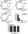
- Figure 1 shows that the VEGF165 rabbit antibody VEGF-R988 blocks the binding of VEGF165 to VEGFR2 protein.
- FIG. 2 shows that the VEGF165 rabbit antibody VEGF-R988 neutralizes the proliferation of HUVEC cells by VEGF165.
- Figure 3 shows the binding of humanized antibody VEGF-H988 Fab to VEGF165 by ELISA.
- Figure 4 shows the species cross-binding between VEGF-H988 Fab and mVEGF164 detected by ELISA.
- Figure 5 shows that VEGF-H988 Fab blocked the binding of VEGF165 to VEGFR2 protein detected by ELISA.
- Figure 6 shows the effect of VEGF-H988 Fab versus Lucentis in neutralizing different concentrations of VEGF165.
- Figure 7 shows the effect of VEGF-H988 Fab versus EYLEA in neutralizing different concentrations of VEGF165.
- Figure 8 shows the effect of VEGF-H988 Fab versus Avastin in neutralizing VEGF165 at different concentrations.
- Figure 9 shows the effect of VEGF-H988 Fab in contrast to Conbercept in neutralizing different concentrations of VEGF165.
- Figure 10 shows the effect of VEGF-H988 Fab versus Brolucizumab in neutralizing different concentrations of VEGF165.
- Various aspects of the present invention relate to isolated anti-VEGF Fab antibody fragments, antibody-drug conjugates comprising the antibody fragments or antigen-binding fragments thereof, nucleic acids and expression vectors encoding the Fab antibody fragments, and hosts containing the nucleic acids or expression vectors Cells, methods for producing the anti-VEGF Fab antibody fragments, pharmaceutical compositions containing the anti-VEGF Fab antibody fragments, and methods for using the anti-VEGF Fab antibody fragments to treat diseases related to angiogenesis.
- antibody means an immunoglobulin molecule, and refers to any form of antibody that exhibits the desired biological activity. Including but not limited to monoclonal antibodies (including full-length monoclonal antibodies), polyclonal antibodies and multispecific antibodies (such as bispecific antibodies), and even antibody fragments.
- the full-length antibody structure preferably contains 4 polypeptide chains, usually 2 heavy (H) chains and 2 light (L) chains connected to each other by disulfide bonds. Each heavy chain contains a heavy chain variable region and a heavy chain constant region. Each light chain contains a light chain variable region and a light chain constant region. In addition to this typical full-length antibody structure, its structure also includes other derivative forms.
- the heavy chain variable region and light chain variable region can be further subdivided into more conservative regions (called framework regions (FR)) and hypervariable regions interspersed (called complementarity determining regions (CDR)).
- framework regions FR
- CDR complementarity determining regions
- CDR complementarity determining region
- CDR1, CDR2, and CDR3 refers to the amino acid residues of the variable region of an antibody, the presence of which is necessary for antigen binding.
- Each variable region usually has 3 CDR regions identified as CDR1, CDR2, and CDR3.
- Each complementarity determining region may contain amino acid residues from the “complementarity determining region” defined by Kabat (Kabat et al., Sequences of Proteins of Immunological Interest, 5th Ed. Public Health Service, National Institutes of Health, Bethesda, MD. 1991 )) and/or those residues from the "hypervariable loop” (Chothia and Lesk; J Mol Biol 196:901-917 (1987)).
- framework or "FR” residues are those variable region residues other than the CDR residues as defined herein.
- Each heavy chain variable region and light chain variable region usually contains 3 CDRs and up to 4 FRs.
- the CDRs and FRs are arranged in the following order from the amino terminal to the carboxy terminal, for example: FR1, CDR1, FR2, CDR2, FR3, CDR3, FR4.
- CDR complementarity determining region
- FR framework region
- constant region refers to such amino acid sequences on the light chain and heavy chain of an antibody that do not directly participate in the binding of the antibody to the antigen, but exhibit various effector functions, such as antibody-dependent cytotoxicity.
- the heavy chain of an antibody can be divided into five categories: ⁇ , ⁇ , ⁇ , ⁇ , and ⁇ .
- ⁇ When it forms a complete antibody with the light chain, it can be divided into five categories: IgA , IgD, IgE, IgG and IgM, several of these classes can be further divided into subclasses (isotypes), such as IgG1, IgG2, IgG3, IgG4, IgA and IgA2.
- the light chain of an antibody can be classified into ⁇ and ⁇ .
- an "antigen-binding fragment of an antibody” includes a portion of a complete antibody molecule that retains at least some of the binding specificity of the parent antibody, and usually includes at least a portion of the antigen-binding region or variable region (eg, one or more CDRs) of the parent antibody.
- antigen-binding fragments include, but are not limited to, Fv, Fab, Fab', Fab'-SH, F(ab')2, Fd fragment, Fd' fragment, single-chain antibody molecules (e.g., scFv, di-scFv, or tri-scFv , Diabody or scFab), single domain antibody.
- Fab fragments typically comprise a heavy chain variable region (V H) and a heavy chain constant region 1 (C H 1) and light chain variable region (V L) and a light chain constant region (C L).
- antibody fragment refers to an incomplete antibody molecule that retains at least some biological properties of the parent antibody, and examples thereof include, but are not limited to, Fc fragments in addition to those mentioned in the above-mentioned "antigen-binding fragments".
- antibody-drug conjugate refers to a binding protein such as an antibody or antigen-binding fragment thereof chemically linked to one or more chemical drugs (also referred to herein as agents), which may optionally Ground is a therapeutic or cytotoxic agent.
- the ADC includes an antibody, cytotoxic or therapeutic drug, and a linker that enables the drug to be linked or conjugated to the antibody.
- ADCs usually have any value of 1 to 8 drugs conjugated to antibodies, including 2, 4, 6, or 8 drug-loading substances.
- Non-limiting examples of drugs that can be included in the ADC are mitotic inhibitors, anti-tumor antibiotics, immunomodulators, vectors for gene therapy, alkylating agents, anti-angiogenic agents, antimetabolites, boron-containing agents, chemotherapy protection Agents, hormones, antihormonal agents, corticosteroids, photoactive therapeutic agents, oligonucleotides, radionuclide agents, topoisomerase inhibitors, tyrosine kinase inhibitors and radiosensitizers.
- chimeric antibody refers to an antibody in which a part of the heavy chain and/or light chain is derived from a specific source or species, and the remaining part is derived from a different source or species.
- the “chimeric antibody” may also be a functional fragment as defined above.
- Humanized antibodies are a subset of “chimeric antibodies.”
- humanized antibody or “humanized antigen-binding fragment” is defined herein as an antibody or antibody fragment: (i) derived from a non-human source (for example, a transgenic mouse carrying a heterologous immune system) And based on human germline sequence; or (ii) the variable region is of non-human origin and the constant region is a chimeric antibody of human origin; or (iii) CDR grafted, wherein the CDR of the variable region is derived from a non-human source, and the variable One or more framework regions of the region are of human origin, and the constant region (if any) is of human origin.
- the purpose of "humanization” is to eliminate the immunogenicity of non-human source antibodies in the human body, while retaining the greatest possible affinity.
- a “monoclonal antibody” refers to an antibody obtained from a substantially homogeneous antibody population, that is, the population comprising a single antibody is identical except for possible mutations (such as natural mutations) that may be present in very small amounts. Therefore, the term “monoclonal” indicates the nature of the antibody, that is, it is not a mixture of unrelated antibodies. In contrast to polyclonal antibody preparations which usually include different antibodies directed against different determinants (epitopes), each monoclonal antibody of a monoclonal antibody preparation is directed against a single determinant on the antigen. In addition to their specificity, the advantage of monoclonal antibody preparations is that they are generally not contaminated by other antibodies. The term “monoclonal” should not be understood as requiring the production of the antibody by any specific method.
- the antibody "specifically binds" to an antigen of interest, such as a tumor-associated polypeptide antigen target (herein, VEGF), that is, binds to the antigen with sufficient affinity so that the antibody can be used as a therapeutic agent to target a target expressing the antigen Cells or tissues, and have no significant cross-reactivity with other proteins or with proteins other than homologs and variants (such as mutant forms, splice variants, or truncated forms of proteolysis) of the antigen target mentioned above No significant cross reaction.
- an antigen of interest such as a tumor-associated polypeptide antigen target (herein, VEGF)
- VEGF tumor-associated polypeptide antigen target
- binding affinity refers to the strength of the sum of non-covalent interactions between a single binding site of a molecule and its binding partner. Unless otherwise stated, "binding affinity” as used herein refers to intrinsic binding affinity, which reflects a 1:1 interaction between members of a binding pair (eg, antibody and antigen).
- K D refers to the equilibrium dissociation constant of the antibody-antigen interaction.
- kon refers to the rate constant at which an antibody binds to an antigen.
- the term “koff” refers to the rate constant at which the antibody dissociates from the antibody/antigen complex.
- K D association rate constant k on "and “dissociation rate constant k off” are usually used to describe the affinity between a molecule (such as an antibody) and its binding partner (such as an antigen), that is, the binding of a ligand to a specific protein Tightness. Binding affinity is affected by interactions between non-covalent molecules, such as hydrogen bonds, electrostatic interactions, hydrophobicity and van der Waals forces between two molecules. In addition, the binding affinity between the ligand and its target molecule may be affected by the presence of other molecules. Affinity can be analyzed by conventional methods known in the art, including the ELISA described herein.
- epitope includes any protein determinant capable of specifically binding to an antibody or T cell receptor.
- Epitope determinants usually consist of chemically active surface groups of molecules (for example, amino acids or sugar side chains, or combinations thereof), and usually have specific three-dimensional structural characteristics and specific charge characteristics.
- isolated antibody is an antibody that has been identified and isolated from a component of the cell that expresses it. Isolated antibodies include antibodies in situ within recombinant cells where at least one component of the antibody's natural environment is absent. However, usually, the isolated antibody is prepared through at least one purification step.
- sequence identity between two polypeptide or nucleic acid sequences means the number of identical residues between the sequences as a percentage of the total number of residues, and is calculated based on the size of the smaller of the compared molecules.
- sequences being compared are aligned to produce the largest match between the sequences, and the gaps in the alignment (if any) are resolved by a specific algorithm.
- Preferred computer program methods for determining the identity between two sequences include, but are not limited to, the GCG program package, including GAP, BLASTP, BLASTN, and FASTA (Altschul et al., 1990, J. Mol. Biol. 215: 403-410) .
- the above program can be publicly obtained from the International Center for Biotechnology Information (NCBI) and other sources.
- NCBI International Center for Biotechnology Information
- Smith Waterman algorithm can also be used to determine identity.
- Fc receptor refers to a receptor that binds to the Fc region of an antibody.
- Human FcR of natural sequence is preferred, and receptors ( ⁇ receptors) that bind to IgG antibodies are preferred, which include Fc ⁇ RI, Fc ⁇ RII, and Fc ⁇ RIII subtypes, and variants of these receptors.
- Other FcRs are included in the term “FcR”.
- the term also includes the neonatal receptor (FcRn) which is responsible for the transfer of maternal IgG to the fetus (Guyer et al., J. Immunology 117:587 (1976) and Kim et al., J. Immunology 24:249 (1994)).
- FcRn neonatal Fc receptor
- the neonatal Fc receptor (FcRn) plays an important role in the metabolic fate of IgG antibodies in the body. FcRn functions to rescue IgG from the lysosomal degradation pathway, thereby reducing its clearance in serum and increasing its half-life. Therefore, the in vitro FcRn binding properties/characteristics of IgG indicate its in vivo pharmacokinetic properties in the blood circulation.
- effector functions refers to those biological activities attributable to the Fc region of an antibody, which differ by antibody isotype.
- antibody effector functions include: C1q binding and complement-dependent cytotoxicity (CDC), Fc receptor binding, antibody-dependent cytotoxicity (ADCC), antibody-dependent phagocytosis (ADCP), cytokine secretion, immune complexes Mediated antigen uptake by antigen-presenting cells, down-regulation of cell surface receptors (such as B cell receptors), and B cell activation.
- effector cells refers to leukocytes that express one or more FcRs and perform effector functions.
- the effector cell at least expresses FcyRIII and performs ADCC effector function.
- human leukocytes that mediate ADCC include peripheral blood mononuclear cells (PBMC), natural killer (NK) cells, monocytes, cytotoxic T cells, and neutrophils.
- PBMC peripheral blood mononuclear cells
- NK natural killer cells
- monocytes cytotoxic T cells
- neutrophils effector cells can be isolated from natural sources, for example, blood. Effector cells are usually lymphocytes associated with the effector stage and function to produce cytokines (helper T cells), kill cells infected by pathogens (cytotoxic T cells) or secrete antibodies (differentiated B cells) .
- Immune cells include cells that have hematopoietic origin and play a role in immune responses. Immune cells include: lymphocytes, such as B cells and T cells; natural killer cells; myeloid cells, such as monocytes, macrophages, eosinophils, mast cells, basophils, and granulocytes.
- ADCC antibody-dependent cell-mediated cytotoxicity
- cytotoxic cells such as NK cells, neutrophils, and macrophages
- the secreted Ig on the Fc ⁇ receptor enables these cytotoxic effector cells to specifically bind to the target cell carrying the antigen, and then kill the target cell using, for example, a cytotoxin.
- an in vitro ADCC assay can be performed, such as the in vitro ADCC assay described in U.S. Patent No. 5,500,362 or 5,821,337 or U.S. Patent No. 6,737,056 (Presta).
- Useful effector cells for such assays include PBMC and NK cells.
- “Complement dependent cytotoxicity” or “CDC” refers to the lysis of target cells in the presence of complement.
- the activation of the typical complement pathway is initiated by combining the first component of the complement system (C1q) with an antibody (of the appropriate subclass) that binds to its corresponding antigen.
- a CDC assay can be performed, such as the CDC assay described in Gazzano-Santoro et al., J. Immunol Methods 202:163 (1996).
- polypeptide variants with an altered Fc region amino acid sequence polypeptides with a variant Fc region
- polypeptide variants with enhanced or reduced C1q binding are described.
- Human Umbilical Vein Endothelial Cells are isolated from umbilical cord veins and are generally used in physiology and pharmacological research, such as macromolecular transport, blood coagulation, angiogenesis and fibrinolysis. In particular, it can be used as a model for angiogenesis research and other VEGF-dependent signal pathway research (related endothelial growth factor) research.
- the present invention firstly uses recombinant human VEGF165 protein to immunize rabbits, and then obtains the antibody clone VEGF165-R988 which specifically binds to recombinant human VEGF165 protein through phage display library screening. Then, the nucleotide sequences encoding the heavy chain and light chain variable regions of the VEGF165-R988scFv antibody were inserted into the pSTEP2 vector with the nucleotide sequence of the rabbit heavy chain IgG1 constant region or the rabbit light chain kappa constant region, and Perform culture expression. Use protein A purification column for purification to obtain high-purity antibody. ELISA test showed that the rabbit antibody VEGF165-R988 can effectively inhibit the binding of VEGF165 protein to VEGFR2 protein, and VEGF165-R988 can effectively neutralize the ability of VEGF165 to promote HUVEC cell proliferation.
- the light chain or heavy chain variable region of a human antibody that is closer to the rabbit light chain or heavy chain variable region is selected as the template, and each of the rabbit antibody light chain or heavy chain is selected as the template.
- Three CDRs (Table 1) were inserted into the variable region of the human antibody to obtain humanized light chain variable region (VL) and heavy chain variable region (VH) sequences. Since the rabbit-derived framework region has key points that are essential for supporting the activity of CDR, the key points are backmutated to the corresponding sequence of the rabbit antibody.
- the VEGF-H988-10 light chain/heavy chain expression vector was obtained by the method of full gene synthesis, transfected into HEK-293 cells and cultured and expressed, and the culture supernatant was purified by a protein A purification column to obtain high-purity VEGF-H988-10 antibody .
- an SDM library of the CDR regions of the heavy and light chain variable domains (including LCDR1, LCDR3, HCDR2 and HCDR3) was constructed, and the 4 mutant libraries were constructed into scFv sequences to be fused with gene 3.
- the protein form was cloned into a phagemid vector, and the best clones were screened for each CDR through the binding ability of the soluble antigen VEGF, and finally the VEGF-H988 Fab antibody fragment with optimized CDR affinity and stability was obtained and tested.
- the antibody of the Fab fragment Compared with the full-length structure, the antibody of the Fab fragment has stronger penetrability, lower gastrointestinal perforation, high blood pressure and bleeding and other side effects and cannot stimulate the complement cascade reaction, thereby reducing the occurrence of endophthalmitis and The risk of autoimmune inflammation.
- the invention also relates to nucleic acid molecules encoding the antibodies of the invention or parts thereof.
- the sequences of these nucleic acid molecules include but are not limited to SEQ ID NO: 2-3, 4-7, 16-17, 20-21, 41-49, and 52-53.
- nucleic acid molecules of the present invention are not limited to the sequences disclosed herein, but also include variants thereof.
- the variants of the present invention can be described with reference to their physical characteristics in hybridization. Those skilled in the art will recognize that using nucleic acid hybridization techniques, nucleic acids can be used to identify their complements and their equivalents or homologs. It will also be recognized that hybridization can occur with less than 100% complementarity. However, considering the proper selection of conditions, hybridization techniques can be used to distinguish DNA sequences based on their structural correlation with specific probes.
- the invention also provides a recombinant construct comprising one or more nucleotide sequences of the invention.
- the recombinant construct of the present invention is constructed by inserting a nucleic acid molecule encoding the antibody of the present invention into a vector, such as a plasmid, phagemid, phage, or viral vector.
- the antibody of the present invention can be prepared by recombinantly expressing nucleotide sequences encoding the light chain and the heavy chain or parts thereof in a host cell.
- one or more recombinant expression vectors carrying the nucleotide sequence encoding the light chain and/or heavy chain or part thereof can be used to transfect the host cell so that the light chain and the heavy chain are in the Expressed in host cells.
- Standard recombinant DNA methodology is used to prepare and/or obtain nucleic acids encoding heavy and light chains, incorporate these nucleic acids into recombinant expression vectors and introduce the vectors into host cells, such as Sambrook, Fritsch and Maniatis (eds.
- Suitable host cells are prokaryotic cells and eukaryotic cells.
- prokaryotic host cells are bacteria, and examples of eukaryotic host cells are yeast, insect or mammalian cells. It should be understood that the design of an expression vector including a selection regulatory sequence is affected by many factors, such as the choice of host cell, the desired protein expression level, and whether the expression is constitutive or inducible.
- a usable expression vector for bacteria By inserting the structural DNA sequence encoding the desired antibody together with suitable translation initiation and termination signals and a functional promoter into an operable reading frame, a usable expression vector for bacteria can be constructed.
- the vector will contain one or more phenotypic selectable markers and an origin of replication to ensure the maintenance of the vector and provide amplification in the host as needed.
- Suitable prokaryotic hosts for transformation include E. coli, Bacillus subtilis, Salmonella typhimurium, and Pseudomonas, Streptomyces, and grapes. Multiple species in the genus Staphylococcus.
- Bacterial vectors can be, for example, phage, plasmid or phagemid based. These vectors may contain a selection marker and a bacterial origin of replication, which are derived from commercially available plasmids that usually contain elements of the well-known cloning vector pBR322 (ATCC37017). After transforming an appropriate host strain and growing the host strain to an appropriate cell density, the selected promoter is de-repressed/induced by an appropriate method (for example, temperature change or chemical induction), and the cells are cultured for an additional time. The cells are usually harvested by centrifugation, broken down by physical or chemical methods, and the resulting crude extract is retained for further purification.
- an appropriate method for example, temperature change or chemical induction
- a variety of expression vectors can be advantageously selected according to the intended use of the expressed protein. For example, when a large number of such proteins are to be produced for antibody production or for screening peptide libraries, for example, a vector that directs high-level expression of a fusion protein product that is easy to purify may be required.
- Preferred regulatory sequences for expression in mammalian host cells include viral elements that direct high-level protein expression in mammalian cells, such as promoters and/or enhancers derived from cytomegalovirus (CMV) (e.g. CMV promoter/enhancer) Promoter), simian virus 40 (SV40) promoter and/or enhancer (e.g. SV40 promoter/enhancer), adenovirus promoter and/or enhancer (e.g. adenovirus major late promoter (AdMLP)) and Polyoma virus promoter and/or enhancer.
- CMV cytomegalovirus
- SV40 simian virus 40
- AdMLP adenovirus major late promoter
- Polyoma virus promoter and/or enhancer e.g. adenovirus major late promoter (AdMLP)
- the recombinant expression vector may also include an origin of replication and a selection marker (see, for example, U.S. 4,399,216, U.S. 4,634,665 and U.S. 5,179,017 of Axel et al.).
- Suitable selection markers include genes that confer resistance to drugs such as G418, hygromycin, or methotrexate to host cells into which the vector has been introduced.
- drugs such as G418, hygromycin
- methotrexate to host cells into which the vector has been introduced.
- the dihydrofolate reductase (DHFR) gene confers resistance to methotrexate
- the neo gene confers resistance to G418.
- Transfection of the expression vector into host cells can be performed using standard techniques such as electroporation, calcium phosphate precipitation, and DEAE-dextran transfection.
- Suitable mammalian host cells for expressing the antibodies provided herein include Chinese Hamster Ovary (CHO cells) [including dhfr-CHO cells, as described in Urlaub and Chasin, (1980) Proc. Natl. Acad. Sci. USA 77:4216 In -4220, DHFR selection markers are used, such as those described in RJ Kaufman and PASharp (1982) Mol. Biol. 159:601-621], NSO myeloma cells, COS cells, and SP2 cells.
- Chinese Hamster Ovary CHO cells
- DHFR selection markers are used, such as those described in RJ Kaufman and PASharp (1982) Mol. Biol. 159:601-621]
- NSO myeloma cells such as those described in RJ Kaufman and PASharp (1982) Mol. Biol. 159:601-621
- NSO myeloma cells such as those described in RJ Kaufman and PASharp (1982) Mol. Biol. 159
- the antibody of the present invention can be recovered and purified from recombinant cell culture by known methods, including but not limited to, ammonium sulfate or ethanol precipitation, acid extraction, protein A affinity chromatography, protein G affinity chromatography, anion Or cation exchange chromatography, phosphocellulose chromatography, hydrophobic interaction chromatography, affinity chromatography, hydroxyapatite chromatography, and lectin chromatography.
- High performance liquid chromatography (“HPLC”) can also be used for purification. See, for example, Colligan, Current Protocols in Immunology or Current Protocols in Protein Science, John Wiley&Sons, NY, NY, (1997-2001), such as Chapters 1, 4, 6, 8, 9, and 10, each of which is fully cited Include this article.
- the antibodies of the present invention can be used to treat diseases related to angiogenesis, including ocular diseases characterized by choroidal neovascularization, including but not limited to age-related age-related macular degeneration (AMD), diabetic macular edema (DME), retinal edema, and degeneration The occurrence of sexual myopia, choroidal neovascularization (CNV).
- AMD age-related age-related macular degeneration
- DME diabetic macular edema
- CNV choroidal neovascularization
- the antibody of the present invention and at least one other agent can be prepared into a pharmaceutical composition, which includes the antibody of the present invention and one or more pharmaceutically acceptable carriers, diluents or excipients.
- the pharmaceutical composition may include additional therapeutic agents.
- the invention also relates to a pharmaceutical package and a kit comprising one or more containers containing the aforementioned pharmaceutical composition of the invention. It is accompanied by a reminder of the form prescribed by the government agency that regulates the production, use or sale of drugs or biological products, which reflects that the drug has been approved by the above-mentioned agencies for human administration.
- the pharmaceutical composition of the present invention can be prepared in a manner known in the art, for example, by conventional mixing, dissolving, granulating, tablet preparation, grinding, emulsifying, coating, embedding or freeze-drying methods.
- compositions containing the compound of the present invention formulated in an acceptable carrier After the pharmaceutical composition containing the compound of the present invention formulated in an acceptable carrier has been prepared, they can be placed in an appropriate container and labeled for the treatment of the indicated condition.
- labels would include the amount, frequency, and method of administration.
- composition containing the antibody of the present invention is also combined with one or more other therapeutic agents, such as anti-tumor agents, wherein the resulting combination does not cause unacceptable adverse effects.
- Example 1 Screening of rabbit antibodies that block the binding of VEGF165 and VEGFR2 using phage antibody display library
- Recombinant human VEGF165 protein (source: Beijing Yiqiao Shenzhou Technology Co., Ltd., Cat.11066-HNAH) was used to immunize rabbits.
- the amino acid sequence of the extracellular region Met1-Arg191 of the human VEGF165 protein (UniProtKB P15692-4) is SEQ ID NO:1.
- the specific method is: mixing recombinant human VEGF165 protein with Freund's adjuvant, and using the mixture for four immunizations, each with a dose of 500 ⁇ g, subcutaneous injection, and the immunization interval is 3 weeks, 2 weeks, and 2 weeks.
- blood was collected from the orbital intracanthal venous plexus four days after immunization.
- the ELISA method was used to coat recombinant human VEGF165 protein to detect the titer of rabbit anti-VEGF165 serum.
- the serum titer of the fifth immunization reached 25000 times dilution.
- 25 ⁇ g of recombinant human VEGF165 protein was injected intravenously for boost. Seven days later, the rabbits were sacrificed and the spleen tissues of the rabbits were frozen and stored in liquid nitrogen.
- TriPure Isolation Reagent reagent (source: Roche, Cat. No. 11 667 165 001) to extract RNA from rabbit spleen tissue, and use reverse transcription kit (source: Invitrogen, Cat. No. 18080-051) for reverse transcription.
- cDNA Design 10 pairs of primers to amplify the light chain variable region sequence of the rabbit antibody, and 4 pairs of primers to amplify the heavy chain variable region sequence (Barbas C F et al., CSHL Press. 2004).
- the overlapping extension splicing PCR method was used to splice the sequences encoding the light chain and heavy chain variable regions of the rabbit antibody into a nucleotide sequence encoding scFv, and the light and heavy chain variable regions were connected through a linker:
- phage vector pComb3x source: Beijing Yiyi
- restriction endonuclease Sfi I source: Fermentas
- X-Blue competent cells were electrotransformed to construct a phage display scFv antibody library for immunizing rabbits.
- the recombinant human VEGF165 protein was coated on the ELISA plate, and the phage library enriched with anti-VEGF165 positive antibodies was screened according to the phage antibody panning process (O'Brien, PM, & Aitken, R.
- Monoclonal phages were selected from the enriched library for expression, and the binding to recombinant human VEGF165 protein was detected by ELISA method.
- the antibody clones that specifically bind to recombinant human VEGF165 were screened and the clones obtained were sent to the sequencing company for sequencing to obtain VEGF165.
- the nucleotide sequence of the R988scFv antibody SEQ ID NO: 3).
- the nucleotide sequence of the heavy chain variable region of the VEGF165-R988scFv antibody was amplified by PCR, and inserted into the heavy chain signal peptide (SEQ ID NO: 43) and the rabbit heavy chain IgG1 constant region (SEQ ID NO: 6)
- the heavy chain (SEQ ID NO: 52) expression vector is obtained from the pSTEP2 vector digested with Sca I+Kpn I (Fermentas).
- the nucleotide sequence of the light chain variable region of the VEGF-R988scFv antibody was amplified by PCR, and inserted into the light chain signal peptide (SEQ ID NO: 44) and the rabbit light chain kappa constant region (SEQ ID NO: 7) by in-fusion method.
- the light chain (SEQ ID NO: 53) expression vector was obtained from the pSTEP2 vector digested with Sca I+BamH I (Fermentas).
- the recombinant plasmid was extracted, transfected into HEK-293 cells and cultured and expressed for 7 days, and the culture supernatant was purified by a protein A purification column to obtain high-purity antibodies.
- the VEGF165 protein (source: Beijing Yiqiao Shenzhou Technology Co., Ltd.) at a concentration of 1 ⁇ g/mL was coated on a 96-well plate, 100 ⁇ L per well, and coated overnight at 4°C. The plate was washed the next day, sealed at room temperature for 1 hour, and then 100 ⁇ L 5 ⁇ g/mL VEGFR2-biotin protein (source: Beijing Yiqiao Shenzhou Technology Co., Ltd.) and different concentrations of VEGF-R988 antibody were added and incubated together. Wash the plate to remove the unbound antibody, add streptavidin/HRP (source: Beijing Zhongshan Jinqiao) to incubate the plate repeatedly, and finally add the substrate color solution for color development.
- streptavidin/HRP source: Beijing Zhongshan Jinqiao
- Inhibition rate PI% (OD blank-OD sample) / OD blank ⁇ 100%, where OD blank represents the OD value of the group with only VEGFR2-biotin and no antibody added, and OD sample represents the waiting for VEGFR2-biotin and antibody Test the OD value of the group.
- the VEGF-R988 antibody can effectively bind to the coated VEGF165 protein, and can effectively inhibit the binding of VEGFR165 protein to VEGFR2 protein.
- WST-8 method was used to detect the proliferation of umbilical vein endothelial cell (HUVEC) cells by neutralizing VEGF165 by chimeric antibody.
- a 96-well plate was seeded with HUVEC cells 4 ⁇ 10 3 cells/well, cultured in M199 medium containing 10% FBS and 5% L-Gln for 4 hours, and then added with different concentrations of VEGF-R988 antibody, 50 ⁇ L/well. Subsequently, 10 ⁇ L/well of VEGF165 with a final concentration of 10 ng/mL was added. Place the 96-well plate in a CO 2 incubator at 37° C. and 5% CO 2 for 3 days.
- HUVEC umbilical vein endothelial cell
- test blank well B no cells
- negative control group M seeding cells, no sample, plus VEGF165
- M' seeding cells, no sample and VEGF165
- Neutralization rate% (Negative control M group OD value-Sample OD value) / (Negative control M group OD value -M' group OD value) ⁇ 100%, using the automatic analysis function of the statistical software GraphPad Prism to calculate the standard curve, the abscissa is the concentration of the sample, the ordinate is the neutralization rate, the regression equation is a four-parameter equation, and the "S" type is obtained Curve, calculate the half effective concentration (EC 50 ) of the sample.
- the VEGF-R988 antibody can effectively reduce the ability of VEGF165 to promote HUVEC proliferation.
- Example 1.2 According to the nucleotide sequence of the VEGF-R988scFv antibody determined in Example 1.2, the amino acid sequences of the heavy chain and light chain variable regions of the VEGF-R988scFv were deduced, see SEQ ID NO: 8/9.
- Rabbit antibody humanization adopts the classic humanization method CDR transplantation method, selects the human antibody light chain or heavy chain variable region that is closer to the rabbit light chain or heavy chain variable region as the template, and combines the rabbit antibody light chain or heavy chain variable region as the template.
- Each of the three CDRs of the chain (Table 1) was inserted into the variable region of the human antibody to obtain humanized light chain variable region (VL) and heavy chain variable region (VH) sequences.
- the human template of the light chain variable region of VEGF-R988 used was IGKV1-27*01, which had 65.30% homology with the light chain of VEGF-R988, and the human template of the heavy chain variable region was IGHV4-4 *08, the template has 53.20% homology with the heavy chain of VEGF-R988.
- the key points are backmutated to the corresponding sequence of the rabbit antibody, where the first back mutation of the light chain is E and the second back mutation is L ,
- the back mutation at position 4 is L
- the back mutation at position 63 is K
- the back mutation at position 3 of the heavy chain is V
- the back mutation at position 37 is V
- the back mutation at position 47 is Y
- the back mutation at position 78 is V
- the 79th back mutation is D
- the 91th back mutation is F
- the reversion sites are all Kabat numbering.
- Humanized antibody VEGF-H988-10 was obtained by CDR humanization transplantation and framework region back mutation.
- the VEGF-H988-10 heavy chain variable region (SEQ ID NO: 20) was obtained by the method of full gene synthesis, and inserted into the heavy chain signal peptide (SEQ ID NO: 43) and human IgG1 constant region (
- the VEGF-H988-10 heavy chain (SEQ ID NO: 16) expression vector was obtained from the pSTEP2 vector digested with Sca I+Nhe I (Fermentas) of SEQ ID NO: 47).
- the VEGF-H988-10 light chain variable region (SEQ ID NO: 21) was obtained by the method of full gene synthesis, and inserted into the light chain signal peptide (SEQ ID NO: 44) and the human kappa constant region (
- the VEGF-H988-10 light chain (SEQ ID NO: 17) expression vector was obtained from the pSTEP2 vector digested with Sca I+BsiW I (Fermentas) of SEQ ID NO: 48). After the plasmid was extracted, it was transfected into HEK-293 cells and cultured and expressed for 7 days. The culture supernatant was purified by a protein A purification column to obtain high-purity antibodies.
- the heavy chain and light chain variable domain CDR regions construct an SDM library, including three saturation mutation libraries of LCDR1, LCDR3, and HCDR2; at the same time, in order to improve the chemical stability of the antibody, it is necessary to remove the The amide or isomerization site is changed to another amino acid residue. Deamidation of asparagine can lead to the production of isoaspartic acid residues on NG, NS, NA, NT and other sequences, which affects the stability or biological function of the antibody.
- VEGF-H988 variable domain HCDR3 has easy deamidation sites.
- an SDM library was constructed at the same time.
- VEGF-H988 light chain and heavy chain CDR sequences are shown in Table 2.
- VEGF-H988 Fab light chain nucleotide sequence (SEQ ID NO: 42) that encodes the signal peptide, it contains the nucleotide sequence of the light chain signal peptide (SEQ ID NO: 44) connected in sequence ,
- the VEGF-H988 Fab heavy chain Fab nucleotide sequence contains the nucleotide sequence of the heavy chain signal peptide (SEQ ID NO: 43), humanized anti- The nucleotide sequence of the variable region of the heavy chain (SEQ ID NO: 45) and the nucleotide sequence of the CH1 constant region of the human IgG1 heavy chain (SEQ ID NO: 49) were inserted by in-fusion method to construct the correct containing light In the pGS vector (Nhe I+Not I) of the chain, the correct VEGF-H988 Fab light and heavy chain expression vector was verified by sequencing.
- the expression vector contains eukaryotic expression vector of GS screening gene and antibody light and heavy chain expression elements.
- the expression vector was transfected into CHO-K1-GS-deficient cells, and VEGF-H988 Fab high-expressing cell line was obtained by MSX screening. ELISA is used to select clones with high antibody expression, and the high-expressing cell lines are screened by combining the cell growth status and the key quality attribute analysis results of antibody drugs.
- the VEGF-H988 Fab-producing CHO cell line was cultivated in a serum-free feeding suspension culture method to obtain high-purity and high-quality VEGF-H988 Fab.
- VEGF-H988 Fab specifically binds to VEGF165
- the plate was washed the next day and blocked at room temperature for 1 hour, then 100 ⁇ L of 1 ⁇ g/mL VEGF165-H988 Fab, Lucentis (source: Novartis) and negative control antibody H7N9-R1 Fab (source: Beijing Yiqiao Shenzhou Technology Co., Ltd.) were added and incubated for 1 hour Then wash the plate to remove unbound antibody, add the secondary antibody goat F(ab')2 anti-human IgG F(ab')2/HRP (source: Jackson ImmunoResearch), incubate the plate repeatedly, add the substrate color development solution for visualization After termination, the microplate reader reads OD450.
- a streptavidin-coated Sensor was used to immobilize the biotin-labeled VEGF165 protein, and the affinity of VEGF-H988 Fab and Lucentis to VEGF165 at multiple concentrations was detected.
- the method is as follows: Use a 5000MW ultrafiltration tube to pass through the buffer solution of the recombinant VEGF165 protein (20mM Tris, 150mM NaCl, pH8.0) Replaced with PBS. After UV quantification, 567.57 ⁇ g protein was obtained. The obtained protein was mixed with 20mM biotin solution at a molar ratio of 1:2, and reacted for 30 minutes in the dark at room temperature. Filtered again through a 5000MW ultrafiltration tube to remove unlabeled biotin. UV After quantification, an equal volume of glycerol and a final concentration of 0.1% BSA were added to obtain a biotin-labeled protein. Its concentration was 2.08mg/mL by UV detection.
- VEGF-H988 Fab to recombinant human VEGF165 protein binding affinity KD value of 1.54E-10 (M), the binding constants k on value of 2.74E + 05 (1 / Ms) , the value of the dissociation constant k dis Lucentis VEGF165 protein binding affinity KD value of 5.78E-11 (M), the binding constants k on value of 5.36E + 04 (1 / Ms) , dissociation constant value from k dis; is 4.21E-05 (1 / s) It is 3.10E-05 (1/s).
- the results show that the affinity of VEGF-H988 Fab is stronger than that of Lucentis, which is about 3.75 times that of Lucentis. Therefore, VEGF-H988 Fab has a stronger ability to bind VEGF165 protein than Lucentis.
- Dilute recombinant human VEGF165 protein and recombinant mouse mVEGF164 protein (source: Beijing Yiqiao Shenzhou Technology Co., Ltd.) to 0.1 ⁇ g/mL, 1 ⁇ g/mL and 10 ⁇ g/mL and coat them on 96-well plates with 100 ⁇ L per well. Coated overnight at 4°C. Wash the plate the next day, block at room temperature for 1 hour, add 100 ⁇ L of VEGF165-H988 Fab, Lucentis and the negative control antibody H7N9-R-Fab at a concentration of 2 ⁇ g/mL, and incubate for 1 hour.
- VEGF165-H988 Fab has specific binding to recombinant human VEGF165 protein, and cross-binding with recombinant mouse mVEGF164 protein. Lucentis does not cross-binding with recombinant mouse mVEGF164 protein.
- the VEGF165 protein at a concentration of 1 ⁇ g/mL was coated on a 96-well plate, with 100 ⁇ L per well, and coated overnight at 4°C. Wash the plate the next day and block at room temperature for 1 hour, add 100 ⁇ L 2 ⁇ g/mL VEGFR2-his protein (source: Beijing Yiqiao Shenzhou Technology Co., Ltd.), and then add different concentrations of humanized VEGF-H988 Fab, Lucentis and negative control antibodies H7N9-R1-Fab (source: Beijing Yiqiao Shenzhou Technology Co., Ltd.) was incubated together. Wash the plate to remove unbound antibody, add C-his-R023/HRP to incubate and repeat the washing. Finally, add the substrate color developing solution for color development. After termination, the microplate reader reads OD450, and each group is twice in parallel.
- Inhibition rate PI% (OD blank- OD sample )/OD blank ⁇ 100, where OD blank represents the OD value of the group with only VEGFR2-his added without antibody, and OD sample represents the OD of the test group with both VEGFR2-his and antibody added value.
- VEGFR2 protein can effectively bind to the coated VEGF165 protein
- VEGF-H988 Fab can effectively inhibit the binding of VEGFR2 protein to VEGF165 protein, and the blocking ability of VEGF-H988 Fab is significantly better than Lucentis, and the negative control antibody has no inhibition. effect.
- VEGF-H988 Fab blocks the growth inhibitory activity of VEGF165 at different concentrations
- the WST-8 method was used to detect the proliferation effect of humanized antibody neutralizing VEGF165 on HUVEC cells.
- a 96-well plate was seeded with HUVEC cells 4 ⁇ 10 3 /well, cultured in M199 medium containing 10% FBS and 5% L-Gln for 4 hours, and then antibodies of different concentrations were added at 50 ⁇ L/well. Subsequently, 10 ⁇ L/well of VEGF165 with final concentrations of 1000 ng/mL, 100 ng/mL, and 10 ng/mL were added. Place the 96-well plate in a CO 2 incubator at 37° C. and 5% CO 2 for 3 days.
- test blank well B no cells
- negative control group M seeding cells, no sample, plus VEGF165
- M' seeding cells, no sample and VEGF165
- Neutralization rate% (Negative control M group OD value-Sample OD value) / (Negative control M group OD value -M' group OD value) ⁇ 100%, using the automatic analysis function of the statistical software GraphPad Prism to calculate the standard curve, the abscissa is the concentration of the sample, the ordinate is the neutralization rate, the regression equation is a four-parameter equation, and the "S" type is obtained Curve, calculate the half effective concentration (EC 50 ) of the sample.
- the neutralizing effect of VEGF-H988Fab is stronger than Lucentis under the conditions of different concentrations of recombinant human VEGF165.
- concentration of VEGF165 is 1000ng/mL, 100ng/mL
- the neutralizing activity of VEGF-H988 Fab is stronger than EYLEA
- concentration of VEGF165 is 10ng/mL
- VEGF-H988 Fab is slightly weaker than EYLEA.
- the activity of VEGF-H988 Fab is stronger than that of Bevacizumab and Brolucizumab, but is equivalent to the activity of Conbercept.
- concentration of VEGF165 increased, VEGF-H988 Fab maintained a high maximum neutralization rate, while EYLEA and Avastin decreased.
Landscapes
- Health & Medical Sciences (AREA)
- Chemical & Material Sciences (AREA)
- Organic Chemistry (AREA)
- Life Sciences & Earth Sciences (AREA)
- General Health & Medical Sciences (AREA)
- Medicinal Chemistry (AREA)
- Immunology (AREA)
- Biochemistry (AREA)
- Biophysics (AREA)
- Genetics & Genomics (AREA)
- Molecular Biology (AREA)
- Proteomics, Peptides & Aminoacids (AREA)
- Bioinformatics & Cheminformatics (AREA)
- Veterinary Medicine (AREA)
- Ophthalmology & Optometry (AREA)
- Chemical Kinetics & Catalysis (AREA)
- General Chemical & Material Sciences (AREA)
- Nuclear Medicine, Radiotherapy & Molecular Imaging (AREA)
- Pharmacology & Pharmacy (AREA)
- Animal Behavior & Ethology (AREA)
- Public Health (AREA)
- Engineering & Computer Science (AREA)
- Peptides Or Proteins (AREA)
- Medicines Containing Antibodies Or Antigens For Use As Internal Diagnostic Agents (AREA)
- Preparation Of Compounds By Using Micro-Organisms (AREA)
- Micro-Organisms Or Cultivation Processes Thereof (AREA)
- Medicinal Preparation (AREA)
- Medicines That Contain Protein Lipid Enzymes And Other Medicines (AREA)
- Pharmaceuticals Containing Other Organic And Inorganic Compounds (AREA)
- Medicines Containing Material From Animals Or Micro-Organisms (AREA)
Priority Applications (9)
| Application Number | Priority Date | Filing Date | Title |
|---|---|---|---|
| EP20844600.5A EP4043490A4 (en) | 2019-07-19 | 2020-07-17 | HUMANIZED ANTI-VEGF FAB ANTIBODY FRAGMENT AND USE THEREOF |
| MX2022000780A MX2022000780A (es) | 2019-07-19 | 2020-07-17 | Fragmento fab del anticuerpo anti-vegf humanizado y uso del mismo. |
| KR1020227004898A KR20220034868A (ko) | 2019-07-19 | 2020-07-17 | 인간화 항-VEGF Fab 항체 단편 및 이의 용도 |
| CA3150046A CA3150046C (en) | 2019-07-19 | 2020-07-17 | Humanized anti-vegf fab antibody fragment and use thereof |
| CN202080051385.8A CN114127106B (zh) | 2019-07-19 | 2020-07-17 | 人源化抗VEGF Fab抗体片段及其用途 |
| BR112022001021A BR112022001021A2 (pt) | 2019-07-19 | 2020-07-17 | Fragmento fab de anticorpo anti-vegf humanizado e uso do mesmo |
| JP2022503863A JP7229419B2 (ja) | 2019-07-19 | 2020-07-17 | ヒト化抗vegf fab抗体断片及びその使用 |
| AU2020316495A AU2020316495A1 (en) | 2019-07-19 | 2020-07-17 | Humanized anti-VEGF Fab antibody fragment and use thereof |
| US17/578,424 US20220135665A1 (en) | 2019-07-19 | 2022-01-18 | Humanized anti-VEGF antibody Fab fragment and use thereof |
Applications Claiming Priority (2)
| Application Number | Priority Date | Filing Date | Title |
|---|---|---|---|
| CN201910657311.8 | 2019-07-19 | ||
| CN201910657311 | 2019-07-19 |
Related Child Applications (1)
| Application Number | Title | Priority Date | Filing Date |
|---|---|---|---|
| US17/578,424 Continuation US20220135665A1 (en) | 2019-07-19 | 2022-01-18 | Humanized anti-VEGF antibody Fab fragment and use thereof |
Publications (1)
| Publication Number | Publication Date |
|---|---|
| WO2021013065A1 true WO2021013065A1 (zh) | 2021-01-28 |
Family
ID=74192837
Family Applications (1)
| Application Number | Title | Priority Date | Filing Date |
|---|---|---|---|
| PCT/CN2020/102560 WO2021013065A1 (zh) | 2019-07-19 | 2020-07-17 | 人源化抗VEGF Fab抗体片段及其用途 |
Country Status (10)
| Country | Link |
|---|---|
| US (1) | US20220135665A1 (ja) |
| EP (1) | EP4043490A4 (ja) |
| JP (1) | JP7229419B2 (ja) |
| KR (1) | KR20220034868A (ja) |
| CN (1) | CN114127106B (ja) |
| AU (1) | AU2020316495A1 (ja) |
| BR (1) | BR112022001021A2 (ja) |
| CA (1) | CA3150046C (ja) |
| MX (1) | MX2022000780A (ja) |
| WO (1) | WO2021013065A1 (ja) |
Cited By (1)
| Publication number | Priority date | Publication date | Assignee | Title |
|---|---|---|---|---|
| WO2024055996A1 (zh) * | 2022-09-14 | 2024-03-21 | 寻济生物科技(北京)有限公司 | 一种抗vegfa抗体或其抗原结合片段及其应用 |
Families Citing this family (2)
| Publication number | Priority date | Publication date | Assignee | Title |
|---|---|---|---|---|
| MX2022000779A (es) * | 2019-07-19 | 2022-02-14 | Sinocelltech Ltd | Anticuerpo monoclonal anti-vegf humanizado. |
| JP7380914B2 (ja) | 2020-12-18 | 2023-11-15 | 日産自動車株式会社 | 内燃機関の触媒暖機制御方法および触媒暖機制御装置 |
Citations (15)
| Publication number | Priority date | Publication date | Assignee | Title |
|---|---|---|---|---|
| US4399216A (en) | 1980-02-25 | 1983-08-16 | The Trustees Of Columbia University | Processes for inserting DNA into eucaryotic cells and for producing proteinaceous materials |
| US4510245A (en) | 1982-11-18 | 1985-04-09 | Chiron Corporation | Adenovirus promoter system |
| US4634665A (en) | 1980-02-25 | 1987-01-06 | The Trustees Of Columbia University In The City Of New York | Processes for inserting DNA into eucaryotic cells and for producing proteinaceous materials |
| US4816397A (en) | 1983-03-25 | 1989-03-28 | Celltech, Limited | Multichain polypeptides or proteins and processes for their production |
| US4968615A (en) | 1985-12-18 | 1990-11-06 | Ciba-Geigy Corporation | Deoxyribonucleic acid segment from a virus |
| US5168062A (en) | 1985-01-30 | 1992-12-01 | University Of Iowa Research Foundation | Transfer vectors and microorganisms containing human cytomegalovirus immediate-early promoter-regulatory DNA sequence |
| US5179017A (en) | 1980-02-25 | 1993-01-12 | The Trustees Of Columbia University In The City Of New York | Processes for inserting DNA into eucaryotic cells and for producing proteinaceous materials |
| US5500362A (en) | 1987-01-08 | 1996-03-19 | Xoma Corporation | Chimeric antibody with specificity to human B cell surface antigen |
| US5821337A (en) | 1991-06-14 | 1998-10-13 | Genentech, Inc. | Immunoglobulin variants |
| WO1999051642A1 (en) | 1998-04-02 | 1999-10-14 | Genentech, Inc. | Antibody variants and fragments thereof |
| US6194551B1 (en) | 1998-04-02 | 2001-02-27 | Genentech, Inc. | Polypeptide variants |
| US6737056B1 (en) | 1999-01-15 | 2004-05-18 | Genentech, Inc. | Polypeptide variants with altered effector function |
| CN1946422A (zh) * | 2003-11-26 | 2007-04-11 | 埃博马可西斯公司 | 针对血管内皮生长因子的人源化抗体 |
| CN102286101A (zh) * | 2011-08-08 | 2011-12-21 | 常州亚当生物技术有限公司 | 抗VEGF单克隆抗体Fab片段Vasculizumab及其应用 |
| CN104761643A (zh) * | 2003-08-01 | 2015-07-08 | 健泰科生物技术公司 | 抗-vegf抗体 |
Family Cites Families (4)
| Publication number | Priority date | Publication date | Assignee | Title |
|---|---|---|---|---|
| US9388239B2 (en) * | 2014-05-01 | 2016-07-12 | Consejo Nacional De Investigation Cientifica | Anti-human VEGF antibodies with unusually strong binding affinity to human VEGF-A and cross reactivity to human VEGF-B |
| MA42924A (fr) * | 2015-09-23 | 2018-08-01 | Hoffmann La Roche | Variants optimisés d'anticorps anti-vegf |
| WO2017119435A1 (ja) * | 2016-01-06 | 2017-07-13 | 株式会社オーダーメードメディカルリサーチ | 高親和性抗vegf抗体 |
| CN110248674B (zh) * | 2016-09-07 | 2021-10-26 | 萨辛生命科学有限公司 | 针对vegf的合成抗体及其用途 |
-
2020
- 2020-07-17 WO PCT/CN2020/102560 patent/WO2021013065A1/zh unknown
- 2020-07-17 CN CN202080051385.8A patent/CN114127106B/zh active Active
- 2020-07-17 MX MX2022000780A patent/MX2022000780A/es unknown
- 2020-07-17 AU AU2020316495A patent/AU2020316495A1/en active Pending
- 2020-07-17 BR BR112022001021A patent/BR112022001021A2/pt unknown
- 2020-07-17 JP JP2022503863A patent/JP7229419B2/ja active Active
- 2020-07-17 EP EP20844600.5A patent/EP4043490A4/en active Pending
- 2020-07-17 CA CA3150046A patent/CA3150046C/en active Active
- 2020-07-17 KR KR1020227004898A patent/KR20220034868A/ko active Search and Examination
-
2022
- 2022-01-18 US US17/578,424 patent/US20220135665A1/en active Pending
Patent Citations (15)
| Publication number | Priority date | Publication date | Assignee | Title |
|---|---|---|---|---|
| US5179017A (en) | 1980-02-25 | 1993-01-12 | The Trustees Of Columbia University In The City Of New York | Processes for inserting DNA into eucaryotic cells and for producing proteinaceous materials |
| US4634665A (en) | 1980-02-25 | 1987-01-06 | The Trustees Of Columbia University In The City Of New York | Processes for inserting DNA into eucaryotic cells and for producing proteinaceous materials |
| US4399216A (en) | 1980-02-25 | 1983-08-16 | The Trustees Of Columbia University | Processes for inserting DNA into eucaryotic cells and for producing proteinaceous materials |
| US4510245A (en) | 1982-11-18 | 1985-04-09 | Chiron Corporation | Adenovirus promoter system |
| US4816397A (en) | 1983-03-25 | 1989-03-28 | Celltech, Limited | Multichain polypeptides or proteins and processes for their production |
| US5168062A (en) | 1985-01-30 | 1992-12-01 | University Of Iowa Research Foundation | Transfer vectors and microorganisms containing human cytomegalovirus immediate-early promoter-regulatory DNA sequence |
| US4968615A (en) | 1985-12-18 | 1990-11-06 | Ciba-Geigy Corporation | Deoxyribonucleic acid segment from a virus |
| US5500362A (en) | 1987-01-08 | 1996-03-19 | Xoma Corporation | Chimeric antibody with specificity to human B cell surface antigen |
| US5821337A (en) | 1991-06-14 | 1998-10-13 | Genentech, Inc. | Immunoglobulin variants |
| WO1999051642A1 (en) | 1998-04-02 | 1999-10-14 | Genentech, Inc. | Antibody variants and fragments thereof |
| US6194551B1 (en) | 1998-04-02 | 2001-02-27 | Genentech, Inc. | Polypeptide variants |
| US6737056B1 (en) | 1999-01-15 | 2004-05-18 | Genentech, Inc. | Polypeptide variants with altered effector function |
| CN104761643A (zh) * | 2003-08-01 | 2015-07-08 | 健泰科生物技术公司 | 抗-vegf抗体 |
| CN1946422A (zh) * | 2003-11-26 | 2007-04-11 | 埃博马可西斯公司 | 针对血管内皮生长因子的人源化抗体 |
| CN102286101A (zh) * | 2011-08-08 | 2011-12-21 | 常州亚当生物技术有限公司 | 抗VEGF单克隆抗体Fab片段Vasculizumab及其应用 |
Non-Patent Citations (22)
| Title |
|---|
| "Current Protocols in Molecular Biology", 1995, JOHN WILEY AND SONS |
| "UniProt", Database accession no. P15692-4 |
| ALTSCHUL ET AL., J. MOL. BIOL., vol. 215, 1990, pages 403 - 410 |
| CHOTHIALESK, J MOLBIOL, vol. 196, 1987, pages 901 - 917 |
| COLLIGAN: "Current Protocols in Immunology, or Current Protocols in Protein Science", 1997, JOHN WILEY & SONS |
| FERRARA, N ET AL., RETINA, vol. 26, no. 8, 2006, pages 859 - 870 |
| GAZZANO-SANTORO ET AL., J. IMMUNOL METHODS, vol. 202, 1996, pages 163 |
| GOEL H L ET AL., NAT REV CANCER, vol. 13, no. 12, 2013, pages 871 |
| GUYER ET AL., JOURNAL OF IMMUNOLOGY, vol. 117, 1976, pages 587 |
| JONES S T ET AL., BIO/TECHNOLOGY, vol. 9, no. 1, 1991, pages 88 |
| KIM ET AL., JOURNAL OF IMMUNOLOGY, vol. 24, 1994, pages 249 |
| MELINCOVICI C S ET AL., ROM J MORPHOL EMBRYOL, vol. 59, no. 2, 2018, pages 455 - 467 |
| MITCHELL P ET AL., LANCET, vol. 392, no. 10153, 2018, pages 1147 - 1159 |
| PATEL J R ET AL., CURR OPIN OPHTHALMOL, vol. 27, no. 5, 2016, pages 387 - 392 |
| R.J. KAUFMANP.A. SHARP, MOL. BIOL., vol. 159, 1982, pages 601 - 621 |
| SAMBROOK ET AL.: "Current Protocols in Molecular Biology", 1989, GREENE PUBLISHING ASSOCIATES |
| SAMSON M ET AL., J CLIN ENDOCRINOL METAB, vol. 89, no. 8, 2004, pages 4078 - 4088 |
| SEBASTIAN, K ET AL., ONCOLOGIST, vol. 14, no. 12, 2009, pages 1242 - 1251 |
| See also references of EP4043490A4 |
| TAN G S ET AL., LANCET DIABETES ENDO, vol. 5, no. 2, 2017, pages 143 - 155 |
| URLAUBCHASIN, PROC. NATL. ACAD. SCI. USA, vol. 77, 1980, pages 4216 - 4220 |
| VAN WIJNGAARDEN ET AL., CLIN EXP OPTOM, vol. 91, no. 5, 2008, pages 427 - 437 |
Cited By (1)
| Publication number | Priority date | Publication date | Assignee | Title |
|---|---|---|---|---|
| WO2024055996A1 (zh) * | 2022-09-14 | 2024-03-21 | 寻济生物科技(北京)有限公司 | 一种抗vegfa抗体或其抗原结合片段及其应用 |
Also Published As
| Publication number | Publication date |
|---|---|
| MX2022000780A (es) | 2022-02-14 |
| CN114127106A (zh) | 2022-03-01 |
| CA3150046C (en) | 2023-11-14 |
| KR20220034868A (ko) | 2022-03-18 |
| BR112022001021A2 (pt) | 2022-04-12 |
| AU2020316495A1 (en) | 2022-02-03 |
| JP7229419B2 (ja) | 2023-02-27 |
| EP4043490A1 (en) | 2022-08-17 |
| EP4043490A4 (en) | 2023-08-16 |
| CN114127106B (zh) | 2024-01-12 |
| US20220135665A1 (en) | 2022-05-05 |
| CA3150046A1 (en) | 2021-01-28 |
| JP2022537072A (ja) | 2022-08-23 |
Similar Documents
| Publication | Publication Date | Title |
|---|---|---|
| CA3080120C (en) | Anti-galectin-9 antibodies and uses thereof | |
| US11472882B2 (en) | Anti-B7-H4 antibody, antigen-binding fragment thereof and pharmaceutical use thereof | |
| WO2021013065A1 (zh) | 人源化抗VEGF Fab抗体片段及其用途 | |
| EP3632932A1 (en) | Anti-cd40 antibody, antigen binding fragment thereof and medical use thereof | |
| TWI750554B (zh) | 人源化抗pd-1抗體及其用途 | |
| JP7397897B2 (ja) | ヒトpd-l1に結合する抗体 | |
| US20220144930A1 (en) | Humanized Anti-VEGF Monoclonal Antibody | |
| WO2022161282A1 (zh) | 抗cldn18.2抗体及其应用 | |
| WO2021143914A1 (zh) | 一种激活型抗ox40抗体、生产方法及应用 | |
| WO2021013064A1 (zh) | 一种人源化vegfr2抗体及其应用 | |
| WO2021023274A1 (zh) | 人源化抗TNFα抗体及其用途 | |
| WO2021018035A1 (zh) | 人源化抗il17a抗体及其应用 | |
| JP2019530661A (ja) | Cd20抗体 | |
| AU2023200779A1 (en) | TGF-beta-RII binding proteins | |
| CN114667296B (zh) | 一种双特异性抗体及其用途 | |
| RU2809746C2 (ru) | Гуманизированное моноклональное антитело против vegf | |
| WO2021013061A1 (zh) | 一种人源化抗vegfr2抗体及其应用 | |
| WO2023109888A1 (zh) | 抗ang2-vegf双特异性抗体及其用途 | |
| TWI833227B (zh) | 靶向pd-l1和cd73的特異性結合蛋白及其應用 | |
| WO2022247826A1 (zh) | 靶向pd-l1和cd73的特异性结合蛋白 | |
| CN116724054A (zh) | 抗Dectin-1抗体和其使用方法 |
Legal Events
| Date | Code | Title | Description |
|---|---|---|---|
| 121 | Ep: the epo has been informed by wipo that ep was designated in this application |
Ref document number: 20844600 Country of ref document: EP Kind code of ref document: A1 |
|
| ENP | Entry into the national phase |
Ref document number: 2022503863 Country of ref document: JP Kind code of ref document: A Ref document number: 3150046 Country of ref document: CA |
|
| NENP | Non-entry into the national phase |
Ref country code: DE |
|
| REG | Reference to national code |
Ref country code: BR Ref legal event code: B01A Ref document number: 112022001021 Country of ref document: BR |
|
| ENP | Entry into the national phase |
Ref document number: 2020316495 Country of ref document: AU Date of ref document: 20200717 Kind code of ref document: A |
|
| ENP | Entry into the national phase |
Ref document number: 20227004898 Country of ref document: KR Kind code of ref document: A |
|
| ENP | Entry into the national phase |
Ref document number: 2020844600 Country of ref document: EP Effective date: 20220221 |
|
| ENP | Entry into the national phase |
Ref document number: 112022001021 Country of ref document: BR Kind code of ref document: A2 Effective date: 20220119 |