WO2021010442A1 - タンパク質とrnaとの相互作用評価方法、相互作用調節物質評価方法、及び相互作用調節物質検出方法、並びに、これらに用いる融合タンパク質、キット、及びバイオセンサ - Google Patents
タンパク質とrnaとの相互作用評価方法、相互作用調節物質評価方法、及び相互作用調節物質検出方法、並びに、これらに用いる融合タンパク質、キット、及びバイオセンサ Download PDFInfo
- Publication number
- WO2021010442A1 WO2021010442A1 PCT/JP2020/027710 JP2020027710W WO2021010442A1 WO 2021010442 A1 WO2021010442 A1 WO 2021010442A1 JP 2020027710 W JP2020027710 W JP 2020027710W WO 2021010442 A1 WO2021010442 A1 WO 2021010442A1
- Authority
- WO
- WIPO (PCT)
- Prior art keywords
- protein
- rna
- fusion
- test
- marker
- Prior art date
- Legal status (The legal status is an assumption and is not a legal conclusion. Google has not performed a legal analysis and makes no representation as to the accuracy of the status listed.)
- Ceased
Links
Images


Classifications
-
- C—CHEMISTRY; METALLURGY
- C12—BIOCHEMISTRY; BEER; SPIRITS; WINE; VINEGAR; MICROBIOLOGY; ENZYMOLOGY; MUTATION OR GENETIC ENGINEERING
- C12Q—MEASURING OR TESTING PROCESSES INVOLVING ENZYMES, NUCLEIC ACIDS OR MICROORGANISMS; COMPOSITIONS OR TEST PAPERS THEREFOR; PROCESSES OF PREPARING SUCH COMPOSITIONS; CONDITION-RESPONSIVE CONTROL IN MICROBIOLOGICAL OR ENZYMOLOGICAL PROCESSES
- C12Q1/00—Measuring or testing processes involving enzymes, nucleic acids or microorganisms; Compositions therefor; Processes of preparing such compositions
- C12Q1/68—Measuring or testing processes involving enzymes, nucleic acids or microorganisms; Compositions therefor; Processes of preparing such compositions involving nucleic acids
-
- G—PHYSICS
- G01—MEASURING; TESTING
- G01N—INVESTIGATING OR ANALYSING MATERIALS BY DETERMINING THEIR CHEMICAL OR PHYSICAL PROPERTIES
- G01N33/00—Investigating or analysing materials by specific methods not covered by groups G01N1/00 - G01N31/00
- G01N33/48—Biological material, e.g. blood, urine; Haemocytometers
- G01N33/50—Chemical analysis of biological material, e.g. blood, urine; Testing involving biospecific ligand binding methods; Immunological testing
- G01N33/68—Chemical analysis of biological material, e.g. blood, urine; Testing involving biospecific ligand binding methods; Immunological testing involving proteins, peptides or amino acids
-
- C—CHEMISTRY; METALLURGY
- C12—BIOCHEMISTRY; BEER; SPIRITS; WINE; VINEGAR; MICROBIOLOGY; ENZYMOLOGY; MUTATION OR GENETIC ENGINEERING
- C12N—MICROORGANISMS OR ENZYMES; COMPOSITIONS THEREOF; PROPAGATING, PRESERVING, OR MAINTAINING MICROORGANISMS; MUTATION OR GENETIC ENGINEERING; CULTURE MEDIA
- C12N15/00—Mutation or genetic engineering; DNA or RNA concerning genetic engineering, vectors, e.g. plasmids, or their isolation, preparation or purification; Use of hosts therefor
- C12N15/09—Recombinant DNA-technology
- C12N15/11—DNA or RNA fragments; Modified forms thereof; Non-coding nucleic acids having a biological activity
Definitions
- the present invention relates to a method for evaluating the interaction between protein and RNA, a method for evaluating an interaction regulator between protein and RNA, a method for detecting an interaction regulator between protein and RNA, and fusion proteins, kits, and fusion proteins used thereto. Regarding biosensors.
- RNA molecules that bind to specific proteins called aptamers have been artificially created (Wu YX, Kwon YJ, Methods). , 106: 21-28 (2016) (Non-Patent Document 1)).
- aptamers have been applied as molecules that inhibit protein function or as molecular target molecules that constitute drug complexes (Zhu G., Chen X. Adv Drag Dev. 134: 65-78 (2016). ) (Non-Patent Document 2)).
- RNA-binding protein fused to the DNA-binding domain of a split transcription factor constituting the yeast-to-hybrid method and a known RNA that binds to this RNA-binding protein (target RNA of the RNA-binding protein)
- target RNA of the RNA-binding protein RNA that binds to this RNA-binding protein
- the RNA to be verified fused to the molecule is allowed to interact with the RNA to be verified, and the interaction between the RNA-binding protein and the target RNA molecule is resolved to include the DNA-binding domain of the split transcription factor and the RNA to be verified.
- Form ribonucleoprotein Form ribonucleoprotein.
- the protein to be verified in this ribonucleoprotein comes into contact with the protein to be verified fused to the transcriptional activation domain of the split transcription factor, when a protein-RNA interaction occurs, a complex consisting of three molecules A body is formed, and as a result, the DNA binding domain and the transcription activation domain in the complex approach each other, and the complex acquires the transcription activation ability. Therefore, in this method, the protein-RNA interaction can be evaluated using the induction of the expression of the reporter gene by the complex that has acquired the transcriptional activation ability as an index.
- Non-Patent Document 3 the yeast three-hybrid method has been used to measure known protein-RNA interactions, search for new proteins that bind to specific RNA molecules, and search for new RNA molecules that bind to specific proteins.
- the protein-RNA interaction field to be verified is in the cytoplasm, whereas these yeast three-hybrid methods evaluate the interaction in the nuclear environment and the interaction field. There is a divergence in.
- the existing yeast three-hybrid method is known to have a problem that many false positives are detected, which is a serious problem especially in the detection of exploratory interactions (Non-Patent Document 3). ).
- the present invention has been made in view of the above-mentioned problems of the prior art, and is capable of specifically and easily measuring the interaction between protein and RNA in a cytoplasmic or cell-free system, and the interaction between protein and RNA. It is an object of the present invention to provide a method for evaluating an interaction regulator between a protein and RNA, a method for detecting an interaction regulator between a protein and RNA, and a fusion protein, a kit, and a biosensor used for these methods.
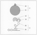
- a system that utilizes a split-type marker protein or a combination-type marker protein that functions in a cytoplasmic or cell-free system specifically, (a) a fusion protein of a test protein and a first element of the marker protein (first (B) Fusion protein of RNA-binding protein and the second element of the marker protein (second fusion protein), and (c) RNA forming a complex with the RNA-binding protein (c)
- a fusion RNA of a complex-forming RNA and a test RNA
- a signal generated due to the expression of the function of the marker protein is detected in a cytoplasmic or cell-free system.
- the present inventors measure the protein-RNA interaction in this system in the presence of a desired substance to increase, decrease, or eliminate the signal caused by the expression of the function of the marker protein. As an index, it was found that it is also possible to evaluate the regulation of protein-RNA interaction by the substance.
- [1] A method for evaluating the interaction between protein and RNA.
- (1) In the cytoplasm or cell-free system (A) A first fusion protein obtained by fusing the test protein and the first element of the marker protein, (B) A second fusion protein obtained by fusing an RNA-binding protein and a second element of the marker protein, and (c) an RNA forming a complex with the RNA-binding protein and a test RNA. Ribonucleoprotein consisting of fused RNA, The process of contacting, (2) A step of detecting a signal caused by the marker protein whose function is expressed by the proximity of the first element and the second element, and (3) A step of determining the interaction between the test protein and the test RNA by detecting the signal. Including When the test protein and the test RNA interact, the first fusion protein and the ribonucleoprotein form a complex, and the first element and the second element A method in which the function of the marker protein is expressed by the proximity of the markers.
- [4] A method for evaluating substances that regulate the interaction between proteins and RNA.
- (1) In the presence of the test substance, in the cytoplasm or cell-free system (A) A first fusion protein formed by fusing a specific protein with the first element of a marker protein, (B) A second fusion protein obtained by fusing an RNA-binding protein and a second element of the marker protein, and (c) an RNA forming a complex with the RNA-binding protein and a specific RNA.
- Ribonucleoprotein consisting of fused RNA
- the process of contacting (2) A step of detecting a signal caused by the marker protein whose function is expressed by the proximity of the first element and the second element, and (3) When the signal is increased as compared with the case where the test substance is absent, the test substance is evaluated as a substance that promotes the interaction between the specific protein and the specific RNA. , The test substance is evaluated as a substance that suppresses the interaction between the specific protein and the specific RNA when the signal is reduced or eliminated as compared with the case where the test substance is absent.
- the first fusion protein and the ribonucleoprotein form a complex
- the first element and the second element A method in which the function of the marker protein is expressed by the proximity of the markers.
- [5] A method for detecting a substance that regulates the interaction between protein and RNA in a sample.
- A A first fusion protein formed by fusing a specific protein with the first element of a marker protein
- B A second fusion protein obtained by fusing an RNA-binding protein and a second element of the marker protein
- c an RNA forming a complex with the RNA-binding protein and a specific RNA.
- Ribonucleoprotein consisting of fused RNA
- the process of contacting (2) A step of detecting a signal caused by the marker protein whose function is expressed by the proximity of the first element and the second element, and (3) A step of detecting a target substance in the test sample by the presence / absence, increase, or decrease of the signal.
- Including The first fusion occurs when the specific RNA interacts with the specific protein in the presence or absence of the target substance, and when the specific protein interacts with the specific RNA.
- a vector containing an RNA-encoding polynucleotide that forms a complex with a sex protein and an insertion site for an adjacent polynucleotide encoding the specific RNA Including, kit.
- C' A polynucleotide encoding a fusion RNA obtained by fusing an RNA forming a complex with the RNA-binding protein and a specific RNA, or a vector expressing the polynucleotide.
- B' A polynucleotide encoding a second fusion protein obtained by fusing an RNA-binding protein and a second element of the marker protein, or a vector expressing the polynucleotide.
- C' A polynucleotide encoding a fusion RNA obtained by fusing an RNA forming a complex with the RNA-binding protein and a specific RNA, or a vector expressing the polynucleotide.
- Transformed cells into which the cells have been introduced, as well as (2) A means for detecting a signal caused by the marker protein whose function is expressed by the proximity of the first element and the second element. Including When the specific protein interacts with the specific RNA, the first fusion protein, the second fusion protein, and the ribonucleoprotein composed of the fusion RNA form a complex, and the said.
- a biosensor in which the function of the marker protein is expressed by the proximity of the first element and the second element.
- the second fusion protein and the fusion RNA have strong binding properties through the binding between the RNA-binding protein contained therein and the complex-forming RNA. And forms a complex (ribonucleoprotein) that can exist stably in the cytoplasm or in a cell-free system.
- a fusion protein first fusion protein and second
- a test protein and the RNA-binding protein are fused to each of the marker proteins divided into a first element and a second element.
- the fusion protein) and the test RNA fused with the complex-forming RNA are brought into contact with each other.
- the function of the marker protein is exhibited only when the first element and the second element interact in close proximity to each other, but at this time, when the test RNA and the test protein interact with each other, the function is exhibited. So to speak, the test protein becomes a capture site (Prey), the test RNA becomes a capture site (Bait), and finally the first fusion protein and the ribonucleoprotein, that is, the above (a).
- the three molecules of (c) to (c) form a complex, and in a cytoplasmic or cell-free system, the first element and the second element are in close proximity to express the function of the marker protein.
- the interaction between the test protein to be verified and the test RNA in the cytoplasmic or cell-free system is caused by the expression of the function of the marker protein, more specifically, the marker protein expressing the function. It is possible to easily evaluate, detect, and measure using the signal generated as an index.
- the expression of background reporter genes and the like is amplified through a two-step gene expression process of transcription and translation, resulting in high levels of false positives.
- the detection of false positives can be sufficiently suppressed, and the interaction between protein and RNA can be sufficiently suppressed as compared with the above-mentioned conventional method. It becomes possible to measure specifically.
- a method for evaluating an interaction between a protein and RNA and a method for evaluating an interaction regulator between a protein and RNA, which can specifically and easily measure the interaction between a protein and RNA in a cytoplasmic or cell-free system.
- methods for detecting protein-RNA interaction regulators, as well as fusion proteins, kits, and biosensors used for them are also useful.
- protein means a molecule in which two or more amino acids are bound by a peptide bond and a modified product thereof. Therefore, it includes not only full-length proteins but also so-called oligopeptides and polypeptides. Modifications of the protein include, for example, phosphorylation, glycosylation, palmitoylation, prenylation (eg, geranylgeranylation), methylation, acetylation, ubiquitination, SUMOylation, hydroxylation, amidation.
- RNA means a molecule in which two or more ribonucleotides are bound by a phosphodiester bond and a modified product thereof.
- modification of RNA include methylation and deamination, and whether it is single-stranded or double-stranded, it has a three-dimensional structure such as a hairpin structure and a hammer head structure. You may be doing it.
- the "interaction between protein and RNA” indicates the proximity or binding of protein and RNA, and more preferably, the protein and RNA form a complex. Such interactions include not only direct proximity and binding of the protein (test protein 11 or specific protein 11) and RNA (test RNA 32 or specific RNA 32), but also between the protein and the RNA.
- Other molecules eg, other proteins, other nucleic acids, sugars, lipids, low molecular weight compounds (vitamins, coenzymes, hormones, toxins, antibiotics, antibacterial agents, antiviral agents, anticancer agents, carcinogens, It also includes indirect interactions such as forming a complex via (drugs, coenzymes, etc.), metal ions, metal complexes, and complex molecules containing two or more of these molecules.
- the "cytoplasm” is the inside of a eukaryotic cell and a prokaryotic cell (eubacterial cell, archaeal cell), and in the eukaryotic cell, the portion of the cell excluding the nucleus, the prokaryotic cell. Indicates the part of the cell excluding the nucleoid.
- the cytosol according to the present invention may be the cytosol of eukaryotic cells or the cytosol of prokaryotic cells, but the formation of a complex of stable protein and RNA having a lower risk of affecting cell function.
- the cytosol of the eukaryotic cell or the cytosol of the prokaryotic cell is preferable, and more preferably the cytosol of the eukaryotic cell. More preferably, it is the cytosol of eukaryotic cells (the portion of the cytosol of eukaryotic cells excluding intracellular small organs).
- the eukaryotic cells include animal cells (mammalian, fish, birds, reptiles, amphibians, insect cells, etc.), plant cells, algae cells, yeast, and examples of the eubacterial cells include lactic acid bacteria and thermophiles.
- Bacterial bacterium, lactic acid bacterium, hyperthermophilic bacterium, and examples of the archaeal cell include methane bacterium, highly thermophilic bacterium, hyperthermophilic bacterium, and hyperthermophilic bacterium. These cells are present in vivo (eg, DNA encoding a first fusion protein) even when cultured in vitro (eg, cells growing in or on medium). And the DNA in the transgenic animal into which the DNA encoding the second fusion protein and the DNA encoding the fusion RNA have been introduced).
- the "cell-free system” refers to a system without living cells (the eukaryotic cells and prokaryotic cells).
- the cell-free system according to the present invention is not particularly limited as long as it is a system in which the first fusion protein according to the present invention and the ribonucleoprotein (or the second fusion protein and the fusion RNA) can come into contact with each other.
- Reconstituted cell-free protein synthesis system such as PURE System (Shimizu Y. et al., Nature Biotechnology 19: 751-755 (2001)); the molecule of the cell crushed solution or cell extract of the eukaryotic cell or prokaryotic cell. Examples include an in vitro reconstitution system that can be contacted with.
- the "marker protein” refers to a protein or a set of proteins capable of expressing one function as a whole, that is, expressing one function as a whole and finally producing at least one signal. ..
- a marker protein can be divided into at least a first fragment and a second fragment, and its function can be regenerated (expressed) when the first fragment and the second fragment are in close proximity to each other.
- the expression of the function can be detected as a signal (sometimes referred to herein as a "split marker protein”); a combination of two independent first and second proteins.
- the function is expressed when the first protein and the second protein are in close proximity to each other, and the expression of the function can be detected as a signal (in the present specification, in some cases, ". "Combination marker protein”) is included.
- the first element according to the present invention comprises the first fragment and the first protein
- the second element according to the present invention contains the second fragment and the second protein, but the "first".
- Each element, fragment, and protein in an element, fragment, and protein and each element, fragment, and protein in a "second" element, fragment, and protein are such that the first and second correspond to each other. It may be independent of each other and vice versa.
- the function is not particularly limited as long as it can directly or indirectly generate a signal due to the function, and for example, a nutrient synthesis function, a color development (color development) function, a light emitting function, a fluorescence function, and a quenching function.
- a nutrient synthesis function for example, a nutrient synthesis function, a color development (color development) function, a light emitting function, a fluorescence function, and a quenching function.
- Examples include a proteolytic function, a nucleic acid decomposition function, a drug decomposition function, and a redox reaction catalytic function.
- those that can be used as a split-type marker protein that can be divided into a first fragment and a second fragment include, for example, a nutritional requirement marker (tryptophan synthase (TRP1), histidine synthesis).
- Enzyme HIS3
- LYS2 lysine synthase
- MENU2 leucine synthase
- ADE2 adenin synthase
- UAA3 uracil synthase
- fluorescent protein green fluorescent protein (GFP))
- YFP Yellow Fluorescent Protein
- CFP Light Blue Fluorescent Protein
- BFP Blue Fluorescent Protein
- RFP Red Fluorescent Protein
- Clover Ruby, Cherry, Zami Green (AG), Sea Mushroom-Green (UkG), Kusabi Laorange (KO), Midoriishi-cyan (MiCy), etc.
- Luminescent enzyme Luciferase, Iquorin, etc.
- Drug resistance markers Drug resistance markers
- the combinational marker proteins that can be used as a combination of the first protein and the second protein include fluorescence source / quencher pair, fluorescence resonance energy transfer (FRET), and bioluminescence resonance energy.
- FRET fluorescence resonance energy transfer
- BRET acceptor pairs for transfer
- kinase / substrate protein pairs for signal transduction pathway activation and the like.
- fluorescence by the fluorescence source is detected when the fluorescence source and the quencher are separated, but when the fluorescence source and the quencher are close to each other, fluorescence by the fluorescence source is detected. Quenching (ie, reducing or extinguishing fluorescence).
- the fluorescence source include ATTO dye, cyanine dye (eg, Cy3, Cy5), tetramethylrhodamine (eg, TRITC), carboxyfluorescein (FAM), tetrachlorofluorescein (TET), hexachlorofluorescein (HEX), Texas. Examples include, but are not limited to, red and Yakima Yellow.
- the quencher examples include, but are not limited to, dark quencher, BHQ (Black Hole Quencher), IBFQ (Iowa Black FQ), IBRQ (Iowa Black RQ), Eclipse and the like.
- the first protein and the second protein may be independently fluorescent proteins, and when the fluorescent source and / or quencher is other than a protein, the fluorescent sources are independent of each other. Alternatively, it may be a protein that specifically presents a quencher intracellularly. Such a protein can be appropriately prepared by a known method or a method similar thereto.
- FRET fluorescence resonance energy transfer
- Donor / acceptor pairs for FRET include, for example, Clover / mRubi2, BFP / eGFP, BFP / YFP, BFP / DsRed2, CFP / YFP, CFP / DsRed2, Midori Cyanine / Clavira Orange, eGFP / DsRed, eGFP / Rhod-2, FITC / TRITC, FITC / Rhod-2, FITC / Cy3, Alexa488 / Alexa546, Alexa488 / Alexa555, Alexa488 / Cy3, YFP / TRITC, YFP / Cy3, Cy3 / Cy5, Cy3 / Cy5, Cy3 / Cy5.
- the first protein and the second protein may be independently fluorescent proteins, and when the donor and / or acceptor is a fluorescent dye, the fluorescent dye is specifically used intracellularly. It may be the protein to be presented. Such a protein can be appropriately prepared by a known method or a method similar thereto.
- donor / acceptor pair for bioluminescence resonance energy transfer fluorescence by BRET is not observed when the donor and acceptor are separated, but when the donor and acceptor are in close proximity, the donor's emission is emitted. causes excitation and fluorescence of the acceptor (ie, fluorescence by BRET occurs).
- donor / acceptor pairs for BRET include equolin / GFP, firefly-derived luciferase / RFP, firefly-derived luciferase / DsRed2, sea pansy-derived luciferase / YFP, sea pansy-derived luciferase / eGFP, and sea pansy-derived luciferase.
- the first protein and the second protein may be independently luminescent proteins or fluorescent proteins, and when the acceptor is a fluorescent dye, the fluorescent dye is specifically used in the cell. It may be the protein to be presented. Such a protein can be appropriately prepared by a known method or a method similar thereto.
- Each of the amino acid sequences of the marker protein can be mutated in nature (that is, non-artificially), and the mutation can be artificially introduced.
- such a mutant is also used as long as the expression of the function can be detected, that is, the function of the marker protein can be expressed by the proximity of the first element and the second element. be able to.
- the marker protein is a split-type marker protein
- the split position is particularly limited as long as the function of the split-type marker protein can be expressed (regenerated) by the proximity of the first fragment and the second fragment. However, it can be appropriately prepared.
- the marker protein one type may be used alone or two or more types may be used in combination.
- the expression of the function of the marker protein can be detected using this as an index by detecting a signal generated by the marker protein that expresses the function due to the proximity of the first element and the second element.
- the "signal caused by the marker protein expressing the function” is not particularly limited as long as it can be expressed by a changing physical quantity, and is directly generated from the marker protein (for example, quenching, fluorescence, etc.). It may be extinguished (such as quenching) or indirectly generated by the expression of the function of the marker protein (for example, a colony indicating that the cell has survived by the expression of the nutrient synthesis function).
- Examples of such a signal include colonies indicating cell survival, coloration (color development), luminescence, fluorescence, quenching, and the like. Further, the signals include those that can be confirmed with the naked eye and those that can be confirmed by a detection method / device according to the type of signal.
- detection of signal includes detection for confirming the presence or absence of the signal, and quantification or semi-quantification of the amount of the signal.
- the method for detecting the signal is not particularly limited, and a method according to the function of the marker protein and the type of signal derived from the marker protein can be appropriately selected. Further, the increase or decrease of the signal can be measured by quantifying or semi-quantifying the signal. When the signal is quenching, the increase and decrease of the signal indicate an increase and decrease of the degree of quenching, respectively.
- the detection of the signal can be divided into two fragments, for example, in the cytoplasm of yeast, at position 2-44 (second fragment) and position 45-224 (first fragment), and these
- tryptophan synthase TRP1
- TRP1 tryptophan synthase
- TRP1 which regenerates (expresses) the function as tryptophan synthase when the fragments are close to each other
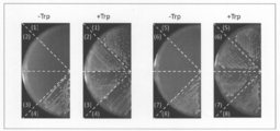
- the marker protein when the second fragment and the first fragment are close to each other Since the function as tryptophan synthase is regenerated (expressed), colonies showing the growth of the yeast in a tryptophan-free medium can be detected as a signal.
- the expression of the function is detected, that is, the function of the marker protein (TRP1) is expressed, while when it is not observed, the expression is said. It can be determined that the expression of the function is not detected, that is, the function of the marker protein is not expressed, or the amount of the signal is quantified or semi-quantified by comparing the amount of the colony with the calibration curve or the like. The degree of expression of can be quantified or semi-quantified.
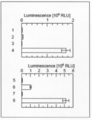
- luciferase (NanoLuc) derived from deep-sea luminescent shrimp is used as the marker protein, it is divided into two fragments, for example, 13 amino acids at the C-terminal (first fragment) and the other (second fragment). And since the function as luciferase is regenerated (expressed) when these fragments are close to each other, luminescence by luciferase can be detected as a signal in cells.
- the expression of the function of the marker protein (NanoLuc) can be detected, or the degree of expression can be quantified or semi-quantified depending on whether or not luminescence is observed (detected) as the signal or the intensity of the luminescence. ..
- the detection of light emission and the quantification or semi-quantification of the intensity can be appropriately selected by a conventionally known method, and can be performed by, for example, a CCD image sensor or a CMOS image sensor equipped with a photomultiplier tube and an optical lens. Further, a method of processing the obtained image by an image analysis program can also be adopted.
- the tryptophan synthase and the luciferase are used in combination as the marker protein, for example, detection of regeneration (expression) of the function of TRP1 by observation of the yeast colony in the tryptophan-free medium and the above-mentioned It is possible to simultaneously detect the regeneration (expression) of the function of NanoLuc by observing the luminescence by luciferase.
- Clover / mRubi2 which is a donor / acceptor pair of FRET
- the function as a combined marker protein when these proteins are close to each other. Is expressed, fluorescence by FRET in cells can be detected as a signal.
- the expression of the function of the marker protein can be detected, or the degree of expression can be quantified or semi-quantified depending on whether or not fluorescence is observed (detected) as the signal or the intensity of the fluorescence.
- the detection of fluorescence and the quantification or semi-quantification of the intensity can be appropriately selected by a conventionally known method, and can be performed by, for example, a fluorescence microscope, a fluorescence spectrophotometer, or a fluorescence plate reader. Further, a method of processing the obtained image by an image analysis program can also be adopted.
- the "RNA-binding protein” is a protein capable of forming a complex with the target RNA by specifically recognizing and binding to the base sequence and structure of the target RNA (complex-forming RNA). is there.
- RNA-binding protein examples include Cas (CRISPER-associated) protein, ribosome protein, spryisosome protein, telomerase protein, ribonuclease P protein, capsid protein derived from RNA virus, and coat protein.
- the RNA-binding protein according to the present invention may be a protein artificially imparted with binding property to RNA.
- RNA-binding protein examples include the tetracycline repressor protein whose target RNA is an RNA aptamer that specifically binds to the tetracycline repressor.
- Cas protein is preferable from the viewpoint of forming a complex of RNA and a stable protein having a lower risk of affecting cell function.
- Cas protein examples include Cas9, Cpf1 (Cas12), Cas12b, CasX (Cas12e), Cas13, and Cas14.
- Cas protein when Cas protein is used as the RNA-binding protein in the present invention, nuclease activity (DNA cleaving ability) is unnecessary. Therefore, as the Cas protein, a Cas protein that has lost a part or all of the nuclease activity may be used (hereinafter, the Cas protein that has lost a part of the nuclease activity is referred to as "nCas” and the nuclease activity. The Cas protein that has lost all of the above is referred to as "dCas").
- the Cas protein typically comprises a domain involved in the cleavage of the target strand (RuvC domain) and a domain involved in the cleavage of the non-target strand (HNH domain), but when the Cas protein is used in the present invention, the Cas protein is included. It is preferable that the nuclease activity of the domain is lost by introducing the mutation into at least one domain.
- a mutation in the case of spCas9 protein (Cas9 protein derived from S. pyogenes), for example, a mutation of the 10th amino acid (aspartic acid) from the N-terminal to alanine (D10A: mutation in the RuvC domain).
- Cas9 proteins of various origins are known (eg, WO2014 / 131833), and their nCas or dCas can be utilized.
- the amino acid sequence and base sequence of Cas9 protein are registered in a public database, for example, GenBank (http://www.ncbi.nlm.nih.gov) (for example, accession number: Q99ZW2.1). Etc.), these can be used in the present invention.
- further mutations for example, mutations for altering PAM recognition, may be introduced into the Cas protein (Benjamin, P. et al., Nature 523, 481-485 (2015); Hirano, S. et al. , Molecular Cell 61,886-894 (2016)).
- RNA forming a complex with an RNA-binding protein has a sequence and structure recognized by the RNA-binding protein. It is an RNA that forms a complex with the RNA-binding protein.
- complex-forming RNA include a guide RNA when the RNA-binding protein is a Cas protein, and the RNA-binding protein is a ribosome protein, a spryisosome protein, a telomerase protein, or a ribonuclease P.
- RNA virus packaging sequences of ribosome RNA, UsnRNA, telomea RNA, ribonuclease P RNA, and RNA virus can be mentioned, respectively.
- a guide RNA is preferable from the viewpoint of forming a complex of a stable protein and RNA having a lower risk of affecting cell function.
- the guide RNA is a combination of crRNA (CRISPR RNA) and tracrRNA (trans-activated CRISPR RNA).
- CRISPR RNA CRISPR RNA
- tracrRNA trans-activated CRISPR RNA
- the crRNA and tracrRNA may be in the form of one molecule or in the form of two molecules.
- crRNA contains a base sequence complementary to a specific base sequence on genomic DNA (targeted RNA sequence) and a base sequence capable of interacting with tracrRNA in this order from the 5'side.
- the targeting RNA sequence is not always necessary because the ability to target genomic DNA is not required.
- the crRNA forms a double-stranded RNA with the tracrRNA in a base sequence capable of interacting with the tracrRNA, and the formed double-stranded RNA interacts with the Cas9 protein.
- the guide RNA may not contain tracrRNA. Examples of the CRISPR / Cas system using such a guide RNA as a component include the CRISPR / Cpf1 system.
- the "fusion protein” is one in which two or more of the above proteins are fused to form one (one molecule) protein.
- the fusion between proteins may be direct or indirect via a linker or spacer protein.
- linker or spacer protein its length is not particularly limited, but it is preferably 1 to 32 amino acid residues independently, and 1 to 17 amino acid residues independently. Is more preferable.
- the fusion protein according to the present invention other functional proteins (for example, epitope tag, affinity tag, solubility improving tag, etc.) are used as long as they do not inhibit the interaction between the target protein and RNA and the expression of the function of the marker protein. It may contain a signal peptide, degron, protease recognition peptide, kinase recognition peptide, etc.).
- the other functional protein can be fused directly or indirectly between the N-terminal, C-terminal, or both sides of the fusion protein, or between the proteins to be fused.
- the other functional protein is not particularly limited, and is appropriately selected according to the function to be imparted to the fusion protein according to the present invention.
- the fusion protein can be obtained, for example, by transcribing and expressing two or more genes encoding the protein as a unit.
- the fusion protein according to the present invention can be produced by appropriately adopting and improving a conventionally known method. For example, as described in the (1) contact step of the following protein-RNA interaction evaluation method, the fusion protein thereof. It can be obtained by a method of chemically synthesizing based on an amino acid sequence by a commercially available synthesizer, or a method of introducing a polynucleotide encoding a fusion protein or a vector expressing the polynucleotide into the cell and expressing it.
- the "first fusion protein” is a fusion protein obtained by fusing a test protein or a specific protein with the first element of the marker protein.
- the first element of the marker protein may be one kind or two or more kinds.
- these elements correspond to at least one of the second elements contained in the second fusion protein.
- the first elements of two or more kinds may be elements derived from the same kind of marker proteins or elements derived from different kinds of marker proteins.
- the fusion of the test protein or the specific protein with the first element is the fusion of the first element to either the N-terminal or C-terminal of the test protein or the specific protein. It may be present, or the first element may be fused to both ends of the test protein or the specific protein.
- the "second fusion protein” is a fusion protein obtained by fusing the RNA-binding protein and the second element of the marker protein.
- the second element of the marker protein may be one kind or two or more kinds.
- the two or more kinds of second elements may be elements derived from the same kind of marker proteins or elements derived from different kinds of marker proteins.
- the fusion of the RNA-binding protein and the second element may be one in which the second element is fused to either the N-terminal or the C-terminal of the RNA-binding protein.
- the RNA-binding protein is the Cas protein
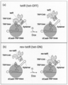
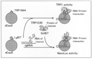
- the amino acid at the 2-44 position of tryptophan synthase (TRP1) (TRP1N44) and tryptophan synthase (TRP1) are added to the C-terminal of the Cas protein as the second element.
- Amino acid at position 45-224 (TRP1C45) 13 amino acids at the C-terminal of luciferase (NanoLuc) derived from deep-sea luminescent shrimp (SmBiT), and 13 amino acids at the C-terminal of luciferase (NanoLuc) derived from deep-sea luminescent shrimp. LgBiT), respectively, are fused.
- these Cas proteins dCas9 and dCas13b are preferable, and dCas9 is more preferable.
- various mutations may be introduced as long as they can bind to the complex-forming RNA.
- these second elements are preferably fused to the Cas protein via a linker, and the length of the linker is not particularly limited, but is preferably 1 to 17 amino acid residues.
- the second fusion protein may be a C-terminal, an N-terminal, or one in which a tag sequence is added at both ends.
- the dCas9-TRP1N44 fusion protein represented by the amino acid sequence of SEQ ID NO: 1
- the dCas9-TRP1C45 fusion protein represented by the amino acid sequence of SEQ ID NO: 2.
- Linker: RS dCas9-LgBiT fusion protein
- linker: RS represented by the amino acid sequence of SEQ ID NO: 3
- Each of these second fusion proteins is capable of binding to the complex-forming RNA, and the function of the marker protein is expressed by the proximity of the first element and the second element.
- it may be a homologue, a mutant, or a partial peptide of the amino acid sequence shown above.
- the homologs include, for example, 85% or more, preferably 90% or more, more preferably 95% or more (for example, 96% or more, 97% or more, 98% or more, 99% or more) with the amino acid sequence shown above.
- a protein consisting of an amino acid sequence having the same identity is included. The identity of the sequence can be evaluated numerically when calculated using BLAST or the like (eg, default or default parameters).
- the mutant consists of an amino acid sequence in which one or more amino acids are substituted, deleted, added or inserted with respect to the amino acid sequence shown above, and can bind to the complex-forming RNA.
- a protein in which the function of the marker protein is expressed by the proximity of the first element and the second element is included.
- plural means, for example, 2 to 150 pieces, preferably 2 to 100 pieces, more preferably 2 to 50 pieces (for example, 2 to 30 pieces, 2 to 10 pieces, 2 to 5 pieces, 2). ⁇ 3 pieces, 2 pieces).
- the "fusion RNA" is one in which two or more of the above RNAs are fused to form one (one molecule) RNA, and in the present invention, the complex-forming RNA and the subject Indicates a fused RNA obtained by fusing a test RNA or a specific RNA.
- the fusion between RNAs may be direct or indirect as long as it does not inhibit the interaction between the protein of interest and RNA and the expression of the function of the marker protein.
- the test RNA or a specific RNA may be fused to either the 3'side or the 5'side of the complex-forming RNA, but the complex-forming RNA may be fused.
- the guide RNA of the Cas protein Is the guide RNA of the Cas protein, the 3'side of the guide RNA (ie, the 3'side if the guide RNA contains trRNA, and the 3'side if it does not contain trRNA and consists only of crRNA. ) Is fused with the test RNA or a specific RNA.
- the fusion RNA can be obtained, for example, by transcribing and expressing two or more genes encoding the RNA as a unit.
- the fusion RNA according to the present invention can be produced by appropriately adopting and improving a conventionally known method.
- the fusion RNA thereof can be obtained by a method of chemically synthesizing based on a base sequence by a commercially available synthesizer, or a method of introducing a polynucleotide encoding fusion RNA or a vector expressing the polynucleotide into the cell and expressing it.
- the "ribonucleoprotein” is a complex of a protein and a ribonucleotide, and in the present invention, it indicates a complex of a second fusion protein and a fusion RNA, and more specifically, the first fusion protein.
- the complex formed through the binding between the RNA-binding protein contained in the fusion protein of 2 and the complex-forming RNA contained in the fusion RNA is shown.
- the binding between the second fusion protein and the fusion RNA that is, the binding between the RNA-binding protein and the complex-forming RNA
- the binding between the second fusion protein and the fusion RNA is the interaction between the target protein and the RNA and the function of the marker protein.
- RNA-binding protein e.g, other proteins, other nucleic acids, sugars, lipids, small molecules
- RNA-binding protein e.g., other proteins, other nucleic acids, sugars, lipids, small molecules
- Compounds (vitamins, coenzymes, hormones, toxins, antibiotics, antibacterial agents, antiviral agents, anticancer agents, carcinogens, drugs, psychotropic agents, etc.), metal ions, metal complexes, and 2 of these It may be indirect, such as forming a complex via a complex molecule containing more than one species of molecule.
- the method for evaluating the interaction between the protein and RNA of the present invention is (1) In the cytoplasm or cell-free system (A) A first fusion protein obtained by fusing the test protein and the first element of the marker protein, (B) A second fusion protein obtained by fusing an RNA-binding protein and a second element of the marker protein, and (c) an RNA forming a complex with the RNA-binding protein and a test RNA.
- Ribonucleoprotein consisting of fused RNA
- the process of contacting (2) A step of detecting a signal caused by the marker protein whose function is expressed by the proximity of the first element and the second element, and (3) A step of determining the interaction between the test protein and the test RNA by detecting the signal.
- the first fusion protein and the ribonucleoprotein form a complex
- the first element and the second element It is a method in which the function of the marker protein is expressed by the proximity of.
- test protein As the "test protein” according to the protein-RNA interaction evaluation method of the present invention, any protein among the above proteins that is desired to measure the interaction with the target RNA (test RNA 32) is used. be able to. Further, as the "test RNA” according to the protein-RNA interaction evaluation method of the present invention, any of the above RNAs that is desired to measure the interaction with the test protein 11 can be used. it can.
- the first fusion protein 1 and the ribonucleoprotein 4 are used as a method of contacting the first fusion protein 1 and the ribonucleoprotein 4.
- the cell those listed as the eukaryotic cell and the prokaryotic cell described above can be used, and the cell-free system is as described above. Further, these introduction into the cytoplasm and expression in the cytoplasm may be transient or constitutive expression depending on the purpose. As the method of introduction or expression, those skilled in the art can appropriately adopt a conventionally known method or a method similar thereto, depending on the type of cell and the like.
- Ribonucleoprotein, and fusion RNA can be used by those skilled in the art, for example, based on the amino acid sequence of the protein, in a cell-free protein synthesis system (eg, reticulated red erythrocyte extract, wheat germ extract), Escherichia coli, animal.
- a polynucleotide encoding a first fusion protein 1 and a ribonucleoprotein 4 (or a second fusion protein 2 and a fusion RNA 3) into a cell or a cell-free system having a gene expression function, respectively.
- Each of these polynucleotides can be chemically synthesized and prepared by a commercially available synthesizer based on its base sequence.
- the expression vector contains one or more regulatory elements that are operably linked to a gene to be expressed (a gene encoded by each of the polynucleotides).
- operably bound means that the gene is operably bound to a regulatory element.
- regulatory elements include promoters, enhancers, internal ribosome entry sites (IRES), and other expression control elements (eg, transcription termination signals such as polyadenylation signals and polyU sequences).
- the regulatory element directs gene expression only in a specific cell, tissue, or organ, even if it directs constitutive expression of the gene in various host cells, for example. It may be a thing. In addition, it may be directed to the expression of a gene only at a specific time, or may be directed to the expression of an artificially inducible gene.
- promoters include polIII promoters (eg, U6 promoter, H1 promoter, and SNR52 promoter), polII promoters (eg, sprouting yeast ADH promoter, CYC promoter, TEF promoter, GPD promoter, GAL promoter, CUP promoter, retrovirus.
- Raus sarcoma virus (RSV) LTR promoter cytomegalovirus (CMV) promoter, SV40 promoter, dihydrofolate reductase promoter, ⁇ -actin promoter, phosphoglycerol kinase (PGK) promoter, and EF1 ⁇ promoter
- polI promoter lac promoter
- trp Examples include a promoter, an araBAD promoter, a T7 promoter, an S6 promoter, or a combination thereof. These promoters can be appropriately selected depending on the type of cell and vector used. For example, when a vector expressing a polynucleotide encoding the fusion RNA according to the present invention is used in yeast, the polII promoter is preferable. ..
- the expression vector examples include a plasmid vector, an episomal vector, and a virus vector.
- the proteins and RNAs encoded in the expression vector are the above-mentioned fusion proteins and fusion RNAs, etc., but from the viewpoint of further improving the expression efficiency of these, the expression vector encoding these is the protein.
- codon-optimized DNA eg, codon-humanized DNA
- a polynucleotide encoding a self-cleaving RNA for example, hammerhead ribozyme
- such an expression vector can be prepared by those skilled in the art by appropriately using known techniques such as a DNA chemical synthesis method and a gene recombination technique.
- a method for introducing the above-mentioned fusion protein, ribonucleoprotein, fusion RNA, polynucleotide (RNA, DNA), and expression vector into cells conventionally known methods are available depending on the type of molecule to be introduced, the type of target cell, and the like. It can be appropriately selected from the method or a method similar thereto. Examples of such an introduction method include a method using a protein introduction reagent, an electroporation method, a microinjection method, a particle gun method, a calcium phosphate method, a liposome method (lipofection method), and a DEAE-dextran method.
- viruses adenovirus, lentivirus, adeno-associated virus, baculovirus, etc.
- agrobacterium method particle gun method
- lithium acetate method lithium acetate method
- spheroplast method heat shock method (chloride) Calcium method
- rubidium chloride method rubidium chloride method
- the first fusion protein 1 and the ribonucleoprotein 4 can be contacted in a cytoplasmic or cell-free system by the above method.
- Ribonucleoprotein 4 is a complex containing a second fusion protein 2 and fusion RNA3.
- Ribonucleoprotein 4 is prepared extracellularly or extracellularly and into a cell or cell-free system. It may be introduced, or the second fusion protein 2 and the fusion RNA 3 may interact with each other in a cell or cell-free system to form a ribonucleoprotein 4.
- the second fusion protein 2 and the fusion RNA 3 form a complex between the RNA-binding protein 21 inherent in each molecule and the complex-forming RNA 31, so that the second fusion protein 2 and the fusion RNA 3 are seconded through such an interaction.
- the fusion protein 2 and the fusion RNA 3 of the above form a ribonucleoprotein 4.
- one molecule of the first fusion protein and one molecule of the second fusion Each element of the marker protein contained in the protein may be one kind alone or two or more kinds, and may be included as the first fusion protein and the second fusion protein, respectively. Depending on the element or combination of proteins, one type may be used alone or may be two or more types, and the number of types of the first fusion protein and the number of types of the second fusion protein may be different. For example, when two types of marker proteins A and B are used, the first element is a fusion of the test protein, the first element A of the marker protein A, and the first element B of the marker protein B.
- the second fusion protein B, which is fused with the second element B, or the ribonucleoprotein B containing the second fusion protein B can be contacted in a cytoplasmic or cell-free system.
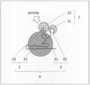
- a signal caused by the marker protein whose function is expressed by the proximity of the first element 12 and the second element 22 is detected.
- the combination of such a marker protein, a signal, and a detection method can be appropriately selected, and examples thereof include the above-mentioned combinations.
- the test protein and the marker protein A first.
- the first fusion protein formed by fusing the element A of 1 and the first element B of the marker protein B; the RNA-binding protein and the second element A of the divided marker protein A are fused.
- Ribonucleoprotein A containing a second fusion protein A Ribonucleoprotein A containing a second fusion protein A
- Ribonucleoprotein B containing a second fusion protein B formed by fusing an RNA-binding protein with a second element B of marker protein B
- the test protein and the test RNA interact with each other, the first fusion protein and the ribonucleoprotein A and the other first fusion protein and the ribonucleoprotein B are brought into contact with each other. And, respectively, form complexes A and B, respectively.
- the functions of the marker proteins A and B are expressed when the first element A and the second element A and the first element B and the second element B are close to each other, respectively.
- Multiple detections can be made simultaneously based on the function of each marker protein and the resulting signals (eg, FIG. 9 of the Examples).
- the interaction between the test protein 11 and the test RNA 32 is determined based on the signal detected in the (2) detection step. For example, in the detection step, when a signal caused by the marker protein expressing the function is detected, that is, when the function of the marker protein is detected, the test protein 11 and the test are tested. It can be determined that there is an interaction with RNA32, while if it is not detected, it can be determined that there is no such interaction.
- the detection method is a method that can be quantified or semi-quantitative
- the magnitude of the interaction between the test protein 11 and the test RNA 32 can be determined according to the magnitude of the value (Aspect 1). ).
- a specific protein is used as the test protein 11 and an RNA library is used as the test RNA 32 to interact with the specific protein.
- RNAs that act can be screened (Aspect 2 below).
- a specific RNA as the test RNA 32 and using the protein library as the test protein 11, a protein that interacts with the specific RNA can be screened (Aspect 3 below).
- (Aspect 2) A method for screening RNA that interacts with a particular protein.
- a first fusion protein formed by fusing a specific protein with the first element of a marker protein (B) A second fusion protein obtained by fusing an RNA-binding protein and a second element of the marker protein, and (c) an RNA forming a complex with the RNA-binding protein and a test RNA.
- Ribonucleoprotein consisting of fused RNA The process of contacting, (2) A step of detecting a signal caused by the marker protein whose function is expressed by the proximity of the first element and the second element, and (3) A step of selecting a test RNA that interacts with the specific protein by detecting the signal.
- the first fusion protein and the ribonucleoprotein form a complex
- the first element and the second element A method in which the function of the marker protein is expressed by the proximity of the markers.
- a method for screening proteins that interact with specific RNA (1) In the cytoplasm or cell-free system (A) A first fusion protein obtained by fusing the test protein and the first element of the marker protein, (B) A second fusion protein obtained by fusing an RNA-binding protein and a second element of the marker protein, and (c) an RNA forming a complex with the RNA-binding protein and a specific RNA. Ribonucleoprotein consisting of fused RNA, The process of contacting, (2) A step of detecting a signal caused by the marker protein whose function is expressed by the proximity of the first element and the second element, and (3) A step of selecting a test protein that interacts with the specific RNA by detecting the signal. Including When the test protein and the specific RNA interact, the first fusion protein and the ribonucleoprotein form a complex, and the first element and the second element A method in which the function of the marker protein is expressed by the proximity of the markers.
- a specific protein 11 is used instead of the test protein 11 of the aspect 1.
- a specific RNA 32 is used instead of the test RNA 32 of the aspect 1.
- any protein that desires to search for RNA that interacts with the above protein can be used, and as the specific RNA 32 of the third aspect, the above RNA Of these, any RNA that desires to search for proteins that interact with it can be used.
- the kit for use in the method of evaluating the interaction between the protein and RNA of the present invention is The following (a) to (c): (A) A first fusion protein obtained by fusing a test protein and a first element of a marker protein, a polynucleotide encoding the first fusion protein, a vector expressing the polynucleotide, or a marker protein. A vector containing an insertion site for a polynucleotide encoding the first element of the above and a polynucleotide encoding the test protein adjacent thereto.
- kit for evaluating protein-RNA interaction is the above-mentioned protein-RNA interaction evaluation method.
- a vector expressing a first fusion protein, a polynucleotide encoding the first fusion protein, and a polynucleotide encoding the first fusion protein (a) a vector expressing a first fusion protein, a polynucleotide encoding the first fusion protein, and a polynucleotide encoding the first fusion protein. (B) A second fusion protein, a polynucleotide encoding a second fusion protein, a vector expressing a polynucleotide encoding a second fusion protein, and (c) a fusion RNA, a polynucleotide encoding a fusion RNA.
- the vectors expressing the polynucleotide encoding the fusion RNA are as described above.
- the insertion site of the polynucleotide encoding the first element of the marker protein and the polynucleotide encoding the test protein adjacent thereto as (a).
- a vector containing the above, and a vector containing the insertion site of the polynucleotide encoding the RNA forming a complex with the RNA-binding protein as (c) and the polynucleotide encoding the test RNA adjacent thereto, respectively. can include.
- vectors are, for example, a polynucleotide encoding the test protein 11 of interest (or the specific protein 11 of aspect 2 or the protein library of aspect 3) and / or the test RNA 32 (or the specific RNA 32 of aspect 3).
- the polynucleotide encoding the RNA library of Aspect 2 can be arbitrarily selected and inserted into the insertion site according to the purpose. Examples of the insertion site include restriction enzyme recognition sequences.
- a polynucleotide library may be inserted in advance at the insertion site.
- the kit for evaluating the protein-RNA interaction of the present invention is a first fusion protein in a cytoplasmic or cell-free system by introducing the above (a) to (c) into the cell or cell-free system. Since 1 and ribonucleoprotein 4 (or the second fusion protein 2 and fusion RNA3) can be brought into contact with each other, cells for introducing these, and media and stabilizers necessary for storing and culturing the cells, It may further comprise other ingredients such as preservatives, preservatives and the like.
- cell-free solutions for introducing the above (a) to (c) cell disruption solution, cell extract, reconstituted cell-free protein synthesis solution such as PURE System, the above (a) to (c) It may further be provided with a contactable in vitro reconstitution solution, etc.).
- at least one of the above (a) to (c) may be in a form previously introduced into the cell or cell-free fluid.
- the cells for introducing the above (a) to (c) may or may not be alive, and the above-mentioned (a) to (c) are efficient in advance. Processing for the purpose of introduction, preservation, etc. may be performed.
- a membrane permeation treatment with a surfactant and the like can be mentioned.
- the kit for evaluating protein-RNA interaction of the present invention may further include a standard such as a reagent (reagent for signal detection) for detecting a signal caused by the expression of the function of the marker protein.
- a standard such as a reagent for preparing a calibration curve or an enzyme (a reagent for preparing a calibration curve) may be further provided.
- each fusion protein, fusion RNA, polynucleotide, expression vector, the cell, the cell-free solution, the signal detection reagent, and the calibration line preparation As a standard such as a reagent, other components such as a buffer solution, a stabilizer, a preservative, and a preservative may be added.
- the kit for evaluating the protein-RNA interaction of the present invention may further include an instruction manual which is an instruction for utilizing the expression vector, cells, and cell-free fluid in the method of the present invention.
- the instruction manual describes, for example, the experimental method and conditions of the method of the present invention, and information on each standard of the present invention (for example, information such as a vector map showing a base sequence of a vector, a cloning site, etc.). Information on the origin, properties, culture conditions, etc. of cells) can be included.
- the method for evaluating a substance (test substance) that regulates the interaction between the protein and RNA of the present invention is (1) In the presence of the test substance, in the cytoplasm or cell-free system (A) A first fusion protein formed by fusing a specific protein with the first element of a marker protein, (B) A second fusion protein obtained by fusing an RNA-binding protein and a second element of the marker protein, and (c) an RNA forming a complex with the RNA-binding protein and a specific RNA.
- Ribonucleoprotein consisting of fused RNA
- the process of contacting (2) A step of detecting a signal caused by the marker protein whose function is expressed by the proximity of the first element and the second element, and (3) When the signal is increased as compared with the case where the test substance is absent, the test substance is evaluated as a substance that promotes the interaction between the specific protein and the specific RNA. , The test substance is evaluated as a substance that suppresses the interaction between the specific protein and the specific RNA when the signal is reduced or eliminated as compared with the case where the test substance is absent.
- method for evaluating a substance that regulates the interaction between a protein and RNA of the present invention (hereinafter, sometimes referred to as "method for evaluating a protein-RNA interaction regulator"), (a) a first fusion protein, (b). ) The second fusion protein, (c) the fusion RNA, and the ribonucleoprotein, and the marker protein whose function is expressed by the step of contacting them and the proximity of the first element 12 and the second element 22.
- the steps for detecting the resulting signal include (a) first fusion protein, (b) second fusion protein, (c) fusion RNA, and ribo, respectively, in the above protein-RNA interaction evaluation method.
- the test protein 11 and the test RNA 32 are used. Instead, a specific protein 11 and a specific RNA 32 are used, respectively.
- a combination desired to obtain an evaluation of the effect of the test substance on their interaction is adopted.
- a combination includes, for example, evaluating whether the test substance promotes or inhibits the interaction of a specific protein 11 and a specific RNA 32 known to interact with each other.
- a combination desired to evaluate and search for whether the test substance promotes the interaction can be mentioned.
- RNA virus particle a protein constituting a ribonucleoprotein such as a ribosome, a spryisosome, a telomerase, a ribonuclease P, or an RNA virus particle and an RNA.
- splicing regulators proteins
- microRNA regulators proteins
- RNA LIN28, hnRNP A1, hnRNP L, KSRP and microRNA precursors (RNA)
- RNA microRNA regulators
- test substance examples include proteins, nucleic acids, sugars, lipids, and low molecular weight compounds (vitamins, coenzymes, hormones, toxins, antibiotics, etc.).
- vitaminss, coenzymes, hormones, toxins, antibiotics, etc. include proteins, nucleic acids, sugars, lipids, and low molecular weight compounds (vitamins, coenzymes, hormones, toxins, antibiotics, etc.).
- Antibacterial agents, antiviral agents, anticancer agents, carcinogens, drugs, psychotropic agents, etc.), metal ions, metal complexes, and complex molecules containing two or more of these molecules can be mentioned.
- the test substance is compared with the case where the test substance is not present.
- the test substance is evaluated as a substance that promotes the interaction between the specific protein 11 and the specific RNA 32, while the other
- the test substance is identified as a specific protein 11. It can be evaluated as a substance that suppresses the interaction with RNA32.
- promoting or suppressing the interaction between a specific protein 11 and a specific RNA 32 and "promoting or suppressing the expression of the function of a marker protein” directly make the promotion or suppression (for example).
- the interaction between the specific protein 11 and the specific RNA 32 may be promoted or suppressed).
- a specific protein 11 can be obtained by carrying out the method in parallel using a drug library or the like composed of a large number of chemical substances as a test substance. It is possible to search for a drug that promotes or suppresses the interaction between the protein and a specific RNA32.
- the method of the present invention for detecting a substance (target substance) that regulates the interaction between a protein and RNA in a sample (test sample) is (1) In the presence of the test sample, in the cytoplasm or cell-free system (A) A first fusion protein formed by fusing a specific protein with the first element of a marker protein, (B) A second fusion protein obtained by fusing an RNA-binding protein and a second element of the marker protein, and (c) an RNA forming a complex with the RNA-binding protein and a specific RNA.
- Ribonucleoprotein consisting of fused RNA
- the process of contacting (2) A step of detecting a signal caused by the marker protein whose function is expressed by the proximity of the first element and the second element, and (3) A step of detecting a target substance in the test sample by the presence / absence, increase, or decrease of the signal.
- Including The first fusion occurs when the specific RNA interacts with the specific protein in the presence or absence of the target substance, and when the specific protein interacts with the specific RNA.
- method for detecting a substance that regulates the interaction between a protein and RNA in the sample of the present invention (a) a first fusion protein. , (B) a second fusion protein, (c) a fusion RNA, and a ribonucleoprotein, and the step of contacting them, and the proximity of the first element 12 to the second element 22.
- steps for detecting the signal caused by the marker protein in the above-mentioned protein-RNA interaction evaluation method, (a) first fusion protein, (b) second fusion protein, and (c) fusion RNA, respectively.
- test protein 11 and the subject A specific protein 11 and a specific RNA 32 are used instead of the test RNA 32, respectively.
- the protein and the RNA may interact in the presence or absence of the target substance to be detected.
- a known combination that is, a combination of a specific protein 11 and a specific RNA 32 known to interact in the presence of the substance to be detected, or an interaction (target to be detected) in the absence of the substance to be detected. It is a combination of a particular protein 11 and a particular RNA 32 that is known to (do not interact in the presence of a substance).
- the target substances include, for example, proteins, nucleic acids, sugars, lipids, low molecular weight compounds (vitamins, coenzymes, hormones, toxins, antibiotics, antibacterial agents, etc.
- Antiviral agents, anticancer agents, carcinogens, drugs, psychotropic drugs, etc. metal ions, metal complexes, and complex molecules containing two or more of these molecules can be mentioned.
- the test sample is not particularly limited as long as it is a sample in which the target substance can exist, and for example, human and animal body fluids (saliva, tears, sweat). , Urine, blood, lymph, etc.), plant biofluids, biological culture fluids, extracts of organisms (individuals, organs, tissues, cells, etc.), water in the environment (rivers, lakes, harbors, waterways, groundwater, purified water, sewage) , Drainage, etc.), suspensions of solids (soil, cinders, etc.), suspensions of wiped samples, etc.
- human and animal body fluids saliva, tears, sweat
- Urine, blood, lymph, etc. plant biofluids
- biological culture fluids extracts of organisms (individuals, organs, tissues, cells, etc.)
- water in the environment rivers, lakes, harbors, waterways, groundwater, purified water, sewage) , Drainage, etc.
- suspensions of solids suspensions of wiped samples, etc.
- the combination of these specific proteins 11, the specific RNA 32, and the target substance is, for example, a combination of TetR (protein), TetR aptamer (RNA), and doxycycline; a neurotrophin receptor (protein). ), Neurotrophin receptor aptamer (RNA), and combination of neurotrophins; combinations of ribosomal proteins, ribosomal RNA, and antibiotics.
- the target substance is the sample.
- the signal is not detected, or when the signal is reduced, that is, when the expression of the function of the marker protein is suppressed, the target substance is present. It can be determined that it does not exist in the sample.
- the detection method is a method capable of quantifying or semi-quantitating, it is possible to quantify or semi-quantify the target substance in the sample by using a calibration curve or the like according to the magnitude of the value. it can.
- the signal is detected in the (2) detection step. If, or if the signal is increased, it can be determined that the target substance is not present in the sample, while if it is not detected or decreased, the target substance is said. It can be determined that it is present in the sample.
- the detection method is a method capable of quantifying or semi-quantitating, it is possible to quantify or semi-quantify the target substance in the sample by using a calibration curve or the like according to the magnitude of the value. it can.
- kit for detection method of protein-RNA interaction regulator The kit for use in the method for evaluating a protein-RNA interaction regulator of the present invention and the kit for use in the method for detecting a protein-RNA interaction regulator of the present invention are respectively.
- B A second fusion protein in which an RNA-binding protein and a second element of the marker protein are fused, a polynucleotide encoding the second fusion protein, or a vector expressing the polynucleotide.
- C A fusion RNA formed by fusing an RNA forming a complex with the RNA-binding protein and a specific RNA, a polynucleotide encoding the fusion RNA, a vector expressing the polynucleotide, or the RNA binding.
- (A') A polynucleotide encoding a first fusion protein, which is a fusion of a specific protein and a first element of a marker protein, or a vector expressing the polynucleotide.
- (B') A polynucleotide encoding a second fusion protein obtained by fusing an RNA-binding protein and a second element of the marker protein, or a vector expressing the polynucleotide.
- kits A polynucleotide encoding a fusion RNA obtained by fusing an RNA forming a complex with the RNA-binding protein and a specific RNA, or a vector expressing the polynucleotide. It is also preferable that the kit contains the transformed cells into which the cells have been introduced.
- a kit for use in the method for evaluating a protein-RNA interaction regulator of the present invention and a kit for use in a method for detecting a protein-RNA interaction regulator (hereinafter, in some cases, "evaluation of a protein-RNA interaction regulator”.
- the “detection method kit”) is a cytoplasmic or cell-free system in the (1) contact step of these methods, respectively, in which the first fusion protein 1 and the ribonucleoprotein 4 (or the second fusion protein 2 and) are used.
- a kit used for contacting fusion RNA3 is a kit for use in the method for evaluating a protein-RNA interaction regulator of the present invention and a kit for use in a method for detecting a protein-RNA interaction regulator.
- (a) to (c) refer to the test protein 11 and the test RNA 32 as the above-mentioned specific protein 11 and the specific RNA 32, respectively. Except for the above, the same applies to (a) to (c) mentioned in the above-mentioned kit for evaluating protein-RNA interaction.
- the kit for evaluating / detecting a protein-RNA interaction regulator of the present invention includes cells for introducing the above (a) to (c), and a medium, stabilizer, and storage necessary for storing and culturing the cells. Other ingredients such as agents and preservatives may be further provided.
- cell-free solutions for introducing the above (a) to (c) cell disruption solution, cell extract, reconstituted cell-free protein synthesis solution such as PURE System, the above (a) to (c) It may further be provided with a contactable in vitro reconstitution solution, etc.).
- at least one of the above (a) to (c) may be in a form previously introduced into the cell or cell-free fluid.
- the cells for introducing the above (a) to (c) may or may not be alive, and the above-mentioned (a) to (c) are efficient in advance. Processing for the purpose of introduction, preservation, etc. may be performed.
- a membrane permeation treatment with a surfactant and the like can be mentioned.
- kits for evaluating / detecting a protein-RNA interaction regulator of the present invention is (a') a polynucleotide encoding a first fusion protein, or a polynucleotide expressing the polynucleotide.
- a vector, a polynucleotide encoding (b') a second fusion protein, or a vector expressing the polynucleotide, and (c') a polynucleotide encoding a fusion RNA, or a vector expressing the polynucleotide examples include kits containing transformed cells into which, has been introduced.
- the transformed cell may or may not be alive, and may be a cell disruption solution or a cell extract. Further, the processing for the purpose of preserving the above (a') to (c') may be performed.
- the kit for evaluating / detecting a protein-RNA interaction regulator of the present invention further comprises a standard such as a reagent (reagent for signal detection) for detecting a signal caused by the expression of the function of the marker protein. You may have it.
- a test substance that promotes the interaction between a specific protein and a specific RNA contained in the kit, a test substance that suppresses the reaction, and a test substance that does not promote or suppress the interaction may be further provided.
- the test substance detected by the kit, the test substance not detected, and a standard such as a reagent or an enzyme (reagent for preparing a calibration curve) for preparing a calibration curve used for quantification or semi-quantification may be further provided.
- each fusion protein, fusion RNA, polynucleotide, expression vector, the cell, the cell-free fluid, the transformed cell, and the signal detection Other components such as a buffer solution, a stabilizer, a preservative, and an antiseptic may be added as a standard such as a protein for use, each of the test substances, and the reagent for preparing a calibration line.
- an instruction manual for using the expression vector, the cell, the cell-free solution, the transformed cell, etc. in each method of the present invention may be further provided.
- the instruction manual describes, for example, information on the experimental method and conditions of each method of the present invention, and information on each standard of the present invention (for example, a vector map showing a base sequence of a vector, a cloning site, etc.).
- Information on the origin, properties, culture conditions, etc. of the cells and transformed cells can be included.
- the biosensor of the present invention is a biosensor used in the method for evaluating a protein-RNA interaction regulator and / or the method for detecting a protein-RNA interaction regulator of the present invention.
- B' A polynucleotide encoding a second fusion protein obtained by fusing an RNA-binding protein and a second element of the marker protein, or a vector expressing the polynucleotide.
- C' A polynucleotide encoding a fusion RNA obtained by fusing an RNA forming a complex with the RNA-binding protein and a specific RNA, or a vector expressing the polynucleotide.
- Transformed cells into which the cells have been introduced, as well as (2) A means for detecting a signal caused by the marker protein whose function is expressed by the proximity of the first element and the second element.
- a ribonucleo consisting of the first fusion protein of (a'), the second fusion protein of (b'), and the fusion RNA of (c') It is a biosensor in which the function of the marker protein is expressed when the protein forms a complex and the first element and the second element are in close proximity to each other.
- examples of the transformed cell include those similar to those mentioned in the kit for evaluating / detecting a protein-RNA interaction regulator of the present invention.
- the marker protein and the type of signal are used as a means (detection means) for detecting a signal caused by the marker protein whose function is expressed by the proximity of the first element and the second element. It can be appropriately selected depending on the situation, and is not particularly limited.
- the marker protein is a tryptophan synthase and the signal is a colony indicating cell survival, the growth of yeast in a tryptophan-free medium. Examples include a container capable of discriminating the situation, an optical microscope, and the like.
- the marker protein is luciferase and the signal is fluorescence
- an image sensor or the like equipped with an optical lens capable of observing light emission by luciferase is mentioned.
- the marker protein is a donor / acceptor pair of FRET and the signal is fluorescent
- an image sensor equipped with a fluorescence microscope capable of observing the fluorescence can be mentioned.
- the determination step (3) of the above-mentioned protein-RNA interaction regulator evaluation method or protein-RNA interaction regulator detection method is carried out according to the result of the detection means. Further, the determination means may be provided.
- the determination means is not particularly limited, and examples thereof include an image analysis program and the like.
- the above-mentioned method for evaluating a protein-RNA interaction regulator and / or a method for detecting a protein-RNA interaction regulator can be carried out, for example, a subject to be detected.
- the detection means By simply adding a test substance or a test sample in which a target substance may be present to the growth medium of the transformed cell, the detection means causes the expression of the function of the marker protein in the transformed cell containing the marker protein. It is possible to determine the determination step (3) of the above-mentioned protein-RNA interaction regulator evaluation method or protein-RNA interaction regulator detection method using the detection result as an index.
- the present invention is used in at least one of a method for evaluating the interaction between a protein and RNA of the present invention, a method for evaluating a protein-RNA interaction regulator, and a method for detecting a protein-RNA interaction regulator.
- a fusion protein obtained by fusing the RNA-binding protein and the second element of the marker protein.
- the function of the marker protein is expressed by the proximity of the second element of the marker protein and the other element (that is, the first element) constituting the marker protein.
- the fusion protein is as described as the second fusion protein, including its preferred embodiment.
- a budding yeast strain expressing a fusion protein (dCas9-TRP1N44 fusion protein represented by the amino acid sequence of SEQ ID NO: 1).
- the parent strain of Saccharomyces cerevisiae includes BY4727 [Reference III: Brachmann C.I. B. et al. , Yeast 14: 115-132 (1998)], and for introduction to the HO locus, HO-poly-KanMX4-HO [Reference IV: Voth W. et al. P. et al. , Nucleic Acids Res.
- the region from the promoter to the terminator is the pRS400 series expression vector [Reference V: Mumberg D. et al. et al. , Gene 156: 119-122 (1995)], respectively.
- the Cas9 gene an endonuclease-inactivated Cas9 (dCas9) derived from Streptococcus pyogenes codon-optimized for Saccharomyces cerevisiae [Reference VI: Farzadfard F. et al. et al. , ACS Synth. Biol. (2013)] was adopted.
- the nuclear localization signal added to Cas9 in the document VI was removed.
- the constructed strain will be referred to as "A-share".
- p5G-TRP1C45-GSG5 a protein in which a 17-amino acid glycine-serine linker sequence (amino acid sequence shown in SEQ ID NO: 4) is fused to the C-terminal side of 45-224 amino acids (TRP1C45) of Saccharomyces cerevisiae Trp1 protein is used in the Saccharomyces cerevisiae GPD promoter. It was inserted into an expression vector p415GPD [described in Document V] expressed under control. Hereinafter, the constructed vector will be referred to as "p5G-TRP1C45-GSG5".
- MS2 phage coat protein (MS2CP) codon-optimized for budding yeast on the C-terminal side of the linker of p5G-TRP1C45-GSG5 Non-aggregating mutant (dlFG A81G) [Reference VII: Keryer-Bibens C.I. et al. , Biol Cell, 100: 125-138 (2008)] was inserted into the plasmid "p5G-TRP1C45-GSG5-yMS2CP".
- fusion RNA expression vector An expression vector of fusion RNA in which MS2 stem loop (MS2SL) was linked to the 3'end side of the single-strand guide RNA (gRNA) of dCas9 was constructed.
- An expression unit of fusion RNA excised by a hammerhead ribozyme added to the 5'end and a human hepatitis D virus ribozyme added to the 3'end [Reference VIII: Zalatan J. et al. G. et al. , Cell 160: 339-350 (2015)] was PCR-amplified with the addition of BamHI on the 5'end side and SalI restriction enzyme recognition sites on the 3'end side, and these restriction enzyme sites were used.
- RNA polymerase III For comparison, the vector pJZC583 [Reference VIII] in which a fusion RNA of a guide RNA (gRNA) and an MS2 stem loop (MS2SL) is expressed under the control of a small nucleolar RNA SNR52 promoter transcribed by RNA polymerase III. Description] was adopted.
- gRNA guide RNA
- MS2SL MS2 stem loop
- ⁇ Construction of transformed yeast strain> Competitive cells of strain A were prepared using Frozen-EZ Yeast Transformation II Kit (ZYMO RESEARCH), and a fusion protein expression vector derived from p415GPD (p5G-TRP1C45-GSG5 or p5G-TRP1C45-GSG5-yMS2CP)
- the fusion RNA expression vector p6A-HHRz-gRNA9-MS2SL-HDVRz or p6A-HHRz-gRNA9-MCS-HDVRz
- pJZC583, and pJZC625 were transformed with two types of plasmids.
- the transformed cells were applied to leucine-uracil-free synthetic agar medium SC-LU and statically cultured at 30 ° C. to obtain transformant colonies.
- the same transformant colonies were striked on tryptophan-leucine-uracil-free synthetic agar media SC-WLU (-Trp) and SC-LU (+ Trp), and statically cultured at 30 ° C.
- Table 1 below shows photographs of the appearance of the colonies after culturing under each condition (+ Trp / -Trp) of the transformants into which each vector has been introduced, respectively.
- the numbers in FIG. 2 correspond to the test numbers in Table 1, respectively.
- Test Example 2 Verification of interaction between Cas13b protein and Cas13b guide RNA (gRNA) ⁇ Construction of first fusion protein expression vector> Similar to Test Example 1, expression in which a protein in which a 17-amino acid glycine-serine linker sequence (amino acid sequence shown in SEQ ID NO: 4) is fused to the C-terminal side of TRP1C45 is expressed under the control of the Saccharomyces cerevisiae TEF promoter. It was inserted into the vector p413TEF [described in Document V]. Hereinafter, the constructed vector will be referred to as "p3T-TRP1C45-GSG5".
- RNA-binding protein for verifying the interaction with RNA
- test protein RNA-binding protein
- Prevotella sp. Derived Endonuclease Inactive Cas13b [Reference IX: Cox D. B. T. et al. , Science, 358: 1019-1027 (2017)] was inserted into the C-terminal side of the linker of p3T-TRP1C45-GSG5 to construct a plasmid "p3T-TRP1C45-GSG5-dCas13".
- ⁇ Construction of fusion RNA expression vector> Prevotella sp.
- the plasmid "p6A-HHRz-gRNA9-gCas13-HDVRz” was constructed by inserting the above-mentioned nucleotide sequence into the EcoRI and HindIII sites of p6A-HHRz-gRNA9-MCS-HDVRz of Test Example 1.
- Competent cells of strain A were used as a fusion protein expression vector derived from p413TEFPD (p3T-TRP1C45-GSG5 or p3T-TRP1C45-GSG5-dCas13) and a fusion RNA expression vector derived from p416ADH (p6A-HHRz-gRNA9-MCS-HDVRz or It was transformed with two types of plasmids (p6A-HHRz-gRNA9-gCas13-HDVRz). The transformed cells were applied to a histidine / uracil-free synthetic agar medium SC-HU and statically cultured at 30 ° C. to obtain transformant colonies.
- FIG. 3 shows photographs of the appearance of the colonies after culturing under each condition (+ Trp / -Trp) of the transformant into which each vector was introduced.
- the numbers in FIG. 3 correspond to the test numbers in Table 2, respectively.
- RNA aptamer (TetR Aptamer) that specifically binds to a tetracycline repressor that originally binds to double-stranded DNA [Reference XI: Goldfless S.A. J. .. et al. , Nucleic Acids Res. , 40: described in e64 (2012)] was inserted into the EcoRI and HindIII sites of p6A-HHRz-gRNA9-MCS-HDVRz of Test Example 1 to construct a plasmid "p6A-HHRz-gRNA9-TetRapt-HDVRz".
- Competent cells of strain A of Test Example 1 were used as a tetracycline repressor fusion protein expression vector derived from p415GPD (p5G-TRP1C45-GSG5-ytetR or p5G-TRP1C45-GSG5-yrtetR) and a fusion RNA expression vector derived from p416ADH (p6A).
- p415GPD p5G-TRP1C45-GSG5-ytetR or p5G-TRP1C45-GSG5-yrtetR
- p6A fusion RNA expression vector derived from p416ADH
- FIG. 4 shows photographs of the appearance of the colonies after culturing under each condition (-Dox / + Dox) of the transformant into which each vector was introduced.
- the numbers in FIG. 4 correspond to the test numbers in Table 3, respectively.
- Test Example 4 Quantification of interaction intensity by luciferase assay ⁇ Construction of experimental strain> A protein fragment (11S, LgBiT) in which the dCas9 protein was removed from the HO locus and the 13 amino acids at the C-terminal were removed from NanoLuc (promega) using the same method as for the construction of the A strain of Test Example 1 [Reference XII: Dixon].
- A. S. et al. ACS Chem. Biol.
- strain B TRP1N44 inserted at the HO locus of strain A is replaced with LgBiT (yLgBiT) codon-optimized for Saccharomyces cerevisiae.
- ⁇ Construction of the first fusion protein expression vector > C-terminal 13 amino acids (114, SmBiT) of NanoLuc codon-optimized for Saccharomyces cerevisiae at the N-terminus of the non-aggregating variant (dlFG A81G) of MS2 phage coat protein (MS2CP) codon-optimized for Saccharomyces cerevisiae.
- the gene fragment of the protein fused with [described in XII] was inserted into an expression vector p415GPD [described in Document V] expressed under the control of Saccharomyces cerevisiae GPD promoter to obtain "p5G-ySmBiT-yMS2CP".
- ⁇ Luciferase assay> Transformant colonies were seeded in 1 mL of leucine-uracil-free synthetic liquid medium and cultured with shaking at 30 ° C. overnight. The turbidity of the culture solution was measured, diluted in 1 mL of the same liquid medium so that the turbidity became 0.2, and further cultured with shaking at 30 ° C. for 4 hours. After replacing the culture solution with water, 25 ⁇ L of a sample having a turbidity of 2.8 was prepared, and 25 ⁇ L of the luminescent reagent of Nano-Glo Luciferase Assay Kit prepared at the time of use was added to determine the amount of luminescence (Intensity) after 5 minutes. It was measured. GloMax 96 was used for the measurement, and the exposure time was 60 seconds. Two colonies were prepared for each transformant, and the mean value and standard deviation measured at two points for each sample are shown in FIG. 6 as a result.
- Test Example 5 Simultaneous use of two types of marker proteins ⁇ Construction of experimental strain> Using the B strain of Test Example 4, a fusion protein of dCas9 protein and TRP1N44 described in Test Example 1 (dCas9-TRP1N44 fusion protein) was obtained from the AUR1 gene locus on chromosome 11 under the control of the budding yeast CYC promoter. A budding yeast strain that was further expressed was constructed. PAUR101 (Takara Bio) was used for introduction to the AUR1 locus. Hereinafter, the constructed strain will be referred to as "C strain”.
- the green fluorescent protein Clover which originally does not bind to nucleic acids [Reference XIII: Lee S. et al. , PLoS ONE, 8 (7): described in e67902, 2013] was used as a test protein to construct a plasmid "p5T-TRP1C45-ySmBiT-yClover" inserted into p5T-TRP1C45-ySmBiT.
- ⁇ Construction of fusion RNA expression vector> The plasmid "p26G-HHRz-gRNA9-MS2SL-" in which the fusion RNA in which MS2SL was ligated to the gRNA of dCas9 of Test Example 1 or the fusion RNA in which the MS2 stem-loop portion thereof was replaced with an MCS sequence was inserted into p426GPD [described in Document V].
- "HDVRz” and "p26G-HHRz-gRNA9-MCS-HDVRz” were constructed. Furthermore, the sequence of RNA aptamer (GFP Aptamer) that specifically binds to the green fluorescent protein GFP, which originally does not bind to nucleic acid [Reference XIV: Shii B.
- a competent cell of strain C was prepared using Frozen-EZ Yeast Transformation II Kit (ZYMO RESEARCH), and a fusion protein expression vector derived from p415TEF (p5T-TRP1C45-ySmbiT, p5T-TRP1C45-ySmBiT, p5T-TRP1C45-ySmBiT- -YSmBiT-yClover) and a fusion RNA expression vector derived from p426GPD (p26G-HHRz-gRNA9-MS2SL-HDVRz, p26G-HHRz-gRNA9-MCS-HDVRz, or p26G-HHRz-gRNA9-GFPApt-HDVRz).
- p415TEF p5T-TRP1C45-ySmbiT, p5T-TRP1C45-ySmBiT, p5T-TRP1C45-ySmBiT-
- ⁇ Luciferase assay> The transformant colony was cultured in the same manner as in Test Example 4, and the luciferase activity was measured. Measure the turbidity of the culture medium shake-cultured overnight at 30 ° C. in 1 mL of leucine-uracil-free synthetic liquid medium, dilute to 1 mL of the same liquid medium so that the turbidity becomes 0.2, and further at 30 ° C. It was cultured with shaking. A 25 ⁇ L sample having a turbidity of 2 was prepared, 25 ⁇ L of a luminescent reagent of Nano-Glo Luciferase Assay Kit prepared at the time of use was added, and the luminescence amount (Intensity) after 20 minutes was measured. Each transformant per average samples were measured by preparing triplicate colonies (Luminecence (10 6 RLU)) and its standard deviation as a result is shown in FIG. The numbers in FIG. 8 correspond to the test numbers in Table 4, respectively.
- Test Example 6 Quantitative detection of volume-dependent interactions of ligands (substances that regulate interactions) ⁇ Construction of first fusion protein expression vector> Using the tetR described in Test Example 3 as a test protein, a plasmid "p5T-TRP1C45-ySmBiT-ytetR" inserted into p5T-TRP1C45-ySmBiT was constructed.
- a plasmid "p26G-HHRz-gRNA9-TetRapt-HDVRz" was constructed by inserting the sequence of the fusion RNA of p6A-HHRz-gRNA9-TetRapt-HDVRz of Test Example 3 (gRNA of dCas9, fusion RNA linked with TetR Aptamer) into p426GPD. did.
- Dox is added to the luminescent reagent of Nano-Glo Luciferase Assay Kit prepared at the time of use so that the Dox concentration in the luminescent reagent becomes a 5-fold dilution series from 2 g / L to 64 ⁇ g / L. And used it.
- Samples were prepared for each of the transformant into which p26G-HHRz-gRNA9-MCS-HDVRz was introduced (Comparative Example 19) and the transformant into which p26G-HHRz-gRNA9-TetRapt-HDVRz was introduced (Example 9).
- Figure 10 shows the average value measured (Luminescence (10 5 RLU)) and standard deviation as a result.
- FIG. 11 shows the fusion RNA (a) in which MS2SL is ligated to the gRNA of dCas9, the sequence (b) excised from the deletion substance 1 by the ribozyme, and the sequence (c) excised from the deletion substance 2 by the ribozyme, respectively. ..
- the transformed cells were applied to SC-LU and statically cultured at 30 ° C. to obtain transformant colonies.
- the same transformant colonies were drawn on SC-WLU and SC-LU and statically cultured at 30 ° C.
- the combination of the marker protein (first element (first fragment)) and the test protein in the first fusion protein expression vector, and the combination of the complex-forming RNA and the test RNA in the fusion RNA expression vector are as follows.
- Table 5 shows a photograph of the appearance of the colony after culturing under each condition (+ Trp / ⁇ Trp) of the transformant into which each vector was introduced, respectively.
- the numbers in FIG. 12 correspond to the test numbers in Table 5, respectively.
- TRP1 marker protein Another fusion mode of TRP1 marker protein ⁇ Construction of experimental strain> Using the same method as the construction of strain A described in Test Example 1, the dCas9 protein is expressed as a fusion protein with TRP1C45 (dCas9-TRP1C45 fusion protein represented by the amino acid sequence of SEQ ID NO: 2) from the HO locus. Saccharomyces cerevisiae strain was constructed. Hereinafter, the constructed strain will be referred to as "D strain”.
- ⁇ Construction of the first fusion protein expression vector> Using MS2CP as a test protein, a protein in which a 17-amino acid glycine-serine linker sequence (amino acid sequence shown in SEQ ID NO: 4) and TRP1N44 are fused to the C-terminal side thereof is expressed under the control of the Saccharomyces cerevisiae GPD promoter. It was inserted into the expression vector p415GPD.
- the constructed vector will be referred to as "p5G-yMS2CP-GSG5-TRP1N44".
- the competent cell of strain A was subjected to the first fusion protein expression vector p5G-TRP1C45-GSG5-yMS2CP and the fusion RNA expression vector derived from p416ADH (p6A-HHRz-gRNA9-MCS-HDVRz). Alternatively, it was transformed with two types of plasmids (p6A-HHRz-gRNA9-MS2SL-HDVRz).
- the competent cells of the D strain were used as the first fusion protein expression vector p5G-yMS2CP-GSG5-TRP1N44 and the fusion RNA expression vector derived from p416ADH (p6A-HHRz-gRNA9-MCS-HDVRz or p6A-HHRz-gRNA9-. MS2SL-HDVRz) and two types of plasmids were transformed.
- the transformed cells were applied to SC-LU and statically cultured at 30 ° C. to obtain transformant colonies. The same transformant colonies were drawn on SC-WLU and SC-LU and statically cultured at 30 ° C.
- a yeast strain a combination of a marker protein (first element (first fragment)) and a test protein in the first fusion protein expression vector, and a combination of a complex-forming RNA and a test RNA in the fusion RNA expression vector.
- Table 6 the appearance photographs of the colonies after culturing under each condition (+ Trp / -Trp) of the transformants into which each vector was introduced are shown in FIG.
- the numbers in FIG. 13 correspond to the test numbers in Table 6, respectively.
- Test Example 9 Detection of interaction by FRET ⁇ Construction of experimental strain> Saccharomyces cerevisiae expressing the dCas9 protein as a fusion protein with the green fluorescent protein Clover (second protein) described in Test Example 5 from the HO locus using the same method as the construction of the A strain described in Test Example 1.
- a yeast strain was constructed. Hereinafter, the constructed strain will be referred to as "E strain”.
- a transformant (Example 13) transformed with the vector p26G-HHRz-gRNA9-TetRapt-HDVRz was prepared. Each of the obtained transformants was applied to SC-HU and allowed to stand at 30 ° C. to obtain colonies of the transformants.
- ⁇ FRET assay> The transformant colonies were seeded in 1 mL of a synthetic liquid medium free of histidine and uracil, and cultured with shaking at 30 ° C. overnight. The turbidity of the culture solution was measured, diluted in 1 mL of the same liquid medium so that the turbidity became 0.2, and further cultured with shaking at 30 ° C. for 8 hours. Yeast cells were precipitated by centrifugation, the medium was removed, and 1 mL of sterile water was added for resuspension. Again, yeast cells were precipitated by centrifugation, the supernatant was removed, and 500 ⁇ L of sterile water was added for resuspension.
- fluorescence 1 excitation wavelength 475 nm, fluorescence wavelength 650 nm
- fluorescence 2 excitation wavelength 475 nm, fluorescence wavelength 515 nm
- TRP1N44 as a marker protein (second element (second fragment)
- a 17-amino acid glycine-serine linker sequence as an RNA-binding protein.
- MS2CP as an RNA-binding protein.
- the fused protein was inserted into the expression vector p413TEF expressed under the control of the sprouting yeast TEF promoter.
- the constructed vector will be referred to as "p3T-TRP1N44-GSG5-yMS2CP”.
- ⁇ Construction of fusion RNA expression vector> The single-strand guide RNA (gRNA) of p26G-HHRz-gRNA9-MCS-HDVRz of Test Example 5 and dCas9 of p26G-HHRz-gRNA9-TetRapt-HDVRz of Test Example 6 were replaced with MS2SL, respectively. -HHRz-MS2SL-MCS-HDVRz "and" p26G-HHRz-MS2SL-TetRAPt-HDVRz "were constructed.
- Competent cells of the BY4727 strain were prepared using the Frozen-EZ First Transformation II Kit (ZYMO RESEARCH), and the fusion protein expression vector p5T-TRP1C45-ySmBiT-ytetR of Test Example 6 and the second fusion protein expression vector p3T- It was transformed with two types of plasmids, TRP1N44-GSG5-yMS2CP.
- competent cells of the obtained transformants were prepared and again with a fusion RNA expression vector derived from p426GPD (p26G-HHRz-MS2SL-MCS-HDVRz or p26G-HHRz-MS2SL-TetRapt-HDVRz). Transformed. Further, the transformation was performed in the same manner except that p26G-HHRz-gRNA9-MS2SL-HDVRz of Test Example 5 or p26G-HHRz-gRNA9-TetRapt-HDVRz described in Test Example 6 was used instead of the fusion RNA expression vector. Converted.
- the transformed cells were applied to a synthetic agar medium SC-HLU free of histidine, leucine, and uracil and statically cultured at 30 ° C. to obtain transformant colonies.
- the same transformant colonies were plotted on histidine, tryptophan, leucine, and uracil-free synthetic agar media SC-HWLU (-Trp) and SC-HLU (+ Trp), and statically cultured at 30 ° C.
- a method for evaluating the interaction between protein and RNA which can specifically and easily detect the interaction between protein and RNA in a cytoplasmic or cell-free system, and a method for evaluating the interaction between protein and RNA. It is possible to provide a method for evaluating an interaction regulator, a method for detecting an interaction regulator between a protein and RNA, and a fusion protein, kit, and biosensor used for these methods.
- fusion protein 11 ... test protein / specific protein, 12 ... marker protein (first element, first fragment), 2 ... second fusion protein, 21 ... RNA binding protein, 22 ... Marker protein (second element, second fragment), 3... fusion RNA, 31... complex-forming RNA, 32... test RNA / specific RNA, 4... ribonucleoprotein.
Landscapes
- Life Sciences & Earth Sciences (AREA)
- Health & Medical Sciences (AREA)
- Chemical & Material Sciences (AREA)
- Engineering & Computer Science (AREA)
- Immunology (AREA)
- Molecular Biology (AREA)
- Biomedical Technology (AREA)
- Urology & Nephrology (AREA)
- Hematology (AREA)
- Biotechnology (AREA)
- Microbiology (AREA)
- General Health & Medical Sciences (AREA)
- Organic Chemistry (AREA)
- Proteomics, Peptides & Aminoacids (AREA)
- Physics & Mathematics (AREA)
- Biochemistry (AREA)
- Analytical Chemistry (AREA)
- Zoology (AREA)
- Cell Biology (AREA)
- Wood Science & Technology (AREA)
- Food Science & Technology (AREA)
- Medicinal Chemistry (AREA)
- General Physics & Mathematics (AREA)
- Pathology (AREA)
- General Engineering & Computer Science (AREA)
- Genetics & Genomics (AREA)
- Bioinformatics & Cheminformatics (AREA)
- Biophysics (AREA)
- Measuring Or Testing Involving Enzymes Or Micro-Organisms (AREA)
Priority Applications (1)
| Application Number | Priority Date | Filing Date | Title |
|---|---|---|---|
| JP2021533101A JPWO2021010442A1 (https=) | 2019-07-16 | 2020-07-16 |
Applications Claiming Priority (2)
| Application Number | Priority Date | Filing Date | Title |
|---|---|---|---|
| JP2019131198 | 2019-07-16 | ||
| JP2019-131198 | 2019-07-16 |
Publications (1)
| Publication Number | Publication Date |
|---|---|
| WO2021010442A1 true WO2021010442A1 (ja) | 2021-01-21 |
Family
ID=74210886
Family Applications (1)
| Application Number | Title | Priority Date | Filing Date |
|---|---|---|---|
| PCT/JP2020/027710 Ceased WO2021010442A1 (ja) | 2019-07-16 | 2020-07-16 | タンパク質とrnaとの相互作用評価方法、相互作用調節物質評価方法、及び相互作用調節物質検出方法、並びに、これらに用いる融合タンパク質、キット、及びバイオセンサ |
Country Status (2)
| Country | Link |
|---|---|
| JP (1) | JPWO2021010442A1 (https=) |
| WO (1) | WO2021010442A1 (https=) |
Cited By (1)
| Publication number | Priority date | Publication date | Assignee | Title |
|---|---|---|---|---|
| EP4061853A4 (en) * | 2019-11-22 | 2023-12-27 | The Regents of the University of California | SPLIT ENZYME SYSTEM TO DETECT SPECIFIC DNA IN LIVING CELLS |
Citations (3)
| Publication number | Priority date | Publication date | Assignee | Title |
|---|---|---|---|---|
| JP2009513141A (ja) * | 2005-10-27 | 2009-04-02 | トラスティーズ オブ ボストン ユニバーシティ | タンパク質相補によるリアルタイムインビボ核酸検出 |
| JP2009515510A (ja) * | 2005-10-27 | 2009-04-16 | トラスティーズ オブ ボストン ユニバーシティ | 活性化分割ポリペプチドならびにその生成法および使用 |
| JP2010509194A (ja) * | 2006-10-27 | 2010-03-25 | トラスティーズ オブ ボストン ユニバーシティ | 疾患、悪性病変、および障害の処置のための標的化された分裂生体分子コンジュゲート、ならびにそれらの生成方法 |
-
2020
- 2020-07-16 WO PCT/JP2020/027710 patent/WO2021010442A1/ja not_active Ceased
- 2020-07-16 JP JP2021533101A patent/JPWO2021010442A1/ja active Pending
Patent Citations (3)
| Publication number | Priority date | Publication date | Assignee | Title |
|---|---|---|---|---|
| JP2009513141A (ja) * | 2005-10-27 | 2009-04-02 | トラスティーズ オブ ボストン ユニバーシティ | タンパク質相補によるリアルタイムインビボ核酸検出 |
| JP2009515510A (ja) * | 2005-10-27 | 2009-04-16 | トラスティーズ オブ ボストン ユニバーシティ | 活性化分割ポリペプチドならびにその生成法および使用 |
| JP2010509194A (ja) * | 2006-10-27 | 2010-03-25 | トラスティーズ オブ ボストン ユニバーシティ | 疾患、悪性病変、および障害の処置のための標的化された分裂生体分子コンジュゲート、ならびにそれらの生成方法 |
Non-Patent Citations (1)
| Title |
|---|
| SENGUPTA, D. J. ET AL.: "A three-hybrid system to detect RNA-protein interactions in vivo", PROCEEDINGS OF THE NATIONAL ACADEMY OF SCIENCES OF THE UNITED STATES OF AMERICA, vol. 93, 1996, pages 8496 - 8501, XP002038710, DOI: 10.1073/pnas.93.16.8496 * |
Cited By (1)
| Publication number | Priority date | Publication date | Assignee | Title |
|---|---|---|---|---|
| EP4061853A4 (en) * | 2019-11-22 | 2023-12-27 | The Regents of the University of California | SPLIT ENZYME SYSTEM TO DETECT SPECIFIC DNA IN LIVING CELLS |
Also Published As
| Publication number | Publication date |
|---|---|
| JPWO2021010442A1 (https=) | 2021-01-21 |
Similar Documents
| Publication | Publication Date | Title |
|---|---|---|
| US12454690B2 (en) | Type VI-E and type VI-F CRISPR-Cas system and uses thereof | |
| Vickers et al. | Development of a quantitative BRET affinity assay for nucleic acid-protein interactions | |
| US20220195514A1 (en) | Construct for continuous monitoring of live cells | |
| Bai et al. | A protein-independent fluorescent RNA aptamer reporter system for plant genetic engineering | |
| US8975042B2 (en) | Fluorescent and colored proteins and methods for using them | |
| WO2019143529A1 (en) | Engineered promiscuous biotin ligases for efficient proximity labeling | |
| US9771402B2 (en) | Fluorescent and colored proteins and methods for using them | |
| Hall et al. | Heterologous reporter expression in the planarian Schmidtea mediterranea through somatic mRNA transfection | |
| Ah-Fong et al. | Vectors for fluorescent protein tagging in Phytophthora: tools for functional genomics and cell biology | |
| US10683559B2 (en) | G-protein coupled receptor (GPCR)-based biosensors and uses thereof | |
| Chao et al. | Fluorogenic label to quantify the cytosolic delivery of macromolecules | |
| US8552151B2 (en) | Mutant blue fluorescent protein and method of using the same for fluorescence energy transfer and blue fluorescent fish | |
| US20180356408A1 (en) | Methods and materials for sensitive detection of target molecules | |
| WO2021010442A1 (ja) | タンパク質とrnaとの相互作用評価方法、相互作用調節物質評価方法、及び相互作用調節物質検出方法、並びに、これらに用いる融合タンパク質、キット、及びバイオセンサ | |
| US20240110231A1 (en) | Rna tagging system for visualization of single mrna molecules | |
| EP2689018B1 (fr) | Constructions et procede pour reguler l'expression des genes ou pour detecter et controler un locus d'adn chez les eucaryotes | |
| Blanchard et al. | A differential cytolocalization assay for analysis of macromolecular assemblies in the eukaryotic cytoplasm | |
| US6635440B1 (en) | Method of isolation of RNA-binding compounds | |
| WO2009009908A1 (en) | A recombmase based screening method for detecting molecular interactions comprising a single plasmid vector with two unique recombmase sites | |
| Valencia‐Burton et al. | Visualization of RNA Using Fluorescence Complementation Triggered by Aptamer‐Protein Interactions (RFAP) in Live Bacterial Cells | |
| US20230204572A1 (en) | Engineered Biosensors in an Encapsulated and Deployable System (EBEADS) for Environmental Chemical Detection | |
| Maekiniemi | Shedding Light on mRNA Localization in S. cerevisiae: Mechanisms and Methodologies | |
| Goldner et al. | Tissue recoil in the early Drosophila embryo is a passive not active process | |
| Mitra et al. | Mutational analysis of the F plasmid partitioning protein ParA reveals novel residues required for oligomerisation and plasmid maintenance | |
| WO2014188335A1 (en) | Biosensor |
Legal Events
| Date | Code | Title | Description |
|---|---|---|---|
| 121 | Ep: the epo has been informed by wipo that ep was designated in this application |
Ref document number: 20839996 Country of ref document: EP Kind code of ref document: A1 |
|
| ENP | Entry into the national phase |
Ref document number: 2021533101 Country of ref document: JP Kind code of ref document: A |
|
| NENP | Non-entry into the national phase |
Ref country code: DE |
|
| 122 | Ep: pct application non-entry in european phase |
Ref document number: 20839996 Country of ref document: EP Kind code of ref document: A1 |